NATIONAL ENERGY TECHNOLOGY LABORATORY Albany, OR • Anchorage, AK • Houston, TX • Morgantown, WV • Pittsburgh, PA The National Energy Technology Laboratory (NETL) Geomaterials group uses unique facilities to analyze natural and manmade material samples and characterize the geologic framework of natural systems using the following tools: • Petrography • Scanning electron microscopy • X-ray microanalysis • X-ray- and micro-x-ray diffraction • Permeability measurements • Thermogravimetric analysis • Differential scanning calorimetry • Infrared and Raman spectroscopy R&D176, September 2017 NETL GEOMATERIALS RESEARCH FACILITIES
Transcript
NATIONAL ENERGY TECHNOLOGY LABORATORY
Albany, OR • Anchorage, AK • Houston, TX • Morgantown, WV • Pittsburgh, PA
The National Energy Technology Laboratory (NETL) Geomaterials group uses unique facilities to analyze natural and manmade material samples and characterize the geologic framework of natural systems using the following tools:
• Petrography
• Scanning electron microscopy
• X-ray microanalysis
• X-ray- and micro-x-ray diffraction
• Permeability measurements
• Thermogravimetric analysis
• Differential scanning calorimetry
• Infrared and Raman spectroscopy
R&D176, September 2017
NETL GEOMATERIALS RESEARCH FACILITIES
NETL GEOMATERIALS RESEARCH FACILITIES
www.NETL.DOE.gov
Using these tools, as well as applying expertise in geology, fluid-rock geophysics, and fluid-rock geochemistry, researchers examine geologic materials, including shales, coals, clays, limestone, sandstone, igneous rocks, carbonates, and basalts. Analyses are also performed on cements, ceramics, nanomaterials, catalysts, corrosion deposits, metals, synthesis products, biological materials, and unknown samples.
These analyses are used to characterize the fundamental properties of unconventional natural gas and oil reservoirs, ultra-deepwater and frontier-region reservoirs, and reservoirs that offer potential for CO2 storage. Information gained helps NETL and its partners better understand field test sites and feeds computational models, simulations, risk assessments, and experimental studies.
PETROGRAPHYNETL makes several petrographic microscopes accessible to researchers:
• The laboratory’s optical petrographic and visible light microscopes are capable of transmitting light petrographic analysis using polarized light and transparent rock sections cut and polished to 30 micrometers. Standard features include crossed-polars, a Bertrand lens, and a first-order red plate. Digital cameras on these scopes send captured images to computers for detailed analyses.
• A reflected light microscope illuminates polished opaque samples in a variety of wavelengths to induce fluorescence in organic or mineral components. Pore structures impregnated with fluorescent-dyed epoxy can be viewed under the correct illumination. In-house modification has adapted the microscope for particle analysis, allowing the shape and size of solid particles to be rapidly measured.
Image analysis software and computer-controlled stages allow for high-powered scanning, creation of image mosaics, enhancement of contrast, color, features, and illumination, and digital analysis of data. Automated detection and volumetric assays of porosity and mineral content provide rapid data analyses.
Figure 1. SEM backscattered electron image of Class H cement exposed to a CO2-H2S acid gas mixture and showing carbonation and pyrite formation.
Figure 3. Olympus BX41 microscope with image analysis software.
Figure 2. Brightfield photomicrograph of Columbia River Basalt vug filled with iron-hydroxide.
NETL GEOMATERIALS RESEARCH FACILITIES
www.NETL.DOE.gov
SCANNING ELECTRON MICROSCOPYNETL uses scanning electron microscopes (SEMs) to analyze samples by scanning them with a high-energy beam of electrons in a raster scan pattern. The beam interacts with the atoms that make up the sample, and the signals provide information about the sample’s surface topography, composition, electrical conductivity, and other properties. NETL uses SEMs to gain information about material morphology, elemental composition and distribution, crystalline phase orientation and distribution, thermally induced morphological changes, and mineral-ceramic characterization.
Both the FEI Inspect F and FEI Quanta 600 FEG SEMs are used to image and analyze material surface structure to 10 nanometers, identify elemental concentrations qualitatively or quantitatively, and provide spot analysis, elemental maps, and line profiles. Software allows for the collection of x-ray data unattended for preselected locations and enables data from multiple locations to be stitched together to produce a high-resolution image of an entire sample.
The SEMs are equipped with three detector types: (1) secondary electron, (2) backscattered electron, and (3) energy dispersive spectroscopy (EDS). In addition, the Quanta 600 FEG operates in three vacuum modes. High-vacuum mode is used for the imaging and microanalysis of typically prepared samples. Low-vacuum mode is used for the imaging and microanalysis of non-conductive specimens without special preparation, such as coating with a conductive material. Environmental (ESEM™) mode is used for high-vacuum-incompatible samples, such as hydrated materials. The sample chambers can accommodate large samples, allowing non-destructive investigations of a variety of sizes and materials.
The ASPEX Personal SEM (PSEM) is equipped with a fully integrated EDS system, as well as secondary and backscattered electron detectors. The PSEM can be switched between two vacuum modes (low and high) and has automated imaging capabilities allowing for quick and easy characterization of a variety of materials. It is equipped with an x-y-z-stage and has a chamber capable of accommodating large samples.
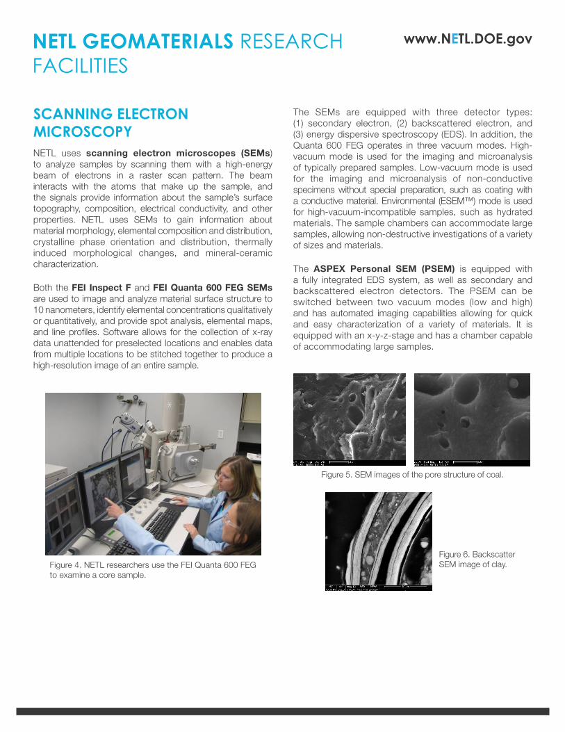
Figure 4. NETL researchers use the FEI Quanta 600 FEG to examine a core sample.
Figure 6. Backscatter SEM image of clay.
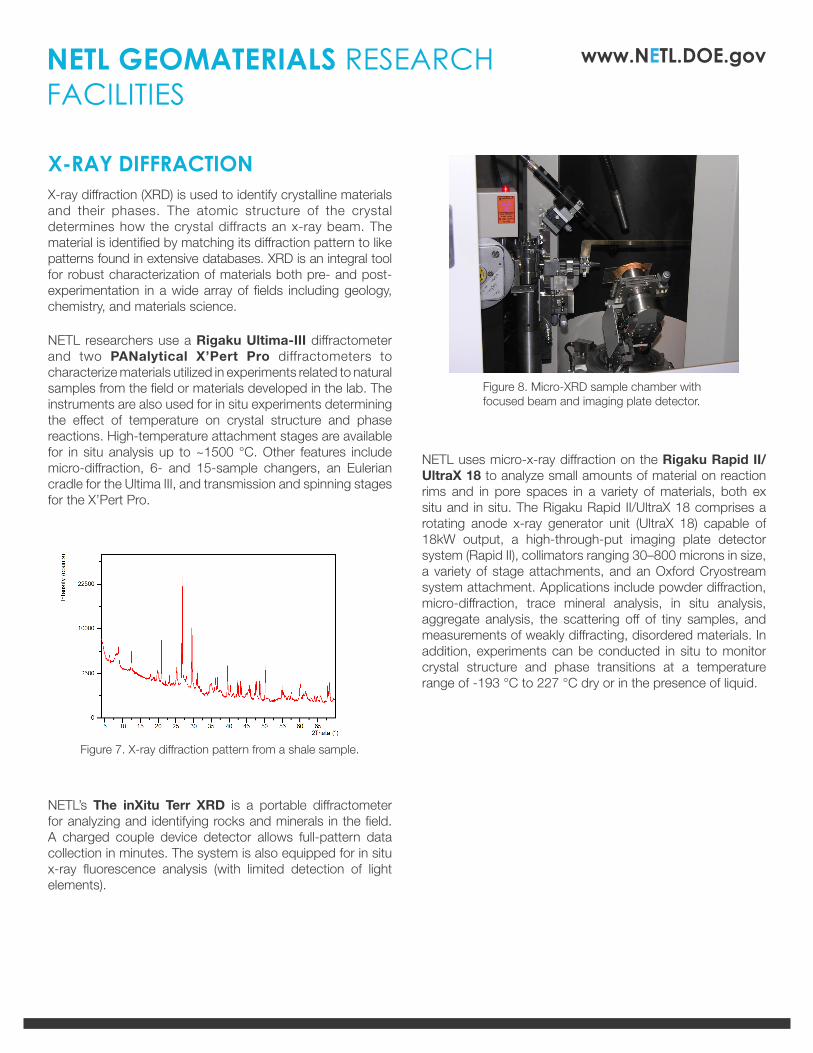
Figure 5. SEM images of the pore structure of coal.
NETL GEOMATERIALS RESEARCH FACILITIES
www.NETL.DOE.gov
X-RAY DIFFRACTIONX-ray diffraction (XRD) is used to identify crystalline materials and their phases. The atomic structure of the crystal determines how the crystal diffracts an x-ray beam. The material is identified by matching its diffraction pattern to like patterns found in extensive databases. XRD is an integral tool for robust characterization of materials both pre- and post-experimentation in a wide array of fields including geology, chemistry, and materials science.
NETL researchers use a Rigaku Ultima-III diffractometer and two PANalytical X’Pert Pro diffractometers to characterize materials utilized in experiments related to natural samples from the field or materials developed in the lab. The instruments are also used for in situ experiments determining the effect of temperature on crystal structure and phase reactions. High-temperature attachment stages are available for in situ analysis up to ~1500 °C. Other features include micro-diffraction, 6- and 15-sample changers, an Eulerian cradle for the Ultima III, and transmission and spinning stages for the X’Pert Pro.
NETL’s The inXitu Terr XRD is a portable diffractometer for analyzing and identifying rocks and minerals in the field. A charged couple device detector allows full-pattern data collection in minutes. The system is also equipped for in situ x-ray fluorescence analysis (with limited detection of light elements).
NETL uses micro-x-ray diffraction on the Rigaku Rapid II/UltraX 18 to analyze small amounts of material on reaction rims and in pore spaces in a variety of materials, both ex situ and in situ. The Rigaku Rapid II/UltraX 18 comprises a rotating anode x-ray generator unit (UltraX 18) capable of 18kW output, a high-through-put imaging plate detector system (Rapid II), collimators ranging 30–800 microns in size, a variety of stage attachments, and an Oxford Cryostream system attachment. Applications include powder diffraction, micro-diffraction, trace mineral analysis, in situ analysis, aggregate analysis, the scattering off of tiny samples, and measurements of weakly diffracting, disordered materials. In addition, experiments can be conducted in situ to monitor crystal structure and phase transitions at a temperature range of -193 °C to 227 °C dry or in the presence of liquid.
Figure 7. X-ray diffraction pattern from a shale sample.
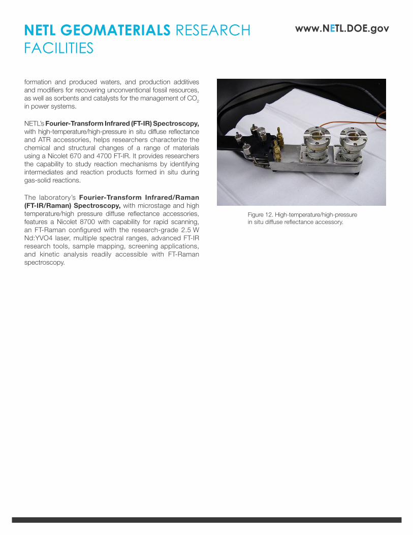
Figure 8. Micro-XRD sample chamber with focused beam and imaging plate detector.
NETL GEOMATERIALS RESEARCH FACILITIES
www.NETL.DOE.gov
PERMEABILITY MEASUREMENTSThe Precision Petrophysical Analysis Lab (PPAL) allows researchers to measure flows of gas through very low-permeability rocks under net pressures approximating those encountered underground. Temperature-controlled gas reference pressures, stable to about one part in 500,000, allow for actual steady-state gas flow measurements as low as one millionth of a standard cm3 per second. Effects of increased net stress on gas permeability, such as those experienced during drawdown, can be duplicated, and the hysteresis of gas flow under stress cycling can be investigated.
Data gained through the PPAL are helping researchers better understand gas shale reservoir properties and the ability of a drained shale to accept CO2 for storage. Measurements on rocks partially saturated with liquids are contributing to the knowledge of how shales behave as reservoir seals and how retrograde condensate shuts off gas flow in some shales.
THERMOGRAVIMETRIC ANALYSIS, DIFFERENTIAL SCANNING CALORIMETRY, AND INFRARED AND RAMAN SPECTROSCOPYUsing thermogravimetry and differential scanning calorimetry, NETL researchers test geological and environmental material samples to determine degradation and decomposition temperatures, absorbed moisture content, solvent residues, levels of various components, and reactivity toward CO2. Materials routinely tested include formation rocks, various minerals, hydrocarbon-rich or source rocks, coal and coal by-products, soil samples,
Figure 10. PPAL coreholder components.
Figure 11. Standard ATR-FTIR spectra of fusain, clarain, and vitrain lithotypes from Springfield Coal and Lower Block Coal.
Figure 9. PPAL undergoing electronics testing.
NETL GEOMATERIALS RESEARCH FACILITIES
www.NETL.DOE.gov
formation and produced waters, and production additives and modifiers for recovering unconventional fossil resources, as well as sorbents and catalysts for the management of CO2 in power systems.
NETL’s Fourier-Transform Infrared (FT-IR) Spectroscopy, with high-temperature/high-pressure in situ diffuse reflectance and ATR accessories, helps researchers characterize the chemical and structural changes of a range of materials using a Nicolet 670 and 4700 FT-IR. It provides researchers the capability to study reaction mechanisms by identifying intermediates and reaction products formed in situ during gas-solid reactions.
The laboratory’s Fourier-Transform Infrared/Raman (FT-IR/Raman) Spectroscopy, with microstage and high temperature/high pressure diffuse reflectance accessories, features a Nicolet 8700 with capability for rapid scanning, an FT-Raman configured with the research-grade 2.5 W Nd:YVO4 laser, multiple spectral ranges, advanced FT-IR research tools, sample mapping, screening applications, and kinetic analysis readily accessible with FT-Raman spectroscopy.
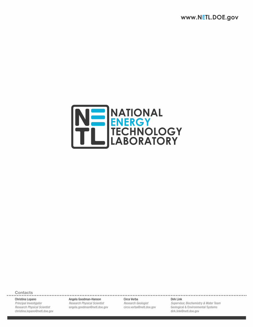
Figure 12. High-temperature/high-pressure in situ diffuse reflectance accessory.
NETL GEOMATERIALS RESEARCH FACILITIES
www.NETL.DOE.gov
For more information on evaluating geologic materials at NETL, please see our NETL Geoimaging Characterization Fact Sheet (R&D178)