Neurochemical mechanisms of sleep-dependent memory consolidation Dissertation der Mathematisch-Naturwissenschaftlichen Fakultät der Eberhard Karls Universität Tübingen zur Erlangung des Grades eines Doktors der Naturwissenschaften (Dr. rer. nat.) vorgelegt von Gordon Benedikt Feld aus Lauterbach (Hessen) Tübingen 2014
Transcript
Neurochemical mechanisms of sleep-dependent memory consolidation
Dissertation
der Mathematisch-Naturwissenschaftlichen Fakultät
der Eberhard Karls Universität Tübingen
zur Erlangung des Grades eines
Doktors der Naturwissenschaften
(Dr. rer. nat.)
vorgelegt von
Gordon Benedikt Feld
aus Lauterbach (Hessen)
Tübingen
2014
Tag der mündlichen Qualifikation: 16.05.2014
Dekan: Prof. Dr. Wolfgang Rosenstiel
1. Berichterstatter: Prof. Dr. Martin Hautzinger
2. Berichterstatter: Prof. Dr. Jan Born
I
I. Index
I. Index .............................................................................................................. I
II. Abbreviations ............................................................................................... V
Figure 1.2. A model for the transfer of memory from the hippocampus, which initially has a hub-like function binding the new trace and disengages as soon as the cortical trace can represent the memory without aid (Frankland & Bontempi, 2005).
Memory
5
System consolidation that involves the transfer of traces from one system to another
is one of two forms of consolidation that are generally differentiated and is distin-
guished from synaptic consolidation (Dudai, 2004). Synaptic consolidation involves
the strengthening of those synapses involved in encoding of the trace per se and can
be perturbed, e.g., by administering protein synthesis blockers shortly after learning
(Bourtchouladze et al., 1998; Xia, Feng, & Guo, 1998). However, synaptic (re-
)consolidation also occurs after reactivation (Milekic & Alberini, 2002). System con-
solidation is assumed to be most effective during sleep, when the brain is offline, i.e.,
deprived from the constant influx of sensory information (Diekelmann & Born, 2010),
evidence for this notion will be discussed in the section on Memory and sleep.
1.2.3 Long term potentiation and other forms of synaptic plasticity
In 1949 Donald O. Hebb postulated that learning in neuronal networks is established
by increasing the connection between two neurons that fire together. Synaptic long
term potentiation (LTP) describes a process of strengthening the connection between
two neurons by increasing their signal transduction efficacy and requires correlated
firing. Signal transduction efficacy in this model is expressed by the amplitude and
duration of post synaptic currents generated by action potentials arriving at the syn-
apse. The most thoroughly investigated form of LTP is related to glutamatergic sig-
nalling. At the glutamatergic synapse presynaptic and postsynaptic modifications
have been shown to be responsible for the expression of LTP (Bliss & Collingridge,
2013). Presynaptic mechanisms are thought to involve the amount of glutamate re-
leased into the synaptic cleft and the speed of clearance therefrom. Postsynaptic
mechanism include modifications of α-amino-3-hydroxy-5-methyl-4-
isoxazolepropionic-acid (AMPA) receptor properties, e.g., changes in opening time
or opening probability due to glutamate binding and, the amount of AMPA receptors
in the active zone. Changes in synapse morphology probably involve increase in size
of both pre- and postsynaptic structures.
A popular mechanism of N-methyl-D-aspartate (NMDA) receptor dependent LTP
in the hippocampus is described in this section and serves to explain the empirical
findings (see Figure 1.3). The basis of this model is that NMDA receptors act as coin-
cidence detectors that detect correlated activity and react by strengthening the syn-
aptic connection (Malenka & Nicoll, 1999). For the opening of their ion channels
NMDA receptors require binding of glutamate and a co-agonist, i.e., glycine or d-
Memory
6
serine (Kleckner & Dingledine, 1988; Mothet et al., 2000). At resting membrane po-
tentials NMDA receptor Ca2+ permeability is blocked by Mg2+. Time and/or location
summation of post synaptic potentials that rely on AMPA receptor activation can push
the membrane potential across the threshold, which releases the Mg2+-block. Open-
ing of the NMDA receptor ion channels allows the influx of Ca2+, which combines with
calmodulin and activates downstream targets such as calmodulin dependent kinase
II. This signalling cascade can lead to, e.g., phosphorylation of AMPA receptors and
trafficking of AMPA receptors to the active zone and, thus, increases signal transduc-
tion.
A discussion of all types of synaptic plasticity goes beyond the scope of the pre-
sent work, but, importantly, plasticity is not a one way street and mechanisms of
NMDA receptor dependent long term depression have been identified (Malenka &
Bear, 2004). Also decay of LTP at potentiated synapses relies on NMDA receptors
(Villarreal, Do, Haddad, & Derrick, 2002). There is evidence that the exact timing of
firing of the pre- and postsynaptic neuron can influence what kind of plasticity is ex-
hibited and this model has been termed spike timing dependent plasticity (Caporale &
Dan, 2008). Finally, considerable evidence is amounting that NMDA receptor subunit
composition can account for differences in the direction of plasticity (Paoletti, Bellone,
& Zhou, 2013).
Figure 1.3. Schematic overview of processes for long term potentiation. (A) Repeated activation of the post synaptic AMPA receptor induced Na+-currents leads to the de-polarization of the post synaptic membrane; (B) the release of the magnesium block from the NMDA receptor. The NMDA receptor can now induce Ca2+-influx, which starts signalling cascades that lead to plastic processes.
Sleep
7
1.2.4 Neuromodulation of synaptic plasticity
Neuromodulators are neurotransmitters that are not involved in direct synaptic trans-
mission but rather modulate ongoing synaptic communication of the brain. Molecules
that are considered neuromodulators for some cells can also be involved in direct
communication at others, e.g., acetylcholine is responsible for signal transduction at
the neuromuscular junction but can influence learning and attention by modulating
neuronal function in the brain (e.g., Sarter, Bruno, & Givens, 2003).
In mammals the adaptive effect of rewards is thought to be induced by dopamine
signalling modulating ongoing plastic processes in the brain (Schultz, 2000, 2007,
2013; Wise, 2004; Wise & Rompre, 1989). Plasticity in the hippocampus is gated by
voke processes, like enhancing cellular calcium influx, in neocortical networks that
prime plastic processes underlying the longer-term storage of the reactivated infor-
mation in these networks (Ribeiro et al., 2007).
Figure 1.4. Model of the interplay between different oscillations during slow wave sleep. Reactivation of memory traces in the hippocampus, in form of sharp-wave ripples, occur most prominently during the slow oscillation up-state. The coupling of fast spindles to the up-state leads to the occurence of spindle-ripple events. This allows the reactivated memories to reach the cortex, possibly represented by gamma-oscillations, during windows of high plasticity (Feld & Born, 2012).
1.4.3 Selective benefit of sleep for memory
The role of sleep for memory is not limited to a mere stabilization of traces, but also
leads to qualitative changes of the trace. For example, Wagner and colleagues
(2004) showed that memory transformation during sleep can lead to insight. This was
demonstrated by letting participants solve mathematical problems that were con-
structed to have a long and a short way to their solution. Participants solved some of
Memory and sleep
12
these problems before sleeping or staying awake. The sleep group showed signifi-
cantly greater rates of detecting the short-cut. Another demonstration of this qualita-
tive change is the induction of false memories by sleep. In these studies participants
learn a list of words that are congruent with a lure that is presented within the retriev-
al list. Sleep seems to increase the participants’ susceptibility to falsely remember
these words (Diekelmann, Born, & Wagner, 2010; Payne et al., 2009).
Another way in which sleep’s benefit for memory is specific is that it only facili-
tates the retention of memories that will be retrieved at a later time point (Wilhelm,
Diekelmann, et al., 2011). This was shown by letting participants learn word pairs and
instructing half of the participants that the words will be retrieved at a later time point.
Sleep only benefited retention, if participants knew they would be tested again,
whereas uninformed participants performed as badly as the wake control group.
Sleep’s effect on memory can also be manipulated by granting rewards for success-
ful retrieval (Fischer & Born, 2009). In this study participants significantly increased
their retention of a finger tapping task, if they were told they would receive a reward
for successful retrieval the next day. Oudiette, Antony, Creery, and Paller (2013)
showed that this enhancing reward effect can be levelled out by externally cueing low
rewarded memory traces, which may suggest that reactivation probability is influ-
enced by rewards. Interestingly, during sleep, cueing half of the low rewarded items
in this study improved retention of the whole set of low reward items, whereas cueing
whilst awake specifically enhanced the cued items.
1.4.4 Pharmacological influences on sleep-dependent memory consolidation
While the specific roles of different neurotransmitters during sleep for memory is not
fully understood, a number of studies that have pharmacologically manipulated neu-
rotransmitter systems demonstrate their influence on sleep-dependent memory con-
solidation. Usually, in these studies, participants learn a task in the evening before a
retention interval containing sleep and the pharmacological agent is administered
thereafter. This leads to a manipulation of sleep-dependent mechanisms by the
agent and after it is removed from the system, retrieval is tested to reveal the agents
influence on consolidation.
As mentioned above, SWS is typically accompanied by low levels of neuromodu-
lators such as acetylcholine (Marrosu et al., 1995) and the direction of information
flow between hippocampus and neocortex is thought to rely on cholinergic tone, as
Memory and sleep
13
low levels of acetylcholine release feedback synapses in the hippocampus (Buzsaki,
1986; Hasselmo, 1999). Consequently, increasing cholinergic activity with cholines-
terase blocker physostigmine during SWS disrupts declarative memory consolidation
(Gais & Born, 2004b). The opposite procedure, blocking muscarinic and nicotinergic
receptors, improves the consolidation of declarative memory during a wake interval
(Rasch, Born, & Gais, 2006), but interferes with motor memory consolidation during
REM sleep (Rasch, Gais, & Born, 2009).
Similar findings have been reported for low levels of cortisol that are commonly
found during SWS, inasmuch as, administration of hydrocortisone or dexamethasone
receptor activation by administration of DCS, i.e., a co-agonist at the glycine binding
site of the receptor, benefited declarative memory encoding (Onur et al., 2010). Yet,
the role of glutamatergic neurotransmission for sleep-dependent offline consolidation
of memories has been scarcely examined. In the developing cortex of cats, sleep-
dependent ocular dominance plasticity was inhibited after blocking NMDA-receptors
(Aton et al., 2009). In adult humans, sleep-dependent consolidation of visual texture
discrimination skill is deteriorated by the non-competitive NMDA-receptor blocker
ketamine or the competitive AMPA-receptor blocker caroverine (Gais et al., 2008).
However, these findings pertain to non-declarative types of memory not essentially
relying on hippocampal networks.
Here, we tested contributions of glutamatergic neurotransmission to sleep-
dependent consolidation of hippocampus-dependent declarative memory. As sleep-
dependent consolidation of these memories is caused by the reactivation of firing
patterns during SWS in neuron assemblies likely comprising glutamatergic activation,
we expected that consolidation would be sensitive to blocking or enhancing glutama-
tergic neurotransmission during retention sleep. First we investigated the effects of
blocking AMPA-receptors (by caroverine) or NMDA-receptors (by ketamine) during
1 Published as: Feld, G. B., Lange, T., Gais, S., & Born, J. (2013). Sleep-dependent declarative
memory consolidation--unaffected after blocking NMDA or AMPA receptors but enhanced by NMDA coagonist D-cycloserine. Neuropsychopharmacology, 38(13), 2688-2697.
Study 1 – The role of glutamatergic neuroplasticity for sleep-dependent memory consolidation
22
retention sleep. Then we tested the effects of enhancing NMDA-receptor function by
post-learning administration of DCS.
2.2.2 Methods
2.2.2.1 Participants
Altogether, 58 participants completed the study (caroverine: n = 15, ketamine: n = 13,
DCS n = 30; see Supplementary Methods for details of methods). Participants were
healthy, non-smoking, native German speaking men (18-30 years). The experiments
were approved by the ethics committee of the University of Luebeck. Written in-
formed consent was obtained from all participants prior to participation. One partici-
pant revoked his consent after data acquisition in the DCS experiment and his data
were deleted.
2.2.2.2 Design and procedures
Each of the experiments followed a randomized, double-blind, placebo-controlled,
within-subject, crossover design. In the DCS study, two different groups were recruit-
ed to compare effects of DCS (versus placebo) during retention intervals of sleep (n
= 16) and wakefulness (n = 14), respectively. Participants took part in two experi-
mental sessions scheduled at least 14 days apart. Both sessions were identical but
for the administration of placebo or substance (caroverine: Calmaverine®, intrave-
nously, 16 mg/h, corresponding to a total dose of 40 mg/kg, Taphlan, Switzerland,
Dement, 1973) and the Positive and Negative Affective Schedule (PANAS; Watson et
al, 1988). In the DCS experiments vigilance was additionally assessed by mean reac-
tion times in a 5-min version of the psychomotor vigilance task (PVT; Dinges et al.,
1997) that required pressing a button as fast as possible whenever a bright millisec-
ond clock presented on a dark computer screen started counting upward. After the
button press, this clock displayed the reaction time. General capabilities of long-term
memory retrieval were also tested in these experiments using a word generation
task. Participants had to generate as many words as possible starting with a certain
letter (P or M) or belonging to a defined category (hobby or profession) during a time
of 2 minutes each.
Only in the wake control group of the DCS experiments, encoding (of a list of 16
three digit numbers) was measured. This measure was applied to test if DCS has an
effect on encoding at high plasma concentrations during the wake retention interval.
At the end of a session all participants were asked if they believed to have received
an active agent or placebo.
2.2.2.5 Analyses of adrenocorticotropin (ACTH) and cortisol
Because blockers of glutamatergic transmission like ketamine can stimulate pitui-
tary adrenal activity (Herman et al, 2004), we sampled blood once before and after
learning as well as after retrieval. Additionally, blood was sampled during the reten-
tion interval, i.e., hourly during the first four hours after substance intake and, in the
DCS study, every two hours during the second four hours. Sampling during the reten-
tion interval was performed via a long plastic tube from an adjacent room, leaving the
participant’s sleep undisturbed. Blood samples were immediately centrifuged and
then stored at -80°C until assay. Serum cortisol concentrations were assessed using
the Immulite (Siemens Medical Solutions Diagnostics, Los Angeles, CA; serum sensi-
tivity, 0.2 µg/dl, interassay coefficient of variation < 10 %). ACTH was assessed in
Study 1 – The role of glutamatergic neuroplasticity for sleep-dependent memory consolidation
25
plasma (Immulite, Siemens Medical Solutions Diagnostics, Los Angeles, CA; sensi-
tivity, 9 pg/ml, interassay coefficient of variation < 9.6 %).
2.2.3 Results
2.2.3.1 Memory Tasks
Neither caroverine nor ketamine significantly changed retention of word pairs in com-
parison with respective placebo treatments (all p > 0.53, see Table 2.1 and Figure
2.1 for a summary of results). Learning performance also did not differ between the
active agents and respective placebo conditions (all t ≤ 1.61, p ≥ 0.13).
DCS administration before the sleep-retention interval distinctly improved recall of
word pairs at retrieval testing after the retention interval. This effect was confirmed by
significance for the ANOVA treatment x time point interaction (F(1,12) = 9.33, p ≤ 0.01,
Table 2.1 and Figure 2.1). By contrast, DCS administered before a wake retention
interval did not improve word pair retention (p ≥ 0.99). During learning there were no
evident differences between placebo and DCS conditions concerning amounts of
learned word pairs and trials to criterion (all t ≤ 1.19, p ≥ 0.19). An ANOVA including
both the sleep and wake groups of the DCS study (represented by an additional
‘sleep/wake’ factor) revealed a trend for the treatment x time point x sleep/wake in-
teraction (F(1,25) = 3.15, p = 0.09).
The emotional memory task did not reveal any differences as measured by the
amount of freely recalled emotional and neutral pictures between DCS and placebo
conditions both in the sleep group and in the wake group of this study (p ≥ 0.29, for
respective treatment main and interaction effects, Table 2.2). Independent of the
treatment condition, generally more emotional than neutral pictures were remem-
bered (sleep: F(1,13) = 21.06 and p ≤ 0.01, wake: F(1,13) = 26.74, p ≤ 0.01).
Procedural finger sequence tapping was not differentially affected by DCS or pla-
cebo. The overnight gains in tapping speed and accuracy were comparable in both
conditions (all p ≥ 0.27, Table 2.2), and this was also true for the wake control group
(all p ≥ 0.16). There was also no difference evident between DCS and placebo condi-
tions at training or concerning performance on the untrained control sequence during
the retrieval phase (all p ≥ 0.26). The ANOVA including both the sleep and the wake
condition of the DCS study revealed that the sleep group improved their performance
more during the retention interval (F(1,24) = 8.64, p ≤ 0.01 for time point x sleep/wake).
Study 1 – The role of glutamatergic neuroplasticity for sleep-dependent memory consolidation
26
Figure 2.1. (A) Study design: In the caroverine and ketamine studies par-ticipants learned at 9:00 pm and went to bed at 11:00 pm. The retention interval was 3.5 hours and half an hour after waking the participant re-trieval was tested at 2:30 am. In the DCS study sleep condition partici-pants also learned at 9:00 pm and went to bed from 11:00 pm to 7:30 am. The retention interval was 22 hours and retrieval was tested at 9:00 pm. In the wake condition learning was shifted 10 hours to 11:00 am and participants remained awake the whole retention interval until retrieval was tested at 11:00 am the next day. Approximate times of learning and retrieval are indicated, during learning criterion trials were the last cued re-call during learning the word pairs and the last three blocks of learning the finger sequence. p.o. – oral ad-ministration, i.v. – intravenous admin-istration, PAL - word pair associates task, FTT –finger sequence tapping task, Pics – emotional and neutral pictures. (B) Overnight retention of word pairs and finger sequence tap-ping skills in the substance (black bars) and placebo condition (empty bars). Retention of word pairs is indi-cated by the mean (�SEM) percent-age of word pairs recalled at retrieval testing after the retention interval relative to recall performance on the criterion trial at learning before sleep (Please note that retention in the DCS experiments is generally lower than in the caroverine and ketamine experiments due to the longer reten-tion interval). Overnight gains in fin-ger sequence tapping are indicated
by the mean (±SEM) percentage of correctly tapped sequences per 30-sec trial at retrieval testing relative to the average performance on the last three trials during training before the retention interval. ** p ≤ 0.01, for pairwise comparisons between the effects of the treatments (caroverine: n = 15, ketamine: n = 12, DCS: n = 13 for sleep condition, n = 14 for wake condition).
Study 1 – The role of glutamatergic neuroplasticity for sleep-dependent memory consolidation
27
Table 2.1. Word Pair Memory Task: Mean (± SEM) values are given for the active agent and placebo conditions. Total amount of recalled words is given for criterion trials at learning and at retrieval, additionally, percent values of retrieved words are provided relative to learning performance (set to 100%). ** p ≤ 0.01 and ns = not significant.
Caroverine + sleep Substance Placebo p
Blocks to criterion 1.53 ±0.17 1.60 ±0.16 ns
Learning 29.60 ±0.83 28.13 ±0.84 ns
Retrieval 32.53 ±0.98 31.67 ±0.98 ns
Absolute difference 2.93 ±0.64 3.53 ±1.0 ns
% of learning 110.14 ±2.27 113.43 ±4.00 ns
Ketamine + sleep Substance Placebo p
Blocks to criterion 1.50 ±0.15 1.58 ±0.15 ns
Learning 29.19 ±0.98 27.93 ±1.13 ns
Retrieval 31.35 ±0.91 30.23 ±0.69 ns
Absolute difference 2.16 ±0.94 2.29 ±0.93 ns
% of learning 108.12 ±3.58 109.65 ±3.90 ns
DCS + sleep Substance Placebo p
Blocks to criterion 1.85 ±0.15 2.00 ±0.25 ns
Learning 27.85 ±0.83 28.31 ±1.11 ns
Retrieval 27.00 ±0.87 25.08 ±1.16 ns
Absolute difference -0.85 ±0.55 -3.23 ±0.59 **
% of learning 97.10 ±1.99 88.56 ±2.27 **
DCS + wake Substance Placebo p
Blocks to criterion 1.50 ±0.14 1.71 ±0.19 ns
Learning 28.64 ±0.90 29.21 ±0.82 ns
Retrieval 27.36 ±1.19 27.93 ±1.32 ns
Absolute difference -1.29 ±1.01 -1.29 ±1.07 ns
% of learning 95.78 ±3.34 95.68 ±3.69 ns
Study 1 – The role of glutamatergic neuroplasticity for sleep-dependent memory consolidation
28
2.2.3.2 Sleep
Infusion of ketamine compared with placebo, reduced the time spent in stage 2,
SWS, and REM sleep and increased time in wakefulness (Wake: t(12) = 2.55, p ≤
p ≤ 0.05, REM: t(12) = -2.50, p ≤ 0.05, Table 2.3). Under caroverine there was a trend
towards less time spent in stage 4 sleep (t(13) = 1.79, p = 0.10). Oral administration of
DCS before sleep increased time in wakefulness (t(12) = 2.66, p ≤ 0.05) and stage 1
sleep (t(12) = 2.27, p ≤ 0.05), and reduced REM sleep (t(12) = -3.51, p ≤ 0.01; Table 3).
There was no evident correlation between DCS induced changes in sleep architec-
ture and improvements in the retention of word pairs (all r ≤ 0.34 and p ≥ 0.26,
changes were calculated individually with reference to the placebo condition).
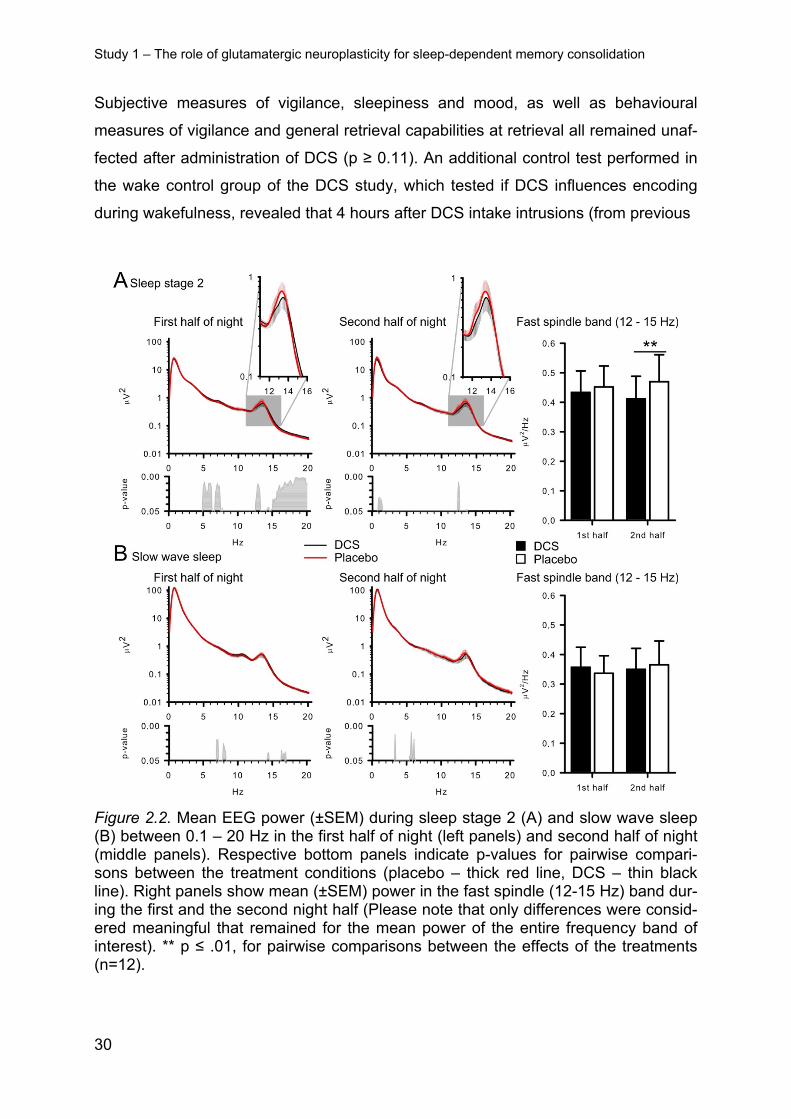
More fine-grained analyses of EEG power spectra at Cz revealed a power reduction
around the spindle maximum during stage 2 sleep following DCS (Figure 2.2). ANO-
VA on the fast spindle band (12-15 Hz) confirmed significance for the sleep stage x
treatment interaction (F(1,11) = 9.47, p ≤ 0.01; t(11) = 2.71, p ≤ 0.05 for post hoc com-
parison between the treatments for stage 2 spindle power). The reducing effect of
DCS on stage 2 sleep spindle power appeared to be less pronounced during the first
than the second half of sleep (t(11) = -2.95, p ≤ 0.01, for pairwise comparison between
the effects of treatment, F(1,11) = 7.87, p ≤ 0.05, for treatment x night half). However,
this was due to the fact that, during the first half, DCS simultaneously enhanced beta
power with this effect extending into the upper (> 14-Hz) range of fast spindle fre-
quencies (Figure 2). Analyses on the other bands did not show any significant effects
of treatment (p ≥ 0.16). There was no correlation between DCS induced changes in
the spindle band and differences in the retention of word pairs (all r ≤ 0.26 and p ≥
0.39).
2.2.3.3 Control measures
In the caroverine study we found no differences between the treatments in subjective
measures of vigilance, alertness, sleepiness or mood during the retrieval phase (p ≥
0.19). These measures also did not differ between treatments in the ketamine study
(p ≥ 0.58); however, three participants reported slight nausea after awakening on the
ketamine nights. Cortisol and ACTH levels were not differentially affected by carover-
ine or placebo (p ≥ 0.31). Under ketamine, cortisol was increased at the end of the
Study 1 – The role of glutamatergic neuroplasticity for sleep-dependent memory consolidation
29
infusion (between 01:00 am and 02:00 pm; ketamine:4.54 ± 3.67 µg/dl; placebo 2.80
± 1.96 µg/dl ; p ≤ 0.05) and ACTH concentrations showed a corresponding trend (p
= 0.10).
Table 2.2. Emotional and Procedural Memory: Mean (± SEM) values are given for the DCS and placebo condition. Top: number of correctly remembered emotional, neutral and of total pictures in the emotional memory task. Bottom: average number of correctly tapped sequences for the finger sequence tapping during the last three 30-sec trials at learning, the three trials at retrieval and for the untrained sequence at retrieval. Additionally, percent values of correctly tapped sequences at retrieval are provided relative to learning performance (set to 100%). ** p ≤ 0.01, * p ≤ 0.05 and ns = not significant.
Emotional and neutral pictures
Sleep DCS Placebo p
Emotional 7.54 ±0.83 8.23 ±0.74 ns
Neutral 4.77 ±0.86 4.85 ±0.85 ns
Total 12.31 ±1.29 13.08 ±1.36 ns
Wake DCS Placebo p
Emotional 6.14 ±0.72 6.79 ±0.88 ns
Neutral 4.00 ±0.60 4.57 ±0.60 ns
Total 10.14 ±1.08 11.36 ±1.34 ns
Finger sequence tapping
Sleep DCS Placebo p
Learning 17.11 ±1.18 16.36 ±1.21 ns
Retrieval 21.03 ±1.26 19.75 ±1.26 ns
Absolute difference 3.91 ±0.72 3.39 ±0.89 ns
% of learning 124.87 ±5.69 122.66 ±5.75 ns
Untrained sequence 13.50 ±1.55 13.47 ±0.83 ns
Wake DCS Placebo p
Learning 18.83 ±1.00 18.78 ±1.00 ns
Retrieval 20.60 ±1.21 20.10 ±1.29 ns
Absolute difference 1.76 ±0.54 1.31 ±0.93 ns
% of learning 109.56 ±3.18 107.86 ±5.04 ns
Untrained sequence 12.86 ±0.72 12.66 ±0.72 ns
Study 1 – The role of glutamatergic neuroplasticity for sleep-dependent memory consolidation
30
Subjective measures of vigilance, sleepiness and mood, as well as behavioural
measures of vigilance and general retrieval capabilities at retrieval all remained unaf-
fected after administration of DCS (p ≥ 0.11). An additional control test performed in
the wake control group of the DCS study, which tested if DCS influences encoding
during wakefulness, revealed that 4 hours after DCS intake intrusions (from previous
Figure 2.2. Mean EEG power (±SEM) during sleep stage 2 (A) and slow wave sleep (B) between 0.1 – 20 Hz in the first half of night (left panels) and second half of night (middle panels). Respective bottom panels indicate p-values for pairwise compari-sons between the treatment conditions (placebo – thick red line, DCS – thin black line). Right panels show mean (±SEM) power in the fast spindle (12-15 Hz) band dur-ing the first and the second night half (Please note that only differences were consid-ered meaningful that remained for the mean power of the entire frequency band of interest). ** p ≤ .01, for pairwise comparisons between the effects of the treatments (n=12).
Study 1 – The role of glutamatergic neuroplasticity for sleep-dependent memory consolidation
31
Table 2.3. Sleep parameters: Mean (± SEM) values of minutes spent in the different sleep stages are given for the active agent and placebo conditions. REM – rapid eye movement sleep; SWS – slow wave sleep, TST – total sleep time; ** p ≤ .01, * p ≤ .05, t p ≤ .10 and ns = not significant.
Caroverine Substance Placebo p
Wakefulness 2.80 ±1.21 2.41 ±0.60 ns
Stage 1 22.29 ±4.59 29.5 ±4.78 ns
Stage 2 96.18 ±5.63 87.57 ±7.65 ns
Stage 3 44.61 ±4.39 42.14 ±3.35 ns
Stage 4 5.46 ±1.64 8.07 ±2.11 t
REM 11.67 ±2.51 9.96 ±3.32 ns
SWS 50.07 ±5.18 50.21 ±4.49 ns
TST 184.28 ±2.05 182.07 ±3.36 ns
Ketamine Substance Placebo p
Wakefulness 47.96 ±13.87 14.69 ±4.29 *
Stage 1 26.38 ±4.49 20.00 ±2.76 ns
Stage 2 77.69 ±9.32 104.12 ±5.05 *
Stage 3 19.50 ±4.61 23.92 ±3.25 ns
Stage 4 2.58 ±1.18 6.08 ±1.96 *
REM 12.62 ±3.61 22.96 ±4.17 *
SWS 22.08 ±5.53 30.00 ±4.35 *
TST 186.73 ±1.73 191.77 ±5.5 ns
DCS Substance Placebo p
Wakefulness 19.54 ±3.43 9.84 ±2.62 *
Stage 1 31.27 ±2.89 25.30 ±2.03 *
Stage 2 213.77 ±8.88 215.69 ±10.52 ns
Stage 3 60.46 ±6.37 56.69 ±6.59 ns
Stage 4 32.96 ±7.61 33.19 ±7.77 ns
REM 83.73 ±5.39 101.85 ±6.95 **
SWS 93.42 ±10.07 89.88 ±10.47 ns
TST 445.38 ±3.18 447.08 ±3.76 ns
Study 1 – The role of glutamatergic neuroplasticity for sleep-dependent memory consolidation
32
testing immediately after and 2 hours after substance intake) were reduced (DCS:
0.21 ± 0.11; placebo: 0.93 ± 0.27, t(13) = -2.92, p ≤ 0.01, F(1,13) = 8.29, p ≤ 0.01 for
treatment x time point). Levels of cortisol and ACTH did not differ between treatment
conditions (p ≥ 0.26). A positive relation between differences in cortisol level and dif-
ferences in word pair retention in the sleep condition was found (2:00 am: r = 0.62
and p ≤ 0.05), however, it did not survive multiple comparison correction.
Participants could differentiate ketamine and placebo (Χ2(1) = 22.29 p ≤ 0.01).
However, caroverine and placebo as well as DCS and placebo could not be discrimi-
nated (p ≥ 0.25).
2.2.4 Discussion
Evidence from animal and humans studies indicates that the consolidation of hippo-
campus-dependent declarative memory relies on the reactivation of newly encoded
neuronal representations during post-learning SWS (Diekelmann & Born, 2010;
Rasch et al., 2007; Ribeiro et al., 2007; Wilson & McNaughton, 1994). In hippocam-
pal neuron assemblies the same sequential firing patterns are observed during SWS
as during encoding during preceding wake (O'Neill, Pleydell-Bouverie, Dupret, &
Csicsvari, 2010). These reactivations that typically coincide with sharp wave-ripples
in the hippocampal EEG are thought to involve excitatory glutamatergic synapses
(Behrens, van den Boom, de Hoz, Friedman, & Heinemann, 2005; Dupret, O'Neill,
and memories associated with high rewards benefit more from this process (Fischer
& Born, 2009; Wilhelm, Diekelmann, et al., 2011). However, it remains unclear if
sleep leads to the preferential consolidation of highly rewarded memories because
reward present at learning tags these memories so that they are reactivated more
frequently during subsequent sleep, or rather the sleep-associated consolidation pro-
cess itself involves reactivation of the dopaminergic reward circuitry associated with a
specific memory. Here we probed the latter assumption by testing the effects of a
dopaminergic agonist (pramipexole) on the sleep-associated consolidation of memo-
ries, which were associated with high or low rewards.
Correlated activity of neurons active that encoded information during wake pre-
dicts their firing together during subsequent sleep in rodents (Wilson & McNaughton,
1994). This replay of neural representations during sleep occurs in the same se-
quence as during wakefulness and is coordinated between the hippocampus and the
neocortex (Ji & Wilson, 2007; Skaggs & McNaughton, 1996). In humans a causal role
for these reactivations has been shown for declarative and skill memory (Antony et
al., 2012; Rasch et al., 2007; Rudoy et al., 2009). During SWS, hippocampal reacti-
vations lead striatal reactivations of place-reward information in rats, consistent with
the view that striatal dopaminergic activation contributes to the consolidation of re-
2 Accepted for publication as: Feld GB, Besedovsky L, Kaida K, Münte TF, & Born J (2014). Do-
pamine D2-like receptor activation wipes out preferential consolidation of high over low reward memo-ries during human sleep. J Cogn Neurosci. doi: 10.1162/jocn_a_00629
Study 2 – The role of dopaminergic neuromodulation for sleep-dependent memory consolidation
40
ward-related memory traces during sleep (Lansink et al., 2008; Lansink, Goltstein,
Lankelma, McNaughton, & Pennartz, 2009).
Dopamine is a major neuromodulator and has been put forward as the main neu-
rotransmitter mediating the preferential encoding of highly rewarding stimuli (Schultz,
2007; Wise, 2004; Wise & Rompre, 1989), by influencing plasticity in the hippocam-
proved effective in several forgoing studies (e.g.,Riba, Kramer, Heldmann, Richter, &
Münte, 2008; Ye, Hammer, Camara, & Münte, 2011).
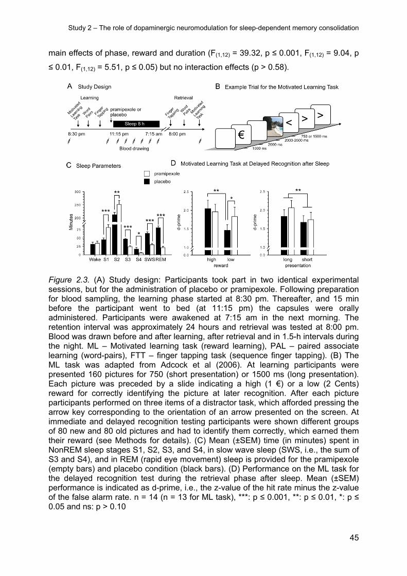
Figure 2.3 A summarizes the experimental procedure. On experimental nights,
participants arrived at the laboratory at 07:30 pm. Following insertion of an intrave-
Study 2 – The role of dopaminergic neuromodulation for sleep-dependent memory consolidation
42
nous catheter and preparations for EEG and polysomnography, the participants
learned the ML task between 08:30 and 09:30 pm. Afterwards, they learned control
tasks (declarative word pair associates and procedural sequence finger tapping) with
a 10-min break between each of the tasks. This order was chosen so that partici-
pants would be most attentive during encoding of the reward task. Fifteen minutes
before lights were turned off (at 11:15 pm) to enable sleep, the participants were oral-
ly administered a capsule containing pramipexole or placebo, as well as, the domper-
idone tablet. They were woken at 07:15 am and left the lab. During the following day
participants engaged in their usual activities. They were instructed to refrain from any
stressful mental or physical activities, and to keep a record of their activities during
this day. In the evening they returned to the lab at 08:00 pm and retrieval of the
memory tasks was tested – in reverse order of learning. (This was done as retrieval
procedures for the word pairs and the sequence finger tapping were short (i.e., < 8
min) compared to the longer picture recognition test taking about 30 min). At learning
and retrieval, as a control, tests of vigilance, mood and subjective sleepiness were
also performed. Blood was sampled before and after learning, after retrieval and at
1.5 h intervals during the night. For this purpose the intravenous catheter was con-
nected to a long thin tube to enable blood collection from an adjacent room without
disturbing the participant’s sleep.
2.3.2.3 Control measures – general retrieval performance, vigilance, sleepiness
and mood
At retrieval, to exclude effects of the drug on general retrieval performance, partici-
pants were tested on a WFT (table 2 for means and SEMs of the control measures).
They were asked to generate as many words as possible within a two minute interval
after being cued with either a letter (p or m) or a category (professions or hobbies).
The following control measures were assessed once before and once after each
learning and retrieval phase. Mean reaction times were assessed as a measure of
vigilance in a 5-min version of the PVT (Dinges et al., 1997) that required pressing a
button as fast as possible whenever a bright millisecond clock presented on a dark
computer screen started counting upward. After the button press this clock displayed
the reaction time. The median reaction speed (i.e., 1/[reaction time in msec]) was
calculated for each participant. Mood was measured using the 10 positive and 10
negative items of the PANAS (Watson, Clark, & Tellegen, 1988), where participants
Study 2 – The role of dopaminergic neuromodulation for sleep-dependent memory consolidation
43
respond to items (e.g., “Do you momentarily feel scared?”) on a 5 point Likert scale
ranging from 1 = “not at all” to 5 = “very much”. Subjective sleepiness was assessed
with the 1-item SSS (Hoddes et al., 1973) ranging from 1 = “Feeling active, vital,
alert, or wide awake” to 8 = “Asleep”. At the end of the experiment participants were
asked if they believed to have received an active agent or placebo.
2.3.2.4 Control measures – blood samples
Samples for measuring hormone concentrations were kept frozen at -80°C until as-
say. Cortisol, growth hormone, and prolactin levels were determined in serum using
commercial assays (Immulite, Siemens Medical Solutions Diagnostics, Los Angeles,
USA). Intra- and interassay coefficients of variation were < 10 %.
2.3.2.5 Data reduction and statistical analysis
Data from two participants were completely discarded because of poor sleep during
the placebo night. Data from one participant were not included in the analysis of the
ML task, as he remembered an unusual amount more low reward items than high
reward items in the placebo condition (i.e., the difference between high and low re-
ward was more than 2 standard deviations from the group mean, probably reflecting
a misunderstanding of the rather complex task instruction or an unusual encoding
strategy). For two participants, hormonal data sets were incomplete because of
problems with blood sampling during sleep. Statistical analyses generally relied on
analyses of variance (ANOVA; SPSS version 21.0.0 for Windows) including a re-
peated measures factor ‘treatment’ (substance vs placebo) and, where appropriate,
the factor ‘phase’ (learning vs retrieval). As analyses revealed a strong suppressive
influence of pramipexole on both SWS and REM sleep, main analyses of memory
performance included the individual difference in wake time between treatment condi-
tions as covariate to account for this sleep disruption. Wake time (i.e., the amount of
time spent awake between sleep onset and lights on) was used for these analyses
because it did not differ significantly between treatment conditions. For analysis of
pictures additional ‘reward’ and ‘duration’ factors were introduced, representing
recognition of high vs low reward pictures and long vs short stimulus presentation,
respectively. The analyses of the pictures did not include a factor ‘phase’ as immedi-
ate and delayed recognition were performed on different sets of stimuli. Significant
Study 2 – The role of dopaminergic neuromodulation for sleep-dependent memory consolidation
44
ANOVA interactions were specified by post-hoc t-tests. Degrees of freedom were
corrected according to Greenhouse-Geisser where appropriate.
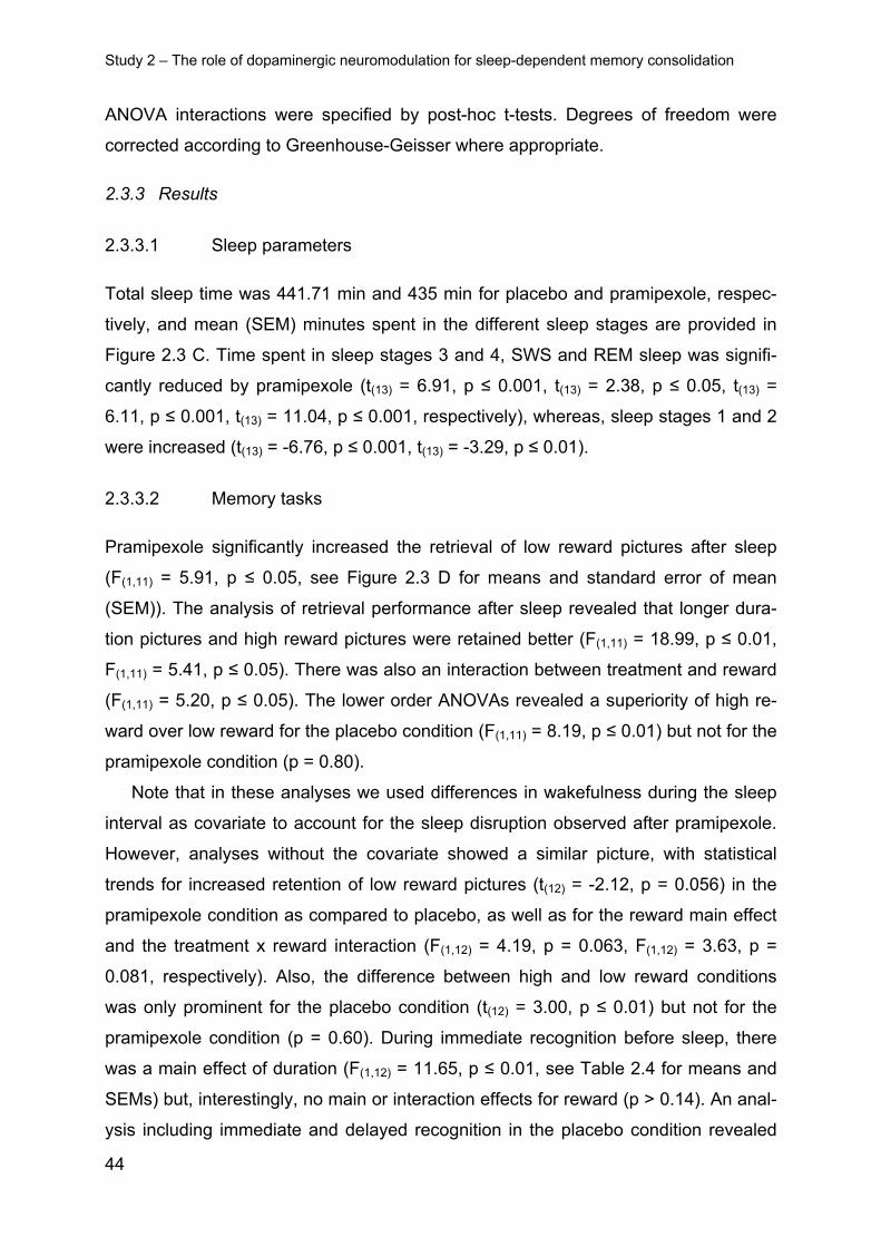
2.3.3 Results
2.3.3.1 Sleep parameters
Total sleep time was 441.71 min and 435 min for placebo and pramipexole, respec-
tively, and mean (SEM) minutes spent in the different sleep stages are provided in
Figure 2.3 C. Time spent in sleep stages 3 and 4, SWS and REM sleep was signifi-
cantly reduced by pramipexole (t(13) = 6.91, p ≤ 0.001, t(13) = 2.38, p ≤ 0.05, t(13) =
6.11, p ≤ 0.001, t(13) = 11.04, p ≤ 0.001, respectively), whereas, sleep stages 1 and 2
were increased (t(13) = -6.76, p ≤ 0.001, t(13) = -3.29, p ≤ 0.01).
2.3.3.2 Memory tasks
Pramipexole significantly increased the retrieval of low reward pictures after sleep
(F(1,11) = 5.91, p ≤ 0.05, see Figure 2.3 D for means and standard error of mean
(SEM)). The analysis of retrieval performance after sleep revealed that longer dura-
tion pictures and high reward pictures were retained better (F(1,11) = 18.99, p ≤ 0.01,
F(1,11) = 5.41, p ≤ 0.05). There was also an interaction between treatment and reward
(F(1,11) = 5.20, p ≤ 0.05). The lower order ANOVAs revealed a superiority of high re-
ward over low reward for the placebo condition (F(1,11) = 8.19, p ≤ 0.01) but not for the
pramipexole condition (p = 0.80).
Note that in these analyses we used differences in wakefulness during the sleep
interval as covariate to account for the sleep disruption observed after pramipexole.
However, analyses without the covariate showed a similar picture, with statistical
trends for increased retention of low reward pictures (t(12) = -2.12, p = 0.056) in the
pramipexole condition as compared to placebo, as well as for the reward main effect
and the treatment x reward interaction (F(1,12) = 4.19, p = 0.063, F(1,12) = 3.63, p =
0.081, respectively). Also, the difference between high and low reward conditions
was only prominent for the placebo condition (t(12) = 3.00, p ≤ 0.01) but not for the
pramipexole condition (p = 0.60). During immediate recognition before sleep, there
was a main effect of duration (F(1,12) = 11.65, p ≤ 0.01, see Table 2.4 for means and
SEMs) but, interestingly, no main or interaction effects for reward (p > 0.14). An anal-
ysis including immediate and delayed recognition in the placebo condition revealed
Study 2 – The role of dopaminergic neuromodulation for sleep-dependent memory consolidation
45
main effects of phase, reward and duration (F(1,12) = 39.32, p ≤ 0.001, F(1,12) = 9.04, p
≤ 0.01, F(1,12) = 5.51, p ≤ 0.05) but no interaction effects (p > 0.58).
Figure 2.3. (A) Study design: Participants took part in two identical experimental sessions, but for the administration of placebo or pramipexole. Following preparation for blood sampling, the learning phase started at 8:30 pm. Thereafter, and 15 min before the participant went to bed (at 11:15 pm) the capsules were orally administered. Participants were awakened at 7:15 am in the next morning. The retention interval was approximately 24 hours and retrieval was tested at 8:00 pm. Blood was drawn before and after learning, after retrieval and in 1.5-h intervals during the night. ML – Motivated learning task (reward learning), PAL – paired associate learning (word-pairs), FTT – finger tapping task (sequence finger tapping). (B) The ML task was adapted from Adcock et al (2006). At learning participants were presented 160 pictures for 750 (short presentation) or 1500 ms (long presentation). Each picture was preceded by a slide indicating a high (1 €) or a low (2 Cents) reward for correctly identifying the picture at later recognition. After each picture participants performed on three items of a distractor task, which afforded pressing the arrow key corresponding to the orientation of an arrow presented on the screen. At immediate and delayed recognition testing participants were shown different groups of 80 new and 80 old pictures and had to identify them correctly, which earned them their reward (see Methods for details). (C) Mean (±SEM) time (in minutes) spent in NonREM sleep stages S1, S2, S3, and S4, in slow wave sleep (SWS, i.e., the sum of S3 and S4), and in REM (rapid eye movement) sleep is provided for the pramipexole (empty bars) and placebo condition (black bars). (D) Performance on the ML task for the delayed recognition test during the retrieval phase after sleep. Mean (±SEM) performance is indicated as d-prime, i.e., the z-value of the hit rate minus the z-value of the false alarm rate. n = 14 (n = 13 for ML task), ***: p ≤ 0.001, **: p ≤ 0.01, *: p ≤ 0.05 and ns: p > 0.10
Study 2 – The role of dopaminergic neuromodulation for sleep-dependent memory consolidation
46
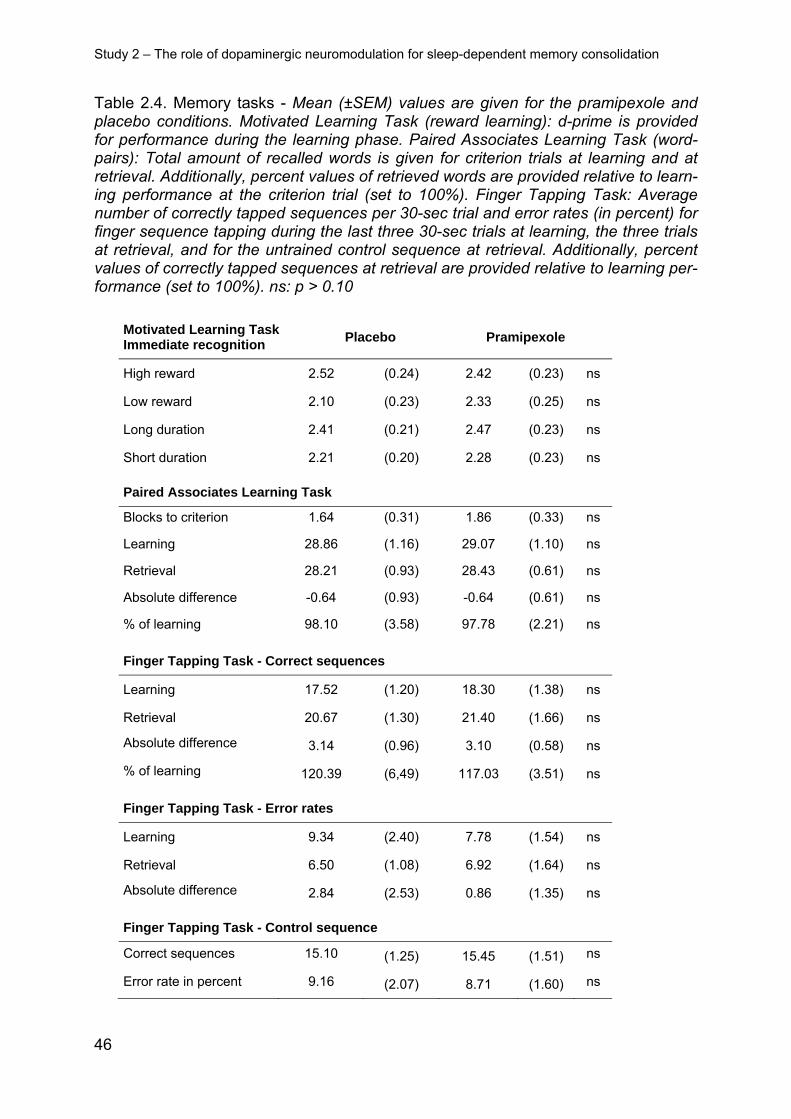
Table 2.4. Memory tasks - Mean (±SEM) values are given for the pramipexole and placebo conditions. Motivated Learning Task (reward learning): d-prime is provided for performance during the learning phase. Paired Associates Learning Task (word-pairs): Total amount of recalled words is given for criterion trials at learning and at retrieval. Additionally, percent values of retrieved words are provided relative to learn-ing performance at the criterion trial (set to 100%). Finger Tapping Task: Average number of correctly tapped sequences per 30-sec trial and error rates (in percent) for finger sequence tapping during the last three 30-sec trials at learning, the three trials at retrieval, and for the untrained control sequence at retrieval. Additionally, percent values of correctly tapped sequences at retrieval are provided relative to learning per-formance (set to 100%). ns: p > 0.10
Motivated Learning Task Immediate recognition
Placebo Pramipexole
High reward 2.52 (0.24) 2.42 (0.23) ns
Low reward 2.10 (0.23) 2.33 (0.25) ns
Long duration 2.41 (0.21) 2.47 (0.23) ns
Short duration 2.21 (0.20) 2.28 (0.23) ns
Paired Associates Learning Task
Blocks to criterion 1.64 (0.31) 1.86 (0.33) ns
Learning 28.86 (1.16) 29.07 (1.10) ns
Retrieval 28.21 (0.93) 28.43 (0.61) ns
Absolute difference -0.64 (0.93) -0.64 (0.61) ns
% of learning 98.10 (3.58) 97.78 (2.21) ns
Finger Tapping Task - Correct sequences
Learning 17.52 (1.20) 18.30 (1.38) ns
Retrieval 20.67 (1.30) 21.40 (1.66) ns
Absolute difference 3.14 (0.96) 3.10 (0.58) ns
% of learning 120.39 (6,49) 117.03 (3.51) ns
Finger Tapping Task - Error rates
Learning 9.34 (2.40) 7.78 (1.54) ns
Retrieval 6.50 (1.08) 6.92 (1.64) ns
Absolute difference 2.84 (2.53) 0.86 (1.35) ns
Finger Tapping Task - Control sequence
Correct sequences 15.10 (1.25) 15.45 (1.51) ns
Error rate in percent 9.16 (2.07) 8.71 (1.60) ns
Study 2 – The role of dopaminergic neuromodulation for sleep-dependent memory consolidation
47
Response bias calculated as the negative mean of the z-value of the hit rate and
the z-value of the false alarm rate were comparable between the treatment conditions
at delayed recognition (p > 0.19, see Table 2.5 for a summary of means and SEMs
of hits, false alarms and response bias). Analysis of response bias during immediate
recognition, however, revealed that participants were more conservative for high re-
ward pictures (F(1,12) = 4.88, p ≤ 0.05), there was also an interaction between treat-
ment and reward (F(1,12) = 6.55, p ≤ 0.05), which was reflected by a more conserva-
tive strategy for high reward pictures in the placebo condition (t(12) = 2.68, p ≤ 0.05).
This argues toward concentrating the analyses on the d-prime measures reported
above, as they are independent of response bias. At delayed recognition and imme-
diate recognition, hit rates were higher for longer duration pictures (F(1,12) = 14.11, p
≤ 0.01 and (F(1,12) = 9.82, p ≤ 0.01, respectively). There were statistical trends for
false alarm rates being reduced for high reward pictures during delayed (F(1,12) =
3.00, p ≤ 0.10) and immediate recognition (F(1,12) = 4.48, p ≤ 0.10). No main or inter-
action effects for participants’ accurate categorization of hits to reward category were
found at immediate (pramipexole: high 0.48 ± 0.06 low 0.49 ±0.07, placebo: high 0.57
± 0.05 low 0.47 ±0.06, p > 0.46) or delayed recognition (pramipexole: high 0.50 ±
0.09 low 0.46 ±0.07, placebo: high 0.52 ± 0.07 low 0.49 ±0.08, p > 0.46) and accura-
cy did not differ from chance (p > 0.20, tested against 0.5 chance level).
The declarative and procedural memory tasks did not yield differences between
placebo and pramipexole (see Table 1 for means and SEMs). Neither the difference
between word-pairs recalled at learning and retrieval (p > 0.99), nor performance at
learning, blocks needed to reach criterion, or performance at retrieval in the word
paired associates task (p > 0.34) differed between placebo and pramipexole condi-
tions. An analysis comparing learning and retrieval for individual treatment conditions
revealed no significant differences (p > 0.31). Likewise, in the finger sequence tap-
ping task, the differences between correctly tapped sequences as well as error rate
at learning and retrieval (p > 0.46) were not significantly affected by treatment, and
performance at learning before treatment, at retrieval after treatment and on the con-
trol sequence at retrieval was comparable between treatments (p > 0.28). However,
at retrieval participants tapped more correct sequences than during learning (F(1,13) =
22.03, p ≤ 0.001) and this was also true in an individual analysis for both of the
treatment conditions (pramipexole: t(13) = 5.30, p ≤ 0.001 and placebo: t(13) = 3.28, p ≤
0.01). No such effect was evident for error rates (p > 0.26).
Study 2 – The role of dopaminergic neuromodulation for sleep-dependent memory consolidation
48
Table 2.5. Motivated Learning Task - additional response information: Mean (±SEM) values are given for the pramipexole and placebo conditions. *: p ≤ 0.05 and ns: p > 0.10.
Hits
Immediate recognition Placebo Pramipexole
High reward 0.72 (0.05) 0.74 (0.05) ns
Low reward 0.73 (0.05) 0.76 (0.04) ns
Long duration 0.75 (0.05) 0.78 (0.04) ns
Short duration 0.69 (0.05) 0.73 (0.05) ns
Delayed recognition
High reward 0.61 (0.07) 0.62 (0.06) ns
Low reward 0.57 (0.07) 0.64 (0.06) ns
Long duration 0.61 (0.07) 0.67 (0.06) ns
Short duration 0.57 (0.07) 0.58 (0.06) ns
False alarms
Immediate recognition
High reward 0.04 (0.01) 0.06 (0.01) ns
Low reward 0.11 (0.03) 0.10 (0.03) ns
Delayed recognition
High reward 0.06 (0.02) 0.08 (0.02) ns
Low reward 0.13 (0.02) 0.11 (0.03) ns
Response Bias
Immediate recognition
High reward 0.59 (0.08) 0.44 (0.10) *
Low reward 0.34 (0.12) 0.35 (0.11) ns
Delayed recognition
High reward 0.67 (0.15) 0.63 (0.13) ns
Low reward 0.51 (0.16) 0.49 (0.13) ns
Study 2 – The role of dopaminergic neuromodulation for sleep-dependent memory consolidation
49
2.3.3.3 General retrieval performance, vigilance, mood and subjective sleepi-
ness
There were no significant differences in general retrieval performance (as measured
by the word fluency task), in reaction times on the PVT, and mood (as assessed by
the PANAS) between pramipexole and placebo conditions at learning or retrieval (p >
0.25, Table 2.6 for means and SEMs). At retrieval, there was a trend toward in-
creased subjective sleepiness in the pramipexole condition (before retrieval: t(13) = -
1.75, p ≤ 0.10, after retrieval: t(13) = -2.11, p ≤ 0.06). Participants could not differenti-
ate if they had received placebo or an active substance (X2(1) = 0.14, p = 0.70).
2.3.3.4 Blood hormone concentrations
For cortisol and growth hormone levels, there was a trend for main effect of treatment
(F(1,11) = 4.68 and p = 0.054, F(1,11) = 3.89 and p = 0.074, respectively). This was due
to increased cortisol (pramipexole: 7.09 µg/dL ±0.90, placebo: 3.67 µg/dL ±0.97 at
ng/mL ±0.11 at 05:00 am) concentrations at night following pramipexole intake (t(11) =
3.30, p ≤ 0.01, t(11) = 2.44, p ≤ 0.05). Serum prolactin levels were not significantly dif-
ferent between pramipexole and placebo conditions (p = 0.45).
2.3.4 Discussion
In the present study, we aimed to clarify whether the preferential consolidation of
memories associated with reward during sleep involves the reactivation of dopamin-
ergic reward circuitry during sleep. For this purpose, we enhanced dopaminergic ac-
tivity during a period of retention sleep by administration of the D2-like receptor ago-
nist pramipexole, which, if reactivation of dopaminergic circuitry is of relevance,
should enhance memory consolidation during sleep, in particular for memories asso-
ciated with low rather than high reward. Our data of the placebo condition replicate
findings by Adcock et al. (2006) in showing a robust reward effect on memory 24
hours after learning. Importantly, as we expected, rather than enhancing memories
that were associated with a high reward, pramipexole wiped out differences in reten-
tion performance between low and high reward memories. Unexpectedly, overall
memory consolidation in the reward task, and also in the procedural and declarative
control tasks, was not increased by pramipexole, which may be due to the fact that
Study 2 – The role of dopaminergic neuromodulation for sleep-dependent memory consolidation
50
the D2-like receptor agonist distinctly impaired SWS and REM sleep (Dzirasa et al.,
2006). This direct effect of pramipexole on sleep limits the explanatory power of the
present study.
Table 2.6. Control measures: Mean (±SEM) values are given for the pramipexole and placebo conditions. SSS – Stanford sleepiness scale (subjective sleepiness), PANAS – Positive and negative affective scale (mood), PVT – Psychomotor vigilance task (reaction speed = 1/[reaction time in msec]) and WFT – Word fluency test (Regens-burger Wortfluessigkeitstest) measuring general retrieval capabilities. t (trend): 0.05 ≤ p ≤ 0.10 and ns: p > 0.10.
Sleepiness (SSS) Placebo Pramipexole
Before learning 2.71 (0.24) 2.71 (0.24) ns
After learning 3.57 (0.43) 4.00 (0.26) ns
Before retrieval 2.43 (0.20) 2.71 (0.28) t
After retrieval 2.64 (0.23) 3.00 (0.26) t
Positive affect (PANAS)
Before learning 26.71 (1.80) 25.21 (1.30) ns
After learning 21.79 (1.68) 19.93 (1.53) ns
Before retrieval 25.43 (1.77) 25.36 (1.61) ns
After retrieval 24.21 (1.83) 24.64 (1.66) ns
Negative affect (PANAS)
Before learning 11.14 (0.39) 10.71 (0.29) ns
After learning 11.36 (0.55) 11.21 (0.43) ns
Before retrieval 10.64 (0.17) 11.36 (0.62) ns
After retrieval 10.50 (0.17) 11.00 (0.55) ns
Psychomotor Vigilance Task (PVT)
Before learning 3.40 (0.07) 3.35 (0.09) ns
After learning 3.22 (0.10) 3.20 (0.10) ns
Before retrieval 3.48 (0.09) 3.49 (0.10) ns
After retrieval 3.36 (0.10) 3.35 (0.11) ns
Word Fluency Test (WFT)
Category 19.36 (1.18) 18.71 (0.87) ns
Letter 16.50 (1.37) 15.64 (1.59) ns
Study 2 – The role of dopaminergic neuromodulation for sleep-dependent memory consolidation
51
The finding in the placebo condition, that reward only differentially affected recog-
nition performance of pictures at delayed recognition after sleep, but not at immediate
recognition testing right after learning before sleep, lends to the idea that sleep sub-
stantially contributes to forming memories specifically associated to reward, beyond
supporting the preferential maintenance of memories associated with high reward,
which corresponds to findings that monetary reward effects are stronger after reten-
tion intervals of several days (Murayama & Kuhbandner, 2011). However, the lack of
clear differential effects of low versus high reward at immediate recognition could al-
so be due to ceiling effects as here all recognition scores were rather high, additional-
ly, the treatment conditions differed regarding bias at immediate recognition. In an
analysis of the placebo condition the respective phase x reward interaction term
failed to reach significance, however, this analysis is limited by the fact that different
recognition stimuli were tested at immediate and delayed recognition. All in all, the
issue of sleep being critical for the formation of representations distinctly differing in
strength depending on the associated reward remains to be further explored.
Whatever the cause for the absence of differences in immediate recognition of
memories associated with low and high reward, at the delayed recognition after
sleep, high reward memories were clearly better recognized than low reward memo-
ries in the placebo condition, and this difference was wiped out by pramipexole. In
rats during sleep reactivation of cell assemblies that were active together during prior
wake has been shown in the hippocampus (Ji & Wilson, 2007; Skaggs &
McNaughton, 1996) and ventral striatum (Lansink et al., 2008; Pennartz et al., 2004).
Therefore, the preferential consolidation of high reward memories might be mediated
by reactivation within the hippocampus that initiates the reactivation of the striatal
reward centres (Lansink et al., 2009) leading to a feedback of reward signals from the
striatum to the hippocampus during sleep, via a feedback-loop that may also include
the VTA (Lisman & Grace, 2005). Another possibility is that reward-associated mem-
ories that are deemed important for future behaviour are already tagged before sleep
by prefrontal processes for preferential reactivation during sleep (Wilhelm et al.,
2011). Indeed, it has been shown that the reactivation frequency of cells within the
hippocampus during sleep can be preferentially enhanced by exogenous cues
(Bendor & Wilson, 2012), and that such reactivations induced by exogenous cues in
particular benefit low value representations (Oudiette et al., 2013). However, differen-
tial effects of reward on consolidation during sleep being solely conveyed by a tag-
Study 2 – The role of dopaminergic neuromodulation for sleep-dependent memory consolidation
52
ging that takes place prior to sleep, would not explain that enhancing D2-like receptor
activation during sleep nullifies any difference in recognition between memories as-
sociated with low and high reward.
It is probable that the reward promised for later retrieval increased encoding
strength and we, therefore, additionally manipulated this factor by presenting pictures
for a short or a long duration. Consequently, the longer duration led to a robust in-
crease in recognized pictures. However, our finding that the reward related effect of
pramipexole did not depend on or interact with the duration of stimulus presentation,
precludes that effects of D2-like receptor activation were conveyed via encoding
strength per se, as a mechanism that might directly regulate reactivation frequency in
Thomas et al., 2000). The action of pramipexole obliterating this adaptation process
by wiping out reward contingencies during consolidation sleep opens the possibility
of manipulating maladaptive but highly rewarding behaviour after its encoding, e.g.,
to buffer effects of relapse in drug addicts.
Study 3 – The role of GABA for the induction of slow wave sleep and sleep-dependent memory consolidation
55
2.4 Study 3 – The role of GABA for the induction of slow wave sleep and
sleep-dependent memory consolidation3
2.4.1 Introduction
The enhancing effect on memory consolidation appears to be mediated in particular
by the neocortical <1 Hz slow oscillation that hallmarks the EEG during SWS, and
synchronizes the neuronal reactivation of newly acquired memory representations
that takes place during SWS in distributed networks, to the excitable depolarizing up-
state of these slow oscillations (Molle & Born, 2011). This allows the redistribution of
the reactivated memory representations and their stabilization for the longer term
(Diekelmann & Born, 2010). Recent evidence suggests that procedural memory can
also benefit from reactivation during NonREM sleep and that this effect is related to
sleep spindles (Antony et al., 2012).
GABAergic mechanism in the preoptic region of the hypothalamus contribute to
the generation of NonREM sleep and SWS (Benedetto, Chase, & Torterolo, 2012).
Time spent in sleep is proportional to the activity of GABA producing neurons in the
ventrolateral preoptic region of the hypothalamus (Sherin, Shiromani, McCarley, &
Saper, 1996). GABA A agonists generally enhance SWS and also slow wave activity
(0.5-4.0 Hz, including the < 1 Hz slow oscillations) during NonREM sleep, although
this enhancement can be accompanied by a reduction in spindle activity (Lancel,
1999). Notably, these effects are opposite to those of benzodiazepines (and
zolpidem) that are considered positive modulators of the GABA A receptor and in-
crease spindle activity but reduce SWS or slow wave activity (Lancel, 1999). While
these discrepant effects are difficult to reconcile they speak in favour of the use of
agents non-specifically increasing extracellular GABA for investigating the role of
GABAergic tone in the regulation of sleep and memory, rather than specific GABA
receptor agonists. Against this backdrop, we tested here the effect of the GABA
reuptake inhibitor tiagabine on sleep and associated memory consolidation in healthy
young volunteers. tiagabine acts by selectively blocking the GABA-transporter GAT 1
(Borden et al., 1994; Fink-Jensen et al., 1992), and has been shown to improve sleep
efficacy in healthy elderly, inasmuch as it strongly increased SWS without affecting
other sleep stages or subjective sleep parameters (Mathias, Wetter, Steiger, & 3 Published as: Feld, G. B., Wilhelm, I., Ma, Y., Groch, S., Binkofski, F., Molle, M., & Born, J.
(2013). Slow wave sleep induced by GABA agonist tiagabine fails to benefit memory consolidation. Sleep, 36(9), 1317-1326.
Study 3 – The role of GABA for the induction of slow wave sleep and sleep-dependent memory consolidation
56
Lancel, 2001). With higher doses it also decreases time in rapid eye movement
(REM) sleep (Walsh et al., 2005). We expected that the SWS promoting effects of
tiagabine would be associated with an enhanced overnight consolidation of memory,
and in particular of declarative materials which proved to be highly sensitive to SWS
in previous studies (Marshall et al., 2006; Plihal & Born, 1997). We expected no ben-
efits from tiagabine for overnight consolidation of procedural skills which in previous
studies proved more sensitive to spindles rather than slow wave activity (Nishida &
Walker, 2007; Rasch, Pommer, et al., 2009; Tamaki et al., 2009). As a control, we
also examined effects on the retention of emotional materials which is known to profit
from REM sleep (Baran, Pace-Schott, Ericson, & Spencer, 2012; Nishida, Pearsall,
Buckner, & Walker, 2009; Wagner et al., 2001). While our study replicated a profound
increase in SWS the data, unexpectedly, do not show an equivalent increase in
sleep’s beneficial effect on memory consolidation, but an impaired gain in motor
memory performance, possibly related to a concurrent decrease in slow oscillation
phase-locked spindle activity after tiagabine.
2.4.2 Methods
2.4.2.1 Participants
Fourteen healthy young men aged 21.9 years (range 18 - 28 years) completed the
study. Participants were non-smoking, native German speaking. Only males were
included to reduce variance as cycling estradiol and progesterone levels in women
can influence plasticity and GABA A receptors (Baudry, Bi, & Aguirre, 2012). They
underwent a routine health examination prior to participation to exclude any mental or
physical disease, did not take any medication at the time of the experiments, and re-
ported a normal sleep–wake cycle. One additionally recruited subject did not com-
plete the study due to adverse side effects. The participants were instructed to get up
at 07:00 am on experimental days, and during these days not to take any naps and
not to ingest alcohol or (after 01:00 pm) caffeine-containing drinks. Before the exper-
iment proper, participants took part in an adaption night under conditions of the ex-
periment (i.e., including the placement of electrodes for polysomnographic record-
ings). The experiments were approved by the ethics committee of the University of
Luebeck. Written informed consent was obtained from all participants prior to partici-
pation.
Study 3 – The role of GABA for the induction of slow wave sleep and sleep-dependent memory consolidation
57
2.4.2.2 Design and procedure
The study followed a randomized, double-blind, placebo-controlled within-subject
crossover design. Participants took part in two experimental sessions scheduled at
least 14 days apart. Both sessions were identical but for the oral administration of
Figure 2.4. Mean (±SEM) of overnight retention of memories (A) for word pair associates, (B) neutral and emotional pictures, and (C) for sequence finger tapping skills in the Tiagabine (empty bars) and Placebo condition (black bars). Retention of word pairs is indicated by the difference in the number of word pairs recalled at retrieval testing after sleep minus recall performance on the criterion trial at learning before sleep. Recall of pictures is indicated by the total number of pictures recalled during retrieval testing after sleep. Overnight gains in sequence finger tapping
(C, left panel) are indicated by the difference in performance (number of correctly tapped sequences per 30-sec trial) at retrieval testing after sleep minus average performance on the last trials during training before sleep. Right panel indicates performance after sleep on a control sequence not trained before sleep. * p ≤ .05, for pairwise comparisons between the effects of the treatments (n=12).
Study 3 – The role of GABA for the induction of slow wave sleep and sleep-dependent memory consolidation
61
Memory for emotional and neutral pictures was also not significantly affected by Ti-
Descriptive data for all sleep stages is provided in Table 2.7. During the tiagabine
condition, participants spent distinctly more time in SWS (t(10) = -3.10, p ≤ .01) but
less time in stage 1 sleep (t(10) = -3.46,p ≤ .01) than in the placebo condition. REM
sleep was also reduced in the Tiagabine condition (t(10) = - 2.54, p ≤ .05).
A more fine grained analysis of EEG power during NonREM sleep stages 2 and
SWS indicated a significantly increased mean power density in the slow oscillation
(0.5–1 Hz), delta (1 – 4 Hz), and theta (4 – 8 Hz) frequency bands during tiagabine in
comparison with placebo (Fig. 2.5 A). All effects were apparent at Fz (slow oscilla-
tion: t(9) = 3.01, p ≤ .05, delta: t(9) = 11.64, p ≤ .01, theta: t(9) = 2.35 and p ≤ .05, Fig.
2B left) and Cz (slow oscillation: t(9) = 5.22, p ≤ .001, delta: t(9) = 4.58, p ≤ .001, theta:
t(9) = 3.76 and p ≤ .01, Fig. 2.5 B right). There was no significant difference between
the treatment conditions for fast (12-15 Hz) and slow (9-12 Hz) spindle power (t(9) <
1.69, p > .13).
Study 3 – The role of GABA for the induction of slow wave sleep and sleep-dependent memory consolidation
62
Figure 2.5. Mean (±SEM) power spectra of EEG signal during NonREM sleep at Fz (left) and Cz (right) for the tiagabine (red thick line) and placebo condition (black thin line). Bottom panels indicate significance between the effects of tiagabine and place-bo. (B) Average power for frequency bands of interest: 0.5-1 Hz slow oscillation, 1-4 Hz delta, 4-8 Hz theta, 9-12 Hz slow spindle, and 12-15 Hz fast spindle bands. *** p ≤ .001, ** p ≤ .01 and * p ≤ .05, for pairwise comparisons between the effects of the treatment (n=10).
Analysis of discrete fast spindles, with power maxima between 13-14 Hz in this sam-
ple, showed that overall fast spindle density (spindles per 30-sec epoch) during Non-
REM sleep was reduced in the tiagabine condition (t(9) = -3.24, p ≤ .01; Fig. 2.6).
When differentiating sleep stage 2 and SWS, this effect was more consistent for
stage 2 sleep (post-hoc pairwise comparisons for stage 2 sleep: t(9) = -2.71, p ≤ .05)
than SWS (t(9) = -0.74, p = .47, F(1,9) = 12.20, p ≤ .01, for Treatment x Sleep stage
interaction). A reducing effect of tiagabine was similarly apparent for absolute spindle
counts (F(1,9) = 12.64, p ≤ .01, for Sleep stage x Treatment, t(9) = -2.00, p = 0.08, and
t(9) = 1.45, p = .18, for pairwise comparisons between the treatments for stage 2
sleep and SWS, respectively).
Study 3 – The role of GABA for the induction of slow wave sleep and sleep-dependent memory consolidation
63
Figure 2.6. (A) Mean (±SEM) density of (fast) spindles during entire NonREM sleep, and separately for sleep stage 2 and SWS, and (B) slow oscillation density during NonREM sleep, separately for recordings from Fz and Cz, in the tiagabine (empty bars) and placebo condition (black bars). *** p ≤ .001 ** p ≤ .01 and * p ≤ .05 for pairwise comparisons between the effects of the treatment (n=10).
In order to further characterize the effect of tiagabine on NonREM sleep, the mor-
phology of slow oscillations as well as spindle activity occurring phase-locked during
the slow oscillation cycle were analysed. Compared with placebo, the density of slow
oscillations (slow oscillations per 30-sec epoch) detected during NonREM sleep un-
der tiagabine was increased in Fz and Cz (t(9) = 2.91, p ≤ .05 and t(9) = 9.24, p ≤ .001,
Fig. 2.7 B). The slow oscillation waveform detected at Cz did not differ significantly
between placebo and tiagabine. At Fz, there were marginal differences occurring
mainly during the increasing and decreasing flanks of the oscillation (Fig. 4A for
waveforms and p-values). However, peak to peak amplitude, negative half wave am-
plitude and slope of the slow oscillation did not differ between the treatment condi-
tions (t(9) < 1.41 p > .19). Under tiagabine, fast spindle activity (RMS) was significant-
ly reduced during the slow oscillation up state, i.e., 200-600 msec following the nega-
tive half-wave peak of the slow oscillation, and this effect was more pronounced at
Cz than Fz (Fig. 4B for data and p-values). Slow spindle activity was also reduced
under tiagabine during the negative half wave at Fz (Fig. 4C).
2.4.3.3 Reaction times, mood and subjective sleepiness
There were no significant differences in reaction times, mood or subjective sleepi-
ness between tiagabine and placebo at learning or retrieval (all t(11) < 1.54 p > .15,
Table 2.7 for means and SEMs). There was a trend toward participants being able to
tell, if they had received tiagabine or placebo, i.e., ~50 % of the sample correctly
identified the active treatment and placebo in the respective conditions (Χ2(1) = 3.50,
p = 0.06).
Study 3 – The role of GABA for the induction of slow wave sleep and sleep-dependent memory consolidation
64
Figure 2.7. (A) Averaged EEG signal within ±1.3 sec around the negative half-wave peak (0.0 sec) of identified slow oscillations. (B) Mean (±SEM) root mean square fast spindle (12-15 Hz) and (C) slow spindle band (9-12 Hz) activity averaged time-locked to negative half-wave peak of identified slow oscillations. Data are shown separately
Study 3 – The role of GABA for the induction of slow wave sleep and sleep-dependent memory consolidation
65
for recordings from Fz (left) and Cz (right), and separately for the Tiagabine (red thick lines, negative going error bars indicate SEM) and Placebo (black thin lines, positive going error bars indicate SEM) conditions. Respective bottom panels indicate signifi-cance between the effects of the Tiagabine and Placebo treatment for consecutive 5-ms bins.
2.4.4 Discussion
A consistent finding in sleep and memory research is that memory consolidation dur-
ing sleep essentially relies on SWS, and in this regard especially on the synchroniz-
ing feature of the <1 Hz slow oscillations during this sleep stage (Diekelmann & Born,
2010; Marshall et al., 2006). The aim of this study was, through stimulating GABAer-
gic neurotransmission, to enhance SWS in order to improve the consolidating effect
on memory. The present data show that the administration of the GABA re-uptake
inhibitor tiagabine (10 mg) has indeed the same effect on SWS in young adults as it
had in previous studies in elderly (Mathias et al., 2001; Walsh et al., 2005), inasmuch
as it promoted SWS in favour of REM sleep. Although the sample size of our study
was relatively small, the more detailed analyses of the EEG signal during SWS that
relied on the artefact free datasets indicated that tiagabine increases power in the
lower frequency bands between 0.5-8 Hz and increased the density of slow oscilla-
tions. However, spindle activity was simultaneously reduced following tiagabine ad-
ministration. Unexpectedly, analysis of the memory tasks show that the increase in
SWS did not reflect in an enhancement of sleep’s beneficial effect on declarative
memories, which in previous studies proved to be most consistently benefited by
SWS (Gais & Born, 2004a). There also was no influence of tiagabine on the over-
night retention of emotional memories. Procedural motor memory consolidation in
terms of correctly tapped finger sequences was even significantly impaired by the
GABA agonist which corresponds to findings in cats of impaired sleep-dependent
ocular dominance plasticity after administration of the GABA A agonist zolpidem
(Seibt et al., 2008).
The failure of tiagabine to improve overnight retention of declarative memory can-
not be attributed to confounding effects of the substance on vigilance and sleepiness
at the time of retrieval testing. Testing took place almost 24 hours after oral admin-
istration of tiagabine or Placebo, i.e., a time when most of the substance had cleared
the system (plasma half time 7 – 9 h). Also, measures of vigilance (PVT), mood
(PANAS) and self-reported tiredness as well as performance on a control finger tap-
ping sequence were comparable in both treatment conditions at retrieval testing.
Study 3 – The role of GABA for the induction of slow wave sleep and sleep-dependent memory consolidation
66
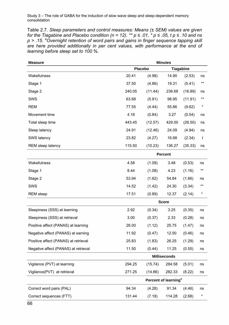
Table 2.7. Sleep parameters and control measures: Means (± SEM) values are given for the Tiagabine and Placebo condition (n = 12). ** p ≤ .01, * p ≤ .05, t p ≤ .10 and ns p > .15. aOvernight retention of word pairs and gains in finger sequence tapping skill are here provided additionally in per cent values, with performance at the end of learning before sleep set to 100 %.
Measure Minutes
Placebo Tiagabine
Wakefulness 20.41 (4.98) 14.90 (2.53) ns
Stage 1 37.50 (4.86) 19.31 (5.41) **
Stage 2 240.05 (11.44) 236.68 (16.89) ns
SWS 63.68 (5.91) 98.95 (11.91) **
REM 77.55 (4.44) 55.86 (9.62) *
Movement time 4.18 (0.84) 3.27 (0.54) ns
Total sleep time 443.45 (12.57) 429.00 (26.50) ns
Sleep latency 24.91 (12.46) 24.09 (4.94) ns
SWS latency 23.82 (4.27) 16.68 (2.34) t
REM sleep latency 115.50 (10.23) 136.27 (35.33) ns
Percent
Wakefulness 4.58 (1.09) 3.48 (0.53) ns
Stage 1 8.44 (1.08) 4.23 (1.16) **
Stage 2 53.94 (1.62) 54.84 (1.66) ns
SWS 14.52 (1.42) 24.30 (3.34) **
REM sleep 17.51 (0.89) 12.37 (2.14) *
Score
Sleepiness (SSS) at learning 2.92 (0.34) 3.25 (0.35) ns
Sleepiness (SSS) at retrieval 3.00 (0.37) 2.33 (0.28) ns
Positive affect (PANAS) at learning 26.00 (1.12) 25.75 (1.47) ns
Negative affect (PANAS) at earning 11.92 (0.47) 12.00 (0.46) ns
Positive affect (PANAS) at retrieval 25.83 (1.83) 26.25 (1.29) ns
Negative affect (PANAS) at retrieval 11.50 (0.44) 11.25 (0.55) ns
Milliseconds
Vigilance (PVT) at learning 294.25 (15.74) 284.58 (5.01) ns
Vigilance(PVT) at retrieval 271.25 (14.86) 282.33 (8.22) ns
Percent of learninga
Correct word pairs (PAL) 94.34 (4.28) 91.34 (4.46) ns
Against this background, the present negative finding that tiagabine-induced increas-
es in SWS and slow wave activity fail to enhance these memories, indicates that
phenotypic SWS per se is not a critical mechanism in the consolidation of these
memories. Also, comparison of slow oscillations showed comparable amplitudes,
slopes and morphology for these oscillations in the tiagabine and placebo condition,
which questions the primary relevance of slow oscillations for memory consolidation.
Figure 2.8 Schematic overview over the phase relationships between the slow oscillation and slow and fast sleep spindles; x-axis in milliseconds is relative to the (surface) negative slow oscillation peak. Fast spindle activity (12-15 Hz) increases during the down-to-up transition, is most pronounced during the up state of the slow oscillation and coincides with memory reactivations in the hippocampus and neocortex (Ji & Wilson, 2007; Mölle et al., 2011). Slow spindle activity (9-12 Hz), on the other hand, has its maximum during the up-to-down state transition (Mölle et al., 2011). The surface EEG negative half wave of the slow oscillation (down state), which is related to reduced neuronal firing, corresponds to a positive field potential wave in deeper cortical layers, while the surface EEG positive half wave (up state), which is related to increased neuronal firing, corresponds to a depth negative wave (Contreras & Steriade, 1995; Mölle et al., 2002; Molle, Yeshenko, Marshall, Sara, & Born, 2006).
Study 3 – The role of GABA for the induction of slow wave sleep and sleep-dependent memory consolidation
68
However, tiagabine distinctly reduced spindle activity. Importantly, this suppres-
sive influence was most prominent when analysing spindle activity in synchrony with
the slow oscillation cycle (Fig. 5 provides a schematic illustration of slow oscillation
and spindle coupling) and its presumed relationship to hippocampal memory reacti-
vations. Consistent with previous studies (e.g., Andrillon et al., 2011; Mölle et al.,
2011; Mölle et al., 2002), classical fast spindle activity in the 12-15 Hz range which
typically displays a more widespread centro-partietal cortical distribution, showed a
distinct increase during the depolarizing up-phase of the slow oscillation, reflecting a
driving influence slow oscillations exert on the thalamic generation of these spindles
(Steriade, 2006). By contrast slow (9-12 Hz) frontal spindle activity, which is a sepa-
rate kind of spindle activity whose function is less well understood, was synchronized
to the up-to-down transition of the slow oscillation (Mölle et al., 2011). Tiagabine pro-
foundly suppressed both types of synchronized spindle activity during the slow oscil-
lation cycle. In particular the classical fast spindle activity has been consistently found
to be associated with overnight retention of both declarative and procedural memo-
ries (Clemens, Fabo, & Halasz, 2005; Gais et al., 2002; Schabus et al., 2004; Tamaki
et al., 2009). Recent studies suggest that the synchronization of fast spindle activity
to the depolarizing slow oscillation up-state is critical to the consolidation effect (Cox,
Hofman, & Talamini, 2012; Ruch et al., 2012). Specifically it has been proposed that
the fast spindles represent a mechanism involved in the redistribution of memory rep-
resentations that become reactivated during SWS, to neocortical and striatal sites of
long-term storage (Bergmann et al., 2012; Clemens et al., 2011; Diekelmann & Born,
2010). Against this background, although density of slow oscillations was increased
after tiagabine, the reduced efficacy of these slow oscillations to drive and synchro-
nize fast spindle activity to the depolarizing upstate of these oscillations could well
explain the failure of tiagabine to produce any improvement in declarative memory
consolidation. An alternative explanation may be that SWS is already maximally ben-
eficial in its unmedicated quantity. However this is not supported by data that show
increasing slow oscillations and spindles above normal physiological levels by anoth-
er method, i.e., through transcranial direct current stimulation, which did increase de-
clarative memory consolidation in young healthy student participants (Marshall et al.,
2006). Also, it could be speculated that tiagabine failed to enhance SWS-dependent
memory consolidation because such enhancement was counteracted by immediate
(retrograde) amnestic effects of the GABA agonist on hippocampal memory traces
Study 3 – The role of GABA for the induction of slow wave sleep and sleep-dependent memory consolidation
69
(Chang & Liang, 2012). Moreover, in studies of fear memory in mice, hippocampal
microinjection of the GABA A receptor agonist muscimol impaired consolidation of
fear context when given 4 or 6 hours following training, but not when given 1 hour
after training (Misane, Kruis, Pieneman, Ogren, & Stiedl, 2013). Indeed, further re-
search seems necessary to explore immediate GABAergic effects on consolidation in
hippocampal networks.
Considering the particular robust association that has been revealed for overnight
gains in procedural skills and spindle activity during retention sleep, the suppressing
effect of tiagabine can also account for the significantly diminished sleep-associated
increases in sequence finger tapping speed in this condition. Alternatively, the dimin-
ished gains in sequence finger tapping could be a consequence of tiagabine reducing
REM sleep as REM sleep has been assumed to contribute to motor memory consoli-
consolidation during sleep and even impairs indicators of procedural memory consol-
idation.
Summary of the main results
71
3 Conclusions and general discussion
3.1 Summary of the main results
Sleeps role for memory has become clearer over the last decade, but the question
remains which plastic processes are involved. To shed some light the present work
manipulated the main excitatory neurotransmitter glutamate, the major neuromodula-
tor dopamine and the main inhibitory neurotransmitter GABA to elucidate their role for
sleep-dependent memory consolidation. The main points of the three studies are vis-
ualized in Figure 3.1. Study 1 revealed that NMDA receptor related plasticity is rele-
vant for sleep’s impact on memory, probably by translating glutamatergic reactivation
of memory traces into plastic changes within the hippocampus. Interestingly, blocking
NMDA or AMPA receptors had no effect on the declarative word pair memory task.
Nevertheless, increasing glutamate’s efficacy at the NMDA receptor using DCS in-
creased the amount of retained word pairs only if the participants were allowed to
sleep in the retention interval. This seemingly contradictory result is explained by the
higher efficacy of DCS at NMDA receptors containing the NR2A subunit. Study 2
suggests that dopaminergic circuits are reactivated during post learning sleep. During
a sleep containing retention interval pramipexole, a dopamine agonist, wiped out dif-
ferences between high and low rewarded items learned before sleep, which was ei-
ther because of interference at dopamine receptors within the hippocampus or be-
cause of inhibition of reward centres by autoreceptor activation. In study 3, tiagabine
was used to strongly increase SWS, but this failed to enhance memory. This was
probably due to disturbed spindle to slow oscillation coupling. Together these find-
ings indicate that the major players in neuronal communication also participate in
sleep-dependent memory consolidation. Specifically, glutamatergic plasticity is in-
volved in the transformation of reactivation into plastic changes. Dopamine signalling
seems to be important to distinguish between more or less important information and,
thus, modulates sleep-dependent memory consolidation. The tiagabine results high-
light that the effects of sleep’s oscillatory EEG pattern on memory relies on the intri-
cate interplay of different frequency bands.
Consequences for the active system consolidation hypothesis
72
Figure 3.1. Summary of main results. DCS improves declarative sleep-dependent memory consolidation by fa-cilitating plasticity, which is triggered by reactivations during SWS. Pramipexole wipes out reward related differences dur-ing retention by inhibiting reward centres and/or interfering with reward related sig-nalling. Tiagabine increases slow oscilla-tions during sleep but has no effect on memory, probably due to inhibition of spindle generation and spindle to slow oscillation coupling.
3.2 Consequences for the active system consolidation hypothesis
3.2.1 Initial consolidation within hippocampal networks
The active system consolidation hypothesis in its latest form (Rasch & Born, 2013)
establishes a model, where memory traces encoded into hippocampal ensembles
during prior wakefulness are redistributed to cortical stores during SWS. This theory,
based on the system consolidation concept (Dudai, 2004), constitutes a valid frame-
work fitting a variety of findings of sleep’s effect on memory that can explain findings
like insight after sleep (Wagner et al., 2004) or increased false memories
(Diekelmann et al., 2010; Payne et al., 2009). Nevertheless, there are also accounts
of initial hippocampal strengthening that could take place before memory traces are
transferred to the cortex (Inostroza & Born, 2013). And while there is evidence for
fast hippocampal disengagement if information relates to an existing schema (Tse et
al., 2007; Tse et al., 2011), Gais et al. (2007) show that hippocampus activation is
increased at retrieval if sleep was allowed directly after encoding. The latter is also in
keeping with evidence for hippocampal contribution to retrieval five days after acqui-
sition that disengages within 25 days (Bontempi, Laurent-Demir, Destrade, & Jaffard,
1999).
An important aspect for researching memory that is thought to rely on different
systems depending on its age is to consider if different plastic mechanisms are at
Consequences for the active system consolidation hypothesis
73
work in the anatomically different brain regions. One hint in this direction is that the
present data were unable to replicate the findings that sleep-dependent consolidation
of sensory memory can be blocked by AMPA and NMDA receptor antagonists in our
hippocampus dependent memory task (Gais et al., 2007). In fact, the acquisition of
sensory memory tasks relies on many repetitions of the same material during learn-
ing that are necessary to form connections between sparsely connected cortical neu-
The present experiments were carried out to a high standard. Nevertheless, future
experiments will be able to correct some shortcomings and extend on the results.
Generally, future research should go beyond the major players identified in the pre-
sent studies by identifying auxiliary roles of neurotransmitters and/or receptors that
have been shown to influence plasticity in the hippocampus, e.g., endocannabinoids,
ghrelin, metabotropic glutamate receptors or gap junctions,
Specifically, Study 1 summarizes data where retention intervals of different length
are used. As the ketamine and caroverine conditions had retention intervals only dur-
ing the early SWS rich half of the night it would be desirable to rule out that the sec-
ond half of the night is important to produce effects in the glutamatergic system. Also
Critical appraisal and future directions
78
it will be important to experimentally prove the influence of subunit composition of
NMDA receptors on the effect of sleep on forgetting and consolidation, e.g., by apply-
ing specific blockers in a rat model. This would also allow establishing whether con-
solidation is happening in the hippocampus or the cortex, by infusing directly into the
structure of interest.
Further, Study 2 found effects of a d2-like receptor agonist on reward memory.
Due to the drug applied there is no possibility to extract if inhibition of reward circuitry
or interference led to the effects. Therefore, it would be important to modulate other
dopamine receptors to see if a similar effect can be observed. Additionally it would be
interesting to see if other inputs to the dopaminergic system, e.g., by the prefrontal
cortex, also contribute to sleep-dependent consolidation. This could be done by
combining the pharmacological approach with psychological manipulations of rele-
vance.
Finally, Study 3 showed that pharmacologically increasing slow wave activity does
not improve memory. To rule out that the GABAergic agent had a disruptive influence
on LTP, which was countered by increased slow wave activity it would be of interest
to directly infuse tiagabine into the different brain areas, which could be done in an
animal model.
Abstract
79
4 Abstract
The beneficial influence of sleep on memory has received considerable support dur-
ing the last decade. The most widely accepted mechanism for the sleep-dependent
strengthening of memories acquired during preceding wakefulness is that of their re-
activation during slow wave sleep. The present thesis focused on pharmacological
manipulations of sleep-dependent memory consolidation to elucidate neurochemical
mechanisms that translate this reactivation into plastic changes. To this end, partici-
pants learned memory tasks before a retention interval, during which an active agent
was administered.
The most abundant excitatory neurotransmitter in the human brain glutamate par-
ticipates in many forms of plasticity. The best studied form of plasticity is that of the
glutamatergic synapse and relies on AMPA and NMDA receptor interaction. Howev-
er, it is unclear if glutamatergic neurotransmission mediates the sleep-dependent
consolidation of declarative memories. Hence, altering the action of these receptors
during sleep, promises insights into the neurochemical mechanisms of plasticity that
lead to sleep-dependent memory consolidation. Using AMPA and NMDA receptor
blockers during sleep did not influence the consolidation of a declarative word pair
associates task. Participant’s performance on the same task, however, was signifi-
cantly increased, if d-cycloserine, a NMDA receptor co-agonist, was given during
sleep. This result indicates that NMDA receptor mediated plasticity is important for
sleep-dependent declarative memory consolidation and that the processes involved
may differ from those observed during wakefulness.
Dopamine is an important neuromodulator that can influence memory strength by
facilitating synaptic plasticity. Activity of dopaminergic reward circuitry leads to better
learning of rewarding information. The goal of the second study was to elucidate if
dopaminergic circuitry is also involved during the reactivation of reward memory dur-
ing sleep. Participants had to learn pictures, for which they were promised a high or a
low reward at retrieval. Giving pramipexole, a d2-like receptor agonist, during the
sleep retention interval wiped out the high over low reward benefit observed under
placebo. Importantly, this effect was independent of encoding depth and thus speaks
Abstract
80
for reactivation of dopaminergic reward circuitry during sleep influencing the fate of
memory encoded during prior wakefulness.
Inhibitory neurotransmission in most cases involves the neurotransmitter GABA.
GABA has also been shown to be involved in switching between states of arousal
and sleep. It is important for the generation of the slow frequency EEG oscillations
characteristic of slow wave sleep that have been shown to benefit sleep-dependent
consolidation of declarative memory. In the third study administration of the GABA
reuptake inhibitor tiagabine during retention sleep was used to manipulate this gen-
erating mechanism of slow wave sleep with the aim of boosting declarative memory
retention. As expected, this manipulation highly increased the amount of slow wave
sleep and slow oscillations that participants displayed, but the associated sleep spin-
dle activity was dampened. As declarative memory was unexpectedly not enhanced
by this treatment, it seems probable that effective slow wave sleep must also be ac-
companied by sleep spindle activity phase-locked to slow oscillations.
Together these studies demonstrate how the pharmacological approach can yield
information about the neurochemical underpinnings of sleep-dependent memory
consolidation and identified neurotransmitter systems that are involved in this pro-
cess. Future pharmacological experiments, however, also in animal models, will have
to specify the mechanisms we are only beginning to understand.
Abstract (German)
81
5 Abstract (German)
Der positive Einfluss von Schlaf auf das Gedächtnis hat im letzten Jahrzehnt breite
Unterstützung gefunden. Der am weitesten akzeptierte Mechanismus für die schlaf-
abhängige Stärkung von Gedächtnisinhalten, die in der vorhergehenden Wachheit
aufgenommen wurden, ist ihre Reaktivierung während des Tiefschlafs. Die vorlie-
gende Arbeit hat sich mit der pharmakologischen Manipulation von schlafabhängiger
Gedächtniskonsolidierung beschäftigt, um neurochemische Prozesse offenzulegen,
die diese Reaktivierung in plastische Veränderungen umsetzen. Zu diesem Ziel lern-
ten Teilnehmer Gedächtnistests vor einem Retentionsintervall, während dessen eine
aktive Substanz verabreicht wurde.
Der am weitesten verbreitete exzitatorische Neurotransmitter im Gehirn Glutamat
ist an vielen Formen von Plastizität beteiligt. Die bestuntersuchte Art von Plastizität
kommt durch die Interaktion von AMPA- und NMDA-Rezeptoren zustande. Es ist je-
doch unklar ob glutamaterge Neurotransmission die schlafabhängige Konsolidierung
von deklarativen Gedächtnisinhalten vermittelt. Die Abläufe an diesen Rezeptoren im
Schlaf zu beeinflussen verspricht daher Einsichten in die neurochemischen Prozes-
se von Plastizität, die zu schlafabhängiger Gedächtniskonsolidierung führen. Der
Einsatz von AMPA- und NMDA-Rezeptorblockern hatte im Schlaf keinen Einfluss auf
eine deklarative Wort-Paar-Assoziationsaufgabe. Im Gegensatz dazu erhöhte sich
die Leistung von Teilnehmern, die den NMDA-Rezeptor-Co-Agonisten d-Cycloserine
vor dem Schlafengehen erhalten hatten. Dieses Ergebnis legt nahe, dass NMDA-
Rezeptor vermittelte Plastizität für schlafabhängige Gedächtniskonsolidierung von
Bedeutung ist und dass die Prozesse sich von denen im Wachzustand unterschei-
den.
Dopamin ist ein wichtiger Neuromodulator, der die Gedächtnisstärke durch die
Manipulation von synaptischer Plastizität beeinflussen kann. Die Aktivierung von do-
paminergen Belohnungsschaltkreisen führt zum besseren Lernen von belohnter In-
formation. Das Ziel der zweiten Studie war es herauszufinden, ob dopaminerge
Schaltkreise von der Reaktivierung belohnter Informationen im Schlaf betroffen sind.
Teilnehmer lernten Bilder, für die Ihnen eine hohe oder niedrige Belohnung verspro-
chen wurde. Die Einnahme von Pramipexol, einem d2-like Rezeptoragonisten, wäh-
Abstract (German)
82
rend des Schlafs führte zu einer Nivellierung des Effekts von Belohnung, der eine
bessere Behaltensleistung für hochbelohnte Bilder in der Placebobedingung bedingt
hatte. Wichtigerweise war dieser Effekt unabhängig von Enkodierungstiefe und
spricht daher für die Reaktivierung dopaminerger Schaltkreise im Schlaf, die das
Schicksal von Gedächtnisinhalten aus der vorangehenden Wachphase beeinflussen.
Inhibitorische Neurotransmission wird meist durch den Neurotransmitter GABA
vermittelt. GABA ist auch am Wechsel zwischen Stadien der Aufmerksamkeit und
Schlaf beteiligt. Der Neurotransmitter ist ebenfalls wichtig für die Generation der
langsamen EEG-Oszillationen, die den Tiefschlaf charakterisieren und die sich güns-
tig auf die schlafabhängige Gedächtniskonsolidierung auswirken. In der dritten Studie
wurde die Gabe des GABA-Wiederaufnahmehemmers Tiagabine während Schlafs
genutzt, um die tiefschlafgenerierenden Mechanismen zu aktivieren und somit dekla-
rative Gedächtnisleistung zu verbessen. Wie erwartet, wurde die Menge an Tiefschlaf
und langsamen Oszillationen stark erhöht, aber assoziierte Spindelaktivität wurde
unterdrückt. Da deklaratives Gedächtnis unerwarteterweise nicht beeinflusst wurde,
scheint es wahrscheinlich, dass effektiver Tiefschlaf mit der Phasenkopplung von
Schlafspindeln und langsamen Oszillation einhergehen muss.
Zusammen demonstrieren diese Studien, wie die pharmakologische Herange-
hensweise Einblicke in die neurochemischen Vorgänge der schlafabhängigen Ge-
dächtniskonsolidierung liefern kann und wie dadurch Neurotransmittersysteme identi-
fiziert werden können, die an diesem Prozess beteiligt sind. Zukünftige Experimente,
auch in Tiermodellen, werden die identifizierten Mechanismen, die wir gerade erst zu
verstehen beginnen, weiter spezifizieren müssen.
Reference list
83
6 Reference list
Abe, M., Schambra, H., Wassermann, E. M., Luckenbaugh, D., Schweighofer, N., & Cohen, L. G. (2011). Reward improves long-term retention of a motor memory through induction of offline memory gains. Curr Biol, 21(7), 557-562.
Adcock, R. A., Thangavel, A., Whitfield-Gabrieli, S., Knutson, B., & Gabrieli, J. D. (2006). Reward-motivated learning: mesolimbic activation precedes memory formation. Neuron, 50(3), 507-517.
Aeschbach, D., Cutler, A. J., & Ronda, J. M. (2008). A role for non-rapid-eye-movement sleep homeostasis in perceptual learning. J Neurosci, 28(11), 2766-2772.
Aiken, C. B. (2007). Pramipexole in psychiatry: a systematic review of the literature. J Clin Psychiatry, 68(8), 1230-1236.
Albouy, G., Sterpenich, V., Balteau, E., Vandewalle, G., Desseilles, M., Dang-Vu, T., . . . Maquet, P. (2008). Both the hippocampus and striatum are involved in consolidation of motor sequence memory. Neuron, 58(2), 261-272.
Albouy, G., Sterpenich, V., Vandewalle, G., Darsaud, A., Gais, S., Rauchs, G., . . . Maquet, P. (2013). Interaction between hippocampal and striatal systems predicts subsequent consolidation of motor sequence memory. PLoS One, 8(3), e59490.
Andrillon, T., Nir, Y., Staba, R. J., Ferrarelli, F., Cirelli, C., Tononi, G., & Fried, I. (2011). Sleep spindles in humans: insights from intracranial EEG and unit recordings. J Neurosci, 31(49), 17821-17834.
Antonenko, D., Diekelmann, S., Olsen, C., Born, J., & Molle, M. (2013). Napping to renew learning capacity: enhanced encoding after stimulation of sleep slow oscillations. Eur J Neurosci, 37(7), 1142-1152.
Antonini, A., & Calandrella, D. (2011). Pharmacokinetic evaluation of pramipexole. Expert Opin Drug Metab Toxicol, 7(10), 1307-1314.
Antony, J. W., Gobel, E. W., O'Hare, J. K., Reber, P. J., & Paller, K. A. (2012). Cued memory reactivation during sleep influences skill learning. Nat Neurosci, 15(8), 1114-1116.
Arzi, A., Shedlesky, L., Ben-Shaul, M., Nasser, K., Oksenberg, A., Hairston, I. S., & Sobel, N. (2012). Humans can learn new information during sleep. Nat Neurosci, 15(10), 1460-1465.
Aserinsky, E., & Kleitman, N. (1953). Regularly occurring periods of eye motility, and concomitant phenomena, during sleep. Science, 118(3062), 273-274.
Aston-Jones, G., & Bloom, F. E. (1981). Activity of norepinephrine-containing locus coeruleus neurons in behaving rats anticipates fluctuations in the sleep-waking cycle. J Neurosci, 1(8), 876-886.
Aton, S. J., Seibt, J., Dumoulin, M., Jha, S. K., Steinmetz, N., Coleman, T., . . . Frank, M. G. (2009). Mechanisms of sleep-dependent consolidation of cortical plasticity. Neuron, 61(3), 454-466.
Baez, M. V., Oberholzer, M. V., Cercato, M. C., Snitcofsky, M., Aguirre, A. I., & Jerusalinsky, D. A. (2013). NMDA Receptor Subunits in the Adult Rat Hippocampus Undergo Similar Changes after 5 Minutes in an Open Field and after LTP Induction. PLoS One, 8(2), e55244.
Baran, B., Pace-Schott, E. F., Ericson, C., & Spencer, R. M. (2012). Processing of emotional reactivity and emotional memory over sleep. J Neurosci, 32(3), 1035-1042.
Barrett, T. R., & Ekstrand, B. R. (1972). Effect of sleep on memory. 3. Controlling for time-of-day effects. J Exp Psychol, 96(2), 321-327.
Bast, T., da Silva, B. M., & Morris, R. G. (2005). Distinct contributions of hippocampal NMDA and AMPA receptors to encoding and retrieval of one-trial place memory. J Neurosci, 25(25), 5845-5856.
Reference list
84
Battaglia, F. P., Benchenane, K., Sirota, A., Pennartz, C. M., & Wiener, S. I. (2011). The hippocampus: hub of brain network communication for memory. Trends Cogn Sci, 15(7), 310-318.
Baudry, M., Bi, X., & Aguirre, C. (2012). Progesterone-estrogen interactions in synaptic plasticity and neuroprotection. Neuroscience, 239, 280-294.
Behrens, C. J., van den Boom, L. P., de Hoz, L., Friedman, A., & Heinemann, U. (2005). Induction of sharp wave-ripple complexes in vitro and reorganization of hippocampal networks. Nat Neurosci, 8(11), 1560-1567.
Ben Mamou, C., Gamache, K., & Nader, K. (2006). NMDA receptors are critical for unleashing consolidated auditory fear memories. Nat Neurosci, 9(10), 1237-1239.
Bendor, D., & Wilson, M. A. (2012). Biasing the content of hippocampal replay during sleep. Nat Neurosci, 15(10), 1439-1444.
Benedetto, L., Chase, M. H., & Torterolo, P. (2012). GABAergic processes within the median preoptic nucleus promote NREM sleep. Behav Brain Res, 232(1), 60-65.
Benson, K., & Feinberg, I. (1975). Sleep and memory: retention 8 and 24 hours after initial learning. Psychophysiology, 12(2), 192-195.
Berberich, S., Jensen, V., Hvalby, O., Seeburg, P. H., & Kohr, G. (2007). The role of NMDAR subtypes and charge transfer during hippocampal LTP induction. Neuropharmacology, 52(1), 77-86.
Bergmann, T. O., Molle, M., Diedrichs, J., Born, J., & Siebner, H. R. (2012). Sleep spindle-related reactivation of category-specific cortical regions after learning face-scene associations. Neuroimage, 59(3), 2733-2742.
Billard, J. M., & Rouaud, E. (2007). Deficit of NMDA receptor activation in CA1 hippocampal area of aged rats is rescued by D-cycloserine. Eur J Neurosci, 25(8), 2260-2268.
Bliss, T. V., & Collingridge, G. L. (2013). Expression of NMDA receptor-dependent LTP in the hippocampus: bridging the divide. Mol Brain, 6, 5.
Bontempo, A., Panza, K. E., & Bloch, M. H. (2012). D-cycloserine augmentation of behavioral therapy for the treatment of anxiety disorders: a meta-analysis. J Clin Psychiatry, 73(4), 533-537.
Borden, L. A., Murali Dhar, T. G., Smith, K. E., Weinshank, R. L., Branchek, T. A., & Gluchowski, C. (1994). Tiagabine, SK&F 89976-A, CI-966, and NNC-711 are selective for the cloned GABA transporter GAT-1. Eur J Pharmacol, 269(2), 219-224.
Born, J., & Feld, G. B. (2012). Sleep to upscale, sleep to downscale: balancing homeostasis and plasticity. Neuron, 75(6), 933-935.
Born, J., Lange, T., Hansen, K., Molle, M., & Fehm, H. L. (1997). Effects of sleep and circadian rhythm on human circulating immune cells. J Immunol, 158(9), 4454-4464.
Bourtchouladze, R., Abel, T., Berman, N., Gordon, R., Lapidus, K., & Kandel, E. R. (1998). Different training procedures recruit either one or two critical periods for contextual memory consolidation, each of which requires protein synthesis and PKA. Learn Mem, 5(4-5), 365-374.
Brown, R. M., & Robertson, E. M. (2007). Off-line processing: reciprocal interactions between declarative and procedural memories. J Neurosci, 27(39), 10468-10475.
Buchanan, A. (2011). Beyond humanity? The ethics of biomedical enhancement. New York: Oxford University Press.
Buzsaki, G. (1986). Hippocampal sharp waves: their origin and significance. Brain Res, 398(2), 242-252.
Caporale, N., & Dan, Y. (2008). Spike timing-dependent plasticity: a Hebbian learning rule. Annu Rev Neurosci, 31, 25-46.
Carpenter, G. A., & Grossberg, S. (1988). The Art of Adaptive Pattern-Recognition by a Self-Organizing Neural Network. Computer, 21(3), 77-88.
Chang, S. D., & Liang, K. C. (2012). Roles of hippocampal GABA(A) and muscarinic receptors in consolidation of context memory and context-shock association in
Reference list
85
contextual fear conditioning: a double dissociation study. Neurobiol Learn Mem, 98(1), 17-24.
Chen, H. H., Liao, P. F., & Chan, M. H. (2011). mGluR5 positive modulators both potentiate activation and restore inhibition in NMDA receptors by PKC dependent pathway. J Biomed Sci, 18, 19.
Chen, Z., Duan, M., Lee, H., Ruan, R., & Ulfendahl, M. (2003). Pharmacokinetics of caroverine in the inner ear and its effects on cochlear function after systemic and local administrations in Guinea pigs. Audiol Neurootol, 8(1), 49-56.
Chklovskii, D. B., Mel, B. W., & Svoboda, K. (2004). Cortical rewiring and information storage. Nature, 431(7010), 782-788.
Cirelli, C., & Tononi, G. (2008). Is sleep essential? PLoS Biol, 6(8), e216. Clemens, Z., Fabo, D., & Halasz, P. (2005). Overnight verbal memory retention correlates
with the number of sleep spindles. Neuroscience, 132(2), 529-535. Clemens, Z., Molle, M., Eross, L., Jakus, R., Rasonyi, G., Halasz, P., & Born, J. (2011). Fine-
tuned coupling between human parahippocampal ripples and sleep spindles. Eur J Neurosci, 33(3), 511-520.
Connor, J. R., Wang, X. S., Allen, R. P., Beard, J. L., Wiesinger, J. A., Felt, B. T., & Earley, C. J. (2009). Altered dopaminergic profile in the putamen and substantia nigra in restless leg syndrome. Brain, 132(Pt 9), 2403-2412.
Contreras, D., & Steriade, M. (1995). Cellular basis of EEG slow rhythms: a study of dynamic corticothalamic relationships. J Neurosci, 15(1 Pt 2), 604-622.
Cox, R., Hofman, W. F., & Talamini, L. M. (2012). Involvement of spindles in memory consolidation is slow wave sleep-specific. Learn Mem, 19(7), 264-267.
Crunelli, V., & Hughes, S. W. (2010). The slow (<1 Hz) rhythm of non-REM sleep: a dialogue between three cardinal oscillators. Nat Neurosci, 13(1), 9-17.
Dash, M. B., Douglas, C. L., Vyazovskiy, V. V., Cirelli, C., & Tononi, G. (2009). Long-term homeostasis of extracellular glutamate in the rat cerebral cortex across sleep and waking states. J Neurosci, 29(3), 620-629.
Dauer, W., & Przedborski, S. (2003). Parkinson's disease: mechanisms and models. Neuron, 39(6), 889-909.
Day, M., Langston, R., & Morris, R. G. (2003). Glutamate-receptor-mediated encoding and retrieval of paired-associate learning. Nature, 424(6945), 205-209.
Debas, K., Carrier, J., Orban, P., Barakat, M., Lungu, O., Vandewalle, G., . . . Doyon, J. (2010). Brain plasticity related to the consolidation of motor sequence learning and motor adaptation. Proc Natl Acad Sci U S A, 107(41), 17839-17844.
Diekelmann, S., & Born, J. (2010). The memory function of sleep. Nat Rev Neurosci, 11(2), 114-126.
Diekelmann, S., Born, J., & Wagner, U. (2010). Sleep enhances false memories depending on general memory performance. Behav Brain Res, 208(2), 425-429.
Diekelmann, S., Buchel, C., Born, J., & Rasch, B. (2011). Labile or stable: opposing consequences for memory when reactivated during waking and sleep. Nat Neurosci, 14(3), 381-386.
Diekelmann, S., Landolt, H. P., Lahl, O., Born, J., & Wagner, U. (2008). Sleep loss produces false memories. PLoS One, 3(10), e3512.
Dinges, D. F., Pack, F., Williams, K., Gillen, K. A., Powell, J. W., Ott, G. E., . . . Pack, A. I. (1997). Cumulative sleepiness, mood disturbance, and psychomotor vigilance performance decrements during a week of sleep restricted to 4-5 hours per night. Sleep, 20(4), 267-277.
Drosopoulos, S., Schulze, C., Fischer, S., & Born, J. (2007). Sleep's function in the spontaneous recovery and consolidation of memories. J Exp Psychol Gen, 136(2), 169-183.
Dudai, Y. (2004). The neurobiology of consolidations, or, how stable is the engram? Annu Rev Psychol, 55, 51-86.
Reference list
86
Dupret, D., O'Neill, J., Pleydell-Bouverie, B., & Csicsvari, J. (2010). The reorganization and reactivation of hippocampal maps predict spatial memory performance. Nat Neurosci, 13(8), 995-1002.
Dzirasa, K., Ribeiro, S., Costa, R., Santos, L. M., Lin, S. C., Grosmark, A., . . . Nicolelis, M. A. (2006). Dopaminergic control of sleep-wake states. J Neurosci, 26(41), 10577-10589.
Ebbinghaus, H. (1885). Über das Gedächtnis Leipzig: Duncker und Humblot. Edelmann, E., & Lessmann, V. (2013). Dopamine regulates intrinsic excitability thereby
gating successful induction of spike timing-dependent plasticity in CA1 of the hippocampus. Front Neurosci, 7, 25.
Ekstrand, B. R. (1967). Effect of sleep on memory. J Exp Psychol, 75(1), 64-72. Ekstrand, B. R., Barrett, T. R., West, J. N., & Maier, W. G. (1977). The effect of sleep on
human long-term memory. In R. Drucker-Colin & J. McGaugh (Eds.), Neurobiology of sleep and memory (pp. 419-438). New York: Academic Press.
Engeln, M., Ahmed, S. H., Vouillac, C., Tison, F., Bezard, E., & Fernagut, P. O. (2012). Reinforcing properties of Pramipexole in normal and parkinsonian rats. Neurobiol Dis, 49C, 79-86.
Eschenko, O., & Sara, S. J. (2008). Learning-dependent, transient increase of activity in noradrenergic neurons of locus coeruleus during slow wave sleep in the rat: brain stem-cortex interplay for memory consolidation? Cereb Cortex, 18(11), 2596-2603.
Espinosa, F., & Kavalali, E. T. (2009). NMDA receptor activation by spontaneous glutamatergic neurotransmission. J Neurophysiol, 101(5), 2290-2296.
Farah, M. J., Illes, J., Cook-Deegan, R., Gardner, H., Kandel, E., King, P., . . . Wolpe, P. R. (2004). Neurocognitive enhancement: what can we do and what should we do? Nat Rev Neurosci, 5(5), 421-425.
Faul, F., Erdfelder, E., Lang, A. G., & Buchner, A. (2007). G*Power 3: a flexible statistical power analysis program for the social, behavioral, and biomedical sciences. Behav Res Methods, 39(2), 175-191.
Feenstra, M. G., Botterblom, M. H., & Mastenbroek, S. (2000). Dopamine and noradrenaline efflux in the prefrontal cortex in the light and dark period: effects of novelty and handling and comparison to the nucleus accumbens. Neuroscience, 100(4), 741-748.
Feld, G. B., Besedovsky, L., Kaida, K., Munte, T. F., & Born, J. (2014). Dopamine D2-like Receptor Activation Wipes Out Preferential Consolidation of High over Low Reward Memories during Human Sleep. J Cogn Neurosci, 10.1162/jocn_a_00629.
Feld, G. B., & Born, J. (2012). Sleep EEG Rhythms and System Consolidation of Memory. In M. G. Frank (Ed.), Sleep and Brain Activity (1st ed., pp. 187-226). Waltham, MA: Academic Press.
Feld, G. B., Lange, T., Gais, S., & Born, J. (2013). Sleep-dependent declarative memory consolidation--unaffected after blocking NMDA or AMPA receptors but enhanced by NMDA coagonist D-cycloserine. Neuropsychopharmacology, 38(13), 2688-2697.
Feld, G. B., Wilhelm, I., Ma, Y., Groch, S., Binkofski, F., Molle, M., & Born, J. (2013). Slow wave sleep induced by GABA agonist tiagabine fails to benefit memory consolidation. Sleep, 36(9), 1317-1326.
Fink-Jensen, A., Suzdak, P. D., Swedberg, M. D., Judge, M. E., Hansen, L., & Nielsen, P. G. (1992). The gamma-aminobutyric acid (GABA) uptake inhibitor, tiagabine, increases extracellular brain levels of GABA in awake rats. Eur J Pharmacol, 220(2-3), 197-201.
Fischer, S., & Born, J. (2009). Anticipated reward enhances offline learning during sleep. J Exp Psychol Learn Mem Cogn, 35(6), 1586-1593.
Fogel, S. M., & Smith, C. T. (2011). The function of the sleep spindle: a physiological index of intelligence and a mechanism for sleep-dependent memory consolidation. Neurosci Biobehav Rev, 35(5), 1154-1165.
Frankland, P. W., & Bontempi, B. (2005). The organization of recent and remote memories. Nat Rev Neurosci, 6(2), 119-130.
Fuentealba, P., & Steriade, M. (2005). The reticular nucleus revisited: intrinsic and network properties of a thalamic pacemaker. Prog Neurobiol, 75(2), 125-141.
Reference list
87
Gais, S., Albouy, G., Boly, M., Dang-Vu, T. T., Darsaud, A., Desseilles, M., . . . Peigneux, P. (2007). Sleep transforms the cerebral trace of declarative memories. Proc Natl Acad Sci U S A, 104(47), 18778-18783.
Gais, S., & Born, J. (2004a). Declarative memory consolidation: mechanisms acting during human sleep. Learn Mem, 11(6), 679-685.
Gais, S., & Born, J. (2004b). Low acetylcholine during slow-wave sleep is critical for declarative memory consolidation. Proc Natl Acad Sci U S A, 101(7), 2140-2144.
Gais, S., Molle, M., Helms, K., & Born, J. (2002). Learning-dependent increases in sleep spindle density. J Neurosci, 22(15), 6830-6834.
Gais, S., Plihal, W., Wagner, U., & Born, J. (2000). Early sleep triggers memory for early visual discrimination skills. Nat Neurosci, 3(12), 1335-1339.
Gais, S., Rasch, B., Dahmen, J. C., Sara, S., & Born, J. (2011). The memory function of noradrenergic activity in non-REM sleep. J Cogn Neurosci, 23(9), 2582-2592.
Gais, S., Rasch, B., Wagner, U., & Born, J. (2008). Visual-procedural memory consolidation during sleep blocked by glutamatergic receptor antagonists. J Neurosci, 28(21), 5513-5518.
Gore, A. C. (1998). Diurnal rhythmicity of gonadotropin-releasing hormone gene expression in the rat. Neuroendocrinology, 68(4), 257-263.
Goto, Y., & Grace, A. A. (2005). Dopamine-dependent interactions between limbic and prefrontal cortical plasticity in the nucleus accumbens: disruption by cocaine sensitization. Neuron, 47(2), 255-266.
Groch, S., Wilhelm, I., Diekelmann, S., Sayk, F., Gais, S., & Born, J. (2011). Contribution of norepinephrine to emotional memory consolidation during sleep. Psychoneuroendocrinology, 36(9), 1342-1350.
Groch, S., Wilhelm, I., Lange, T., & Born, J. (2013). Differential contribution of mineralocorticoid and glucocorticoid receptors to memory formation during sleep. Psychoneuroendocrinology, 38(12), 2962-2972.
Hall-Porter, J. M., Schweitzer, P. K., Eisenstein, R. D., Ahmed, H. A., & Walsh, J. K. (2014). The effect of two benzodiazepine receptor agonist hypnotics on sleep-dependent memory consolidation. J Clin Sleep Med, 10(1), 27-34.
Hardt, O., Migues, P. V., Hastings, M., Wong, J., & Nader, K. (2010). PKMzeta maintains 1-day- and 6-day-old long-term object location but not object identity memory in dorsal hippocampus. Hippocampus, 20(6), 691-695.
Hardt, O., Nader, K., & Nadel, L. (2013). Decay happens: the role of active forgetting in memory. Trends Cogn Sci, 17(3), 111-120.
Hartvig, P., Valtysson, J., Lindner, K. J., Kristensen, J., Karlsten, R., Gustafsson, L. L., . . . et al. (1995). Central nervous system effects of subdissociative doses of (S)-ketamine are related to plasma and brain concentrations measured with positron emission tomography in healthy volunteers. Clin Pharmacol Ther, 58(2), 165-173.
Hasselmo, M. E. (1999). Neuromodulation: acetylcholine and memory consolidation. Trends Cogn Sci, 3(9), 351-359.
Hebb, D. O. (1949). The Organization of Behavior: A Neuropsychological Theory. New York: Wiley.
Hefti, K., Holst, S. C., Sovago, J., Bachmann, V., Buck, A., Ametamey, S. M., . . . Landolt, H. P. (2013). Increased metabotropic glutamate receptor subtype 5 availability in human brain after one night without sleep. Biol Psychiatry, 73(2), 161-168.
Hoddes, E., Zarcone, V., Smythe, H., Phillips, R., & Dement, W. C. (1973). Quantification of sleepiness: a new approach. Psychophysiology, 10(4), 431-436.
Hrabetova, S., & Sacktor, T. C. (2001). Transient translocation of conventional protein kinase C isoforms and persistent downregulation of atypical protein kinase Mzeta in long-term depression. Brain Res Mol Brain Res, 95(1-2), 146-152.
Huber, R., Ghilardi, M. F., Massimini, M., & Tononi, G. (2004). Local sleep and learning. Nature, 430(6995), 78-81.
Huber, R., Hill, S. L., Holladay, C., Biesiadecki, M., Tononi, G., & Cirelli, C. (2004). Sleep homeostasis in Drosophila melanogaster. Sleep, 27(4), 628-639.
Reference list
88
Hulme, S. R., Jones, O. D., & Abraham, W. C. (2013). Emerging roles of metaplasticity in behaviour and disease. Trends Neurosci, 36(6), 353-362.
Ikeda, K., & Morotomi, T. (1996). Classical conditioning during human NREM sleep and response transfer to wakefulness. Sleep, 19(1), 72-74.
Inostroza, M., & Born, J. (2013). Sleep for Preserving and Transforming Episodic Memory. Annual Review of Neuroscience, 36, 79-102.
Janke, W., & Debus, G. (1978). Die Eigenschaftswörterliste EWL. Göttingen: Hogrefe. Jankovic, J., & Poewe, W. (2012). Therapies in Parkinson's disease. Curr Opin Neurol, 25(4),
433-447. Jenkins, John G, & Dallenbach, Karl M. (1924). Obliviscence during sleep and waking. The
American Journal of Psychology, 35(4), 605-612. Ji, D., & Wilson, M. A. (2007). Coordinated memory replay in the visual cortex and
hippocampus during sleep. Nat Neurosci, 10(1), 100-107. Kaestner, E. J., Wixted, J. T., & Mednick, S. C. (2013). Pharmacologically increasing sleep
spindles enhances recognition for negative and high-arousal memories. J Cogn Neurosci, 25(10), 1597-1610.
Kalisch, R., Holt, B., Petrovic, P., De Martino, B., Kloppel, S., Buchel, C., & Dolan, R. J. (2009). The NMDA agonist D-cycloserine facilitates fear memory consolidation in humans. Cereb Cortex, 19(1), 187-196.
Karni, A., Meyer, G., Rey-Hipolito, C., Jezzard, P., Adams, M. M., Turner, R., & Ungerleider, L. G. (1998). The acquisition of skilled motor performance: fast and slow experience-driven changes in primary motor cortex. Proc Natl Acad Sci U S A, 95(3), 861-868.
Karni, A., Tanne, D., Rubenstein, B. S., Askenasy, J. J., & Sagi, D. (1994). Dependence on REM sleep of overnight improvement of a perceptual skill. Science, 265(5172), 679-682.
King, C., Henze, D. A., Leinekugel, X., & Buzsaki, G. (1999). Hebbian modification of a hippocampal population pattern in the rat. J Physiol, 521 Pt 1, 159-167.
Kleckner, N. W., & Dingledine, R. (1988). Requirement for glycine in activation of NMDA-receptors expressed in Xenopus oocytes. Science, 241(4867), 835-837.
Kochlamazashvili, G., Bukalo, O., Senkov, O., Salmen, B., Gerardy-Schahn, R., Engel, A. K., . . . Dityatev, A. (2012). Restoration of synaptic plasticity and learning in young and aged NCAM-deficient mice by enhancing neurotransmission mediated by GluN2A-containing NMDA receptors. J Neurosci, 32(7), 2263-2275.
Kuriyama, K., Honma, M., Koyama, S., & Kim, Y. (2011). D-cycloserine facilitates procedural learning but not declarative learning in healthy humans: a randomized controlled trial of the effect of D-cycloserine and valproic acid on overnight properties in the performance of non-emotional memory tasks. Neurobiol Learn Mem, 95(4), 505-509.
Kuriyama, K., Honma, M., Shimazaki, M., Horie, M., Yoshiike, T., Koyama, S., & Kim, Y. (2011). An N-methyl-D-aspartate receptor agonist facilitates sleep-independent synaptic plasticity associated with working memory capacity enhancement. Sci Rep, 1, 127.
Kuriyama, K., Honma, M., Soshi, T., Fujii, T., & Kim, Y. (2011). Effect of D-cycloserine and valproic acid on the extinction of reinstated fear-conditioned responses and habituation of fear conditioning in healthy humans: a randomized controlled trial. Psychopharmacology (Berl), 218(3), 589-597.
Lancel, M. (1999). Role of GABAA receptors in the regulation of sleep: initial sleep responses to peripherally administered modulators and agonists. Sleep, 22(1), 33-42.
Lancel, M., Faulhaber, J., & Deisz, R. A. (1998). Effect of the GABA uptake inhibitor tiagabine on sleep and EEG power spectra in the rat. Br J Pharmacol, 123(7), 1471-1477.
Lansink, C. S., Goltstein, P. M., Lankelma, J. V., Joosten, R. N., McNaughton, B. L., & Pennartz, C. M. (2008). Preferential reactivation of motivationally relevant information in the ventral striatum. J Neurosci, 28(25), 6372-6382.
Reference list
89
Lansink, C. S., Goltstein, P. M., Lankelma, J. V., McNaughton, B. L., & Pennartz, C. M. (2009). Hippocampus leads ventral striatum in replay of place-reward information. PLoS Biol, 7(8), e1000173.
Lanthorn, T. H. (1994). D-Cycloserine: Agonist turned antagonist. Amino Acids, 6(3), 247-260.
Lee, J. L., Milton, A. L., & Everitt, B. J. (2006). Reconsolidation and extinction of conditioned fear: inhibition and potentiation. J Neurosci, 26(39), 10051-10056.
Lewis, P. A., & Durrant, S. J. (2011). Overlapping memory replay during sleep builds cognitive schemata. Trends Cogn Sci, 15(8), 343-351.
Lisman, J. E., & Grace, A. A. (2005). The hippocampal-VTA loop: controlling the entry of information into long-term memory. Neuron, 46(5), 703-713.
Liu, L., Wong, T. P., Pozza, M. F., Lingenhoehl, K., Wang, Y., Sheng, M., . . . Wang, Y. T. (2004). Role of NMDA receptor subtypes in governing the direction of hippocampal synaptic plasticity. Science, 304(5673), 1021-1024.
Malenka, R. C., & Bear, M. F. (2004). LTP and LTD: an embarrassment of riches. Neuron, 44(1), 5-21.
Malenka, R. C., & Nicoll, R. A. (1999). Long-term potentiation--a decade of progress? Science, 285(5435), 1870-1874.
Manahan-Vaughan, D., & Kulla, A. (2003). Regulation of depotentiation and long-term potentiation in the dentate gyrus of freely moving rats by dopamine D2-like receptors. Cereb Cortex, 13(2), 123-135.
Mander, B. A., Rao, V., Lu, B., Saletin, J. M., Lindquist, J. R., Ancoli-Israel, S., . . . Walker, M. P. (2013). Prefrontal atrophy, disrupted NREM slow waves and impaired hippocampal-dependent memory in aging. Nat Neurosci, 16(3), 357-364.
Maret, S., Faraguna, U., Nelson, A. B., Cirelli, C., & Tononi, G. (2011). Sleep and waking modulate spine turnover in the adolescent mouse cortex. Nat Neurosci, 14(11), 1418-1420.
Marr, D. (1971). Simple memory: a theory for archicortex. Philos Trans R Soc Lond B Biol Sci, 262(841), 23-81.
Marrosu, F., Portas, C., Mascia, M. S., Casu, M. A., Fa, M., Giagheddu, M., . . . Gessa, G. L. (1995). Microdialysis measurement of cortical and hippocampal acetylcholine release during sleep-wake cycle in freely moving cats. Brain Res, 671(2), 329-332.
Marshall, L., Helgadottir, H., Molle, M., & Born, J. (2006). Boosting slow oscillations during sleep potentiates memory. Nature, 444(7119), 610-613.
Marshall, L., Kirov, R., Brade, J., Molle, M., & Born, J. (2011). Transcranial electrical currents to probe EEG brain rhythms and memory consolidation during sleep in humans. PLoS One, 6(2), e16905.
Marshall, L., Molle, M., Hallschmid, M., & Born, J. (2004). Transcranial direct current stimulation during sleep improves declarative memory. J Neurosci, 24(44), 9985-9992.
Mathias, S., Wetter, T. C., Steiger, A., & Lancel, M. (2001). The GABA uptake inhibitor tiagabine promotes slow wave sleep in normal elderly subjects. Neurobiol Aging, 22(2), 247-253.
McCabe, C., Harwood, J., Brouwer, S., Harmer, C. J., & Cowen, P. J. (2013). Effects of pramipexole on the processing of rewarding and aversive taste stimuli. Psychopharmacology (Berl), 228(2), 283-290.
McClelland, J. L., McNaughton, B. L., & O'Reilly, R. C. (1995). Why there are complementary learning systems in the hippocampus and neocortex: insights from the successes and failures of connectionist models of learning and memory. Psychol Rev, 102(3), 419-457.
McGeoch, John A. (1932). Forgetting and the law of disuse. Psychological Review, 39(4), 352.
McNaughton, B. L., Barnes, C. A., Rao, G., Baldwin, J., & Rasmussen, M. (1986). Long-term enhancement of hippocampal synaptic transmission and the acquisition of spatial information. J Neurosci, 6(2), 563-571.
Reference list
90
Mednick, S. C., McDevitt, E. A., Walsh, J. K., Wamsley, E., Paulus, M., Kanady, J. C., & Drummond, S. P. (2013). The critical role of sleep spindles in hippocampal-dependent memory: a pharmacology study. J Neurosci, 33(10), 4494-4504.
Melendez, J., Galli, I., Boric, K., Ortega, A., Zuniga, L., Henriquez-Roldan, C. F., & Cardenas, A. M. (2005). Zolpidem and triazolam do not affect the nocturnal sleep-induced memory improvement. Psychopharmacology (Berl), 181(1), 21-26.
Menz, M. M., Rihm, J. S., Salari, N., Born, J., Kalisch, R., Pape, H. C., . . . Buchel, C. (2013). The role of sleep and sleep deprivation in consolidating fear memories. Neuroimage, 75, 87-96.
Migues, P. V., Hardt, O., Wu, D. C., Gamache, K., Sacktor, T. C., Wang, Y. T., & Nader, K. (2010). PKMzeta maintains memories by regulating GluR2-dependent AMPA receptor trafficking. Nat Neurosci, 13(5), 630-634.
Milekic, M. H., & Alberini, C. M. (2002). Temporally graded requirement for protein synthesis following memory reactivation. Neuron, 36(3), 521-525.
Misane, I., Kruis, A., Pieneman, A. W., Ogren, S. O., & Stiedl, O. (2013). GABA(A) receptor activation in the CA1 area of the dorsal hippocampus impairs consolidation of conditioned contextual fear in C57BL/6J mice. Behav Brain Res, 238, 160-169.
Missale, C., Nash, S. R., Robinson, S. W., Jaber, M., & Caron, M. G. (1998). Dopamine receptors: from structure to function. Physiol Rev, 78(1), 189-225.
Mölle, M., Bergmann, T. O., Marshall, L., & Born, J. (2011). Fast and Slow Spindles during the Sleep Slow Oscillation: Disparate Coalescence and Engagement in Memory Processing. Sleep, 34(10), 1411-1421.
Molle, M., & Born, J. (2011). Slow oscillations orchestrating fast oscillations and memory consolidation. Prog Brain Res, 193, 93-110.
Mölle, M., Marshall, L., Gais, S., & Born, J. (2002). Grouping of spindle activity during slow oscillations in human non-rapid eye movement sleep. J Neurosci, 22(24), 10941-10947.
Molle, M., Yeshenko, O., Marshall, L., Sara, S. J., & Born, J. (2006). Hippocampal sharp wave-ripples linked to slow oscillations in rat slow-wave sleep. J Neurophysiol, 96(1), 62-70.
Mothet, J. P., Parent, A. T., Wolosker, H., Brady, R. O., Jr., Linden, D. J., Ferris, C. D., . . . Snyder, S. H. (2000). D-serine is an endogenous ligand for the glycine site of the N-methyl-D-aspartate receptor. Proc Natl Acad Sci U S A, 97(9), 4926-4931.
Murayama, K., & Kuhbandner, C. (2011). Money enhances memory consolidation--but only for boring material. Cognition, 119(1), 120-124.
Nader, K., & Hardt, O. (2009). A single standard for memory: the case for reconsolidation. Nat Rev Neurosci, 10(3), 224-234.
Ngo, H. V., Martinetz, T., Born, J., & Molle, M. (2013). Auditory closed-loop stimulation of the sleep slow oscillation enhances memory. Neuron, 78(3), 545-553.
Nishida, M., Pearsall, J., Buckner, R. L., & Walker, M. P. (2009). REM sleep, prefrontal theta, and the consolidation of human emotional memory. Cereb Cortex, 19(5), 1158-1166.
Nishida, M., & Walker, M. P. (2007). Daytime naps, motor memory consolidation and regionally specific sleep spindles. PLoS One, 2(4), e341.
O'Neill, J., Pleydell-Bouverie, B., Dupret, D., & Csicsvari, J. (2010). Play it again: reactivation of waking experience and memory. Trends Neurosci, 33(5), 220-229.
Onur, O. A., Schlaepfer, T. E., Kukolja, J., Bauer, A., Jeung, H., Patin, A., . . . Hurlemann, R. (2010). The N-methyl-D-aspartate receptor co-agonist D-cycloserine facilitates declarative learning and hippocampal activity in humans. Biol Psychiatry, 67(12), 1205-1211.
Oudiette, D., Antony, J. W., Creery, J. D., & Paller, K. A. (2013). The role of memory reactivation during wakefulness and sleep in determining which memories endure. J Neurosci, 33(15), 6672-6678.
Oudiette, D., & Paller, K. A. (2013). Upgrading the sleeping brain with targeted memory reactivation. Trends Cogn Sci, 17(3), 142-149.
Reference list
91
Pace-Schott, E. F., & Hobson, J. A. (2002). The neurobiology of sleep: genetics, cellular physiology and subcortical networks. Nat Rev Neurosci, 3(8), 591-605.
Payne, J. D., Schacter, D. L., Propper, R. E., Huang, L. W., Wamsley, E. J., Tucker, M. A., . . . Stickgold, R. (2009). The role of sleep in false memory formation. Neurobiol Learn Mem, 92(3), 327-334.
Peineau, S., Taghibiglou, C., Bradley, C., Wong, T. P., Liu, L., Lu, J., . . . Collingridge, G. L. (2007). LTP inhibits LTD in the hippocampus via regulation of GSK3beta. Neuron, 53(5), 703-717.
Pennartz, C. M., Lee, E., Verheul, J., Lipa, P., Barnes, C. A., & McNaughton, B. L. (2004). The ventral striatum in off-line processing: ensemble reactivation during sleep and modulation by hippocampal ripples. J Neurosci, 24(29), 6446-6456.
Pizzagalli, D. A., Evins, A. E., Schetter, E. C., Frank, M. J., Pajtas, P. E., Santesso, D. L., & Culhane, M. (2008). Single dose of a dopamine agonist impairs reinforcement learning in humans: behavioral evidence from a laboratory-based measure of reward responsiveness. Psychopharmacology (Berl), 196(2), 221-232.
Plihal, W., & Born, J. (1997). Effects of early and late nocturnal sleep on declarative and procedural memory. Journal of Cognitive Neuroscience, 9(4), 534-547.
Plihal, W., & Born, J. (1999). Memory consolidation in human sleep depends on inhibition of glucocorticoid release. Neuroreport, 10(13), 2741-2747.
Plihal, W., Pietrowsky, R., & Born, J. (1999). Dexamethasone blocks sleep induced improvement of declarative memory. Psychoneuroendocrinology, 24(3), 313-331.
Pourcher, E., Remillard, S., & Cohen, H. (2010). Compulsive habits in restless legs syndrome patients under dopaminergic treatment. J Neurol Sci, 290(1-2), 52-56.
Quartermain, D., Mower, J., Rafferty, M. F., Herting, R. L., & Lanthorn, T. H. (1994). Acute but not chronic activation of the NMDA-coupled glycine receptor with D-cycloserine facilitates learning and retention. Eur J Pharmacol, 257(1-2), 7-12.
Raizen, D. M., Zimmerman, J. E., Maycock, M. H., Ta, U. D., You, Y. J., Sundaram, M. V., & Pack, A. I. (2008). Lethargus is a Caenorhabditis elegans sleep-like state. Nature, 451(7178), 569-572.
Rasch, B., & Born, J. (2013). About sleep's role in memory. Physiol Rev, 93(2), 681-766. Rasch, B., Born, J., & Gais, S. (2006). Combined blockade of cholinergic receptors shifts the
brain from stimulus encoding to memory consolidation. J Cogn Neurosci, 18(5), 793-802.
Rasch, B., Gais, S., & Born, J. (2009). Impaired off-line consolidation of motor memories after combined blockade of cholinergic receptors during REM sleep-rich sleep. Neuropsychopharmacology, 34(7), 1843-1853.
Rasch, B., Pommer, J., Diekelmann, S., & Born, J. (2009). Pharmacological REM sleep suppression paradoxically improves rather than impairs skill memory. Nat Neurosci, 12(4), 396-397.
Rechtschaffen, A., & Kales, A. (1968). A manual of standardized terminology, technique and scoring system for sleep stages of human sleep. Los Angeles Brain Information Service. Brain Information Institute, UCLA.
Riba, J., Kramer, U. M., Heldmann, M., Richter, S., & Münte, T. F. (2008). Dopamine agonist increases risk taking but blunts reward-related brain activity. PLoS One, 3(6), e2479.
Ribeiro, S., Shi, X., Engelhard, M., Zhou, Y., Zhang, H., Gervasoni, D., . . . Nicolelis, M. A. (2007). Novel experience induces persistent sleep-dependent plasticity in the cortex but not in the hippocampus. Front Neurosci, 1(1), 43-55.
Riddle, J. L., Rokosik, S. L., & Napier, T. C. (2012). Pramipexole- and methamphetamine-induced reward-mediated behavior in a rodent model of Parkinson's disease and controls. Behav Brain Res, 233(1), 15-23.
Rosenzweig, E. S., Barnes, C. A., & McNaughton, B. L. (2002). Making room for new memories. Nat Neurosci, 5(1), 6-8.
Ruch, S., Markes, O., Duss, S. B., Oppliger, D., Reber, T. P., Koenig, T., . . . Henke, K. (2012). Sleep stage II contributes to the consolidation of declarative memories. Neuropsychologia, 50(10), 2389-2396.
Rudoy, J. D., Voss, J. L., Westerberg, C. E., & Paller, K. A. (2009). Strengthening individual memories by reactivating them during sleep. Science, 326(5956), 1079.
Saletu, M., Anderer, P., Saletu-Zyhlarz, G., Hauer, C., & Saletu, B. (2002). Acute placebo-controlled sleep laboratory studies and clinical follow-up with pramipexole in restless legs syndrome. Eur Arch Psychiatry Clin Neurosci, 252(4), 185-194.
Santesso, D. L., Evins, A. E., Frank, M. J., Schetter, E. C., Bogdan, R., & Pizzagalli, D. A. (2009). Single dose of a dopamine agonist impairs reinforcement learning in humans: evidence from event-related potentials and computational modeling of striatal-cortical function. Hum Brain Mapp, 30(7), 1963-1976.
Sarter, M., Bruno, J. P., & Givens, B. (2003). Attentional functions of cortical cholinergic inputs: what does it mean for learning and memory? Neurobiol Learn Mem, 80(3), 245-256.
Schabus, M., Gruber, G., Parapatics, S., Sauter, C., Klosch, G., Anderer, P., . . . Zeitlhofer, J. (2004). Sleep spindles and their significance for declarative memory consolidation. Sleep, 27(8), 1479-1485.
Schonauer, M., Geisler, T., & Gais, S. (2014). Strengthening procedural memories by reactivation in sleep. J Cogn Neurosci, 26(1), 143-153.
Schotanus, S. M., & Chergui, K. (2008). Dopamine D1 receptors and group I metabotropic glutamate receptors contribute to the induction of long-term potentiation in the nucleus accumbens. Neuropharmacology, 54(5), 837-844.
Schultz, W. (2000). Multiple reward signals in the brain. Nat Rev Neurosci, 1(3), 199-207. Schultz, W. (2007). Multiple dopamine functions at different time courses. Annu Rev
Neurosci, 30, 259-288. Schultz, W. (2013). Updating dopamine reward signals. Curr Opin Neurobiol, 23(2), 229-238. Scoville, W. B., & Milner, B. (1957). Loss of recent memory after bilateral hippocampal
lesions. J Neurol Neurosurg Psychiatry, 20(1), 11-21. Seibt, J., Aton, S. J., Jha, S. K., Coleman, T., Dumoulin, M. C., & Frank, M. G. (2008). The
Sheinin, A., Shavit, S., & Benveniste, M. (2001). Subunit specificity and mechanism of action of NMDA partial agonist D-cycloserine. Neuropharmacology, 41(2), 151-158.
Sherin, J. E., Shiromani, P. J., McCarley, R. W., & Saper, C. B. (1996). Activation of ventrolateral preoptic neurons during sleep. Science, 271(5246), 216-219.
Shohamy, D., & Adcock, R. A. (2010). Dopamine and adaptive memory. Trends Cogn Sci, 14(10), 464-472.
Siapas, A. G., & Wilson, M. A. (1998). Coordinated interactions between hippocampal ripples and cortical spindles during slow-wave sleep. Neuron, 21(5), 1123-1128.
Silber, M. H., Ancoli-Israel, S., Bonnet, M. H., Chokroverty, S., Grigg-Damberger, M. M., Hirshkowitz, M., . . . Iber, C. (2007). The visual scoring of sleep in adults. J Clin Sleep Med, 3(2), 121-131.
Skaggs, W. E., & McNaughton, B. L. (1996). Replay of neuronal firing sequences in rat hippocampus during sleep following spatial experience. Science, 271(5257), 1870-1873.
Sokoloff, P., Diaz, J., Le Foll, B., Guillin, O., Leriche, L., Bezard, E., & Gross, C. (2006). The dopamine D3 receptor: a therapeutic target for the treatment of neuropsychiatric disorders. CNS Neurol Disord Drug Targets, 5(1), 25-43.
Reference list
93
Sowers, J. R., & Vlachakis, N. (1984). Circadian variation in plasma dopamine levels in man. J Endocrinol Invest, 7(4), 341-345.
Spiegel, K., Follenius, M., Simon, C., Saini, J., Ehrhart, J., & Brandenberger, G. (1994). Prolactin secretion and sleep. Sleep, 17(1), 20-27.
Spratt, D. I., O'Dea, L. S., Schoenfeld, D., Butler, J., Rao, P. N., & Crowley, W. F., Jr. (1988). Neuroendocrine-gonadal axis in men: frequent sampling of LH, FSH, and testosterone. Am J Physiol, 254(5 Pt 1), E658-666.
Squire, L. R. (1992). Declarative and nondeclarative memory: multiple brain systems supporting learning and memory. J Cogn Neurosci, 4(3), 232-243.
Squire, L. R., & Wixted, J. T. (2011). The cognitive neuroscience of human memory since H.M. Annu Rev Neurosci, 34, 259-288.
Squire, L. R., & Zola, S. M. (1996). Structure and function of declarative and nondeclarative memory systems. Proc Natl Acad Sci U S A, 93(24), 13515-13522.
Steriade, M. (2003). The corticothalamic system in sleep. Front Biosci, 8, d878-899. Steriade, M. (2006). Grouping of brain rhythms in corticothalamic systems. Neuroscience,
137(4), 1087-1106. Tadavarty, R., Rajput, P. S., Wong, J. M., Kumar, U., & Sastry, B. R. (2011). Sleep-
deprivation induces changes in GABA(B) and mGlu receptor expression and has consequences for synaptic long-term depression. PLoS One, 6(9), e24933.
Tamaki, M., Matsuoka, T., Nittono, H., & Hori, T. (2009). Activation of fast sleep spindles at the premotor cortex and parietal areas contributes to motor learning: a study using sLORETA. Clin Neurophysiol, 120(5), 878-886.
Thomas, M. J., Malenka, R. C., & Bonci, A. (2000). Modulation of long-term depression by dopamine in the mesolimbic system. J Neurosci, 20(15), 5581-5586.
Thorndike, Edward Lee. (1913). Educational Psychology, Vol 2: The Psychology of Learning. New York: Teacher’s College Press.
Timofeev, I., Grenier, F., & Steriade, M. (2000). Impact of intrinsic properties and synaptic factors on the activity of neocortical networks in vivo. J Physiol Paris, 94(5-6), 343-355.
Tononi, G., & Cirelli, C. (2003). Sleep and synaptic homeostasis: a hypothesis. Brain Res Bull, 62(2), 143-150.
Tononi, G., & Cirelli, C. (2006). Sleep function and synaptic homeostasis. Sleep Med Rev, 10(1), 49-62.
Tononi, G., & Cirelli, C. (2014). Sleep and the price of plasticity: from synaptic and cellular homeostasis to memory consolidation and integration. Neuron, 81(1), 12-34.
Trachtenberg, J. T., Chen, B. E., Knott, G. W., Feng, G., Sanes, J. R., Welker, E., & Svoboda, K. (2002). Long-term in vivo imaging of experience-dependent synaptic plasticity in adult cortex. Nature, 420(6917), 788-794.
Tse, D., Langston, R. F., Kakeyama, M., Bethus, I., Spooner, P. A., Wood, E. R., . . . Morris, R. G. (2007). Schemas and memory consolidation. Science, 316(5821), 76-82.
Tse, D., Takeuchi, T., Kakeyama, M., Kajii, Y., Okuno, H., Tohyama, C., . . . Morris, R. G. (2011). Schema-dependent gene activation and memory encoding in neocortex. Science, 333(6044), 891-895.
Unden, M., & Schechter, B. R. (1996). Next day effects after nighttime treatment with zolpidem: A review. European Psychiatry, 11, S21-S30.
Van Cauter, E., Kerkhofs, M., Caufriez, A., Van Onderbergen, A., Thorner, M. O., & Copinschi, G. (1992). A quantitative estimation of growth hormone secretion in normal man: reproducibility and relation to sleep and time of day. J Clin Endocrinol Metab, 74(6), 1441-1450.
Van Der Werf, Y. D., Altena, E., Schoonheim, M. M., Sanz-Arigita, E. J., Vis, J. C., De Rijke, W., & Van Someren, E. J. (2009). Sleep benefits subsequent hippocampal functioning. Nat Neurosci, 12(2), 122-123.
Vanini, G., Lydic, R., & Baghdoyan, H. A. (2012). GABA-to-ACh ratio in basal forebrain and cerebral cortex varies significantly during sleep. Sleep, 35(10), 1325-1334.
Reference list
94
Vervliet, B. (2008). Learning and memory in conditioned fear extinction: effects of D-cycloserine. Acta Psychol (Amst), 127(3), 601-613.
Villarreal, D. M., Do, V., Haddad, E., & Derrick, B. E. (2002). NMDA receptor antagonists sustain LTP and spatial memory: active processes mediate LTP decay. Nat Neurosci, 5(1), 48-52.
Vyazovskiy, V. V., Cirelli, C., Pfister-Genskow, M., Faraguna, U., & Tononi, G. (2008). Molecular and electrophysiological evidence for net synaptic potentiation in wake and depression in sleep. Nat Neurosci, 11(2), 200-208.
Wagner, U., Degirmenci, M., Drosopoulos, S., Perras, B., & Born, J. (2005). Effects of cortisol suppression on sleep-associated consolidation of neutral and emotional memory. Biol Psychiatry, 58(11), 885-893.
Wagner, U., Gais, S., & Born, J. (2001). Emotional memory formation is enhanced across sleep intervals with high amounts of rapid eye movement sleep. Learn Mem, 8(2), 112-119.
Wagner, U., Gais, S., Haider, H., Verleger, R., & Born, J. (2004). Sleep inspires insight. Nature, 427(6972), 352-355.
Walker, M. P., Brakefield, T., Seidman, J., Morgan, A., Hobson, J. A., & Stickgold, R. (2003). Sleep and the time course of motor skill learning. Learn Mem, 10(4), 275-284.
Walsh, J. K., Randazzo, A. C., Frankowski, S., Shannon, K., Schweitzer, P. K., & Roth, T. (2005). Dose-response effects of tiagabine on the sleep of older adults. Sleep, 28(6), 673-676.
Watson, D., Clark, L. A., & Tellegen, A. (1988). Development and validation of brief measures of positive and negative affect: the PANAS scales. J Pers Soc Psychol, 54(6), 1063-1070.
Watts, A., Gritton, H. J., Sweigart, J., & Poe, G. R. (2012). Antidepressant Suppression of Non-REM Sleep Spindles and REM Sleep Impairs Hippocampus-Dependent Learning While Augmenting Striatum-Dependent Learning. J Neurosci, 32(39), 13411-13420.
Weintraub, D., Koester, J., Potenza, M. N., Siderowf, A. D., Stacy, M., Voon, V., . . . Lang, A. E. (2010). Impulse control disorders in Parkinson disease: a cross-sectional study of 3090 patients. Arch Neurol, 67(5), 589-595.
Weitzman, E. D., Fukushima, D., Nogeire, C., Roffwarg, H., Gallagher, T. F., & Hellman, L. (1971). Twenty-four hour pattern of the episodic secretion of cortisol in normal subjects. J Clin Endocrinol Metab, 33(1), 14-22.
Wilhelm, I., Diekelmann, S., Molzow, I., Ayoub, A., Molle, M., & Born, J. (2011). Sleep selectively enhances memory expected to be of future relevance. J Neurosci, 31(5), 1563-1569.
Wilhelm, I., Wagner, U., & Born, J. (2011). Opposite effects of cortisol on consolidation of temporal sequence memory during waking and sleep. J Cogn Neurosci, 23(12), 3703-3712.
Wilson, M. A., & McNaughton, B. L. (1994). Reactivation of hippocampal ensemble memories during sleep. Science, 265(5172), 676-679.
Winocur, G., Moscovitch, M., & Bontempi, B. (2010). Memory formation and long-term retention in humans and animals: convergence towards a transformation account of hippocampal-neocortical interactions. Neuropsychologia, 48(8), 2339-2356.
Wise, R. A. (2004). Dopamine, learning and motivation. Nat Rev Neurosci, 5(6), 483-494. Wise, R. A., & Rompre, P. P. (1989). Brain dopamine and reward. Annu Rev Psychol, 40,
191-225. Xia, S. Z., Feng, C. H., & Guo, A. K. (1998). Multiple-phase model of memory consolidation
confirmed by behavioral and pharmacological analyses of operant conditioning in Drosophila. Pharmacol Biochem Behav, 60(4), 809-816.
Ye, Z., Hammer, A., Camara, E., & Münte, T. F. (2011). Pramipexole modulates the neural network of reward anticipation. Hum Brain Mapp, 32(5), 800-811.
Reference list
95
Zhang, J. C., Lau, P. M., & Bi, G. Q. (2009). Gain in sensitivity and loss in temporal contrast of STDP by dopaminergic modulation at hippocampal synapses. Proc Natl Acad Sci U S A, 106(31), 13028-13033.
Zhu, M., Nix, D. E., Adam, R. D., Childs, J. M., & Peloquin, C. A. (2001). Pharmacokinetics of cycloserine under fasting conditions and with high-fat meal, orange juice, and antacids. Pharmacotherapy, 21(8), 891-897.
Zola-Morgan, S., & Squire, L. R. (1990). The neuropsychology of memory. Parallel findings in humans and nonhuman primates. Ann N Y Acad Sci, 608, 434-450; discussion 450-436.
Acknowledgements
97
7 Acknowledgements
After all this, it is now time for a heartfelt thank you to all those people, who have been essential for the execution and completion of this project. First, I would like to thank my supervisors Prof. Jan Born and Prof. Martin Hautzinger. I am grateful to Jan for giving me the opportunity of experiencing how science is done in his lab and for teaching me how to do good experiments and then write them up. He has been a great boss, giving me the much needed freedom in my research so I could be pro-ductive and work to my interests. However, he also made sure I did not get stuck in projects that were clearly not for me. I am also thankful for many rounds of football played together in Luebeck and would be happy to play again in Tuebingen. I would like to thank Prof. Hautzinger for help and support and his advice to finally hand in my work.
I would like to thank all my co-authors and, while I would like to also thank all the great colleagues in Luebeck and in Tuebingen for a great time and for the relaxed atmosphere in the lab, I want to single out Anja O., Susi, Ines, Manfred, Hongi, Fabi, Felix, Volker and Björn. Many thanks to Anja O. for organising the lab, the ordering of material and the traveling, and of course for the coffee and sweets that kept me go-ing. Thank you to Susi for always listening to my new ideas and for promising that, one day, we will perform this research (“Just put it on the list...”). I also want to thank Ines and Susi for their help and patience whenever I would come with my many questions regarding how to reliably test memory and sleep. Manfred I would like to thank first most for being a great roommate making coming to work a pleasure, I very much enjoyed our political and cultural debates. Among other things his help was invaluable when commenting on the sensitive professional communication I had to engage in. Thank you also to Hongi, who always answers my EEG-related questions and has been a great friend and competitor, but I gotta tell ya: “you work too much”. And of course thank you to Fabi, who cheered me up, when things didn’t go the way I wanted. Thank you for putting up with my ranting and my absent mindedness during our evening adventures. Thank you also to Volker and Felix for lending their medical expertise but also for the bantering. Also thanks to Björn, who’s “encouragement” made me engage in the organisation of the young scientists’ activities of the biologi-cal psychology.
Finally, thank you to my family and friends. Thank you Katha for being there and for sticking with me through this. I am looking forward to autumn, when our child will be born. Thanks to my mum for supporting me during my dissertation and for proof reading these approximately 90 pages of prattle. I also thank my sisters and my good friends, who make it worth my time.