Neuroimmune crosstalk in asthma: Dual role of the neurotrophin receptor p75NTR Christina Nassenstein, MD, PhD, a Thomas Kammertoens, PhD, b,c Tibor Zoltan Veres, MD, a Wolfgang Uckert, PhD, d Emma Spies, a Barbara Fuchs, MSc, a Norbert Krug, MD, a and Armin Braun, PhD a Hannover and Berlin, Germany Background: Neurotrophins have been implicated in the pathogenesis of asthma because of their ability to induce airway inflammation and to promote hyperreactivity of sensory neurons, which reflects an important mechanism in the pathogenesis of airway hyperreactivity. Neurotrophins use a dual-receptor system consisting of Trk-receptor tyrosine kinases and the structurally unrelated p75NTR. Previous studies revealed an important role of p75NTR in the pathogenesis of allergic asthma. Objectives: The aim of the study was to investigate the precise mechanisms of neurotrophins in neuroimmune interaction, which can lead to both airway inflammation and sensory nerve hyperreactivity in vivo. Methods: Mice selectively expressing p75NTR in immune cells or nerves, respectively, were generated. After sensitization and allergen provocation, hyperreactivity of sensory nerves was tested in response to capsaicin. Airway inflammation was analyzed on the basis of differential cell counts and cytokine levels in bronchoalveolar lavage fluids. Results: Allergic mice selectively expressing p75NTR in immune cells showed normal inflammation but no sensory nerve hyperreactivity, whereas mice selectively expressing p75NTR in nerve cells had a diminished inflammation and a distinct sensory nerve hyperreactivity. Conclusion: Our data indicate that p75NTR plays a dual role by promoting hyperreactivity of sensory nerves and airway inflammation. Additionally, our study provides experimental evidence that development of sensory nerve hyperreactivity depends on an established airway inflammation in asthma. In contrast, development of airway inflammation seems to be independent from sensory nerve hyperreactivity. Clinical implications: Because of its dual function, antagonization of p75NTR-mediated signals might be a novel approach in asthma therapy. (J Allergy Clin Immunol 2007;120:1089-96.) Key words: Neuroimmune interaction, airway hyperreactivity, airway inflammation, sensory nerves, neurotrophin receptors, p75NTR, capsaicin, asthma, bone marrow chimeras, neurotrophins Bidirectional neuroimmune interaction is believed to be involved in several immunologic disorders that are accompanied by acute or chronic inflammation. The basis for this assumption is provided by structural and func- tional associations of different subsets of nerves and immune cells in several inflammatory diseases, 1 including allergic asthma. 2,3 Allergic asthma is characterized by re- current and reversible episodes of broncho-obstruction, airway inflammation, and airway hyperreactivity in re- sponse to various unspecific stimuli, including smoke, ozone, and cold air. In addition to changes in airway smooth muscle cells, which are reflected by an enhanced bronchoconstriction in response to methacholine, it has been postulated that allergen-induced sensory neuroplas- ticity in the airways might contribute to airway hyperreac- tivity. 4 Sensory neuroplasticity is associated with changes in neuronal excitability and characterized by an enhanced synthesis of neuropeptides in nodose primary afferent neu- rons, which is, at least in part, due to an increasing number of tachykinin-immunoreactive nodose ganglion neurons projecting to the airways. 5 The rapid release of neuropep- tides from peripheral nerve endings is thought to result in airway smooth muscle contraction 6 and might also modu- late allergic airway inflammation through direct interac- tion with immune cells (neurogenic inflammation). 7 In experimental studies capsaicin provocation has been used to study the functional effects of sensory neuroplas- ticity (‘‘sensory nerve hyperreactivity’’) in the airways 8,9 because its receptor, TrpV1, is selectively expressed in sensory nerves. Although allergic airway inflammation contributes to changes in the excitability of sensory nerve endings and the induction of neuropeptide production in airway sensory nerves, 5,10 the mechanisms of allergen-induced nerve activation have not been completely elucidated. Mediators released by immune cells recruited to the air- ways after allergen exposure might be responsible for the functional changes of pulmonary sensory C-fibers in asthma. 11 Growing evidence suggests that neurotro- phins, which are elevated in bronchoalveolar lavage fluid From a the Department of Immunology, Allergology and Immunotoxicol- ogy, Fraunhofer Institute of Toxicology and Experimental Medicine, Hannover, Germany; b the Institute of Immunology, Charite ´ Campus Benjamin Franklin, Berlin; c Molecular Immunology and Gene Therapy, Max-Delbru ¨ck Center for Molecular Medicine, Berlin; and d the Institute of Biology, Humboldt-University Berlin. Supported by a grant from the Deutsche Forschungsgemeinschaft (SFB 587, B4) and the Fraunhofer Society. Disclosure of potential conflict of interest: The authors have declared that they have no conflict of interest. Received for publication April 12, 2007; revised June 21, 2007; accepted for publication July 9, 2007. Available online August 23, 2007. Reprint requests: Armin Braun, PhD, Department of Immunology, Allergology and Immunotoxicology, Fraunhofer Institute of Toxicology and Experimen- tal Medicine, Nikolai-Fuchs-Str. 1, 30625 Hannover, Germany. E-mail: [email protected]. 0091-6749/$32.00 Ó 2007 American Academy of Allergy, Asthma & Immunology doi:10.1016/j.jaci.2007.07.007 1089 Mechanisms of asthma and allergic inflammation
Transcript
Neuroimmune crosstalk in asthma: Dual roleof the neurotrophin receptor p75NTR
Christina Nassenstein, MD, PhD,a Thomas Kammertoens, PhD,b,c Tibor Zoltan Veres,
MD,a Wolfgang Uckert, PhD,d Emma Spies,a Barbara Fuchs, MSc,a Norbert Krug, MD,a
and Armin Braun, PhDa Hannover and Berlin, Germany
1089
Mech
anis
ms
ofast
hm
aand
allerg
icin
flam
mation
Background: Neurotrophins have been implicated in the
pathogenesis of asthma because of their ability to induce airway
inflammation and to promote hyperreactivity of sensory
neurons, which reflects an important mechanism in the
pathogenesis of airway hyperreactivity. Neurotrophins use a
dual-receptor system consisting of Trk-receptor tyrosine
kinases and the structurally unrelated p75NTR. Previous
studies revealed an important role of p75NTR in the
pathogenesis of allergic asthma.
Objectives: The aim of the study was to investigate the precise
mechanisms of neurotrophins in neuroimmune interaction,
which can lead to both airway inflammation and sensory nerve
hyperreactivity in vivo.
Methods: Mice selectively expressing p75NTR in immune cells
or nerves, respectively, were generated. After sensitization and
allergen provocation, hyperreactivity of sensory nerves was
tested in response to capsaicin. Airway inflammation was
analyzed on the basis of differential cell counts and cytokine
levels in bronchoalveolar lavage fluids.
Results: Allergic mice selectively expressing p75NTR in
immune cells showed normal inflammation but no sensory
Bidirectional neuroimmune interaction is believed tobe involved in several immunologic disorders that areaccompanied by acute or chronic inflammation. The basisfor this assumption is provided by structural and func-tional associations of different subsets of nerves andimmune cells in several inflammatory diseases,1 includingallergic asthma.2,3 Allergic asthma is characterized by re-current and reversible episodes of broncho-obstruction,airway inflammation, and airway hyperreactivity in re-sponse to various unspecific stimuli, including smoke,ozone, and cold air. In addition to changes in airwaysmooth muscle cells, which are reflected by an enhancedbronchoconstriction in response to methacholine, it hasbeen postulated that allergen-induced sensory neuroplas-ticity in the airways might contribute to airway hyperreac-tivity.4 Sensory neuroplasticity is associated with changesin neuronal excitability and characterized by an enhancedsynthesis of neuropeptides in nodose primary afferent neu-rons, which is, at least in part, due to an increasing numberof tachykinin-immunoreactive nodose ganglion neuronsprojecting to the airways.5 The rapid release of neuropep-tides from peripheral nerve endings is thought to result inairway smooth muscle contraction6 and might also modu-late allergic airway inflammation through direct interac-tion with immune cells (neurogenic inflammation).7 Inexperimental studies capsaicin provocation has beenused to study the functional effects of sensory neuroplas-ticity (‘‘sensory nerve hyperreactivity’’) in the airways8,9
because its receptor, TrpV1, is selectively expressed insensory nerves.
Although allergic airway inflammation contributes tochanges in the excitability of sensory nerve endings andthe induction of neuropeptide production in airwaysensory nerves,5,10 the mechanisms of allergen-inducednerve activation have not been completely elucidated.Mediators released by immune cells recruited to the air-ways after allergen exposure might be responsible forthe functional changes of pulmonary sensory C-fibersin asthma.11 Growing evidence suggests that neurotro-phins, which are elevated in bronchoalveolar lavage fluid
(BALF) after allergen provocation,12,13 are importantcandidates. These factors are synthesized by differentstructural and inflammatory cells within the lung.14 Theycan sensitize afferent C-fibers to various irritants and arepotent regulators of neuropeptide synthesis in sensoryneurons.10,15
The relevance of the neurotrophins nerve growth factor(NGF), brain-derived neurotrophic factor (BDNF), neuro-trophin 3, and neurotrophin 4 in allergic asthma has beendemonstrated in animal models, as well as in patients,indicating that these factors contribute to an alteredneuronal control of the airways,16,17 induce allergic air-way inflammation by promoting survival and activationof eosinophils,18 and augment T-cell cytokine synthesis.12
A large proportion of the effects of neurotrophins areprobably dependent on the low-affinity pan-neurotrophinreceptor p75NTR because disruption of p75NTR signal-ing inhibited the development of a hyperreactivity ofsensory C-fibers and diminished airway inflammation.8
These data were supported by means of immunohisto-logic analyses, which revealed a distinct immunoreactiv-ity for p75NTR exclusively in nerve fibers surroundingthe airways and in immune cells of the inflammatoryinfiltrate.8
Although recent data indicate a critical involvementof neurotrophins in asthma by binding to p75NTR, theprecise mechanisms by which neurotrophins lead to bothairway inflammation and sensory nerve hyperreactivity invivo are not known. p75NTR-dependent airway inflam-mation can be induced by interaction of neurotrophinswith immune cells or indirectly by neurotrophin-mediatedneuropeptide release from sensory nerves. Conversely,hyperreactivity of sensory nerves might depend directlyon neurotrophins or on neurotrophin-mediated mediatorrelease in immune cells, including neuropeptides or eosin-ophil cationic protein.11
We therefore investigated the effect of p75NTR-mediated neurotrophin signaling selectively in nervesand immune cells by using a bone marrow (BM) transferapproach. Our results indicate that p75NTR plays a dualrole in allergic asthma. This receptor is involved in air-way inflammation by mediating neurotrophin signalsin immune cells, most likely T lymphocytes, and inthe development of sensory nerve hyperreactivity bymediating neurotrophin signaling in afferent C-fibers.
Because of the observation that p75NTR plays a dualrole in asthma, the BM transfer experiment also repre-sented an excellent model to provide experimentalevidence that the allergen-dependent development ofsensory nerve hyperreactivity critically depends on anestablished airway inflammation. Moreover, we demon-strate that changes in sensory nerve function do notcontribute to the development of airway inflammation(neurogenic inflammation) in vivo.
METHODS
Animals
Ngfrtm1Jae (p75NTR knockout [KO]) mice and their wild-type
(WT) littermates (C57BL/6J) were purchased from Jackson
Laboratory (Bar Harbor, Me). KO mice were generated through
targeted mutation of the gene encoding p75NTR.19 Mice were
backcrossed for 12 generations to a C57BL/6J background and
maintained in a homozygous fashion. They were fed with ovalbumin
(OVA)–free laboratory food and tap water ad libitum and held in a
regular 12-hour dark/light cycle at a temperature of 228C. All animal
care practices and experimental procedures were performed in accor-
dance with the German animal protection law under a protocol
approved by the appropriate governmental authority (Bezirksregier-
ung Hannover).
Irradiation and BM transfer
T cells from female donor mice (WT or KO mice, respectively)
were depleted by anti-Thy 1.2 antibodies (clone 30H12) before
cervical dislocation to avoid graft-versus-host disease in recipients.
BM was flushed from donor femurs, passed through sterile mesh
filters, washed, and resuspended in PBS.
Recipient mice (female, 6-8 weeks of age) obtained a single
intravenous injection of 5 3 106 BM cells after irradiation (11 Gy)
on day 256 followed by antibiotic treatment (Borgal, Intervet,
Unterschleissheim, Germany; 120 mg/mL sulfadoxine and 24 mg/
mL trimethoprim in drinking water) for 42 days (see Fig E1 in the
Online Repository at www.jacionline.org).
Genotyping
A detailed protocol for the genotyping by means of real-time PCR
is described in the Methods section of the Online Repository at
www.jacionline.org. Genotyping was performed in at least 3 animals
per transfer group on blood leukocytes on day 27 to confirm complete
reconstitution of peripheral blood cells before sensitization in recipient
mice. Complete reconstitution of BALF cells was verified by means of
genotyping in at least 3 animals per transfer group after dissection on
day 29 (see Fig E2 in the Online Repository at www.jacionline.org).
Sensitization and allergen challenge of mice
Mice of each group were randomized according to their body
weights at day 27 and subdivided into 2 subgroups (Table I).
The OVA groups were sensitized with 10 mg of OVA/1.5 mg of
Al(OH)3 in PBS, and the PBS groups were treated with PBS/1.5
mg of Al(OH)3. All animals received OVA aerosol provocation on
days 28 and 29 (see Fig E1 in the Online Repository at
www.jacionline.org).
Immunohistochemistry and flow cytometry
Detailed protocols are shown in the Online Repository at
limits were 31.25 pg/mL for all cytokines and 15 pg/mL for all neuro-
trophins. All measurements were performed in duplicate.
Statistical analysis
Statistical analysis was performed with GraphPad Prism Version
4.0 software (GraphPad Software, Inc, San Diego, Calif). Results are
presented as means 6 SEM.
Normality distribution was assessed by using the Kolmogorov-
Smirnov test. All data followed a normal distribution. One-way
ANOVA combined with the Bonferroni test for selected pairs of
columns was performed to detect differences among selected groups.
Two-way ANOVA was used to detect differences among groups in
their response to capsaicin during lung-function measurement.
RESULTS
Generation of BM chimeras
BM cells from donor mice were transferred into lethallyirradiated (11Gy) recipient mice (see Fig E1 in the OnlineRepository at www.jacionline.org). Control experimentsrevealed that application of Thy1.2 antibodies led to de-pletion of CD41 and CD81 T cells in peripheral blood,
whereas Sca1 stem cells in the BM were not affected(data not shown). Reconstitution of blood leukocytes(day 27) and BALF cells (day 29) was verified by usingLightCycler PCR in at least 3 animals per transfer group.Both melting curves and PCR product lengths revealedcomplete reconstitution of all WT BM recipients, aswell as KO BM recipients (see Fig E2 in the OnlineRepository at www.jacionline.org).
Localization of p75NTR expression inmurine airways
Cellular distribution of p75NTR in the murine lungwas investigated by means of immunohistochemistry(WT [PBS] and WT [OVA] mice) and flow cytometry(WT [OVA] mice) on day 29.
Immunoreactivity for p75NTR was detected in largebundles of nerve fibers, as well as on terminal subepithe-lial varicosities, as previously described.17 Many nervefibers were positive for both p75NTR and Substance P,indicating that p75NTR is expressed in sensory nervesadjacent to the airways (see Fig E3, A-C, in the OnlineRepository at www.jacionline.org).
Furthermore, p75NTR was expressed in cells within theinflammatory infiltrate in WT (OVA) mice, although notin parenchymal cells (eg, the epithelium of intrapulmo-nary airways; see Fig E3, D-I, in the Online Repository atwww.jacionline.org). Quantitative analysis of p75NTRsurface expression in different BALF cell subpopulationsby means of flow cytometry revealed that this receptor isexpressed in CD31 lymphocytes. In contrast, p75NTRsurface expression was not seen in BALF granulocytes,consisting of more than 80% eosinophils and macro-phages, respectively (see Fig E3, J-L, in the OnlineRepository at www.jacionline.org).
Expression of p75NTR on sensory nerves isrequired for the development of sensorynerve hyperreactivity
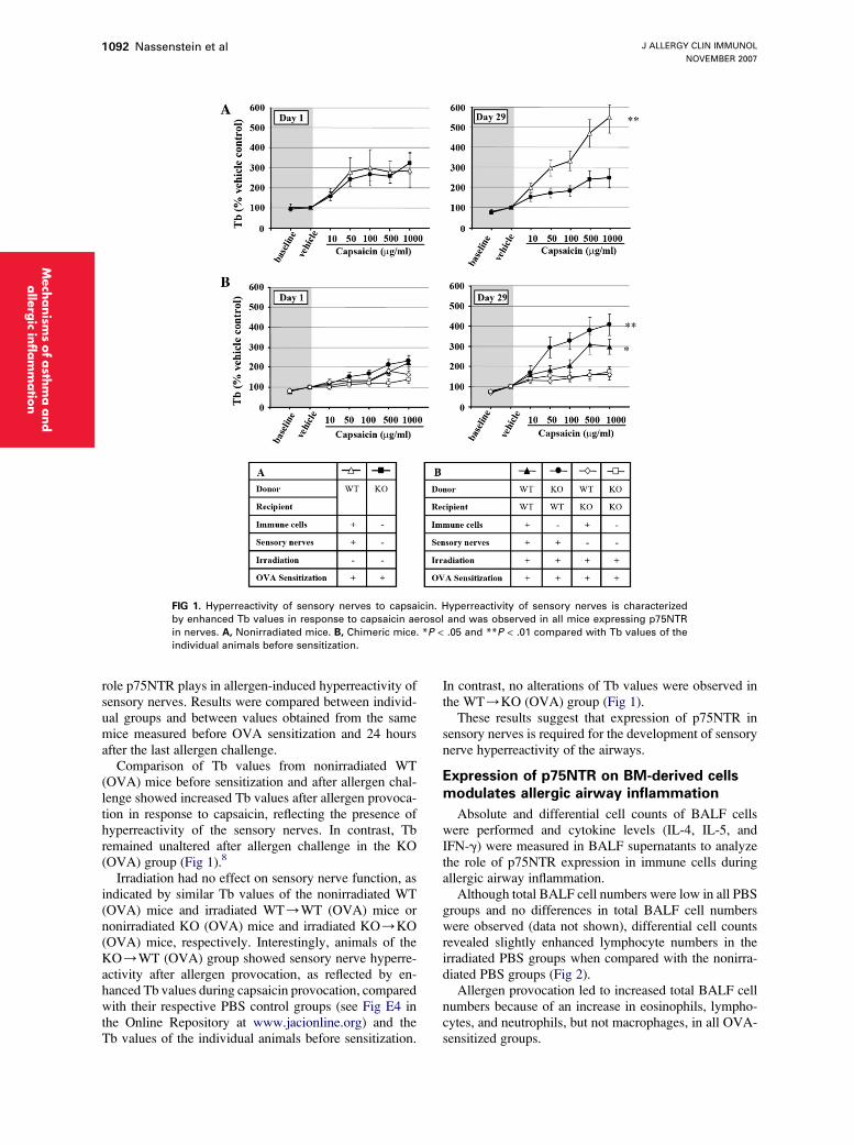
The capsaicin-induced time of braking (Tb) after aller-gen challenge was analyzed in all groups to delineate the
FIG 1. Hyperreactivity of sensory nerves to capsaicin. Hyperreactivity of sensory nerves is characterized
by enhanced Tb values in response to capsaicin aerosol and was observed in all mice expressing p75NTR
in nerves. A, Nonirradiated mice. B, Chimeric mice. *P < .05 and **P < .01 compared with Tb values of the
individual animals before sensitization.
role p75NTR plays in allergen-induced hyperreactivity ofsensory nerves. Results were compared between individ-ual groups and between values obtained from the samemice measured before OVA sensitization and 24 hoursafter the last allergen challenge.
Comparison of Tb values from nonirradiated WT(OVA) mice before sensitization and after allergen chal-lenge showed increased Tb values after allergen provoca-tion in response to capsaicin, reflecting the presence ofhyperreactivity of the sensory nerves. In contrast, Tbremained unaltered after allergen challenge in the KO(OVA) group (Fig 1).8
Irradiation had no effect on sensory nerve function, asindicated by similar Tb values of the nonirradiated WT(OVA) mice and irradiated WT/WT (OVA) mice ornonirradiated KO (OVA) mice and irradiated KO/KO(OVA) mice, respectively. Interestingly, animals of theKO/WT (OVA) group showed sensory nerve hyperre-activity after allergen provocation, as reflected by en-hanced Tb values during capsaicin provocation, comparedwith their respective PBS control groups (see Fig E4 inthe Online Repository at www.jacionline.org) and theTb values of the individual animals before sensitization.
In contrast, no alterations of Tb values were observed inthe WT/KO (OVA) group (Fig 1).
These results suggest that expression of p75NTR insensory nerves is required for the development of sensorynerve hyperreactivity of the airways.
Expression of p75NTR on BM-derived cellsmodulates allergic airway inflammation
Absolute and differential cell counts of BALF cellswere performed and cytokine levels (IL-4, IL-5, andIFN-g) were measured in BALF supernatants to analyzethe role of p75NTR expression in immune cells duringallergic airway inflammation.
Although total BALF cell numbers were low in all PBSgroups and no differences in total BALF cell numberswere observed (data not shown), differential cell countsrevealed slightly enhanced lymphocyte numbers in theirradiated PBS groups when compared with the nonirra-diated PBS groups (Fig 2).
Allergen provocation led to increased total BALF cellnumbers because of an increase in eosinophils, lympho-cytes, and neutrophils, but not macrophages, in all OVA-sensitized groups.
FIG 2. Differential cell count in BALF. Increased numbers of eosinophils, neutrophils, and lymphocytes were
observed in the BALF of OVA-sensitized animals (OVA group, right row) compared with those seen in PBS-
treated control mice (left row). Mice that received WT BM exhibited higher numbers than recipients of BM
from KO mice. *P < .05.
However, the increase was significantly diminished innonirradiated KO (OVA) mice compared with that seenin WT (OVA) mice.8 A similar reduction was seen whenWT/WT (OVA) and KO/KO (OVA) mice were com-pared. A decreased influx of eosinophils, lymphocytes,and neutrophils could also be seen in KO/WT (OVA)mice (Fig 2). Because WT/KO (OVA) mice possesssimilar numbers of eosinophils, lymphocytes, andneutrophils compared with WT/WT (OVA) mice, air-way inflammation seems to depend, at least in part, onp75NTR expression in hemopoietic cells.
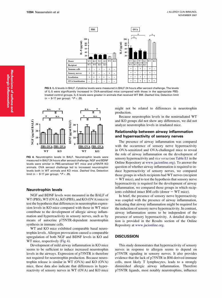
To extend on our findings concerning inflammatorycell numbers in the BALF, we also investigated cyto-kines associated with allergic airway inflammation inBALF. IL-5 levels in BALF supernatants were less thanthe detection limit in all examined PBS groups. Allergenprovocation in all OVA groups led to increased IL-5levels. However, IL-5 levels were lower in irradiated
mice compared with those in nonirradiated animals (Fig3). Comparison of IL-5 cytokine levels in nonirradiatedWT (OVA) mice and KO (OVA) mice revealed signifi-cantly reduced levels in KO (OVA) animals, as previ-ously described.8 Similar decreases in IL-5 levels couldbe observed in KO/KO (OVA) compared withWT/WT (OVA) mice. Interestingly, IL-5 levels ofKO/WT (OVA) mice were similar to those of KO/KO (OVA) mice and significantly different from thoselevels observed in WT/WT (OVA) mice. Conversely,WT/KO (OVA) mice possessed IL-5 levels that werenot significantly different from those of WT/WT(OVA) mice (Fig 3). The levels of IL-4 and IFN-gwere less than the lower limit of quantification in allgroups (data not shown).
In conclusion, these results suggest that p75NTRexpression on BM-derived cells is involved in the devel-opment of allergic airway inflammation.
J ALLERGY CLIN IMMUNOL
NOVEMBER 2007
1094 Nassenstein et al
Mech
anism
sofasth
ma
and
alle
rgic
infl
am
matio
n
FIG 3. IL-5 levels in BALF. Cytokine levels were measured in BALF 24 hours after aerosol challenge. The levels
of IL-5 were significantly increased in OVA-sensitized mice compared with those in the appropriate PBS-
treated control groups. IL-5 levels were greater in animals that received WT BM. Dashed line, Detection limit
(n 5 9-17 per group). *P < .05.
Neurotrophin levels
NGF and BDNF levels were measured in the BALF ofWT (PBS), WT (OVA), KO (PBS), and KO (OVA) mice totest the hypothesis that differences in neurotrophin expres-sion levels in KO mice compared with those in WT micecontribute to the development of allergic airway inflam-mation and hyperreactivity in sensory nerves, such as bymeans of autocrine p75NTR-dependent neurotrophinsynthesis in immune cells.
WT and KO mice exhibited comparable basal neuro-trophin levels. Allergen provocation caused a comparableupregulation of both NGF and BDNF levels in KO andWT mice, respectively (Fig 4).
Development of mild airway inflammation in KO miceseems to be sufficient to induce increased neurotrophinlevels in the airways. Expression of p75NTR is thereforenot required for neurotrophin production. Because neuro-trophin release is similar in WT (OVA) and KO (OVA)mice, these data also indicate that differences in hyper-reactivity of sensory nerves in WT (OVA) and KO mice
FIG 4. Neurotrophin levels in BALF. Neurotrophin levels were
measured in BALF 24 hours after aerosol challenge. NGF and BDNF
levels were similar in PBS-sensitized WT mice and p75NTR KO
animals. OVA aerosol challenge led to increased neurotrophin
levels both in WT animals and KO mice. Dashed line, Detection
limit (n 5 9-17 per group). *P < .05.
might not be related to differences in neurotrophinproduction.
Because neurotrophin levels in the nonirradiated WTand KO groups did not show any differences, we did notanalyze neurotrophin levels in irradiated mice.
Relationship between airway inflammationand hyperreactivity of sensory nerves
The presence of airway inflammation was comparedwith the occurrence of sensory nerve hyperreactivityin OVA-sensitized and OVA-challenged mice to revealthe role of airway inflammation on the development ofsensory hyperreactivity and vice versa (see Table E1 in theOnline Repository at www.jacionline.org). To answer thequestion of whether airway inflammation is required to in-duce hyperreactivity of sensory nerves, we comparedthose groups in which recipients had WT nerves (recipient5 WT mice), and to test the hypothesis that sensory nervehyperreactivity is required for the development of airwayinflammation, we compared those groups in which recip-ients exhibited intact BM cells (donor 5 WT mice).
In brief, the presence of sensory nerve hyperreactivitywas coupled with the presence of airway inflammation,indicating that airway inflammation might be required forthe induction of sensory nerve hyperreactivity. In contrast,airway inflammation seems to be independent of thepresence of sensory hyperreactivity. A detailed descrip-tion is provided in the Results section of the OnlineRepository at www.jacionline.org.
DISCUSSION
This study demonstrates that hyperreactivity of sensorynerves in response to allergen seems to depend onp75NTR signaling in sensory nerves. It also providesevidence that the lack of p75NTR in BM-derived immunecells, most likely T lymphocytes, leads to a stronglydiminished allergic airway inflammation. Thereforep75NTR ligands, most notably neurotrophins, influence
allergic asthma through 2 independent mechanisms in vivoand play an important role in the control of allergic asthmaby affecting neuroimmune interaction.12,16,17
Previous studies have demonstrated that neurotrophinsdirectly act on immune cells. It has been shown thatneurotrophins promote survival and activation of eosino-phils, induce differentiation in B lymphocytes, augmentcytokine synthesis in T cells, and increase proliferation,differentiation, and production of various mediators inmast cells. However, the effects of neurotrophins on mastcells have been related to Trk receptors rather thanp75NTR.14 Nevertheless, it was unclear what relevanceneurotrophins might have in airway inflammation in vivo.We could demonstrate that the lack of p75NTR-mediatedsignaling caused a decrease in IL-5 cytokine productionand a concomitant decrease of eosinophil numbers in theBALF. Because p75NTR expression was detected in Tcells, but not in eosinophils, our study supports the hypoth-esis that eosinophil infiltration is caused by TH2 cytokines,although the clinical relevance of IL-5 itself has beencontroversially discussed in the past.20 However, NGF-transgenic mice, which constitutively overexpressedNGF in the airways, displayed enhanced airway inflamma-tion after allergen provocation, and this inflammation wasdiminished by application of a dual neurokinin 1/neuroki-nin 2 receptor antagonist.21 These data indicate that neuro-trophin-induced neurogenic inflammation might beanother relevant mechanism causing airway inflammation.In this scenario the cellular sources of neuropeptides havenot been firmly identified. However, there is evidence thatNGF production within the airways increases the density oftachykinin-producing nerve fibers surrounding the air-ways.22 Furthermore, concomitant synthesis of tachykininsby hemopoietic cells has been postulated to contribute toinflammatory responses within the airways in vivo, as hasbeen shown in immune complex–mediated and stretch-mediated injuries.23 Although the relative contribution ofneuropeptides has not been investigated in our model, thereis evidence that neuropeptides might be involved in neuro-trophin synthesis24 but also that neurotrophins mightregulate the production of neuropeptides in immune cells.25
Neurotrophin levels were measured in the BALF of WTmice and p75NTR KO mice to test the hypothesis thatimpaired neurotrophin production in vivo might contributeto diminished airway inflammation in p75NTR KO mice.There were no differences between basal NGF and BDNFlevels in WT animals or p75NTR KO animals. The aller-gen-induced increase in NGF and BDNF supports the ideathat diminished allergic airway inflammation in p75NTRKO mice is not caused by differences in neurotrophinproduction but rather is due to the absence of p75NTR-mediated signals in immune cells.
Although our observation that hyperreactivity ofsensory nerves in response to capsaicin is mediatedby p75NTR agrees with that described by Kerzel et al,8
our study provides additional evidence for the cellularmechanism underlying the hypersensitivity of pulmonaryafferent nerves. We show for the first time that the devel-opment of sensory nerve hyperreactivity after allergen
provocation depends on p75NTR signaling in sensorynerves. The mechanisms by which p75NTR mediates sen-sory nerve hyperreactivity are not completely understoodbut might include facilitation of Trk-mediated signals.9,26
Because our study shows that allergic airway inflam-mation and hyperreactivity of sensory nerves are 2 entitiesthat can be selectively influenced by p75NTR signaling,and p75NTR expression in the murine lung is restricted toimmune cells and airway nerves,8 we used the BM transferexperiment to further estimate the in vivo relevance ofneuroimmune interaction in the murine asthma model.Neuroimmune interaction within the lung has been charac-terized anatomically by intimate associations between var-ious immune cells and different subtypes of neurons.27,28
Functionally, this might result in bidirectional interactions.Thus it is suggested that immune cell–derived mediatorscause depolarization of neurons and decrease their mem-brane resistance,29 potentiate transmitter release,30 andpromote survival.31 Conversely, activated afferent nervefibers amplify airway inflammation by means of localrelease of neuropeptides, which then is also termedneurogenic inflammation.7 Neuropeptides promote the che-motaxis, activation, and degranulation of eosinophils; in-crease lymphocyte proliferation and adhesion; and causean imbalance in TH1/TH2-derived cytokines in favor of aTH2 profile. Additionally, neuropeptides promote the re-lease of mast cell mediators.32
The in vivo relevance of neuropeptide-induced neuro-genic inflammation in allergic asthma is unclear. Ourdata indicate that if hyperreactivity of sensory nerves islacking, the development of airway inflammation after al-lergen challenge remains unimpaired. These data suggestthat airway inflammation in the murine asthma model isprobably not caused or modulated by neurogenic inflam-mation. The importance of neurogenic inflammation hasbeen controversially discussed and might vary betweenupper and lower airways or species. Stimulation of nasalsensory nerves by capsaicin in patients with allergic rhini-tis caused leukocyte influx, albumin leakage, and glandu-lar secretion.33 However, depletion of sensory nerves inrats followed by allergen sensitization and challenge didnot affect the number of inflammatory cells in peripheralblood, the number of eosinophils in lung tissue, or the dis-tribution of eosinophils in the adventitial tissue of blood.34
In addition, we could demonstrate that the presence ofsensory nerve hyperreactivity was always coupled withairway inflammation, which supports the hypothesis thatairway inflammation is required for the development ofsensory nerve hyperreactivity. The underlying mecha-nisms have not yet been characterized in detail. Althoughit has been shown that vagal C-fiber afferents in lungs canbe stimulated by exogenously added eosinophil cationicprotein eliciting pulmonary chemoreflex responses,11 ourdata suggest that neurotrophins might be critically in-volved in the development of sensory nerve hyperreactiv-ity in vivo. Thus the selective lack of p75NTR in neuronsprevented the development of sensory nerve hyperreactiv-ity, even when severe airway inflammation was present.Interestingly, mild airway inflammation was sufficient to
J ALLERGY CLIN IMMUNOL
NOVEMBER 2007
1096 Nassenstein et al
Mech
anism
sofasth
ma
and
alle
rgic
infl
am
matio
n
induce distinct hyperreactivity of sensory nerves. Inanimals with mild and severe airway inflammation, thenumber of BALF cells and cytokine levels were different.However, neurotrophin levels were similar, as indicatedby comparable neurotrophin levels in WT mice exhibitingsevere airway inflammation and p75NTR KO mice exhib-iting mild airway inflammation after allergen challenge.These results support the hypothesis that neurotrophinsplay a crucial role in the development of sensory nervehyperreactivity.
In conclusion, our study provides evidence for a dualrole of p75NTR in allergic asthma in vivo. p75NTR partic-ipates in mediating neurotrophin signals to immune cells,as well as to nerve cells. Furthermore, it supports thehypothesis that immune cells influence sensory nervefunction in vivo. This immunoneuronal interaction mightbe involved in the pathogenesis of allergic asthma.
We thank Thomas Blankenstein for providing the mice used in the
experiments and Sabine Schild, Olaf Macke, and Petra Straub for
excellent technical assistance.
REFERENCES
1. Tracey KJ. The inflammatory reflex. Nature 2002;420:853-9.