66
Natthapon Rattanathamsakul, MD. December 14 th , 2017 Neurovascular Anatomy (1) : Anterior Circulation Anatomy

Natthapon Rattanathamsakul, MD.December 14th, 2017
Neurovascular Anatomy (1):Anterior Circulation Anatomy

Contents:
Neurovascular Anatomy
Arterial supply of the brain
Anterior circulation
Posterior circulation
Arterial supply of the spinal cord
Venous system of the brain
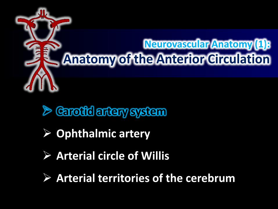
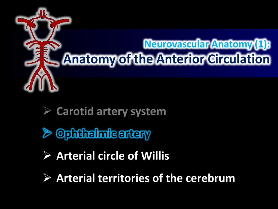
Neurovascular Anatomy (1):
Anatomy of the Anterior Circulation
Carotid artery system
Ophthalmic artery
Arterial circle of Willis
Arterial territories of the cerebrum
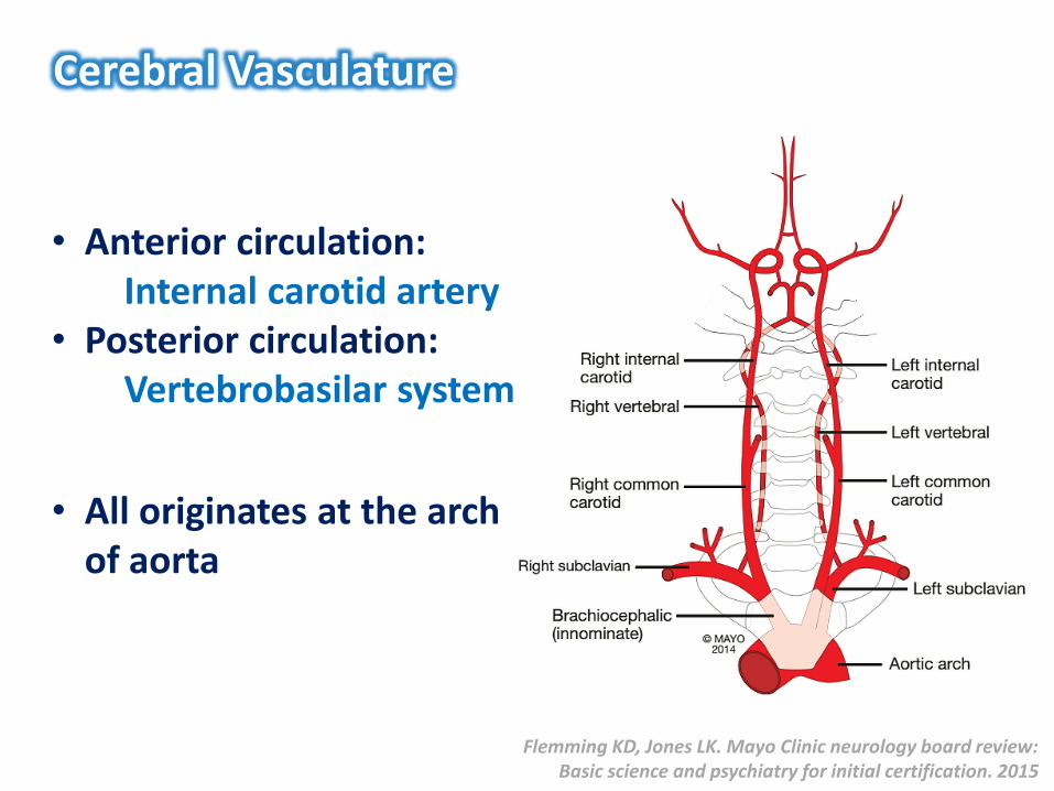
Cerebral Vasculature
• Anterior circulation: Internal carotid artery
• Posterior circulation: Vertebrobasilar system
• All originates at the arch of aorta
Flemming KD, Jones LK. Mayo Clinic neurology board review: Basic science and psychiatry for initial certification. 2015
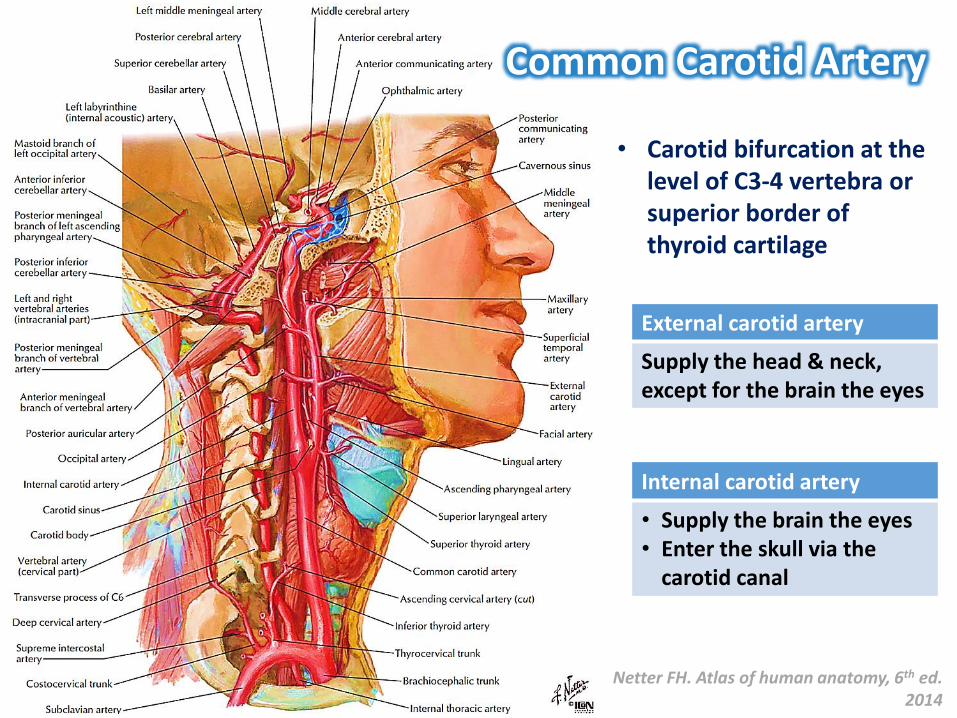
• Carotid bifurcation at the level of C3-4 vertebra or superior border of thyroid cartilage
External carotid artery
Supply the head & neck, except for the brain the eyes
Internal carotid artery
• Supply the brain the eyes• Enter the skull via the
carotid canal
Common Carotid Artery
Netter FH. Atlas of human anatomy, 6th ed. 2014

Uflacker R. Atlas of vascular anatomy: an angiographic approach, 2007
Angiographic Correlation

External carotid artery
• Superior thyroid artery• Lingual artery• Facial artery• Ascending pharyngeal
artery• Posterior auricular artery• Occipital artery• Maxillary artery• Superficial temporal artery
External Carotid Artery
• Middle meningeal artery – epidural hemorrhage
Netter FH. Atlas of human anatomy, 6th ed. 2014
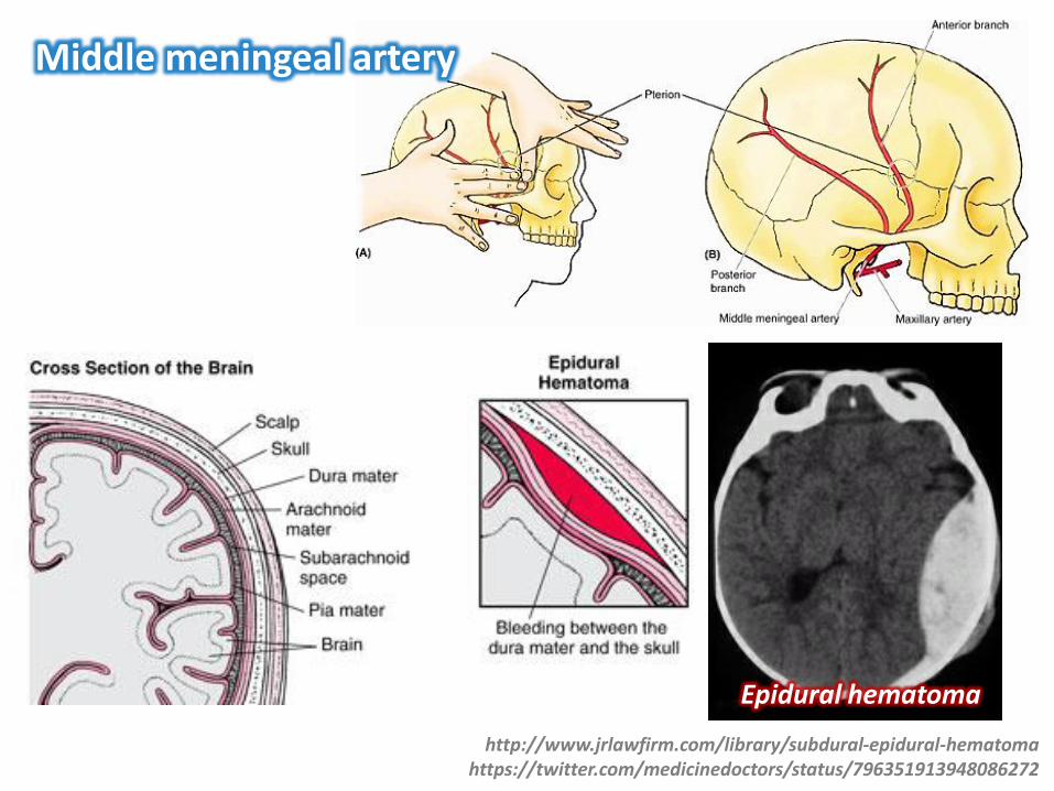
Middle meningeal artery
http://www.jrlawfirm.com/library/subdural-epidural-hematomahttps://twitter.com/medicinedoctors/status/796351913948086272
Epidural hematoma
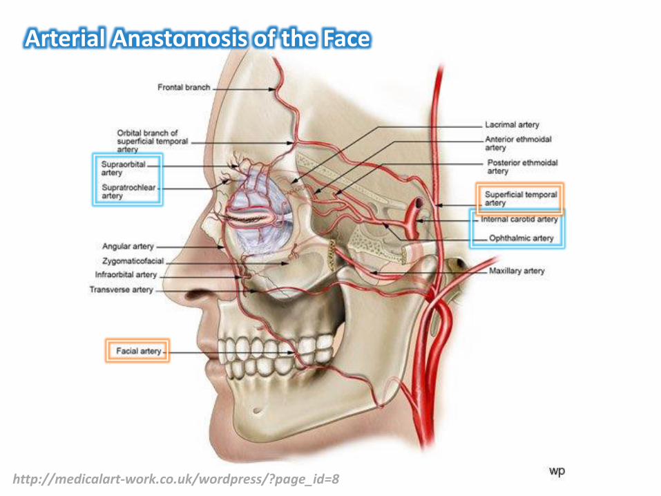
Arterial Anastomosis of the Face
http://medicalart-work.co.uk/wordpress/?page_id=8
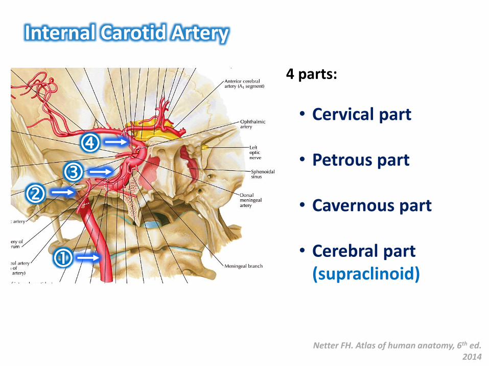
Internal Carotid Artery
4 parts:
• Cervical part
• Petrous part
• Cavernous part
• Cerebral part (supraclinoid)
Netter FH. Atlas of human anatomy, 6th ed. 2014
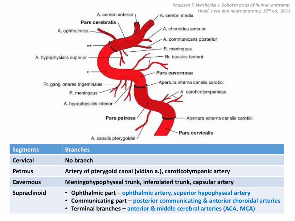
Pauslsen F, Washchke J. Sobotta atlas of human anatomy: Head, neck and neuroanatomy, 15th ed, 2011
Segments Branches
Cervical No branch
Petrous Artery of pterygoid canal (vidian a.), caroticotympanic artery
Cavernous Meningohypophyseal trunk, inferolaterl trunk, capsular artery
Supraclinoid • Ophthalmic part – ophthalmic artery, superior hypophyseal artery• Communicating part – posterior communicating & anterior choroidal arteries• Terminal branches – anterior & middle cerebral arteries (ACA, MCA)

https://image.slidesharecdn.com/brainvascularsupply-140701071122-phpapp01/95/brain-vascular-supply-8-638.jpg?cb=1404217076

External carotid artery Internal carotid artery
https://openi.nlm.nih.gov/detailedresult.php?img=PMC3284869_1752-1947-6-45-3&req=4
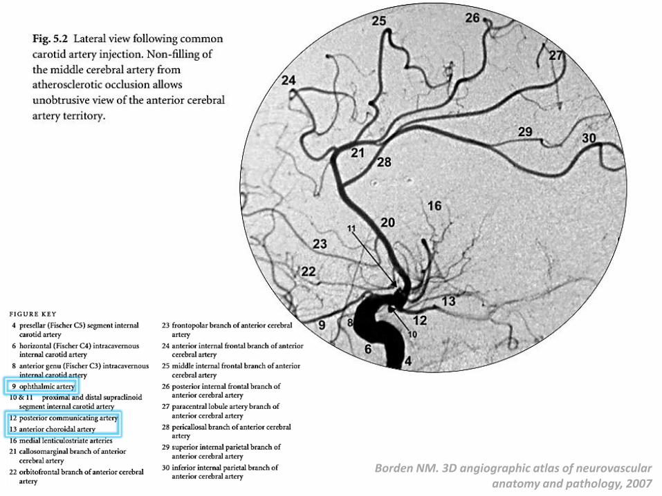
Borden NM. 3D angiographic atlas of neurovascular anatomy and pathology, 2007
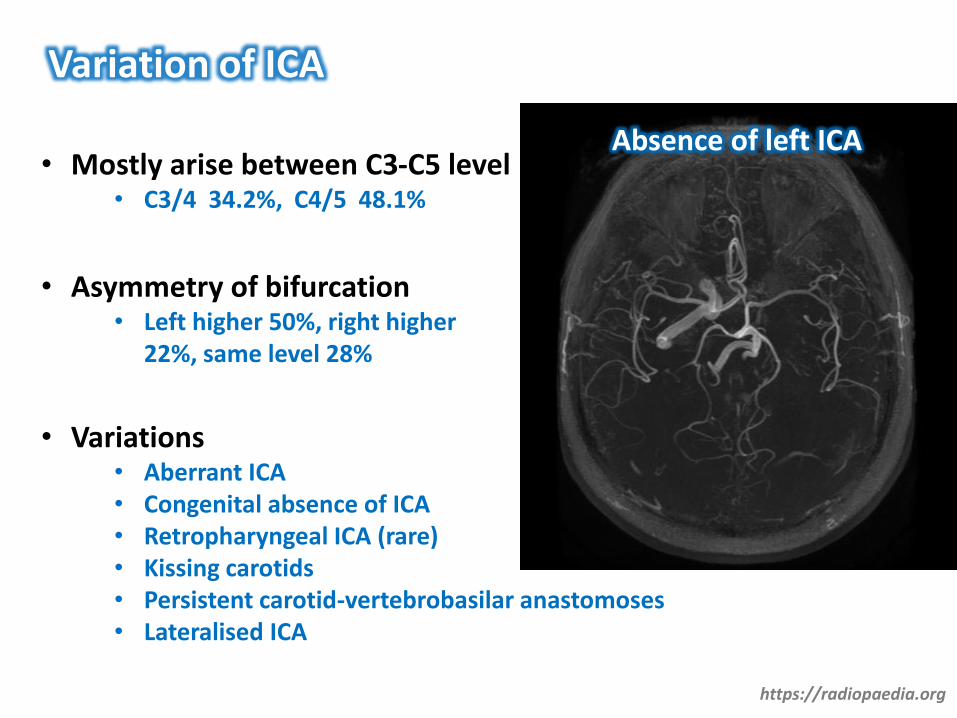
Variation of ICA
• Mostly arise between C3-C5 level• C3/4 34.2%, C4/5 48.1%
• Asymmetry of bifurcation• Left higher 50%, right higher
22%, same level 28%
• Variations• Aberrant ICA• Congenital absence of ICA• Retropharyngeal ICA (rare)• Kissing carotids• Persistent carotid-vertebrobasilar anastomoses• Lateralised ICA
Absence of left ICA
https://radiopaedia.org

https://radiopaedia.orghttp://dx.doi.org/10.1590/S0004-282X2007000200034
Kissing carotids (tortuous) Aberrant left ICA (pulsatile tinnitus)
Persistent carotid-vertebrobasilaranastomoses

Persistent Carotid-Vertebrobasilar Anastomoses
http://www.neuroradiologycases.com/2012/03/carotid-vertebrobasilar-anastomoses.htmlhttp://dx.doi.org/10.1136/neurintsurg-2013-010703.rep
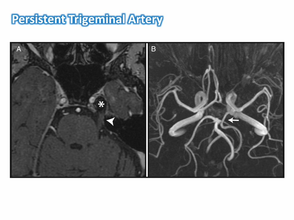
Persistent Trigeminal Artery
CD
US
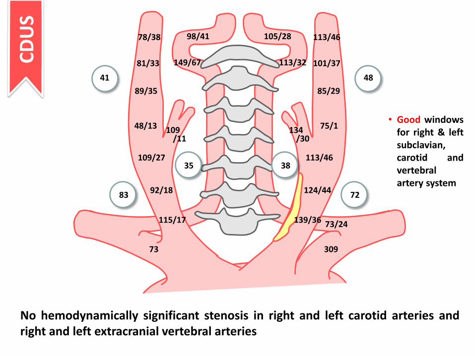
No hemodynamically significant stenosis in right and left carotid arteries and right and left extracranial vertebral arteries
• Good windows for right & left subclavian, carotid and vertebral artery system
98/41 105/28
113/32149/67
78/38 113/46
81/33 101/37
89/35 85/29
48/13 75/1134/30
109/11
109/27 113/46
92/18
115/17
124/44
139/36
73 309
73/24
7283
4841
35 38
3111
2911
6433
4612
8027
8435 66
41
4221
8640
5326
7333
9444
7042
7126 72
36 3014
3919
4826
2715
0.620.73
0.58 0.750.45
1.01
0.54
0.80 0.80
0.80 0.82
0.70
0.70 1.46
1.16 1.09
1.170.94
0.94
TCC
D
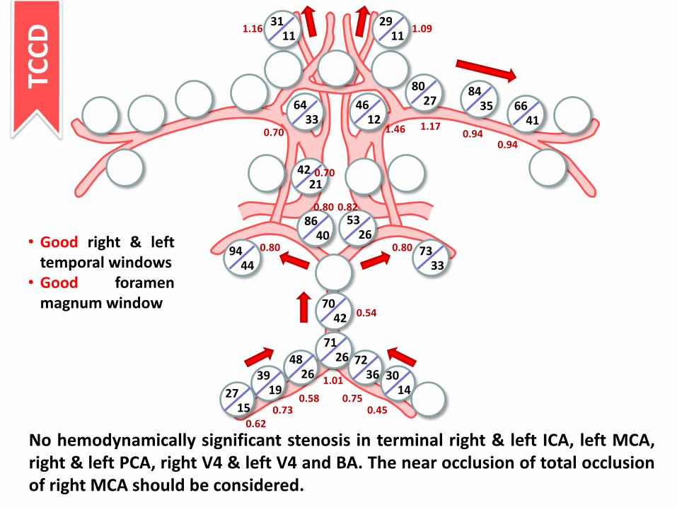
No hemodynamically significant stenosis in terminal right & left ICA, left MCA, right & left PCA, right V4 & left V4 and BA. The near occlusion of total occlusion of right MCA should be considered.
• Good right & left temporal windows
• Good foramen magnum window
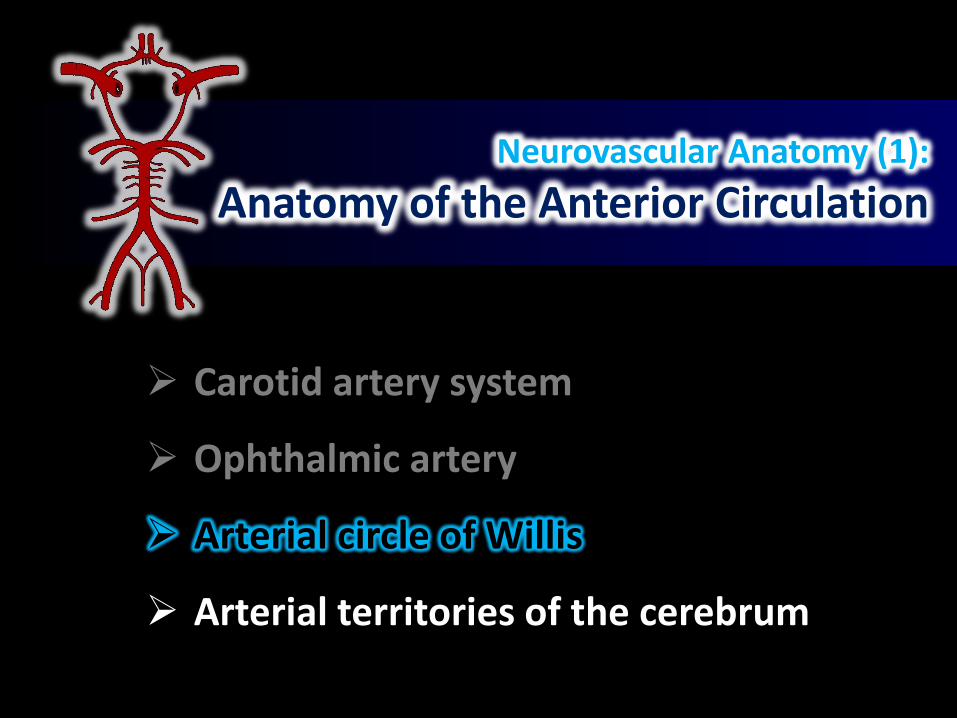
Neurovascular Anatomy (1):
Anatomy of the Anterior Circulation
Carotid artery system
Ophthalmic artery
Arterial circle of Willis
Arterial territories of the cerebrum

http://anesthesiology.pubs.asahq.org/article.aspx?articleid=2566231
Ophthalmic Artery
Branches of ophthalmic artery
• Central retinal artery**• Long & short posterior ciliary
arteries**• Supratrochlear artery**• Supraorbital artery**• Lacrimal artery• Anterior & posterior ethmoidal
arteries• Etc.

https://www.opticianonline.net/cet-archive/150https://www.pinterest.com/pin/488922103268831757/?lp=true
Ophthalmic Artery
• Central retinal artery –supply most part of the retina
• Posterior ciliary artery –supply choroid plexus and macula lutea (forming circle of Zinn-Haller)
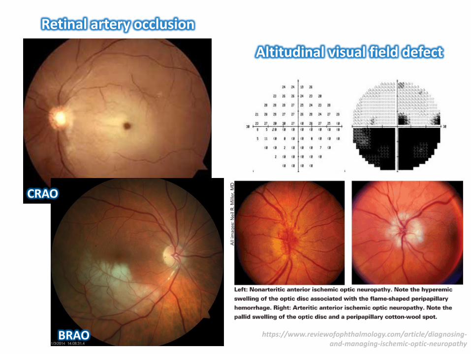
Altitudinal visual field defect
Retinal artery occlusion
CRAO
BRAO https://www.reviewofophthalmology.com/article/diagnosing-and-managing-ischemic-optic-neuropathy
Neurovascular Anatomy (1):
Anatomy of the Anterior Circulation
Carotid artery system
Ophthalmic artery
Arterial circle of Willis
Arterial territories of the cerebrum
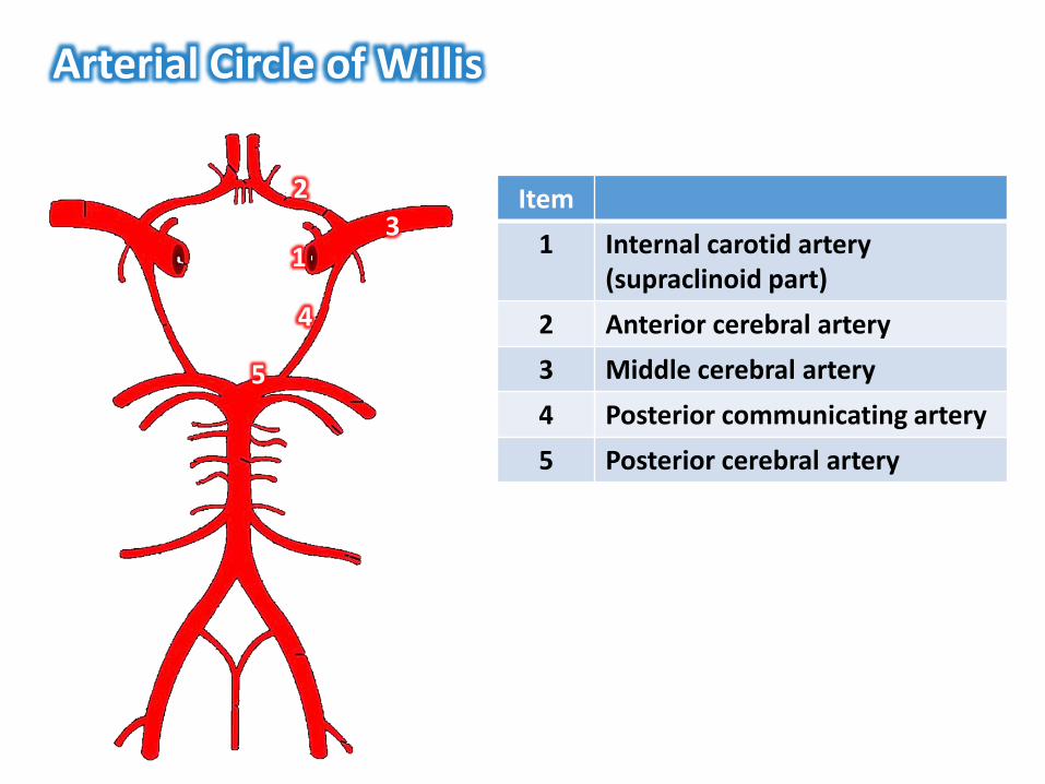
Arterial Circle of Willis
5
1
2
3
4
Item
1 Internal carotid artery(supraclinoid part)
2 Anterior cerebral artery
3 Middle cerebral artery
4 Posterior communicating artery
5 Posterior cerebral artery

https://radiologykey.com/ultrasound-assessment-of-the-intracranial-arteries/https://www.rainwaterharvesting.co.uk/accessories/overflow-siphon-22
Segments of the Arterial CircleCarotid siphon
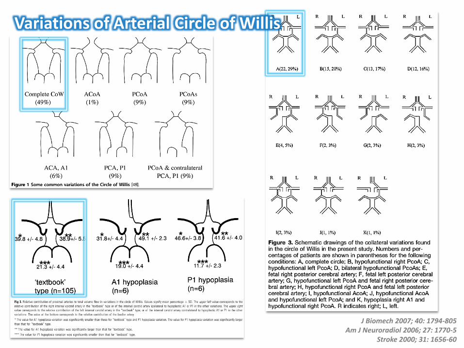
J Biomech 2007; 40: 1794-805Am J Neuroradiol 2006; 27: 1770-5
Stroke 2000; 31: 1656-60
Variations of Arterial Circle of Willis
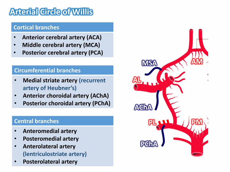
Arterial Circle of Willis
Central branches
• Anteromedial artery• Posteromedial artery• Anterolateral artery
(lentriculostriate artery)• Posterolateral artery
Cortical branches
• Anterior cerebral artery (ACA)• Middle cerebral artery (MCA)• Posterior cerebral artery (PCA)
Circumferential branches
• Medial striate artery (recurrent artery of Heubner’s)
• Anterior choroidal artery (AChA)• Posterior choroidal artery (PChA)
MSA AM
PMPL
AL
AChA
PChA
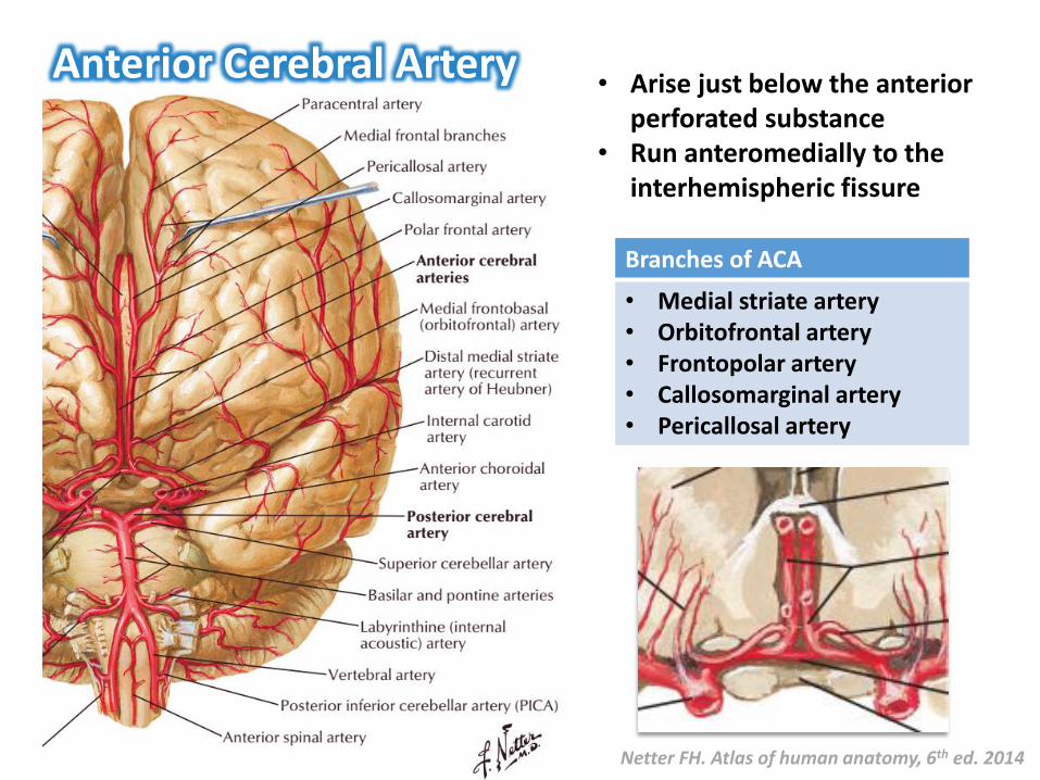
Anterior Cerebral Artery
Netter FH. Atlas of human anatomy, 6th ed. 2014
• Arise just below the anterior perforated substance
• Run anteromedially to the interhemispheric fissure
Branches of ACA
• Medial striate artery• Orbitofrontal artery• Frontopolar artery• Callosomarginal artery• Pericallosal artery
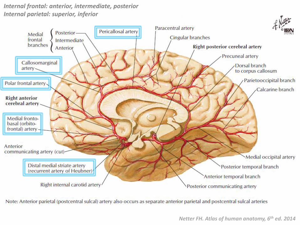
Netter FH. Atlas of human anatomy, 6th ed. 2014
Internal frontal: anterior, intermediate, posteriorInternal parietal: superior, inferior
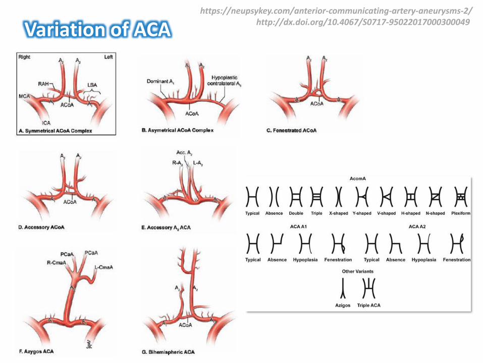
Variation of ACAhttps://neupsykey.com/anterior-communicating-artery-aneurysms-2/
http://dx.doi.org/10.4067/S0717-95022017000300049
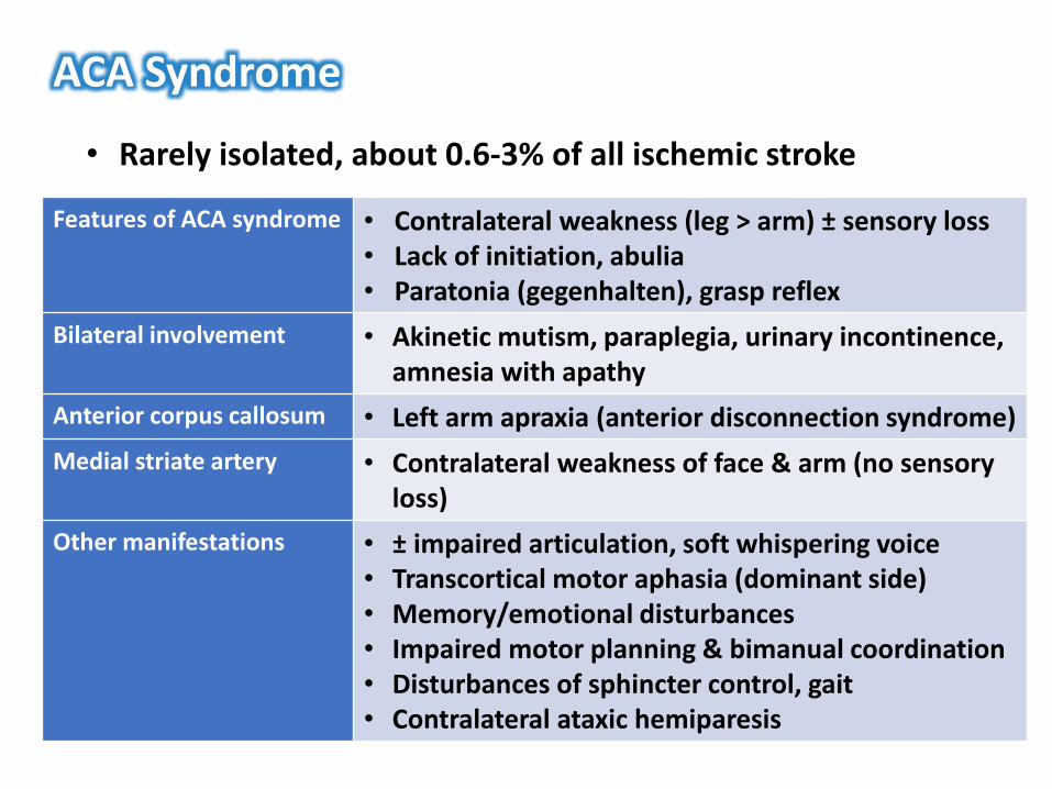
ACA Syndrome
• Rarely isolated, about 0.6-3% of all ischemic stroke
Features of ACA syndrome • Contralateral weakness (leg > arm) ± sensory loss• Lack of initiation, abulia• Paratonia (gegenhalten), grasp reflex
Bilateral involvement • Akinetic mutism, paraplegia, urinary incontinence, amnesia with apathy
Anterior corpus callosum • Left arm apraxia (anterior disconnection syndrome)
Medial striate artery • Contralateral weakness of face & arm (no sensory loss)
Other manifestations • ± impaired articulation, soft whispering voice• Transcortical motor aphasia (dominant side)• Memory/emotional disturbances• Impaired motor planning & bimanual coordination• Disturbances of sphincter control, gait• Contralateral ataxic hemiparesis
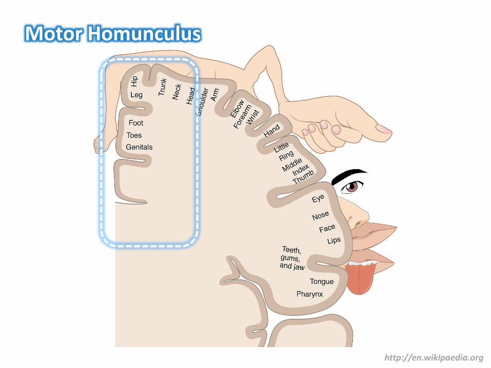
Motor Homunculus
http://en.wikipaedia.org
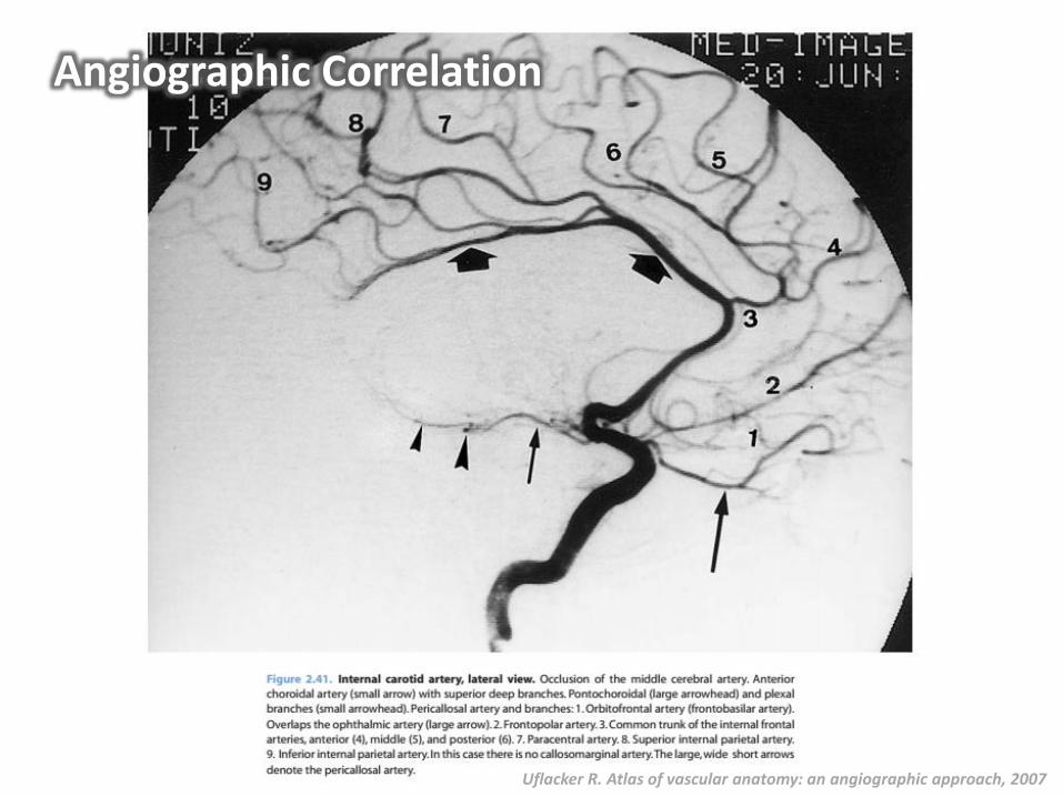
Uflacker R. Atlas of vascular anatomy: an angiographic approach, 2007
Angiographic Correlation

Middle Cerebral ArteryNetter FH. Atlas of human anatomy, 6th ed. 2014
http://www.radiologyassistant.nl
• Arise just below the anterior perforated substance
• Supplies most of the lateral surface of the cerebral hemisphere
• 3 parts :• Proximal (M1)• Sylvian (M2)• Distal (M3)

Supplies • Corona radiate, superior portion of anterior/posterior limb of internal capsule
• External capsule, claustrum, putamen, part of the globus pallidus, body of the caudate nucleus
Syndromes • Contralateral hemiparesis (mainly upper extremity)• Cortical symptoms (aphasia, neglect, apraxia)
Lenticulostriate Artery (Lateral Striate Artery)
Netter FH. Atlas of human anatomy, 6th ed. 2014Acta Neurochir 2015; 157: 743-54
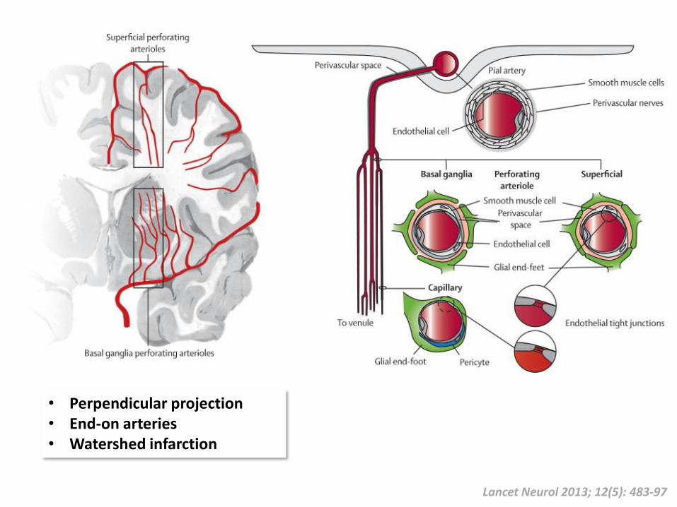
Lancet Neurol 2013; 12(5): 483-97
• Perpendicular projection• End-on arteries• Watershed infarction
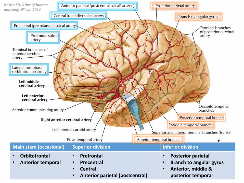
Main stem (occasional) Superior division Inferior division
• Orbitofrontal• Anterior temporal
• Prefrontal• Precentral• Central• Anterior parietal (postcentral)
• Posterior parietal• Branch to angular gyrus• Anterior, middle &
posterior temporal
Netter FH. Atlas of human anatomy, 6th ed. 2014
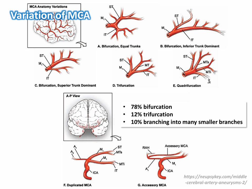
Variation of MCA
https://neupsykey.com/middle-cerebral-artery-aneurysms-2/
• 78% bifurcation• 12% trifurcation• 10% branching into many smaller branches
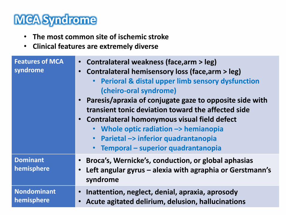
MCA Syndrome• The most common site of ischemic stroke• Clinical features are extremely diverse
Features of MCA syndrome
• Contralateral weakness (face,arm > leg)• Contralateral hemisensory loss (face,arm > leg)
• Perioral & distal upper limb sensory dysfunction (cheiro-oral syndrome)
• Paresis/apraxia of conjugate gaze to opposite side with transient tonic deviation toward the affected side
• Contralateral homonymous visual field defect• Whole optic radiation –> hemianopia• Parietal –> inferior quadrantanopia• Temporal – superior quadrantanopia
Dominanthemisphere
• Broca’s, Wernicke’s, conduction, or global aphasias• Left angular gyrus – alexia with agraphia or Gerstmann’s
syndrome
Nondominanthemisphere
• Inattention, neglect, denial, apraxia, aprosody• Acute agitated delirium, delusion, hallucinations
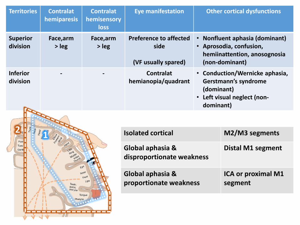
Territories Contralathemiparesis
Contralathemisensory
loss
Eye manifestation Other cortical dysfunctions
Superior division
Face,arm> leg
Face,arm> leg
Preference to affected side
(VF usually spared)
• Nonfluent aphasia (dominant)• Aprosodia, confusion,
hemiinattention, anosognosia(non-dominant)
Inferior division
- - Contralathemianopia/quadrant
• Conduction/Wernicke aphasia,Gerstmann’s syndrome (dominant)
• Left visual neglect (non-dominant)
12 Isolated cortical M2/M3 segments
Global aphasia & disproportionate weakness
Distal M1 segment
Global aphasia & proportionate weakness
ICA or proximal M1 segment

Other MCA Syndrome
Insular cortex • Somatosensory deficits, gustatory disorders• Vestibular-like manifestations• Cardiovascular disorders, including arterial hypertension
and arrhythmias (increased risk of MI, sudden death)• Language & neuropsychological disorders (aphasia,
dysarthria, somatoparaphrenia)
Double infarcts of dominant MCA
• Global aphasia without hemiparesis• Hemianopic hemiplegia without sensory impairment• Conduction aphasia with hemiparesis
Bilateral anterioropercular infarcts
• Foix-Chavany-Marie syndrome• Bilateral supranuclear facio-pharyngeal-
glossomasticatory paresis with automatic-voluntary dissociation
Bilateral temporal infarcts
• Cortical deafness: Awareness of sound, but cannot interpret verbal or identify nonverbal auditory stimulus
• Klüver-Bucy syndrome
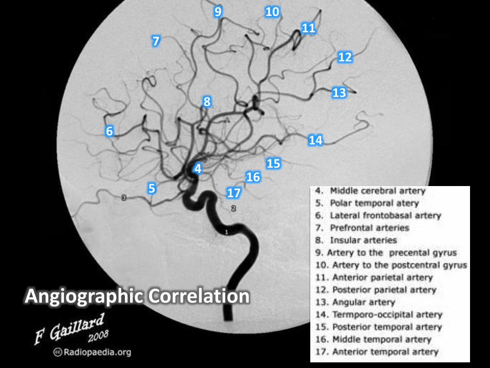
4
5
6
7
8
9 1011
12
13
14
1516
17
Angiographic Correlation
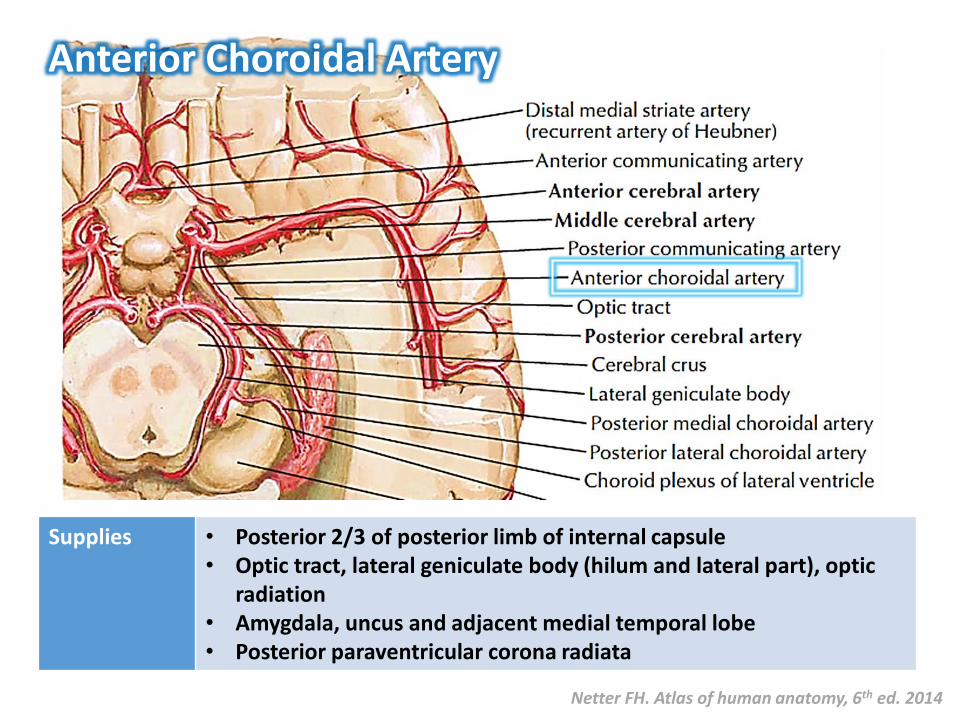
Anterior Choroidal Artery
Supplies • Posterior 2/3 of posterior limb of internal capsule• Optic tract, lateral geniculate body (hilum and lateral part), optic
radiation• Amygdala, uncus and adjacent medial temporal lobe• Posterior paraventricular corona radiata
Netter FH. Atlas of human anatomy, 6th ed. 2014
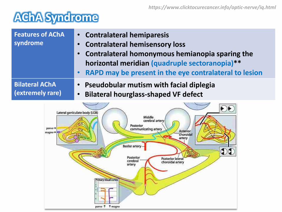
AChA SyndromeFeatures of AChAsyndrome
• Contralateral hemiparesis• Contralateral hemisensory loss• Contralateral homonymous hemianopia sparing the
horizontal meridian (quadruple sectoranopia)**• RAPD may be present in the eye contralateral to lesion
Bilateral AChA(extremely rare)
• Pseudobular mutism with facial diplegia• Bilateral hourglass-shaped VF defect
https://www.clicktocurecancer.info/optic-nerve/iq.html

http://www.cybersight.org/bins/volume_page.asp?cid=1-2630-2760-2763&print=true
PChAPChA
Horizontal sectoranopia(Posterior choroidal artery)
Quadruple sectoranopia(Anterior choroidal artery)

Anterior Choroidal Artery
www.radiopaedia.org
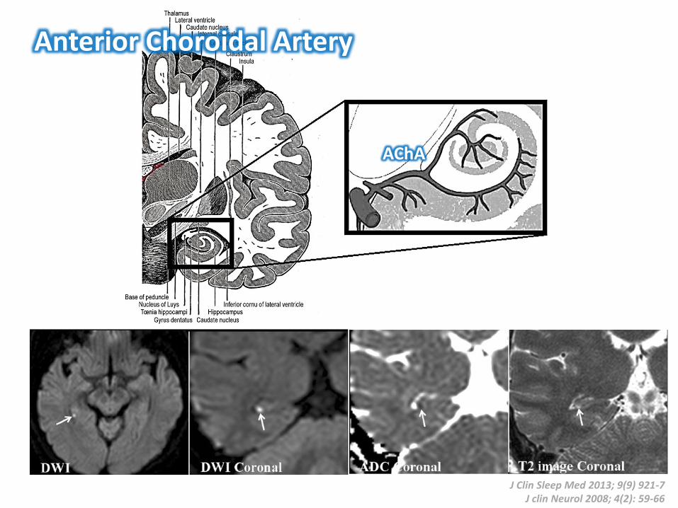
J Clin Sleep Med 2013; 9(9) 921-7J clin Neurol 2008; 4(2): 59-66
AChA
Anterior Choroidal Artery

Posterior Communicating Artery
https://neupsykey.com/posterior-communicating-artery-aneurysms-2/
• Interconnection between the anterior and posterior circulation• Lied adjacent to CN.III• Supplies the optic tract, CN.III and anterior part of thalamus (anterior
thalamoperforate branch)

Variations of Posterior Communicating Artery
Fetal origin of PCA PCom Infundibulum
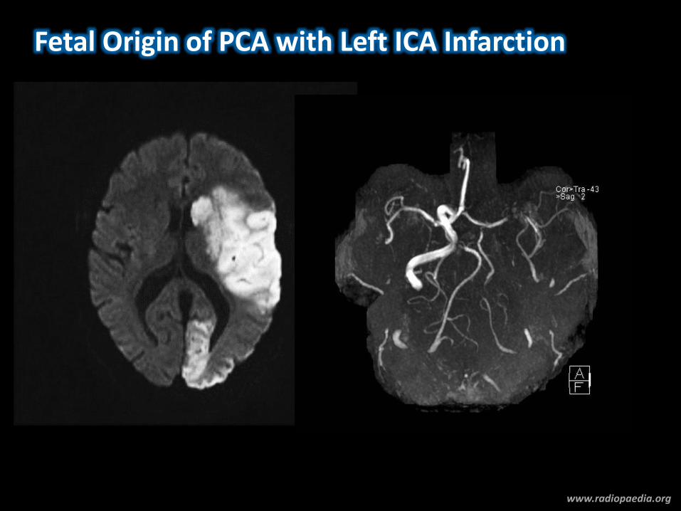
Fetal Origin of PCA with Left ICA Infarction
www.radiopaedia.org
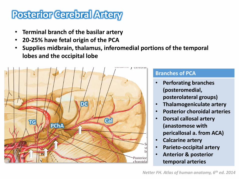
Posterior Cerebral Artery
Netter FH. Atlas of human anatomy, 6th ed. 2014
• Terminal branch of the basilar artery• 20-25% have fetal origin of the PCA• Supplies midbrain, thalamus, inferomedial portions of the temporal
lobes and the occipital lobe
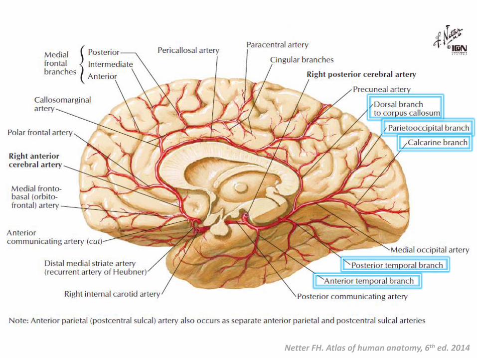
Branches of PCA
• Perforating branches (posteromedial, posterolateral groups)
• Thalamogeniculate artery• Posterior choroidal arteries• Dorsal callosal artery
(anastomose with pericallosal a. from ACA)
• Calcarine artery• Parieto-occipital artery• Anterior & posterior
temporal arteries
TGPChA
DC
Cal
Netter FH. Atlas of human anatomy, 6th ed. 2014
PCA Syndrome
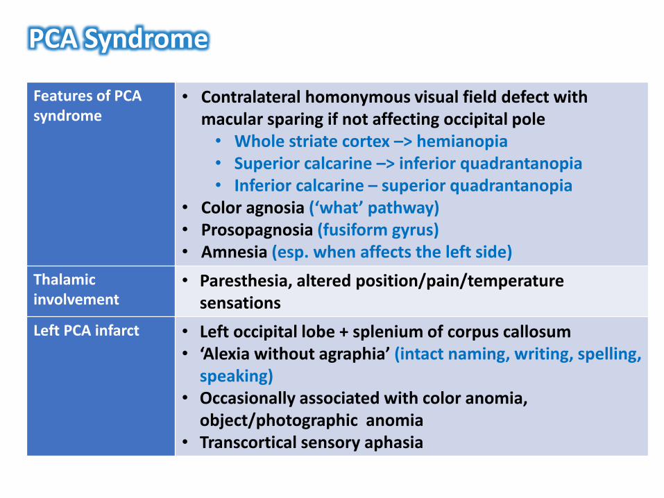
Features of PCA syndrome
• Contralateral homonymous visual field defect with macular sparing if not affecting occipital pole• Whole striate cortex –> hemianopia• Superior calcarine –> inferior quadrantanopia• Inferior calcarine – superior quadrantanopia
• Color agnosia (‘what’ pathway)• Prosopagnosia (fusiform gyrus)• Amnesia (esp. when affects the left side)
Thalamic involvement
• Paresthesia, altered position/pain/temperature sensations
Left PCA infarct • Left occipital lobe + splenium of corpus callosum• ‘Alexia without agraphia’ (intact naming, writing, spelling,
speaking)• Occasionally associated with color anomia,
object/photographic anomia• Transcortical sensory aphasia
PCA Syndrome
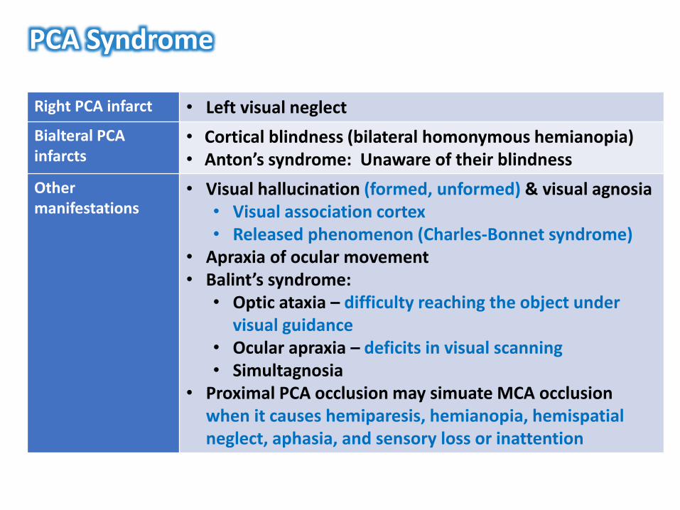
Right PCA infarct • Left visual neglect
Bialteral PCAinfarcts
• Cortical blindness (bilateral homonymous hemianopia)• Anton’s syndrome: Unaware of their blindness
Other manifestations
• Visual hallucination (formed, unformed) & visual agnosia • Visual association cortex• Released phenomenon (Charles-Bonnet syndrome)
• Apraxia of ocular movement• Balint’s syndrome:
• Optic ataxia – difficulty reaching the object under visual guidance
• Ocular apraxia – deficits in visual scanning• Simultagnosia
• Proximal PCA occlusion may simuate MCA occlusion when it causes hemiparesis, hemianopia, hemispatialneglect, aphasia, and sensory loss or inattention
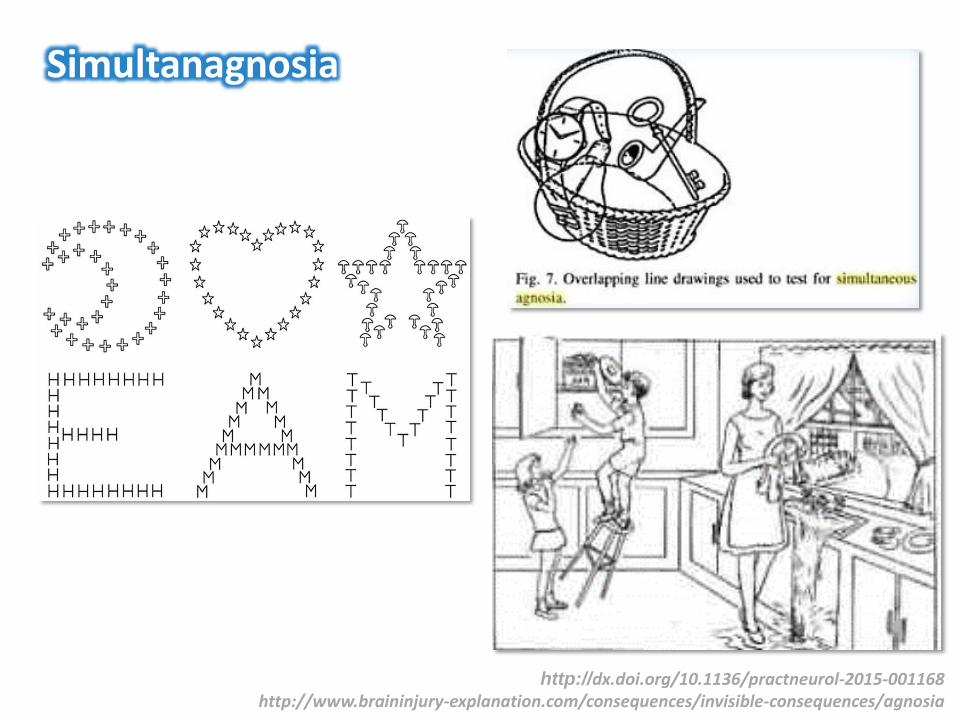
Simultanagnosia
http://dx.doi.org/10.1136/practneurol-2015-001168http://www.braininjury-explanation.com/consequences/invisible-consequences/agnosia

Neurovascular Anatomy (1):
Anatomy of the Anterior Circulation
Carotid artery system
Ophthalmic artery
Arterial circle of Willis
Arterial territories of the cerebrum
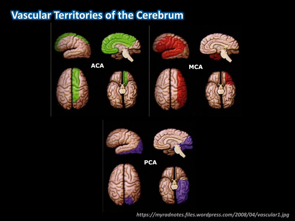
https://myradnotes.files.wordpress.com/2008/04/vascular1.jpg
Vascular Territories of the Cerebrum
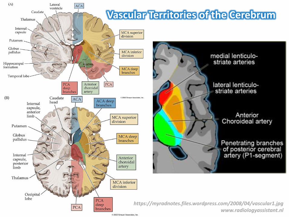
https://myradnotes.files.wordpress.com/2008/04/vascular1.jpgwww.radiologyassistant.nl
Vascular Territories of the Cerebrum
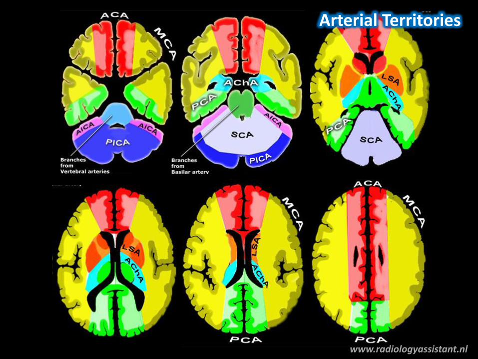
Arterial Territories
www.radiologyassistant.nl
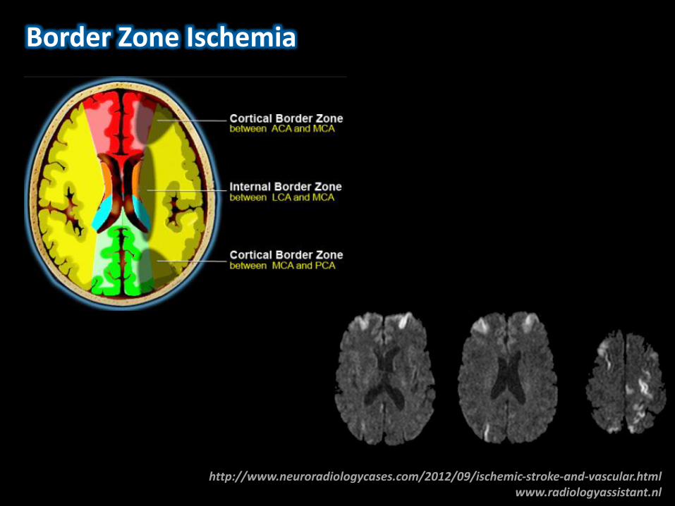
http://www.neuroradiologycases.com/2012/09/ischemic-stroke-and-vascular.htmlwww.radiologyassistant.nl
Border Zone Ischemia

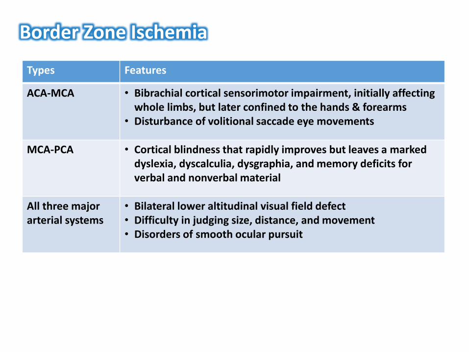
Border Zone Ischemia
Types Features
ACA-MCA • Bibrachial cortical sensorimotor impairment, initially affecting whole limbs, but later confined to the hands & forearms
• Disturbance of volitional saccade eye movements
MCA-PCA • Cortical blindness that rapidly improves but leaves a marked dyslexia, dyscalculia, dysgraphia, and memory deficits for verbal and nonverbal material
All three major arterial systems
• Bilateral lower altitudinal visual field defect• Difficulty in judging size, distance, and movement• Disorders of smooth ocular pursuit

Neurovascular Anatomy (1):
Anatomy of the Anterior Circulation
Carotid artery system
Ophthalmic artery
Arterial circle of Willis
Arterial territories of the cerebrum

Natthapon Rattanathamsakul, MD.December 14th, 2017
Neurovascular Anatomy (1):Anterior Circulation Anatomy


