Page 1
RESEARCH ARTICLE
NIMA-related kinase 1 (NEK1) regulates
meiosis I spindle assembly by altering the
balance between α-Adducin and Myosin X
Miguel A. Brieño-Enrı́quez*, Stefannie L. Moak, J. Kim Holloway, Paula E. Cohen
Department of Biomedical Sciences and Center for Reproductive Genomics, Cornell University, Ithaca, New
York, United States of America
* [email protected]
Abstract
NIMA-related kinase 1 (NEK1) is a serine/threonine and tyrosine kinase that is highly
expressed in mammalian germ cells. Mutations in Nek1 induce anemia, polycystic kidney
and infertility. In this study we evaluated the role of NEK1 in meiotic spindle formation in
both male and female gametes. Our results show that the lack of NEK1 provokes an abnor-
mal organization of the meiosis I spindle characterized by elongated and/or multipolar spin-
dles, and abnormal chromosome congression. The aberrant spindle structure is
concomitant with the disruption in localization and protein levels of myosin X (MYO10) and
α-adducin (ADD1), both of which are implicated in the regulation of spindle formation during
mitosis. Interaction of ADD1 with MYO10 is dependent on phosphorylation, whereby phos-
phorylation of ADD1 enables its binding to MYO10 on mitotic spindles. Reduction in ADD1
protein in NEK1 mutant mice is associated with hyperphosphorylation of ADD1, thereby pre-
venting the interaction with MYO10 during meiotic spindle formation. Our results reveal a
novel regulatory role for NEK1 in the regulation of spindle architecture and function during
meiosis.
Introduction
Meiosis is a specialized cell division characterized by a single round of DNA replication fol-
lowed by two rounds of chromosome segregation, resulting in the formation of haploid gam-
etes for sexual reproduction. At the first male meiotic division (MI), homologous (maternal
and paternal) chromosomes must segregate equally into two daughter cells, each of which then
undergoes a mitosis-like second meiotic division (MII) in which sister chromatids separate
into the haploid gametes. In mammals, meiosis in males results in four haploid gametes, while
meiosis in female results in one haploid gamete per meiosis, the remaining genetic material
being distributed between two polar bodies. Regardless of the sex of the individual, in order to
achieve accurate segregation at both divisions, tension must be established on the meiotic spin-
dles and this is achieved by the formation of crossovers between homologous chromosomes in
meiosis I, and by cohesion between sister chromatids in both meiosis I and meiosis II [1].
PLOS ONE | https://doi.org/10.1371/journal.pone.0185780 October 5, 2017 1 / 18
a1111111111
a1111111111
a1111111111
a1111111111
a1111111111
OPENACCESS
Citation: Brieño-Enrı́quez MA, Moak SL, Holloway
JK, Cohen PE (2017) NIMA-related kinase 1
(NEK1) regulates meiosis I spindle assembly by
altering the balance between α-Adducin and
Myosin X. PLoS ONE 12(10): e0185780. https://
doi.org/10.1371/journal.pone.0185780
Editor: Claude Prigent, Institut de Genetique et
Developpement de Rennes, FRANCE
Received: May 22, 2017
Accepted: September 19, 2017
Published: October 5, 2017
Copyright: © 2017 Brieño-Enrı́quez et al. This is an
open access article distributed under the terms of
the Creative Commons Attribution License, which
permits unrestricted use, distribution, and
reproduction in any medium, provided the original
author and source are credited.
Data Availability Statement: All relevant data are
within the paper and its Supporting Information
files.
Funding: M.A.B-E is supported by a Postdoctoral
fellowship by the Empire State Stem Cell Fund
through New York State Department of Health
Contract # C30293GG. This project is funded by
grants from the National Institute of Child Health
and Human Development (NICHD) to J.K.H
(5R00HD065870), and from National Institute of
General Medical Sciences (NIGMS) and March of
Page 2
Cohesion is also necessary for sister kinetochore attachment to microtubules during meiotic
spindle formation [2].
In eukaryotes, the microtubule spindle is the structure that orchestrates chromosome align-
ment and segregation during both mitotic and meiotic divisions [3]. In mitosis, the microtu-
bules that compose the spindle are mostly nucleated from centrosomes, which act as a major
microtubule organizing center (MTOC) [2–4]. Centrosomes are composed of pairs of centri-
oles surrounded by pericentriolar material (PCM) that posses microtubule nucleation activity.
When mitosis starts, centrosomes separate to opposite sides of the nuclear envelope, defining
the poles of the spindle and allowing the bipolar spindle [2,4]. However, there is a sexual dis-
morphism during the meiosis, where the male spindle behaves like a mitotic spindle, while in
oocytes, the spindle it is formed without centrosomes [4,5]. Another important difference is
the stringency of the spindle checkpoint in females, in which the oocyte still proceeds through
the first meiotic division despite disruptions in the spindle and chromosome congression at
metaphase plate [6]. Such differences in spindle dynamics in male and female mammals sug-
gest that different regulatory proteins may predominate in each sex.
Several families of kinases such as Polo kinases, Aurora kinases and NEK kinases have been
implicated in the regulation of cell cycle events in both mitosis and meiosis [7,8]. The founding
member of the NEK family is the Aspergillus nidulans never-in-mitosis-gene-A (NIMA) pro-
tein [9]. NIMA is a serine/threonine kinase involved in the initiation of mitosis and in promot-
ing chromosome condensation. Genetic ablation of NIMA results in cell cycle arrest at G2,
while overexpression of NIMA leads to premature entry into mitosis [7]. In mammals, there
are 11 orthologs of NIMA that comprise the NIMA-like (NEK) family of kinases. Nek1 is
unique in that it encodes both the serine/threonine kinase activity typical of the NEK family as
well as tyrosine kinase activity that is not a feature of other NEKs [10]. NEK1 has been impli-
cated in ciliogenesis [11] and DNA damage response [12–15]. Additionally, NEK1 is highly
expressed in mouse germ cells [16], where it appears to play essential roles in meiosis I and
possibly also at later stages [17,18].
The Kat2J allele of NEK1 is a spontaneous point mutation within the coding sequence of
Nek1 that results in a premature stop codon leading to a truncated protein lacking the both
kinase domains [16]. Nek1kat2j/kat2j mice display polycystic kidneys, dwarfism, anemia and
male sterility [16,19]. Interestingly, while initial reports mention that Nek1kat2j/kat2j females
had low fertility rates [16,19], in our animal facility females no pregnancies are observed [17]
possibly reflecting subtle background effects among mouse strains. Our previous studies of
meiosis in Nek1kat2j/kat2j mice showed that the loss of NEK1 activity induces aberrant retention
of cohesin subunit Structural Maintenance of Chromosomes protein 3 (SMC3), at the end of
meiotic prophase I [17]. More recently, we showed NEK1 regulates cohesin removal, in part,
through regulation of wings apart-like homolog (WAPL) during meiotic prophase I [18], as
part of the so-called Prophase pathway for cohesin removal. This regulation of WAPL by
NEK1 is mediated through WAPL interactions with the cohesin-associated protein, PDS5
homolog B (PDS5B), and protein phosphatase 1 gamma (PP1γ), both of which also interact
with NEK1 [18].
In the current study, and given the conserved function of NEK proteins in spindle assembly
dynamics, we investigated the role of NEK1 downstream of prophase I events. Our results
demonstrate that loss of NEK1 induces failure of meiotic spindle organization in both males
and females, leading to elongated spindles, multipolar spindles, and abnormal chromosome
congression. Concurrent with this, we observe altered levels and localization of two proteins
known to regulate mitotic spindle dynamics: the unconventional myosin, myosin X (MYO10),
and α-adducin (ADD1). Interaction of ADD1 with MYO10 is dependent on phosphorylation
of ADD1 which allows for the interaction of these two proteins on mitotic spindles [20].
NEK1 regulates the meiotic spindle
PLOS ONE | https://doi.org/10.1371/journal.pone.0185780 October 5, 2017 2 / 18
Dimes to P.E.C. (1R01GM097263, MOD2006-
844). The funders had no role in study design, data
collection and analysis, decision to publish, or
preparation of the manuscript.
Competing interests: The authors have declared
that no competing interest exist.
Page 3
Depletion of ADD1 or changes in its phosphorylation status results in abnormal mitotic spin-
dles [20]. Our studies demonstrate that loss of NEK1 kinase causes changes in MYO10 and
ADD1 protein levels, and hyperphosphorylation and deamination of ADD1 caused at least in
part by the abnormal function of PP1 during meiotic spindle formation. Interestingly spindle
phenotypes seem not directly related to the role of NEK1 in prophase I events.
Material and methods
Mice and genotyping
All mouse studies were conducted with the prior approval of the Cornell Institutional Animal
Care and Use Committee (Protocol 2010–0054). The mice were bred under pathogen-free,
controlled temperature (22±1˚C) and regulated humidity (50–55%) conditions with periods of
light/dark of 12 h and food available ad libitum. Mice were euthanized using CO2 administra-
tion method.
The Nek1kat2j/kat2j line was obtained originally from Jackson Laboratory (Bar Harbor,
Maine), and has been established in our mouse colony for more than nine years. Genotyping
of this mouse line was performed following the protocol described elsewhere [17]. For the pur-
poses of these studies, male mice were 8 weeks old and female where used at 24–28 days old.
Homozygous mutant animals of (Nek1kat2J/kat2J) were compared with wild type (Nek1+/+) lit-
termates on a C57Bl/6J background.
Male and female spindle preparation
Male spindle analysis was performed in testis from 8 week old mice following the protocol
described elsewhere [21,22]. Briefly, following euthanasia, the testes were removed, decapsu-
lated and placed in PBS. Using fine forceps, tubules were separated and analysed under a ste-
reomicroscope. Differential light absorption of the tubules creates different zones that can be
defined by different cell populations [22]. The differentent zones are consequence of the para-
crine regualtion by Sertoli cells, spermatogenesis proceeds in synchronized waves along the
seminiferus tubules, and every given cross-section contains only certain cell types, that pro-
duces a diferent light absorbation creating zones [22]. These zones are the weak spot that cor-
respond to the spermatogenic stages XII-I, the strong spot (stages II-VI), the dark zone (stages
VII-VIII) and the pale zone (stage IX-XI) We selected the zone corresponding to the sperma-
togenetic development XI and XII that are rich in meiotic cells during both meiosis I and mei-
osis II. Tubules were placed in fresh PBS and these required zones were excised and placed on
poly-lysine-coated slides (P4981, Thermo Fisher). Tubule sections were fixed for 10 minutes
(1% paraformaldehyde and 0.15% Triton-X, pH 9.2) and then squashed under a coverslip.
Slides with the squashed tubules were put into liquid nitrogen for 20 seconds and thereafter
the coverslip removed and washed with PBS-0.4% Kodak Photo flo (Kodak) (3 times for 5
minutes each). Slides were kept at -80˚C or stained immediately.
Female spindle analysis was performed with oocytes from 24–28 day old mice following
our previously published protocol [23]. Briefly, after dissection, ovaries were collected in col-
lection medium (Waymouths’s medium (11220035, Gibco), 10% Fetal bovine serum
(26140079, Gibco), 1% penicillin-streptomycin (P4333-100ML, Sigma-Aldrich) and 0.1%
sodium pyruvate (11360070, Gibco)). Oocytes were released from ovarian follicles using
30-gauge needles, and oocytes with germinal vesicles placed in EmbryoMax KSOM medium
(MR-121-D, EMD Millipore). Oocytes were cultured for 6hr in EmbryoMax KSOM medium
drops at 37˚C, 5% CO2. After incubation, oocytes were placed in fibrinogen-thrombin clot,
incubated for 60 seconds at 37˚C, and rinsed with 1XPBS containing 2% Triton-X (BP151-
500, Fisher Scientific) for 3 min. Oocytes were fixed for 30 minutes at 37˚C. Fixation was
NEK1 regulates the meiotic spindle
PLOS ONE | https://doi.org/10.1371/journal.pone.0185780 October 5, 2017 3 / 18
Page 4
followed by a 15 minute wash in 0.1% NGS (5 ml 10X PBS, 50 μL goat serum and 50 ml Milli-
Q water). Oocytes were stored at 4˚C or stained immediately.
Spindle immunofluorescence staining
Male and female fixed spindles slides were washed in PBS containing 0.4%Kodak Photo flo
(Kodak) for 5 min, followed by 0.1% PBS-Triton X and blocked in 1XPBS-antibody dilution
buffer (ADB) before being incubated over night at 4˚C with the primary antibody. Primary
antibodies used were: rabbit anti-ADD1 (GTX101600, Genetex. Dilution 1:100), rabbit anti-
MYO10 (24565-1-AP, Proteintech. Dilution 1:100) and rabbit anti-β-tubulin (T8328, Sigma.
Dilution 1:500). After overnight incubation, slides were washed to remove the unbound anti-
bodies and incubate for 2 hours at 37˚C with Alexafluor™ secondary antibodies (Molecular
Probes Eugene OR, USA). Slides were washed and mounted with Prolong Gold antifade
(Molecular Probes). Image acquisition was performed using a Zeiss Imager Z1 microscope
under 20X, 40X or 63X magnifying objectives, at room temperature. Images were processed
using ZEN 2 (Carl Zeiss).
Mass spectrometry
Mass spectrometry was performed in the Cornell University Proteomics and Mass Spectrome-
try facility. 2D LC-MS/MS raw data files were acquired using Orbitrap Elite (Thermo Scien-
tific). We performed a database search using Mascot searching against the SwissProt mouse
database from Uniprot website (http://www.uniprot.org) using Mascot software version 2.3.02
(Matrix Science, UK). The default Mascot search settings were as follows: one missed cleavage
site by trypsin allowed with fixed MMTS modification of cysteine, fixed four-plex iTRAQ
modifications on Lys and N-terminal amines and variable modifications of methionine oxida-
tion, deamidation of Asn and Gln residues, and 4-plex iTRAQ on Tyr for iTRAQ 4-plex analy-
sis. One or two-missed cleavage site by trypsin allowed with fixed carboxamidomethyl
modification of cysteine, fixed six-plex TMT modifications on Lys and N-terminal amines and
variable modifications of methionine oxidation, deamidation of Asn and Gln residues, and
6-plex TMT on Tyr for TMT 6-plex analysis. The quantitative protein ratios were weighted
and normalized by the median ratio with outlier removal set automatic in Mascot for each set
of experiments. Only those proteins with ratios equivalents to two-fold increase or two-fold
reduction were considered significant. To obtain the specific post-traslational modification of
the peptides we perfumed TiO2 enrichment followed by the proteomics analysis as was
described above.
Western blotting
Whole testis protein and oocyte protein were extracted by sonication in RIPA buffer. Samples
were boiled for 5 min in sample buffer, electrophoresed on SDS-polyacrylamide gels (8%) and
transferred to nitrocellulose membranes. Primary antibody incubation was performed over-
night at 4˚C at 1:1000 dilution (antibodies are the same used in spindle staining). Incubation
with secondary antibodies was performed for two hours at room temperature (secondary HRP
conjugated antibodies were obtained from Pierce, Life Technologies). Signal-detection was
carried out using the superSignal substrate (Thermo Scientific). Loading control was per-
formed using GAPDH-HPR (PA1-987-HRP, from ThermoFisher). Images were captured with
BIO RAD Image Lab 5.1and analyzed by ImageJ version 1.49v (http://rsbweb.nih.gov/ij).
NEK1 regulates the meiotic spindle
PLOS ONE | https://doi.org/10.1371/journal.pone.0185780 October 5, 2017 4 / 18
Page 5
Results
Loss of NEK1 induces abnormal meiotic spindle formation and failed
chromosome congression at the first meiotic division in males and
females
We analyzed the role of NEK1 in spindle formation and chromosome orientation at MI in
oocytes and spermatocytes from Nek1+/+ and Nek1kat2j/kat2j mice. In male mice, we evaluated
the shape, number of poles and misaligned chromosomes in both Nek1+/+ (n = 100) and
Nek1kat2j/kat2j (n = 100) spermatocytes. MI spindles from Nek1+/+ spermatocytes showed a
bipolar structure with the chromosomes aligned at metaphase plate (Fig 1a), while spindles
from Nek1kat2j/kat2j spermatocytes showed defects in the structure of the spindle as well as in
chromosome congression. These defects included MI spindles without a pole, with only one
pole, with misaligned chromosomes (Fig 1b), and with multiple poles (Fig 1c). A total of 55%
of spindles from Nek1kat2j/kat2j males were abnormal, while only 18% of the spindles from
Nek1+/+ male spermatocytes showed abnormalities (Fig 1d). Quantification of spindle abnor-
malities in Nek1+/+ and Nek1kat2j/kat2j spermatocytes reveals that the most common abnormal-
ity is misaligned chromosomes. The percentage of spindles with misaligned chromosomes in
Nek1+/+ was 18%, while in Nek1kat2j/kat2j mice the percentage reached 38%. Other observed
spindle abnormalities were absence of poles, one pole and multiple poles (Fig 1e)
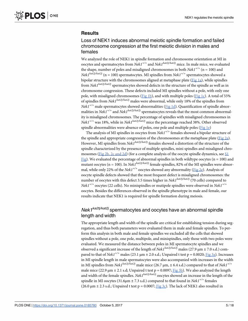
The analysis of MI spindles in oocytes from Nek1+/+ females showed a bipolar structure of
the spindle and appropriate congression of the chromosomes at the metaphase plate (Fig 2a).
However, MI spindles from Nek1kat2j/kat2j females showed a distortion of the structure of the
spindle characterized by the presence of multiple spindles, mini spindles and misaligned chro-
mosomes (Fig 2b, 2c and 2d) (for a complete analysis of the oocyte spindle dynamics, see S1
Fig). We evaluated the percentage of abnormal spindles in both wildtype oocytes (n = 100) and
mutant oocytes (n = 100). In Nek1kat2j/kat2j female spindles, 82% of the MI spindles were abnor-
mal, while only 22% of the Nek1+/+ oocytes showed any abnormality (Fig 2e). Analysis of
oocyte spindle defects showed that the most frequent defect is misaligned chromosomes: the
number of oocytes with this defect 3.5 times higher in Nek1kat2j/kat2j (70 cells) compared to
Nek1+/+ oocytes (22 cells). No minispindles or mutipole spindles were observed in Nek1+/+
oocytes. Besides the differences observed in the spindle phenotype in male and female, our
results indicate that NEK1 is required for spindle formation during meiosis.
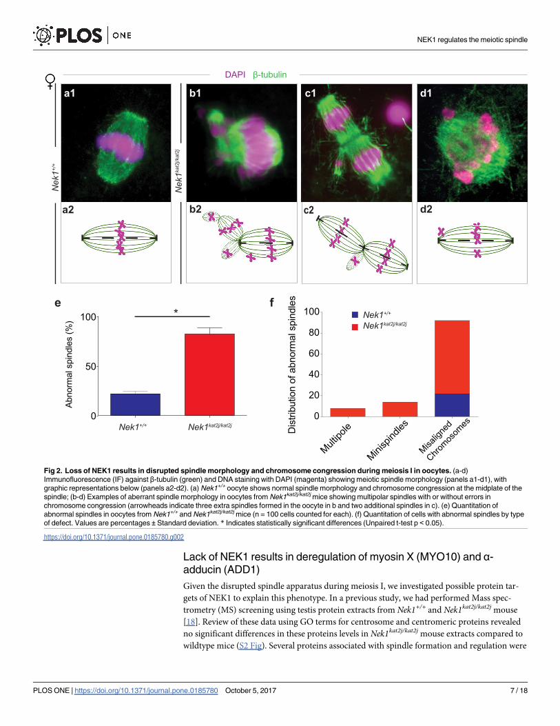
Nek1ka2tj/kat2j spermatocytes and oocytes have an abnormal spindle
length and width
The appropriate length and width of the spindle are critical for establishing tension during seg-
regation, and thus both parameters were evaluated them in male and female spindles. To per-
form this analysis in both male and female spindles we excluded all the cells that showed
spindles without a pole, one pole, multipole, and minispindles, only those with two poles were
evaluated. We measured the distance between poles in MI spermatocyte spindles and we
observed a significant increase of the length of Nek1kat2j/kat2j males (27.9 μm ± 7.0 s.d.) com-
pared to that of Nek1+/+ males (23.1 μm ± 2.0 s.d.; Unpaired t test p = 0.0020; Fig 3a). Increases
in MI spindle length in male spermatocytes were also accompanied with increases in the width
in MI spindles from Nek1kat2j/kat2j male mice (26.7 μm, ± 6.4 s.d.) compared to that of Nek1+/+
male mice (22.9 μm ± 2.1 s.d; Unpaired t test p = 0.0097; Fig 3b). We also analyzed the length
and width of the female spindles. Nek1kat2j/kat2j oocytes showed an increase in the length of the
spindle in MI oocytes (31.6μm ± 7.3 s.d.) compared to that found in Nek1+/+ females
(26.0 μm ± 2.3 s.d.; Unpaired t test p = 0.0007; Fig 3c). The lack of NEK1 also resulted in
NEK1 regulates the meiotic spindle
PLOS ONE | https://doi.org/10.1371/journal.pone.0185780 October 5, 2017 5 / 18
Page 6
altered width of the MI spindles in female meiosis. Nek1kat2j/kat2j oocytes showed a decrease in
the width of the spindle (15.1 μm ± 4.0 s.d.) compared to the Nek1+/+ oocyte spindles
(17.0 μm ± 1.7 s.d.; Unpaired t test p = 0.039; Fig 3d). Taken together our results show that
NEK1 is required for the establishment of the correct spindle length and width during meiosis
in both male and female gametes.
Fig 1. Loss of NEK1 results in disrupted spindle morphology and chromosome congression during meiosis I in spermatocytes.
(a-c) Immunofluorescence (IF) against β-tubulin (green) and DNA staining with DAPI (magenta) showing meiotic spindle morphology
(panels a1-c1), with graphic representations below (panels a2-c2). (A) Nek1+/+ spermatocyte shows normal spindle morphology and
chromosome congression at the midplate of the spindle; (b, c) Examples of aberrant spindle morphology in spermatocytes from Nek1kat2j/
kat2j mice showing monopolar spindles with mislocalized chromosomes resulting from failed chromosome congression (arrowheads) in b and
multipolar spindles in c (arrowheads). (d) Quantitation of abnormal spindles in spermatocytes from Nek1+/+ and Nek1kat2j/kat2j mice (n = 100
cells counted for each). (e) Quantitation cells with abnormal spindles by type of defect. Values are percentages ± Standard deviation. *Indicates statistically significant differences (Unpaired t-test p < 0.05).
https://doi.org/10.1371/journal.pone.0185780.g001
NEK1 regulates the meiotic spindle
PLOS ONE | https://doi.org/10.1371/journal.pone.0185780 October 5, 2017 6 / 18
Page 7
Lack of NEK1 results in deregulation of myosin X (MYO10) and α-
adducin (ADD1)
Given the disrupted spindle apparatus during meiosis I, we investigated possible protein tar-
gets of NEK1 to explain this phenotype. In a previous study, we had performed Mass spec-
trometry (MS) screening using testis protein extracts from Nek1+/+ and Nek1kat2j/kat2j mouse
[18]. Review of these data using GO terms for centrosome and centromeric proteins revealed
no significant differences in these proteins levels in Nek1kat2j/kat2j mouse extracts compared to
wildtype mice (S2 Fig). Several proteins associated with spindle formation and regulation were
Fig 2. Loss of NEK1 results in disrupted spindle morphology and chromosome congression during meiosis I in oocytes. (a-d)
Immunofluorescence (IF) against β-tubulin (green) and DNA staining with DAPI (magenta) showing meiotic spindle morphology (panels a1-d1), with
graphic representations below (panels a2-d2). (a) Nek1+/+ oocyte shows normal spindle morphology and chromosome congression at the midplate of the
spindle; (b-d) Examples of aberrant spindle morphology in oocytes from Nek1kat2j/kat2j mice showing multipolar spindles with or without errors in
chromosome congression (arrowheads indicate three extra spindles formed in the oocyte in b and two additional spindles in c). (e) Quantitation of
abnormal spindles in oocytes from Nek1+/+ and Nek1kat2j/kat2j mice (n = 100 cells counted for each). (f) Quantitation of cells with abnormal spindles by type
of defect. Values are percentages ± Standard deviation. * Indicates statistically significant differences (Unpaired t-test p < 0.05).
https://doi.org/10.1371/journal.pone.0185780.g002
NEK1 regulates the meiotic spindle
PLOS ONE | https://doi.org/10.1371/journal.pone.0185780 October 5, 2017 7 / 18
Page 8
Fig 3. Lack of NEK1 results in abnormal meiosis I spindle morphology in spermatocytes and oocytes. (a)
Immunofluorescence (IF) against β-tubulin (green) and DNA staining with DAPI (magenta) showing a wildtype (top) and Nek1kat2j/
kat2j spermatocytes (bottom), along with measurements of spindle length (b) and width (c). Black bars are means (μm) ± Standard
deviation. (d) Immunofluorescence (IF) against β-tubulin (green) and DNA staining with DAPI (magenta) showing a wildtype (top)
and Nek1kat2j/kat2j (bottom) oocytes, along with measurements of spindle length (b) and width (c). Black bars are means (μm) ±Standard deviation. * Indicates statistically significant differences (Unpaired t-test p < 0.05).
https://doi.org/10.1371/journal.pone.0185780.g003
NEK1 regulates the meiotic spindle
PLOS ONE | https://doi.org/10.1371/journal.pone.0185780 October 5, 2017 8 / 18
Page 9
identified and quantified, however no differences between Nek1+/+ and Nek1kat2j/kat2j mouse
were observed for tubulins, actins and SAC proteins (S3–S5 Figs).
Previous reports showed that MYO10 has a direct function in spindle formation [24–26].
In contrast to the tubulins, actins, and other spindle proteins that did not appear to be altered
in the absence of NEK1, we observed that the levels of the myosins, MYO5, MYO7A, MYO10
and MYO15, were significantly increased in testis protein extracts from Nek1 mutant mice
compared to wildtype littermates (S1 Table). By contrast, the myosins, MYO1B, MYO1C,
MYO1D, MYO7A and MYO9B were unaltered. The function of MYO10 in spindle formation
and integrity is related to its binding partner α-adducin (ADD1)[20]. Our MS results revealed
that ADD1 showed lower protein levels in Nek1kat2j/kat2j male mice compared to wildtype male
mice (S1 Table). The changes in protein levels observed by MS indicates that the loss of NEK1
results in an imbalance in the ratio MYO10/ADD1, and this could be affecting the proper spin-
dle formation and function.
Nek1kat2j/kat2j spermatocytes and oocytes show aberrant localization of
myosin X (MYO10) on meiotic spindles
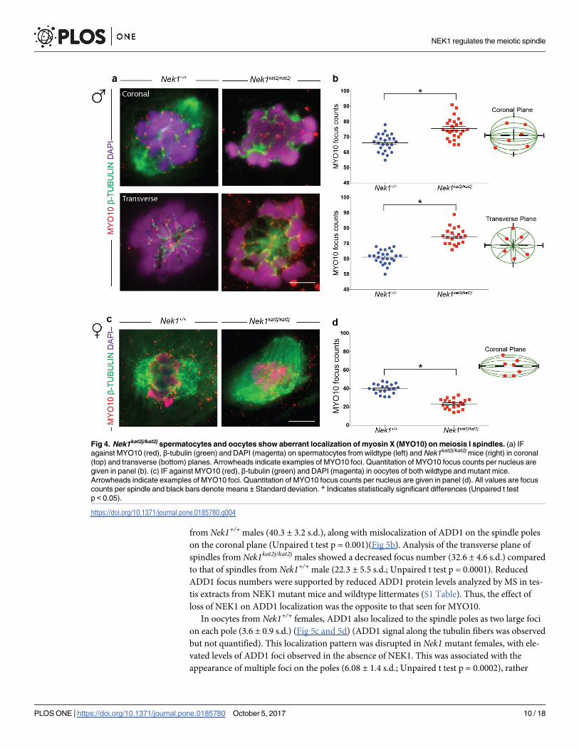
We evaluated the localization of MYO10 on MI spindles from male spermatocytes and female
oocytes. Immunofluorescence (IF) analysis in Nek1+/+ spindles revealed that MYO10 localizes
along the β-tubulin fibers specifically in mouse spermatocytes (Fig 4a). We analyzed the num-
ber of MYO10 foci per nucleus in the coronal plane (Fig 4b) and found an increase in the
number of foci in the Nek1kat2j/kat2j mice (75.4 ± 6.7 s.d.) compared to Nek1+/+ animals
(66.3 ± 5.5 s.d.; Unpaired t test p = 0.0001). Analysis of transverse sections of male spindles
revealed that there is a subtle accumulation of MYO10 at the spindle pole in Nek1+/+ male
mice (Fig 4a), and this was disrupted in Nek1kat2j/kat2j males (Fig 4b). In the transverse plane,
we observed an increase in the MYO10 foci number in Nek1kat2j/kat2j mice (74.7 ± 5.4 s.d.)
compared to the Nek1+/+ mice (61.7 ± 4.4 s.d.; Unpaired t test p = 0.0001). The increase in
MYO10 focus counts was supported by our mass spectrometry analysis showing increased
MYO10 protein in testis extracts from Nek1kat2j/kat2j males compared to Nek1+/+ mice (S1
Table).
We also evaluated the MYO10 foci in Nek1+/+ oocytes (Fig 4c), where MYO10 localizes on
tubulin fibers as foci during MI (39.7 ± 5.1 s.d.). In contrast to the situation in males, however,
oocytes obtained from Nek1kat2j/kat2j females showed more small and dispersed foci that do not
co-localize with β-tubulin. However, counts of MYO10 foci that co-localized with β-tubulin
were significantly reduced (23.3 ± 5.4 s.d.; Unpaired T test p = 0.0001)(Fig 4d). This could
indicate that the MYO10 is present but its capacity to bind to tubulin is reduced. Taken
together, our results indicate that loss of NEK1 leads to mislocalization of MYO10 along mei-
otic spindles in both male and females germ cells, but that the regulation of MYO10 by NEK1
may be sexually dimorphic.
Localization of ADD1 on spermatocytes and oocytes is disrupted in
absence of NEK1
Our previous MS results showed that ADD1 proteins levels are reduced in testis extracts from
Nek1kat2/kat2j mouse compared to wildtype littermates (S1 Table)[18]. Thus, we evaluated the
localization and protein levels of ADD1 in both male and female spindles during meiosis I.
ADD1 localizes both to the poles of the spermatocyte spindles (Fig 5a, asterisk) and with β-
tubulin fibers of the spindle (Fig 5a, arrow heads). We evaluated the ADD1 focus number asso-
ciated with the spindle structure in coronal and transverse planes. MI spindles from Nek1kat2j/
kat2j males showed a decreased ADD1 focus number (25.7 ± 7.8 s.d.) compared to MI spindles
NEK1 regulates the meiotic spindle
PLOS ONE | https://doi.org/10.1371/journal.pone.0185780 October 5, 2017 9 / 18
Page 10
from Nek1+/+ males (40.3 ± 3.2 s.d.), along with mislocalization of ADD1 on the spindle poles
on the coronal plane (Unpaired t test p = 0.001)(Fig 5b). Analysis of the transverse plane of
spindles from Nek1kat2j/kat2j males showed a decreased focus number (32.6 ± 4.6 s.d.) compared
to that of spindles from Nek1+/+ male (22.3 ± 5.5 s.d.; Unpaired t test p = 0.0001). Reduced
ADD1 focus numbers were supported by reduced ADD1 protein levels analyzed by MS in tes-
tis extracts from NEK1 mutant mice and wildtype littermates (S1 Table). Thus, the effect of
loss of NEK1 on ADD1 localization was the opposite to that seen for MYO10.
In oocytes from Nek1+/+ females, ADD1 also localized to the spindle poles as two large foci
on each pole (3.6 ± 0.9 s.d.) (Fig 5c and 5d) (ADD1 signal along the tubulin fibers was observed
but not quantified). This localization pattern was disrupted in Nek1 mutant females, with ele-
vated levels of ADD1 foci observed in the absence of NEK1. This was associated with the
appearance of multiple foci on the poles (6.08 ± 1.4 s.d.; Unpaired t test p = 0.0002), rather
Fig 4. Nek1kat2j/kat2j spermatocytes and oocytes show aberrant localization of myosin X (MYO10) on meiosis I spindles. (a) IF
against MYO10 (red), β-tubulin (green) and DAPI (magenta) on spermatocytes from wildtype (left) and Nek1kat2j/kat2j mice (right) in coronal
(top) and transverse (bottom) planes. Arrowheads indicate examples of MYO10 foci. Quantitation of MYO10 focus counts per nucleus are
given in panel (b). (c) IF against MYO10 (red), β-tubulin (green) and DAPI (magenta) in oocytes of both wildtype and mutant mice.
Arrowheads indicate examples of MYO10 foci. Quantitation of MYO10 focus counts per nucleus are given in panel (d). All values are focus
counts per spindle and black bars denote means ± Standard deviation. * Indicates statistically significant differences (Unpaired t test
p < 0.05).
https://doi.org/10.1371/journal.pone.0185780.g004
NEK1 regulates the meiotic spindle
PLOS ONE | https://doi.org/10.1371/journal.pone.0185780 October 5, 2017 10 / 18
Page 11
than the two distinct large aggregations of ADD1. Again, as with the males, the change in
ADD1 focus frequency on the meiotic spindles was the opposite to that seen for MYO10
(ADD1 foci increased on oocyte spindles in the Nek1 mutant animals, whereas MYO10 foci
decreased on oocyte spindles in the absence of NEK1). Taken together, these results demon-
strate that loss of NEK1 alters the profile and ratios of ADD1 and MYO10 protein on meiosis I
spindles in males and females, but that the effect may be sexually dimorphic.
Loss of NEK1 activity induces abnormal phosphorylation of ADD1
During mitotic spindle formation mammalian cell lines, ADD1 binds to the motor domain of
MYO10 on the spindle. ADD1 and MYO10 interaction on mitotic spindles is negatively regu-
lation by phosphorylation. Phosphorylation of ADD1 induces the loss of the interaction
between both proteins and therefore the complex unloads from the mitotic spindle [20]. Phos-
phorylation of ADD1 results in abnormal mitotic spindle morphology (elongation, multipolar
Fig 5. Nek1kat2j/kat2j spermatocytes and oocytes show aberrant localization of Adducin 1 (ADD1) on meiosis I spindles. (a) IF
against ADD1 (red), β-tubulin (green) and DAPI (magenta) on spermatocytes from wildtype (left) and Nek1kat2j/kat2j mice (right) in coronal
(top) and transverse (bottom) planes. Arrowheads indicate examples of ADD1 foci. Quantitation of ADD1 focus counts per nucleus are given
in panel (b). (c) IF against ADD1 (red), β-tubulin (green) and DAPI (magenta) in oocytes of both wildtype and mutant mice. Arrowheads
indicate examples of ADD1 foci. Quantitation of ADD1 focus counts per nucleus are given in panel (d). All values are focus counts per
spindle and black bars denote means ± Standard deviation. * Indicates statistically significant differences (Unpaired t test p < 0.05).
https://doi.org/10.1371/journal.pone.0185780.g005
NEK1 regulates the meiotic spindle
PLOS ONE | https://doi.org/10.1371/journal.pone.0185780 October 5, 2017 11 / 18
Page 12
spindles and aberrant chromosome alignment) and loss of its interaction with MYO10 [20].
We evaluated the proteins levels of ADD1 in testis lysates and isolated oocytes. Analysis of pro-
tein levels from testis lysates reveled a decrease in the protein levels in extracts from mutant
mice compared to wildtype testis (Fig 6a; Unpaired t test p = 0.0031). We observed a clear dou-
ble band in the Nek1kat2j/kat2j lysates, however this double band is not that obvious in the lysates
from Nek1+/+ testes. Decreased levels of ADD1 and the presence of a heavier band in WB anal-
ysis of Nek1kat2j/kat2j mouse compared to wildtype littermates suggested that the lack of NEK1
activity affects ADD1 phosphorylation. To test this hypothesis, we screened our previous phos-
phoproteomics data obtained from Nek1 mutant animals, specifically focusing on changes in
ADD1. Loss of NEK1 results in a significant increase in the phosphorylation of ADD1, but not
MYO10 (Fig 6b)[18]. Specifically, loss of NEK1 results in hyperphosphorylation of ADD1 on
serine 465 (S465) (Fig 6c). Interestingly, ADD1 also showed an increase in deamidation of
asparagine 462 (N462). Thus, both post-translational modifications could be acting as a trigger
to induce the degradation of the protein.
The results described above show that the lack of NEK1 induces changes in spindle forma-
tion and protein localization in both spermatocytes and oocytes. However, the phenotype in
both sexes is slightly different. We evaluated the relative protein levels of ADD1 in oocytes of
both Nek1+/+ and Nek1kat2j/kat2j oocytes. WB analysis revealed a decrease in ADD1 protein lev-
els in Nek1kat2j/kat2j oocytes compared to Nek1+/+ oocytes (Fig 6d; Unpaired t test p = 0.0003).
As in male, GV oocytes from Nek1kat2j/kat2j mice showed a double band, however oocytes from
Nek1+/+ also showed a double band. In both wildtype and mutant, the slower migrating band
had a stronger signal indicating that probably in oocytes the basal status of ADD1 is its phos-
phorylated state, at least at this stage of development. To evaluate the changes in the ADD1
protein levels according to oocyte maturation, we performed oocyte culture followed by WB.
Our results showed that after 6h in culture, Nek1+/+ oocytes showed the loss of the heavier
band, possibly indicating a change in ADD1 phosphorylation. However in the Nek1kat2j/kat2j
oocytes we observe that both bands almost disappear indicating that there is a change in the
phosphorylation in the mutants but a faster degradation of the protein in the mutant oocytes.
These results suggest that the phosphorylation levels of ADD1 in the mutant mice depends on
active phosphorylation of the protein by a kinase that is mis-regulated in the absence of NEK1,
or by loss of activity of a phosphatase that controls the phosphorylated status of ADD1. We
previously reported the interaction of PP1γ with NEK1 and its function regulating the pro-
phase pathway during meiosis [18]. To test if the changes of ADD1 phosphorylation similarly
depends on PP1γ activity, we cultured Nek1+/+ oocytes for 6h in the presence of the PP1γinhibitor, Calyculin (Fig 6e). The WB analysis of these cultures showed that the inhibition of
PP1γ induces a statistically significant increase in ADD1 protein levels, predominantly of the
slower migrating band, suggesting that the inhibition of PP1γ maintains the phosphorylation
status of ADD1. Thus, these results suggest that the lack of NEK1 action on ADD1 phosphory-
lation status may be the result of changes to PP1γ activity on ADD1.
Discussion
In eukaryotes, the structure orchestrating chromosome alignment and segregation during cell
division is the microtubular spindle [3]. To ensure correct spindle dynamics, the spindle
assembly checkpoint (SAC) becomes activated in situations where tension is not appropriately
established between the chromosomes and the spindle poles, and this will block the progres-
sion of the cell cycle to prevent aberrant segregation. In addition to the canonical SAC, other
levels of control have been described, including MYO10 [24–26]. In oocytes and frog embryos,
disruption of MYO10 negatively affects nuclear anchoring and meiotic spindle formation [26],
NEK1 regulates the meiotic spindle
PLOS ONE | https://doi.org/10.1371/journal.pone.0185780 October 5, 2017 12 / 18
Page 13
Fig 6. Loss of NEK1 during meiosis is associated with hyper-phosphorylation and reduced ADD1 protein in a PP1γ-dependent
manner. (a) Quantitation of ADD1 protein levels in Nek1+/+ testis extracts relative to GAPDH control. (b) ADD1 and MYO10 protein levels,
expressed as a ratio of Nek1kat2j/kat2j / Nek1+/+, as determined by mass spectrometry. (c) ADD1 phosphopeptide and deaminated peptide
sequence determined by mass spectrometry to be higher in Nek1kat2j/kat2j testis extracts. (d) Quantitation by western blotting of ADD1 protein
levels in oocytes at 0h and after 6h of culture. (e) Quantification of ADD1 protein levels in wildtype oocytes cultured for 6h in vehicle (ethanol)
and the PP1 inhibitor Calyculin A (CLA) (at doses of 2 and 20 nM). All values are means ± Standard deviation. * Indicate statistically significant
differences (Unpaired t test p < 0.05 and one-way ANOVA followed by Dunnett’s multiple comparisons test, p < 0.05, for cultures with inhibitor)
https://doi.org/10.1371/journal.pone.0185780.g006
NEK1 regulates the meiotic spindle
PLOS ONE | https://doi.org/10.1371/journal.pone.0185780 October 5, 2017 13 / 18
Page 14
while depletion of MYO10 in frog epithelial cells induces abnormal spindle movements, spin-
dle elongation, multi- spindle and pole fragmentation [25,27]. MYO10 has an amino-terminal
globular head domain that harbors acting biding and ATPase activities, giving to the protein
the capacity to bind F-actin. The tail of MYO10 has the MyTH4 and FERM domains that give
to the protein the ability to interact with microtubules [26,28]. Accordingly, recent studies
have demonstrated that loss of Myo10 results in spindle dysfunction, while overexpression of
only the MyTH4 domain alone affects the structure and shape of the spindle [24].
In the current study, we present evidence to suggest that loss of NEK1 induces increases in
MYO10 protein levels in spermatocytes, and that this in turn may result in spindle defects.
Our interpretation that overexpression of Myo10 leads to spindle defects is in line with the sug-
gestion that over-expression of the MyTH4 domain of MYO10 results in competition with the
wildtype protein, leading to the displacement of the latter from the spindle [28]. Furthermore,
it has been suggested that over-expression of only the MyTH4 may disrupt the functional link
between F-actin, microtubules and MYO10. In our data (S1 Table), we observed an increase in
the MYO10 protein levels without changes in F-actin or microtubules that could be inducing
an imbalance in the protein levels provoking a similar phenotype to the overexpression of the
MyTH4 domain. However, we could not eliminate the possibility that the lack of NEK1 could
induce changes in other proteins or on the structure of MYO10 that induces mislocalization
and/or abnormal function of MYO10. However, more studies using a MYO10 conditional
knockout or siRNA against MYO10 will help to answer these possibilities.
ADD1 is an actin-biding protein that is important for membrane stabilization [29] and for
cell-cell adhesion [30,31]. There are 3 isoforms of ADD with similar domain structures that
are formed by NH2-terminal head domain, a neck domain and a C-terminal domain. The C-
terminal domain is characterized by it high contain of myristoylated alanine-rich C kinase sub-
strate (MARCKS), this MARCKS related domain is necessary for its interaction with F-actin,
spectrin, and calmodulin [28,32–34]. Previous studies in somatic cells showed that loss of
ADD1 leads to a failure in mitotic spindle formation characterized by distortion, elongation,
multipolar spindles and abnormal chromosome alignment [20]. Here, we show that loss of
NEK1 leads to a reduction in ADD1 protein levels in spermatocytes, abnormal ADD1 distribu-
tion on the meiotic spindle of spermatocytes, and consequent disruption of spindle formation
and integrity. These phenotypes observed in meiotic cells are highly reminiscent of the pheno-
types observed in somatic cells lacking Add1 [20].
ADD1 and MYO10 interact via the MyTH4 domain of MYO10, and this interaction is criti-
cal for the correct spindle formation and function in mitotic cells [20,25,26,28]. Interaction of
ADD1 to MYO10 is dependent on phosphorylation whereby phosphorylation of ADD1 by
cyclin-dependent kinase 1 (CDK1) enables ADD1 to bind MYO10 on mitotic spindle [20].
Depletion of ADD1 or changes in phosphorylation status results in abnormal mitotic spindles
(elongation, multipolar spindles and aberrant chromosome alignment) [20]. In the absence of
NEK1 we observe an increase in the phosphorylation of ADD1 at S465 and deamination of
N462. Such modifications could induce premature loss of the ADD1-MYO10 interaction, first
by altered phosphorylation of the protein and then via degradation of the protein as a conse-
quence of the deamidation, similar to the phenotypes observed in somatic cells [20]. However,
it is unclear what kinase is phosphorylating ADD1 during meiosis, since the increased phos-
phorylation in the absence of NEK1 suggests the up-regulation of a kinase that is itself regu-
lated by NEK1 (either directly or indirectly). Clear candidates could be CDKs, however, our
MS results did not show any significant change in the profile of CDK in the absence of NEK1,
and specifically no differences in CDK1 indicating that increases in phosphorylation of ADD1
does not depend of CDK1 pathway. Alternatively, the increased phosphorylation of ADD1
could suggest the existence of a phosphatase that is inactivated by the loss of NEK1, allowing
NEK1 regulates the meiotic spindle
PLOS ONE | https://doi.org/10.1371/journal.pone.0185780 October 5, 2017 14 / 18
Page 15
the hyperphosphorylation of protein. We previously showed that in Nek1 mutants, there is a
down regulation and abnormal phosphorylation in whole testis lysate of PP1γ, and our results
of oocyte cultures in the presence of a PP1 inhibitor suggest that the increase in ADD1 phos-
phorylation could be mediated by a similar mechanism.
Our results showed the disruption of the spindle, however some inconsistences between
male and female gametes were observed. In both cases is clear that the lack of NEK1 is affecting
ADD1 and MYO10, but the phenotype is slightly different. Thus, loss of NEK1 results in an
increase in MYO10 localization on male meiotic spindles and a decrease in MYO10 localiza-
tion on female meiotic spindles. Conversely, loss of NEK1 induces a decrease in ADD1 locali-
zation on male spindles, while in females the lack of NEK1 induces an increment in focus
number but smaller in size, suggesting a disruption in the protein loading or acceleration in
protein degradation. It is possible, therefore, that the increased focus count for ADD1 associ-
ated with the oocyte spindle reflects lower amounts of total protein per focus and/or fragmen-
tation of the structures with which ADD1 is associating. This is supported by the different
appearance of ADD1 foci in NEK1-deficient oocytes compared to wildtype oocytes. Another
important difference in Nek1+/+ and Nek1kat2j/kat2j oocytes compared to spermatocytes is the
presence of a double band of ADD1 in the WB analysis, suggesting that in wildtype oocytes
there is a basal phosphorylated state of ADD1. However the main difference between WT and
mutant is that in the wildtype after culture the heavier band disappear but the lighter band still
present while in the mutant both bands disappear indicating the instability of the protein in
the absence of NEK1. Further analysis of ADD1 dynamics during oocyte meiosis will require
proteomics analysis of thousands of mouse oocytes, in order to understand the distribution
and status of spindle regulatory proteins in the presence and absence of NEK1.
Taken together, our data indicate that effect of NEK1 is sexually dimorphic with respect to
spindle association of key spindle-related proteins, as is often the case in other aspects of mei-
otic regulation, including recombination, chromosome pairing and synapsis [6,35–37]. Our
results showed, how the lack of NEK1 induced changes in both male and female spindle how-
ever the variation in the female phenotype could be related to how the spindle is formed. In
males the spindle formation depend on the centrosomes. In mitotic cells, centrosome’s func-
tions are regulated by other proteins of the NEK family such as NEK2, NEK6, NEK7 and
NEK9 that in the absence of NEK1 could be modifying the interactions and function of tubu-
lins or myosins [38–40]. However, female spindle formation does not depend of centrosomes,
thus a potential compensatory function of other NEK’s during the spindle formation is not
present; inducing the differences observed in the female phenotype. The broad spectrum of
abnormal spindles in female Nek1kat2j/kat2j oocytes, also could be associated to the “leakiness”
of spindle check point in females [41]. Besides, differences observed in the phenotype, the lack
of NEK1 ends up in male and female infertility [16,17].
In summary, we propose that the loss of NEK1 activity induces abnormal spindle formation
through a mechanism mediated by the imbalance of MYO10 and ADD1. The decreases in
ADD1 and it abnormal localization on the meiotic spindle is mediated by an increase its phos-
phorylation and deamination that leads to loss of interaction of MYO10 with ADD1. Taken
together our results show the importance of NEK1 in the control of spindle formation during
male and female meiosis.
Supporting information
S1 Table. Myosin proteins quantitation by mass spectrometry.
(DOCX)
NEK1 regulates the meiotic spindle
PLOS ONE | https://doi.org/10.1371/journal.pone.0185780 October 5, 2017 15 / 18
Page 16
S1 Fig. Dynamics of spindle formation in Nek1kat2j/kat2j oocytes. (a-f) Immunofluorescence
againts β-tubulin (green) and DNA staining with DAPI (magenta) showing the spindle
dynamics in Nek1kat2j/kat2j oocytes. a) Germinal vesicle (GV); b) Germinal vesicle break-
down (GVBD); c) Prometaphase I (ProI); d and e) Metaphase I (MI); f) Telophase I (TeI).
Scale bar 20μm
(EPS)
S2 Fig. Analysis of centromere and centrosomal protein ratio Nek1kat2j/kat2j/Nek1+/+ by
massspectrometry in whole testis lysates.
(EPS)
S3 Fig. Analysis of tubulins protein ratio Nek1kat2j/kat2j/Nek1+/+ by massspectrometry in
whole testis lysates.
(EPS)
S4 Fig. Analysis of actins protein ratio Nek1kat2j/kat2j/Nek1+/+ by massspectrometry in
whole testis lysates.
(EPS)
S5 Fig. Analysis of spindle assembly checkpoint protein ratio Nek1kat2j/kat2j/Nek1+/+ by
massspectrometry in whole testis lysates.
(EPS)
Acknowledgments
We are grateful to Peter Borst for assistance in animal care and genotyping. We thank Dr.
Sheng Zhang from Cornell Proteomics and Mass Spectrometry Facility for his advice in devel-
opment of mass spectrometry experiments.
Author Contributions
Conceptualization: Miguel A. Brieño-Enrı́quez, J. Kim Holloway, Paula E. Cohen.
Data curation: Miguel A. Brieño-Enrı́quez, Stefannie L. Moak, Paula E. Cohen.
Formal analysis: Miguel A. Brieño-Enrı́quez, Paula E. Cohen.
Funding acquisition: J. Kim Holloway, Paula E. Cohen.
Investigation: Miguel A. Brieño-Enrı́quez, Stefannie L. Moak, J. Kim Holloway, Paula E.
Cohen.
Writing – original draft: Miguel A. Brieño-Enrı́quez, Paula E. Cohen.
Writing – review & editing: Miguel A. Brieño-Enrı́quez, Paula E. Cohen.
References1. Gray S, Cohen PE. Control of Meiotic Crossovers: From Double-Strand Break Formation to Designa-
tion. Annu Rev Genet 2016; 50: 175–210. https://doi.org/10.1146/annurev-genet-120215-035111
PMID: 27648641
2. Duro E, Marston AL. From equator to pole: splitting chromosomes in mitosis and meiosis. Genes Dev
2015; 29: 109–122. https://doi.org/10.1101/gad.255554.114 PMID: 25593304
3. Bennabi I, Terret ME, Verlhac MH. Meiotic spindle assembly and chromosome segregation in oocytes.
J Cell Biol 2016; 215: 611–619. https://doi.org/10.1083/jcb.201607062 PMID: 27879467
4. Ohkura H. Meiosis: an overview of key differences from mitosis. Cold Spring Harb Perspect Biol 2015;
7.
NEK1 regulates the meiotic spindle
PLOS ONE | https://doi.org/10.1371/journal.pone.0185780 October 5, 2017 16 / 18
Page 17
5. McKim KS, Hawley RS. Chromosomal control of meiotic cell division. Science 1995; 270: 1595–1601.
PMID: 7502068
6. Morelli MA, Cohen PE. Not all germ cells are created equal: aspects of sexual dimorphism in mamma-
lian meiosis. Reproduction 2005; 130: 761–781. https://doi.org/10.1530/rep.1.00865 PMID: 16322537
7. Fry AM, O’Regan L, Sabir SR, Bayliss R. Cell cycle regulation by the NEK family of protein kinases. J
Cell Sci 2012; 125: 4423–4433. https://doi.org/10.1242/jcs.111195 PMID: 23132929
8. Meirelles GV, Perez AM, de Souza EE, Basei FL, Papa PF, Melo Hanchuk TD, et al. "Stop Ne(c)king
around": How interactomics contributes to functionally characterize Nek family kinases. World J Biol
Chem 2014; 5: 141–160. PMID: 24921005
9. Oakley BR, Morris NR. A mutation in Aspergillus nidulans that blocks the transition from interphase to
prophase. J Cell Biol 1983; 96: 1155–1158. PMID: 6339527
10. Letwin K, Mizzen L, Motro B, Ben-David Y, Bernstein A, Pawson T. A mammalian dual specificity pro-
tein kinase, Nek1, is related to the NIMA cell cycle regulator and highly expressed in meiotic germ cells.
EMBO J 1992; 11: 3521–3531. PMID: 1382974
11. White MC, Quarmby LM. The NIMA-family kinase, Nek1 affects the stability of centrosomes and cilio-
genesis. BMC Cell Biol 2008; 9: 29. https://doi.org/10.1186/1471-2121-9-29 PMID: 18533026
12. Hilton LK, White MC, Quarmby LM. The NIMA-related kinase NEK1 cycles through the nucleus. Bio-
chem Biophys Res Commun 2009; 389: 52–56. https://doi.org/10.1016/j.bbrc.2009.08.086 PMID:
19699716
13. Patil M, Pabla N, Ding HF, Dong Z. Nek1 interacts with Ku80 to assist chromatin loading of replication
factors and S-phase progression. Cell Cycle 2013; 12: 2608–2616. https://doi.org/10.4161/cc.25624
PMID: 23851348
14. Liu S, Ho CK, Ouyang J, Zou L. Nek1 kinase associates with ATR-ATRIP and primes ATR for efficient
DNA damage signaling. Proc Natl Acad Sci U S A 2013; 110: 2175–2180. https://doi.org/10.1073/pnas.
1217781110 PMID: 23345434
15. Chen Y, Chen CF, Chiang HC, Pena M, Polci R, Wei RL, et al. Mutation of NIMA-related kinase 1
(NEK1) leads to chromosome instability. Mol Cancer 2011; 10: 5. https://doi.org/10.1186/1476-4598-
10-5 PMID: 21214959
16. Upadhya P, Birkenmeier EH, Birkenmeier CS, Barker JE. Mutations in a NIMA-related kinase gene,
Nek1, cause pleiotropic effects including a progressive polycystic kidney disease in mice. Proc Natl
Acad Sci U S A 2000; 97: 217–221. PMID: 10618398
17. Holloway K, Roberson EC, Corbett KL, Kolas NK, Nieves E, Cohen PE. NEK1 Facilitates Cohesin
Removal during Mammalian Spermatogenesis. Genes (Basel) 2011; 2: 260–279.
18. Brieño-Enriquez MA, Moak SL, Toledo M, Filter JJ, Gray S, Barbero JL, et al. Cohesin Removal along
the Chromosome Arms during the First Meiotic Division Depends on a NEK1-PP1gamma-WAPL Axis
in the Mouse. Cell Rep 2016; 17: 977–986. https://doi.org/10.1016/j.celrep.2016.09.059 PMID:
27760328
19. Janaswami PM, Birkenmeier EH, Cook SA, Rowe LB, Bronson RT, Davisson MT. Identification and
genetic mapping of a new polycystic kidney disease on mouse chromosome 8. Genomics 1997; 40:
101–107. https://doi.org/10.1006/geno.1996.4567 PMID: 9070925
20. Chan PC, Hsu RY, Liu CW, Lai CC, Chen HC. Adducin-1 is essential for mitotic spindle assembly
through its interaction with myosin-X. J Cell Biol 2014; 204: 19–28. https://doi.org/10.1083/jcb.
201306083 PMID: 24379415
21. Page J, Suja JA, Santos JL, Rufas JS. Squash procedure for protein immunolocalization in meiotic
cells. Chromosome Res 1998; 6: 639–642. PMID: 10099877
22. Kotaja N, Kimmins S, Brancorsini S, Hentsch D, Vonesch JL, Davidson I, et al. Preparation, isolation
and characterization of stage-specific spermatogenic cells for cellular and molecular analysis. Nat
Methods 2004; 1: 249–254. https://doi.org/10.1038/nmeth1204-249 PMID: 16144087
23. Sun X, Cohen PE. Studying recombination in mouse oocytes. Methods Mol Biol 2013; 957: 1–18.
https://doi.org/10.1007/978-1-62703-191-2_1 PMID: 23138941
24. Sandquist JC, Larson ME, Hine KJ. Myosin-10 independently influences mitotic spindle structure and
mitotic progression. Cytoskeleton (Hoboken) 2016; 73: 351–364.
25. Woolner S, O’Brien LL, Wiese C, Bement WM. Myosin-10 and actin filaments are essential for mitotic
spindle function. J Cell Biol 2008; 182: 77–88. https://doi.org/10.1083/jcb.200804062 PMID: 18606852
26. Weber KL, Sokac AM, Berg JS, Cheney RE, Bement WM. A microtubule-binding myosin required for
nuclear anchoring and spindle assembly. Nature 2004; 431: 325–329. https://doi.org/10.1038/
nature02834 PMID: 15372037
NEK1 regulates the meiotic spindle
PLOS ONE | https://doi.org/10.1371/journal.pone.0185780 October 5, 2017 17 / 18
Page 18
27. Kwon M, Bagonis M, Danuser G, Pellman D. Direct Microtubule-Binding by Myosin-10 Orients Centro-
somes toward Retraction Fibers and Subcortical Actin Clouds. Dev Cell 2015; 34: 323–337. https://doi.
org/10.1016/j.devcel.2015.06.013 PMID: 26235048
28. Hirano Y, Hatano T, Takahashi A, Toriyama M, Inagaki N, Hakoshima T. Structural basis of cargo rec-
ognition by the myosin-X MyTH4-FERM domain. EMBO J 2011; 30: 2734–2747. https://doi.org/10.
1038/emboj.2011.177 PMID: 21642953
29. Anong WA, Franco T, Chu H, Weis TL, Devlin EE, Bodine DM, et al. Adducin forms a bridge between
the erythrocyte membrane and its cytoskeleton and regulates membrane cohesion. Blood 2009; 114:
1904–1912. https://doi.org/10.1182/blood-2009-02-203216 PMID: 19567882
30. Abdi KM, Bennett V. Adducin promotes micrometer-scale organization of beta2-spectrin in lateral mem-
branes of bronchial epithelial cells. Mol Biol Cell 2008; 19: 536–545. https://doi.org/10.1091/mbc.E07-
08-0818 PMID: 18003973
31. Franco T, Chu H, Low PS. Identification of Adducin-Binding Residues on the Cytoplasmic Domain of
Erythrocyte Membrane Protein, Band 3. Biochem J 2016.
32. Scaramuzzino DA, Morrow JS. Calmodulin-binding domain of recombinant erythrocyte beta-adducin.
Proc Natl Acad Sci U S A 1993; 90: 3398–3402. PMID: 8475088
33. Kaiser HWO’Keefe E, Bennett V. Adducin: Ca++-dependent association with sites of cell-cell contact. J
Cell Biol 1989; 109: 557–569. PMID: 2503523
34. Matsuoka Y, Hughes CA, Bennett V. Adducin regulation. Definition of the calmodulin-binding domain
and sites of phosphorylation by protein kinases A and C. J Biol Chem 1996; 271: 25157–25166. PMID:
8810272
35. Herran Y, Gutierrez-Caballero C, Sanchez-Martin M, Hernandez T, Viera A, Barbero JL, et al. The
cohesin subunit RAD21L functions in meiotic synapsis and exhibits sexual dimorphism in fertility.
EMBO J 2011; 30: 3091–3105. https://doi.org/10.1038/emboj.2011.222 PMID: 21743440
36. Hayashi T, Kageyama Y, Ishizaka K, Kihara K, Oshima H. Sexual dimorphism in the regulation of mei-
otic process in the rabbit. Biol Reprod 2000; 62: 1722–1727. PMID: 10819776
37. Handel MA, Schimenti JC. Genetics of mammalian meiosis: regulation, dynamics and impact on fertility.
Nat Rev Genet 2010; 11: 124–136. https://doi.org/10.1038/nrg2723 PMID: 20051984
38. Bertran MT, Sdelci S, Regue L, Avruch J, Caelles C, Roig J. Nek9 is a Plk1-activated kinase that con-
trols early centrosome separation through Nek6/7 and Eg5. EMBO J 2011; 30: 2634–2647. https://doi.
org/10.1038/emboj.2011.179 PMID: 21642957
39. Sdelci S, Schutz M, Pinyol R, Bertran MT, Regue L, Caelles C, et al. Nek9 phosphorylation of NEDD1/
GCP-WD contributes to Plk1 control of gamma-tubulin recruitment to the mitotic centrosome. Curr Biol
2012; 22: 1516–1523. https://doi.org/10.1016/j.cub.2012.06.027 PMID: 22818914
40. Hames RS, Crookes RE, Straatman KR, Merdes A, Hayes MJ, Faragher AJ, et al. Dynamic recruitment
of Nek2 kinase to the centrosome involves microtubules, PCM-1, and localized proteasomal degrada-
tion. Mol Biol Cell 2005; 16: 1711–1724. https://doi.org/10.1091/mbc.E04-08-0688 PMID: 15659651
41. Gorbsky GJ. The spindle checkpoint and chromosome segregation in meiosis. FEBS J 2015; 282:
2471–2487. https://doi.org/10.1111/febs.13166 PMID: 25470754
NEK1 regulates the meiotic spindle
PLOS ONE | https://doi.org/10.1371/journal.pone.0185780 October 5, 2017 18 / 18