CLH REPORT FOR ACETALDEHYDE 1 CLH report Proposal for Harmonised Classification and Labelling Based on Regulation (EC) No 1272/2008 (CLP Regulation), Annex VI, Part 2 Acetaldehyde EC Number: 200-836-8 CAS Number: 75-07-0 Index Number: 605-003-00-6 Contact details for dossier submitter: RIVM, The Netherlands National Institute for Public Health and the Environment Centre for Safety of Substances and Products Bilthoven, The Netherlands Version number: 2.0 Date: June 2015
Transcript
CLH REPORT FOR ACETALDEHYDE
1
CLH report
Proposal for Harmonised Classification and Labelling
Based on Regulation (EC) No 1272/2008 (CLP Regulation),
Annex VI, Part 2
Acetaldehyde
EC Number: 200-836-8
CAS Number: 75-07-0
Index Number: 605-003-00-6
Contact details for dossier submitter:
RIVM, The Netherlands
National Institute for Public Health and the Environment Centre for Safety of Substances and Products Bilthoven, The Netherlands
Version number: 2.0 Date: June 2015
CLH REPORT FOR ACETALDEHYDE
2
CLH REPORT FOR ACETALDEHYDE
3
CONTENTS
Part A. 1 PROPOSAL FOR HARMONISED CLASSIFICATION AND LABELLING ................................................. 5
1.1 SUBSTANCE ........................................................................................................................................................... 5 1.2 HARMONISED CLASSIFICATION AND LABELLING PROPOSAL .................................................................................. 5 1.3 PROPOSED HARMONISED CLASSIFICATION AND LABELLING BASED ON CLP REGULATION .................................... 6
2 BACKGROUND TO THE CLH PROPOSAL ..................................................................................................... 8
2.1 HISTORY OF THE PREVIOUS CLASSIFICATION AND LABELLING .............................................................................. 8 2.2 SHORT SUMMARY OF THE SCIENTIFIC JUSTIFICATION FOR THE CLH PROPOSAL .................................................... 8 2.3 CURRENT HARMONISED CLASSIFICATION AND LABELLING .................................................................................... 9
2.3.1 Current classification and labelling in Annex VI, Table 3.1 in the CLP Regulation .................................. 9 2.3.2 Current classification and labelling in Annex VI, Table 3.2 in the CLP Regulation .................................. 9
2.4 CURRENT SELF-CLASSIFICATION AND LABELLING ................................................................................................. 9 2.4.1 Current self-classification and labelling based on the CLP Regulation criteria ........................................ 9 2.4.2 Current self-classification and labelling based on DSD criteria .............................................................. 10
3 JUSTIFICATION THAT ACTION IS NEEDED AT COMMUNITY LEVEL .............................................. 10
Part B. SCIENTIFIC EVALUATION OF THE DATA ........................................................................................................... 11
1 IDENTITY OF THE SUBSTANCE .................................................................................................................... 11
1.1 NAME AND OTHER IDENTIFIERS OF THE SUBSTANCE ............................................................................................ 11 1.2 COMPOSITION OF THE SUBSTANCE ...................................................................................................................... 12
1.2.1 Composition of test material ..................................................................................................................... 12 1.3 PHYSICO-CHEMICAL PROPERTIES ........................................................................................................................ 12
2 MANUFACTURE AND USES ............................................................................................................................ 13
4.9.1 Non-human information ............................................................................................................................ 17 4.9.1.1 In vitro data ............................................................................................................................................................ 17 4.9.1.2 In vivo data ............................................................................................................................................................ 26
4.9.2 Human information ................................................................................................................................... 30
CLH REPORT FOR ACETALDEHYDE
4
4.9.3 Other relevant information ....................................................................................................................... 31 4.9.4 Summary and discussion of mutagenicity ................................................................................................. 37 4.9.5 Comparison with criteria .......................................................................................................................... 39 4.9.6 Conclusions on classification and labelling ............................................................................................. 40
4.10 CARCINOGENICITY ......................................................................................................................................... 41 4.10.1 Non-human information ....................................................................................................................... 41
4.10.2 Human information .............................................................................................................................. 46 4.10.3 Other relevant information .................................................................................................................. 46 4.10.4 Summary and discussion of carcinogenicity ........................................................................................ 48 4.10.5 Comparison with criteria ..................................................................................................................... 49 4.10.6 Conclusions on classification and labelling ......................................................................................... 49
4.11 TOXICITY FOR REPRODUCTION ....................................................................................................................... 49 4.12 OTHER EFFECTS .............................................................................................................................................. 49
5.1 DEGRADATION .................................................................................................................................................... 49 5.2 ENVIRONMENTAL DISTRIBUTION ......................................................................................................................... 49 5.3 AQUATIC BIOACCUMULATION ............................................................................................................................ 50 5.4 AQUATIC TOXICITY ............................................................................................................................................. 50 5.5 COMPARISON WITH CRITERIA FOR ENVIRONMENTAL HAZARDS (SECTIONS 5.1 – 5.4) .......................................... 50 5.6 CONCLUSIONS ON CLASSIFICATION AND LABELLING FOR ENVIRONMENTAL HAZARDS (SECTIONS 5.1 – 5.4) ....... 50
6 OTHER INFORMATION .................................................................................................................................... 50
1.3 Proposed harmonised classification and labelling based on CLP Regulation
Table 3: Proposed classification according to the CLP Regulation
CLP Annex I
ref
Hazard class Proposed classification
Proposed SCLs and/or M-factors
Current classification 1)
Reason for no classification 2)
2.1. Explosives None None Not evaluated
2.2. Flammable gases None None Not evaluated
2.3. Flammable aerosols None None Not evaluated
2.4. Oxidising gases None None Not evaluated
2.5. Gases under pressure None None Not evaluated
2.6. Flammable liquids Flam. Liq. 1
2.7. Flammable solids None None Not evaluated
2.8. Self-reactive substances and mixtures
None None Not evaluated
2.9. Pyrophoric liquids None None Not evaluated
2.10. Pyrophoric solids None None Not evaluated
2.11. Self-heating substances and mixtures
None None Not evaluated
2.12. Substances and mixtures which in contact with water emit flammable gases
None None Not evaluated
2.13. Oxidising liquids None None Not evaluated
2.14. Oxidising solids None None Not evaluated
2.15. Organic peroxides None None Not evaluated
2.16. Substance and mixtures corrosive to metals
None None Not evaluated
3.1. Acute toxicity - oral None None Not evaluated
Acute toxicity - dermal None None Not evaluated
Acute toxicity - inhalation None None Not evaluated
3.2. Skin corrosion / irritation None None Not evaluated
3.3. Serious eye damage / eye irritation
Eye Irrit. 2
3.4. Respiratory sensitisation None None Not evaluated
3.4. Skin sensitisation None None Not evaluated
3.5. Germ cell mutagenicity Muta. 1B
3.6. Carcinogenicity Carc. 1B Carc. 2
3.7. Reproductive toxicity None None Not evaluated
3.8. Specific target organ toxicity –single exposure
STOT SE 3
3.9. Specific target organ toxicity – repeated exposure
None None Not evaluated
3.10. Aspiration hazard None None Not evaluated
4.1. Hazardous to the None None Not evaluated
CLH REPORT FOR ACETALDEHYDE
7
aquaticenvironment
5.1. Hazardous to the ozone layer None None Not evaluated 1) Including specific concentration limits (SCLs) and M-factors
2) Data lacking, inconclusive, or conclusive but not sufficient for classification
Labelling: Signal word: Danger Hazard statements: H224, H319, H335, H350, H340 Precautionary statements: not harmonized
Proposed notes assigned to an entry:
: none
CLH REPORT FOR ACETALDEHYDE
8
2 BACKGROUND TO THE CLH PROPOSAL
2.1 History of the previous classification and labelling
Acetaldehyde is classified for carcinogenicity in Annex VI of regulation (EC) No 1272/2008 as follows: Carc 2 (suspected human carcinogen; H351: suspected of causing cancer). The substance is not classified for mutagenic activity. The classification by the European Commission dates from 1991. The existing classification with Carc. Cat 2 is based on the same carcinogenicity studies as in this proposal. However, there is new information regarding mutagenicity. This proposal for changing the harmonised classification is based on the report of the Health Council of the Netherlands.(1)
2.2 Short summary of the scientific justification for the CLH proposal
In 1999, IARC concluded that there was inadequate evidence in humans for the carcinogenicity of acetaldehyde, and that there was sufficient evidence in experimental animals.(2) Therefore, IARC classified the substance in Group 2B (‘possibly carcinogenic to humans’).
In 2010, IARC evaluated the risk of cancer due to alcohol consumption, including acetaldehyde. It confirmed that there was sufficient evidence in animal experiments for the carcinogenicity of acetaldehyde.(3) Moreover, in 2012 IARC concluded that ‘acetaldehyde associated with alcohol consumption’ is carcinogenic to humans (Group 1).(4)
Acetaldehyde is an intermediate substance in the metabolism of ethanol, and it has been suggested that acetaldehyde accounts for a great part of the toxic effects of ethanol. However, this proposal focuses on acetaldehyde alone and does not consider combined exposure with ethanol and ethanol-related adverse health effects.
On mutagenicity, sufficient evidence has been found for in vivo mutagenicity testing in somatic cells of mammals. There is limited evidence that acetaldehyde is genotoxic (sister chromatid exchanges) in germ cells of mice (Madrigal-Bujaidar et al. 2002), when the substance was given by intraperitoneal injection.(5) These findings indicate that acetaldehyde is able to reach the germ cells, and interacts with the genetic material, which would be in line with the findings on absorption and distribution kinetics. However, in another animal study no abnormal sperm cells, and no meiotic micronuclei in spermatids were observed at dose levels inducing acute toxicity (Lähdetie et al. 1988).(6) Overall, it is considered that some evidence exists that acetaldehyde has potential to cause mutations in germ cells. Therefore, it is recommended to classify the substance in category 1B.
On carcinogenicity, there is little or no epidemiological data to support statements concerning an association between exposure to acetaldehyde and cancer. Therefore, human data are considered insufficient to make a final conclusion on the carcinogenic potential of acetaldehyde in humans. For animal data, there is sufficient evidence of carcinogenicity, since a causal relationship was established between malignant tumours in animals and chronic inhalation to acetaldehyde in two studies (Woutersen et al. 1986, Feron et al. 1982), the main route of exposure in an occupational environment.(7, 8) According to the CLP classification criteria, acetaldehyde should, therefore, be classified as “presumed to have carcinogenic potential for humans”, which corresponds to classification in category 1B. Supporting evidence for its carcinogenic potential is that the substance has mutagenic properties.
CLH REPORT FOR ACETALDEHYDE
9
2.3 Current harmonised classification and labelling
2.3.1 Current classification and labelling in Annex VI, Table 3.1 in the CLP Regulation
The classification of acetaldehyde is harmonised in Annex VI of CLP under the index number 605-003-00-6 as follows:
Table 3.1 CLP Regulation
Flam. Liq. 1 - H224
Eye Irrit. 2 - H319
STOT SE 3 - H335
Carc. 2 - H351
2.3.2 Current classification and labelling in Annex VI, Table 3.2 in the CLP Regulation
This paragraph is considered irrelevant seen the repeal of Directive 67/548/EEC with effect from 1 June 2015.
2.4 Current self-classification and labelling
2.4.1 Current self-classification and labelling based on the CLP Regulation criteria
The registrants and most notifiers use the harmonised classification:
Flam. Liq. 1 - H224
Eye Irrit. 2 - H319
STOT SE 3 - H335
Carc. 2 - H351
However, the following additional classifications were applied by some of the other notifiers:
Acute Tox. 4 – H302
Acute Tox. 3 – H311
Eye Dam. 1 - H318
Skin Sens. 1 – H317
Muta 2 – H341
CLH REPORT FOR ACETALDEHYDE
10
STOT SE 2 – H371
Aquatic Chronic 2 – H411
2.4.2 Current self-classification and labelling based on DSD criteria
This paragraph is considered irrelevant seen the repeal of Directive 67/548/EEC with effect from 1 June 2015.
3 JUSTIFICATION THAT ACTION IS NEEDED AT COMMUNITY LEVEL
A change in the harmonised classification of acetaldehyde is proposed because there is new data especially on mutagenicity, which warrants a more severe classification for germ cell mutagenicity and carcinogenicity compared to the current harmonised classification.
Impurity Typical concentration Concentration range Remarks
confidential The known impurities are not expected to affect the classification.
Current Annex VI entry:
Table 7: Additives (non-confidential information)
Additive Function Typical concentration Concentration range Remarks
confidential
Current Annex VI entry:
1.2.1 Composition of test material
Relevant information on the purity is given in the respective study summaries when available.
1.3 Physico-chemical properties
CLH REPORT FOR ACETALDEHYDE
13
Table 8: Summary of physico - chemical properties
Property Value Reference Comment (e.g. measured or estimated)
State of the substance at 20°C and 101,3 kPa
Liquid IUCLID 2000
Melting/freezing point -123.5 °C SCCNFP 20042
Boiling point 20.4 °C SCCNFP 20042
Relative density 0.78 g/cm3 at 20 °C IUCLID 2000
Vapour pressure 98 kPa at 20 °C SCCNFP 20042
Surface tension - IUCLID 2000
Water solubility Miscible at 20 °C IUCLID 2000
Partition coefficient n-octanol/water
log P, 0.43 IARC 19993
Flash point -40 °C (open cup), -38 °C (closed cup)
IARC 19993
Flammability Extremely flammable IUCLID 2000
Explosive properties - IUCLID 2000
Self-ignition temperature -
Oxidising properties -
Granulometry -
Stability in organic solvents and identity of relevant degradation products
-
Dissociation constant 13.6 at 25 °C NTP 2010
Viscosity 0.2456 mPa x sec at 15 °C
SCCS 2012
2 MANUFACTURE AND USES
2.1 Manufacture
Not relevant for classification.
2.2 Identified uses
Acetaldehyde is an aldehyde, occurring widely in nature. For instance, it occurs naturally in coffee, bread, and ripe fruit, and is produced by plants as part of their normal metabolism. Acetaldehyde is also formed endogenously in humans in small amounts, for instance during the breakdown of ethanol in the body. It is, furthermore, present in tobacco smoke.
Acetaldehyde is produced on a large industrial scale for many purposes and uses.(9) For instance, it is used as an intermediate in the production of acetic acid; in the production of cellulose acetate, pyridine derivates, perfumes, paints (aniline dyes), plastics and synthetic rubber; in leather tanning
CLH REPORT FOR ACETALDEHYDE
14
and silvering mirrors; as a denaturant for alcohol; in fuel mixtures; as a hardener for gelatine fibres; in glue and casein products; as a preservative for fish and fruit; in the paper industry; and as a flavouring agent.
Acetaldehyde has a full registration. However, no use information is publicly available from the registration.
3 CLASSIFICATION FOR PHYSICO-CHEMICAL PROPERTIES
Not evaluated in this dossier.
4 HUMAN HEALTH HAZARD ASSESSMENT
4.1 Toxicokinetics (absorption, metabolism, distribution and elimination)
The data presented below is a summary from evaluations and reviews by others, such as IARC,(2-4) IPCS,(10) DFG,(11), CERI (12), and SCCNFP.(13)
Absorption, distribution and elimination In human volunteers, a significant uptake (45-70%) by the respiratory tract of inhaled acetaldehyde (100 to 800 mg/m3) was observed after a very short exposure duration of 45 to 75 seconds. In an inhalation study (1 litre/minute for 1-hr, between 1-20 mM) in 3 male SD rats, acetaldehyde was distributed in the blood, liver, kidney, spleen, heart, myocardium and skeletal muscle. Levels of acetaldehyde in the blood were reduced quickly, with a half-life of 3.1 minutes. Following acetaldehyde inhalation, peripheral blood acetaldehyde levels were highest; other tissue levels were similar except for the liver, which had a much lower level (Table 9). The concentration in the liver was relatively low due to the rapid metabolism of acetaldehyde. In the same study, acetaldehyde was also measured after a single intragastric ethanol administration (3 gr/kg bw). Acetaldehyde was found in the same tissues compared to inhalation exposure, but the liver levels were higher instead of lower, due to the formation of acetaldehyde in the metabolism of ethanol (Table 9) (14). Table 9: The tissue distribution of acetaldehyde following acetaldehyde inhalation and intragastric ethanol administration (14) Tissue Acetaldehyde inhalation
(nmol/g) Ethanol administration (nmol/g)
Blood* 1210 4.2 Liver 55 9.4 Kidney 213 2.1 Spleen 183 2.1 Heart muscle 277 2.3 Skeleton-muscle 345 1.7 *Blood levels were expressed as nmol/ml. Rats were exposed to acetaldehyde gas for 1 hour (1-20 mM). The acetaldehyde levels were determined immediately after discontinuation of inhalation and 3 hours after the intragastric administration of ethanol (3 g/kg body weight).
CLH REPORT FOR ACETALDEHYDE
15
Limited data obtained from animal experiments suggest that acetaldehyde (administered by intraperitoneal injection) may be partially transferred from maternal to foetal blood. It is also found in foetal liver. In a few studies acetaldehyde was detected in the blood and brain of animals, which were given the substance by intragastric administration or intraperitoneal injections. After an oral administration of ethanol at a dose of 4,500 mg/kg in male and female Wistar rats, it was confirmed that produced acetaldehyde was distributed in the blood and brain interstitial fluid. No data are available on dermal or percutaneous absorption.
Data on elimination are very limited. In one study using dogs, a single administration of acetaldehyde via a stomach tube revealed the presence of the substance in urine in minor quantities, but in most dogs no urinary acetaldehyde could be detected at all. Most likely this is due to the rapid metabolism of the substance in the liver. This was supported by studies in rabbits and rats, where metabolites were found in urine after intravenous administration of acetaldehyde. Metabolism Acetaldehyde is metabolized to acetic acid by nicotinamide adenine dinucleotide (NAD)-dependent aldehyde dehydrogenase (ALDH), which exists in the cells of most tissues, including the liver, mucosal tissue of the respiratory tract, and the testes of mice. Eventually it is degraded to carbon dioxide and water by the citric acid cycle. A minor part of the substance is probably oxidized by cytochrome P450 2E1, and by different aldehyde oxidases. There are two types of ALDH, a mitochondrial and a cytosolic form. The kinetic characteristics of the enzymatic reaction of liver mitochondrial ALDH are similar among human, rat and Syrian hamster. The Km value of human cytosolic ALDH1 was approximately 180 ìM, but those of rat and Syrian hamster were 15 and 12 ìM, respectively. In human liver, mitochondrial ALDH alone oxidizes acetaldehyde at physiological concentrations, but in rodent liver, both mitochondrial and cytosolic ALDHs have a role in acetaldehyde metabolism.
Acetaldehyde dehydrogenases show genetic polymorphism that gives rise to differences in vulnerability in humans concerning toxicity. Approximately 40% of Oriental population is inactive in mitochondrial ALDH2, which is associated with alcohol intolerance.
In general, data indicate a highly effective metabolism, in that half-time values in the blood for acetaldehyde were found to be three minutes in rats (after repeated exposure by inhalation) and mice (single intraperitoneal injection). For humans, no reliable data on half-times are available.
Acetaldehyde is a highly reactive electrophile, which reacts with nucleophilic groups of cellular macromolecules, such as proteins and DNA, to form adducts. It is shown that acetaldehyde (purity: 99%) that is incubated with ribonucleosides and deoxyribonucleosides forms adducts with cytosine or purine nucleoside, and one of acetaldehyde guanosine adducts is N2-ethylguanosine. Conclusion The available information from laboratory animals and humans indicate that acetaldehyde becomes systemically available after oral and inhalation exposure. However, the data also show that due to the rapid metabolism as indicated by the half-time values in blood of 3 minutes the systemic exposure can be expected to be low and to decrease quickly after the end of exposure. There is no direct evidence that acetaldehyde reaches the germ cells or the testes and ovaries after exposure via physiological routes of exposure. However, as acetaldehyde reaches the systemic circulation and several organs it is considered likely that acetaldehyde will also reach the testes and ovaries.
CLH REPORT FOR ACETALDEHYDE
16
4.2 Acute toxicity
Not evaluated in this dossier.
4.3 Specific target organ toxicity – single exposure (STOT SE)
Not evaluated in this dossier.
4.4 Irritation
Not evaluated in this dossier.
4.4.1 Skin irritation
Not evaluated in this dossier.
4.4.2 Eye irritation
Not evaluated in this dossier.
4.4.3 Respiratory tract irritation
This paragraph is considered irrelevant seen the repeal of Directive 67/548/EEC with effect from 1 June 2015.
4.5 Corrosivity
Not evaluated in this dossier.
4.6 Sensitisation
Not evaluated in this dossier.
CLH REPORT FOR ACETALDEHYDE
17
4.6.1 Skin sensititsation
Not evaluated in this dossier.
4.6.2 Respiratory sensitisation
Not evaluated in this dossier.
4.7 Repeated dose toxicity
Not evaluated in this dossier.
4.8 Specific target organ toxicity (CLP Regulation) – repeated exposure (STOT RE)
Not evaluated in this dossier.
4.9 Germ cell mutagenicity (Mutagenicity)
4.9.1 Non-human information
4.9.1.1 In vitro data
Data on in vitro mutagenicity testing are presented in Table 10.
Table 10 Summary of in vitro mutagenicity studies Method Cell type Concentration
Range*
Results
- negative
+ positive
Klimisch(15)
Score**
References
Micro-organisms
Reverse
mutation; multi-
substance study
S. typhimurium
TA98, TA100,
TA1535, TA1537
0 – 10,000
μg/plate
- (tested in two
laboratories)
2 Mortelmans et al.
1986(16)
Reverse mutation S. typhimurium
TA98, TA100,
TA1535, TA1537,
TA1538
0.005, 0.01, 0.1,
1.0, 5.0, and 10
μg/plate: + and –
S9
- 2 ECHA registration
data, in vitro.001,
study report 1979
(echa.europe.eu;)
Reverse mutation S. typhimurium
TA100, TA102,
TA104
0.1 – 1.0
ml/chamber,
vapour; - and +
S9
- 2 Dillon et al.
1998(17)
Reverse mutation S. typhimurium Max. non-toxic - 3; only one Marnett et al.
* + or - S9, with or without metabolic activation system.
** Klimisch score is expressed in reliability levels (cited from original publication):
Reliability 1 (reliably without restriction). For example, guideline study (OECD, etc.); comparable to guideline
study; test procedure according to national standards (DIN, etc.).
Reliability 2 (reliable with restrictions). For example, acceptable, well-documented publication/study report which
meets basic scientific principles; basic data given: comparable to guidelines/standards; comparable to guideline
study with acceptable restrictions.
Reliability 3 (not reliable). For example, method not validated; documentation insufficient for assessment; does
not meet important criteria of today standard methods; relevant methodological deficiencies; unsuitable test
system.
Reliability 4 (not assignable). For example, only short abstract available; only secondary literature (review,
tables, books, etc.).
Micro-organisms
Acetaldehyde was not mutagenic to Salmonella typhimurium or E. coli WP2 uvrA, with or without metabolic activation. It induced chromosome malsegregation in Aspergillus nidulans and forward mutations in yeast. Mammalian cells
CLH REPORT FOR ACETALDEHYDE
22
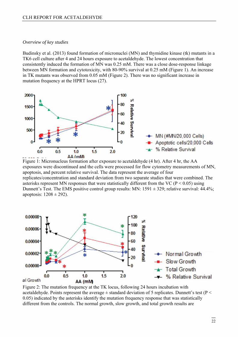
Overview of key studies Budinsky et al. (2013) found formation of micronuclei (MN) and thymidine kinase (tk) mutants in a TK6 cell culture after 4 and 24 hours exposure to acetaldehyde. The lowest concentration that consistently induced the formation of MN was 0.25 mM. There was a close dose-response linkage between MN formation and cytotoxicity, with 80-90% survival at 0.25 mM (Figure 1). An increase in TK mutants was observed from 0.05 mM (Figure 2). There was no significant increase in mutation frequency at the HPRT locus (27).
Figure 1: Micronucleus formation after exposure to acetaldehyde (4 hr). After 4 hr, the AA exposures were discontinued and the cells were processed for flow cytometry measurements of MN, apoptosis, and percent relative survival. The data represent the average of four replicates/concentration and standard deviation from two separate studies that were combined. The asterisks represent MN responses that were statistically different from the VC (P < 0.05) using Dunnett’s Test. The EMS positive control group results: MN: 1591 ± 329; relative survival: 44.4%; apoptosis: 1208 ± 292).
Figure 2: The mutation frequency at the TK locus, following 24 hours incubation with acetaldehyde. Points represent the average ± standard deviation of 5 replicates. Dunnett’s test (P < 0.05) indicated by the asterisks identify the mutation frequency response that was statistically different from the controls. The normal growth, slow growth, and total growth results are
CLH REPORT FOR ACETALDEHYDE
23
represented by the blue circles, red squares, and green triangles, respectively. Total growth represents the combined results for normal and slow growth mutants. The inverted black triangles represent the % relative survival. Separate positive controls, using EMS at 20 and 200 3M, were conducted. The 20 and 200 3M EMS positive controls in the AA study showed a normal growth MF of 1.87E - 05 and 2.44E - 04, respectively; a slow growth MF of 1.68E - 05 and 6.46E - 05, and a total MF of 3.55E - 05 and 3.09E - 04. In a study by Mechilli et al (2008), induction of chromosomal aberrations (CAs) and sister chromatid exchanges (SCEs) by acetaldehyde (AA) was evaluated in parental and different DNA repair-deficient Chinese hamster ovary (CHO) cell lines to elucidate the mechanisms involved in the protection against AA-induced chromosome damage. Cell lines employed included the parental (AA8), nucleotide excision repair (UV4, UV5, UV61), base excision repair (EM9), homologous recombination repair (HRR) (irs1SF, 51D1)-deficient and Fanconilike (KO40) ones. Concentration dependent increases in both CAs and SCEs were observed. The ranking of different cell lines for sensitivity to induction of CAs by AA was 51D1 > irs1SF > KO40 > UV4 > V33-EM9-AA8 > UV61-UV5 in a descending order (Table 11). Cells deficient in HRR were most sensitive followed by Fanconi anaemia like (KO40) suggesting these pathways, especially HRR is very important for the repair of AA-induced lesions. These observations also suggest that interstrand cross links are primary biologically relevant DNA lesions induced by AA for induction of CAs. Only marginal differences were found between the cell lines for induction of SCEs (34). Table 11. Relative sensitivity values for induction of CAs; relative sensitivity values for induction of abnormal cell and SCEs (34)
Cell line CAs Abnormal cells or SCEs
1 mM 1.8 mM Fab, 0.6 mM FSCE 0.6 mM
AA8 1 1 1 1
EM9 1.43 2.50 1 1.25
V3-3 1.78 0 1.29 1.29
KO40 2.96 6.70 2.36 1.21
51D1 31.9 67.1 27.28 0.93
irs-1SF 9.52 0 3.50 0.70
UV61 0.42 0.94 0.36 1.68
UV4 2.6 4.40 2.36 0.68
UV5 0.27 0.63 0.21 1.20
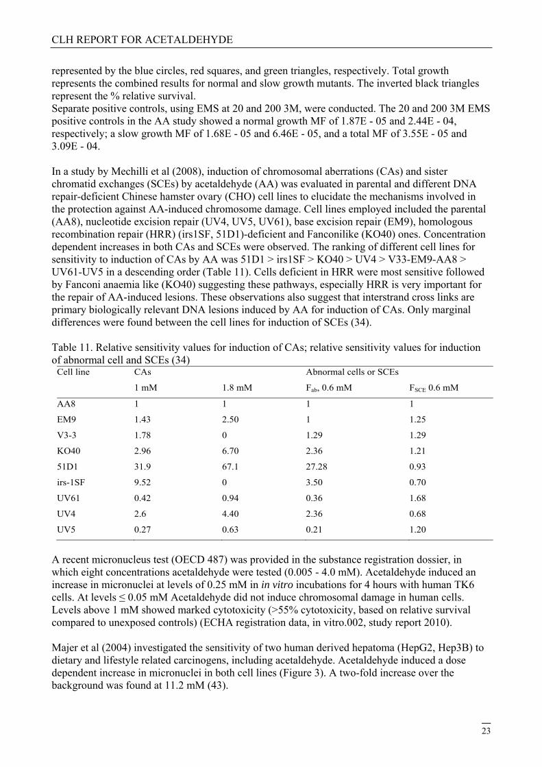
A recent micronucleus test (OECD 487) was provided in the substance registration dossier, in which eight concentrations acetaldehyde were tested (0.005 - 4.0 mM). Acetaldehyde induced an increase in micronuclei at levels of 0.25 mM in in vitro incubations for 4 hours with human TK6 cells. At levels ≤ 0.05 mM Acetaldehyde did not induce chromosomal damage in human cells. Levels above 1 mM showed marked cytotoxicity (>55% cytotoxicity, based on relative survival compared to unexposed controls) (ECHA registration data, in vitro.002, study report 2010). Majer et al (2004) investigated the sensitivity of two human derived hepatoma (HepG2, Hep3B) to dietary and lifestyle related carcinogens, including acetaldehyde. Acetaldehyde induced a dose dependent increase in micronuclei in both cell lines (Figure 3). A two-fold increase over the background was found at 11.2 mM (43).
CLH REPORT FOR ACETALDEHYDE
24
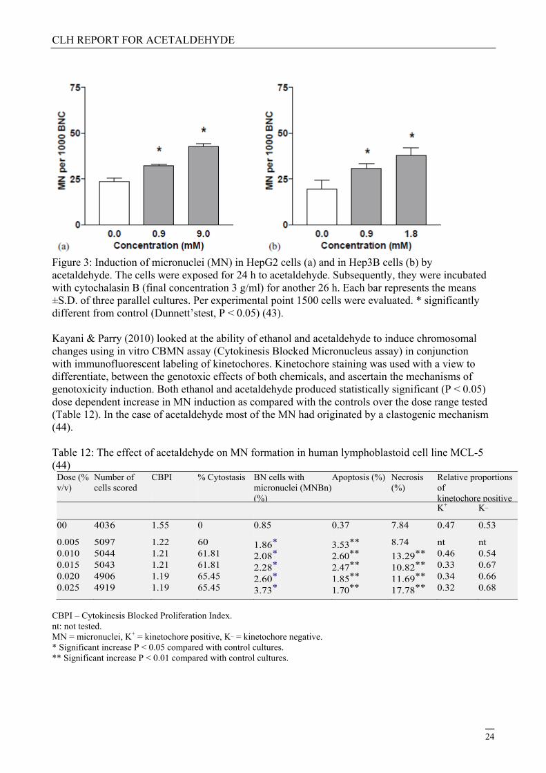
Figure 3: Induction of micronuclei (MN) in HepG2 cells (a) and in Hep3B cells (b) by acetaldehyde. The cells were exposed for 24 h to acetaldehyde. Subsequently, they were incubated with cytochalasin B (final concentration 3 g/ml) for another 26 h. Each bar represents the means ±S.D. of three parallel cultures. Per experimental point 1500 cells were evaluated. * significantly different from control (Dunnett’stest, P < 0.05) (43). Kayani & Parry (2010) looked at the ability of ethanol and acetaldehyde to induce chromosomal changes using in vitro CBMN assay (Cytokinesis Blocked Micronucleus assay) in conjunction with immunofluorescent labeling of kinetochores. Kinetochore staining was used with a view to differentiate, between the genotoxic effects of both chemicals, and ascertain the mechanisms of genotoxicity induction. Both ethanol and acetaldehyde produced statistically significant (P < 0.05) dose dependent increase in MN induction as compared with the controls over the dose range tested (Table 12). In the case of acetaldehyde most of the MN had originated by a clastogenic mechanism (44). Table 12: The effect of acetaldehyde on MN formation in human lymphoblastoid cell line MCL-5 (44)
Dose (% v/v)
Number of cells scored
CBPI % Cytostasis BN cells with micronuclei (MNBn) (%)
CBPI – Cytokinesis Blocked Proliferation Index. nt: not tested. MN = micronuclei, K+ = kinetochore positive, K_ = kinetochore negative. * Significant increase P < 0.05 compared with control cultures. ** Significant increase P < 0.01 compared with control cultures.
CLH REPORT FOR ACETALDEHYDE
25
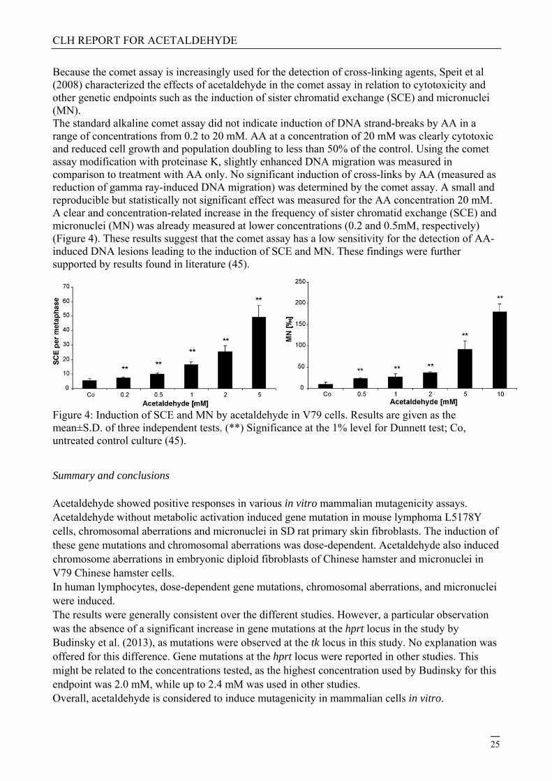
Because the comet assay is increasingly used for the detection of cross-linking agents, Speit et al (2008) characterized the effects of acetaldehyde in the comet assay in relation to cytotoxicity and other genetic endpoints such as the induction of sister chromatid exchange (SCE) and micronuclei (MN). The standard alkaline comet assay did not indicate induction of DNA strand-breaks by AA in a range of concentrations from 0.2 to 20 mM. AA at a concentration of 20 mM was clearly cytotoxic and reduced cell growth and population doubling to less than 50% of the control. Using the comet assay modification with proteinase K, slightly enhanced DNA migration was measured in comparison to treatment with AA only. No significant induction of cross-links by AA (measured as reduction of gamma ray-induced DNA migration) was determined by the comet assay. A small and reproducible but statistically not significant effect was measured for the AA concentration 20 mM. A clear and concentration-related increase in the frequency of sister chromatid exchange (SCE) and micronuclei (MN) was already measured at lower concentrations (0.2 and 0.5mM, respectively) (Figure 4). These results suggest that the comet assay has a low sensitivity for the detection of AA-induced DNA lesions leading to the induction of SCE and MN. These findings were further supported by results found in literature (45).
Figure 4: Induction of SCE and MN by acetaldehyde in V79 cells. Results are given as the mean±S.D. of three independent tests. (**) Significance at the 1% level for Dunnett test; Co, untreated control culture (45).
Summary and conclusions Acetaldehyde showed positive responses in various in vitro mammalian mutagenicity assays. Acetaldehyde without metabolic activation induced gene mutation in mouse lymphoma L5178Y cells, chromosomal aberrations and micronuclei in SD rat primary skin fibroblasts. The induction of these gene mutations and chromosomal aberrations was dose-dependent. Acetaldehyde also induced chromosome aberrations in embryonic diploid fibroblasts of Chinese hamster and micronuclei in V79 Chinese hamster cells. In human lymphocytes, dose-dependent gene mutations, chromosomal aberrations, and micronuclei were induced. The results were generally consistent over the different studies. However, a particular observation was the absence of a significant increase in gene mutations at the hprt locus in the study by Budinsky et al. (2013), as mutations were observed at the tk locus in this study. No explanation was offered for this difference. Gene mutations at the hprt locus were reported in other studies. This might be related to the concentrations tested, as the highest concentration used by Budinsky for this endpoint was 2.0 mM, while up to 2.4 mM was used in other studies. Overall, acetaldehyde is considered to induce mutagenicity in mammalian cells in vitro.
CLH REPORT FOR ACETALDEHYDE
26
4.9.1.2 In vivo data
A summary on the in vivo mutagenicity of acetaldehyde is shown in Table 13.
Table 13 Summary of in vivo mutagenicity studies (animal studies) Method Animal Exposure
conditions
Results Klimisch(15)
score*
References
Somatic cell mutagencicity
Gene mutation
and micronuclei
Wildtype and
knock-out
mice with
inactive
ALDH21
gene; micro-
nuclei deter-
mined in reti-
culocytes;
mutations
were deter-
mined by T-
cell receptor
(TCR) gene
mutation
assay
Inhalation, 125 and
500 ppm vapour,
continuously for two
weeks; negative
control was
inhalation of clean
air
Micronuclei:
+ in knock-out mice
(p<0.05);
- in wild-type mice.
Mutation (TCR
mutant frequency):
+ in knock-out mice
(p<0.05);
- in wild-type mice.
2 Kunugita et al.
2008(46)
Gene mutation
and micronuclei
Wildtype and
knock-out
mice with
inactive
ALDH2 gene;
micronuclei
determined in
reticulocytes;
mutations
were deter-
mined by
TCR gene
mutation
assay
Oral administration,
0 and 100 mg/kg
bw, daily, once a
day for two weeks; 5
– 10 animals/group
Micronuclei:
+ in knock-out mice
(p<0.05);
- in wild-type mice.
Mutation (TCR
mutant frequency):
+ in knock-out mice
(p<0.05);
- in wild-type mice
2 Kunugita et al.
2008(46)
Micronuclei; multi-
substance study
Male SD and
F344 rats,
bone marrow
erythrocytes
250 mg/kg bw,
intraperitoneal
injection. Highest
dose tested was
+ ( both cell types) 2; only highest
dose tested
Wakata et al.
1998(47)
1 ALDH2, aldehyde dehydrogenase 2 family (mitochondrial), converts acetaldehyde into acetate.
CLH REPORT FOR ACETALDEHYDE
27
and
peripheral
blood
erythrocytes
maximum tolerated
dose; at least four
animals/group
Micronuclei 5 male CD-1
mice
0 – 400 mg/kg bw,
Intraperitoneal
injection, three dose
levels; tests on
acute toxicity
performed
+ (dose-related
increase)
2 Morita et al.
1997(48)
Micronuclei Male Han
rats, 5
animals/group
Single
intraperitoneal
injection of 125 or
250 mg/kg bw; blood
samples collected
after 0, 24, 48 and
72 hours
+ (at 24 and 48
hours), dose-related
increase; no data at
72 hours due to
toxicity
2 Hynes et al.
2002(49)
Chromosomal
aberrations
Rat embryos Single intra-amniotic
injection of 7,800
mg/kg bw
+ 4; original
publication
available in
Russian only
Bariliak and
Kozachuk
1983(50)
Germ cell mutagenicity
Meiotic
micronuclei in
spermatids
C57BL/6J x
C3H/He
mouse early
spermatids
125, 250, 375 and
500 mg/kg bw per
day, single dose,
intraperitoneal
injection; 4
animals/group
- ; survival rate was
significantly
decreased in highest
exposure group
2 Lähdetie 1988(6)
Sex-linked
recessive lethal
mutations; multi-
substance study
Drosophila
melanogaster
1) Single injection of
22,500 ppm; 2)
25,000 ppm in feed;
data presented on
mortality and sterility
+ (injection)
- (feed)
2 Woodruff et al.
1985(51)
* See footnote in Table 10 for explanation of the Klimisch-scores.
Germ cells
Lähdetie (1988) studied the induction of meiotic micronuclei in spermatids of mice.(6) Mice (4 animals per group) were given a single intraperitoneal injection of acetaldehyde at a concentration of 0 (control vehicle), 125, 250, 375 and 500 mg/kg bw. A group of mice served as positive control (cyclophosphamide injection). Thirteen days after treatment the mice were killed to examine the presence of meiotic micronuclei in early spermatids (1,000 spermatids scored per mouse). Compared to the vehicle control, the number of spermatids with micronuclei did not increase after acetaldehyde treatment, whereas in the positive control it did. The author reported that at a dose of
CLH REPORT FOR ACETALDEHYDE
28
500 mg/kg bw all animals died due to acute toxicity, whereas all survived at lower doses. In a separate experiment, the author also investigated the sperm morphology in mice treated with acetaldehyde for a short period (up to 250 mg/kg bw; 5-day exposure regimen). However, acetaldehyde did not decrease sperm count, testis weight or seminal vesicle weight, nor did it induce abnormal sperm at the doses. The highest administered dose was lethal to half of the animals in the group.
In a sex-linked recessive lethal mutation assay, acetaldehyde was positive after injection (Woodruff et al. 1985).(51) This shows that the substance induces mutations in germ lines of the insect.
Somatic cells
Kunugita et al. (2008) studied the induction of gene mutations and micronuclei in knock-out mice having an inactive acetaldehyde dehydrogenase (Aldh2, converts acetaldehyde into acetate) gene.(46) Both wildtype and the knockout mice inhaled acetaldehyde at concentrations of 0, 225 or 900 mg/m3, continuously for two weeks. In addition, groups of mice (5-10 animals per group) were given acetaldehyde orally at doses of 0 or 100 mg/kg bw, once a day for two weeks. Two weeks after the last exposure, all animals were killed and the number of reticulocytes with micronuclei was determined. Also the mutations in the TCR gene of T-lymphocytes was measured. Irrespective the route of exposure, in knockout mice, the number of micronuclei positive cells, and the frequency of TCR gene mutations in lymphocytes was statistically significantly increased compared to the respective controls. In wildtype animals, acetaldehyde did not cause any effects on these endpoints. See Table 14 for a summary of the results.
In a well-performed study, Wakata et al. (1998) showed that in bone marrow polychromatic and peripheral blood erythrocytes of SD and F344 rats, micronuclei were induced after exposure to acetaldehyde by a single intraperitoneal injection of 250 mg/kg bw.(47) Bone marrow and blood cells were harvested 24 hours after the treatment. The maximal micronucleated polychromatic erythrocyte frequency in bone marrow was 0.43%; the mean for the negative control (saline) was 0.15 ± 0.13%, the mean positive control (cyclophosphamide, 20 mg/kg) was 2.9 ± 1.5%. The highest frequency of micronucleated reticulocytes in peripheral blood was 0.33; the negative control had a mean of 0.07 ±0.08%, the positive control a mean of 0.67 ±0.46%.
In addition, Morita et al. (1997) reported on acetaldehyde-induced micronuclei in bone marrow polychromatic erythrocytes of male CD-1 mice.(48) Five/six mice received the substance by a single intraperitoneal injection. Dose levels were based on acute toxicity test results. Two different lots were used, because the experiment was performed in two different laboratories. Twenty four hours after injections, bone marrow cells were harvested for the micronucleus assay. In Table 15 a summary of the results is shown.
Hynes et al. (2002) exposed male Wistar Han rats (5 animals per group) to acetaldehyde by a single intraperitoneal injection of 125 or 250 mg/kg bw.(49) For micronuclei testing, peripheral blood cells were harvested 0, 24, 48 and 72 hours after the injection. Micronuclei were scored by flow cytometric analysis. The study included negative (vehicle) and positive (cyclophosphamide) controls. Acetaldehyde at a dose of 250 mg/kg bw induced micronuclei, with maximum increases at 48 hours (see Table 16).
CLH REPORT FOR ACETALDEHYDE
29
Table 14 Induction factors of micronuclei and TCR gene mutations in knockout mice (Kunugita et al
2008).(46)
Exposure route Exposure level Micronuclei in reticulocytes Mutant frequency in T-cell
receptor gene
Knock-out mice (Aldh2 -/-)
Inhalation 0 (control) - -
225 mg/m3 1.8 * Not determined
900 mg/m3 1.9/unspecified **/*** 1.7**
Oral administration 0 (control) - -
100 mg/kg bw 2/1.7 **/*** 2.4/1.6 **/***
Wildtype mice (Aldh2 +/+)
Inhalation 0 (control) - -
225 mg/m3 - -
900 mg/m3 - -
Oral administration 0 (control) - -
100 mg/kg bw - -
* compared to Aldh2 +/+ control mice (p<0.05); ** compared to Aldh2 +/+ control mice (p<0.01); ***
compared to Aldh2 -/- control mice (p<0.05).
Table 15 Induction of micronuclei in male CD mice (Morita et al. 1997).(48)
Manufact. lot LD50 Dose Percentage of micronuclei in bone marrow cells
mg/kg bw mg/kg bw mean SD p-value*
Wako 470 0 0.12 0.08 -
95 0.22 0.15 0.132
190 0.33 0.10 0.010
380 0.85 0.21 0.000
Merck 338 0 0.12 0.08 -
100 0.10 0.07 0.726
200 0.44 0.11 0.002
300 0.62 0.16 0.000
400 1.10 0.25 0.000
* P-value of pairwise comparisons. Table 16 Induction of micronuclei in blood cells of rats treated with acetaldehyde (Hynes et al. 2002).(49)
reticulocytes; MNNCE, micronucleated monochromatic erythrocytes. No data on statistical significance
presented.
These studies show that acetaldehyde is inducing mutation in the bone marrow after intraperitoneal injection or in ALDH2 knock-out mice after inhalation but not in wild-type mice after inhalation, suggesting metabolism is an important factor in the ability of acetaldehyde to reach distant sites. No mutations were found in spermatids of mice, although this was endpoint was investigated in only one study.
4.9.2 Human information
Table 17 summarizes a few studies performed on humans, in which effects were related to acetaldehyde. Acetaldehyde exposure in these studies was due to alcohol abuse and/or smoking.
Table 17 Summary of human studies
Method Population Cells Results and remarks Quality and/or
reliability of
study
References
DNA-adducts
(32P-postlabelling)
Alcohol abusers
(n=24) and
controls (n=12)
Peripheral
white blood
cells (granulo-
cytes and
lymphocytes)
+ in alcohol abusers
compared to controls
(p<0.001). Average
adduct levels in
abusers (adducts /107
nucleotides):
- granulocytes: 3.4 ±
3.8
- lymphocytes: 2.1 ±
0.8
Levels in controls
were below LOD
Reliability low
in that
subjects in the
alcoholic group
were heavy
smokers; in
control group
one moderate
smoker.
Fang and Vaca
1997(52)
DNA-adducts Cancer-free
male Japanese
alcoholic
patients with
different
acetaldehyde
dehydrogenase
(ALDH)
genotypes
Peripheral
white blood
cells
+, adduct level was
significantly higher in
alcoholics with
ALDH2*1*2 genotype
compared to
alcoholics with
ALDH2*1*1 genotype.
Past exposure
to ethanol; no
non-alcoholic
healthy
controls
included
Matsuda et al.
2006(53)
Acetaldehyde
specific DNA-
adducts (N2-
Smokers,
before and after
smoking
Leucocytes Decrease in number of
N2-ethylidene-dGuo
adducts after
Reliability low,
because of
smoking
Chen et al.
2007(54)
CLH REPORT FOR ACETALDEHYDE
31
ethylidene-
deoxiguanosine)
cessation cessation (28%). Note:
cigarette smoke
contains acetalde-
hyde, but also other
potential carcinogens.
history
participants
and co-
exposure
Acetaldehyde–DNA adducts have been observed in granulocytes and lymphocytes of human alcohol abusers (52, 53) and leucocytes of smokers (54). In comparison with controls, Fang and Vaca (1997) (52) found 13- and 7-fold higher adduct levels in respectively granulocytes and lymphocytes of alcohol abusers. However, the alcohol abusers were also heavy smokers, and the values of the controls were all below the limit of detection, limiting the reliability of these percentages. Matsuda et al. (2006) enrolled 19 alcoholic patients with the ALDH2*1/2*1 genotype and 25 alcoholic patients with the ALDH2*1/2*2 genotype. The averages of age, daily ethanol consumption, duration of drinking, and daily cigarette consumption were not significantly different between the two groups. The average levels of three acetaldehyde-derived adducts were significantly higher in ALDH2*1/2*2 alcoholics. The average level of blood N2-Et-dG adducts in ALDH2*1/2*2 and ALDH2*1/2*1 alcoholics were 28.3 and 3.9 adducts per 109 bases, respectively. Chen et al. (2008) (54) found a decrease in DNA-adducts of 28% in leucocytes of volunteers after 4 weeks of smoking cessation. Levels of acetaldehyde in mainstream cigarette smoke typically range from 500 – 1000 μg/cigarette. The most important confounder was alcohol consumption, for this reason, subjects were eligible only if they consumed less than six alcoholic beverages per month and abstained during the study. Nevertheless, occasional drinking might have been undetected and could potentially contribute to acetaldehyde DNA adducts. The only modifier in this study was the race of the participants. When the data were stratified by race, there was no change in adduct levels in whites, but a significant 57% decrease was observed in the black plus other group (consisting of 7 blacks, 1 American Indian, and one person of mixed racial background).
The data indicate the intrinsic property of acetaldehyde to react in vivo in humans with DNA.
4.9.3 Other relevant information
In the Tables 18 and 19 data are shown on the DNA damaging and genotoxic (other than mutagenicity) properties of acetaldehyde.
Table 18 Summary of other information on DNA damage
Method Cell type Concentration Results Klimisch(15)
score**
References
In vivo studies
DNA-protein
crosslinks
Male Fischer-
344 rats; DNA-
protein cross-
links studied in
nasal respiratory
mucosa and
olfactory cells
1) Inhalation; 100,
300, 1,000 and
3,000 ppm; single
6-hour exposure
2) inhalation; 1,000
ppm; 6-hours/day,
daily, 5-days
1) + (respiratory
mucosa; dose-
dependent increase,
p<0.05);
- (olfactory mucosa)
2) + (respiratory
mucosa); + (olfactory
2 Lam et al.
1986(55)
CLH REPORT FOR ACETALDEHYDE
32
samples of three
rats were
combined
mucosa, p<0.05)
In vitro tests using human cells
DNA single and
double strand
breaks
Human
lymphocytes
from two healthy
donors
0, 1.56, 6.25, 25
and 100 mM for
one hour; for each
dose 50 cells were
analysed from
each subject
+ (single strand
breaks at all
exposures)
+ (double strand
breaks at 100mM
only)
Authors reported that
> 80% of cells were
not viable after
exposure to 100 mM
for 2 hours
2; no positive
control
Singh and
Khan 1995(56)
Comet assay* Human
peripheral blood
lymphocytes
3, 10, 30 and 100
mM for one hour;
doses were based
on cytotoxicity data
+ (dose-dependent) 2 Blasiak et al.
1999(57)
Comet assay* Human
lymphocytes,
gastric and
colonic mucosa
cells
3 mM (lympho-
cytes), 100 mM
(gastric and colonic
mucosa cells)
+ No differences were
noted among the
different cell types;
viability was over 70%
at the tested doses
2; one dose
tested only
Blasiak et al.
2000(58)
Comet assay* Human bronchial
epithelial cells
Exposure to 3, 10,
30 and 100 mM for
1 hour in thiol free
medium
+, dose-dependent
effects
- for single strand
breaks
2 Grafström et
al. 1994(33)
DNA-adducts DNA form
primary human
liver cells,
samples from
normal liver
Incubation of cells
with 5.7 mM
[13C2]acetaldehyde;
12 liver samples
analysed
+ (N2-ethyl-
deoxiguanosine
adducts)
3 Wang et al.
2006(59)
Alkaline elution
assay*
Human
lymphocytes
10 – 20 mM for 4
hours
+, DNA cross-links
- ,DNA strand-breaks
3; No data on
cytotoxicity; no
positive
controls
Lambert et al.
1985(60)
Alkaline elution
assay*; multi-
substance study
Normal human
bronchial
epithelial cells
and humane
leucocytes
1 mM for 1 hour
- (without metabolic
activation); at 1 mM
no significant growth
reduction noted
3; only one
concentration
used
Saladino et al.
1985(61)
Alkaline elution
assay*
Human bronchial
epithelial cells
10 mM for 1 hour - 3; only one
dose tested; no
data on
Grafström et
al. 1986(62)
CLH REPORT FOR ACETALDEHYDE
33
controls; 10
mM
acetaldehyde
induced 50%
cytotoxicity
DNA-protein
crosslinks
EBV-transformed
human Burkitt’s
lymphoma cells
(EBV, Epstein
Barr virus)
0.035, 0.175,
0.875, 3.5 and 17.5
mM for 2 hours;
Maximum tolerated
dose was 17.5 mM
+ (> 5 mM, p<0.05) 2 Costa et al.
1997(63)
DNA-adducts normal epithelial
cells, and SV40T
antigen-immor-
talized human
buccal epithelial
cells
1-100 mM for one
hour; 32P-
postlabeling assay
+ (N2-ethyl-3’-dG-
monophosphate
adducts, dose-
dependent
2 Vaca et al.
1998(64)
In vitro tests using rodent cells
Comet assay* V79 Chinese
hamster cells
0.2 – 20 mM -; authors reported
more than 50%
reduction of cell
viability at 20 mM
2; no positive
control
Speit et al.
2008(45)
Cell
transformation
Mouse C3H
10T1/2 cells
10-100 μg/ml - 4 Abernathy et
al. 1982 (65)
Cell
transformation
Mammalian cells 0.44 μg/ml (3
hours)
- 4
Eker & Sanner
1986 (66)
Alkaline elution
assay*
Chinese hamster
ovary cells (K1
cells)
0.5, 1.5 and 4.5
mM for 90 minutes
- (strand breaks);
+ (crosslinks);
cell viability > 80%
2; no positive
control
Marinari et al.
1984(67)
Alkaline elution
assay*; multi-
substance study
Primary rat
hepatocytes
0.03, 0.3 and 3 mM
for 3 hours;
cytotoxicity < 55%
- 3 Sina et al.
1983(68)
Other test systems
DNA-adducts Calf thymus DNA 1 M for 30 minutes
at 37 °C; negative
control included
+ (without metabolic
activation)
3; only one
concentration
tested
Ristow and
Obe 1978(69)
DNA-adducts Calf thymus DNA 0.01-40 mM for 20
to 96 hours
+ (mainly N2-
ethylidene-deoxi-
guano-sine DNA-
adducts, but also (<
10%) 1,N-propano-
deoxi-guanosine, N2-
dimethyldioxane-
deoxiguanosine, and
a cross-link adduct
detected).
2 Wang et al.
2000(70)
DNA-adducts Calf thymus DNA 1.8 mM for 92
hours; 32P-
+ (N2-ethyl-3’-dG-
monophosphate
3 Fang and Vaca
1995(71)
CLH REPORT FOR ACETALDEHYDE
34
postlabeling assay adducts)
DNA-adducts Calf thymus DNA
in 2’-deoxy-
guanosine-3’-
monophosphate
Up to 79,000
μg/ml;
+ 3 Fang and Vaca
1997(52)
DNA-protein
crosslinks
Calf thymus DNA
in 2’-deoxy-
guanosine-3’-
monophosphate
100, 300 and 1,000
mM for one hour
+ 3 Lam et al.
1986(55)
Alkaline elution
assay*
Saccharomyces
cerevisiae
(yeast)
0.85 M for 2 or 4
hours
+ 3; no positive
control; no data
on statistical
analysis
Ristow et al.
1995(72)
DNA damage E. coli polA 7800 μg/ml - 3
Rosenkranz,
1977 (20)
DNA repair
host-mediated
assay, in vivo;
multi-substance
study
repair-deficient
E.coli K-12
uvrB/recA; tests
performed in
mice
Highest tested
concentration 370
mM/L; - and + S9
- (- and + S9) 3; method not
validated
Hellmer and
Bolcsfoldi
1992(73)
* Comet assay and alkaline elution assay: DNA single and double strand breaks, DNA cross-links.
** See footnote in Table 10 for explanation of the Klimisch-scores.
Table 19 Summary of genotoxicity studies
Method Cell type Concentration Results and
remarks
Klimisch(15)
Score*
References
In vitro tests using rodent cells
Sister chromatid
exchange
Different DNA-
repair deficient
Chinese hamster
ovary cells
0.3, 0.6, 1.0, 1.8,
2.5 and 3.6 mM for
2 hours; 250
metaphases
scored/group
+ 2; no positive
control
Mechilli et al.
2008(34)
Sister chromatid
exchange
Chinese hamster
ovary cells
0, 30, 100 and 300
μM; - S9
+ (dose-
dependent
increase
2 Brambilla et al.
1986(74)
Sister chromatid
exchange
V79 Chinese
hamster cells
0.2 – 5 mM
+ (dose-
dependent
increase)
2; No positive
control
Speit et al.
2008(45)
Sister chromatid
exchange
Chinese hamster
ovary cells
0, 0.8, 2, 4, 7.8,
39.4 and 78 μg/ml;
+ and – S9; 20
metaphases/sample
scored
+, dose-related
response
3; no data on
cytotoxicity; no
positive control
de Raat et al.
1983(75)
Sister chromatid
exchange
Chinese hamster
ovary cells
0.25x10⁻³, 0.5x10⁻³,
1x10⁻³, and 1.5x10⁻³
% (v/v); - S9; 100
+ 3; no positive
controls, no
data on
Obe et al.
1979(40)
CLH REPORT FOR ACETALDEHYDE
35
mitoses scored/
sample
cytotoxicity
In vitro tests using human cells
Sister chromatid
exchange
Human
peripheral
lymphocytes
0 – 1,080 μM; -S9;
reduction of cell
growth noted above
720 μM
+, dose-related
response
2; no positive
controls
Böhlke et al.
1983(76)
Sister chromatid
exchange
Human
peripheral
lymphocytes
1 – 100 μM + 2; no positive
controls
Knadle 1985(77)
Sister chromatid
exchange
Human
lymphocytes and
fibroblast of
normal subjects
40, 400 and 800
μM;
+ 3; limited
information on
test protocol
Véghelyi and
Osztovics
1978(78)
Sister chromatid
exchange
Human
lymphocytes
0, 63, 125, 250 500
and 2,000 μM; -S9
+ (dose-
dependent
increase)
3; no positive
controls; no
data on
cytotoxicity
Norppa et al.
1985(79)
Sister chromatid
exchange
Human
lymphocytes
0, 0.0005, 0.001,
and 0.002 % (v/v);
-S9
+, dose-related
response
3; no positive
controls; no
data on
cytotoxicity
Ristow and Obe
1978(69)
Sister chromatid
exchange
Human
lymphocytes
0 – 500 μM; - S9 +, dose-related
response
3; no data on
cytotoxicity; no
positive
controls
Sipi et al.
1992(80)
Sister chromatid
exchange
Human
peripheral
lymphocytes
100 – 400 μM; - S9;
exposure performed
in capped bottles
+ (dose-
dependent
increase)
3; no positive
controls; no
data on
cytotoxicity
Helander and
Lindahl-
Kiessling
1991(81)
Sister chromatid
exchange
Human
peripheral
lymphocytes
2x10⁻³ % (v/v);
+ or – acetaldehyde
metabolizing
enzyme ALDH
+ 3; no positive
controls, no
data on
cytotoxicity
Obe et al.
1986(82)
Sister chromatid
exchange
Human
lymphocytes
100 – 2,400 μM;
- S9
+ (dose-
dependent
increase
3; no positive
controls used,
no data on
cytotoxicity
He and Lambert
1985(83)
Sister chromatid
exchange
Human
peripheral
lymphocytes
0 – 0.001% (v/v); -
S9
+ (dose-
dependent
increase)
3; limited
information on
test protocol
Jansson
1982(84)
Rodents (in vivo somatic cell tests)
Sister chromatid
exchange
Bone-marrow
cells of Chinese
hamsters (strain
not specified)
Single intra-
peritoneal injection
of 0.01, 0.1 and 0.5
mg/kg bw; 6-7
animals/ dose;
+ at the highest
exposure level
only; at this level
signs of intoxica-
tion were noted;
2 Korte et al.
1981(85)
CLH REPORT FOR ACETALDEHYDE
36
negative and
positive control
included
no signs of
intoxication at 0.1
and 0.01 mg/kg
bw
Sister chromatid
exchange
Male mouse
(NIH) bone
marrow cells
0.4, 4.0, 40 and 400
mg/kg bw, single
intraperitoneal
injection
+ (40 and 400
mg/kg bw, p<0.05)
Mitotic index and
average
generation time
did not differ from
control
3; number of
mice per group
not given; no
positive control
Torres-Bezauri
et al. 2002(86)
Sister chromatid
exchange
Male CBA mouse Single intraperi-
toneal injection of 1
or 0.5 mL of a
10-4 % (v/v) solu-
tion; one animal/
dose
+ 3; low number
of animals in
study, no
positive
controls
Obe et al.
1979(37)
Rodents (in vivo germ cell tests)
Sister chromatid
exchange
Mouse
spermatogonial
cells
Single
intraperitoneal
injection; 0.4, 4.0,
40 and 400 mg/kg
bw; 4 – 5 animals/
concentration; cells
were isolated, 53 h
after injection.
+ (all doses
applied, p<0.05);
no clear
exposure-
response
relationship
observed
2; authors did
test for
intoxication;
concentrations
used were
considered
non-toxic/-lethal
Madrigal-
Bujaidar et al.
2002(5)
* See footnote in Table 10 for explanation of the Klimisch-scores.
In vitro studies: DNA damage and genotoxicity Acetaldehyde caused DNA strand breaks and cross-links in human lymphocytes in vitro without metabolic activation, but not in human bronchial epithelial cells and in human leukocytes, or in rodent cells. Acetaldehyde–DNA adducts have been found in vitro in calf thymus DNA and in 2′-deoxyguanosine-3′-monophosphate. It induced dose-dependent sister chromatid exchanges in Chinese hamster ovary cells and human lymphocytes in a wide range of studies. Overall, these studies show the intrinsic property of acetaldehyde to react with DNA in vitro.
In vivo studies
Germ cells
Madrigal-Bujaidar et al. (2002) injected NIH mice (4-5 mice per group) with acetaldehyde at concentrations of 0 (vehicle control), 0.4, 4, 40 and 400 mg/kg bw (single treatment), or cyclophosphamide (positive control).(5) Fifty-three hours later, the animals were killed, and the tunica albuginea was removed from each testes to obtain spermatogonial cells in the seminiferous tubules. A statistically significant increase in the number of cells with sister chromatid exchange
CLH REPORT FOR ACETALDEHYDE
37
and a clear dose response relationship was reported (30 metaphases per mouse scored; see Table 20). The authors determined a LD50-dose of 560 mg/kg bw.
Somatic cells
Lam et al. (1986) reported on the formation of DNA-protein crosslinks in the nose tissue of male Fischer-344 rats after inhalation exposure.(55) The animals were exposed to acetaldehyde at concentrations of 0,180, 540, 1,800 and 5,400 mg/m3 for a single six hours, or to 5,400 mg/m3, 6 hours a day for 5 consecutive days. Immediately after the final exposure the animals were killed, and nasal respiratory mucosa was obtained for further examination. After a single inhalation, a dose dependent increase in DNA-protein crosslinks was observed in the respiratory mucosa, but not in the olfactory mucosa. Short-term repeated inhalation induced DNA-protein crosslinks in the respiratory and the olfactory mucosa.
In bone marrow cells of Chinese hamsters (6-7 animals per group), a single intraperitoneal injection of acetaldehyde increased the number of sister chromatid exchanges at the two highest doses applied (0.1 and 0.5 mg/kg bw; Korte et al., 1981).(85) The authors reported that exposure to concentrations of 0.6 mg/kg bw and higher was lethal.
Table 20 Sister chromatid exchanges in spermatogonial cells of mice treated with acetaldehyde (Madrigal-
Bujaidar et al. 2002).(5)
Dose (mg/kg bw) SCE/cell ± SD SCE increase
0 1.9 ± 0.16
0.4 2.9 ± 0.33* 1.1
4 4.1 ± 0.34* 2.2
40 4.6 ± 0.51* 2.7
400 5.1 ± 0.8* 3.2
50 (cyclophosphamide) 6.0 ± 0.1* 4.1
SCE, sister chromatid exchange. * Statistically significant different compared to control, p< 0.05.
4.9.4 Summary and discussion of mutagenicity
Below, only data are summarized of reliable (with or without restrictions) experimental design (according to the Klimisch criteria (1997)).(15) In vitro studies Numerous data have been presented on the mutagenic and genotoxic properties of acetaldehyde in bacteria and mammalian cells. Overall, negative outcomes were found in bacteria using the reverse mutation assay, whereas most in vitro assays with mammalian cells gave positive outcomes. These included gene mutations, chromosome aberrations, micronuclei, DNA-strand breaks, DNA-adducts, DNA-protein crosslinks, and sister chromatid exchanges in both rodent and human cells (the latter were mainly lymphocytes). In some of these positive studies, also a dose-related response was found. The only mammalian in vitro assay that gave mainly negative outcomes was the alkaline elution assay. However, two these studies had low reliability, as they tested only one concentration and two
CLH REPORT FOR ACETALDEHYDE
38
studies reported positive results for DNA cross-links, together with negative results for DNA strand-breaks. The presence of DNA or DNA-protein crosslinks may affect the outcomes of an alkaline elution test. Taken together, the data show that acetaldehyde can damage DNA directly and induce mutations in vitro. In vivo studies in somatic cells After inhalation of acetaldehyde, a dose-dependent increase of DNA-crosslinks was found in the respiratory and olfactory mucosa of rats. Acetaldehyde also induced micronuclei in bone marrow and blood cells in mice and rats, and sister chromatid exchange in the bone marrow of mice and hamsters after intraperitoneal injection. Gene-mutations and micronuclei were induced in reticulocytes of ALDH2 knock-out mice, after inhalatory or oral administration, but not in wild-type mice. According to Buddinsky et al. (2013), the key event after acetaldehyde exposure involves Schiff's base formation with DNA and proteins to elicit genotoxicity and/or cytotoxicity. DNA repair, apoptosis and other stress-related adaptive responses, and replacement of proteins or redundancy in protein function all act in opposition of these adducts. This is followed by metabolic deactivation of acetaldehyde via ALDH2. If the action of ALDH2 is sufficient, and when it is combined with DNA repair, apoptosis, and other stress-related responses, no increase in genotoxic outcomes will occur. In vivo, tissue acidification occurs, caused by the production of acetic acid, which adds to the cytotoxicity of DNA and protein adducts. Because of the constant presence of acetaldehyde in cells, the dose-response for mutagenicity will depend on the capacity of cells to maintain homeostatic levels of the agent. These data suggest that acetaldehyde is a direct acting mutagen in vivo, of which the potential to induce mutations at distant sites depends strongly on the activity of ALDH2. Data on humans are limited, but show the formation of DNA adducts in white blood cells related to acetaldehyde exposure through alcohol (ab)use and smoking (see Table 17). The available studies also showed that variation in the ALDH2 genotype indeed influenced the occurrence of DNA-adducts (Matsuda et al. 2006 and indirectly via race Chen et al. 2007). The available kinetic data shows that acetaldehyde can reach the systemic circulation and several organs. The intraperitoneal studies show that when sufficient acetaldehyde reaches the systemic circulation it induces genotoxicity and mutagenicity in vivo. This is confirmed by the inhalation studies by Kunugita (2008) which showed that in animals without ALDH2, which most likely have higher systemic acetaldehyde levels, were positive whereas wild type animals were negative for the induction of micronuclei. Germ cell genotoxicity
Two animal studies were found on germ cell genotoxicity by acetaldehyde, both in mice. The first is the study by Lähdetie et al. (1988), in which a single intraperitoneal injection of acetaldehyde did not induce meiotic micronuclei in early spermatids nor sperm abnormalities.(6) The second study is published by Mardigal-Bujaidar et al. (2002), and considers the induction of sister chromatid exchanges in mouse spermatogonial cells.(5) Although no clear dose-response relationship could be
CLH REPORT FOR ACETALDEHYDE
39
assessed, the authors reported that acetaldehyde induced sister chromatid exchanges (see Table 13). This difference in results might be related to a difference in sensitivity between the two assays. In relation to this, degradation of acetaldehyde could be of influence, as Maredigal-Bujaidar showed that blockage of aldehyde dehydrogenase resulted in an increase in SCEs at normally non-genotoxic doses (0.004 and 0.04 mg/kg bw). However, considering these uncertainties and the non-physiological route of exposure, it cannot be concluded that acetaldehyde is genotoxic in germ cells on these studies alone.
4.9.5 Comparison with criteria
Annex VI of CLP states for the hazard class germ cell mutagenicity that “the classification in Category 2 is based on positive evidence obtained from experiments in mammals and/or in some cases from in vitro experiments, obtained from: - Somatic cell mutagenicity tests in vivo, in mammals; or - Other in vivo somatic genotoxicity tests which are supported by positive results from in vitro mutagenicity assay” In vivo in somatic cells, the following effects were observed:
- increases of DNA-crosslinks at local sites after inhalation - micronuclei and sister chromatid exchanges in bone marrow and blood cells after
intraperitoneal injection - gene-mutations and micronuclei in reticulocytes of ALDH2 knock-out mice, after inhalatory
or oral administration - DNA adducts in humans after exposure through alcohol and/or smoking
These findings are supported by in vitro studies in mammalian cells, which showed gene mutations, chromosome aberrations, micronuclei, DNA-strand breaks, DNA-adducts, DNA-protein crosslinks, and sister chromatid exchanges in both rodent and human cells. Also the available kinetic information shows that acetaldehyde is systemically available after exposure via relevant routes. Thus the genotoxic and mutagenic effect of acetaldehyde warrants at least classification in category 2. According to the criteria in Annex VI of the European regulation No. 1272/2008, classification as a mutagen in category 1 is warranted when positive evidence for in vivo heritable germ cell mutagenicity in humans (1A) or mammals (1B) has been reported. No data have been presented on human germ cell mutagenicity, and the only animal germ cell mutagenicity study did not show mutagenic activity (Lähdetie et al., 1988).(6) Overall, due to a lack of data it is concluded that there is no positive direct evidence for in vivo heritable germ cell mutagenicity of acetaldehyde. In addition, substances may be categorized in 1B if there are “positive results from in vivo somatic cell mutagenicity tests in mammals, in combination with some evidence that the substance has potential to cause mutations to germ cells”. The latter may be based on a) “supporting evidence from mutagenicity/genotoxicity tests in germ cells in vivo”, or b) “by demonstrating the ability of the substance or its metabolites to interact with the genetic material of germ cells”. Sufficient evidence has been found for in vivo mutagenicity testing in somatic cells of mammals. Regarding the second part of the criterion, there is limited evidence that acetaldehyde is genotoxic (sister chromatid exchanges) in germ cells of mice (Madrigal-Bujaidar et al. 2002), when the substance was given by intraperitoneal injection.(5) These findings indicate that acetaldehyde is able to reach
CLH REPORT FOR ACETALDEHYDE
40
the germ cells, and interacts with the genetic material, which would be in line with the findings on absorption and distribution kinetics. As described in 4.1, acetaldehyde is rapidly taken up after inhalation and oral exposure. In rats, acetaldehyde was distributed in the blood, liver, kidney, spleen, heart, myocardium and skeletal muscle. However, in another animal study no abnormal sperm cells, and no meiotic micronuclei in spermatids were observed at dose levels inducing acute toxicity (Lähdetie et al. 1988).(6) An important factor for the distribution of acetaldehyde in the body is the activity of the enzyme acetaldehyde dehydrogenase (ALDH2). It is known that this enzyme has a high degree of genetic polymorphism in humans, which influences the occurrence DNA adducts in white blood cells due to exposure to acetaldehyde through alcohol (ab)use and smoking. Thus it cannot be excluded that acetaldehyde may reach the germ cells, especially in humans with a mutated form of ALDH2. Overall, it is considered that some evidence exists that acetaldehyde has potential to cause mutations in germ cells. Therefore, it is recommended to classify the substance in category 1B.
4.9.6 Conclusions on classification and labelling
Based on the available data, it is recommended to classify acetaldehyde as a germ cell mutagen in category 1B, “substance to be regarded as if they induce heritable mutations in the germ cells of humans”.
CLH REPORT FOR ACETALDEHYDE
41
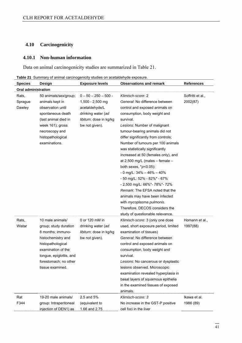
4.10 Carcinogenicity
4.10.1 Non-human information
Data on animal carcinogenicity studies are summarized in Table 21.
Table 21 Summary of animal carcinogenicity studies on acetaldehyde exposure.
Species Design Exposure levels Observations and remark References
Oral administration
Rats,
Sprague
Dawley
50 animals/sex/group;
animals kept in
observation until
spontaneous death
(last animal died in
week 161); gross
necroscopy and
histopathological
examinations.
0 – 50 – 250 – 500 -
1,500 - 2,500 mg
acetaldehyde/L
drinking water (ad
libitum; dose in kg/kg
bw not given).
Klimisch-score: 2
General: No difference between
control and exposed animals on
consumption, body weight and
survival.
Lesions: Number of malignant
tumour-bearing animals did not
differ significantly from controls;
Number of tumours per 100 animals
was statistically significantly
increased at 50 (females only), and
at 2,500 mg/L (males – female –
both sexes, *p<0.05):
- 0 mg/L: 34% – 46% – 40%
- 50 mg/L: 52% - 82%* - 67%
- 2,500 mg/L: 66%*- 78%*- 72%
Remark: The EFSA noted that the
animals may have been infected
with mycoplasma pulmonis.
Therefore, DECOS considers the
study of questionable relevance.
Soffritti et al.,
2002(87)
Rats,
Wistar
10 male animals/
group; study duration
8 months; immuno-
histochemistry and
histopathological
examination of the
tongue, epiglottis, and
forestomach; no other
tissue examined.
0 or 120 mM in
drinking water (ad
libitum; dose in kg/kg
bw not given).
Klimisch-score: 3 (only one dose
used, short exposure period, limited
examination of tissues)
General: No difference between
control and exposed animals on
consumption, body weight and
survival.
Lesions: No cancerous or dysplastic
lesions observed. Microscopic
examination revealed hyperplasia in
basal layers of squamous epithelia
in the examined tissues of exposed
animals.
Homann et al.,
1997(88)
Rat
F344
19-20 male animals/
group: Intraperitoneal
injection of DEN1) as
2.5 and 5%
(equivalent to
1.66 and 2.75
Klimisch-score: 3
No increase in the GST-P positive
cell foci in the liver
Ikawa et al.
1986 (89)
CLH REPORT FOR ACETALDEHYDE
42
initiator, followed by
acetaldehyde
administration for 4
weeks from 2 weeks
after the start of study
mg/kg/day) in drinking
water
Inhalation
Rats,
Wistar
105 animals/sex/
group; six hours/day,
five days/week for 28
months; gross
necroscopy and
histopathological
examination.
0 - 1,350 - 2,700 -
5,400 mg/m3; due to
toxicity, the highest
exposure level was
reduced to 1,800
mg/m3 over a period
of 11 months.
Klimisch-score: 2
General: lower survival and body
weights were observed in exposed
animals compared to controls.
Lesions: exposure induced malignant
tumour in the respiratory tract. See
main text and Table 17.
Note: only the respiratory tract was
examined for the presence of
abnormalities.
Woutersen et
al., 1986(8)
Rats Number of animals
not given, exposure
for 52 weeks followed
by 26 weeks (n=20)
and 52 weeks (n=10)
recovery
750, 1500, 3000/1000
ppm
Klimisch-score: 3
Increased incidence of nasal tumors
Woutersen
and Feron,
1987 (90)
Hamster,
Syrian
golden
36 animals/sex/group;
seven hours/day, five
days/week for 52
weeks, week 53-81,
post-exposure period;
gross necroscopy and
histopathological
examination; 6
animals/sex were
killed for interim
examination.
4,500 mg/m3 (week 1-
9), 4,050 mg/m3
(week 10-20), 3,600
mg/m3 (week 21-29),
3,240 mg/m3 (week
30-44) and 2,970
mg/m3 (week 45-52);
due to considerable
growth retardation
and to avoid early
death, exposures
were reduced
gradually during
experiment.
Klimisch-score: 2 (no standard
procedure of doses applied)
General: from week 4 onwards,
exposed animals showed significant
reduced body weight compared to
controls; reduction diminished partly
in the post-exposure period.
Lesions: exposure induced rhinitis,
hyperplasia and metaplasia in the
nasal, laryngeal and tracheal
epithelium. Also laryngeal and nasal
carcinomas and polyps were
observed; respiratory tract tumours:
0/30–8/29 (male, control-exposed)
0/28–5/29 (female, control-exposed)
Feron et al.,
1982(7)
Hamster,
Syrian
golden
35 animals/group
(males only); 7
hours/day, five
days/week for 52
weeks, animals killed
after 78 weeks; at
week 52, 5 animals
were killed for interim
examination; gross
necroscopy and
histopathological
0 or 2,700 mg/m3 Klimisch-score: 2 (only one sex used,
only one dose applied)
General: in exposed animals, body
weights were slightly lower than in
controls. In the last part of the
exposure period mortality increased
more rapidly in exposed animals than
in controls.
Lesions: no substance-related
tumours found. Acetaldehyde induced
hyperplastic, metaplastic and
Feron et al.,
1979(91)
CLH REPORT FOR ACETALDEHYDE
43
examination. inflammatory changes.
Note: exposure level may have been
too low to induce adverse health
effects.
Dermal exposure
Rats 14 to 20 animals;
subcutaneous
injection
(Total) dose not
known; repeated
injections.
Klimisch-score: 4 (data from
secondary source; original study in
Japanese; no abstract available))
General: no data.
Lesions: spindle-cell sarcomas at
site of injections (in four animals
that survived the period up to 554
days).
Watanabe and
Sugimoto
1956(92)
Intratracheal installation
Hamsters,
Syrian
golden
35 animals/sex/group;
weekly installations
for 52 weeks,
experiment was
terminated at week
104.
0 or 2% acetaldehyde
(installation volume,
0.2 mL)
Klimisch-score: 3 (only one dose
applied; experiment not performed
according to today’s standard
methods).
General: no clear effects on body
weight or mortality.
Lesions: No substance-related
tumours found. Hyperplastic and
inflammatory changes observed in
the bronchioalveolar region of
exposed animals.
Feron et al.,
1979(91)
4.10.1.1 Carcinogenicity: oral
Male and female Sprague-Dawley rats (50 animals/sex/group) were exposed to 0, 50, 250, 500, 1500 and 2500 mg/L acetaldehyde in drinking water (dose in kg bw not given), beginning at six weeks of age (Soffritti et al., 2002).(87) Animals were kept under observation until spontaneous death. In various organs and tissues neoplastic lesions were observed. However, no clear increase in number of tumour-bearing animals was found in any of the exposed groups compared to the control group. The investigators reported a significantly increased total number of tumours (per 100 animals) in groups exposed to 50 mg/L (females only), and 2,500 mg/L (males; females). There was a lack of statistical analysis, and the limited examination of non-neoplastic end-points. Furthermore, the European Food Safety Authority (EFSA) has evaluated the studies performed by the European Ramazzi Foundation of Oncology and Environmental Sciences, who performed this study, and noted that the animals used by this foundation, may have been infected with Mycoplasma pulmonis. This may have resulted in chronic inflammatory changes.(93) For these reasons, the findings of the study are considered of questionable relevance.
Homann et al. (1997) have given male Wistar rats (N=10/group) either water containing acetaldehyde (120 mM) or tap water to drink for eight months.(88) Animals were then sacrificed, and of each animal tissue samples were taken from the tongue, epiglottis, and forestomach. No tumours were observed. However, in these organs, microscopic examination revealed statistically significant hyperplasia of the basal layers of squamous epithelia in rats receiving acetaldehyde (compared to controls). Furthermore, in the three organs of the treated animals, cell proliferation
CLH REPORT FOR ACETALDEHYDE
44
was significantly increased, and the epithelia were significantly more hyperplastic, than in control animals.
4.10.1.2 Carcinogenicity: inhalation
In a carcinogenicity study by Woutersen et al. (1986), Wistar rats (105 animals/sex/group) inhaled acetaldehyde at a concentration of 0, 750, 1,500 or 3,000 ppm (0, 1,350, 2,700 or 5,400 mg/m3) for six hours a day, five days per week for a maximum of 28 months.(8) The highest exposure level was reduced progressively over a period of eleven months to 1,000 ppm (1,800 mg/m3) due to toxicity. The study focussed on lesions in the respiratory tract.
In general, animals exposed to acetaldehyde showed lower survival rates and body weights compared to controls. This was most pronounced in males exposed to the highest concentration of acetaldehyde. Gross examination at autopsy did not reveal acetaldehyde-related lesions, except for decolourisation of the fur and nasal swellings in all exposed groups. Microscopic examination revealed several non-neoplastic lesions in the respiratory tract of males and females, such as: hyperplasia in the respiratory nasal and olfactory epithelium; squamous metaplasia in the respiratory nasal epithelium; and, squamous metaplasia/hyperplasia in the larynx. These lesions were mainly noted in the mid and/or high exposure groups, and were statistically significantly increased compared to controls. No lesions were found in the lungs.
In a second publication on the same study (Woutersen & Feron, 1987), the progression and regression of nasal lesions were studied. Major compound-related nasal lesions found at the end of the exposure period comprised thinning of the olfactory epithelium with loss of sensory and sustentacular cells at all concentrations; this condition was accompanied by focal basal cell hyperplasia in low- and mid-concentration animals. The top concentration group showed hyper- and metaplasia of the respiratory epithelium frequently accompanied by keratinisation and occasionally by proliferations of atypical basal cells and rhinitis in several top-concentration rats (90).
In the nose, also exposure-related neoplastic lesions were observed (see Table 21). It concerned squamous cell carcinoma in the respiratory epithelium of the nose, and adenocarcinomas in the olfactory epithelium. The relative lower tumour incidences in the high exposure groups were explained by the investigators by early mortality due to other causes than cancer. According to the authors, the observations support the hypothesis that nasal tumours arise from degeneration of the nasal epithelium. The same research group reported earlier on degeneration of the olfactory epithelium in rats inhaling acetaldehyde for four weeks, under comparable experimental conditions (Appelman et al., 1986).85
In a separate publication, the same authors reported on the interim results obtained in the first 15 month of the study (Woutersen et al. 1984).(94) In short, nasal lesion were reported in exposed animals, indicating chronic and permanent inflammation.
In a study by Feron et al. (1982), Syrian golden hamsters (n=36/sex/group) inhaled decreasing concentrations of acetaldehyde (from 2,500 ppm to 1,650 ppm (equal to 4,500 to 2,970 mg/m3)) or clean room air, for seven hours a day, five days per week for 52 weeks.(7) The concentrations were reduced during the study because of considerable growth retardation and to avoid early death. Acetaldehyde induced rhinitis, hyperplasia and metaplasia of the nasal, laryngeal and tracheal epithelium. The exposed animals also developed laryngeal carcinomas with a few laryngeal polyps, and nasal polyps and carcinomas. The incidences of respiratory tract tumours were 0/30 (males, control), 8/29 (males, exposed), 0/28 (females, control) and 5/29 (females, exposed) (see Table 23).
CLH REPORT FOR ACETALDEHYDE
45
It is noted that the study by Feron et al. supports the findings of the carcinogenicity study by Woutersen et al. (1986) with rats.
Male Syrian golden hamsters (n=35/group) were exposed to 1,500 ppm (2,700 mg/m3) acetaldehyde combined with weekly intratracheal instillations of benzo[a]pyrene (0.0625, 0.125, 0.25, 0,5 or 1 mg/kg bw) (Feron et al., 1979).(91) The exposure was for seven hours a day, five days per week for 52 weeks. No tumours were found in hamsters exposed to acetaldehyde alone, whereas in animals treated with benzo[a]pyrene alone, or with a combination of acetaldehyde and benzo[a]pyrene, a dose-related increase in respiratory-tract tumours were found.
Table 22 Respiratory tract tumour incidences in rats, which were exposed by inhalation to acetaldehyde for 28
Table 23 Respiratory tract tumour incidences in hamsters, which were exposed by inhalation to
acetaldehyde for 52 weeks (Feron et al., 1982).(7)
Incidence of tumours: males Incidence of tumours: females
Control Acetaldehyde Control Acetaldehyde
Nose
Adenoma 0/24 1/27 0/23 0/26
Adenocarcinoma 0/24 0/27 0/23 1/26
CLH REPORT FOR ACETALDEHYDE
46
Anaplastic carcinoma 0/24 1/27 - -
Larynx
Polyp/papilloma 0/20 1/23 0/22 1/20
Carcinoma in situ 0/20 3/23 0/22 0/20
Squamous cell carcinoma 0/20 2/23 0/22 1/20
Adeno-squamous cell carcinoma - - 0/22 2/20
Total 0/30 8/29* 0/28 5/29
* Statistical significance (Fisher’s exacttest).
4.10.1.3 Carcinogenicity: dermal
Watanabe et al. (1956) reported on the induction of sarcomas in rats given acetaldehyde by subcutaneous injections.(92) However, the study design had limitations, such as the small number of animals and the lack of a control group.
4.10.1.4 Carcinogenicity: other routes of exposure
No tumours were found in Syrian golden hamsters (n=35/sex/dose), which were given acetaldehyde by intratracheal installations, weekly or biweekly, for 52 weeks, followed by a recovery period for another 52 weeks (Feron et al., 1979).(91) Doses applied were 0.2 mL of 2% or 4% solutions. In positive controls, which were given benzo[a]pyrene and N-nitrosodiethylamine, a variety of tumours in the respiratory tract were found.
4.10.2 Human information
No human studies addressing the carcinogenicity of acetaldehyde alone have been retrieved from public literature.
In East-Germany, nine cancer cases were found in a factory where the main process was dimerization of acetaldehyde, and where the main exposures were to acetaldol, acetaldehyde, butyraldehyde, crotonaldehyde and other higher, condensed aldehydes, as well as to traces of acrolein.(95, 96) Of these cancer cases, five were bronchial tumours and two were carcinomas of the oral cavity. All nine patients were smokers. The relative frequencies of these tumours were reported to be higher than those observed in the population of East-Germany. A matched control group was not included. The combined exposure with other potential carcinogenic substances, the small number of cases, and the poorly defined exposed population have been considered when evaluating this study.
4.10.3 Other relevant information
Alcohol consumption
Regarding the general population, some investigators suggest a role for acetaldehyde in cancer development (and other disorders) in humans after alcohol consumption, in particular in people with
CLH REPORT FOR ACETALDEHYDE
47
a genetic predisposition of one of the enzymes that are involved in ethanol metabolism.(2, 3, 97-103) Acetaldehyde is the major metabolite of ethanol (ethyl alcohol).(3,92,96-98) First, ethanol is oxidized by alcohol dehydrogenase (ADH) to acetaldehyde, and subsequently acetaldehyde is converted by aldehyde dehydrogenase (ALDH2) to acetate. Both enzymes show genetic polymorphisms. This means that depending on the genotype, the enzymes may lead to a faster breakdown of ethanol to acetaldehyde, and/or to a slower breakdown of acetaldehyde to acetate. Thus, people having unfavourable genotypes of these enzymes are likely to be exposed internally to higher levels of acetaldehyde after alcohol consumption than would be the case when not having one of these isoenzymes. This would increase the susceptibility to cancer development after alcohol consumption, since it is suggested that acetaldehyde possesses carcinogenic properties (see also Chapter 4.9).
Several studies reported on the association between genetic polymorphism and ethanol-related cancer development, all suggesting a role for acetaldehyde. As a result, a few meta-analyses have been performed to get more clarity. For instance, Chang et al. (2012) performed a meta-analysis to study the association between ADH1B2 and ADH1C genotypes in head and neck cancer risk.(104) The analysis included twenty-nine studies. According to the authors, having at least one of the fast alleles ADH1B*2 or ADH1C*1 reduced the risk for head and neck cancer (odds ratios: 0.50 (95% confidence interval (CI), 0.37-0.68) for ADH1B*2; 0.87 (95%CI, 0.76-0.99).
Wang et al. (2012) performed a meta-analysis to derive a more precise estimate of the relationship between ADH1C genotypes, and breast cancer risk.(105) Twelve case-control studies were included in the analysis, covering 6,159 cases and 5,732 controls (all Caucasians). The investigators did not find any significantly increased breast cancer risk that could be related to any ADH1C genotype.
Boccia et al. (2009) reported on a meta-analysis to study the relationship between ALDH2 homozygous and heterozygous genotypes, alcohol consumption, and head and neck cancer.(106) The analysis included six case-control studies, covering 945 Japanese cases and 2,917 controls. For the analysis, the investigators used a Mendelian randomization approach. The homozygous genotype ALDH2*2*2 (unable to metabolize acetaldehyde) reduced the risk of head and neck cancer, whereas the heterozygous genotype ALDH2*1*2 (partly able to metabolize acetaldehyde) did significantly increase the risk compared to the homozygous ALDH2*1*1 genotype (able to metabolize acetaldehyde). According to the authors, the reduction of cancer risk in ALDH2*2*2 was most likely explained by the fact that people having this genotype consumed markedly lower levels of alcohol compared to the other genotypes, probably due to discomfort. Therefore, the authors conclude that their study supports the hypothesis that alcohol increases head and neck cancer risk through the carcinogenic action of acetaldehyde.
The same results were obtained by Fang et al. (2011), who carried out a meta-analysis of ALDH2 genotypes and esophageal cancer development.(107) Data from sixteen studies (hospital- or population-based, one multicenter study) were analysed, covering 2,697 Asian cases and 6,344 controls. The analysis showed that the heterozygous ALDH2*1*2 genotype increased the risk of esophageal cancer, whereas the homozygous ALDH2*2*2 genotype reduced the risk.
2 ADH has seven isoenzymes, which are divided into five classes. Most relevant for alcohol metabolism in the liver of adults are the class one isoenzymes ADH1B and ADH1C (formerly known as ADH2 and ADH3 isoenzymes).104. Chang JS, Straif K, Guha N. The role of alcohol dehydrogenase genes in head and neck cancers: a systematic review and meta-analysis of ADH1B and ADH1C. Mutagenesis. 2012;27(3):275-86. For each isoenzyme two or three different alleles are known, leading to different genotypes and thus to functional polymorphism. The genotypes of the isoenzyme ADH1B are expressed as ADH1B*1, ADH1B*2 and ADH1B*3; those for the isoenzyme ADH1C are expressed as ADH1C*1 and ADH1C*2. The metabolic speed is highest for homozygote genotypes ADH1B*2, ADH1B*3 and ADH1C*1. ADH1B*1 and ADH1C*2 are considered slow metabolisers.
CLH REPORT FOR ACETALDEHYDE
48
Yokoyama and Omori (2005) reviewed a number of case-control studies (including those performed by themselves) on the relationship of genetic polymorphism of ADH1B, ADH1C and ALDH2 genotypes and esophageal, and head and neck cancer risk.(108) They found positive associations between the less-active ADH1B*1 genotype and inactive heterozygous ALDH2*1*2 genotype, and the risk for esophageal cancer in East Asian heavy drinkers. Light-to-moderate drinkers showed a higher vulnerability. According to the authors, some studies suggest similar associations for the risk for head and neck cancer in moderate-to-heavy-drinking Japanese. Data on ADH1C genotype were controversial.
It has to be emphasized that in none of the studies on genetic polymorphism and alcohol-related cancer risk, a direct association was found between acetaldehyde and cancer, although the indirect data are suggestive for this.
Cell transformation tests
Koivisto and Salaspuro (1998) reported on a transformation test in which human colon adenocarcinoma cell line Caco-2 were used to study changes in cell proliferation, cell differentiation, and adhesion due to exposure to acetaldehyde.(109) In the absence of cell cytotoxicity, on acute exposure (for 72 hours), acetaldehyde (0.5 or 1 mM) inhibited the cell proliferation rate, but on chronic exposure (for five weeks) it stimulated cell proliferation. Furthermore, acetaldehyde clearly disturbed the cell differentiation (concentration applied was 1 mM for 7, 14 or 21 days); and, a clear decrease of adhesion of Caco-2 cells to collagens was observed when acetaldehyde was applied to the cells at a concentration of 0.5 or 1 mM for four days. According to the authors, the increased proliferation rate, disturbed differentiation, and reduced adhesion, would in vivo predict more aggressive and invasive tumour behaviour.
Eker and Sanner (1986) used a rat kidney cell line in a two-stage cell transformation assay.(66) Acetaldehyde (up to 3 mM) did not affect cytotoxicity nor did it induce colony formation of the cells. When acetaldehyde treatment (3 mM) was followed by a tumour promoter 12-O-tetradecanoylphorbol-13-acetate (TPA), the ability of the cells to form colonies was increased.
In a poorly reported study by Abernathy et al. (1982), acetaldehyde (10 – 100 μl/ml (LC50, 25 μg/ml)) induced cell transformation in C3H/10T½ cells, in the presence of TPA.(65) Treatment with acetaldehyde alone did induce transformed foci.
It should be emphasized that the value of transformation test in assessing carcinogenic potential is under debate. Therefore, little value is attached to the outcomes of these tests.
4.10.4 Summary and discussion of carcinogenicity
Epidemiological studies are not available. In the literature, it is suggested that acetaldehyde may play a role in cancer development in humans after alcohol consumption, in particular in combination with a genetic predisposition for enzymes that convert ethanol in acetaldehyde, and for enzymes that convert acetaldehyde in acetate. It should be emphasized that in none of the studies on genetic polymorphism and alcohol-related cancer risk, a direct association was found between acetaldehyde and cancer, although the indirect data are suggestive for this.
Regarding animal carcinogenicity studies, chronic inhalation of acetaldehyde induced squamous cell carcinomas and adenocarcinomas in the nose of male and female rats. In hamsters, inhaling the substance, one study showed the presence of laryngeal and nasal tumours, whereas in another study - using a lower exposure concentration - no tumours were observed at all.
CLH REPORT FOR ACETALDEHYDE
49
4.10.5 Comparison with criteria
For epidemiological data, there is little or no data to support statements concerning an association between exposure to acetaldehyde and cancer. Therefore, it is considered that human data are insufficient to make a final conclusion on the carcinogenic potential of acetaldehyde in humans. For animal data, there is sufficient evidence of carcinogenicity, since a causal relationship was established between malignant tumours in animals and chronic inhalation to acetaldehyde in two studies (Woutersen et al. 1986, Feron et al. 1982), the main route of exposure in an occupational environment.(7, 8) According to the CLP classification criteria, acetaldehyde should, therefore, be classified as “presumed to have carcinogenic potential for humans”, which corresponds to classification in category 1B. Supporting evidence for its carcinogenic potential is that the substance has mutagenic properties.
4.10.6 Conclusions on classification and labelling
It is concluded that acetaldehyde is presumed to be carcinogenic to man, and recommended to classify the substance in category 1B.
4.11 Toxicity for reproduction
Not evaluated in this dossier.
4.12 Other effects
Not evaluated in this dossier.
5 ENVIRONMENTAL HAZARD ASSESSMENT
5.1 Degradation
Not evaluated in this dossier.
5.2 Environmental distribution
Not evaluated in this dossier.
CLH REPORT FOR ACETALDEHYDE
50
5.3 Aquatic Bioaccumulation
Not evaluated in this dossier.
5.4 Aquatic toxicity
Not evaluated in this dossier.
5.5 Comparison with criteria for environmental hazards (sections 5.1 – 5.4)
5.6 Conclusions on classification and labelling for environmental hazards (sections 5.1 – 5.4)
6 OTHER INFORMATION
7 REFERENCES
1. Netherlands HCot. Acetaldehyde - Re-evaluation of the carcinogenicity and genotoxicity. The Hague: Health Council of the Netherlands, 2014 2014/28 Contract No.: 978-94-6281-016-7. 2. IARC. Re-evaluation of Some Organic Chemicals, Hydrazine and Hydrogen Peroxide. 1999. p. 319-35. 3. IARC. Alcohol consumption and ethyl carbamate. Monogrpah on the evalation of carcinogenic risks to humans, International Agency for research on cancer, Lyon, France, Volume 96: 2010 2010. Report No. 4. IARC. A review of human carcinogens: personal habits and indoor combustions. 2012 2012. Report No. 5. Madrigal-Bujaidar E, Velazquez-Guadarrama N, Morales-Ramirez P, Mendiola MT. Effect of disulfiram on the genotoxic potential of acetaldehyde in mouse spermatogonial cells. Teratog Carcinog Mutagen. 2002;22(2):83-91. 6. Lahdetie J. Effects of vinyl acetate and acetaldehyde on sperm morphology and meiotic micronuclei in mice. Mutat Res. 1988;202(1):171-8. 7. Feron VJ, Kruysse A, Woutersen RA. Respiratory tract tumours in hamsters exposed to acetaldehyde vapour alone or simultaneously to benzo(a)pyrene or diethylnitrosamine. Eur J Cancer Clin Oncol. 1982;18(1):13-31. 8. Woutersen RA, Appelman LM, Van Garderen-Hoetmer A, Feron VJ. Inhalation toxicity of acetaldehyde in rats. III. Carcinogenicity study. Toxicology. 1986;41(2):213-31. 9. The Merck Index. 11th ed1989. 10. IPCS. Acetaldehyde. 1995 1995. Report No. 11. Forschungsgemeinschaft D. Acetaldehyde. 2013 2013. Report No. 12. (CERI) CEaRI. Hazard assessment on Acetaldehyde. 2007. 13. (SCCNFP) SCoCPaN-fPIfC. Acetaldehyde. 2004 SCCNFP/0821/04.
CLH REPORT FOR ACETALDEHYDE
51
14. Hobara N, Watanabe A, Kobayashi M, Nakatsukasa H, Nagashima H, Fukuda T, et al. Tissue distribution of acetaldehyde in rats following acetaldehyde inhalation and intragastric ethanol administration. Bull Environ Contam Toxicol. 1985;35(1):393-6. 15. Klimisch HJ, Andreae M, Tillmann U. A systematic approach for evaluating the quality of experimental toxicological and ecotoxicological data. RegulToxicol Pharmacol. 1997;25(1):1-5. 16. Mortelmans K, Haworth S, Lawlor T, Speck W, Tainer B, Zeiger E. Salmonella mutagenicity tests: II. Results from the testing of 270 chemicals. Environ Mutagen. 1986;8 Suppl 7:1-119. 17. Dillon D, Combes R, Zeiger E. The effectiveness of Salmonella strains TA100, TA102 and TA104 for detecting mutagenicity of some aldehydes and peroxides. Mutagenesis. 1998;13(1):19-26. 18. Marnett LJ, Hurd HK, Hollstein MC, Levin DE, Esterbauer H, Ames BN. Naturally occurring carbonyl compounds are mutagens in Salmonella tester strain TA104. Mutat Res. 1985;148(1-2):25-34. 19. Chang HL, Kuo ML, Lin JM. Mutagenic activity of incense smoke in comparison to formaldehyde and acetaldehyde in Salmonella typhimurium TA102. BullEnviron ContamToxicol. 1997;58(3):394-401. 20. Rosenkranz HS. Mutagenicity of halogenated alkanes and their derivatives. Environ Health Perspect. 1977;21:79-84. 21. JETOC. Mutagenenicity Test Data of Existing Chemical Substances. 1997. p. p.94. 22. Sasaki Y, Endo R. Mutagenicity of aldehydes in Salmonella typhimurium. Mutation Research. 1978;54(2):251-2. 23. Hemminki K, Falck K, Vainio H. Comparison of alkylation rates and mutagenicity of directly acting industrial and laboratory chemicals: epoxides, glycidyl ethers, methylating and ethylating agents, halogenated hydrocarbons, hydrazine derivatives, aldehydes, thiuram and dithiocarbamate derivatives. Arch Toxicol. 1980;46(3-4):277-85. 24. Igali S, Gazsó L. Mutagenic effect of alcohol and acetaldehyde on Escherichia coli. Mutation Research. 1980;74:209-10. 25. Crebelli R, Conti G, Conti L, Carere A. A comparative study on ethanol and acetaldehyde as inducers of chromosome malsegregation in Aspergillus nidulans. Mutat Res. 1989;215(2):187-95. 26. Bandas EL. Studies on the role of metabolites and contaminants in the mutagenic action of ethanol on the yeast mitochondria. Genetika. 1982;18:1056-61. 27. Budinsky R, Gollapudi B, Albertini RJ, Valentine R, Stavanja M, Teeguarden J, et al. Nonlinear responses for chromosome and gene level effects induced by vinyl acetate monomer and its metabolite, acetaldehyde in TK6 cells. Environ MolMutagen. 2013;54(9):755-68. 28. He SM, Lambert B. Acetaldehyde-induced mutation at the hprt locus in human lymphocytes in vitro. Environ MolMutagen. 1990;16(2):57-63. 29. Noori P, Hou SM. Mutational spectrum induced by acetaldehyde in the HPRT gene of human T lymphocytes resembles that in the p53 gene of esophageal cancers. Carcinogenesis. 2001;22(11):1825-30. 30. Lambert B, Andersson B, Bastlova T, Hou SM, Hellgren D, Kolman A. Mutations induced in the hypoxanthine phosphoribosyl transferase gene by three urban air pollutants: acetaldehyde, benzo[a]pyrene diolepoxide, and ethylene oxide. Environ Health Perspect. 1994;102 Suppl 4:135-8. 31. Wangenheim J, Bolcsfoldi G. Mouse lymphoma L5178Y thymidine kinase locus assay of 50 compounds. Mutagenesis. 1988;3(3):193-205. 32. Matsuda T, Kawanishi M, Yagi T, Matsui S, Takebe H. Specific tandem GG to TT base substitutions induced by acetaldehyde are due to intra-strand crosslinks between adjacent guanine bases. Nucleic Acids Res. 1998;26(7):1769-74.
CLH REPORT FOR ACETALDEHYDE
52
33. Grafstrom RC, Dypbukt JM, Sundqvist K, Atzori L, Nielsen I, Curren RD, et al. Pathobiological effects of acetaldehyde in cultured human epithelial cells and fibroblasts. Carcinogenesis. 1994;15(5):985-90. 34. Mechilli M, Schinoppi A, Kobos K, Natarajan AT, Palitti F. DNA repair deficiency and acetaldehyde-induced chromosomal alterations in CHO cells. Mutagenesis. 2008;23(1):51-6. 35. Bird RP, Draper HH, Basrur PK. Effect of malonaldehyde and acetaldehyde on cultured mammalian cells. Production of micronuclei and chromosomal aberrations. MutatRes. 1982;101(3):237-46. 36. Dulout FN, Furnus CC. Acetaldehyde-induced aneuploidy in cultured Chinese hamster cells. Mutagenesis. 1988;3(3):207-11. 37. Obe G, Natarajan AT, Meyers M, Hertog AD. Induction of chromosomal aberrations in peripheral lymphocytes of human blood in vitro, and of SCEs in bone-marrow cells of mice in vivo by ethanol and its metabolite acetaldehyde. Mutat Res. 1979;68(3):291-4. 38. Badr FaHF. Action of ethanol and its metabolite acetaldehyde in human lymphocytes: in vivo and in vitro study. 1997. p. S2-S3. 39. Obe GRHaHJ. Mutagenic activity of alcohol in man. 1978. p. 151-61. 40. Obe G, Beek B. Mutagenic activity of aldehydes. Drug Alcohol Depend. 1979;4(1-2):91-4. 41. Bohlke JU, Singh, S. and Goedde, H.W. Cytogenetic effects of acetaldehyde in lymphocytes of Germans and Japanese: SCE, clastogenic activity, and cell cycle delay. Hum Genet. 1983;63:285-9. 42. Migliore L, Cocchi L, Scarpato R. Detection of the centromere in micronuclei by fluorescence in situ hybridization: its application to the human lymphocyte micronucleus assay after treatment with four suspected aneugens. Mutagenesis. 1996;11(3):285-90. 43. Majer BJ, Mersch-Sundermann V, Darroudi F, Laky B, de WK, Knasmuller S. Genotoxic effects of dietary and lifestyle related carcinogens in human derived hepatoma (HepG2, Hep3B) cells. MutatRes. 2004;551(1-2):153-66. 44. Kayani MA, Parry JM. The in vitro genotoxicity of ethanol and acetaldehyde. Toxicol In Vitro. 2010;24(1):56-60. 45. Speit G, Frohler-Keller M, Schutz P, Neuss S. Low sensitivity of the comet assay to detect acetaldehyde-induced genotoxicity. MutatRes. 2008;657(2):93-7. 46. Kunugita N, Isse T, Oyama T, Kitagawa K, Ogawa M, Yamaguchi T, et al. Increased frequencies of micronucleated reticulocytes and T-cell receptor mutation in Aldh2 knockout mice exposed to acetaldehyde. J Toxicol Sci. 2008;33(1):31-6. 47. Wakata A, Miyamae Y, Sato S, Suzuki T, Morita T, Asano N, et al. Evaluation of the rat micronucleus test with bone marrow and peripheral blood: summary of the 9th collaborative study by CSGMT/JEMS. MMS. Collaborative Study Group for the Micronucleus Test. Environmental Mutagen Society of Japan. Mammalian Mutagenicity Study Group. Environ MolMutagen. 1998;32(1):84-100. 48. Morita T, Asano N, Awogi T, Sasaki YF, Sato S, Shimada H, et al. Evaluation of the rodent micronucleus assay in the screening of IARC carcinogens (groups 1, 2A and 2B) the summary report of the 6th collaborative study by CSGMT/JEMS MMS. Collaborative Study of the Micronucleus Group Test. Mammalian Mutagenicity Study Group. MutatRes. 1997;389(1):3-122. 49. Hynes GM, Torous DK, Tometsko CR, Burlinson B, Gatehouse DG. The single laser flow cytometric micronucleus test: a time course study using colchicine and urethane in rat and mouse peripheral blood and acetaldehyde in rat peripheral blood. Mutagenesis. 2002;17(1):15-23. 50. Bariliak IR, Kozachuk SIu. [Embryotoxic and mutagenic activity of ethanol and acetaldehyde in intra-amniotic exposure]. Tsitol Genet. 1983;17(5):57-60. 51. Woodruff RC, Mason JM, Valencia R, Zimmering S. Chemical mutagenesis testing in Drosophila. V. Results of 53 coded compounds tested for the National Toxicology Program. Environ Mutagen. 1985;7(5):677-702.
CLH REPORT FOR ACETALDEHYDE
53
52. Fang JL, Vaca CE. Detection of DNA adducts of acetaldehyde in peripheral white blood cells of alcohol abusers. Carcinogenesis. 1997;18(4):627-32. 53. Matsuda T, Yabushita H, Kanaly RA, Shibutani S, Yokoyama A. Increased DNA damage in ALDH2-deficient alcoholics. Chem Res Toxicol. 2006;19(10):1374-8. 54. Chen L, Wang M, Villalta PW, Luo X, Feuer R, Jensen J, et al. Quantitation of an acetaldehyde adduct in human leukocyte DNA and the effect of smoking cessation. Chem Res Toxicol. 2007;20(1):108-13. 55. Lam CW, Casanova M, Heck HD. Decreased extractability of DNA from proteins in the rat nasal mucosa after acetaldehyde exposure. Fundam Appl Toxicol. 1986;6(3):541-50. 56. Singh NP, Khan A. Acetaldehyde: genotoxicity and cytotoxicity in human lymphocytes. MutatRes. 1995;337(1):9-17. 57. Blasiak J, Gloc-Fudala EWA, Trzeciak A. Formation of DNA crosslinks in human lymphocytes by acetaldehyde revealed by the Comet assay. Cellular and Molecular Biology Letters. 1999;4(2):181-7. 58. Blasiak J, Trzeciak A, Malecka-Panas E, Drzewoski J, Wojewodzka M. In vitro genotoxicity of ethanol and acetaldehyde in human lymphocytes and the gastrointestinal tract mucosa cells. Toxicol In Vitro. 2000;14(4):287-95. 59. Wang M, Yu N, Chen L, Villalta PW, Hochalter JB, Hecht SS. Identification of an acetaldehyde adduct in human liver DNA and quantitation as N2-ethyldeoxyguanosine. Chem Res Toxicol. 2006;19(2):319-24. 60. Lambert B, Chen Y, He SM, Sten M. DNA cross-links in human leucocytes treated with vinyl acetate and acetaldehyde in vitro. Mutat Res. 1985;146(3):301-3. 61. Saladino AJ, Willey JC, Lechner JF, Grafstrom RC, LaVeck M, Harris CC. Effects of formaldehyde, acetaldehyde, benzoyl peroxide, and hydrogen peroxide on cultured normal human bronchial epithelial cells. Cancer Res. 1985;45(6):2522-6. 62. Grafstrom RC, Curren RD, Yang LL, Harris CC. Aldehyde-induced inhibition of DNA repair and potentiation of N-nitrosocompound-induced mutagenesis in cultured human cells. ProgClin Biol Res. 1986;209A:255-64. 63. Costa M, Zhitkovich A, Harris M, Paustenbach D, Gargas M. DNA-protein cross-links produced by various chemicals in cultured human lymphoma cells. J Toxicol Environ Health. 1997;50(5):433-49. 64. Vaca CE, Nilsson JA, Fang JL, Grafstrom RC. Formation of DNA adducts in human buccal epithelial cells exposed to acetaldehyde and methylglyoxal in vitro. Chem Biol Interact. 1998;108(3):197-208. 65. Abernathy D, Frazelle J, Boreiko C. Effects of ethanol, acetaldehyde and acetic acid in the C3H/10T1/2 CI 8 cell transformation assay. Environ Mutagen. 1982;4:331. 66. Eker P, Sanner T. Initiation of in vitro cell transformation by formaldehyde and acetaldehyde as measured by attachment-independent survival of cells in aggregates. EurJ Cancer ClinOncol. 1986;22(6):671-6. 67. Marinari UM, Ferro M, Sciaba L, Finollo R, Bassi AM, Brambilla G. DNA-damaging activity of biotic and xenobiotic aldehydes in Chinese hamster ovary cells. CellBiochemFunct. 1984;2(4):243-8. 68. Sina JF, Bean CL, Dysart GR, Taylor VI, Bradley MO. Evaluation of the alkaline elution/rat hepatocyte assay as a predictor of carcinogenic/mutagenic potential. MutatRes. 1983;113(5):357-91. 69. Ristow H, Obe G. Acetaldehyde induces cross-links in DNA and causes sister-chromatid exchanges in human cells. Mutat Res. 1978;58(1):115-9. 70. Wang M, McIntee EJ, Cheng G, Shi Y, Villalta PW, Hecht SS. Identification of DNA adducts of acetaldehyde. Chem Res Toxicol. 2000;13(11):1149-57.
CLH REPORT FOR ACETALDEHYDE
54
71. Fang JL, Vaca CE. Development of a 32P-postlabelling method for the analysis of adducts arising through the reaction of acetaldehyde with 2'-deoxyguanosine-3'-monophosphate and DNA. Carcinogenesis. 1995;16(9):2177-85. 72. Ristow H, Seyfarth A, Lochmann ER. Chromosomal damages by ethanol and acetaldehyde in Saccharomyces cerevisiae as studied by pulsed field gel electrophoresis. MutatRes. 1995;326(2):165-70. 73. Hellmer L, Bolcsfoldi G. An evaluation of the E. coli K-12 uvrB/recA DNA repair host-mediated assay. II. In vivo results for 36 compounds tested in the mouse. Mutat Res. 1992;272(2):161-73. 74. Brambilla G, Sciaba L, Faggin P, Maura A, Marinari UM, Ferro M, et al. Cytotoxicity, DNA fragmentation and sister-chromatid exchange in Chinese hamster ovary cells exposed to the lipid peroxidation product 4-hydroxynonenal and homologous aldehydes. Mutat Res. 1986;171(2-3):169-76. 75. de Raat WK, Davis PB, Bakker GL. Induction of sister-chromatid exchanges by alcohol and alcoholic beverages after metabolic activation by rat-liver homogenate. Mutat Res. 1983;124(1):85-90. 76. Bohlke JU, Singh S, Goedde HW. Cytogenetic effects of acetaldehyde in lymphocytes of Germans and Japanese: SCE, clastogenic activity, and cell cycle delay. Hum Genet. 1983;63(3):285-9. 77. Knadle S. Synergistic interaction between hydroquinone and acetaldehyde in the induction of sister chromatid exchange in human lymphocytes in vitro. Cancer Res. 1985;45(10):4853-7. 78. Veghelyi PV, Osztovics M. The alcohol syndromes: the intrarecombigenic effect of acetaldehyde. Experientia. 1978;34(2):195-6. 79. Norppa H, Tursi F, Pfaffli P, Maki-Paakkanen J, Jarventaus H. Chromosome damage induced by vinyl acetate through in vitro formation of acetaldehyde in human lymphocytes and Chinese hamster ovary cells. Cancer Res. 1985;45(10):4816-21. 80. Sipi P, Jarventaus H, Norppa H. Sister-chromatid exchanges induced by vinyl esters and respective carboxylic acids in cultured human lymphocytes. Mutat Res. 1992;279(2):75-82. 81. Helander A, Lindahl-Kiessling K. Increased frequency of acetaldehyde-induced sister-chromatid exchanges in human lymphocytes treated with an aldehyde dehydrogenase inhibitor. Mutat Res. 1991;264(3):103-7. 82. Obe G, Jonas R, Schmidt S. Metabolism of ethanol in vitro produces a compound which induces sister-chromatid exchanges in human peripheral lymphocytes in vitro: acetaldehyde not ethanol is mutagenic. Mutat Res. 1986;174(1):47-51. 83. He SM, Lambert B. Induction and persistence of SCE-inducing damage in human lymphocytes exposed to vinyl acetate and acetaldehyde in vitro. Mutat Res. 1985;158(3):201-8. 84. Jansson T. The frequency of sister chromatid exchanges in human lymphocytes treated with ethanol and acetaldehyde. Hereditas. 1982;97(2):301-3. 85. Korte A, Obe G, Ingwersen I, Ruckert G. Influence of chronic ethanol uptake and acute acetaldehyde treatment on the chromosomes of bone-marrow cells and peripheral lymphocytes of Chinese hamsters. Mutat Res. 1981;88(4):389-95. 86. Torres-Bezauri R, Madrigal-Bujaidar E, varez-Gonzalez RI, Zepeda G, Chamorro G. Effects of chlorophyllin on acetaldehyde: lack of modulation of the rate of sister-chromatid exchanges in mouse bone marrow, and of complex formation in aqueous solution. FoodChemToxicol. 2002;40(10):1507-13. 87. Soffritti M, Belpoggi F, Lambertin L, Lauriola M, Padovani M, Maltoni C. Results of long-term experimental studies on the carcinogenicity of formaldehyde and acetaldehyde in rats. Ann N Y Acad Sci. 2002;982:87-105.
CLH REPORT FOR ACETALDEHYDE
55
88. Homann N, Karkkainen P, Koivisto T, Nosova T, Jokelainen K, Salaspuro M. Effects of acetaldehyde on cell regeneration and differentiation of the upper gastrointestinal tract mucosa. J NatlCancer Inst. 1997;89(22):1692-7. 89. Ikawa E, Tsuda, H., Sakata, T., Masui, T., Sato,. K. and Ito, N. Modification potentials of ethyl
alcohol and acetaldehyde on development of preneoplastic glutathione S-transferase
P-form-positive liver cell foci initiated by diethylnitrosamine in the rat. Cancer Lett. 1986;31:53-60. 90. Woutersen RA, Feron VJ. Inhalation toxicity of acetaldehyde in rats. IV. Progression and regression of nasal lesions after discontinuation of exposure. Toxicology. 1987;47(3):295-305. 91. Feron VJ. Effects of exposure to acetaldehyde in syrian hamsters simultaneously treated with benzo(a)pyrene or diethylnitrosamine. Prog Exp Tumor Res. 1979;24:162-76. 92. WATANABE F, SUGIMOTO S. [Study on the carcinogenicity of aldehyde. 3. Four cases of sarcomas of rats appearing in the areas of repeated subcutaneous injections of acetaldehyde.]. Gan. 1956;47(3-4):599-601. 93. Authority EFS. Opinion of the Scientific Panel on Food Additives, Flavourings, Processing Aids and Materials in contact with Food (AFC) on a request from the Commission related to a new long-term carcinogenicity study on aspartame. EFSA Journal. 2006;356:1-44. 94. Woutersen RA, Appelman LM, Feron VJ, Van der Heijden CA. Inhalation toxicity of acetaldehyde in rats. II. Carcinogenicity study: interim results after 15 months. Toxicology. 1984;31(2):123-33. 95. Bittersohl G. [Epidemiologic investigations on cancer incidence in workers contacted by acetaldol and other aliphatic aldehyds (author's transl)]. Arch Geschwulstforsch. 1974;43(2):172-6. 96. Bittersohl G. Epidemiological research on cancer risk by aldol and aliphatic aldehydes. Environ Qual Saf. 1975;4:235-8. 97. Jelski W, Szmitkowski M. Alcohol dehydrogenase (ADH) and aldehyde dehydrogenase (ALDH) in the cancer diseases. ClinChimActa. 2008;395(1-2):1-5. 98. Brooks PJ, Goldman D, Li TK. Alleles of alcohol and acetaldehyde metabolism genes modulate susceptibility to oesophageal cancer from alcohol consumption. HumGenomics. 2009;3(2):103-5. 99. Seitz HK, Stickel F. Acetaldehyde as an underestimated risk factor for cancer development: role of genetics in ethanol metabolism. GenesNutr. 2009. 100. Council TH. Ethanol (ethyl alcohol). Evaluation of the health effects from occupational exposure. 2006 2006. Report No. 101. Boffetta P, Hashibe M. Alcohol and cancer. Lancet Oncol. 2006;7(2):149-56. 102. Seitz HK, Pelucchi C, Bagnardi V, La VC. Epidemiology and pathophysiology of alcohol and breast cancer: Update 2012. AlcoholAlcohol. 2012;47(3):204-12. 103. Sturmer T, Wang-Gohrke S, Arndt V, Boeing H, Kong X, Kreienberg R, et al. Interaction between alcohol dehydrogenase II gene, alcohol consumption, and risk for breast cancer. BrJ Cancer. 2002;87(5):519-23. 104. Chang JS, Straif K, Guha N. The role of alcohol dehydrogenase genes in head and neck cancers: a systematic review and meta-analysis of ADH1B and ADH1C. Mutagenesis. 2012;27(3):275-86. 105. Wang L, Zhang Y, Ding D, He X, Zhu Z. Lack of association of ADH1C genotype with breast cancer susceptibility in Caucasian population: A pooled analysis of case-control studies. Breast. 2012. 106. Boccia S, Hashibe M, Galli P, De FE, Asakage T, Hashimoto T, et al. Aldehyde dehydrogenase 2 and head and neck cancer: a meta-analysis implementing a Mendelian randomization approach. Cancer Epidemiol BiomarkersPrev. 2009;18(1):248-54.
CLH REPORT FOR ACETALDEHYDE
56
107. Fang P, Jiao S, Zhang X, Liu Z, Wang H, Gao Y, et al. Meta-analysis of ALDH2 variants and esophageal cancer in Asians. AsianPacJ Cancer Prev. 2011;12(10):2623-7. 108. Yokoyama A, Omori T. Genetic polymorphisms of alcohol and aldehyde dehydrogenases and risk for esophageal and head and neck cancers. Alcohol. 2005;35(3):175-85. 109. Koivisto T, Salaspuro M. Acetaldehyde alters proliferation, differentiation and adhesion properties of human colon adenocarcinoma cell line Caco-2. Carcinogenesis. 1998;19(11):2031-6.