229
NOTE TO USERS I This reproduction is the best copy available.

NOTE TO USERS I
This reproduction is the best copy available.


Identification and Cbaracterization of the tantalus Gene from
Drosophila melanogaster
Bruce Dietrich
A thesis submitted in conformity with the ~quirements for the degree of Doctor of Philosop hy
Gnduate Department of Moiecular and Medical Genetics University of Toronto
G3 Copyright by Bruce Dietrich 2001

Acquisitions and Acquisitions et Bibiiographic Services se<vicss bibliographiques
The author has &tanted a non- exclusive licence dowing the National Library of Canada to reproduce, Ioan, distriiute or sell copies of this thesis in microfom, paper or electronic formats.
The author retains ownership of the copyright in this thesis. Neither the thesis nor substantial extracts fiom it may be printed or otherwise reproduced without the author's permission.
L'auteur a accordé une licence non exchuive permettant à la Bibliothèqpe natiode du Canada de reproduire, prêter, distridisa ou vendre des copies de cette thèse sous la forme de microfiche/nlm, de reproduction sur papier ou sur format électronique.
L'auteur conserve la propriété du droit d'auteur qui protège cette thèse. Ni Ia thèse ni des extraits substantiels de celle-ci ne doivent ê e imprimés ou autrement reproduits sans son auîorkation.

Identification and Characterization of the tantalus Gene €tom Drosophila melanogaster
Doctor of Philosophy, 200 1
Bruce Dietrich
Department of Molecular and Medicai Genetics. University of Toronto
Abstract
One longstanding and thought-provoking question in developmentai biology is
how an early egg achieves asymmetry, and how this asymmetry is interpreted to produce
pattern in the adult organism. By choosing to study development in the fruit fly
Drosophila melanogaster, 1 had hoped to understand and contribute to the growing field
of knowledge that was unraveling these basic questions. My onginal goal of
understanding the interpretation of asymmetry has been extended to encompass the
mechanisms involved in the maintenance of this asymmetry.
This thesis concems the discovery and andysis of the tuntalus (tan) gene
identified in a yeast two-hybrid screen as a protein interactor for the fwhi tarazu protein,
a pair-rule gene involved in asymmetry interpretation. tan was aiso independentiy
identified in the lab of Dr. Hugh Brock at the University of British Columbia as an
interactor for the Additional sex combs (Am) protein, a gene involved in asymmetry
maintenance.
Approxirnately 50% of Drosophila genes appear to be unique to this species and
tan would seem to fall into this category, as tan is not homologous to any other identified
genes. To understand the role of tan during development, the expression of both the gene

and protein have been foilowed, and a ndi tan aiieIe was created. Studies of over- and
under expression of tan suggest that the gene functions in a tissue-specific mamer.
Interestingiy, Asx dso has tissue-specific activity, and a collaboration between our Iab
and Dr. Brock's lab bas demoostrated a physicd and geneticai interaction between tan
and Asx. These results have led to the proposai that TAN acts as a tissue-specific cofactor
for ASX.

Acknowledgments
I would üke to thank my supervisor Henry Krause for his guidance and patience,
and my supervisory committee members Brenda Andrews, Howard Lipshitz, and Arthur
Roach for their ides and encouragement. 1 would dso iike to thank Hugh Brock for his
valued collaboration.
My introduction into lab üfe was made much easier by the generosity and
invaiuable assistance of John Copeland Andrzej Nasiadka, and Robert Strome. I am very
grateful for the time, effort, and friendship they provided to me while 1 was leaming that
ethmol need not be sterilized. i would also like to thank past and present lab members for
their help throughout the duntion of my stay in the lab. Andrew Sirnmonds, in particular,
has been an invaiuable source of coinputer knowledge for Our lab, and could always be
counted on when our computen could not be.
I would also like to thank my parents, family, and friends who provided much
support and encouragement, even though the nuances of the last eight years of my
scientific life escaped them. Finaiiy. I must thank my wonderful wife, Toolika, who has
been everything that one couid h o p for in a cornpanion. Throughout the f'stration, there
was aiways you.

Table of Contents Abstract
Table of Contents
List of Tables
List of Figures
List of Abbreviations
Genes
Other Abbreviations
CEAITER 1-Introduction
1. Drosophila Development
A) Synopsis of the Drosophila LEe Cycle
v
X
xi
xiii
*. - XLll
xiv
1
3
3
B) Initiai Asymmetry in the Oocyte 4
C) Translation of Axis Polarity
i.) Patterning the A-P Am3
ii.) Patteming the D-V Ms
D) From Asymmetry to Pre-pattern: Making Parasegments L2
i. ) Penodicity
ii.) Defining Parasegments
E) Patterning the Larval Epidermis
i.) Definhg Position Within the Parasegment 17
ii.) Providing Pattern Specificity to the Parasegment: the Homeotic Genes 20

II. Molerular Basis of Ceii Fate Spification
A) T h e k h i tarazu Gene B ) The Homeobox
C) The Importance of the KD in FTZ Function
i* ) HD-lndependen t FlZ Activities
ii.) 1s the HD of FïZ Important for its Function?
D) Achieving Functional Specificity for HD-Containhg Proteins
i-) a 1, C Y ~ , and MCMI Function in Yeast
ii.) extradenticle (exd)
m. Maintenance of CeU Fate Specification
A) The Polycomb and trithorax Group of Genes
i.) nie Polycomb Group
ii.) The inthorax Group
B) The Additional sex combs Gene
A bstract
Introduction
Remiits and Discnssion
Screenhtg for Fushi t a r w interactrng proteins

Tanfalus interacts wirh the HD of Furhi tarazu
Sequence and genomic location of tantdus
7%e tantdus expression pattern Tantalus is a nuclear DNA binding protein
lMateriais and metfiods
The yeast two-hybrid screen
Far Western assay
Southem blots
Northem blots
In situ hybrîdization
DNA binding of Tantalus
Preparation of larval tissues for antibody staining
CEAPTER 3-Tantalus Interacts P h y s i d y and Geneticaliy with the Polycomb- and hithorax-Group Member Additionai sex combs
Abstract
Introduction
Idenmng novel A&itional seex combs cofactots
TAN and AIiX bindug sites overlap on polytene chromosomes
vii

Cellular distribution of Tantulus
Creating a tantdus nul[ allele
tantaius mutant pheno~pes
Developmental defects caused by ectopie tantaius expression
Genetic interaction between Additional sex combs and tantaius
Notch is a genetic modifier of tantalus
Discussion
Tantalus, a new Additional sex combs cofactor
Additional sex cornbs and Tantalus control senson, organ development
Tantalus subcelhlar localizution
Other roles 4 Tantalus
Materiais and methods
GST pull-do wns
Genurnic rescue
Dmsophiia strains, crosses, and analysis
CEbWTER 4Snmmary and Future Directions
Snmmary
Future Directions: Testing the mode1
viii

Redundancy in Tantalus funciion
Hometdomain binding of Tmtalrcs
Conclusions
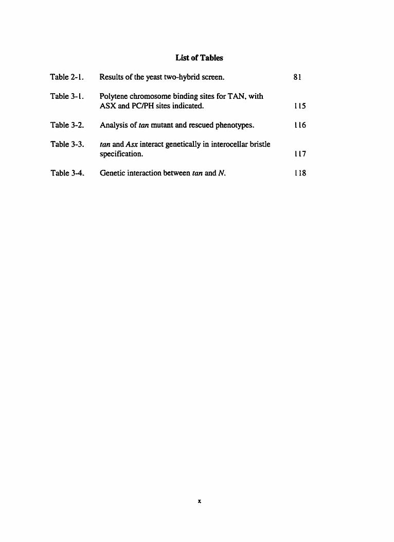
List of Tables
Table 2- 1. Results of the yeast two-hybrid screen. 8 1
Table 3- 1 . Polytene chromosome binding sites for TAN. with ASX and PCPH sites indicated. 115
Table 3-2. Analysis of tan mutant and rescued phenotypes. 116
Table 3-3. tan and Asx ïntenct geneticaiiy in interocehr bristle specification. 117
Table 3-4. Genetic interaction between tan and N. 118

List of Figures
Figure 1- l . The Drosophila melanogairer Me cycle.
Figure 1-2. Embryogenesis.
Figure 1-3. A-P and D-V axes specification during oogenesis.
Figure 1-4. Hiearchy of genes involved in A-P patteming.
Figure L-5. Domains of gap gene expression.
Figure 1-6. D-V axis specification.
Figure 1-7. Parasegments versus segments.
Figure 1-8. fushi tarazu expression.
Figure 1-9. Expression of even-skipped stripe 2.
Figure 1 - 10. Expression pattern of selected pair-de and segment polarity genes.
Figure 1 - 1 1. Specification in the l a r d epidermis.
Figure 2- 1 . Schematic diagram of the yeast two-hybrid screen.
Figure 2-2. Bait constmcts used in yeast two-hybnd screen.
Figure 2-3. Far Western assay.
Figure 2-4. Cloning tan from cDNA and genornic [ibnries.
figure 2-5. Genomic and amino acid sequence of tan.
Figure 2-6. Polytene in situ hybridizations.
Figure 2-7. Noahern blot andysis.
Figure 2-8. Expression pattern of tan.
Figure 2-9. An epitope tagged version of TAN localizes to the nucleus.
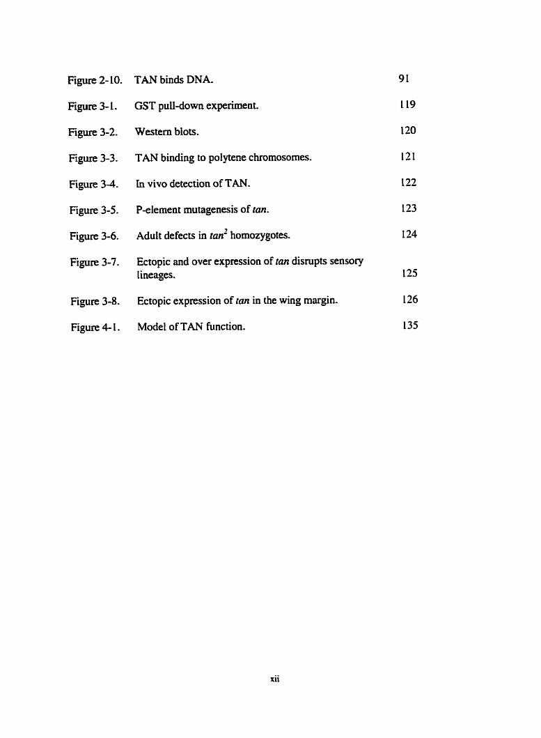
F i p 2- 10. TAN binds DNA.
Figure 3-1.
Fi- 3-2.
Figure 3-3.
Figure 3-4.
Figure 3-5.
Figure 3-6.
Figure 3-7.
Figure 3-8.
Figure 4- 1.
GST pulldown experiment.
Western blots.
TAN binding to pol ytene chromosomes.
in vivo detection of TAN-
P-element mutagenesis of tan.
Adult defects in tan' homozygotes.
Ectopic and over expression of tan disrupts sensory lineages.
Ectopic expression of tan in the wing margin.
Mode1 of TAN function.

List of Abbreviations
Genes
ANT-C Antp Asx bcd b m BX-C cac cad Ofd dl ~ P P EGFR
IR grk gt h hb hh hkb hth kni Kr luc MCMI Mers N nos odd
OPa otd P ~ X Pc ph ph0 prd
Antempedia complex An tennapedia Additional sex combs bicoid b r u h a bithorax comptex cactus caudal Defonned dorsal decapentapiegic Epidermal growth factor receptor engrailed even-skipped extradenticle Enhancer of zeste fmhi tarazu gurken giant hairy hunchback hedgehog huckebein homothorax knirps Krüppel luciferace Minichromosome maintenance Myeloid ecotropic insertion site Notch n a o s O&-skipped odd-paired onhodenticle pre-B ce11 homeobox 2 Polycontb polyhomeotic p leiohomeotic paired
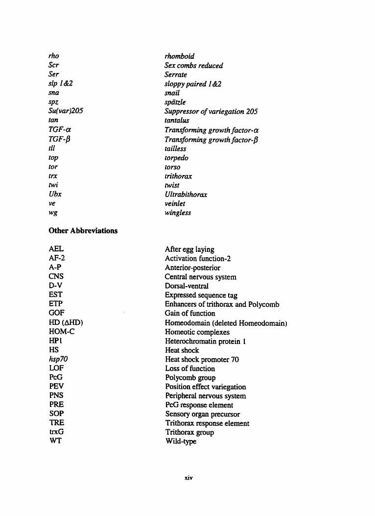
rhomboid rho Scr Ser slp I Br2
for trx twi Ubx ve
Other Abbreviations
AEL AF-2 A-P CNS D-V EST ETP GOF HD (0) HOM-C HP1 HS Iisp 70 LOF PcG PEV PNS PRE SOP TRE trxG WT
Sex combs reduced Serrate sloppy paired 1 &2 mail spatzle Suppressor of variegation 205 tantuttcs Transfo rming growth factor- Transfo rming gr0 wrh factor- /3 tailless torpedo tors0 trithorax twist Ultrubithorax veinlet wingless
After egg laying Activation function-2 Antenor-posterior Centrai nervous system Dorsal-ventral Expressed sequence tag Enhancers of trithorax and Polycomb Gain of fiinction Homeodomain (deleted Homeodomain) Homeotic complexes Heterochromatin protein 1 Heat shock Heat shock prornoter 70 Loss of fiinction Polycomb group Position effect vuiegation Peripherd nervous system PcG response element Sensory organ precursor Trithorax response elemsnt Trithorax group Wid-type
xiv

Introduction
A great success of devefopmental biology has been the fusion of the long history
of embryological observation with the power of molecular biology. In particular,
scientists have pushed classical genetics out of the realm of mathematics and into the
realm of ceil biology, whereby the developmental function of the gene could be deduced.
Without question Drosophila melanoguster has k e n a key experimentai organism that
has allowed this to occur. While the groundbreaking study of phage and bacteria in the
rniddie of the 1st century aiîowed us to understand the nature of the gene. Drosophila has
led the way in extending the concept of the gene to an understanding of development and,
ultimately, an explanation of the evolution of animal complexity.
The contribution of Drosophila research began over 80 years ago, and had
immediate impact when Thomas Hunt Morgan and his students provided the fmt
d e f ~ t i v e evidence for the chromosornai iheory of inhentance. The cecent sequencing of
the Drosophila genome (Adams et ai., 2000)- a monumental achievement surpassed oniy
by the sequencing of the human genome, provides a fuifding bookend to Morgan's feat.
Most interestingly, the sequencing projects have emphasized the similarity between
Drosophila and humm genes. ensuring that Drosophila research will not only continue to
shed iight on the evolution of H e on earth but also throw insight onto what we, rightly or
wroagiy. have decided to be the most important result of that evolution: ourselves.
In the last 20 years, development of the Drosophila embryo has k e n dissected at

the genetic and molecular Ievel to give us one of the most complete and elegant
explanations of how complex patterns derive fiom a single ceil. This advance was
initiated by several large scale genetic screens (Nüsslein-Volhard and Wieschaus, 1980;
Jürgens et al., 1984; Nüsslein-Volhard et al., 1984, Wieschaus et al., 19û4; Schüpbach
and Wieschaus, 1986; Schüpbach and Wieschaus, 1989) which sought to determine how
the anterior-postenor (A-P) and dorsal-ventral (D-V) axes are designated, and how
segments are formed and specified once these axes are demarcated.
This Introduction is divided into three sections. The first section outlines early
developmentûl events in the Drosophila embryo Ieading to the pattemed larva. This
description incorporates the work of hundreds of researchea and npresents a
monumental achievement in the study of development; for the fint time in any organism
a derailed knowledge of the events leading fiom oocyte selection to adult pattern has been
obtained. Although some of the specifics are unique to Drosophila, many fundamental
underlying principles have k e n deduced. These include universal signaiing cascades and
morphogens responsible for translating axis information into pattern. ln the next two
sections I review the events surroundiig asymmetry interpretation and maintenance in
greater detail: the second section focuses on the role of the /tr protein in interpreting
asymmetry, whiie the third section discusses the Asx gene in the context of global
mechaaisms used to maintain States of transcription.

1. Drosophila Development
A) Synopsis of the Drosophila Life Cycle
The Krause laboratory focuses on the events of early development (reviewed in
Wolpert et al.. 1998). providing a fitting place to enter the life cycle of Drosophila
(Figure 1-1). Mer the egg is fertilized the embryo undergoes several rapid rounds of
mitotic division without cytoplasmic cleavage (Figure 1-2). The nuclei then migrate to the
penphery of the egg and become ceiiularized by membranes pinching in from the egg
surface. The migration of nuclei towards the surface begins after 9 mitotic divisions and
by the end of 13 divisions. t h e houn after egg laying (W), cellulûnzation is complete.
During this tirne. the germ anlagen are specified fiom a group of approximately 15 nuclei
derived from the posterior end of the blastoderm embryo. As Drosophila melanogaster is
a long germband insect, al1 future segments an defined by the end of the blastoderm
stage.
During gastnilation, the epitheüai layer from the blastoderrn embryo gives rise to
the mesoderm, ectoderm and at the anterior and posterior ends of the gastrulating tissue,
the endodem. Gastmlation is followed by an extension and retraction of the germ band
around the posterior end of the embryo, to the dorsal side. By the end of retraction the
t h e head, i d e thoracic, and eight abdominal segments are clearly demarcated. Dunng
this t h e the imagina1 discs, which will eventuaiiy give nse to the adult structures, are
also set aside. Approximately 24 hrs AEL the Iarvae hatches, and after two more Iarval
instars pupation occurs. During pupation the imagiaal discs differentiate into the adult
structures of the fly, which ecloses at day 10 AEL.

B) Initial Asymmetry in the Oocyte
The mother play; a major role in produchg the asyrnmetry that specifies the axes
of the developing oocyte during oogenesis. Initialiy, an asymmetnc division of a germ-
line stem ce11 in the germarium of the fernde ovary produces a new stem celi and a
cystobIast (reviewed in Giiinert and St. Johnston, 1996). The cystoblast undergoes four
mitotic divisions without cytokinesis, resulting in 16 cens attached by ring canais. Only
two of these cells will have four such canals and both become pro-oocytes and initiate
meiosis. However, only one is selected to become the oocyte. This selection may involve
the asymmetric segregation of cellular components during the initiai division of the
cystoblast (de Cuevas and Spradling, 1998). The other pro-oocyte and remaining 14 cells
become nurse cells, which deposit large quantities of RNA and protein into the egg.
Dunng the movernent of the 16 ce11 cyst through the germarium it becomes enveloped by
somatic follicle cells, key components of poiarity formation. After the oocyte is selected.
the f i t sign of asymmetcy is increased levels of the ceil adhesion moIecule, DE-cadherin,
in postenor foliicle celis (Godt and Tepass, 1998; Gonz5lez-Reyes and St. Johnston.
1998). DE-cadherin upregulation causes migration of the oocyte to the posterior end of
the egg chamber.
The posterior position of the oocyte and oocyte nucleus within the cyst ailows the
posterior end of the egg to be specified F i t (reviewed in Ray and Schüpbach, 1996; van
Eeden and St. Johnston. 1999); (Figure 1-3). The oocyte signals to the surroundhg
foüicle ceils by releasing a Ligand encoded by gurken (grk), a member of the transforming
growth factor-a (TGF-a) f d y (Godiez-Reyes et al., 1995; Roth et al., 1995). GRK

binds to the posteriorly Iocated follicle ceils through the Torpedo (TOP) receptor, a
homologue to the epidermal growth factor receptor (EGFR). Activated TOP then sends a
retum signal to the oocyte, which causes a major change in the polarity of the oocyte
microtubule network (Lane and Kalderon, 1994; RuohoIa et al., 1991). This polarity
reversal is the & event that specifies the A-P axis: it is required for the locdization of
mRNAs that will determine the antenor end of the embryo (see below), and also for the
re-positioning of the oocyte nucleus to the anterior end of the egg, an event essential for
D-V axis determination (Gonzdez-Reyes et d.. 1995; Roth et al., 1995).
Microtubule reorganization dlows the oocyte nucleus to move to a random
anterior margin location (Theurkauf et al., 1992). Once there, GRK signals again to the
overlying follicle cells through TOP to specify the dorsd side (Neuman-Silberberg and
Schüpbach, 1994; Schüpbach, 1987). TOP activation in dorsal foUicle cells prevents the
expression of pipe. a gene required for modification of the Spaale (SPZ) ligand, which in
tum. is responsible for ventrai fates (Morisato and Anderson, 1994; Sen et ai.. 1998). if
the G W O P pathway is inactivated during dorsd specification, active ventrai ligand is
produced everywhere. resulting in a ventraiized embryo.
To sumrnarize, axes designation in the oocyte uses DE-cadherin based ceU
adhesion to posteriorly locate the oocyte and oocyte nucIeus within the f 6 ceii cyst. This
positionhg diows the G W O P signahg cascade to mrganke the microtubule
network and produce an A-P polariiy in the egg. Microtubde reorganization also causes
the nucleus to migrate to an anterior mugin Location where signahg specifies the D-V
axis by iimiting the release of the ventral-induchg Ligand SPZ to the future ventrai side.

Although the GRK signaling pathway nicely explains the formation of both axes, the
cause of the original asymmetry in the posterior and dorsal foliicle cells. dowing them to
react dinerentially to the GRK Ligand, is still not known (Goazalez-Reyes and St.
Johnston, 1998). How the folücle cens achieve this asymmetry is one of the few
unanswered questions of axis determination.
C) Translation of A x i s Polmity
In order for the newly established oocyte polarity to lead to patterning in the egg.
several matemal gene products must act as morphogens. Morphogens are defined as
factors capable of specifymg dBerent ceIl fates at different concentrations dong an axis.
The positional values created by these morphogens in Drosophila are used to produce the
iimited spatial expression of the fmt zygotic genes dong the axes. These initid zygotic
genes are the first in a hiemhy of genes whose expression wiil translate the initial
asymmetry in the A-P and the D-V axes into segments and g e m Iayers (ectoderm and
mesoderm), respectively (Figure L -4).
i) Patterning the A-P Axis
A-P specification requires three systerns, unlike the D-V axis which requkes only
one (reviewed in St. Johnston and Nüssiein-Voihard, 1992). The anterior, posterior. and
terminai systems are initiated independentiy. but work together to provide pattern to the
A-P &S.
In the mtenor system bicoid (bcd) mRNA is localued to the antenor end of the
oocyte as a result of the microtubde reorganization events that occurred earlier during
oogenesis (Berleth et al., 1988; Frigerio et al., 1986; St. Johnston et al., 1989), and is

translated after the egg is laid to produce a gradient of protein extendlng to the middle of
the embryo (Driever and Nüsslein-Volliard, 1988a; Driever and Nüsslein-Volhard,
1988b); (Figure 14). BCD contains a homeodomain (HD) DNA binding motif (Berleth et
al.. 1988; Frigeno et d.. L986) and functions as a morphogen in anterior patteming
(Driever and Nüsslein-Volhard, 1988b).
BCD regulates several gap genes, including hunchback (hb) and o~horlcnticle
(or&, in the anterior end of the embryo. Gap genes are the first zygotic genes to be
activated and encode transcription factors which pattern Iarge regions of the embryo
(NUsslein-Volhard and Wieschaus, 1980); (Figure 1-5). The hb enhancer contains both
strong and weak binding sites for BCD (Driever and Nüsslein-Volhard, 1989: Driever et
al., 1989; Stnihl et al., 1989) and, consequently, is expressed in a brod domain that
rnirnics BCD expression (Taua, 1988). The otd enhancer, on the other hand. has only
weak BCD sites and is only activated in the most anterior regions of the embryo where
BCD concentrations are highest (Gao and Finkelstein, 1998). In this way, BCD acts as a
morphogen by limiting individual gene expression to specific regions dong the A-P a i s ,
based on the affinity of BCD for each target gene enhancer.
HB, dso a transcription factor, appears to be the pnrnary morphogen required to
speciQ thoncic and abdominal fates (Hülskamp et al., 1990; Schulz and Tautz, 1994;
Suuhl et al., 1992; Wimmer et al., 2000), and acts in conjunction with BCD (which may
be redundmt) to directly regdate transcription of severai additionai gap genes (reviewed
in Rivera-Pomar and Jkkle, 1996). For example, the gap gene Kriïppeï (Kr) is required in
thoracic and abdominal segments (Nüssleh-Volhard and Wieschaus, 1980) and has its
expression boundarïes set by both BCD and HB (Gad and Jackie, 1989; Hoch et al.,
1992; Hoch et d.. 1991; Hülskamp et al.. 1990). Kr is activated at Iow concentrations of
BCD and KB found in the middle of the embryo. but is repressed in more anterior
regions. The anterior repression of Kr is presumably due to repressor pmteins encoded for
by other gap genes, tike giant, which are activated by higher levels of BCD. The posterior
limits of Kr expression are set by yet other gap genes activated by the postenor system.
Therefore, because of the different affuiities of BCD and HB for target gene enhancers
and the ability of gap genes to cross-regulate their expression, severd broad domains of
gap gene activity can be established within the antenor region of the embryo.
Patteming in the posterior end of the embryo requires a posterior to anterior
gradient of nanos (nos) protein (Gavis and Lehmann, 1992; Wang and Lehmann, 199 1) to
inhibit translation of matematly provided hb mRNA (Hülskamp et al.. 1989; Irish et al.,
1989; Stnihl et ai., 1989). In addition to its zygotic expression hb mRNA is deposited
ubiquitousty by the mother in the embryo, and it is the ubiquitous hb mRNA in the
posterior end that NOS must counteract. Suppression of maternai KB in the posterior of
the embryo allows the Caudal ( C m ) transcription factor to function. CAD is found in a
gradient emanating from the posterior end and is responsible for activating severd gap
genes in the posterior region (Rivera-Pomar et ai., 1999, similar to the role of BCD in
the anterîor end. Interestingly, cad mRNA is aiso ubiquitously provided by the mother
and has its gradient fonned by the inhibition of its translation in the antenor end (Mlodtik
et al., 1990). Surprisingiy, this inhibition is performed by BCD, as BCD uses its KD to
bind to 3'UTR sequences in the cod mRNA to preveat translation (Dubnau and Stnihf,

1996; Rivera-Pomar et al., 1996).
Finally, in the termini, the most anterior and posterior follicle cens sectete a
ligand for the Torso (TOR) receptor (reviewed in Duffy and Perrimon. L994). TOR. a
member of the tyrosine kinase class of receptors. is zygotically expressed and found
throughout the surface of the ernbryo (Casanova and Stnihl, 1989; Sprenger et al.. 1989).
The folkle ceUs at the poles retain the ligand in the perivitelline space until after
feailuation, at which time it is released and binds to the receptor. The downstrem result
of TOR activation is derepression of the gap genes ?ailfess and huckebein in the terminal
regions (Liaw et al., 1995; Paroush et al., 1997; Rusch and Levine, 1994). These proteins
iimit expression of other gap genes in the termini of the embryo and pattern the terminal
acron and telson stmctures, with BCD overlap making anterior structures different from
posterior (Pignoni et ai.. 1990; Weigel et ai.. 1990).
To summarize, localized expression of Bm and NOS is required to effect the
designation of the A-P axis by producing opposite gradients of the HB and CAD
transcription factors. BCD/HB and CAD extend h m the anterior and posterior poles,
respectively. of the embryo and provide positional information dong the axis. This
positional information, in conjunction with signais fmm the terminal system, is
interpreted to pmduce the specifk aperiodic expression domains of the individuai gap
genes. The gap genes then cross-regulate each other. refining their expression domains
into sharp on-off patterns.
ii.) Patteming the D- V Axis
Althou* the D-V axis is specified ushg a different initial mechanism (signal

transduction) thm the A-P axis (localized determinants) the end result is the same: the
production of a morphogen to suppIy positional information dong the axis (reviewed in
Morisato and Anderson, 1995; Rusch and Levine, 1996). This rnorphogen is encoded by
the dorsal (dl) gene (Roth et al., 1989), a homologoue of the NF-& gene of the IL-LR-
NF-- pathway found in both the plant and animal kingdoms (reviewed in Belvin and
Anderson. 1996); (Figure 1-6). DL is an outstanding example of the ideal morphogen as it
acts as both a repressor and activator, and induces expression of genes which themselves
limit DL function. DL is ubiquitously provided by the mother but is sequestered in the
cytoplasm through its association with the protein product of the cactus (crict) gene,
CACT (Kidd, 1992; Wasserman, 1993; Whalen and Steward, L993). CACT repression of
DL function is lifted by signding of the ventral ligand SpBitle (SPZ) to the ubiquitously
expressed receptor encoded by Toll (Hashimoto et ai., L 99 1; Hashimoto et al.. 1988). The
resuIt of this signaling is the release of DL from CACT, dlowing DL to translocate to the
nucleus.
As active SPZ extends in a graded fashion from ventrai regions, a sirnilar p d e d
distribution of nuclear DL is produced. This p d e d distribution allows DL to play a role
in specwng d l fates (four in total) dong the D-V a i s , either directly or indirectly. in
v e n d regions high levels of DL cause the expression of twist (mi) and snaii (ma), two
genes required for gastrulation and mesodemi specifcation (Ip et al., 1992; Jiang et ai.,
L99 1; Pan et al., 199 1; Simpson. L983; Thisse et al., 1987; Thisse et ai., 199 1). The
enhancer regions of these genes have low flrnity sites for DL, limiting their expression
to ventral regions.

Foilowing its activation by DL, SNA acts as a direct transcriptional repressor by
Limiting the expression domains of other DL target genes to laterai regions (Boulay et al.,
1987; Ip et al.. 1992; Kosman et al.. 199 1 ; kptin, 199 1). For example. rhomboid (rho) is
required specifically in ventral-laterai regions to speciQ neumectodermai fate. Even
though rho has high affinity sites for DL and DL coactivators in its enhancer (Gray et al.,
1994; Jiang and Levine. 1993), its expression is limited to ventral-lated regions because
this is the only location where the DL activator is present and SNA, its repressor, is not.
DL is not expressed in dorsal regions but plays a role similu to BCD and NOS by
repressing the expression of dorsalizing genes in ventral regions. The primary dorsalizing
signal is the morphogen encoded by clecupentaplegic (dpp). a homologue of the
transfonning growth factor-p (TGF-p) family of cytokines (Irish and Gelbart, 1987:
Padgett et al., 1987). DL uses corepressors to directly dom-regulate transcription of dpp
and severai other dorsai genes in the venual regions of the zygote, thereby lirniting their
domains of expression and activity to dorsal regions (Huang et al.. 1995; Lehming et al..
1994).
Summarizing, TOLL receptor activation by ventral SPZ results in a gradient of
nuclear DL Iocaiization extending Gom the venual surface. At high DL levels the
mesodemi spetifying factors TWI and SNA are induced. SNA then iimits the expression
of genes under DL control, such as rho, to ventrai-laterai regions, thereby specimng
neuroectodermal fates. DL also acts as a repressor of dorsaiizing factors. Like DPP, by
dirrctiy down-regufating their expression in v e n d and ventral-laterai regions. This
aiiows DPP to specify amnioserosa (hi& DPP) and ectodermd fates (low DPP).

Examination of the underlying mechanism of axes formation in Drosophila
reveds that only one deteminant aeed be locaiized in order to initiate a complicated
patteming process. Although postenor NOS functions to inhibit maternai hb mRNA
translation in the posterior end, in hb materna1 (hb""') mutants NOS function is
dispensable and fertile adults c m be obtained in a hb""'lnos double mutant (Hülskamp et
al., 1989; Irish et al., L989; Stnihl et ai.. 1989). Therefore. one localized morphogen
(BCD) cm pattern the A-P mis, excluding the terminal regions. The BCD gradient
patterns the anterior end and aiso produces a gradient of the posterior determinant CAD
(HB can aiso prevent antenor CAD function by an unknown mechanism). DL acts in a
similar fashion by acting as a morphogen to pattern ventral fates and by preventing the
ubiquitous expression of the dorsai morphogen DPP.
D) Frorn Asymmetry to Pre-pattern: Making Pansegments
The activities of BCD/HB, CAD, DL, and DPP have provided the egg with
asymmetries (limited zygotic gene expression) that c m now be used to pattern the
organism. For the sake of brevity and relevance, 1 wiU focus on the events involved in
patternhg the body trunk in the A-P axis. Aithough the differentiation mechanisms used
in each axis have similarities, a major ciifference between the two is the production of
periodicity dong the length of the A-P axis by the aperiodic gap and maternai genes.
Periodicity in the A-P axis is first visible as the altemating suiped expression
patterns of the pair-mle genes. The pair-rule genes represent the next wave of genes f ier
the gûp genes and wîli produce the f k t visible sign of segmentation in the ernbryo, the
parasegments (reviewed in Martinez-Anas and Lawrence, 1985); (Figure 1-7). The 14

parasegments are preludes to the future segmental divisions prominent in larvae and
adults. and are unique in that they display an early lineage restriction. The
"compartments" produced by this lineage restriction prevent celi mixuig and are similar
to rhombomeres and sornites found in vertebrates (reviewed in McGinnis and Krumlauf.
1992). In Drosophila. these cornpartmentai boundaries act as organizing centres for future
patteming events in both larvai and adult structures (reviewed in hgham and Martinez-
Arias. 1992; Lawrence ÿnd Stnihl. 1996). As development of al1 higher organisms
consists of the sequentid division of the egg into "domains" of differentiation, the
discovery and analysis of parasegments has offered a simple system to understand how
regions of an embryo become restncted in their capacity to differentiate. immune from
competing influences in the egg.
i ) Pen'odiciiy
The protein expression pattern of the pair-mle geneficshi tarnzu (ftz) (Kaufman et
al., 1980; Wzkimoto and Kaufman, 1981) at cellular blastoderm is a remarkable image
(Carroll and Scott, L985; Karr and Komberg, 1989). M e d y two hours dter fertilization
seven sharp frz stripes in altemathg parasegments dong the A-P mis of the egg are
visible (Figure 1-8). The segrnented nature of Drosophila implied that regulators of
differentiation would develop periodic patterns of expression during development, but
one marvels at how early in development this expression is achieved and required.
The periodicity of the pair-mle genes results from the regulatory abilities OF the
overlapping matemal and gap proteins (reviewed in Pick, 1998). Initiation of men-
skipped (ore) expression, another pair-rule gene, by the maternai and gap proteins has

k e n weil studied and serves as a mode1 for the activation of other pair-rule genes. The
penodic expression of eve is complex and involves the use of stripe-specific enhancers,
combined-stripe enhancers, and seven-stripe enhancen that act foiiowing the initiation of
a broad band of very weak eve expression in the trunk of the embryo.
In the case of the stripe-specifc enhancers, activation of me stripe 2 expression is
the best understood (Figure 1-9). Stripe 2 expression requires activation by matemal BCD
and zygotic HB and is counter baianced by the repressive effects of the gap proteins Giant
(0 and Krüppel (KR) (Small et al.. 1992; S m d et al., 1991). The peaks of GT and KR
expression are offset, allowing BCD and HB to activate e w expression in a limited
domain between the two represson. The binding sites for ail four proteins are partially
overlapping, and it appears that cornpetition for binding and local repression by
quenching occurs ( h o s t i et ai., 1996: Gray et al., 1994). This elegant mechanism,
sometimes using different playen, provides periodicity for other cve stripes (see Fujioka
et al.. 1999) and is probably used for achieving periodicity in the expression domains of
other pair-rule genes. AU eve stripes, however, are not individually controlled as
composite-stripe enhancen (eg. stnpes 4 and 6) appear to exist (Fujioka et al., 1999).
FoIiowing activation of the eariy stripe enhancers a single late enhancer is xtivated
which functions to increase the level of eve expression in dl seven siripes and to refine
the early broad suipes into sharper domains. The late enhancer requires the auto-
regulaiùig abbitity of nrE and the repressor effects of seved other pair-rule gene products
(Frasch and Levine, 1987; Fujioka et al., 1995; Fujioka et d., 1996).
Although a hierarchy withùi the pair-de class was originaüy proposed based on

genetic interactions between members. the situation appears to be more complicated (see
Pick 1998). This model proposed that "primary" pair-de genes like eve, hairy (h), and
nurt interpret gap gene cues and then were directly responsible for providing the striped
pattern for the "secondary" pair-nile genes such as& because composite seven-stripe, but
not stnpe-specific, enhancers were found for& (Hirocni and Gehring, 1987: Hiromi et
al.. 1985). Also. early resuits suggested that the secondary pair-rule genes did not play a
d e in regulating the expression of the primary genes. Recent studies have indicated that
this model is too simple (Nasiadka and Knuse, 1999; Pick, 1998; Saulier-Le Drean et al.,
1998: Yu and Pick, 1995) and suggest that matemal and gap proteins play an important
role in the early specification of stripes for most pair-rule genes, with cross-regdatory
interactions between pair-mle genes refining and properly positioning the stripes.
ii) Defining Parasegments
As there are numerous pair-rule genes with partially overlapping expression
domains, it was not irnmediately apparent where the 14 pansegments would arise.
Assistance carne from the redization that the parasegmental boundaries Form between
suipes of engrailed (en) and wingless (wg) expression (reviewed in Ingharn and
Mûninez-Arias, 1992), two mernbers of the segment polarity gene f;uniIy. wg is
expressed in the posterior domain of one parasegment while en is expressed in the
anterior region of the next puasegment. It is now clear that the overlapping expression
domains of the pûir-nile genes act as a combinatorid code to delimit the d o m e s of en
and wg expression (DSlardo and OFmU, L987; Ingham et ai., 1988); (Figure 1- 10).
The 14 en stripes form at the anterior borden of the alternathg me (odd-

numbered parasegments) and ftz (even-nurnbered parasegments) stnpes (Harding et al.,
1986; Howard and Ingham, 1986; Macdonald et al., 1986). Although stripes of EVE and
FIL are broad at the time of m initiation (Frasch and Levine, 1987; Harding et al., 1 986),
en expression is lirnited to a few cells in the anterior of each parasegment (Fjose et al.,
1985; Komberg et al., 1985). This important limitation on en expression is created by the
combinatorial code of pair-de genes: odd-numbered en stripes require the overlap of
EVE and Paired (DNardo and O'FarreIi, L987; Momssey et al., 199 1). h contrast, even-
numbered en stripes require the overlap of FIZ and Odd-paired (Benedyk et al.. 1994:
DiNard0 and 0'Fami.i. 1987). Moreover, en expression in the middle of the parasegment
is repressed by other pair-rule proteins (Cadigan et al., 1994; DiNardo and O'Farrell.
1987; Manoukian and Krause, 1992).
The early broad expression of EVE and FTZ initidy acts to cepress wg expression
in the posterior of each segment (hgham et al., 1988). However, as development
proceeds the expression of eve and& decays in the postenor region of the parasegment
and it is at this time that the appropriate activators tum wg expression on there (Baker,
1987; Benedyk et al., 1994; Ingham et ai., 1988). Additionally, other genes, like naked
and sloppypired, provide polarîty to the parasegment by limiting the regions where en
and wg expression cm be activated and rnaintained (Cadigan et al., 1994; Muilen and
DNardo, 1995).
Pair-mle gene expression is hitiated before celIular blastoderm (- 3hrs A m ) and
within an hour the parasegments have k e n defined by the juxtaposition of en and wg
saipes. The importance of the parasegments, as 1 discuss below, is in their orgmizhg

abiiity; the borders of the parasegments act as sources of morphogenic activity to specQ
different ceii fates within the pnrasegments.
E) P a t t e d g the Larval Epidermis
The segmented A-P axis wiii give rise to severai structures, including the larval
epidermis and the adult imaginai discs. Again, for sirnplicity, I will focus on only one of
these tissues. the larval epidermis. The larval epidermis consists of 3 thoncic and 8
abdominal segments (the head segments are involuted). with each segment represented by
a unique denticte pattern (Figure 1-1 1A). In most segments the cuticle consists of rows of
denticles in the anterior half and naked cuticle in the posterior hdf. What also makes the
denticle pattern of the cuticle interesting is that not only are the individual segments
unique, in tenns of the bristle pattern, but there are also differences in denticle
morphology within each segment as one moves from the anterior to postenor end of the
segment. The existence of unique segments and polarity within each segment offers a
simple system to understand cornplex patteming interactions. For example, how are
individual ce11 fates (the different denticles) established within the fnmework of n larger
defined field (the parasegment) which itself must be given a unique identity (the different
segments)?
i) DejTning Position Within the Parasegrnent
Lawrence has proposed that the diffecent venual cuticle pattern obsemed across
the segment couid be achieved by a morphogen gradient specifying different bristle types
at different morphogen concentrations (Lawrence and Sampedro, 1993). In this mode1 the
hinction of the segment polarity genes is solely to estabiish and maintintain the

parasegmental borden, borders which are essentiaf to maintain a "source" and "sink" for
the rnorphogen. Other rnodels, in contnst, suggest that the overlapping domains of
segment polarity gene expression contain enough information to pattern the
approxirnately seven different individual ceil types that exist within a parasegment
(Bejsovec and Wieschaus, 1993; Martinez-Anas et ai.. 1988). Recent results suggest that
both methods - morphogen gradients and cornbinatorial codes - are used to pattern the
ventrd epidermis of the Iarvae.
Denticle specification occurs several hours after the pansegmental boundaries are
established and until this specification c m occur the integrity of the parasegmental
boundaries must be maintûined (reviewed in DNardo et al., 1994). Since expression of
the pair-rule genes decays around the end of genn band extension (-4 AU). a
mechanism is required to maintain expression of en and wg. in k t , they maintain each
other's expression dunng the early phase of their expression, while at Iater stages this
dependence is lifted. EN contains a homeodomain (Poole et al., 1985) and acts as a
transcription factor, while WG is a secreted giycoprotein that participates in signaiing
events (Cabrera et al., 1987; Rijsewijk et al., 1987). During the early phase of their
expression, WG signals across the parasegmental boundary to the en expressing cells and
activates a sigoaüng cascade that results in the continued expression of en (Bejsovec and
Martinez-Anas, 1991; Cumberledge and Krasoow, 1993; DiNardo et al., 1988; Martinez-
M a s et al., 1988). The en expressing celi then produces another signal, encoded by
hedgehog (hh), which signais back to the wg expressing ce11 to maintain wg expression
(Ingham et al., 1991; Ingham, 1993). This reciprocal signaiing occurs Iocaiiy and
consolidates the parasegmental boundary until the process of celi specification cm occur
(Figure 1-1 1B).
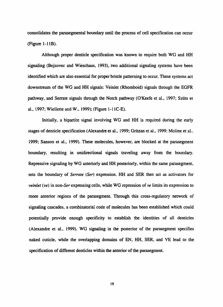
Aithough proper denticle specification was known to require both WG and HH
signahg (Bejsovec and Wieschaus. 1993), two additional signaling systems have k e n
identified which are aiso essentiai for pkper bristle patteming to occur. These systems act
downstrei~m of the WG and HH signals: Veinlet (Rhomboid) signais through the EGFR
pathway. and Sernte signais through the Notch pathway (OtKeefe et al., 1997; Szüts et
al., 1997; Wiellette and W.. 1999); (Figure 1- 1 LC-E).
hitially. a bipartite signai involving WG and HH is required during the early
stages of denticle specification (Alexandre et ai., 1999: Gritzan et al., 1999: Moline et al..
1999; Sanson et al., 1999). These molecules, however. are blocked at the parasegment
boundary? resulting in unidirectionai signals tnveling away from the boundary.
Repressive signaiing by WG anteriorly and HH postenorly, within the same parasegment.
sets the boundary of Serrate (Ser) expression. HH and SER then act as activators for
veinlet (ve) in non-Ser expressing cells, while WG repression of ve limits its expression to
more anterior regions of the parasegment. Through this cross-regulatory network of
signaling cascades, a combinatorid code of molecules has been established which could
potentially provide enough specifcity to establish the identities of di denticles
(Alexandre et al., 1999). WG signalhg in the posterior of the pansegment specifies
naked cuticle, whiie the overiapping domains of EN, KH, SER, and VE lead to the
specification of different denticles within the anterior of the parasegment.

ii) Providing Pattern Specificity to the Parasegment: the Homeotic Genes
The activities of the WG and HH signaüng pathways have led to the specification
of ce11 fates within the parasegment; individual ceiis now know their location dong the
axis of a parasegment. The responsibility for providing segment-specific patterns (iarvd
and adult head, thoracic or abdomen) to these cells lies with the genes of the homeotic
complexes (HOM-C). Interestingly, the HOM-C acts analogously to WGNH signaling
described above in that HOM-C genes speciQ positional vaiues dong the length of the A-
P axis, sirnilar to the positionai values specified dong the length of the individual
parasegments by WGRIH signaling. It is important to stress that HOM-C genes do not
make, for example. abdominal-specific structures, but they tell the organism where such
structures should be located.
HOM-C genes exist within two complexes: the Antemapedia complex (ANT-C),
which contains 5 genes responsible for speciwng head and thoracic identities antenor to
parasegment 5, and the bithonx compfex (BX-C). which contains 3 genes responsible for
speciwng thoncic and abdominal identities posterior to parasegment 4 (reviewed in
Morata, 1993). Expression of these complexes is required throughout development, and is
initiated very early during embryogenesis at the tirne when the gap and pair-rule genes are
active. In particular, the BX-C gene Ultrubithorax (Ubx) is repressed in anterior regions
of the embryo by hunchback protein (White md Lehmann, 1986) while the fi2 pair-mle
proteio activates übx expression outside areas of Hunchback repression (hgham and
Martinez-Arias, 1986; Müller and Bienz, 199 1; Müller and Bieoz, 1992).
The number of pnmary HOM-C genes (8) is not enough to pattern ail 14
parasegments individuaiiy, necessitating the need for a combinatorid code between
different members to pattern at least some of the individual parasegments This is most
clearly understood for the abdominal regions where the combination of Ub.x and
abdominal-A is required to pattern parasegments 7-9, while Ubx alone is responsible for
patterning parasegment 6. Although progress has been made in finding HOM-C target
genes in the imaginai discs (Weatherbee et ai., 1998), targets For patterning the Iarval
epidermis remain Iugely unknown, but experiments are beginning to detemiine which
genes the HOM-C regulate and how the 8 HOM-C genes work together to pattem the 14
parasegments (Casares et al.. 1996; CasteIli-Gair and Akarn. 1995; Li et al., 1999).
It should not go unnoticed that the embryo uses the sarne genes (eve and&) to
define the borders of the parasegmental divisions (determined by en and wg expression)
and to delirnit the domains of expression of genes (HOM-C) required to pattem these
pansegmentai divisions. hportantly, in ternis of evolution. these processes are not
Iinked directiy but exist in paraiiel. The W G m system and HOM-C genes converge
during Iater stages of development to produce the pattern of the parasegment: the W G M
system specifies position dong the parasegment, while the HOM-C specifies position
dong the A-P axis, producing segmentai identity (thorax or abdomen). The parallei
nature of the WG/KH signaiing system and HOM-C specification function aiiows for
evolutionary change by the HOM-C, independent of the W G H signaiing system. The
integrity of the parasegment is maintained by the WGMH signaling system, leaving
HOM-C genes free to evohe and possibly alter their targets. Patteming of the different
segments cm then change without affecthg the cellular coordinates within each

parasegment.
R s-ary
The aspects of Drosophila development descnbed here are cemarkable in that, for
the k t time in any organïsm, one can follow a developmental process uninterrupted
from the initiai specification of the oocyte to the final specification of pattern in the
developed organism. Although great suides have been made there are still obvious
questions to be addressed. but the synthesis ofgenetics and embryology is now well under
way. 1 have only discussed pattern in the larval epidermis, which is somewhat simplified
king basicdly a two-dimensional field. However, the more complicated genetic
interactions underlying the-dimensional structures such as wings and legs ;ue dso well
undentood and utiiize s i d a r principles (reviewed in Lawrence and Struhl, 1996). Tnily,
Drosophila melanogaster has provided ;in exceptional system to understand basic
developmentd questions.
IL. Molecular Basis of Cell Fate Specification
A) The fuîhi taruzu Gene
TheJushi tar- (Fz) locus was identified in the Kaufmm lab by screens for non-
complementing mutations of a deletion for the Antmnapedia region (Lewis et al., L980a;
+ Lewis et al., 1980b) and independentiy by the Nüsslein-Volhard and Wieschaus screen
for segmentation mutants ( 1980). Characterization of the& mutant embryonic phenotype
reveded a deletion of approximately half the number of segments in unhatched f i t instar
l m e (Jürgens et al., 1984; Wakimoto and Kaufman, 1981), and experiments using a
temperature sensitive de le reveaied that the critical penod of& activity required to

produce wild-type segments in the larvae is the 241- penod (cellular blastoderm) AU.
(Wakimoto et al., 1984). This early role for FTZ in segmental patteniing was confirmed
when cloning (Bender et al., 1983; Garber et al., 1983; Kuroiwa et al., 1984; Scott et al.,
1983; Weiner et al., 1984) and in situ analysis reveaied that the 7 stripe expression pattern
of fiz dunng cellular blastoderm (Hnfen et al., 1984) corresponded to those regions
deleted in the larval cuticle o f f i mutants (see Carroll and Scott, 1985; Martinez-Arias
and Lawrence, 1985). Besides this early stage of fiz function, it is also required during
neurogenesis in every segment (Doe et al., L988) and it is also expressed in the
developing hindgut (Krause et al., 1988), although its role there has not been addressed.
fn encodes a 413 rimino acid protein and contains a horneobox motif sirnilar to
that found in many homeotic genes (Kuroiwa et al., 1984) (Laughon and Scott. 1984:
McGinnis et al., 1984a; McGinnis et al., 1984b; Weiner et ai., 1984). FTZ uses its
homeodomain (HD) to directly bind its own enhancer (Schier and Gehring, 1992; Schier
and Gehring, 1993) and that of en (Desplan et al., L988; DiNardo and O'Farrell. 1987:
Howard and Ingham, 1986) to directly increase their Ievels of expression during cellular
blastoderm (Florence et ai.. 1997; Nasiadka and Krause, 1999). F E also directly
increases the expression of severai horneotic genes in the frz-dependent segments at this
time (Ingharn and Maninez-Arias, 1986; Ish-Horowin et al., L989: Müller and Bienz.
1992), while negatively regulating wg expression (Ingham et al., 1988; Ish-Horowia et
al., 1989). dso directiy (Copeland et al., 1996; Nasiadka et al., 2000; Nasiadka and
Krause, 1999). An activating role For ET2 in the embryo is consistent with assays in
tissue cttiture and yeast c e k where FE acts a mscripti~nal activator, dependent on HD

binding sites and the HD of FTZ (Fitzpatnck and hgles, 1989; Han et ai., 1989; Jaynes
and O'Farrell, 1988; Ohkuma et al., 1990; Wislow et al., 1989). However. it has yet to
be determined how FE acts as a repressor.
As described in the Introduction, F E hinction is Fundamentai in defining the
parasegmentai boundaries and producing segment-specific expression patterns for the
homeotic genes. However. a rather surprising discovery suggested that the HD of FTZ is
not required for it to perform the majority of its hnctions in patterning the epidermis
(Fitzpatrick et ai.. 1992). Before discussing this novel finding in greater detail it is
appropriate to review the HD motif. reveding why this novel activity for FTZ is so
surprising.
B) The Homeobox
The term homeosis was fint used by Wüliam Bateson to describe mutations that
transformed one particular segmentai or metameric structure of an organism into the
identity of another (Bateson, 1894). Genetic analysis in Drusophila over the last 80 yem
has identified sevenl loci which produce similar effects. most notably geenees of the BX-C
and ANT-C described by Lewis (1978) and Kauhan (1980), respectively. When these
genes were cloned and found to contain a similar 60 amino acid motif. the DNA sequence
was appropriately named the "homeobox" and the ;unino acid motif a " h o r n e o d ~ r n ~ ~
(McGinnis et al., 19th; McGinnis et ai., L984b; Shepherd et al., L984). This KD motif
was aiso found in MATal and MAT& proteins ~quired for cell-type switching in yeast
and was reminiscent of the hek-turn-heüx motif found in sevenl repressor proteins from
7c phage (Laughon and Scott, 1984), suggesting that the homeobox was highiy conserved


core (reviewed Treisman et ai., 1992). Bicoid, for example, contains a Lysine residue at
this position and prefen a GG dinucieotide, while Antemapedia contains a Glutamine
and prefers a CC dinucleotide (Hanes and Brent, 1989; Treisman et al., 1989). However,
as the HD proteins of the ANT-C and BX-C, including FTZ, have the same residue at this
position, it raises the question as to how HD-containing proteins of this class achieve
specificity in vivo.
Studies of transcriptional regulation in vivo have clearly demonstrated that,
regardess of how HD proteins attain specificity, the HD plays a major role (reviewed in
Hayashi and Scott, 1990). in particular, HD swaps revealed that the KD is crucial in
targeting proteins to the proper targets (Gibson et al., 1990; Kuzion and McGinnis, 1989;
Mann and Hogness. 1990). Experiments which replaced the Deformed (DFD) HD with
the HD of Ultnbithonx (UBX), for example, altes 17 of the DFD residues in the HD.
none of which reside in the recognition helix (Kuzion and McGinnis, 1989). This swap.
which aiso altered 5 residues just C-terminal to the HD, targeted the Dm-üBX hybrid to
a Ubx target, Antennapedia (Anp), resulting in the activation of this gene. This result was
particulûrly interesting because LJBX normaiiy represses Antp expression during
development, suggesting that the KD of UBX targets the protein but that regulation,
either positive or negative, depends on sequences outside of the HD. DFD normdly
autoregulates. potentialiy explainhg the positive eRect of the DFD-LIBX hybrid on Antp
transcription. Interestingiy, the DFD-UBX hybrid was not targeted to the endogenous Dfd
gene, presumably because it is not a nomiil target of UBX.
These results were extended in other HD swap experiments to narrow down the

regions responsible for targeting. Residues in the N-terminai ym of the HD were crucial
for providing target specificity; in some cases only 5 amino acids could aiter the targeting
ability of the HDs of DFD, Sex combs reduced and ANTP (Chan and Mann. 1993;
Furukubo-Tokunaga et al., 1993; Lin and McGinnis, 1992; Zeng et al.. 1993).
interestingly, these residues are not predicted to contact DNA, suggesting the possibility
that they recognize particular cofactoa that assist HD proteins in target recognition.
Regardless of the mechanism, the HD swap experiments support the notion that the HD
and surrounding sequences are criticai for providing target specificity to HD containing
proteins in vivo, making the fuiding of homeodomain-independent activities for FIZ
quite surprising.
C) The uriportance of the HD in FTZ Function
i ) HD-Independent FïZ Activities
The above studies cleariy demonstrate the importance of the HD For gene
regdation in vivo, and similar results were expected in experiments using thefi protein
(Fitzpatrick et al.. 1992). This study analyzed FIZ activity by attaching ftc transgenes to
the hsp70 promoter to drive ubiquitous expression upon transient heat-shock (HS) pulses
(Stnihl, 1985). Expression of wiid-type FIZ using this method produces an "anti-ftz"
phenotype (StniN, L985) in which the&-&dependent segments are deleted, due to the
inappropriate expression of ftz in regions that do not nomally require its activity (kh-
Horowicz et ai., 1989). Additionafly, the HS-j?z uaosgene c m rescue the fe-dependent
segments, as detemiined by the production of larvai cuticle, in a f n mutant background
(Copeland et ai., 1996; Hyduk and Percivai-Smith, 1996). Aithough the anti-ftz phenotype

is s t i l l produced in these embryos due to the ectopic expression of&, the experiment
demonstrates that the tronsgene cm perform many of the functions of endogenousftz and
is not simply acting ihrough endogenous fa. an important consideration since fn auto-
regulates its expression.
Surprisingly, of di the constmcts tested, the only fe-deletion construct that could
produce the anti-ftz phenotype or rescue the fidependent segments in a& mutant was
one in which the majority of the HD is missing (most of helices 1 and 3 and ail of helix 2
are missing) and is incapable of binding DNA (Copeland et al., 1996; Fitzpatrick et al.,
1992; Hyduk and PercivalSmith, 1996). The fact that this constmct, FKUHD. is capable
of rescuingfiz-dependent cuticle in aftz mutant background demonsuates that the HD of
FiZ is not required for it to perfom the majority of its functions in the epidecmis. These
fuactions include activating en and the homeotic genes, while repressing wg. However,
unlike wild-type FIZ, ETZAHD cannot rescue a ftz mutant phenotype when its
expression is solely controlled by endogenous fiz enhancer sequences (Furukubo-
Tokunaga et al., 1992). This is most likely caused by the inability of ETZAKD to auto-
regdate at low concentrations, a step that may require direct F E binding to DNA (Schier
and Gehring, 1992; Schier and Gehring, 1993). The levels produced by the hsp70
promoter appear to bypass the need for auto-regdation, suggesting that this may be the
oniy step that requires the HD.
The finding of HD-independent activities for F E is consistent with observations
demoostrating that its HD is not required in vivo if protein-stabiiizing mutations are

present (1 Duncan, personal communication). Also. the N-terminus of FTZ, which does
not include the HD, c m synergisticdiy activate transcription in tissue culture cells with
the pair-nile protein P k d (PRD) (Ananthan et al.. 1993). These results are consistent
with the HD king required only at low levels, and strongly suggested that FE would
interact with other proteins to carry out its bnctions.
Searches for FTZ-interacting proteins have reveaied at Ieast two promising
candidates to date (Copeland et al., 1996; Guichet et al., 1997; Yu et al., 1997).
Consistent with the resuits of Ananthan et al. (1993), our lab has demonstrated that FTZ
and PRD intenct directly in vitro through an N-terminai domain in FTZ, and that this
FTZ/PRD interaction is involved in wg repression in vivo (Copeland et ai.. 1996). FTZ
and PRD are repressors and activators, respectively, for wg expression. Therefore. FTZ
m g function as a direct repressor by inhibiting PRD function at the bvg promoter. or FTZ
may act by squelching (promoter-independent association) PRD, preventing it from
binding the wg promoter. Further analysis wil1 be required to determine which of these
models is used by FIZ.
A rather exciting FE-interacting protein recently identified is the orphan nuclear
receptor encoded by the F e F I gene (Guichet et d.. 1997; Yu et aI., 199'7). Ftz-FI
encodes two transcripts: a, which is materndy expressed and p, which is tygotically
expressed (Lavorgna et ai., 1993; Ohno and Petkovich, 1993). These isoforms differ in
their N-terminal regions but contain the same DNA- and ligand-binding domains
characteristic of this class of pmtein. a-Ftz-FI mutants (no maternai product) have a pair-
d e phenotype identical to that of frz mutants (Guichet et ai., 1997; Yu et ai.. 1997).

Since a-Ftz-Flbinds the to the& enhancer (Ueda et al., 1990) it seemed Likely that the
pairairrule phenotype produced by the a-Ftz-Flmutant was due to a Iack offiz expression.
However, frz expression is normal in a-Fe-FI mutants (Guichet et ai., 1997; Yu et al.,
1997). h fact, the&-dependent en stnpes are missing and theftr-dependent repression of
wg does not occur. suggesting that a-FTZ-Fl acts as a cofactor for FTZ regulation of en
and wg expression. Consistent with these fmdings, a reporter construct from the en gene
requires juxtaposed FIZ and a-FE-F1 binding sites for activity (Florence et al.. 1997).
a-ETZ-FI interacts strongly with an N-terminal domain of FïZ which includes
an LXXU. motif (where X is any amino acid) (Schwartz et al., 2000). This motif, the
nuclear receptor box. is required by severai coactivators to bind nuclear receptoa (Heery
et al., L997). and a deletion in ET2 that includes this domain prevents the coopentive
regulation of en expression in vivo by ET2 and a-FTZ-FI (Schwartz et ai., 2000). The
region of a-FïZ-FI required for FTZ binding in vitro is the conserved AF-J domain
(Schwartz et al., 1000), a region in the Ligand binding domain of other hormone receptors
that is required to bind coactivators (Durand et ai., 1994; Wurtz et al., 1996).
Most cofactors identified For nucIear hormone receptors hinction as repressors (in
the absence of ligand binding) or activators (in the presence of ligand binding) by acting
as, or recruiting, histone deacetylase or histone acetyItransfense complexes. respectively
(Torchia et al., 1998). Although a Ligand for a-=-Fi has not k e n identified, the
Finding that a-FTZ-F 1 uses its consemed AF-2 domain to bind to the conserved nuclear
receptor box of FIL (Schwartz et al.. 2000) suggests that FTZ may be involved in

recmiting other factors, such as acetyiltraosferases, required for nuclear hormone receptor
activity, a novel finding for HD-containing proteins. Additionaiiy, the cooperative
interaction between ET2 and a-FTZ-F1 provides target specificity to FE, since or-FE-
F1 is required for FE to regdate some genes (eg. en) but not others (eg-pz) (Schwartz et
al., 2000).
ii.) 1s the HD of FR Importmrt for its Function?
The data suggest that the HD is dispensable for al1 knownftz-dependent activities
in the epidennis, although it has yet to be determined whether the HD is required in other
tissues where FTZ is also expressed. On the other hand, it is has also been shown that
FTZAHD, under endogenous enhancer/promoter control, is not sunicent to rescuefi nul1
embryos, presurnably due to the inabzty of FKZ to autoregulate and produce Ievels of
FTZ required for activation of itself and other target genes. Although the implication is
that ET2 only requks its HD to autoregdate at low levels (Copeland et al.. 1996) there
are reasons to be cautious about this conclusion.
F i t . HS-FTZAHD is less efficient thm HS-FïZ at inducing the anti--z
phenotype in either&+ orfi- backgrounds, suggesting that the lower levels of FïZAHD,
besides not king able to autoregulate endogenousftz, may not be sufficient to properly
regulate en and wg (Hyduk and PercivaI-Smith, 1996). Second, most assays for F E
function use anincial expression methods (HS promoten) to produce pmtein, an
important consideration shce Merences of l e s thaa two-fold in protein levels can have
a noticeable effect on the ability of a protein to regtdate transcription (Berleth et d., 1988;

Frohnhofer and Ntisslein-Volhard , 1986; Roth et al., 1989; Thisse et al., 199 1). Thirdly,
it appears that FIZ can be directed to iower affinity sites in the presence of a coactivator
(Florence et al., 1997; Yu et al., 1997). For example, the activation of an en reporter
constnict requires binding sites for both a-FTZ-FI and FI2 in vivo (Florence et al.,
1997), and F E binds a low dfinity endogenous FTZ site cooperatively with a-FTZ-F1 in
vitro (FTZ binding is increased by at least 50 FoId), as long as binding sites for both
proteins are present (Yu et ai., 1997). These results suggest that at lower concentrations of
F E the HD could be important for many of the FlZ-dependent activities, increasingiy
important if cofactoa are required to target FTZ to lower affinity sites. At higher
concentrations the need for DNA-bound FTZ may be bypassed, allowing FTZ to intenct
with the proper cofactors. like a-FlZ-FL and PRD, to regulate transcription. Therefore. a
fine line might exist between HD-dependent and HD-independent dvities.
D) Achieving Functiond Specificity for KD-Containing Proteins
Although severai examples of how protein-protein interactions can provide target
specificity to HD-containing proteins exist (see Wegner et al., 1993; Xue et ai.. 1993),
two weil studied examples provide exquisite insight. Work in yeast has demonstrated
how cooperative interactions between the HD proteins of the MATa and MATa loci
d o w yeast to "differentiate" into seved ceIi types. Additionally, study of the
eCxtradenticle (ex4 protein h m Drosophile, homologous to the a l protein encoded by
MATa, has expanded the yeast findings and demonstrates how layers of regulation can be
used to specifcaiiy target HD-containhg protek to the proper targets.

i.) a 1, a2, and MCMI Function in Yeast
The yeast Saccharomyces cerevisiue can exist as one of three different ceil types:
the haploid types a and cr, and the diploid a/a (reviewed in Johnson, 1995). In haploid
ceiis either the a-specific or a-specifk genes. dong with the haploid-specific genes, are
activated, while in diploid celis the haploid-specific genes must be repressed. The a celi
appears to be the default ce11 type and. therefore. the a-specific genes must be repressed in
both the a and da cells. This repression occvs through the cooperative interaction
between the MATa protein, a2. and the ubiquitous MCMl protein (present in ail t h e
ce11 types), a non-HD containing protein. In the diploid cell a l , from the MATa locus.
and a2 bind coopentively to haploid-specific genes to repress them. These interactions
have proven an exceptionai mode1 for understanding how HD proteins cm be directed to
specific gene targets.
Al1 a-specific genes contain a 32bp operator upstream of their promoters. Within
this sequence is a site that recognizes ;ui MCMl dimer, B d e d on either side by sites for
a2 (Keleher et al., 1988; Keleher et al., 1989). a2 dimerizes in solution using an N-
terminai domain (Sauer et d., 1988) that is attached to the HD by a flexible region. This
BexibiIity causes prorniscuous DNA binding by u2, as it can recognize individuai sites in
dierent head-to-taü orientations as weli as sites s e p ~ e d by variable spacing (Smith
and Johnson, 1992). Order is given to this flexible domain by an interaction between an
a2 dimer and an MCMl dimer flan and Richmond. 1998; Veahoo and Johnson, 1993).
diowing MCMl to set the proper spacing for the cQ sites by iocking or2 into a set

configuration (Smith and Johnson, 1992). This increases the specificity, although not the
;iffinity, of a2 for the 32bp operator sequence and demonstrates how a2 cm discriminate
between different operators by the use of a cofactor.
In dipluid ceiis, a1 and a2 interact to form a heterodimer that coopentively
recognizes the haploid-specific operator sequence (Dranginis, 1990; Goutte and Johnson,
1988). This interaction requires a second flexible domain in a2 located C-temiinal to the
HD that interacts with residues between the fmt and second helices of the a l HD (Mak
and Johnson, 1993). Similar to the situation with MCMl, the interaction between al and
a2 structures the flexible dornain of a2 (Li et al., 1995) and potentiates the binding ability
of al (Stark et al.. 1999), which has low DNA binding specificity and ûffinity on its own.
Interestingly, this coopentivity can occur in vivo even when the recognition helix of the
a2 HD is mutated to prevent DNA binding by cQ (Vershon et al., 1995). These same
mutations in a2, however, severely compromise the ability of dL to interact coopentively
with MCMl, suggesting a different role for the DNA binding ability of a2 in each
complex.
The example of al and a2 nicely illustrates the potential mechanisms by which
HD-containing proteins can achieve specifïcity in vivo: a cd-type specific protein (a)
cm interact with a ubiquitous factor (MCM 1) or another cell-type specific protein (a L ) to
change the DNA binding specificity and affinity of the complex (either uUMCMl or
mal) . Additionally. the potentiating abiiity that dL has on the DNA binding abiiity of
al, even when a2 cm not bind DNA, serves as a potentiai mode1 for some HD-

independent activities of FTZ
ii.) extradenticle (exd)
exd mutations cause mild homeotic transformations in larvae (Jürgens et al., 1984)
without grossly altering HOM-C gene expression patterns (Peifer and Wieschaus, 19901,
suggesting that exd could encode a cofactor for HOM-C proteins (reviewed in Mann and
Affolter, 1998; Mann and Chan, 1996). However, both HOM-C and exd genes have
functions independent of each other, suggesting that DCD does not act solely as a KOM-
C cofactor (Peifer and Wieschaus, 1990; Rauskolb et ai., 1993). a d is ubiquitously
expressed dunng early development, contains a homeobox (Rauskolb et al., 1993), and is
homologous to the cancer causing PBX genes of humans and the C. elegtins gene ceh-20
(Burglin and Ruvkun, 1992). Consistent with its hypothesized funciion. JXD can
selectively raise the specificity of HOM-C proteins in vivo for their targets (Chan et al..
1994b; Chan et ai.. 1997; Ryoo and Mann. 1999). For example, decapentaplegic (dpp)
expression in parasegment 7 of the viscerd mesodem requires both Ultrabithoru (UBX)
and EXD, but not Antennapedia (ANTP). UBX and E?CD binding sites are Found
juxtaposed in an enhancer f'ent of dpp and they bind this site coopentively in vitro.
ANTP, however, which also recognizes the üBX site does not have its binding enhanced
by EXD (Chan et al., 1994b).
Furthemore, EXD can cooperatively bind DNA with other HOM-C proteins in
vitro, depending on the DNA sequence used (Chiin et al., 1994b; van Dijk and Murre,
1994) (Popper1 et ai., 1995), and this cooperativity requires regions within the HD and a
hexapeptide motif located N-terminai to the HD of the HOM-C protein (Chan and Mann.

1996; Chang et al.. 199%; Johnson et al., 1995; Passner et al.. 1999; summarized in
Mann and Chan. 1996). The EXD HD is 65% identicai to the a1 HD and it has been
suggested that the hexapeptide motif of HOM-C proteins is similar to the hydrophobie
patch used by a2 to contact al. since the hexapeptide motif also inserts into a pocket in
the EXD HD surface (Chan and Mann, 1996; Passner et al., 1999). Aiso, sequences
within the N-terminal am of the HD which were suggested to be a source of specificity
for different HOM-C proteins (see Pages 26-27) also appear to be partially involved in
EXD interactions (Chan and Mann, 1996; Chan et al., 1997: Ryoo and Mann. 1999). A
DNA site which binds to EXDRlsX or EXDRABIAL (LAB) equdly well can be made
to specificdiy bind EXDNBX or E X D M when nucleotides in the minor groove,
which are recognized by the N-terminal arm of the HD, are changed appropriately (Chan
and Mann, 1996).
Further regulation of HOM-C proteins is achieved by temporally and spatidly
reguiating the nuclear localization of MD (Aspland and White, 1997; Mann and Abu-
Shaar, L996). Surprisingly. this Locaiization is prirnarily dependent on the direct
interaction between EXD and another HD-containing protein encoded by the homothorair
(hth) gene (Rieckhof et al., i997). The HTH HD is 43% identical to the EXD HD and is
homologous to the murine protein MEIS 1, suggesting that the HOM-C, EXD, and HTH
components have k e n conserved during evolution. hth expression coincides with nuclear
Iocaiization of EXD. aithough not aii nuclear EXD requires hth expression. As EXD is
only active when in the nucleus, the regulation of hth expression is another method by
which HOM-C activity codd be regulated (Henderson and Andrew, 2 0 : Ryoo and

Mann, 1999) since the target specificity of HOM-C proteins changes in the presence of
W.
Interestingly, the presence of a HD motif in HTH may add another level of
specificity to the EXDMOM-C interaction (Ryoo et al., 1999). A teniary complex
containhg HTWEXDILAB fomis on an endogenous enhancer sequence irom the [ab
gene and this complex is required for regulation of a shortened bb enhancer fragment in
vivo. Mutations in any one of the hree separate sites abolishes enhancer activity.
EXD has provided unique insight into how HOX genes obtain specificity in vivo.
EXD can change the specificity and *nity of HOM-C DNA binding by specifically
directing different HOM-C proteins to lower &nity sites not normdly bound by HOM-C
monomen in vitro. The importance of low affinity binding by HOM-C proteins was
largely overlooked because the in vitro DNA-binding studies used for detennining HOM-
C binding sites selected for the highest affinity sites; it was thought that these wouid
represent me HOM-C binding sites in vivo (see Dnganescu et al., 1995 and references
therein). These high afinity sites rnay not be relevant in vivo or, possibly, may not
require EXD for specificity in vivo: several target genes are regulated by the sarne HOM-
C genes in vivo, but to different degrees, and would not require EXD for specificity
(Graba et ai., 1997). The regulation of nuclear EXD by HTH dows for another level of
control to be placed on HOM-C binding, as HOM-C proteins wil1 potentially be targeted
to diflerent sites dependhg on which tissues contain nuclear EXD. Furthermore, the
finding of HTH/D(D/HOM-C complexes provides additional specificity to HOM-C
fimction.

An additionai twist to ihe EXD story is the proposai that MD only regulates the
activity of HOX proteins bound to an euhancer. It has k e n suggested that HOM-C
proteins act as represson when bound as monomea to target sites, but that the presence
of EXD tums the EXD/HOM-C complex into an activator complex (Li and McGinnis,
1999; Li et al.. 1999; Pinso~eault et d.. 1997). Further experirnentation will be required
to verify this hypothesis.
III. Maintenance of Ce11 Fate Specification
A) The Polycomb and trithorair Groups of Genes
Differentiation in higher organisrns requires that cells maintain the activated or
repressed transcnptiond States of specific loci throughout development. In organisrns
ranging Froom fies to humans two conserved protein groups, the products of the Polycomb
group (PcG) and trithorax group (mG) of genes, play an important role (reviewed in
lacobs and van Lohuizen, 1999). Mutations in PcG genes in Drosophila cause posteriorly
directed transformations in embryos and adults because of a failure to maintain the
repressed transcriptionai state of HOM-C genes (Simon, 1995). Conversely, mutations in
trxG genes result in antenorly directed transformations due to a failure to maintain the
active transcriptionai state of HOM-C genes (Kennison, 1995). However, since their
initial characterization as regulators of the HOM-C, trxG and PcG genes have been
shown to be involved in rnmy other developmental processes, including specification of
the CNS and PNS, dorsai ventrai patteming, imagina1 d ix growth, and segmentation (see
Simon, 1995 and ceferences therein; Breen, 1999; Sinclair et al., 1992).

i.) The Polycomb Group
Polycomb (Pc), the Founding member of the PcG, was T i t chancterized by its
dominant extra sex combs phenotype in male fies (Lewis, 1978). This homeotic
phenotype consists of the transformation of legs of the second thonx (T2) (which lack
male sex combs), into legs of TI identity (which possess male sex combs). Since the
characterization of Pc, many genes have been identified which either rnimic or enhance
the extra sex combs phenotype, and hence have been classified as memben of the PcG
(Simon 1995). Approxirnately 15 PcG proteins have been identified and 30-40 are
predicted to exist (Jürgens, 1985; Landecker et al.. 1994).
Most PcG nulYstrong mutants are rygotically lethal and produce unhatched larvae
with posteriorly transformed segmenta1 identities (StruN, 198 1: Duncan. 1982; Jürgens,
1985; Breen and Duncan, 1986). In strong mutants these cuticles are abdominal 8 (Ag) in
appearance, suggesting ubiquitous expression of the most posterior HOM-C gene,
Abdominal-B. throughout the embryo. Andysis of Pc (Beachy et al.. 1985; Carroll et ai.,
1986; Celniker et ai., 1989; Wedeen et ai., 1986) and other PcG mutants (McKeon and
Brock, 1991; Simon et al., 1992; StmM and Akam, 1985) reveded that the initiai
expression of the HOM-C genes was normal but that later expression patterns were
expanded into more anterior regions for most of the HOM-C genes studied. These results
demonsuaied that the PcG genes are not required during the initiation of HOM-C
expression, but for maintainhg the silenced state of HOM-C genes in those tissues where
they have not k e n activated.
Aithough the cloning of PcG members has offered ches as to how these proteins

maintain repressed States of transcription, there is s u a wide gap in the understanding of
how this occun. Originally, it was suggested that PcG proteins create "heterochrornatin"
structures (Paro, 1990). This idea was based largely on the fact that PC contains a region
of homotogy, cdied the chromodomain, with the Heterochromatin protein 1 (HPL)
protein from Drosophifa (Paro and Hogness. 199 1). H P I is encoded by the Suppressor of
variegarion 205 gene and mutations in this gene act as suppresson of position-effect
variegation (PEV) (Eissenberg et ai., 1990; Eissenberg et ai.. 1992), a phenornenon in
which euchromatic regions of DNA that anz juxtaposed (by chromosome inversions etc.)
to heterochromatin become randomly silenced to varied extents in different ceils
(reviewed in Weiler and Wûkimoto 1995). This silencing is believed to occur by the
variable spreading of the heterochromatic state into euchromatic regions. Because HP 1 is
locaiized to heterochromatic regions and is involved in maintaining heterochromatin. it
was proposed that PC, because of its similarity to HPI, woufd be involved in packagîng
HOM-C genes into a heterochromatic state to maintain hem in a silenced state.
Aithough this hypothesis is quite intriguing, the accumuiating data do not support
a simple mode1 in which PEV and PcG repression occur by identical mechanisrns.
Screens for modifien of PEV have not, for the most part, identified memben of the PcG,
suggesting that the heterochromatin associated with PEV does not require PcG members
(Kenaison, 1995; Sinclair et al., 1998a). Additionaiiy, in some cases in which a PcG gene
has been shown to modify PEV it c m o t be rded out that the effect is indirect, since
many of the PcG proteins potentidy regtdate the transcription of PEV modifiers (see
Stankunas et d., 1998). Furtherrnore, some of the PEV modification mociated with PcG
gens is acnidy due to the presence of second site modifers on the mutated chromosome
(Rio, 1999; SincIair et al., 1998a). However, these results do aot d e out the possibility
that PcG proteins act by packaging their DNA targets into heterochromatin (ie. by
expansion of a repressive nucleosome structure), but does suggest that this silencing is
mechanistically different from PEV silencing. Diff'erent types of heterochromatin exist in
Drosophila (Wdlrath and Elgin, 1995) and it is possible that factors involved in PcG
repression or PEV may intenct with the nucleosome structure in unique ways to maintain
the repressed state.
Besides a heterochrornatin-üke model, it hm also been suggested that PcG
proteins might silence genes through DNA looping (Pinotta, 1995). PcG complexes
bound at discrete sites dong the enhancer rnight interact with each other to produce one
large PcG complex that blocks activator binding sites. It has dso been proposed that PcG
complexes could sequester target genes to specialized compartments within the nucleus
that are not transcnptionaüy active (Schlossherr et al., 1994; Strouboulis and Wolffe
1996). Understanding how the PcG group proteins silence genes will require more
biochemical studies addressing the hinction of PcG complexes. To date. the only
biochemicai analysis of an intact PcG complex purified from Drosophila tissues suggests
that at Ieast one hinction of a PcG complex is to stabilize the formation of nucleosome
structures (Shao et al., L999), supportive of a "hetemchromatin-iike" mode1 for PcG
Function. Nucleosome structures are postulated to be a primary target of transcriptional
activators (see beiow) and nucleosome blockage of enhancer binding sites, the TATA
box, etc., represents a plastic mechanism by which genes couid be regulated.

Biochemical analyses (Franke et al., 1992; Horard et ai., 2000; Jones et al., 1998;
Kyba and Brock, 1998; Ng et al., 2000; Peteaon et al., 1997; Shao et ai.. 1999; Stmn and
Paro. 1997; Tie et al., 1998) and the detection of overlapping binding sites for PcG
pmteins on polytene ch.romosomes support the idea that multiple PcG complexes exist
(DeCamiilis et al., 1992; Franke et al., 1992; Lonie et al., 1994; Martin and Adier. 1993;
Peterson et al., 1997; RasteHi et al., 1993; Sinclair et al., 1998b; Stankunas et ai., 1998;
Zink and Paro, 1989). As well. the presence of pleiotropic phenotypes, besides homeotic
defects, in various PcG mutants implies that the different PcG complexes regulate distinct
subsets of target genes. Cunently, it is not known why different complexes are required.
One common component expected of PcG complexes. however, is a member(s)
capable of binding DNA. PcG complexes associate with specific DNA sites on polytene
chromosomes known as PcG response elements (PREs) (Chan et al., 1994a; Gindhart and
Kaufman, L 995; Simon et al., 1993) and these elements are sufficient to induce repression
in a PcG-dependent marner in vivo when attached to a reporter construct (Busturia and
Bienz, 1993; Chan et al., 1994a; Christen and Bienz, 1994; Müller and Bienz, 199 1;
Simon et ai.. 1993; Simon et al., 1990; Zhang and Bienz, 1992). To date the oniy PcG
protein capable of sequence-specific DNA binding is the pleiohomeotic (pho) protein
(Brown et al.. 1998) and sites for PH0 binding are Found in many PRE sequences
(Mihdy et ai., 1998). Whether this protein is required for di PcG complexes is not yet
known, but other DNA binding PcG members are predicted to exist since some PRE
sequences identified by in vivo assays do uot contain consensus PH0 binding sites (Tïliib
et al., 1999).

Although progress is king made in determinhg which PcG complexes contain
which PcG members. more biocbemical analysis is required before it is understood how
these genes function in maintaining repressed States of transcription. In addition, other
aspects of PcG activity need to be addressed, including: how PcG complexes maintain
silencing after mitosis (Buchenau et ai., 1998) when it is expected that most complexes
wiil be displaced from the DNA for replication, and how PcG complexes are initially
recruited to their targets. Recmiunent is an important consideration since. for the most
part. PcG genes are expressed ubiquitously thmughout the ear1y stages of development
but must act to silence different genes at different positions dong the A-P axis. Although
recmitment by repressor proteins is an attractive hypothesis, it has also k e n suggested
that the transcriptional state alone (ie. an "open" or "closed" enhancer/promoter
conformation) could determine whether or not a PcG complex will forrn; no recruitment
by repressor proteins would be required (Poux et al., 1996). However, this mode1 cannot
explain di the complexity associated with PcG function. For example, some PcG genes
act in a tissue-specific manner (McKeon and Brock, 199 1; Simon et al., 1992: Soto et al..
1995) and this activity wifi most likely requke physical interactions with tissue-specific
factors (Soto et al., 1995).
ii.) ntr trithorax Group
In contrast to PcG genes. trxG genes are required to maintain the activated state of
target genes (reviewed in Kennison, 1995). trxG gens were identified in large part by
screening for dominant suppressors of PcG mutant homeotic phenotypes (Kennison and
Tankun. 1988) and, Iike the PcG genes. are also required to reguiate the expression of

non-HOM-C genes (Breen, 1999; Brizuela and Kennison, 1997; Elfnng et al., 1998;
Vazquez et al., 1999). importantly, the identified trxG mutants cause homeotic
transformations in embryos and adults on their own (uigham and Whittle 1980; Kennison
and Tamkun, 1988; Tamkun et ai., 1992), Uidicating that they play a major role in
maintainhg gene function.
A breakthrough, in ternis of understanding how PcG and trxG proteins might
function, resulted from cloning brahma (bm), a gene identified in the Kennison and
Tamkun screen (1988). Unlike rnany of the previously identified PcG and trxG genes,
brm provided immediate insight because it was homologous to a known yeast protein
with an identified function: the S W S N F 2 protein of the SWSNF complex (Tamkun et
ai., 1992). This cornplex, consisting of at least L 1 proteins, is an ATP-dependent modifier
of chromatin structure and is required for normal transcription of seved yeast genes
(reviewed in Kingston et al., 1996). As a general mechanism for transcriptional
activation, it is postulated that chromatin remodeling by the SWUSNF complex, in
conjunction with the cornbined efforts of transcriptional activators and nucIeosome-
acetylating complexes, causes the dissociation or sliding of nucleosomes away from
enhancerlpromoter regions of a target gene, keeping it in an "'open" configuration primed
for transcription (reviewed in Workman and Kingston, 1998). It is important to stress that
not al1 yeast genes require SWVSM: or acetylation functions, and in some instances the
SWVSM and acetylation hinctions rnay act independentiy of each other in gene
regdation.
Evidence ihat acetylation bLrnarks" genes as active in Drosophila has recently

corne from study of the Fub-7 DNA element fiom the BX-C (Cavalli and Paro, 1999).
This element ÛLlows for PcG/trxG-dependent regulation of a reporter constnict in vivo
and conveys epigenetic inheritance of an active transcnp~ionai state through meiosis
(Cavalli and Paro. 1998). Activation of the reporter constnict during embryogenesis, but
not during larval stages, results in the hyperacetylation of H4 histones associated with the
transgene. Interestingly, activation of the reporter transgene is inherited through meiosis
only when the transgene is activated dunng embryogenesis; inheritance does not occur if
activation takes place during larval stages. The authors postulate that the role of PcG/trxG
proteins is to prevent ensure of such epigenetic marks.
The BRM homology to the SWSNF complex suggested a mechanism by which
trxG proteins could countenct PcG activity (Collins et al., 1999; Crosby et al., 1999:
Daubresse et al., 1999; Dingwail et al., 1995: Vazquez et ai., 1999). However the
purification of a BRM complex from Drusophila (Papoulas et al., 1998) identified only
two known members of the trxG f m d y (Crosby et al., 1999: Dingwall et al., 1995: Kal et
al., 2000). suggesting that multiple independent WCG complexes Iikely exist. Consistent
with this, two other complexes. each containing a separate uxC member, were identified
in the Papoulas study (1998). Although the biochemical lunction of these additional two
complexes is not yet known, seved chromatin-remodeting complexes have been
identified in yeast (Cairns et al., 1996; Peterson et al., 1994) and Drosophila (Ito et al.,
1997; Tsukiyama et al., 1994; Varga-Weisz et al., 1997). The Drosophiiu complexes
appear to have diKerent activities in vivo (Ka1 et al., 2000). supporting a mode1 in which
several cüfferent irxG complexes are involved in rnaintaining the activated state of target
gene transcription.
Smdy of chromatin remodeling complexes has offered a mechanism to understand
how an active state cm be createâ, but has yet to address the interplay behveen the PcG
and tn<G complexes. ui pdcuiac, how do the tn<G complexes antagonize PcG activity to
aiiow permanent activation of target genes? It is known that PcG/trxG function is
required throughout development since, without the later function of some trxG
components. PcG repression cm be re-established (Bnzuela and Kennison, 1997;
uigham, 198 1; ùigharn, 1985; Shem et al., 1987). This fact points to a continuous role
for some trxG members in directiy counteracting PcG function.
A potentiai candidate For direct involvement in antagonizing PcG function is the
founding member of the tn<GT trithorax (trx). trx was identified over 30 years ago as a
regulator of HOM-C expression (Inghiim and Whittle, L980: Lewis, 1968) and was dso
identified in the Kennison and Tamkun screen (1988) for represson of PcG mutants.
Interestingly, TRX overlaps mûny PcG protein binding sites on polytene chromosomes
(Chinwaila et al., 1995: Kuzin et al., 1994) although these sites, referred to as Trithorax
response elements (TREs) (Chang et ai., 1995b), appear to be distinct from PRES. at least
in the Ultrabithorax regulatory regions (Tiiiib et al.. 1999). Furthemore, TRX and PC
were found to associate with their identified TREs and PREs in the HOM-C genes very
early during development (Orlando et al., 1998), suggestiog that these complexes are
poised to react to the appropriate cepressors and activators. These results suggest that
TRX does not compete for PC bioding but that TRX may be involved in direct protein-
protein interactions with PcG complexes to counteract their effect
How does TRX, then, function to maintain activated transcription? Although this
is not yet known, an interesting bding is that TRX interacts with the Drosophila
homologue of the SNFS component of the yeast SWSNF complex (Rozenblatt-Rosen et
al., 1998), suggesting that TRX, or a TRX complex (Rozovskaia et al., 1999). may be
involved in recruiting or stabilizing a BRM-type complex at target promoters in reaction
to the proper activators during early development.
B) The Additional sex combs Gene
Additionni sex combs (Asx) was f i t identified by its Iarval head defects Nüsslein-
Volhard et al.. 1984) but was subsequentiy found to enhance Pc and other PcG mutants
(Campbell et al., 1995; Jürgens, 1985). Like genes of the PcG, Asx 10s-of-function (LOF)
mutants cause posteriorly directed homeotic transformations in the cuticle of zygotic nul1
larvae, aithough this phenotype is weaker thon that seen in Pc mutants (Breen and
Duncan, 1986). Heterozygous Asx mutant adults have dominant homeotic
transformations, including occasional ectopic sex combs on T2 kgs in males and
posteriorly directed transformations of the abdominai tergites, while heterozygous adults
and homozygous embryos exhibit occasional segmentation defects (Sinclair et al., 1992).
Also, consistent with Asx acting as a member of the PcG, Asx mutants cause ectopic
derepression of HOM-C genes (McKeon and Brock 1991; Simon et al., 1992; Soto et al.,
1995). Curïously, most krx aileles act as gain-of-function (GOF) mutations for head
phenotypes, since the head defects seen in these mutants are more severe (ie. more Pc-
like) thm nuii LOF deles uncovered by larger deletions of the Asx region (Sinclair et al.,
1992)-
Severai findings suggest that Asx also has trxG activity. Fmt, a P-element induced
d e l e of Asx, sa?, exhibits both antenor and posterior transfomations (Sinclair et al.,
1992). Second, several Asx deles, including a deficiency, enhance thoncic and
abdominal mutations in the trxG gene trithorax ( t a ) (Milne et al., 1999). Finaliy, Asx
mutations were fouad to enhance position effect variegation (PEV), a result not expected
for PcG memben (Sinclair et al., 1998a). In Light of these findings. it has been suggested
that the weriker LOF Asx phenotype, when compared to other PcG genes, may result from
the combined loss of trxG and PcG activities (Sinclair et ai., 1992). This dual role may
explain the complexity of the Asx GOF mutations in the head.
The possibility that a G and PcG complexes have comrnon components is
supported by the discovery that Enhancer of zeste, originally identified as a PcG member,
dso has trxG activity (Weunesse and Sheam, 1996). Also, it was recently demonstnted
that severai genetic enhancers of both tncG and PcG genes were previously identified as
PcG genes (Gildea et d., 2000). These resuits have led Gildea et al. (2000) to suggest that
an additionai group, ETP (Enhiincers of trithorax and Polycomb mutations), should be
included in the class of trxG and PcG genes.
The mechanism by which trxG/PcG proteins are directed to their targets and how
they maintain a "memory" of the desired state is an active area of research (reviewed in
Pinotta., 1998). This question is especiaiiy relevant to Asx. as it has tissue-iirnited effects
even though it is ubiquitously expressed in the early embryo (Sinclair et ai., 1998). Asx
mutants genedy exhiiit misexpression of HOM-C genes in the epidermis and viscenl
mesoderrn, but not in the CNS (Soto et al., 1995). Inierestingiy, ASX is active in the
CNS, as a L4.5kb regulatory fngment fiom UZtr.abithorcur (bndI4) attached to a reporter
constnict is responsive to Asr mutants in the CNS (Soto et ai., 1995). Soto et al. (1995)
have suggested that this type of tissue specificity could be achieved by one of two basic
mechanisms. The first would require tissue-specific Merences in ASX, which could
include modifications of the Asx protein. This mechanism, as they point out, appears
unlikely since ASX is capable of acting on the bxd14 element in the CNS. The second
mechanism would require tissue-specific factors to interact with ASX at the target gene.
Currently, the role that ASX plays in either tmG or PcG complexes is unknown.
Aithough ASX binding in polytene chromosomes overlqs sites for proteins of both the
trxG and PcG (Sinclair et al., 1998b) it is difficult to determine the significance of this, in
terms of a role for ASX in both complexes, since severai uxG and PcG memben dso
have overlapping binding sites in polytene chromosomes. Cloning of Asx has reveded
limited homology to other members of the PcG, as well as two human ESTs (Sinclair et
al., 1998b). The conserved domains include a C-terminal cysteine cluster, but the function
of this domain is not yet known. However ASX acts, the requirement for Asx in both trxG
and PcG functions. as weli as the tissue-lirnited effects of ASX, suggest that physical
interactions with other proteins are important.
W . Thesis Outline
In this thesis, 1 discuss the characterization of the tuntaiuc (tun) gene €rom
Drosophila meimrogaster. Results h m the b u s e Iaboratory and others (Fitzpatrick et
al., L992; Copeland et al., 1996; Hyduk and PercivdSmith. 1996) have demonstrated the
importance of protein-protein interactions in Fushi tanzu (FE) function during early

segmentation in the embryo, and I have used the yeast two-hybrid screen to identify
potential protein partners for FIZ TAN was identified as a protein capable of interacting
with the homeodomain of F E in the yeast two-hybrid and Far Western assays.
Dunng the analysis of tm. it came to our attention that this gene had ais0 been
identified in the Iab of Dr. Hugh Brock at the University of British Columbia as an
interacting protein partner for Additional sex combs (AS X), a member of the Polycomb-
and uithorax-groups (PcG and trxG respectively) of transcriptionai maintenance factors.
The observation in Dr. Brock's lab that endogenous TAN protein binds to numerous sites
on polytene chromosomes that overlap sites of ASX binding provided strong evidence
that these two proteins were interacting in vivo and, in collaboration with Dr. Brock's lab.
precipitated an investigation into the role that TAN plays in development in conjunction
with ASX.
Figure 1-1. The Drosophih melanogaster life cycle. Adapted from Ham~ell , L.H. et al., "Genetics: From Genes to Genomes" Chapter 2 L. The McGraw Hill Cornpimies, 2000.

Drosopfùfu Life Cycle
1 day \
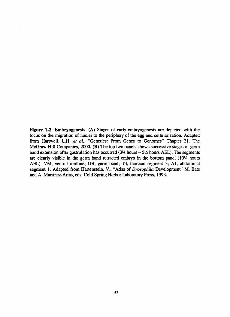
Figure 1-2. Embryogenesis. (A) Stages of early embryogenesis are depicted with the focus on the migration of nuclei to the periphery of the egg and cellularization. Adapted from Hartwell, L.H. et al., "Genetics: From Genes to Genomes" Chapter IL. The McGraw Hiii Cornpimies, 2000. (B) The top two panels shows successive stages of germ band extension after giistnilation has occurred (3% hours - 5% houn AU). The segments are clearly visible in the germ band retracted ernbryo in the bottom panel (10% houa AEL). VM. ventrai midline; GB, germ band; T3, thoracic segment 3; AL, abdominai segment 1. Adapted fmm Hartenstein, V., "Atlas of Drosophila Development" M. Bate and A. Martinez-Arias, eds. Cold Spring Harbor Laboratory Press, 1993.

Diplad zygohc nucleus
Mimtic cyck 7 th 10min
Most nucIei migracc out to cmex
Mimtic cyck 1 O
End of mitotic cycle 13 1 2h30min
Membranes in t5 min egg's cortex grow inward
Cellular blastodem

Figure 1-3. A-P and D-V axes specification during oogenesis. The top panel shows a 16 ce11 cyst during early Gurken signaling. The nucleus (circle) and microtubule network (long strands) are visible. During stage 7 of oogenesis the microtubule reorganization signal required to speciQ the A-P axis is received in the oocyte. allowing the locdization of bicoid RNA to occur (left side of figure). The remmged microtubule network causes the nucleus to migrate to a m e n location. specifying the D-V mis (right side of figure). The late Gurgen signal prevents pipe expression in the future dorsal side, thereby lirniting active Spiit.de to ventrai regions. Adapted from van Eeden, F and St. Johnston, D.. 1999.

A-P d~ patterning
Stage 9
stage 7 Micr
Anterior d
Posterior follick cells f o k k cells
/ \ D-V aicis
Locaiized posterior .
O d dete--u
1 Late Gurke Rearranged Stage 10 siendine
bicoid mRNA
pipe mRNA

Figure 1-4. Hiearchy of genes involveci in A-P patterning. The gene groups are shown on the left side (Maternai etc.). An example of a gene belonging to each group (bicoid etc.), with its conesponding expression pattern, is shown on the nght side. A, anterior: P. posterior. Adapted From Wolpert, L., "Principles of Development" Chapter 5. Cumnt BioIogy Ltd., 1998.

Gene Group Gene Example
Segment engmikd palan*,

Figure 1-5. Domains of gap gene expression. The embryo at top shows the location of the termini (T), anterior (A), postenor (P), and pole ceiis (Pc). The regions of expression for each gap gene are shown in soiid iines, with the dotted lines representing later expression patterns for giant (gr) and hunchback (hb). For simplicity the antenor domains of huckebein (hkb) and tailless (tlE) have not been shown. The domain of active ceaudai protein is ais0 shown (CAD). Md, mandible; Mx, rnaxilla; La, labium: Proct. proctodeum; bcd, bicoid; Kr, Krüppel: hi, knirps. Adapted from Pankratz. M.J. and Jackie, H., 1990.

hb Kr kni
hkb
tll
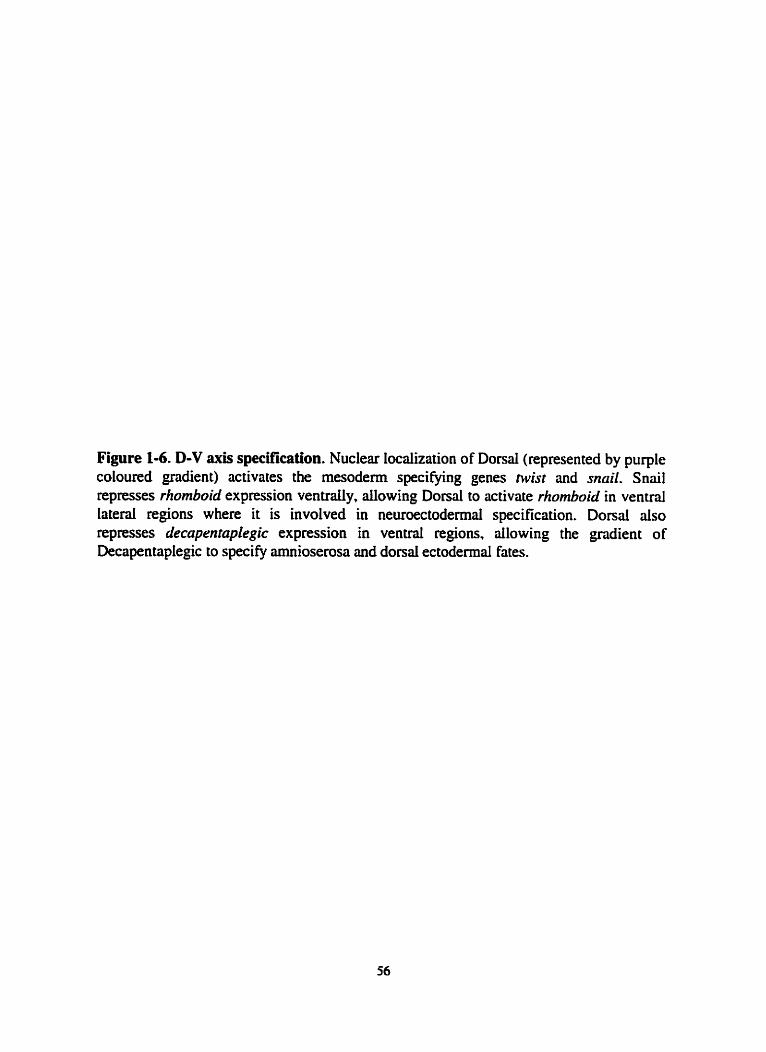
Figure 1-6. D-V axis specification. Nuclear localization of Dorsal (represented by purple coloured gradient) activates the mesoderm specifjhg genes twist and snczil. Snail represses rhornboid expression ventmlly, dowing Donai to activate rhornboid in ventral Iateral regions where it is involved in neuroectodennal specification. Dorsal also represses decapentaplegic expression in ventrai regions, ailowing the gradient of Decapentaplegic to speciQ amnioserosa and dond ectodermai fates.


Figure 1-7. Parasegments versas segments. Segmentai divisions, as defined in the adult, consist of the antenor region of one parasegment and the posterior region of the next in the ernbryo. The posterior (p) part of each segment is defined by the engraiIed expression domain in the early embryo; anterior. (a); head, (H); clypeolabrum (0; thorax, (T); abdomen, (H). Adapted frorn Lawrence, P., 'The Making of a Ry" Chapter 1. BIackweil Scientific hblications, 1992.

/
segments
\. parasegments .I '*\
\ \

Figure 1-8. fushi turuzu expression. Seven stripes of expression are seen during the blastodem stage of embryogenesis. Anterior is to the left and donai is up. [mage courtesy of A. Nasiadka.


Figure 13. Expression of even-skipped stripe 2. Expression of stipe 2 occun between the repressors Giant and Krüppel and the activaton Bicoid and Hunchback. Adapted from Wolpen, Le, "Principles of Development" Chapter 5. Cunent Biology Ltd.. 1998.


Figure 1-10. Expression pattern of selected pair-nile and segment polarity genes. fwhi tarazu Va) and even-skipped (eve) are expressed in the Even- and Odd-numbered pansegments (PS). respectively. At this the, each parasegment is four cells wide (represented by boxes in bottom of figure) The horizontal gradation of colour represents the narrowing of stripes during ernbryogensis. The verticai gradation of colour for the sloppy-pairecl Ir112 (sip IdS ) and odd-skipped (odd) genes represent the Iater appearance of secondary stnpes for these genes. prd; paired, opa; odd-paired, wg; wingless. en;engrailed. Adapted from Copland, I.W.R. 'The role of protein-protein interactions and phosphorylation in the function of the homeodomain protein Fushi tarazu" Ph.D. thesis, 1997.


Figure 1-11. Specification in the larvai epidermis. (A) Top panel shows the cuticle and a corresponding schematic diagnm of a fmt instar Iarvae. The fint and last thoracic (T) and abdominal (A) segments are marked. Each segment is composed of anterior rows of hairs which are unique within in the segment and between segments. The posterior of each segment is devoid of cuticle. hage courtesy of A. Nasiadke (B-E) Role of the Wingless and Hedgehog signaiing pathways in ceH specification within the parasegment. (B) Diagram of genetic interactions. (C) The pansegmental border falls between the Wingless (wg) expressing ceii (blue) and the EngniledMedgehog (en/hh) expressing cell (red). Early negative signoling by Wingless and Hedgehog regulate the spatial domains of Serrate (ser) expression (green). (D) Wingless, Hedgehog and Senate signding regulate the position of the Veiniet (ve) expression domain. (E). Apparent correlations between gene expression and specific cuticular structures. Further expenments are needed to c o n h the correlations in parenthesis. Adapted fmm Alexandre et al., 1999.


CELUTER 2
Identification of Tantalus as a Fushi tarani-Interacting Protein
I performed ail experirnents in ihis chapter. The bait constnicts used in the yeast two- hybrid screen were prepared by Benoît St. Pierre and the yeast two-hybrid reagents were a gift from Roger Brent.

A bstract
The pair-mie gene fushi tarazu ÿiz) is an essential gene in Drosophila
melanogater requind to regdate many target genes. The fiz protein contains a
homeodomain (HD) DNA binding domain. aüowing FTZ to bind DNA sequence-
specifically. However. the study of niany HD-containing proteins has revealed that, on
their own, they are poor discriminaton of DNA target sequences. I have used a yeast-two
hybrid screen to identiw genes encoding potentid cofactors for FTZ function, and
describe here the identification of one such gene that we have subsequently called
tanmius (tan). TAN interacts with the HD of FTZ in both a yeast two-hybrid screen and a
Far Western analysis. Additiondiy, TAN binds DNA in vitro. and ectopic expression
snidies in vivo reved that TAN is a nuclear protein.
Introduction
As described in Chapter 1, the fushi tara= @) gene of Drosophila rnekrnogmter
is a member of the pair-mie class of segmentation genes (Nüssiein-Volhard and
Wieschaus. 1980) and is expressed in an altemating seven-stripe pattern during early
embryogenesis (Hafen et al., 1984). One of the major roles offn in the epidermis is to
position the domains of expression of the segment polarity genes engrailed (en) and
whgless (wg) (reviewed in ingham and Martioez-Arias, 1992). These segment polarity
gens act to define the boundaries of the pmsegments, which act as fundamental
patteming units in the antenor-posterior axis of the embryo. Patternhg of these
parasegments occurs through the parasegment-specinc expression of genes found in the
homeotic complexes (HOM-C). The expression of HOM-C genes defmes the second

major role of&, as FTZ increases the level of expression of several HOM-C genes while
also defining their domains of expression (Ingham et ai.. 1988; kh-Homwia et al., 1989;
Müller and Bienz, 1992).
FïZ contains a homeodornain DNA binding motif (Kuroiwa et al.. 1984; Lûughon
and Scott. 1984; McGinnis et al., 1984a; McGinnis et al., 1984b; Weiner et al., 1984) and
this domain is requïred for FIZ to activate transcription in vitro (Fitzpatrick and Ingles,
1989; Han et ai.. 1989; iaynes and O'Farrell. 1988; Ohkuma et al., 1990; Winsiow et al.,
1989). ET2 directiy regulates its own expression (Schier and Gehring, 1992; Schier and
Gehring, 1993). as well as that of en (Despian et al., 1988; DNardo and O'Farrell. 1987:
Howard and Iogham, 1986; Nasiadka md b u s e , 1999). wg (Copeland et ai., 1996;
uigham et al.. L988; Ish-Horowicz et ai., 1989; Nasiadka et ai., 2000; Nasiadka and
b u s e , 1999) and the HOM-C genes (Ingham et al., 1988; ïsh-Horowicz et ai.. 1989:
Müller and Bienz, 1992). It was expected that these activities would require direct
binding of FTZ, through its HD? to the regulatory regions of these genes. However.
results from our lab and others have demonstrated that the HD of FïZ is not essential for
it to cary out many of its functions in the epidemiis (Copeland et al., 1996; Fitzpatrick et
ai.. 1992; Hyduk and PercivalSmith, 1996). On the other hand, the HD is required for
FïZ to rescue nul1 fn embryos in vivo. in the context of a genomic rescue consmct
(Furukubo-Tokunaga et ai., 1992). Together, these resuits have lead to the suggestion that
the HD is only required at low levels of FEZ, when an autoregdatory Loop is requkd to
increase fU expression Ievels. Later, however, when the Ievels of FI2 are increased, the
HD appears to be dispensable for Fi2 hinction.

These findings strongly suggested that FTZ wouid require additiond cofactors to
regulate its target genes, and research in our Iab and othea has identified two such
cofacton (Copeland et al., 1996; Guichet et al., 1997; Yu et ai., L997). Testing in our lab
for protein-protein interactions between and other pair-mie proteins ~veaied a
strong in vitro interaction with the paired protein. and this interaction appears to be
required for the proper regufation of wg expression in vivo (Copeland et al., 1996).
Additiondly, Fi2 interacts with the orphan nuciear receptor encoded by a-FEFI. a-Ftz-
FI is a requisite cofactor for FTZ regdation of both en and wg, but notftz itself (Guichet
et al., 1997; Yu et al., 1997). FIZ and a-FTZ-F1 bind to lower affinity FE sites
coopentively (Yu et al., 1997) and both FïZ and a-FIZ-FI sites are required for the
expression of an en reporter constnict in vivo (Rorence et al.. 1997). These results
support the idea that ET2 requires cofacton to function. To extend these ~sul ts . I have
used the yeast two-hybrid scnen in a less biased attempt to identify novel protein partners
for FTZ

Resuits and Discussion
Screening for Furhi tarazu interacting protek
Original attempts at screening a O-L2hr cDNA Libnry from Drosophila expressed
in E. coli with radioactively labeled FTZ were unsuccessful in identifjring protein-protein
partners for FïZ (data not show) and the yeast two-hybrid system was employed as an
alternative approach (Figure 2- 1). Because M-lengthftz protein activates transcription in
yeast celis (Fitzpatrick et al.. 1992) and would therefore interfere with the screening
approach (see Figure 24) , severai ''Bait" constructs were created which contained
different portions of the F E protein mgure 2-2). These B a h were selected based on
their hypothesized relative importance for FïZ function. in particular, Bait '2 consisted of
a region of strong homology to the FTZ homologue from Drosophila hydei (Figure 2-2).
Bait *3, on the other hmd, contained 17 amino acids upstream of the highly conserved
FTZ HD and continued untii amino acid 49 of the 60 amino acid HD. The entire HD was
not used because it was feared that the DNA binding activity of the construct might
interfere with the two-hybrid rissay. Of the four bai& tested oniy Baits "2 and "3 were
suitable for the screen. as they did not activate transcription on theû own when introduced
into yeast.
The results of the screen are summ;il.ized in Table 2-1. Of the 9 clones identified
by BaÎt *2 and sequenced, clones for rRNAs (2 clones). cytochrome c (1 clone) and
several novel sequences (6 clones) were identified. Unfortunateiy. most of the novel
clones had oniy short codllig regions in fiame with the acidic activation domain, making
it ciifficuit to assess their significance. Tn some cases, longer coding regions were found

out-of-frame with the activation domain. making these clones unlikely candidates for FTZ
interactors. One explanation for the isolation of these clones was the appearance of a
weak coiled-coi1 protein interaction motif (Hodges, 1992) in both the Bait 9 constmct
and several of these out-of-frame clones. Sequencing of the 3 positive clones identified
using Bait '3 reveaied 2 rRNAs and one novel clone (see below) whose coding region
was in frame with the acidic activation domain.
The isolation of severai rRNAs using both bi ts is a common finding during two-
hybnd screens, most likely due to the abundance of such species in the total amount of
RNA in tissue samples. Unfortunately, clones for the strong interactors Paired
(Pm) (Copeland et al., 1996) and a-FFZ-FI (Guichet et al., 1997; Yu et al., 1997) were
not identified in the screen. This result at f i t seems surprising since ment results from
in vitro studies have demonstrated the importance of the N-terminai regions of FïZ.
specificaily amino acids 100450, for cofactor binding to both PRD (Copeland et ai..
1996) and a-=-FI (Guichet et d., 1997, Schwartz et al., 2000), and these residues were
included in the Bait *2 construct. However, there are several possible explmations as to
why the PRD and a-FTZ-F 1 interactors were not identified in the screen.
First, aithough amino acids 100-150 of FïZ ye required for the interactions with
PRD and a-=-FI, other regions of F E contribute to these interactions (Copeland,
L997; Schwartz et ai., 2000). The absence of these regions in the Bait *2 construct may
lower the FIZ/PRD and FTZ/a-FTZ-FI interactions to Ievels below detection by the two-
hybrîd iissay. As an extension of this idea, it is also possible that the isolated 100-150

residues of F E are not folded properly in the bait constr~~ct to participate in a protein-
protein interaction. Second, yeast ceiîs expressing the PRD and a-FIZ-F1 fusion proteins
rnay not be viable or may be under-represented in the library, making their detection
dificult. FwiallyT erron in the experimentd method must also be considered.
Tantalus intetacts with the HD of Fushi taruzu
Clone 33-13, heredter referred to as tantalcs (tan), was identified using Bait '3.
To venQ the yeast two-hybnd results and to confm that the interaction between TAN
and F E is direct, the Lkb tan cDNA recovered in the screen was subcloned into a T7-
expression vector and used in a Far Western assay with different FTZ deletion constructs.
Bait *3 wûs comprised of 17 amino acids upstream of the FTZ HD and continued until
amino acid 49 of the 60 amino acid HD, suggesting that TAN was making contacts with
at least part of the HD. To test this, several consinicts containing different portions of the
fn polypeptide were created for use in a Far Westem assay. F i t . full-length FïZ, N- and
C-terminal portions of FTZ (the HD is found in the C-terminal construct), GST and GST
fused to the HD of ET2 (4 amino acids upstream and 10 amino acids downstream of the
HD) were expressed in bacteria and mn on SDS-PAGE (Figure 2-3A). To veri@ the
position and identity of each construct on the blot, a Western using a polyclonal antibody
against the FIZ peptide was performed (Figure 2-3B). Protein expressed from the tan
cDNA was then radioactiveiy Iabeled with 3 5 ~ -methionine using a rabbit reticulocyte
Iysate and used to probe a second blot containing the same FTZ constructs as those
detected on the Westem. TAN bound to the N1-length, C-terminal regîon, and isolated
HD of FTZ but did not bind to the N-terminal portion of FTZ or to GST done (Figure 1-

3C). These results c o n f i the yeast two-hybrid interaction and suggest that the regioo of
FTZ suficient for an interaction with TAN includes the 4 amino acids preceding the HD
and the f i t 49 amino acids of the of the HD.
This region of FTZ incorpontes helices 1 and 2 of the HD motif, and studies of
seved HOM-C proteins (Gibson et d., 1990; Kuziora and McGinnis, 1989: Mann and
Hogness, L990), as weli as other HD containing proteins (Chan and Mann, 1996; Mak
and Johnson, 1993; Passner et al., 1999), have suessed the importance of residues within
the N-terminus of these various KDs for target specificity. In particular. these residues are
important for making protein-protein contacts with cofactors, such as Extradenticle
(EXD) and OCA-B (Chan and Mann, 1996; Lai et al., 1992; Mak and Johnson, 1993:
Passner et al., 1999; Pomerantz et al., 1992). For example, the HOM-C proteins in
Drosophila have increased DNA binding specificity when complexed with the HD-
containhg EXD protein (reviewed in M m and Chan, 1996). while the mammalian
O n - 1 HPcontiiining protein has an altered DNA binding specificity. and becomes a
more potent transcriptional activator, when complexed with OCA-B or VP 16 (reviewed
in Wegner et al., 1993). However, what role TAN may play in FE function wili requise
further experimentation.
The idenfication of tan using the HD of ET2 may at fmt seem paradoxicai,
considering that this screen was Uùitiated based on the finding that many activities of FïZ
are HD-independent. However, it is important to remember thai the analyses of FïZ
function were, for the most part, based on results in the epidermis (Ffzpatrick et al.,
1992; Copeland et al.. 1996; Hyduk and Percivai-Smith, 1996).fn is also expressed in

other tissues, iike the CNS, and the role of the HD in these tissues has not been
addressed. Additionally, the HD is cequired for the auto-activating abiiity of FE (Schier
and Gehnng, 1992) and it is possible that cofactors are required for this activation.
Sequence and genornic location of tantalus
Sequencing of the Ikb tan cDNA recovered From the yeast two-hybrid screen
revealed an open reading frame of 269 amui0 acids. Northem analysis (see below)
suggested that the tan transcnpt was approximately 1.6kb in length. so a 0-13hr cDNA
libnry was screened to identify overlapping cDNA clones. One cDNA of approxirnately
lkb was identified which extended the tan sequence in the 5' direction. Combining the
new cDNA sequence with the old produced a total sequence of approximately 1.6kb
(Figure 2-4A&, in close agreement with the size predicted by Northem analysis.
Approximately 7kb of genomic DNA flanking the cDNA sequence was obtained
by screening a pnomic library (Figure 2-42} and 4kb surrounding the tan locus was
sequenced Figure 2-SA shows the sequence of the 1.6kb tan cDNA and the tan genomic
regions. A canonical TATA box sequence was not found in upsveam of the recovered
cDNA sequence. Cornparison of the cDNA and genomic sequences reveals two introns,
one located upstream of the putative ATG translationai sstart site and the second within
the coding region. ï h i s ATG conforms well to Drosophila consensus translation stm
sites (Cavener, 1987) and the predicted open reading frame would encode a protein with a
m a s of 33kDa
TAN lacks homology to any proteins currentiy identifi~ed in GenB;uik; this is not
unusuai since only 50% of Drosophila proteins display sequence similarity to mammaiian

and worm proteins. with even fewer showing homoiogy to yeast proteins (28%) (Rubin et
ai., 2 0 ) . TAN does, however, contain severai regions of interest (Frgure 2-SB). Two
Large basic regions consisting of 32% (21/66) and 41% (19146) basic amino acids are
located at residues 90- L54 and 188-235. Despite these large patches of basic residues, the
overail pI predicted for TAN is 6.4. The protein has two canonicai nuclear Iocalization
signais at residues 90-106 (RRSSTFGARAGVARRRM) and 217-210 (KRRR). The
protein aiso contains two PEST regions (Praline, Glutamate. Senne and Threonine).
which are believed to act as protein degradation signais. These motifs are located at
positions 74-90 and 154- 188. and have "PESTfind" scores of 9.5 and 1 1.7 respectively (a
PESTfind score of +5 or pa t e r is considered significant; Rechsteiner and Rogers, 1996).
Numerous canonicai sites for protein kinase C. CAMP-dependent kinase and casein
kinase II phosphorylation are aiso present (data not shown).
To determine if the tan locus had been identified genetically. in situ hybndizations
to polytene chromosomes were performed (Figure 2-6) and a Pl genomic library was
screened (data not shown). Both methods localized tan to position 65A on the polytene
map, consistent with the ment sequencing of the Drosuphila genome (Adams. 2000).
However. tan does not appear to CO-map with any candidate FTZ-interacting genes.
The tant alus expression panent
Before pursuing additional biochemicai studies of the TAN/FTZ interaction. it
was f i t hponmt to determine whether these two proteins had the potential to interact
in vivo, ie., do their temporal and spatial domains of expression ovedap? As fiz is
expressed during the 2-4,6-8 and IO- 1 Zhr time periods d u ~ g development (Kuroiwa et

al., 1984; Weiner et ai., 1984) tan shouid aiso be expressed at this time if the two proteins
are to interact in vivo. A developrnental Nocthem blot shows high levels of tan mRNA in
early ernbryos, decreased expression in 12-24 hr embryos and high levels of expression in
third instar larvae, pupae, and femdes (figure 2-7).
This early temporal expression pattern for tan overlaps that of FTZ. To determine
whether the spatial expression domains of the two genes also overlap, in situ
hybridizations were perfomed (Figure 2-8). Transcripts were first looked br in ovaries
since the Nonhern blot results indicated the presence of tan expression in early embryos.
before zygotic transcription takes place. uideed, tan mRNA is first detected in stage 10
ovaries in the nurse cells surrounding the oocyte (Figure 2-8A). These cells will
eventudly deposit their mRNA and protein contents into the oocyte. tan is dso expressed
ubiquitously in pre-ceLiularized and cellularizing embryos (Figure 2-8C,D). Signal is not
detected in control embryos hybndized with a sense probe (Figure 2-8B). Transcript
levels begin to decrease after cellular blastodenn but increase again during germ band
extension. At this time, lociilized expression is seen in what appears to be the somatic and
viscenl mesodemal Iayers (Figure 2-8E). tan is also expressed ubiquitously in imaginai
discs of third instar larvae, with higher levels of expression seen in the morphogenetic
furrow of the eye-antennai disc (figure 2-8F). These results demonstrate that the early
expression pattern of tan paniaiiy overlaps that of&. However, the mesodemal and disc
expression of tan suggest ihat at least some TAN functions must be independent of FIZ,
since FTZ is not expressed in these tissues.
Tantah is a nuclear DNA binding protein
As ET2 acts in the nucleus, the subcellular Iocalization of TAN was deterrnined
by using an epitope tagged version of the protein. A DNA sequence encoding the p53
epitope tag (Dalby and Glover, 1993) was inserted in the tan cDNA at amino acid 19, and
the cDNA was cloned into a T7 inducible vector to ver@ that the tag was functional
(Figure 2-9A). The p53-tan cDNA was then cloned into the pCaSpeR-hs vector
(Thummel and Pirrotta, 1992), which uses the hsp70 promoter to permit trmsgene
expression in vivo upon heat shock (HS). Transgenic lines were produced using P-
elecnent mediated gem line transformation (Spndling, 1986). Third instar transgenic or
wild-type (WT) larvae were exposed to HS and the location of p53-TAN was malyzed in
dissected sdivary glands using a monoclonal anti-p53 antibody (Figure 2-9B). No signal
was detected in the salivary glands of WT controls (Figure 2-9C) but a strong nuclear
signal was seen in both the sdivary glands and attached fat bodies of the transgenic Line
(Figure 2-98). At higher mapifications TAN appeared to be associated with the
c hromatin (Figure M D ) .
Careful anaiysis of the TAN sequence nvealed a smdl stretch of residues with
homology to the third recognition helk of the homeodomain (HD) DNA binding motif
(Figure 2-50. This homology may be relevant as the zeste protein dso contains a
somewhat diverged HD sequence required for DNA bhding in vitro (Chen et al., 1992).
This raises the possibiiity that the association of TAN with specific sites on polytene
chromosomes may depend in part on its own ability to bind DNA. To test this possibility,
35~-1abe~ed TAN was produced using a reticulocyte lysate and incubated with salmon

sperm DNA that had previously been attached to nitmceliulose fiters (Figure 2-10). The
Fushi t a u (FTZ) protein, with and without its DNA binding HD (FïZAHD), and
Luciferase (LUC), which does not bind DNA, were used as controls. Of the four
polypeptides. TAN yielded the strongest signal. FuU-Iength FE also bound to the bloned
DNA, and as expecied, FTZAHD and LUC both failed to bind.
The lack of homology between tan and any other genes in the databases makes it
difficult to predict how TAN may affect FIZ function. However, the finding that TAN
binds DNA and is found in the nucleus is consistent with a role for TAN in
assistinglstabilizing the DNA binding of FTZ. It will be important to determine whether
TAN binds DNA sequence specificdly, and how a T A N m interaction rnight affect this
binding.
Summary
The tan protein was identified in a yeast-two hybnd screen and Far Western
andysis as a potential partner for the pair-nile protein FTZ. A 53 amino acid region of
FTZ which includes helices 1 and 2 of the HD is suficient for the interaction with TAN.
however, additionai studies will be ~quired to determine how specific the interaction is
for the FTZ HD. TAN expression partiaiiy overiaps the temporal and spatial window of
FE, consistent with a potential in vivo interaction between the two proteins. Although
TAN does not show strong homology to any sequences in GenBank, our analysis supports
a nuclear role for TAN since P binds DNA saongly and a tagged version of the protein is
Iocalized to the nucleus. Mthough the evidence presented here is consistent with an in
vivo interaction between FIZ and TAN, proof that such an interaction is relevant in vivo


Materials and methods
The yeast two-hybrid screen
The two-hybrid screen has been described in detail (Golemis, E.A*, et al., 1997).
The RnYl iibnry was used in the screen and represents cDNAs from 0-12hr of
embryogenesis (Golemis, E.A., et ai., 1997). The FIZ B i t constructs were made by PCR
amplification using the following pnmers;
Bait ' 1 STCGGAATTCATGGCCACCACAAACAGC'3 and
STCGGGAK~CAAAG~CTGCTCCTGATTGTTGTA'3;
Bait '2 STCGGAATTCCCGCCGCCCAAGGCCACC'3 and
S'CGGGAKflCAAAGCTTGGGGAGCCTTWCACmG'3;
Bait '3 S'CGGAAmCGGCGAmCAATTGGTCG'3 and
S'CGGGATCCTCAAAGCTTGAACCAGATCTTGATCTG'3;
Boit '4 S'CGGAAnCACGCTGGACAGmCCCCGT3 and
Far Western ussay
The Far Western anaiysis was pecfonned as previously described (Guichet et al..
1997) using "s-labeled TAN expressed using a reticulocyte Iysate (Promega TNT).The
described FIZ constructs expressed on the geVblot (Figure 2-3) were cloned into pET L9B
(Novagen). The HD of FTZ (4 amui0 acids upstreûm and 10 amino acids downstream of
the KD) was cloned as a GST fusion into pGEX2T ( g i f t of C. Desplan). Proteins were
expressed in E. coli and nm on SDS-PAGE using standard techniques (Sambrook. et al.,
1989). The polycolond anti-FI"2 antibody has ken described (Krause and Gehring,

1988).
Southern blots
DNA preparation was modified h m Ballinger et al., (1989). Batches of twenty
fies were collected in 1.5~11 eppendod tubes (flies were stored at -20°C until required).
To each tube. 200p.i of TENS buffer was added (TENS is l O O m M Tris-HCl pH 7.6,
1OOrnM EDTA, lOOmM NaCl and 0.5% SDS). The fies were homogenized with a pestle
and incubated at 65OC for 30min to denature protein. 30p.l of 8M Potassium Acetate was
then added (for a finai concentration of 1.2M) to precipitate protein. The tube was spun at
14,000xg in a table top centrifuge after which the supematant was removed and the spin
repeated. An equal volume of 100% EtOH was added to the supematant and the tube was
mixed at room temperature and ailowed to iacubate for Smin to precipitate the DNA. The
tube was again spun at L4,ûûûxg after which the pellet wûs washed in 70% EtOH and
dlowed to dry. The high molecular weight genomic DNA was resuspended in Iûûpl TE
overnight at 4°C. ïhe DNA was ethanol precipitated 2X and resuspended in 40pl TE at
37°C. The DNA was digested with the qpropnate enzymes, blotted onio positively
charged nylon membranes (Boehnnger Mannheim) and W cross-linked. Blotting was
performed as described (Sambrook et ai.. 1989) with the foiiowing modifications. DIG
labeled DNA probes were made by PCR amplification. Labeiing efficiency of the probe
was verified by cornparison to a control DIG labeled DNA sample. Probes were used at a
concentration of 50ng,/ml in 5X SSC, L% (wfv) blocking reagent (Boehringer Mannheim).
0.1% (wlv) N-laurylsarcosine, and 0.02% (wlv) SDS. Incubations were at 65°C overnight

and, d e s s noted washes were 2X Smin at room temperature with 2X SSC, 0.1% (w/v)
SDS followed by 2X LSmin at 6S°C with 0.1X SSC. 0.1% (w/v) SDS. Signal was
detected using CDP-Star according to the manufacturer's instructions (Boehringer
Mannheim).
Northern blots
Northern blots were performed as described for agamse/formaldehyde gels
(Sambrook et ai., 1989). RNA was isolated by direct phenol extraction (Andres and
Thummel. 1994) and 1Opg of total RNA loaded ont0 the gel. After blotting and W
cross-linking the blot was stained with Methylene Blue (Ausubel et ai.. 1997). Probe
preparation and signal detection were perfomed as per the Southem blotting procedure.
Cloning tantaius
Genomic (Tarnkun et al., 1992) and cDNA (Poole et al., 1985) tibmies were
screened using standard procedures (Sambrook et al., 1989). To obtain a full-Iength
cDNA tan construct, two overlapping cDNAs were fused together using a common Bst
EII site Iocated at +582 (site positions refer to the genomic map in Figure 2-5) and
subcloned into pBIuescnpt II (Stratagene). The cDNA was subcloned into the PET L9b
vector (Novagen) for TI expression. The p53 tag was inserted at the Bst EiI site (before
the fint PEST domain) of the tm cDNA. The inserted sequence is KRSRAFRHSW-R
(new sequence between dashes).
In situ hybridization
Hybridization of embryos and imaginai discs with RNA probes was performed as
d e m i i d (Hughes and Krause, L999) except that secondary antibodies were HRP-
conjugated and the ABC system of Pierce was used for detection. [n situ hybridization to
pdytene chromosomes was carried out as follows. SIilivary glands were dissected from
larvae grown at 18°C. Glands were dissected in 1X PBS and placed in 45% acetic acid
and dlowed to fix for 3-Smin. Glands were squashed and then placed in liquid nitrogen
for 30sec followed by 3X lOmin washes in 95% EtOH. Glands were often stored in 95%
EtOH before use. SLides were rehydrated by successive washes of ?min each in 95%
EtOH, 70% EtOH, 50% EtOH, 30% EtOH, 0.1X SSC and 3 2 SSC. Slides wece then
incubated in 2X SSC at 70°C - 80°C for 30min followed by denaturation in O. LN NaOH
for 90sec at room temperature. Slides were washed for 30sec in 2X SSC before another
dehydntioo step of lmin each in 30% EtOH. 50% EtOH, 70% EtOH and 95% EtOH.
Slides were air dried for Smin and incubated with the appropriate probe (made as per the
Southem blotting procedure) in a moist chamber overnight at 37°C. Slides were washed
for 2X LSmin in 2X SSC followed by a 2m.n wash in LX PBS and a 5rnin wash in PBT.
Secondary antibody was added and slides were incubated for lhr at 37°C. Slides were
then washed in 1X PBS briefly followed by 2X LSmin washes in 2X SSC and developed
using standard NBTlBCIP procedures (Sigma). Chromosomes were then stained using
Giernsa (Andrew and Scott, f 994).
DNA bindhg of Tantalus
5pg diquots of sonicated s h o n sperm DNA were vacuum blotted to
Ritroceiiulose (nitrocehiose gave l e s background then other membranes) in a 30pl
mutnire of DNA and 2X SSC foliowed by washes in 500pl 2X SSC. The blot was

blocked with LX Binding Buffer2 ( tX BB2: 2% milic powder, 20mM Tris pH 7.6,
lOOmM NaCI, 0.25rn.M EDTA, 0.25mM DTT, 0.1% Tween-20 and 10% glycerol) for
2hrs on ice. Radioactively Iabeled proteins were synthesized according to the protocol
provided by the manufacturer (Promega TNT '17 kit) and incubated with the biots in IX
BB2 for Emin on ice. Blots were washed with LX BB2 with seved changes over 15min,
then dried and exposed to film.
Prepurution of lama1 tissues for mtibody staining
tan and p53-tan cDNAs were cloned into the pCaSpeR-hs (Thummel and Pirrotta,
1992) or pUAST (Brand and Pemmon, 1993) vecton For in vivo expression. Depending
on constructs used, transgenic or wild-type third instar Iarvae were heat shocked for Zhrs
and dlowed to recover For 30min. W a e were dissected in cold IX PBS plus 0.3% (v/v)
Triton X-100 and then futed in 4% paraformddehyde in LX PBS for 20min. Tissues were
dehydrated using severai changes of methanol followed by rehydntion in PBST ( IX PBS
+ 0.3% Triton X-100) and blocking in PBST + 0.5% BSA for 25hrs. Tissues were
incubated ovemight at 4°C in a 1/50 dilution of a monoclonal anti-p53 antibody (Santa
Cruz Biotechnology, Inc.) in 1 X PBS + 0.5% BSA. Tissues were washed quickly several
iimes followed by 3X 30min washes in PBST + 0.5% BSA. The secondary antibody
(HM) was added at a dilution of 1 / 3 0 for 45min after which tirne samples were washed
in PBT for Lhr. The staining was developed using the ABC system of Pierce. M e r
stainùig, tissues were washed Ïn PBS and a gIycerol solution (50:50 with IX PBS) before
dissection and mounting.

Table 2-1, Results of the veast two-hvbrid smea Bait '2 Bait '3
Colonies screened i ~ l d 1x10~ Colonies seiec ted 300-400 800-900 Gd dependent colonies 35 100 classified into 4 categories* Positive clones after retransformation 9 3 *ody 8 were retransformed
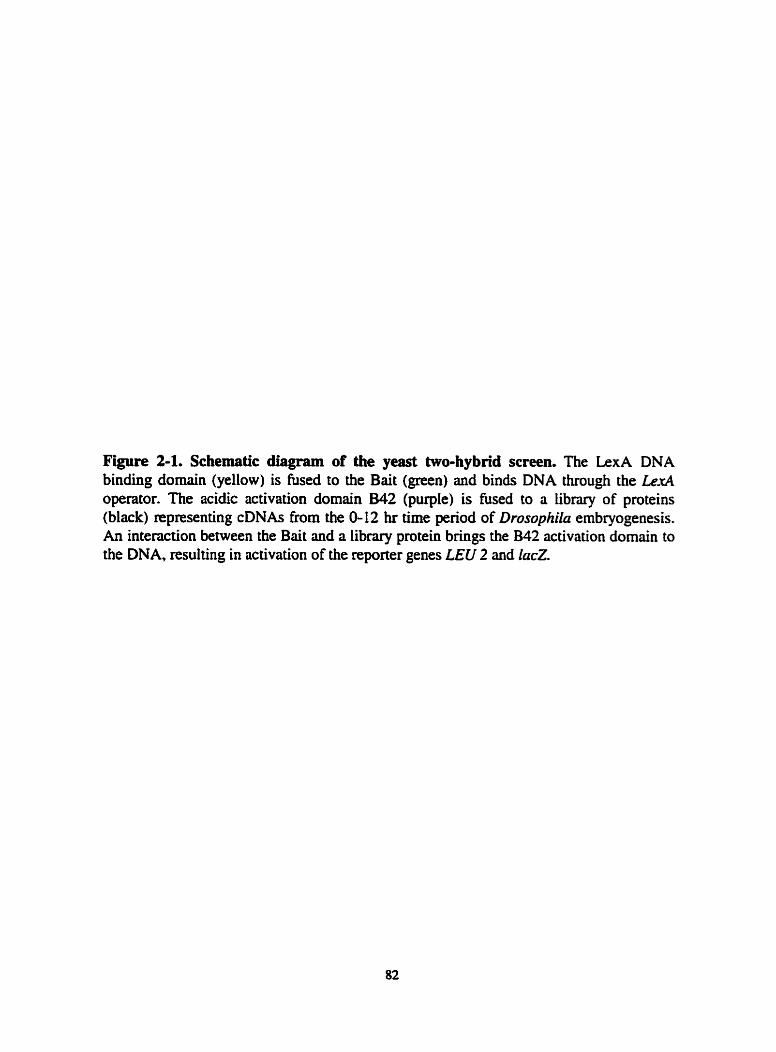
Figure 2-1. Schernatic diagram of the yeast two-hybcid screen. The LexA DNA binding domain (yellow) is fused to the Bait (green) and binds DNA through the LexA operator. The acidic activation domain B42 (purple) is fused to a iibrary of proteins (black) representing cDNAs from the 0- 12 hr t h e period of Drosophila embryogenesis. An interaction between the Bait and a library protein brings the B42 activation domain to the DNA, resulting in activation of the reporter genes LEU 2 and l a d .

Transcription
r--+
Levl Operators ) Reporten lac Z

Figure 2-2. Bait constnicts used in p s t two-hybrid screen. Cornparison of the JI= gene h m Drosophila melanogaster (upper sequence) and Drosophiln hydei (Iower sequence). The highly conserved N-terminal region (yeilow) and homeodomain sequences (green) are indicated. Identities are indicated by lines and similarities are indicated by dots. Regions compnsing each of the Bait constnicts are shown.
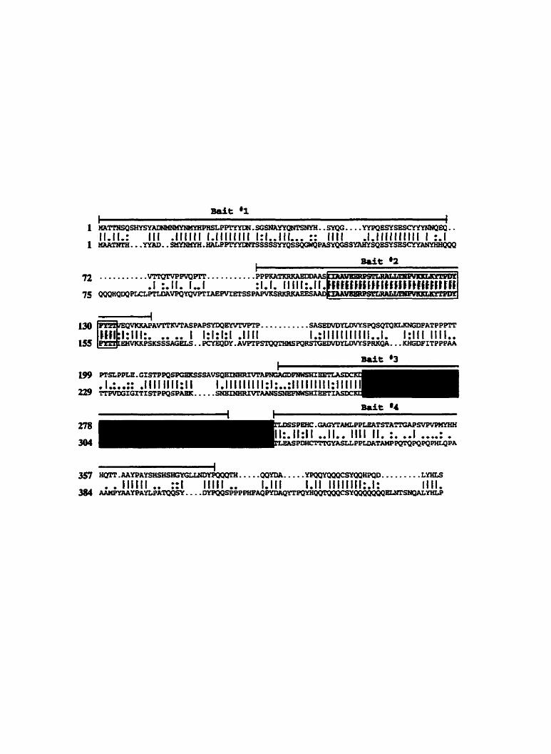
mit @2

Figure 2-3. Far Western assay. (A) FiZ constructs, as descnbed adjacent to the figure were expressed in E. d i and separated by SDS-PAGE and stained with Coomasie blue. (B) Expression of each constmct is verified by a Westem blot using a polyclond FTZ antibody. Fewer epitopes for the antibody are found in the HD, resulting in a weaker signal in the GST-FIZHD lane. (C) Labeled TAN binds to full-Iength FE, a C-terminal portion of FE containhg the HD and the isolated HD of F'TZ fused to GST in a Far Westem assay. interacting FTZ constructs are denoted by an asterisk.

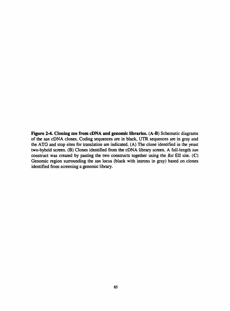
Figure 24. Cloning tan from cDNA and genomic libraries. (A-B) Schematic d i a m of the tan cDNA clones. Coding sequences are in black, UTR sequences are in gray and the ATG and stop sites for translation are indicated. (A) The clone ideotified in the yeast two-hybrid screen. (B) Clones identified from the cDNA library screen. A fuiMength tan construct was created by pasting the two cons~cts together using the Bst EII site. (C) Genomic region surrounding the fan locus (black with intmns in gray) based on clones identified from screening a genomic library.

424 Bst ESI t 505
ATG
1 Bsr Eu
ATG
1 t 505
1 I Fusion Consma
ATG STOP
Bwn HI Eco R1 Saf 1
ATG

Figure 2-5. Genomic and amino acid sequence of tan. (A) Sequence of the tan locus with introns (two boxed sequences) and putative polyadenylation signai (AATAAA) indicated Arrows Iabeled 1 , 2, and 3 denote the 5' ends of the cDNAs identified by, respectively, my screen and two ESTs from the Berkeley EST project (Rybase). (B) Schematic diagnm of TAN indicating PEST (a PEST score of +5 or p a t e r is considered sigoificant; Rechsteiner and Rogers, 1996), Basic, and Acidic (AC) domains. (C) A i ivent of residues 190-201 of TAN with the third helix of the HD from severai different classes of HD containing proteins (see Laughon, 1991). Identities are dark gray while simiiarities are Light gray.

TAN W C 86 BCD EMS ANTP MATa2 ZESTE

Figure 26. Polytene in situ hybridizatious. (A) in situ hybridization using a tan probe detects one signal on polytene chromosomes (arrowhead). The ends of the five large chromosome arms are indicated by arrows. (B) tan maps to 65A.


Figure 2-7. Northem blot analysis. Developmental Northem analysis of tan (top panel) detects an approximately 1.6kb signal. The lower panel shows the same blot stained with Methylene blue as a loading control. Larval, (L); Pupal, (P); Fernale. (F); and Male. (M).


Figure 2-8. Expression pattern of tan, (A) In situ hybridization to oocytes shows tan expression in Stage 10 oocytes. (B) Hybridization to a cleavage stage embryo with a tan sense probe. (C-F) In situ hybridiations using an anti-sense tan probe; (C) cleavage stage; (D) cellular blastoderm; (E) gem band extension; specific expression is seen in the somatic and viscerd mesoderm (mow) in addition to the ubiquitous transcnpt. For ail embryonic stages antenor is to the left and dorsal up. (F) tan expression in an eye- antennai dix: higher expression is seen in the morphogenetic furrow (arrowhead).


Figure 2-9. An epitope tagged version of TAN localizes to the nucleus. (A) Left panel is in vitro transcribed and "s-labeled TAN run on SDS-PAGE. Right panel is a Western blot of in vitro transcribed and translated p53-TAN (unlabeled) detected by anti-p53 antibodies. (B) Ectopic p53-TAN produced by heat shock (see text) is Iocaiized to the nucleus in salivary glands (arrow) and associated fat bodies (arrowhead) of a third instar larme. (C) No signai is detected in wild-type lanrae exposed to heat shock. (D) Higher masnif~cation of the nuclei from (B) reveals TAN staining associated with the chromosomes (arrow).


Figure 2-10. TAN binds DNA. Top panel shows in vitro transcnbed and ndioactively labeled FTZ (Lane l), ETZAHD (Lane 2). TAN (Lane 3) and LUC (Lane 4) proteins analyzed by SDS-PAGE. Bottom panet shows the ability of each protein to bind to siilmon spem DNA blotted to a filter. TAN and FIL bind strongly in this assay while ETZAHD and LUC do not bind. The faster migrating band in Lane 3 most Iikely results from alternate translational start sites.


Tantalus Interacts P h y s i d y and Geneticaiïy with the Polycomb- and trithorax- Group Member Additional sex combs
In this chapter, Dr. Hugh Brock's Iaboratory performed the original yeast two-hybrid assay identifying Tantaius as an Additional sex combs-interacting protein (MichaeI Kyba) and mapped Tantdus binding sites on polytene cbromosornes (Hu* Brock) (Figure 3-3 and Table 3-1). TAN antibodies were made by myself and the Brock [ab. Shelley Lumba assisted with the chmcterization of the TAN antibodies. Experiments described in Figures 3-2B and 3 4 A B were performed by Iocelyn Moore. Gilbert Dos Santos and Fiona WCIoskey assisted in the experiments descnbed in Figure 3-4C-E. 1 performed the experiments described in the remaining Figures and Tables.

Abstract
The Drosophila trithorax- and Polycomb-groups of proteins maintain activated
and repressed tnnscriptional states at specific target gene loci. The Additional sex combs
( A n ) gene is of particular interest as it acts as a member of both protein complexes and
its effects on target genes are tissue-limited. A novel protein, Tantdus (TAN). was
idenWied in a yeast two-hybrid screen for ASX-interacting proteins that might confer
tissue-specific activities. Aithough TAN contains consensus nuclear locdization sites and
binds DNA in vitro. the protein Iocalizes to both the cytoplasm and nucleus. [n salivary
glands. TAN is nuclear and associates with 66 euchromatic sites on the polytene
chromosomes. more than half of which overlap with ASX. Unlike the majority of trxG
and PcG proteins. however, TAN does not appear to ngulate homeobox complex pnes.
Rather. mn mutant phenotypes are specificaiIy limited to sensory lineage defects and one
of these phenotypes. shared by Asx, is geneticaily enhiuiced by Asx. Taken together, the
data suggest that TAN rnay act as a tissue-specific cofactor for ASX, and that its activity
may be controlled by subcelIuiar tmfficking.
Introduction
PcG and trxG genes are required to maintain states of repressed (PcG) or activated
(trxG) transcription during Drosophila development, and mutations in these genes are
most comrnoniy associated with homeotic aaasformations in the aduit fly (Simon. 1995;
Kenoison, 1995). Mutations in PcG genes, such as Poiycomb or poiyhomeotic, cause
ectopic HOM-C activation, whiie mutations in tnrG genes. such as trithora, result in o
loss of expression of HOM-C genes. In both cases, however. HOM-C gene expression is

initiated normaliy but the maintenance stage of HOM-C expression is compromised. Both
groups appear to act as Iarge protein complexes (reviewed in Pirrotta, L998), and ment
mults support the idea that each group is involved in the maintenance of an "open" or
"closed" configuration of nucIeosomes around target genes (Ka1 et al., 2000; Shao et al.,
1 999).
Asx was classified as a member of the PcG because Asx mutations enhance the
defects seen in other PcG mutants and cause PcG-like transformations (Breen and
Duncan, 1986; Sinclair et ai., 1992). As with other PcG members, the homeotic
transformations seen in Asx mutants are associated with an inability to maintain
repression of HOM-C genes (McKeon and Brock, 1991; Simon et al., 1992; Soto et al..
1995). However, one Asx diele, created by a Pelement insertion within the Asx locus
(ASX"), causes opposite - anteriorly directed - homeotic transformations in the adults of
homozygous aies (Sinclair et al., 1992), suggesting an inability to maintain the active
state of HOM-C gene expression. This result, and the finding that several Asx dalles
including As.<' c m geneticaily enhance the homeotic phenotypes associated with the
trxG member trithora, suggested that krn may be a memember of both the PcG and trxG.
ASX is not the fiat protein suggested to have both PcG and tntG activities: the Enhancer
of zeste (E(z)) gene was origindiy identified as a PcG gene, but has since been shown to
have trxG activity (Laieunesse and Sheam, 1996). More recentiy, dieles of sevenl genes
that had previously been characterized as PcG gens were shown to be genetic enhancers
of both trxG and PcG Ioci (Gildea et ai., 2000). These results prompted the suggestion of
an additionai gene class, enhancers of trithora and Polycomb mutations (ETE').

Unfortunately, there is little biochemical data available for ASX. E(Z) or proteins of the
proposed ETP group, making it difficuit to assess what their respective roles might be in
PcGItrxG function. However. ASX associates with a iimited number of sites on polytene
chromosomes. suggesting that it plays a specific role in gene regdation.
Another interesting attribute of Asx is that its effects are tissue-limited. Asx
mutations cause ectopic derepression of HOM-C genes in the epidennis and viscerai
mesoderm. but not in the centrai nervous system (CNS) where other trxG and PcG genes
play important d e s (Soto et al., 1995). Experiments with reporter constructs have
demonstrated that ASX is active in the CNS however. suggesting that it is functional in
this tissue. One interpretation of these results is that cofacactors are required to facilitate
tissue- and promoter-specific functions of pnrticuIar PcG complex components.
A tissue-specific cofactor(s) for ASX activity would be expected to exhibit tissue-
specific defects when mutated and to interact geneticdly with An mutations. ASX
cofactors should dso interact physicaiiy with ASX, either directly or indkctly. TAN and
ASX interact direcdy in yeast two-hybrid and GST pull-down expenments, and co-
locaiize at a subset of sites on polytene chromosomes €rom third instar larvae. Asx and tan
mutations aiso interact geneticaiiy. Interestingiy, TAN is unlike other trxGPcG complex
components in that it exhibits differentiai subceiiular Iocalization. It is aiso unusual in
that it appears to play no role in HOM-C gene regdation. Rather. TAN appem to be a
novel cofactor of ASX thût specifcaüy faciiitates a roie in sensory organ development.

R e d &
Ideniifjing novel Additional sex cornbs cofactors
The As.. protein contains 1668 amino acids (Sinclair et al., 1998b). The C-
terminal 500 amino acids constitute the main homology region with respect to
mammalian counterparts (E. O'Dor and H.W.B., unpubiished results), and inciudes a
cysteine cluster with 25/28 conserved residues. As moût conserved PcG protein domains
are required for protein-protein interactions (Peterson et al., 1997; Gunster et al., 1997:
Kyba and Brock. 1998; Hashimoto et al.. I998), this region (residues 1 139- 1668) was
used to rnake a yeast two-hybrid bait constmct. A 0- 12 hr embryonic cDNA library was
screened. and out of approximately 10' clones, 11 unique interacting constmcts were
recovered (M.K., LM. and H.W.B., in preparation). One of these clones was found to
encode the tantalus gene. The tan clone did not interact with control consuucts, nor did it
interact with 23 of 24 two-hybrid baits constructed from 1 1 PcG or PcG-related proteins.
To verify the two-hybrid interaction between ASX and TAN, a GST pulldown
assay was used. Residues i 139-1668 of ASX were used to make a GST-ASX fusion
protein. GST and GST-ASX were purified and bound to giutathione beads. The two
resins were then incubated with 3S~-~abeled TAN. TAN was not precipitated by GST, but
was efficiently precipitated by the GST-ASX fusion protein (figure 3-1). These results
suggest that ASX and TAN are Likely to interact directly.
TAN and ASX binding sites overlap on pulyrene chromosomes
Staining of poiytene chromosomes with antibodies against PcG and trxG members
reveds Iùnited but overlapping euchromatic binding for many members, and is a

haiimark of P c G h G proteins (see ChinwaDa et al., 1995; Rasteiii et al., 1993). To
determine whether TAN is capable of binding polytene chromosomes in vivo, polyclonal
antibodies were taised against GST fusions of both full-length and peptide portions of
TAN. As a control, the p53 tagged version of TAN was employed (see page 72). tan and
p53-tan cDNAs were then placed in appropriate vectors for induction by heat shock (HS)
or the GALARlAS system. and transgenic lines were produced.
Figure 3 - shows a Western blot fiom wild-type (WT), HS-tan and HS-p53-tan
third instar Iarvae which have k e n exposed to HS. A signal that nins at approximately
55kDa is detected by the TAN antibody, and this signal is dnmaticdly increased when
extncts were made from HS-tan or HS-p53-tan fly lines (Figure 3-2A). Monoclonal
antibodies specific for the p53 epitope tag also recognized this inducible protein (Figure.
3-2A). A protein of simiiar size was detected by affinity purified serum in nuclear
extracts of cultured Drmphi la Kc cells (F~gure 3-2B). and by in vitro transcription and
translation of the tan cDNA (Figure 34). Although 55kDa is significantly larger than the
predicted TAN molecular weight of 33kDa many transcription factors are known to run
aberrantly on SDS-PAGE gels (Krause and Gehring, 1988; Martin and Adler, 1993:
Yamamoto et al., 1997).
The rifFinity purified anti-TAN antibodies were then used to stain third instar
polytene chromosomes to determine whether endogenous TAN binds discrete loci.
Approximately 66 discrete euchromatic sites recognized by TAN were reliabiy detected
and mapped (Figure 3-3 and Table 34). Over haif of these sites (35166) overlap
previously mapped ASX binding sites (Sinclair et ai., i998b). suggesting that a subset of
ASX-regulated loci may be CO-regdated by TAN. Double staining with TAN and ASX
antibodies confi~rmed that a number of these sites ovedap (H.WB unpublished
observations). TAN also binds to a number of Polycomb/polyhomeotic binding sites not
recognized by ASX, indicating that TAN may have functions that are independent of
ASX. Somewhat surpnsingly. two of the PcGlvxG sites that do not appear to interact
with anti-TAN antibodies are those of the ANT-C (84A,B) and BX-C (89E) homeotic
gene clustea (Figure 3-38). These results suggest that TAN function is likely to be
unrelated to homeobox gene activities.
Cellular distribution of Tantalus
Afftnity purified TAN antibodies were also used to follow endogenous TAN.
Although patterns of expression similar to those detected by in situ hybndization were
detected (Figure 34A,B), the ubiquitous nature of TAN expression made it dificult to
definitively determine whether endogenous TAN was locaiized to the nucleus or
cytoplasm. However, while following the expression of TAN and p53-TAN from the HS
and UAS msgenic Iines, it was observed that TAN was not found exclusively Iocdized
to the nucleus. The proteins detected by anti-TAN and anti-p53 antibodies showed similar
subcei.lular distributions and, unexpectedly, this localization varied in different tissues.
For example, when expressed in imaginai discs using a patched-GAIA driver, TAN is
cleariy enriched in the cytoplasm (Figure 3-4C-E). However, when expressed in salivary
glands or associated fat bodies, the protein is nuclear (see Figure 2-9). Further studies will
be required to catalogue protein distributions in different tissues and to determine how
these distributions affect activity.

Creuting a tantaius null allele
As TAN and ASX bind to a subset of shared sites on polytene chromosomes, 1
was interested in obtaining mutations in tan to test for a genetic interaction between the
two genes. tan is located at 65A on chromosome 3. Searches of Fiybase and Sourhem
analysis of P-elements within the 65A region (data not shown) were unsuccessful in
identiQing a mutation within the tan locus. Therefore, a P-element mutagenesis screen
was undertaken. De& and coworkers (1997) have created an extensive library of P-
element inserts on the third chromosome and several of these lines were obtained to begin
the screen. Three different P-elements in the 65A region were mobilized to generate 270
independent lines for screening (see Materials and methods). Using PCR. one line
contûining a P-element inserted near the start site of tan transcription (hereafter referred
to as tan') was identified (Figure 3-5A).
A fan nuil allele was created by mobilizing the Pelement and using PCR to
screen for imprecise excisions that disrupted tan, but not an upstream gene identified
from the genomic clones. After screening over 1000 lines, one deletion was identified
which removed approximateiy 1.4kb of tan sequence, Ieaving al1 sequences upstream of
the P-element insert site intact (Figure 3-SB-E). Sequencing through the deleted region
confmed the removal of 1.4kb of DNA. This diele is designated as tan' and appears to
be a nufl allele (set below) as the deletion removes a i l of the 5' UTR and most of the
coding region, extending to residue 266.
During the Southem andysis to c o b the P-element inserts and excisions,
severd background bands were detected which were originaiiy attrïbuted to probes

detecting homologues of the serine protease gene located upstream to tan (Figure 3-5B).
However. the use of a specific tan probe SU resulted in cross-reaction with the strongest
background band uniier highly stringent conditions (Figure 3-5D), suggesting a
homologue to tan may exist. The annotated Drosuphila genome does not reved such a
homologue to date but numerous gaps still exist in the sequence (Adams et al., 2000:
Flybase).
tantalus mutant phenotypes
Despite the fact that tan is widely expressed dunng much of development, ranz
flies. which have the majority of the gene deleted. are homozygous viable and fertile (as
are their progeny). Examination of both tan ' and tan2 homozygous adults (unless stated
otherwise, tan ' and tan' mutants described from hereon are homoygous), however.
reveals a number of morphologicai defects. These include a rough-eye phenotype. the
loss or duplication of sensory bristles, and shortened veins in the wings. A common
feature of each of these tissues is that they comprise or contain enervated sensory organs.
These defects are discussed further below.
Consistent with the expression of tan in the eye-antennal disc (see Figure 2-8F),
tan' mutants have a ruugh eye appearance due to ommatidia defects and the deletion or
duplication of ommatidinl bnstIes (Figure 3-6A-D). Wild-type ommatidia have a uniforrn
size and hexagonal apperirance with bristïes equdiy spaced at three of the six corners
(Figure 3-6C). In contrast, many tan2 mutants have ommatidia that are smaller, and
occasionaiiy the ommatidia are deleted altogether or fused with neighbon (Figure 3-
6B,D). Bristles tend to be shified adjacent to one another or are missing altogether,
LOO

In wild-type fies the medial part of the adult head contains three iight sensitive
organs caiied oceiii with numerous sensory bristles spaced around (rnacrochaetae) and
between (microchae tae) them (Figure 3-6E). There are approxhatel y eight microc hae tae,
referred to as interoceiiar brisdes, between the oceiii and the number of these bristies is
greatiy reduced in tan' and tan2 mutants (Figure 3-6F and Table 3-2). This phenotype was
rescued by two transgenic copies of genomic tan sequence (referred to as P[tan]) which
included 1.4kb of upstream and 3kb of downstream sequence (Figure 3-6G). Strikingly,
79% of tan2 mutants have five or fewer bnstles compared with 2% of the rescued flies.
with no rescued flies having fewer than five bristles. The mutants averaged 4.8 bristles
per fly venus 7.5 brides per rescued fly, similar to wild-type, demonstrating that the
mutants are deficient in the ability to properly speciQ these microchaetae.
tan mutants ais0 have other bride and sensory organ defects. These include
macrochaetae duplications, predominantly of scuteflar bnstles (Figure 3-6H and Table 3-
2), while males fiequently have ectopic hais in the A6 stemite, a segment normally
devoid of hain (Figure 3-61 and Table 3-2). The penetrance of this phenotype was
increased when fies were r e a d at 30°C (Table 3-2). The bristle pattern of the abdominal
tergite also appeûrs to be modifed variably, with many of the mutants showing smaller,
irreguiarly spaced bristles and ectopic bristles (data mot shown). Finaiiy, tan mutants also
frequentiy have a shortened fifth wing vein (Figure 3-6J and Table 3-2).
These phenotypes are also hlly or partiaüy rescued by two copies of P[tan] (Table
3-2 and data not shown), verifying that the phenotypes are not a consequence of second
site mutations. To fiuther c o & m tan mutations as the cause of the sensory defects

described above. tan mutants were crossed to a deletion that uncovers the 65A region
(Table 3-2 and data not shown). Hemizygous flies display phenotypes with similar
frequencies as those of homozygous tan' and tan' mutants. This suggests that tan' is a
stmng hypomorph and that tanz is a null allele. These results are also consistent with
TAN king the soie source of the observed defects.
Developmental defects caused by ectopic tantalus expression
tan and p53-tan were expressed under the control of sevenl different promoten to
determine whether ectopic expression or over-expression rnight induce additional
developmental defects that shed further light on the function of the protein. As a fint
approach, the genomic tan rescue constnict was introduced into wild-type aies to double
the gene copy number to 4. These flies were viable and had subtle effects on sensory
tissues. For example, of the two Iines generated one (Line A) showed deletions of the
macrochaetae referred to as post-vertical bristles, located just posterior to the ocelli
(Figure 3-7A). The second line (Line B) had a more variable effect on these bristles.
including reductions in size (figure 3-7B). occasional deletions with sockets remaining
(Figure 3-70? and rare deletions where the sockets were dso missing, similar to Line A.
This p d e d effect suggests that ton may function at different stages of bnstle
specifcation and thai the level of tan activity is important for these functions.
HS-tan and HSp53tan lines reared at 30°C to induce low levels of tan expression
dso had frequent deletions of the post-vertical bristies (up to 40% of the fies had one or
both bristies missing). Interestingly, ail HS h e s dso had ectopic bnsties (usually three or
four) in the A6 stemite of males (Figure 3-7E). This phenotype was found ûequently and.

surprisingiy, was more severe than the tanZ mutant phenotype. These phenotypes were not
seen in control crosses (Table 3-2).
In order to increase the levels of tan expression, and to express the protein
ectopically, the UAS-tan and UAS-p53-tan constnicts described earlier were expressed
under the control of various GALA drivers. When tan was expressed under the control of
a patched-GU driver, adults were frequently missing bristles in the ocellar and
scutellum regions, and occasionally di brïstles fmm the oceiiar region were missing
(Figure 3 4 3 ) . Ectopic UAS-tan expression under the control of a hairy-GAL4 driver was
l a n d lethal. However, when grown at 18"C, escapers were recovered from one line. and
these occasionally displayed hypertrophy of the wing veins. Similar wing vein defects
were seen in one (Line A) of the two tines with 4 copies of endogenous tan (Figure 3-7F).
These wing vein effects are reciprocaf to those observed in tan mutants.
Genetic interuction between Additionai sex combs md tantalus
The results above support a lirnited role for TAN during development since both
tan over- and under-expression result in specific defects in sensory lineages. To test
whether ASX and TAN act together to control sensory organ specification, Asx and tan
mutants were combined and tan bnstle phenotypes scored. The highly penetrant and well
characterized ÏnteroceUar bride phenotype was focused on since tan mutants have an
eady quantifiable effect on the number of these bnstles (see Figure 3-6G). To ensure a
strhgent assessrnent, only flies with 4 or fewer interocellar bnstles were scored.
To deterrnine if Asx plays a role in bnstie specincation, several Am gain-of-
function @OF) deles (Sinclair et al., 1992) were crossed to tan mutants to test for a

specific genetic interaction. nie ASX" and ASX' aileles both strongly rescued the tan
interoceliar bristle phenotype (Table 3-3). Because these alleles were made in the same
background, another GOF allele. A d , was also tested. This allele also rescued the tan
phenotype (Table 3-3).
Additionally, a losssf-function (LOI?) Asx dele, Df(2R)tk. was crossed to tan
mutants. In contrast to the GOF alleles, this diele enhanced the penetrance of the tan
bristle phenotype when compared to sibling controls (Table 3-3). Note, however, that the
penetraace of the tan phenotype on its own is reduced in the Df(ZR)tnX/CyO genetic
background (only 2 -58 of CyO;tan/un flies venus 378 of tan' hornozygous nies exhibit
the phenotype). Although the bais of this suppression is unknown, this suppression
effect has been observed previously with this Asx aliele (see Table II in Milne et al.,
1999).
To confirm this genetic enhancement, the genomic tan rescue line was crossed to
~ f ( 2 ~ ) t r ~ . r / ~ ~ 0 ; t a n ~ / t a n ' flies. Introduction of one copy of P[tan] completely rescues the
tan bristle phenotype in CyO progeny and, as expected if Asx is involved in interocellar
bristle specification, oniy p d d y rescues the bnstle phenotype in Df(2R)trk siblings
(Table 3-3). To ver@ that the Df(2R)trik allele affects bnstle specification. this allele
was out-crossed to control fiies. Seven percent (n=167) of the heterozygous Df(2R)tri.r
progeny exhibited the bristie phenotype, whfie ail control siblings were wild-type (n=88),
demonstrating that the Df(2R)tri.x ailele affects interocellar bristle specification.
h x mutants produce homeotic defects including ectopic sex combs on the T2 legs
of males. These transformations were not enhanced by the presence of tan, nor did

deletions of tan alter the expression patterns of the homeotic genes Antennapedia.
Ultrabithorax or Sex combs reduced (data not shown), consistent with the inability to
detect binding of TAN at the sites of the ANT-C and BX-C on polytene chromosomes.
Notch is a genetic modifier of tant dus
The effects of tan on bnstie and wing vein differentiation resemble the effects
genented by certain aileles of Notch (N) (see Lindsley and Zimrn, 1992) while expression
of tan in the wing margin using the mûrgin-specific C96-GAL4 driver caused severe
notching of the wings (Figure 3-8), dso reminiscent of certain alleles of N. As N
functions within a field to specim individual ceil fates (reviewed in Artavanis-Tsakonas
et al., 1999) it is possible that N signaihg could be involved in limiting TAN function. a
potential requirernent since fun is ubiquitously expressed but appean to be active in only
a subset of cells. To test whether N and tan intenct genetically. loss-of-function N alleles
were crossed with tan2 fl ies to generate trans-heterozygotes (Table 3-4). As shown eariier,
tan' heterozygotes exhibit no detectable defects in intemcellar bnstie formation.
However, in the presence of N, deletions of the interocellar bristles were observed with
very high frequency (754 of ff/+;tm2~+ tlies displayed the interocellu br ide
phenotype). This phenotype was ~verted by crossing in the genomic tan rescue consuuct
(Table 3-4).
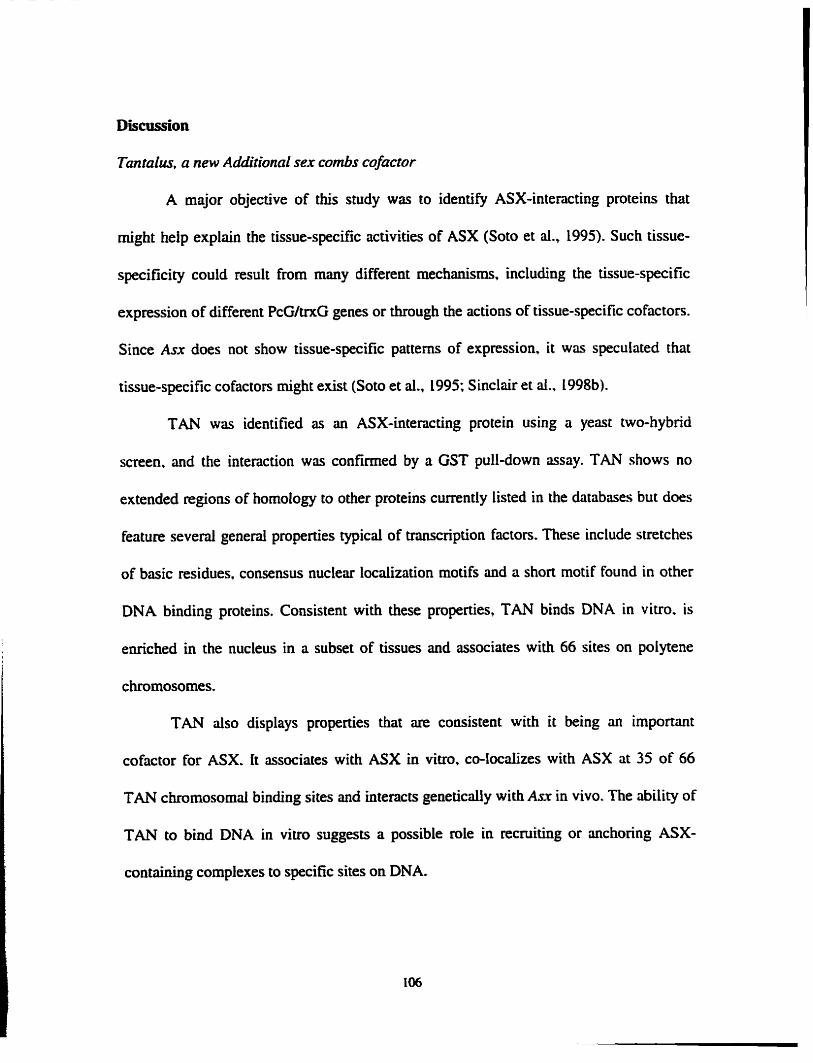
Discussion
Tantalus, a new Additional sex combs cofuctor
A major objective of this study was to identify ASX-interacting proteins that
might heip explilin the tissue-specific activities of ASX (Soto et al., 1995). Such tissue-
specificity could result €rom many different mechanisrns, including the tissue-specific
expression of different PcG/tn<G genes or through the actions of tissue-specific cofactoa.
Since Asx does not show tissue-specific patterns of expression. it was speculated that
tissue-specific cofacton might exist (Soto et al., 1995; Sinclair et al.. 1998b).
TAN was identifed as an ASX-interacting protein using a yeast two-hybrid
screen. and the interaction was confmed by a GST pull-dom assay. TAN shows no
extended regions of homology to other proteins currently listed in the databases but does
feature severai generai properties typical of transcription factors. These include stretches
of basic residues, consensus nuclear localization motifs and a short motif found in other
DNA binding proteins. Consistent with these properties, TAN binds DNA in vitro. is
e ~ c h e d in the nucleus in a subset of tissues and associates with 66 sites on polytene
chromosomes.
TAN dso displays properties that are consistent with it king an important
cofactor for ASX. [t associates with ASX in vitro, CO-localizes with ASX at 35 of 66
TAN chromosornai binding sites and intencts genetically wi th Asx in vivo. The ability of
TAN to bind DNA in vitro suggests a possible role in recruiting or anchoring ASX-
containing complexes to specific sites on DNA.

Additional sex cornbs and Tantalus contrul sensory organ development
Most members of the PcG and trxG protein complexes chmcterized thus far
associate with the both the ANT-C and BX-C gene clusters and cause homeotic
transfomations when mutated However, TAN appean to have no role in the regulation
of these genes. Rather, TAN appears to play a specific role in the differentiation of
sensory organs. The data show that ASX is also required in at least a subset of these
tissues, and when tan and Asx mutations are combined, these sensory organ defecis are
specifcaiiy enhanced or suppressed. Taken together, these results suggest that TAN acts
as a tissue-specific ASX cofactor in sensory orgm differentiation. This study only
examined the role of tan and Asx in ocellar bristle development where. possibly because
of smdl field in which btistle specification occurs, bristle fate is extremely sensitive to
tan over- or under-expression. Further work wili be required to detemine if ASX acts
together with TAN to regulate dl other tan-dependent processes identified. This need not
be the case, as ASX only colocaiizes with 35/66 of the mapped TAN polytene
chromosome binding sites.
Although the rnajority of PcG and tntG components identified to date were
isolated via genetic screens for the suppression or enhancement of homeotic
transformations, loss of sensory bristle phenotypes has been obsenred when other
tn<G/PcG genes are misexpressed Examples include LOF mutations in the uxG genes
brahma (Elfring et ai, 1998), absent smaIUhorneotic diskr2 (Adamson and Sheam. L996),
[eg arista w h g cornpiex (Zorin et ai. 1999) and Asr (this study). Loss of bristle
phenotypes are also obsewed &ter ectopic expression of the tn<G gene usa (Collins et ai.,

1999) and the PcG genes Posterior sex conzbs and Suppressor 2 of Zeste (Sharp et d,
L994). Additionai studies will be required to detemiine which bristle genes TAN and
ASX are acting through, and whether this regulation is positive or negative in nature.
The defects in sensory organ tissues caused by tan misexpression are remarkably
similar to those caused by mutations in the Notch (N) gene (see Lindsley and Zimm,
1992). in addition, expression of tan in the wing margin causes severe notching of the
wings and N mutations specificdiy and strongiy enhance the fan interocellar bnstle
phenotype. N is required at two different stages of sensory organ development. dunng
specification of the sensory organ precursor (SOP) cells and dunng the subsequent
specification of the SOP's daughter cells (reviewed in Artavanis-Tsakonas et ai.. 1999).
Simiiarly, the results presented here demonstrate that TAN cm &ect successive stages of
bristle development; over- or under-expression of TAN can cause b i d e and socket loss.
deletion of bristles only, and reduction in bristle sire.
One additionai observation regarding the effect of tm on sensory bristles needs
comment. The presence of ectopic bristles in the A6 stemite of tan mutant male flies is
characteristic of a loss of Abdominal-B (Abd-B) expresska, and is a phenotype exhibited
by mutations in t a and trithora-like of the trxG (Farkas et ai., 1994; ingham. 1981).
However, ectopic expression of tan using the HS promoter resulted in an even more
severe anterior homeotic transformation of male sternites. Considenng this finding, and
the other evidence presented here for a sensory-specific role for TAN, it seems likely that
the ectopic hairs in the A6 sternïtes of males are a result of TAN activity in bristie
specification and not an effect on Abd43 expression. Such an interpretation is consistent

with the inability to detect TAN binding at the site of the BX-C (where Abd-B is located)
in polytene chromosomes.
Tantalus subcellular Zocalization
An interesting feature of TAN that has not previously been noted for other trxG or
PcG complex components is that its subcelIulat localization varies in a tissue-specific
manner. In ernbryos. protein expressed by the GALA system is pnmarily cytoplasrnic.
white in third instar larvae it is cytoplasmically e ~ c h e d in some tissues and locdized to
the nucleus in others. Although the mecbaaism and importance of this localization has yet
to be addressed. it suggests a novel means of functiond regulation.
To date. the number of transcription factors known to cycle between cytoplasm
and nucleus are relatively few. Other well-characterized examples include proteins such
as Dorsal. Am@-catenin, STAT proteins, MAD proteins and components of the Norch
signding pathway. including N itseIf (Stnihl and Adachi. 1998). The last example is
particularIy notable. given the genetic interaction observed between N and tan.
Control of TAN nuciear localization may explain the restriction of mutant
phenotypes to sensory lineages. despite fairIy ubiquitous patterns of gene expression.
Retention in the cytoplasm rnay be a general way of relegating TAN activity to a subset of
TAN-expressing celis. On the other hand, the cytoplasmical.iy localized protein rnay dso
serve a function that has yet to be elaborated.
Other rules of Tanrulus
Another explanation for the relatively specific nature of tan mutant phenotypes,
despite the prolonged and widespread expression of the gene, is that some of its fiinetions

are obscured by redundantly acting gene products. Several additional observations lend
weight to this argument Fit, tan bristle phenotypes are highly variable in terms of
penetrance and seventy. Second more severe phenotypes cm be induced by ectopic- or
over-expression. Third, siightly less than hdf of the TAN polytene chromosome binding
sites do not appear to colocdize with ASX. The mapped TAN binding sites also show
heterogeneity with respect to other PcG/tn<G protein binding sites. This heterogeneity of
protein complexes nt different loci suggests the iikelihood of different functions and
outputs for each protein complex. Revealing the full extent of these TAN complex
activities wiil likely require the elimination of redundantly acting Factors.

Materiais and methods
GST pull-doms
Amino acids 1 139- 1668 of ASX were subcloned into pGM4T 1. GST beads were
bound to GST and GST-ASX at a concentration of 2pg/pl for GST and 400ng/pl for
GST-ASX. Beads were resuspended in an equal volume of 1X Binding Buffer 1 ( 1 X BB 1 :
25mM HEPES pH 7.5, 5mM KCl, IrnM EDTA, 0.25m.M DIT, 0.05% Tween-20,
lmg/mL BSA, 10% glycerol). The Promega TNT T7 Rabbit Reticulocyte Lysate System
was used to synthesize full length TAN (25pI reaction) which was passed over a
Sephadex G-25 column and cliluted to 100jd with 1X BB 1. The probe was incubated at
4OC for 30min to block and then 50~1 added to each of the GST and GST-ASX beads
(total volume is lûûpl). Reactions were incubated at 4°C with mixing for Zhrs and then
washed 2X with 1X BB 1 and 2X with Wash Buffer (20m.M HEPES pH 7.5.0.15M NaCI.
10% glycerol). Bound probe was eluted with 15pl of 1X SDS buffer and analyzed by
SDS-PAGE,
Antibodies
Either full-length TAN or residues 87-164 were fused to GST and purified for
injection to rabbits. Polyclonal antibodies were purified as descnbed (Sinclair et al.,
1998b) with both antibodies giving sirnilar results. Kc extracts were made as descnbed
previously ( Q b a and Brock 1998). Polytene chromosomes were stained as descnbed
(Sinclair et al. 1998b) wîth a Ln5 dilution of affinity pded anti-TAN antibody. Semm
depleted by passage over columos containing GST-TAN did not react with polytene

chromosomes. demonstrating the specificity of the antibody. Double-staining of polytene
chromosomes was undertaken with the sheep anti-ASX antibody described previously
(Sinclair et al. L998b) and the mbbit mti-TAN antibody described hem Binding was
detected with appropriate secondary antibodies Iabeled with Alexa Ruor 488 (Molecular
Probes) and Cy3 (Jackson Labs). To immunostain imaginai discs, l a m e were dissected
and fixed as described (Hughes and b u s e , 1999). Fixed tissues were dehydnted using
sevenl changes of methanol followed by washes in PBST (LX PBS + 0.3% Triton X-
1 0 ) . Tissues were blocked in PBST + 0.5% BSA for 2.5hrs. Tissues were incubated
ovemight at 4OC in appropriate antibodies (LIS0 dilution of a monoclonal mti-p53
antibody (Santa Cruz Biotechnology, Inc.) or 11500 dilution of anti-TAN against full
length TAN) in LX PBS + 0.5% BSA. Tissues were washed quickiy several times
followed by 3X 30rnin washes in PBST + 0.5% BSA. Tissues were incubated in
secondary antibodies for 45min ( ln00 anti-mouse HRP (Bio-Rad) or 11300 anti-rabbit
Alexa Fluor 488 (Molecular Probes)) followed by washes in PBT For lhr. For fluorescent
staining, tissues were incubated in propidium iodide to stain nuclei. The HRP staining
was developed using the ABC system of Pierce. After staining HRP tissues were washed
in PBS and a giycerol solution (5050 with LX PBS), while fluorescent tissues were
washed in 70% giyceroU 2% DABCO, before dissection and mounting.
P-eiement screen
The screen was performed based on the protocol of Hamilton and Zim (1994).
Lines 0545/ûI, 0666/10 and 1203/07 €rom De& and coworkers (1997) were used For the
P-dement mutagenesis screen. Groups of 20 aies containhg hopped P-elements were

anaiyzed by PCR using the P-element inverted repeat sequence as a primer and several
primea nom the tan genomic sequence. PCR was perfonned according to standard
procedures (Ausubel et al.. 1997).The foiiowing primea were used:
Pelement inverted repeat S'CGACGGGACCACCTïATGTTATTTCATCATG73
13- 1 5'CAGCGATTGCATCAGTGGT3
13-2 S'GATTGTCTCCAGATTGGTGG'3
G-l S'GGCTGAACCCAAGTACACTACCTA73
G-2 S'CCGAACGACAGTGGATGGATATGT'3
One line was obtained h m 0545/01 that contained an insert in tan (see Figure 3-
5). The 054301 line contains a lethai mutation located outside of the 65A region which
was recombined off the chromosome to make homozygously viable tm'/tm' tlies. A
similar procedure was used to screen for deletions of tan after hopping the P-element
from the gene. Southem biots were performed as described in Chapter 2 except high
stringency washes were carried out at 68°C.
Genamic rescue
The rescue constnict is approximately 7kb in size consisting of 1477bp upsueam
of tan (a Barn Hl site) and 3kb downstreiim of tun (a Saï I site) cloned into pW8
(Klemenz et al., 1987). mes were made homozygous for both the rescue consuuct and
tan' de le tion (P [tan]/P[tm];tan2/tan').
Drosophila strains. crosses. and anaïysis
As?, ASX~, AS?? and Df(2R)trir have been described (Sinclair et al.. 1992) and
the wiid-type stnin used is Oregon-R. $ and pif deles of N are desmibed in Lindsley

and Zimm (1992). patched-GALA, hairy-GAIA and C96-GALA are described in Rybase
(http://flybase.bio.inàianaaedu:82). Crosses for genetic malysis were 15-20 males and
fernales mated for 3-4 days. Parents were either transferred to new botties or dumped and
progeny ailowed to hatch until Day 17 for counting (parents are introduced to botties at
Day O). For the genomic rescue of tan' mutant brides (Figure 3-6C) 200 flies were
randomly selected at Day 17. Tissue samples were prepûred as descnbed (Sinclair et al.,
i 992).

Table 3-1. Polytene chromosome binding sites for TAN, with M X and PC/ PH sites indicated.
ASX
ASX PCIPH
ASX PC/PH ASX PCPH
ASX PCPH ASX PCPH MX. PC/PH ASX
PWPH ASX
PUPH PC/PH
ASX PUPH ASX a x ASX PClPH
M X . PUPH
ASX
ASX PCIPH ASX
ASX PUPH
Asx. PUPH
ASX PUPH ASX FUPH ASX
PUPH M X
ASX ASX. PUPH
ASM PCFH ASX PUPH
ASX. PC/PH ASM PCIPH ASX AS%. KYPH
PîiPH
'sx. PCIPH
Asx. r n H PCPH
ASX
ASX. KYPH

Table 3.12. Andysis of tan mutant and rescued phenotypes. A6 Stemire" in- BristIesb Ectopie Scuttiforss Wng Vrind
rd/)an" 24.7 96 (361) 36.7 % (387) 8 3 % (387) 85.1 % (174)
The % of flies displayinp the indicated phenotypes and the number of flies analyzed (in brackets) are shown for each cross. P[tan] is the genomic rescue construct. TM3,Sb is a balancer chromosome and CH4 is a deletion of 64E-6SB1,î. For the CH4 containing cross. reciprocal matings were performed and, as the resuits did noc Vary, the data were pooied. AI1 crosses except those noted were at room temperature. US-tan 4.1, HS-p53-tan 37.3, and HS-p53-tan 29.4 are independent lines with the tan gene (or a p53 epitope tagged version of tan) under the controt of a HS promoter. "refers to flies with at i tat 1 ectopic hair in the A6 sternite of d e s
refers to flies with 4 or fewer interocellar brisdes refers to nies with at itesist 1 ectopic scutelfat bristle cefers to flies with a shortened fifth vein

Table 3-3. ton and Asx interact genetically in
Crosses were ~ s x i ~ ~ ~ ; t a n ~ / t u n ' femnles to tan'ltan' males, except * which were ~ d ~ 0 : t a n ' l t a n ' femaies to tan'ltan' d e s , Gwr/+:tudtan and CyO/+;tan/ton siblings were then compived (coIumns 2 and 3). ~ f ( 2 ~ ) t ~ ~ ~ ~ 0 : t o n ~ / t a n ~ fernales were dso crossed to ~ [ t o n ~ ~ [ t o n ] ; t ~ ~ / t c u 1 ~ d e s to m u e the interocellm bride phenotype associated with the tan mutant (columns 4 ruid 5). Values indicate the percentilge of ffies with four or fewer intemeMar bristles, with the aumber of flies counted in brackets. Ail crosses were repted twice, except for those crosses involving the tan' diele, and the data pooled. Results were verified for significance by analysis (a = 0.05).

Table 3-4. Genetic interaction between ton and M.
The percentage of flies witti 4 or fewer interocellw bristles is shown, with the nurnber of flies counted in brackets. The loss of function allele A? and hypomorphic allele fie" were crossed to eittier tan2 homozygotes or p[tanl;tanZ homozygotes. Both N aileles show a strong genetic interaction when N and tan' are presen! heterozygousiy (column 2). tan' hetemzygotes do not display the interocellar bristle phenotype when heterozygous (see Tables 3-2 and 33). This interaction is strongly fes~led by the addition of the P[tanj construct (column 3).

Figure 3-1. GST pull-dom experiment. Labeled TAN was incubated with GST or GST-ASX and the unbound (5% of sample) and bound (100% of simple) fractions were anaiyzed by SDS-PAGE. Unbound (Un) (Lanes 1 and 3) and Bound (B) (Lanes 2 and 4) hctions of TAN from GST (Lanes 1 and 2) and GST-ASX (Lanes 3 and 4) samples are shown. The two faster migrating bands most iikely result from dtemate translational stm sites. The interaction of the fastest rnigrating band with ASX appeared to be weaker than hiil-length protein (compare nght two lanes). and may uncover part of an ASX interaction domain in TAN.

GST GST-ASX

Figure 3-2. Western Mots. (A) Third instar Iwai extracts from HS-treated samples incubated with anti-TAN antibodies (Fi t panel). A band of approximately 55kDa (arrow) is detected in ail three lanes and is more abundant in HS-tan and HS-p53-tan lanes (WT: wild-type). The p53 tag ad& LO amino acids to the protein. resulting in a slower migrating band. The blot was overexposed to demonstrate equai loading (compare cross nacting bands indicated by anowhead). A 55kDa protein is dso detected by monoclonal p53 antibodies in HS-p53-tan samples, but not in HS-WT samples (second panel). (B) Affinity purified anti-TAN antibodies detect a 55kDa protein in Kc ce11 nuclear (Nu) extracts; cytoplasmic (Cy) hction.

TAN Ab pS3 Ab

Fipre 3-3. TAN binding to polytene chromosomes. (A) Atfinity purified ad-TAN antibodies de tect endogenous TAN at euc hromatic sites on polytene chromosomes from saiivary glands of third instar larvae. TAN binding sites are stained brown and the chromosome is counterstaiaed in blue. Aithough numerous sites mapped show strong TAN binding (arrowheads), no staining is detected at the site of the BX-C (89E) (B). Similas results were obtained for the ANT-C.

Figure 3-4. In vivo detection of TAN. (Ab) Embryos from stage 16-17 stained with affinity purified TAN antibodies. (A) No signai is detected in a homozygous tan' mutant. (B) Staining is seen in the somatic and viscerai (arrow) mesodemi, consistent with tari
expression (see Figure 2-8E). (GE) Confocal images of ectopic TAN locdization. (C) Wing imaginai disc nuclei stained with propidium iodide (red). (D) TAN expression from a UAS-tan constnict driven by patched-GALA (green). (E) Merged image reveds staining predominantly in the cytoplasm of the cells (compare arrow in panels). pntched is expressed in a strong stripe dong the A-P axis of the disc and no TAN staining is detected in non-parched expressing cells.

Figure 3-5. P-element mutagenesis of un. (A) Schematic drawing of tan genomic region and surrounding Eco R1 (E) sites. Directed arrows indicate direction of transcription €rom an endopeptidase gene (gray box) located upstream of tan (white box). The 12kb P-element that inserted into the 5' region of tan is shown above (not to scde). The direction of transcription from the facZ reporter gene within the P-element is indicated by the dkcted mow. G-1, G-2, 13- 1, and 13-2 show primes for PCR analysis and PL and P2 indicate probes used for Southem analysis. The box below the PL/P2 probes indicates the extent of the deletion in tan' (see befow). (B-D) Genornic Southem blots. (B) The staning P-element iine 0545-OU+ (Lane 1) and the trini/+ iine (Lane 2) were digested with Eco RI and probed with Pl and P2 probes. One -8kb E'-E' band (Lane 1) is shifted upwards in the tan1 chromosome (Lme 2) due to the E" site within the P-element. The 3kb E'-E~ band is not affected by the insert but a new - 5kb band (star on right side) is created by E' and the E' site within the P-element. The E'-E* band contains only 200bp of tan sequence, resulting in a weak signai. The lower m w on the nght side shows one background band seen under high stringency washes using oniy the Pl probe (see text). (C) Southem blot with lac2 probe. Eco Ri digested DNA from 0545-01/+ (Lane I), tant/+ ( h e 2) and tan21tan2 (Lane 3) fies probed with a IacZ probe reveal a new band in the tan1/+ iine (the onginai Pelement is still present). This band is completely overlapping with the - 5kb E'-E' band detected in Lane 2 of panel B. as expected. tczn2/tan' flies have Iost the P-element inserted at the tan locus and the P- eiement has not reinserted in the genome. (D) Blot of 0545-OL/+ (Lane l) and tnn2/tan' ( h e 2) using only the Pl probe. The E'-E' band detected in Lane i is weak because of the Limited overlap between P1 and this fngment (- 2ûûbp). The deletion in tan21ran-' fies (Lane 2) created by the jurnp has deleted the E? site leaving a weak -8.3kb E'-E' band. The arrow denotes the background band also detected in panel B. (E) PCR anaiysis c o n f i the deletion of 1.4kb of tan DNA. The G-11G-2 primea amplified a 2kb fngment from 0545-0ll+ genomic DNA (Lane 1) but a 600bp fragment in tan'lm' (Lane 3), while the 13- 1113-2 primers, located within the tun coding sequence, amplified a 2 0 b p fragment in 0545-011+ (Lane 2) but no product in tan'ltan' (Lane 4). The 600bp band (Lane 3) was purified h m the gel and sequenced.


Figure 3-6. Adult defects in tanZ homozygotes. (A-D) Scanning electron rnicrographs with corresponding high magnification views. (AC) Wild-type eye. (I3.D) tan' mutant eye. Notice the rough appearance due to disruption of ornrnatidial spacing and the misplaced. dupiicated or missing bnstles. Both groups of flies were reared at 30°C. (E) Wild-type dorsal head with one of the three ocelii indicated by an asterisk and interocellar btisties indicated by anow (antenor is up). The larger ocellar bnstles (top) and post- vertical brides (bottom) a~ dso visible. (F) A tan' homozygous mutant with only t h e interoceliar bristles (arrow). (G) The number of interocellar brides was counted in 200 tnn2/tan' and 200 ~ [ t a n ] l ~ [ t a n ] ;tan5/tanZ rescued fies and gnp hed. tan' mutants average approximately half the number of interocellar bristles. (EI) Scutellum with a duplicated machrochaetae (arrow). (I) Male sternite with an ectopic br ide in the 61h abdominal (A6) segment (mow). (J) tan2 mutant showing a fgth vein which does not reach the margin (arrow).

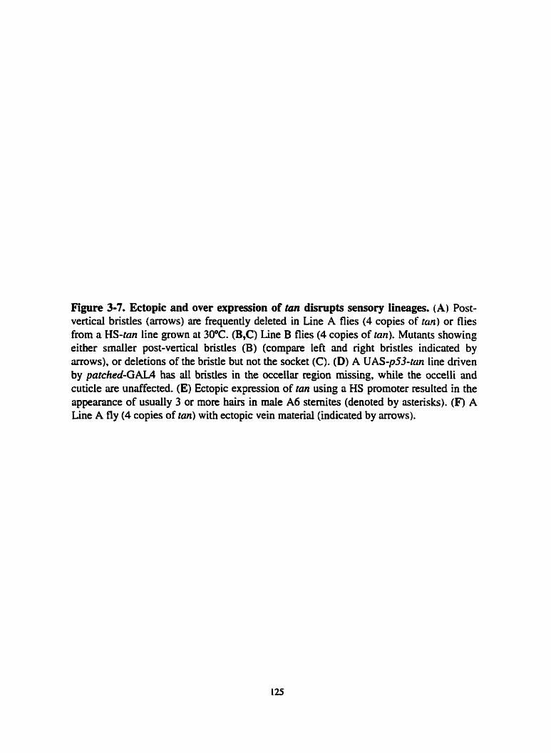
Figure 3-7. Ectopic and over expression of tan disrupts sensoty lineages. (A) Post- verticai brides (arrows) are frequently deleted in Line A flies (4 copies of tari) or flies from a HS-tm line grown at 30°C. (B,C) Line B flies (4 copies of tan). Mutants showing either smaiier post-verticai bristles (B) (compare left and right brktles indicated by arrows), or deletions of the brisde but not the socket (C). (D) A UAS-p53-tan Line driven by patched-GAL4 has dI bristles in the occellar segion missing, while the occelli and cuticle are unaffected. (E) Ectopic expression of tan using a HS prornoter resulted in the appemce of usuaily 3 or more hairs in male A6 sternites (denoted by astetisks). (F) A Line A fly (4 copies of tan) with ectopic vein material (indicated by arrows).


Figure 3-8. Ectopk expression of tan in the wing margin. (A) Wild-type wing. (B) UAS-tan expression driven by a margin specifrc ciriver (C96-GAU) results in severe notching of the wing rnargins.

CHAPTER 4
Summary and Future Directions

Summarg
In this thesis, 1 have descnbed the characterization of the novel gene tantaluî and
outiined experiments which have begun to address its role during Drosophila
developrnent. AIthough the current understanding of TAN'S in vivo function is limiteci,
enough details exist from my analysis of the tantdus gene to put forth a simple model
amenable to further experirnentation (Figure 4- 1). Because ectopic TAN produced by the
GALAKJAS and heat shock systerns is found in both the cytoplasm and nucleus, it is
reasooable to assume that endogenous TAN will also be found locdized to both
compartments. Based on the genetic interaction between tan and Notch (N), and the
similuity of N and tan phenotypes, it is possible that translocation of TAN to the nucleus
could occur through N signaiing, or possibly through another signal transduction
pathway. One possibility is that translocation by these signaling pathways could require
phosphorylation of TAN, since TAN contaios many canonical phosphorylation sites. A
role for signal transduction has recentiy ken proposed for the tn<G member trithora,
based on extensive phenotypic andysis of trx mutants (Breen, 1999) and the finding that
cell maturation and differentiation are promoted by dephosphorylation of HRX. the
human homologue of trithorax (Cui et al., 1998; De Vivo et ai., 1998).
Once translacated to the nucleus, the data support a model in which TAN
associates with the DNA, possibly thmugh direct binding, and interacts with ASX. It is
currentiy impossible though to predict which event might occur first. The specific binding
of TAN and ASX to euchromatic sites on polytene chromosomes suggests that these
factors play a direct role in target gene reguiation. However, the dud d e of ASX in both

PcG and trxG activities does not aHow one to address whether TAN is acting in a PcG or
trxG fasbion.
Future Directions: Testhg the mode1
The mode1 proposed offers several testable predictions, which I discuss below.
Detennining whether TAN cycles between the nucleus and cytoplasm simply requires
close examination of endogenous TAN expression, but couid be supplernented by studies
using the GALANAS system. When tan is overexpressed in the presumptive notum
using the patched-GALA chiver, ail machrochaetae are deleted The p53-tagged version of
TAN would dlow one to easiiy fobw protein distribution and determine when, and if.
TAN becomes localized to the nucleus during brisde development. Additionaily, the
Al0 1 enhancer uap line, which marks seosory organ precunor cells and their descendants
(Beiien et al., 1989; Huang et al.. 199 1). would be useful in following TAN localization
during brisde development. It wouid be quite exciting if TAN nuclear localization was
coupled to sensory lineage specification.
Evidence presented in this thesis suggests that the N pathway may be involved in
TAN hnction: interoceiiar bristie loss is dramatically enhanced in tan/N
transheterozygous mutants when compared to tan heterozygous mutants alone (see Table
3-4). The presence of temperature-sensitive and inducibie dominant forms of N could be
used to determine whether the N pathway affects TAN localization and/or acûvity in SOP
ceiis and their descendants.
Several groups have reported the dissociation of PcG or US members from
polytene chromosomes in the absence of other rnembers (Carrington and Jones, 1996;

KUWi et al., 1994; Rastelli et al., 1993). For example, Enhancer of zeste mutations cause
a demase in the binding of both the SuppressoR of zeste and Posterior sex combs
proteins to polytene chromosomes (RasteHi et al., 1993). The fact that homozygous tan
mutants are viable would make it simple to test whether tan loss-of-function mutations
affect ASX binding to polytene chromosomes. If the DNA binding ability of TAN is used
to stabilize/recruit an ASX complex then one might expect to see a decrease in ASX
binding to at least some of the sites bound by both proteins on polytene chromosomes.
As mentioned, PcG/trxG membea are found as protein complexes. and
experiments such as the Far Western assay or CO-immunoprecipitations could be used to
address whether TAN interacts with other members of the PcG/trxG. tndeed, an
interaction between TAN and Polycomb (PC) has been observed in the yeast two-hybrid
system and by GST pull-dowo assays (Kyba and Brock, unpublished observations). This
finding is rather interesting considering TAN and PC also bind to a large number of
overlapping sites on polytene chromosomes. Several of these overlapping sites are not
bound by ASX, suggesting that TAN could have hnctions that specificaily require an
interaction with Polycomb.
The early identification of HOM-C genes as targets of the PcG/trxG has been a
boon for understanding many questions surrounding the hnction of PcG/trxG genes. and
identification of TAN targets would greatiy aid the understanding of its hnction. The
bristie phenotypes associated with over- and under-expression of tan, combined with the
mapped TAN binding sites on poiytene chromosomes. d o w one to postulate potentiai
TAN target genes. By ident-g potential brisde-regulating genes that map to the 66

sites of TAN binding on poiytene chromosomes, the expression of these genes could be
foiiowed in tan mutants to determine if TAN regulates their expression. Unforninately,
the low penetrancelexpressivity of the associated tan phenotypes rnay hinder the success
of this approach.
One manner by which to overcome this hindrance is to combine a dominant
modifier of the tan bristle phenotype, like Notch (see Table 3 4 , with the tan' mutant to
increase the penetnnce of the phenotype. Another approach would be to study the
phenotypes associated with tan over-expression. The penetrance of the Ioss of bnstle
phenotype in the notum approaches 10096 when tan expression is driven by pntched-
GAIA. However. a potential drawback to this approach is that over-expression of tan may
have siightiy different developmental conseqwnces than underexpression which.
although bristle specific, would not necessarily reflect the role of endogenous TAN.
FinaiIy, it is necessary to look more broadly at the role of TAN in sensory lineage
specifcation, and such experiments could easily be combined with attempts to identib
potential ttarget genes. There are numerous bristle-specific genes and marken (see
bvaier et al.. 1999) that may not be directiy regulated by tan, but would still serve as
usehl molecular markers for SOP ceiis and their differentiated progeny. These markers
could be used to determine the stage at which bristle differentiation is disrupted in tm
mutants.
Redmdancy in TuntaIusfunction
Strong evidence for a tissue-specific role for TAN has k e n presented here.
However, the incomplete penetrance of tan phenotypes is consistent with some
redundancy in tan function. Additionaiiy, results of the Southem anaiysis leave open the
possibiiity that a homologue to tan exists. One main goal of future studies wiU be to
determine if redundancy plays a role in the penetrancdexpressivity and tissue-specific
activities observed for TAN.
The ment sequencing of the Drosophila genome (Adams et al., 2000; R2Sin et
al., 2000) may provide some perspective for this question. Approximately 13,600
Drosuphila genes a~ predicted from the sequence andysis, representing - 8,000 distinct
gene families. The rernaining - 5.500 genes, or roughly 40%. represent duplicated genes.
It is important to note, however, that the 5,500 genes do not represent separate gene
families; for examp le, there are approximately 100 homeobox containing genes included
within the 5,500 genes. The large number of duplicated genes rnay partiy explain the fact
that less than one-third of Drosophila genes give nse to obvious phenotypes when
mutated (Ashbumer and et al., 1999; Mikios and Rubin, 1996). Therefore, it would
appear that tan, with its subtle defects, most likely represents the nom. Once the
remaining gaps in the Drosophila sequence are füled, it should become clear as to
whether or not a second gene with homology to tan exists.
Screens for dominant suppressors or enhancers of tan phenotypes are one way to
search for redundant factors, as weii as genes that may assist in TAN function. Such a
procedure, based on results in Table 3-4 with the N gene, should be capable of identiwing
potentiai rnodifiers of tan function. Also, other identifi~ed PcGhntG members should be
anaiyzed to deterrnine which of them, Like Am, can modify tan mutant phenotypes. As
TAN binding to polytene chromosomes overlaps the binding sites of many PcG/mG

members, it seems likely that at least some of these genes wiü also be involved in sensory
lineage specification.
Homeodomain binding of Tantah
1s there devance to the finding that TAN interacts with the HD of FTZ?
Aithough this interaction may be signif~cant in vivo, it is aiso possible that this interaction
is not specific to the HD of FTZ. The Drosophila genorne project has identified -LOO
HD-containing proteins (Rubin et al., 2000) and it will be important to address the
specificity of the TAN/HD interaction. There are several HD-containing proteins. like D-
PAX2 and Prospero, which are involved in the proper differentiation of sensory bristles
( v e r et al., 1999; Reddy and Rodngues, 1999) and it is possible that TAN could
intenct with these, or with simiiar proteins, during bristle differentiation.
in particular, study of D-Par2 has uncovered some interesting similarities to tan
function (Kavaler et al., 1999). N signaling is required during development of sensory
organ precursor (SOP) ceils, as weil as in the daughter cells of the SOPs. Within these
daughter ceiis N signaling regulates the expression of D - P d . D-Pm2 mutant alleles
cause shortened/stunted bristles as well as ernpty sockets (Kavaler et al., 1999), and over-
expression of D-PAX2 leads to the formation of ectopic bristie stnictures. interestingly,
D-Pax2 mutants also have a rough eye appearance (Fu and Noil, 1997). The similarities
of the D-Pax2 phenotypes to the phenotypes seen when tan is over- or under-expressed
make DPAX2 an ideal candidate to test for a putentid protein-protein interaction with
TAN.
Conclusions
ThÎs project has iîiustsated some of the problems that WU be encountered during
the deciphering of the Drosophila genome. As mentioned, roughly two-thirds of
Drosophila genes may not have easiiy identifiable phenotypes ( Ashbumer et al., 1999;
Miklos and Rubin, 1996) and many of these rnay not possess sequence homology to
currently known genes (Rubin et al, 2000). Both facts make study of new genes in
Drosophila a slow and arduous process, and suggest that future studies will have to exert
great effort to h d detectable and highly penetrant phenotypes. As demonstrated here,
over- and ectopic expression studies combined with genetic modifiers of subtle
phenotypes (Like the effect of N on tan) may be excellent ways to address these
difficul ties,

Figure 4-2. Mode! of TAN function. Cytoplasrnic TAN translocates to the nucleus upon signahg by Notch or other, unidentified (?), pathways. This translocation step could involve phosphorylation (P). Once tnwlocated to the nucleus TAN binds DNA and this step could nquire other cofactors (CoF). TAN then becomes associated with an ASX complex cornposed of either PcG or trxG members to maintain tmscrîptional states of target genes.


REFEREN(3ES
Adams, M.D.. et al. ( 2 0 ) The genome sequence of Drosophila melamgaster. Science,
287,2185-2 195.
Adamson, AL. and Shearn, A. (1996) Molecular genetic anaiysis of Drosophila ash2, a
member of the trithorax group required for imagina1 disc pattern formation.
Genetics, 144.62 1-633.
Alexandre, C., Lecourtois, M. and Vincent, I.P. (1999) Wingless and Hedgehog pattern
Drosophila denticle bel& by regulating the production of short-range signais.
Development, 126.5689-5698.
Ananthan, J., Baler. R., Morrissey, D., Zuo. I.. Lm, Y., Weir, M. and Voellmy, R. (1993)
Synergistic activation of transcription is mediated by the N-terminai domain of
Drosophila fmhi tarau homeoprotein and c m occur without DNA binding by the
protein. Mol. Cell. B O L , 13. 1599-1609.
Andrew, D.J. and Scott, M.P. (L994) Immunologicai Methods for Mapping Protein
Distributions on Polytene Chromosomes. In Goldstein, L.S.B. and Fyrberg, E.A.
(eds.), Methods in Cell Biology, vol 44: Drosophila melanogastec Practical Uses
in Cell and Molecular Biology. Academic Press. San Diego USA, pp. 353-370.
Andres, AJ. and Thummel, C.S. (L994) Methods for Quantitative Andysis of
Transcription in Lûirvae and Prepupae. ui Goldstein, L S B . and Fyrberg, E.A.
(eds.), Methoh in Ce22 Biolugy, vol 44.- Drosophila melanogaster: Practical Uses
in Cell mid Molecuiar Bioiogy. Academic Press, San Diego USA, pp. 565-573.
Amosti, DM., Barolo, S., Levine, M. and Srnail, S. (1996) The eve stripe 2 euhancer
employs multiple modes of transcriptionai synergy. Datelopment, 122,205-2 14.
Artaviuiis-Tsakonas, S., Rand, M.D. and Lake. R.J. (1999) Notch sigiialing: ce11 fate
control and signai integration in development. Science. 284,770-776.
Ashbumer. M. et al.. (1999) An exploration of the sequence of a 2.9-Mb region of the
genome of Drosophila melanogapter: The Adh region. Genetics, 153, 179-2 19.
Aspland, S.E. and White, R.A.H. ( 1997) Nucleocytoplasmic locaiization of exiraclenticle
protein is spatiaily regulated throughout development in Drosophila.
Deve fopment, 124.74 1-747.
Ausubel, F.M.. Brent, R., Kingston, RE-, Moore. D.D.. Seidman, I.G., Smith, I.A., and
Stnihl, K. (1997) Current Protocols in Molecular Bioiogy. John Wiley & Sons.
[nc. New York.
Baker, N.E. (1987) Molecular cloning of sequences from wingless a segment polarity
gene in Drosophila: The spatial distribution of a transcript in embryos. EMBO J..
6, 1765-1773.
Baiünger, DG. and Benzer, S. (1989) Targeted gene mutations in Drosophila. Proc. Nutt.
Acad. Sci. USA, 86.9402-9406.
Bateson, W. ( 1894) Muterials for the SSnuly of Variation Treated with Especiul Regard to
Dhcontinuiîy in the Origin of Species. Macmillan, London.
Beachy, P.A., HeEand, SL. and Hogness, D.S. (1985) Segmental distnbutioa of bithorax
cornplex proteins duruig Drosophila development. Nature, 313,545-55 1.
Bejsovec, A. and Marthez-Anas, A. (1991) Roles of wingless in patternhg the larvd

epidermis of Drosophita. Development, 113,47 1-485.
Bejsovec, A. and Wieschaus, E. (1993) Segment polarity gene interactions modulate
epidermal patteming in Drosophila embryos. Development, ll9,SO 1 -5 17.
Beiien, H.J., O'Kane, C.J., Wilson, C., Grossniklaus, U., Pearson, R.K. and Gehring,
W.J. (1989) P-elernent-mediated enhancer detection: a versatile method to study
developrnent in Drosophila. Genes Dev., 3, 1 288- L 3OO.
Belvin, M.P. and Anderson, K.V. ( 1996) A conserved signaling pathway: The Drosophilu
TOU-dorsal pathway. Annu Rev. Cell Dot. Biol., 12,393-4 16.
Bender, W., Spierer, P. and Hogness, D.S. (1983) Chromosomal walking and jumping to
isolate DNA from the Ace and rosy loci and the Bithorax Complex in Drosophila
melanogcisrer. J. Mol. Bioi., 168, 17-33.
Benedyk, M.J., Mullen, J.R. and DiNardo, S. (1994) odd-pairrd: a zinc finger pair-rule
protein required for the timely activation of engrailed and wingless in Drosophili
embryos. Genes Dev., 8, 105-1 17.
Berleth. T., Bum, M., Thorna, G., Bopp, D., Rechstein, S., Ftigeno, G., Noll. M. and
Nüsslein-Volhard . C. (1988) The role of localization of b h i d RNA in
organizing the anterior pattern of the Drosophila embryo. EMBO J., 7, 1749-
1756.
Boulay, I.L., Dennefeld, C. and Alberga, A. (1987) The Drosophila developmentai gene
snait encodes a protein with nucleic acid binding fingers. Nature, 330,395-398.
Brand, A.H. and Perrimon, N. (1993) Targeted gene expression as a means of altering ceil
fates and generating dominant phenotypes. Developmt, 118,40 1-4 1 5.
Breen, T.R. (1999) Mutant alleles of the Drosophila trithorax gene produce common and
unusual homeotic and other developmentd phenotypes. Genetics, 152.3 19-344.
Breen, T.R. and Duncan. IM. (1986) Matemal expression of genes that reguiate the
bithorux complex of Drosophila melanogaster. Dm. Biol.. 118.442456.
Brizuela, B.J. and Kennison, LA. (1997) The Drosophila homeotic gene moira regulates
expression of engr&ed and HOM genes in imagina1 tissues. Mech. Dev.. 65,209-
220.
Brown, J.L. Mucci, D e . Whiteiey, M., Dirissen, M.L. and Kassis, J.A. (1998) The
Drosophila Polycomb group gene pleiohomeotic encodes a DNA binding protein
with homology to the transcription factor W 1. Md. Ceil. 1. 1 057- 1064.
Buchenau. P., Hodgson. J.. Strutt. K. and Arndt-Jovin, DJ. (1998) The distribution of
polycomb-group proteins during ceil division and development in Drosophih
embryos: impact on models for silencing. J Ce11 Bbl, 141,469-48 1.
Burglin, T.R. and Ruvkun. G. (1992) New motif in PBX genes. Nature Genet., 1. 3 19-
320.
Busturia, A. and Bienz, M. (1993) Silencers in abdominal-B. a homeotic Drosophila
gene. WMBO J., 12, L415-1425.
Cabrera, C.V., Alonso, M.C., lohnston, P.. Phillips, R.G. and Lawrence, P.A. (1987)
Phenocopies induced with antisense RNA identifi the wingiess gene. Celt, 50.
649-663.
Cadigan, W., Grossniklaus. U. and Gehring. W. (1994) Localued expression of sloppy
paired protein maintains the plarity of Drosophila parasegments. Genes Dm., 8.

899-9 1 3.
Cairns, B .Re, Lorch, Y ., Li, Y., Zhang, M., Lacomis, L., Erdjurnent-Bromage, K., Tempst,
P., Du, J., Laurent, B.C. and Komberg, RD. (1996) RSC, an essentid. abundant
chromath-remodeling cornplex. Cell, 87, 1249- 1260.
Campbell, RB., Sinclair, D.A., Couling, M. and Brock. H.W. (1995) Genetic interactions
and dosage effects of Pofycomb group genes of Drosophila. Mol. Gen. Genet..
246,39 1-300.
Carrington. E.A. and Jones. R.S. (1996) Mutant dieles of the Drosophiln trithornr gene
produce cornmon and unusual homeotic and other developmental phenotypes.
Genetics, 152,3 19-344.
Carroll. S. (1995) Homeotic genes and the evolution of arthropods and chordates. Nutrrre.
376,479-485.
Carroll, S.B., Lapon, R.A., McCutcheon, M.A., Riley, P.D. and Scott, M.P. (1986) The
locaiization and replation of Antennapedia protein expression in Drosophila
embryos. Cell, 47, L 13- L 22.
Carroll, S.B. and Scott, M.P. (1985) Localkation of the fwhi tarazu protein dunng
Drosophila emb yrogenesis. Cell, 43-47-57.
Casanova, J. and Struhi, G. (1989) Localued surface activity of torso. a receptor tyrosine
kinase, specifies terminal body pattern in Drosophila. Genes Dm., 3,2025-2038.
Casares, F., CdIeja, M. and Sanchez-Hemro, E. (1996) Functiond similarity in
appendage specifcation by the ü2trubithomx and abdominal-A Drosophila HOX
genes. EMBO J., 15,39343942-
Castelli-Gair, J. and Akam, M. (1995) How the Hox gene Ultrabithorax specifies two
different segments: the significance of spatial and temporal regulation within
metameres. Development, 121,2973-2982.
Cavaiii, G. and Paro, R. (1998) The Drosophila Fab-7 chromosomal element conveys
epigenetic inhentance during mitosis and meiosis. Cell, 93,505-5 18.
Cavaiii, G. and Paro. R. (1999) Epigenetic inheritance of active chromatin &ter removai
of the main transactivator. Science, 286,955-958.
Cavener, D.R. (1987) Comparison of the consensus sequence flanking translational start
sites in Drosophila and vertebrates. Nucleic Acids Res., 15, 1353-136 1.
Celniker, S.E., Keelan, D.I. and Lewis, E.B. (1989) The molecular genetics of the
bithorax complex of Drosophila: chacacterization of the products of the
Abdomind-B domain. Genes Dev., 3,1424- 1436-
Chan, C.S., RasteIli, L. and Pirrotta, V. (1994a) A Polycomb cesponse element in the Ubx
gene that determines an epigeneticaiiy inhented state of repression. EMBO J.. 13,
2553-2564,
Chan, S.K.. Jaffe, L., Capovilla, M., Botas, J. and Mann, R.S. ( 1994b) The DNA binding
specificity of Ukrabithorax is modulated by coopentive interactions with
extradenticle, another homeoprotein. Ce11.78,6036 15.
Chan, S.K. and Mann, R.S. (1993) The segment identity functions of Ultrabithorax are
contained within its homeo domah and carboxy-terminal sequences. Genes Dev..
7,796-8 1 1.
Chao. S.-K. and Mann. R.S. (1996) A structural mode1 for a homeotic protein-

extradenticle-DNA complex account. br the choice of HOX protein in the
heterodimer. Proc. Natl. Acad. Sci. U S A, 93,5223-5228.
Chan, S.K, Ryoo, HD.9 Gould, A., Knimlauf, R. and Mm, R.S. (1997) Switching the in
vivo speci fici ty of a minimal Hox-responsive element. Development, 124, 2007-
20 14.
Chang, C.-P., Shen, W.-F., Rozenfeld, S.. Lawrence, H.J., Largman. C. and Cleary, M.L.
( 199%) Pbx proteins dis play hexapeptide-de pendent coopentive DN A binding
with a subset of Hox proteins. Genes and Dev., 9,663-674.
Chang, Y.L., King, B.O., O'COMO~, M., Mao, A. and Huang, D.H. (1995b) Functional
reconstruction of tram regulation of the Ultrabithornr pmmoter by the products of
two antagonistic genes, trithorax and Polycomb. Moi. Cell. Biol., 15.660 1-66 1 2.
Chen, J.D., Chan, C.S. and Pinotta, V. (1992) Conserved DNA binding and self-
association domains of the Drosophila zeste protein. Mol. Cell. Biol., 12, 598-
608.
Chinwaila. V.. Jane, E.P. and Harte, P.J. ( 1995) The Drosophtla trithorax protein binds to
specific chromosornai sites and is co-localized with Polycomb at many sites.
EMBO J., 14,2056-2065.
Christen, B. and Bienz, M. (1994) haginal cüsc silencers from Ultrabithornr: evidence
for Polycomb response eIements. Mech. Dev., 48,255-266.
Collins, R.T., Furukawa, T., Tanese, N. and Treisman, JE. ( 1999) Osa associates with the
Brahma chromath remodehg complex and promotes the activation of some
iarget genes. EMBO J., 18,7029-7040.

Copeland LW., Nasiadka, A., Dietrich, B.H. and b u s e , HM. (1996) Patteming of the
Drosophila embryo by a homeodomaindeleted Ftz polypeptide. Nature, 379. 162-
165.
Copland, I.W.R. (1997) The d e of protein-protein interactions and phosphorylation in
the function of the homeodomain protein furhi t a r a . Thesis. University of
Toronto, Toronto.
Crosby, M.A., Miller, C., Non. T., Watson, KL.. Verrijzer. C.P., Goldman-Levi. R. and
Zak, N.B. ( 1999) The trithorax group gene moira encodes a brahma-associated
putative chromatin-remodelling factor in Drosophiln mefanogaster. Moi. Ceil.
Biol., 19, 1 159- 1 170,
Cui, X., et al. (1998) Association of SET domain and myotubularin-related proteins
modulates growth control. Nat. Genc, 18.33 1-337.
Cumberiedge, S. and Knsnow, M.A. (1993) Intercellular signaling in Drosophila
segment formation reconstructed in vitro. Nature, 363,549452.
Ddby, B. and Glover, D.M. (1993) Discrete sequence elements control posterior pole
accumulation and tnnslationd repression of matemal cyclin B RNA in
Drosophila. EMBO JO, 12, 12 19- I 227.
Daubresse, G., Deuring, R., Moore, L., Papoulas, O., Zakrajsek, I., Waldrip, W.R., Scott,
M.P., Kennison, I.A. and Tamkun, J.W. (1999) The Drosophila kismet gene is
related to chromath-remodehg factors and is required for both segmentation and
segment identity. Developmenr, 126, 1 175- 1 187.
De&. P. et ai. (1997) P-elernent insertion aiieles of essentid genes on the third
chromosome of Drosophila rnelanogater: correlation of physicd and cytogenetic
maps in chromosomal region 86E-87F. Genetics, 147m 1697- 1722.
de Cuevas, M. and SpradLing. A.C. (1998) Morphogenesis of the Drosophila f'usome and
its implications for oocyte specification. Datelopment, 125.278 1-2789.
DeCamillis, M., Cheng. N.S., Piene. D. and Brock. W.W. (1992) The polyhomeotic gene
of Drosophila encodes a chromatin protein that shares polytene ciuornosome-
binding sites with Polycomb. Genes Dev., 6,223-232.
Desplan, C.. Theis. I. and O'Famll, P.H. (1988) The sequence specificity of
homeodomain-DNA interaction. Cell, 54, 1 O8 1 - 1090.
De Vivo. 1.. Cui. X., Domen. J. and Cleary, M.L. (1998) Growth stimulation of primary B
ceil precursoa by the anti-phosphatase Sbfl. Proc. N d . Acad. Sci. USA. 95.
947 1 -9476.
DiNardo, S., Heemskerk, I., Dougan, S. and O'Farrell, P.H. (1994) The making of a
maggot: patterning the Drosophila embryonic epidermis. Curr. Opin. Genet. Dm..
4,529-534.
DiNwdo, S. and O'Fanell. EH. (1987) Establishment and refinement of segmentai
pattern in the Drosophila embryo: spatial contml of engrailed expression by pair-
mie genes. Genes D m , 1, 12124225.
DiN~do, S., Sher, E., Heemskerk-longens, JeT Kassis, J.A. and O'Farrell, P.H. (1988)
Two-tiered regulation of spatialiy pattemed engrailed gene expression during
Drosophila embryogenesis. Nature, 332,604609.
Dingwail. A.K., Beek, SJ., McCdum, CM., Tamkun, J.W., Kalpana, G.V., Goff, S.P.
and Scott, M.P. (1995) The Drosuphila snrl and brm proteins are related to yeast
SWSNF proteins and are components of a large protein cornplex. Mol. Biol.
Cell, 6,777-79 1.
Dm, C.Q.9 Hirotni, Y., Gehnng, W.J. and Goodman, C.S. (1988) Expression and
function of the segmentation gene furhi tarazu dunng Drosophila neurogenesis.
Science, 239, L 70- 1 75,
Dnganescu, A., Levin, I.R. and Tuliius, T.D. ( 1995) Homeodomain proteins: what
govems their ability to recognize specific DNA sequences? J. Mol. Biol., 250,
595-608.
Dranginis, A.M. (1990) Binding of yeast al and dpha 2 as a hetemdimer to the operator
DNA of a haploid-specific gene. Nature, 347,682-685.
Driever, W. and Nüsslein-Volhard. C. (1988a) A gradient of bicoici protein in Drosophila
embryos. Ceil, 54.83-93.
Driever, W. and Nüsslein-Vothard, C. ( 1988b) The bicoid protein determines position in
the Drosophiln embryo in a, concentration-dependent manner. Ceil, S4,95- 1 M.
Driever, W. and Nüsslein-Volhard, C. (1989) The bicoid protein is a positive regulator of
hmchback transcription in the early Drosophlikz embryo. Nature, 337, 138- 143.
Driever, W., Thorna, G. and Nüsslein-VoIhard, C. ( 1989) Determination of spatial
domains of zygotic gene expression in the Drosophifu embryo by the affinity of
binding sites for the bicoid morphogen. Nature, 340,363-367.
Dubnau, J. and Struhl, G. (1996) RNA recognition and translational regdation by a
homeodomain protein. Nature. 379,694-699.

Du% J.B. and P e h o n , N. (1994) The Torso pathway in Drosophila: Lessons on
receptor tyrosine kinase signaüng and pattern formation. Dev. BioL, 166,380-395.
Duncan, LM. (1982) Polycomblike: a geae that appears to be required for the normal
expression of the bithorax and antennapedia gene complexes of Drosophila
melanogaster. Genetics, 102.49-70.
Durand, B., Saunders, M., Gaudon, C., ROY? B., Losson, R. and Chambon. P. (1994)
Activation function 2 (AF-2) of retinoic acid receptor and 9 4 s retinoic acid
receptor: presence of a conserved autonomous constitutive activating domain and
influence of the nature of the response element on AF-2 activity. EMBO J., 13,
5370-5382.
Eissenberg, J.C., lames, T.C., Foster-Hartnett, D.M., Hartnett, T., Ngan, V. and Elgin,
S.C.R. (1990) Mutation in a heterochromatin-specific chromosornai protein is
associated with suppression of position-effect variegation in Drosuphilu
melanoguster. Proc. Natl. Acud. Sci. USA, 87,9923-9927.
Eissenberg, J-C., Morris, G.D., Reuter, G. and Hartnett, T. (1992) The heterochromatin-
associated protein HP1 is an essentiai protein in Drosophila with dosage-
dependent effects on position-effect variegation. Generics, 131,345-352.
Elfnng, L.K. Daniel, C., Papoulas, O., Deuring, R., Sarte, M., Moseley, S.. Beek, S.S.,
Wddrip, W-R., Daubresse, Ge, DePace, A., Kennison, LA. and Tamkun, J.W.
(1998) Genetic analysis of brahma: the Drosophila homolog of the yeast
chromatin remodeling factor STKlUSNF2. Genetics, 148,25 1-265.
Farkas, G., Gausz, J., Gdlooi, M., Reuter, G., Gyurkovics, H. and IOrch, F. ( 1994) The

Tdhorczx-like gene encodes the Drosophila GAGA factor. Nature, 371,806-808.
Fitzpatnck, V.D. and Ingles, C.J. (1989) The Drosophila furhi turazu polypeptide is a
DNA-binding transcriptionai activator in yeast tek. Nature, 337,666-668.
Fiapatrick, V.D., Percival-Smith, A., hgles, CJ. and b u s e , HM. ( 1992)
Homeodomain-independent activity of the fushi tarazu polypeptide in Drosophilci
embryos. Nature, 356,6 10-6 12.
Fjose, A., McGinnis, W.J. and Gehnng, W. (1985) Isolation of a homoeo box-containing
gene from the engrailed region of Drosophila and the spatial distribution of its
transcripts. Nature. 313,284289.
Fiorence, B., Guichet, A.. Ephrussi, A. and Laughon, A. ( 1997) Ftz-FI is a cofactor in Ftz
activation of the Drosophila engrailed gene. Development, 124,839-847.
Franke, A., DeCamiilis, M., Zink, D., Cheng, N., Brock, H.W. and Paro. R. (1992)
Polycomb and polyhomeotic are constituents of a multimeric protein complex in
chromatin of Drosophila melanogaster. EMBO J.. LI, 2941-2950.
Frasch, M. and Levine. M. (1987) Complementary patterns of men-skipped and fishi
tnrazu expression involve their differential regulation by a common set of
segmentation geaes in Drosophila. Genes Dev., 1.98 1-995.
Frigerio, G., Burri, M., Bopp, D., Bûurngartner, S. and Noll, M. (1986) Structure of the
segmentation gene paired and the Drosophila PRD gene set as part of a gene
network. C d , 47,735-746.
Frohnhofer, H.G. and Nüsslein-Volhard , C. (1986) Organization of anterior pattern in the
Drosophila embryo by the maternai gene bicoid. Nature, 324, 120- 1 25.

Fu, W. and Noil, M. (L997). The P d homolog spanCIing is required for development of
cone and pigment ceus in the Drosophila eye. Genes Dm., 11.2066-2078.
Fujioka, M., Emi-Sarker, Y., Yusibova, GL*, GO~O, T. and Iaynes. J.B. (1999) Analysis
of an men-skipped rescue tnnsgene reveals both composite and discrete neuronal
and earfy blastoderm enhancers, and multi-stripe positioning by gap gene
tepressor gradients. Development, 126,2527-2538.
Fujioka, M., Jaynes, I.B. and Goto, T. (1995) Early even-skipped stripes act as
morphogenetic gradients at the single ce11 level to establish engrctiled expression.
Development, 121,437 1-4382.
Fujioka, M., Miskiewicz, P., Raj, L., Guiîedge, A.A., Weir, M. and Goto. T. ( 1996)
Drosophila Paired regulates late even-skipped expression through a composite
binding site for the pakd domain and the homeodornain. Development, 122.
2697-2707.
Furukubo-Tokunaga, K., Flister, S. and Gehring, WJ. ( 1993) Functiond specificity of the
Antennapedia homeodomain. Proc. Nutl. Acad Sei. USA. 90,6360-6364.
Furukubo-Tokunaga, K., Muller, M., molter, M., Pick, L., Kloter, U. and Gehring, W.J.
(1992) In vivo aaalysis of the heiix-tum-heh motif of the fushi taraztt homeo
domain of Drosophila melanoguster. Gmes Dev., 6, 1082- 1096.
Gao, Q. and Finkelstein, R. ( 1998) Targeting gene expression to the head: the Drosophila
onhohticle gene is a direct target of the Bicoid morphogen. Development. 125,
4185-4193.
Gyber, RL, Kuroiwa, A. and Gehring, WJ. (1983) Genomic and cDNA clones of the

homeotic locus Antennapedio in Drosophila. EMBO J., 2,2027-2036.
Gaul, U. and Jackle, H. (1989) Analysis of matemal effect mutant combinations
elucidates regdation and function of the overlap of hunchback and Krzïppel gene
expression in the Drosophila blastoderm embryo. Development, 107,65 1-663.
Gavis. E.R. and Lehmann, R. (1992) Localization of nanos RNA controls embryonic
poluity. Cell, 71.3 10-3 13.
Gehnng, WJ.. Affolter. M. and Bürglin. T. (1994) Homeodomain proteins. Annu. Rrv.
Biochem, 63,487-526.
Gibson, G., Schier, A., LeMotte, P. and Gehnng, W. (1990) The specificities of Sex
combs reduced and Antennapedia are defined by a distinct portion of each protein
that includes the homeodomain. Cell, 62, 1087-1 103.
GiIdea, J.. Lopez, R. and Shearn, A. (2000) A screen for new Trithorax Group genes
identified little imagina1 discs, the Drosophila melanoguster homologue of
Humm Retinoblastoma Binding Protein 2. Genetics, 156.645-663.
Gindhart, J.G., Jr. and Kaufman, T.C. (1995) Identification of Polycomb and trithom
group responsive elements in the regulatory region of the Drosophikt homeotic
gene Sex cornbs reduced. Genetics, 139.797-8 14.
Godt, D. and Tepass, U. ( 1998) Drosophila oocyte localization is mediated by differential
Cadherin-based adhesion. Nature, 395,387-39 1.
Golemis, E.A., Serebriiskü, I., Gymis, I. and Brent. (1997) Interaction trap/two-hybrid
system to indentify interacting proteins. In Ausubel, FM., Brent. R. Kingston,
R E , Moore, D.D., Seidman, I.G.. Smith, J.A., and Stnihl, K. (eds.) Current

Protocols Protocols in Molecular Biology. John Wiley Br Sons, Inc., New York,
Supplernent 40.
Goozflez-Reyes, A., Elliot, H. and St. Johnston, D. (1995) Polarizatioa of both major
body axes in Drusophila by gurken-topedo signalling. Nature, 375.654-658.
Gotualez-Reyes, A. and St. Johnston, D. (L998) The Drosophila AP axis is polarised by
the cadhenn-mediated positioning of the oocyte. Deve[opment, 125,3635-3644.
Goutte, C. and Johnson, A.D. (1988) al protein aiters the DNA binding specificity of
alpha 2 repressor. Cell, 52,875-882.
Gnba, Y., Angnol, D. and Pndel, J. (1997) Drosophila Hox complex downstream
targets and the function of homeotic genes. BioEssays, 19.379-388.
Gny, S.. Szyrnanski, P. and Levine, M. (1994) Short-range repression permits multiple
enhancers to function autonomously within a complex prornoter. Genes Dev., 8,
1829- 1838.
Gritzan, U., Hatini, V. and DiNado, S. (1999) Mutual antagonism between signals
secreted by adjacent Wingiess and Engrailed cells leads to specification of
cornplementary regions of the Drosophila parasegment. DeveLopment, 126,1107-
41 15.
Grünert, S. and St. Johnston, D. (1996) RNA localization and the development of
asymetry during Drosophila oogenesis. Curr. Opin. Genet. Dm., 6.395402.
Guichet, A., Copland, J.W., ErdeIyi, M., Hlousek, D., Zavorszlq, P.. Ho, J., Brown, S.,
PercivdSmith, A., buse , HM. and Ephrussi, A. (1997) The nuclear receptor
homologue Fa-FI and the homeodomain protein FE are mutualiy dependent

cofactors. Nature, 385,548-552.
Hafen, E., Kuroiwa, A. and Gehring. W.J. (1984) Spatial distribution of transcripts from
the segmentation gene fushi iarazu during Drosophila ernbryonic development.
Cell. 37,833-84 1.
Hamilton, B.A. and Zinn, K. (1994) From Clone to Mutant Gene. in Goldstein, L.S.B.
and Fyrberg, EA. (eds.), Methodr in Ce11 Biology, vol 44: Drosophila
melanogaster: Practical Uses in Ce11 and Molecular Biology. Academic Press,
San Diego USA. pp. 8 1-94.
Han, K., Levine, M. and Manley. J.L. (1989) Synergistic activation and repression of
transcription by Drosophila homeobox proteins. Cell, 56,573-583.
Hanes, S.D. and Brent, R. (1989) DNA specificity of the bicoid activator protein is
deterrnined by homeodomain recognition helix residue 9. Cell. 57, 1 275- 1283.
Harding, K., Rushlow, H., Doyle, H., Hoey, T. and M., L. (1986) Cross-regulatory
interactions among pair-nile genes in Drosophila. Science, 233,953-959.
Hartenstein, V. ( 1993) Atlas of Drosophila Development. Cold Spring Harbor Laboratory
Press, Cold Spnng Harbor, New York.
Hartwell, L.H., Hood. L., Goldberg, ML., Reynolds, AE., Siiver, LM. and Veres, R.C.
(2000) Genetics: From Genes to Genomes. Chapter 21. The McGraw Hill
Companies, Toronto.
Hashimoto, C., Gerttula, S. and Anderson, K-V. (1991) Plasma membrane localization of
the Toll protein in the syncytial Drosophila embryo: importance of
eransmembrane signaiing for dorsal-ventral pattern formation. Development, 111,

102 1-1028,
Hashimoto, C., Hudson, ICL. and Anderson, KV. (1988) The Toll gene of Drosophile,
required for dorsal-vend embryonic polzuity, appears to encode a
transmembrane protein. CelZ, 52,269-279.
Hayashi, S. and Scott, M.P. (1990) What determines the specificity of action of
Drosophila homeodomaîn proteins. Cell, 63.883-894.
Heery, D.M., Kalkhoven, E.. HO=, S. and Parker. M.G. (1997) A signature motif in
transcriptionai CO-activators mediates binding to nuclear recepton. Nature. 387.
733-736,
Hendeaon. K.D. and Andrew. DJ . (2000) Regulation and function of Scr, exd, and hth in
the Drosophila salivary gland. Dev. Bioi., 217,362374-
Hiromi, Y. and Gehnng, WJ. (1987) Regulation and function of the Drosophiln
segmentation gene fuhi tararu. Cell, 50,963-974.
Hirorni, Y., Kuroiwa, A. and Gehring, W.J. ( 1985) Control elements of the Drosophiln
segmentation gene furhi tarazu. Cell, 43,603-6 13.
Hoch, M., Gerwin, N., Taubert, H. and Jackie, H. (1992) Cornpetition for overlapping
sites in the regdatory region of the Drosophila gene Krüppel. Science, 256, 94-
97.
Hoch, M., Seifert, E. and Jiickle. H. (199 1) Gene expression rnediated by ck-regulatory
sequences of the Krüppel gene in respoase to the antenor morphogens bicoid and
hunchback. EMBO J*, 10,2267-2278.
Hodges, R.S. ( 1992) Unzipping the secrets of coiled-coils. Curr. Biol., 2, 122- 1 24.

Horard, B., Tatout, C., Poux, S. and Pinotta, V. (2000) Structure of a polycomb response
element and in vitro binding of polycomb group complexes containing GAGA
factor. Mol. Ceil. Bioi., 20,3 1 87-3 197.
Howard, K. and Ingham. P. (1986) Regdatory interactions between the segmentation
genes fushi tarazu, hairy, and engraifed in the Drosophiln blastoderm. Ceil, 44,
949-957.
Huang, F., DambIy-Chaudière. C. and Ghysen, A. (199 1) The emergence of sense organs
in the wing disc of Drosophila. Deveiopment, 111, 1087-1095.
Huang, J.-D., Dubnicoff, T., Liaw. G.4 , B i T Y., Vaientine. S., Shirokawa. LM.,
Lengyel, J. and Courey. A.J. (1995) Binding sites for transcription factor NTF-
l/Elf-1 contribute to the ventral repression of decapentaplegic. Genes Dev., 9,
3 177-3 189.
Hughes, S.C. and b u s e , H.M. (1999) Single and Double FTSH protocois for
Drosophilii. in Paddock, S.W. (ed.), Metho& NI Moiecular Biology. vol. 122:
Confocal Microscopy Methuds and Protocois. Humana Press, Totowa USA, pp.
93-LOL.
Hülskamp, M., Pfeifle, C. and Tautz, D. (1990) A morphogenetic gradient of hunchback
protein organizes the expression of the gap genes Krüppel and hbrirps in the early
Drosophilu embryo. Nature, 346,577-580.
Hülskmp, M., Schader, C., ffeifle, C., Jackle, H. and T ~ U Q D. (1989) Posterior
segmentation of the Drosuphila embryo in the absence of a matemd postenor
organizer gene. Nature, 338,629-632.

Hyduic, D. and PercivaiSmith, A. (1996) Genetic chancterization of the horneodomain-
independent activity of the Drosophila fushi tarazu gene product. Genrtics, 142.
48 1-492-
Ingham, P. (198L) frifhorar. A oew homeotic mutation of Drosophifu melanogaster.
Wdhelm Roads Arch. Dev, Biol., 190,365-369.
ingham, P.W. (1985) A clonal analysis of the requirement for the trithorax gene in the
diversification of segments in Drosophila. J. Ernbryul. Exp. Morphol.. 89. 349-
365,
ingham, P.W. ( 1993) Localized hedgehog activity controls spatiai limits of wingless
tnnscnption in the Drosophila embryo. Nature. 366,560-562.
Ingham, P.W., Baker, N.E. and Martinez-Arias, A. (1988) Regulation of segment polarity
genes in the Drosophila blastodem by fushi tarazu and even skipped. Nczture.
331,73-75,
ingham, P.W. and Martinez-Anas, A. (1986) The correct activation of the Antennapedia
and bithonx complex genes requires thefishi turazu gene. Nature, 324,592-597.
ingham, P.W. and Martinez-Arias. A. (1992) Boundaries and fields in early embryos.
Cell, 68,22 1-235,
uigham, P.W., Taylor, A. aad Nakano, Y. (199 1) Role of the Drosophitu pafched gene in
positional signdhg. Nature, 353, 184 18%
Inghm, P.W. and Whittle, R. (1980) Tnthorar: a new homoeotic mutation of Drosophila
melanogaster causing uansformations of abdominal md thoncic imagina1
segments. Mol- Gen. Genet., 179,607-6 14.

Ip, Y.T., Park, R E , Kosman, D., Yazdanbakhsh, K. and Levine, M. (1992) dorsal-twist
interactions establish mail expression in the presumptive mesoderm of the
Drosophila embryo. Genes Dev., 6. 15 18- 1530.
Irish, V. and Gelbart. WM. (1987) The decapentaplegic gene is requùed for donai-
ventral patterning of the herosophila embryo. Genes Dev., 1.868-879.
Irish, V.. Lehmann, R. and Akm, M. (1989) The Drosophila postenor-group gene nanos
functions by repressing hunchback activity. Nature, 338,646-648.
ish-Horowitz, D., Pinchin, SM.. ingham, P.W. and Gyurkovics, H.G. (1989)
Autocatdytic jtz activation and metmeric instability induced by ectopic fi:
expression. Cell, 57,223-232.
Ito, T.. Bulger, M.. Pazin, M.J., Kobayashi, R. and Kadonaga, J.T. (1997) ACF. an ISWI-
containing and ATP-utiiizing chromatin assembly and rernodefing factor. Cell, 90,
145-155.
J ~ c o ~ s , J.J. and van Lohuizen, M. (1999) Cellular memory of transcriptiond states by
PoIycomb-grou p proteins. Semin. Cell Dev. Biol. . 10.217-235. laynes, J.B. and O'Farrell. PH. (1988) Activation and cepression of transcription by
homoeodomaia-containing pmteins that bind a common site. Nature. 336, 744-
749.
Jiang, J.. Kosman, D., $, Y-T. and Rushlow, C.A. ( 199 1) The clorsal morphogen gndient
regulates the mesoderm determinant nvist in early Drosophila embryos. Genes
Dw., 5,1881-1891.
Jiang, J. and Levine, M. (1993) Binding affrnities and cooperative interactions with
bHLH activaton delimit threshold responses to the dorsal gradient morphogen.
Cell, 72-74 1-752.
Johnson, AD. ( 1995) Molecular mechanisms of ceU-type determination in budding yeast.
Curr. Biol., 5,552-558.
Johnson, F.B., Parker, E. and Krasnow, M. (1995) extradenticle protein is a selective
cofactor for the Drosophila homeotics: Role of the homeodomain and YPWM
arnino acid motif in the interaction. Proc, Natl. Acad. Sei. USA, 92,739-743.
Jones. C.A., NgT S., Peterson, AJ., Morgan, K., Simon. J. and Jones, R.S. (1998) The
Drosophila esc and E(z) proteins are direct Pamiers in polycomb group-mediated
repression. Mol. Cell. Biol.. 18,2825-2834.
Jürgens, G.. Weischaus, E., Nüsslein-Volhard. C. and EUuding, H. (1984) Mutations
affecting the pattern of the larval cuticle in Drosophila melnnognster II. Zygotic
loci on the third chromosome. Roux's Arch, Dev. Biol,, 193,283-295,
Jürgens, G. (1985) A group of genes controlling the spatial expression of the bithorax
complex in Drosophila. Nature. 316, L53-155.
Jürgens, G., Weischaus, E.. Nüsslein-Volhard, C. and EUuding, H. (1984) Mutations
affecthg the pattern of the larval cuticle in Drosophilu melmogaster II. Zygotic
loci on the third chromosome. Roux's Arch. Dev. Biol., 193,283-295.
KdT AL, Mahmoudi, T., Zak. N.B. and Verrijzer. P.C. ( 2 0 ) The Drosophila Bnhma
complex is an essential coactivator for the trithorax group protein Zeste. Genes
Dat., 14, 1058-1071.
Karr, TL. and Komberg. TB. (1989) fushi turzu protein expression in the cellular

blastoderm of Drumphila detected using a novel imaging technique.
Development, 105.95-103.
Kauhnan. TC., Lewis, R. and Wakimoto, B. (1980) Cytogenetic analysis of chromosome
3 in Drosophila rnelunogaster: the homeotic gene complex in polytene
chromosome intervai @AB. Genetics, 94, 1 15- 133.
Kavaler, J., Fu. W., Duan, H., NoIl, M. and Posakony, J.W. (1999) An essential role for
the Drosophila Par2 homolog in the differentiation of adult sensory organs.
Development, 126.226 L -2272.
Keleher, C.A., Goutte, C. and Johnson, A.D. ( 1988) The yeast cell-type-specific repressor
a2 acts cooperatively with a non-cell-type-specific protein. Cd, 53,927-936.
Keleher, C.A., Passrnote, S. and Johnson, A.D. (1989) Yeast repressor a2 binds to its
operator cooperatively with yeast protein MCM I . Moi. CeII. Bi& 9.5228-5230.
Kennison, LA. ( 1995) The Polycornb and trithorax group proteins of Drosophila: trans-
regulators of homeotic gene function. Annu. Rev. Genet., 29,289-303.
Kennison, LA. and Tamkun, LW. (1988) Dosage-dependent modifiers OF polycomb and
antennapedia mutations in Drosophila. Proc. Natl. Acad. Sci.. USA, 85, 8 136-
8 140.
Kidd, S. (1992) Characterization of the Drusophila cactus locus and analysis of
interactions between cactus and dorsal protek. Cd, 71,623-635.
Kingston, RE., Bunker, C.A. and Imbaizano, AN. (1996) Repression and activation by
multiprotein complexes that alter chromatin structure. Genes Dm., IO, 905-910.

Kissinger, C.R., Liu, B., Martin-BIanco, E., Kornberg. TB. and Pabo, C.O. (1989)
Crystal stnictm of an enngrailed homeodomain-DNA complex at 2.8 A resolution:
a framework for understanding homeodomain-DNA interactions. Cell, 63, 579-
590.
Klemenz, R.. Weber, U. and Gehring, W.J. (1987) The white gene as a marker in a new
P-element vector for gene tmsfer in Drosophilcz. Nucleic Acids Res., 15. 3947-
3959.
Kornberg, T.. L, S., O'Farreli, P.H. and Simon, M. (1985) The engrailed locus of
Drosophila: in situ locaiization of transcripts reveds compartment-specific
expression. Ceil, 40,45-53.
Kosman, D., Ip, Y.T.. Levine, M. and Aron, K. (1991) Establishment of the mesoderm-
neuroectodem boundary in the Drosophila embryo. Science, 254.
b u s e , H.M. and Gehnng, W.J. (1988) The location, modification, and function of the
fushi tamzu protein during Drosophila embryogenesis. Prog. Clin. BioL Res., 284,
105- 123.
Krause, H.M., Klemenz. R. and Gehring, WJ. (1988) Expression, modification, and
lociilization of the fushi tarw protein in DrosophiZa embryos. Genes Dev., 2,
1021-1036.
Kuroiwa, A., Hafen, E. and Gehnng, WJ. (1984) Clouing and transcriptiond analysis of
the segmentation gene jkh i tamzrî of DrosophiZu. Cell, 37,825-83 1.
Kuzin, B., Tüiib, S., Sedkov, Y., Mizrokhi. L and Mazo. A. (1994) The Drosophila
trithorax gene encodes a chromosornai protein and directiy regdates the region-

specific homeotic gene fork head. Genes Dev.. 8.2478-2490.
Kuziora, M.A. and McGinnis, W. (1989) A homeodornain substitution changes the
regulatory specificity of the Dgfonned protein in Drosophila embryos. Cd. 59,
563-57 1.
Kyba, M. and Bmck, H.W. (1998) The Drosophiiu polycomb group protein Psc contacts
ph and Pc through specific conserved domains. Mol. Cell. Biol.. 18.27 12-2720.
Lai, J.S., Cleary, M.A. and Herr, W. (1992) A single amino acid exchange transfen
VP 16-induced positive control from the Oct- 1 to the Oct-2 homeo-domain. Genes
D~v . , 6,2058-2065.
LaJeunesse, D. and Sheam, A. (1996) E(z): a polycomb group gene or a trithonx group
gene? Development, 122,2 189-2 197.
Landecker, M.L.. Sinclair, D.A.R. and Brock, H.W. (1994) A screen for enhancers of
PoIycomb and Pdycombtike in Drosophila melnnogaster. Dev. Genet.. 15. 425-
434-
Lane, M.E. and Kdderon, D. (1994) RNA localization dong the anterioposterior mis of
the Drosophila oocyte r e q k PU-mediated signal transduction to direct
normal microtubule organization. Genes Dev., 8,2986-2995.
Laughon, A. (1991) DNA binding specifcity of homeodomains. Biochemistry, 30,
1 l357-11367.
Laughon, A. and Scott, M.P. (1984) Sequence of a Drosophila segmentation gene:
protein structure homoIogy wîth DNA-binding proteins. Nature, 310,253 1.
Lavorgna, G., Ka&, ED., Thummel. C.S. and Wu, C. (1993) Potentid role for a FIZ-

F1 steroid receptor superfamily member in the control of Drosophila
metamorphosis. Proc. Natl. Acad. Sei. USA, 90,304-3008.
Lawrence, P. ( 1992) The Making of a Fly. Blackwell Scientific Publications, Oxford.
Lawrence, P. and Stnihl, G. ( 1996) Morpbogens, compartments. and pattern: lessons from
Drosophila? Cell, 85.95 1 -96 1.
Lawrence, P.A. and Sampedro, J. ( 1993) Drosophila segmentation: alter the first three
hours. Development. 119.97 1 -976.
Lehming, N.. Thanos, D.. Brickman, LM., Ma, J., Maniatis, T. and Ptiishne, M. (1994)
An HMG-like protein that can switch a transcriptional activator to a repressor.
Nature, 371, 175-179.
Leptin, M. (199 1) nvist and snail as positive and negative regulators during Drosophiln
mesoderm development. Genes Dev., S. 1568- 1576.
Lewis, E.B. (1968) Genetic control of developmentai pathways in Drosophila
melanogaster. Proceedings of the 12th hiemational Congress of Genefics.
Science Council of Japan, Tokyo.
Lewis, ES. ( 1978) A gene complex controlling segmentation in Drosophiln. Natrire. 276,
565-570.
Lewis, R.A., Kaufman, T.C., Dennel, R.E. and Tderico, P. (1980a) Genetic anaiysis of
the Antemapedia gene complex (ANT-C) and adjacent chromosomal regions of
Drosophila rnelanogaster. I Po[ ytene chromosome segments 84B-D. Geneîics.
95,367-38 1.
Lewis, RA., Wakimoto. B.T., Denael, RE. and Kaufman, T.C. (I980b) Genetic andysis

of the Antemapedia gene cornplex (ANT-C) and adjacent chromosomal regions
of Drosophila melanoguster. II. Poiytene chromosome segments 84A-84B1.2.
Genetics, 95,383-397.
Li, T., Stark, M.R.. Johnson, A.D. and Wolberger, C. (1995) Crystd structure of the
MATaI/MATor2 homeodomain hetetodimer bound to DNA. Science, 270, 262-
269.
Li, X. and McGinnis, W. ( 1999) Activity regdation of Hox proteins, a mechanism for
altering functional specificity in development and evolution. Proc. Natl. Acad.
Sci. USA, 96,6802-6807.
Li, X., Murre, C. and McGinnis, W. (1999) Activity regulation of a Kox protein and a
role for the homeodomain in inhibiting transcriptionai activation. EMBO J., 18,
198-2I 1.
Liaw, G., Rudolph, KM., Huang, LD., Dubnicoff, T., Courey, AJ. and Lengyel, I.A.
( 1995) The torso response element binds GAGA and NTF- i/EIf-1. and regdates
railless by relief of repression. Genes Dot., 9, 3 163-3 176.
Lin, L. and McGinnis, W. (1992) Mapping functionai specificity in the M d and übx
home0 domains. Genes Dev., 6, 107 1-108 1.
Lindsfey, D.L. and Zirnrn, G.G. (1992) nie Genome of Drosophila melanogaster.
Academic Press, San Diego USA.
Lonie, A., D'Andrea, R., Paro, R. and Saint, R. (1994) MoIecular characterisation of the
Polycomblke gene of Drosophita melanogamer, a tram-acting negative reguIator
of homeotic gene expression. Development, 120,2629-2636.

Macdoadd. P., uigham, P. and S t d , G. (1986) Isolation, structure. and expression of
men-skipped: A second pair-rule gene of Drosophila containhg a homeo box.
Cell, 47,72 1-734.
Mak, A. and Johnson, A.D. (1993) The carboxy-terminal tail of the homeo dornain
protein O@ is required for function with a second homeo domain protein. Genes
Dev., 7, 1862- 1870.
Mann, R.S. and Abu-Shaar, M. (1996) Nuclear import of the homeodomain protein
Extradenticle in response to Wg and Dpp sipalling. Nature, 383,630-633.
Mann, R.S. and Affolter, M. (1998) Hox proteins meet more partnen. Curr. Opin. Genet.
Dm., 8,423-429.
Mann. R.S. and Chan. S.-K. (1996) Extra specificity from extradenticle: the partnership
between HOX and P B X E W homeodomain proteins. Trends Genet.. 12, 158-
262.
Mann, R.S. and Hogness. D.S. (1990) Functional dissection of Ultnbithonx proteins in
D. melanogaster. Ceil, 60,597-6 10.
Manoukian, A.S. and b u s e , KM. ( 1992) Concentration-dependent activities of the
even-skipped protein in Drosophila embryos. Genes Dev., 6, 1740- L 75 1.
Martin, E.C. and Adler, P.N. (1993) The Polycomb group gene Posterior Sex Combs
encodes a chromosomd protein. Datelopment, 117,64 1-655.
Martinez-Arias, A., Baker, N.E. and Ingham. P. (1988) Role of segment polarity genes in
the definition and maintenance of ceil States in the Drosophiia embryo.


in Drosophila embryos. Development, 126, 15 15- 1526.
Neuman-SiIberberg, F.S. and Schüpbach, T. (1994) Dorsoventnl axis formation in
Drusophiin depends on the conect dosage of the gene gurken. Developrnent, 120.
2457-2463,
Ng, I.. Hart. C.M., Morgan, K. and Simon, I.A. (2000) A Drosophila ESC-E(Z) protein
complex is distinct from other polycomb group complexes and contains
covalently rnodified ESC. Mol. Cell. Biol., 20,3069-3078.
Nüsslein-Volhard. C. and Wieschaus. E. ( 1980) Mutations affecting segment number and
polarity in Drusophila. Nature, 287,795-80 1.
Nüsslein-Volhard, C., Wieschaus, E. and Kiuding, H. (1984) Mutations affecting the
pattern of the lanral cuticle in Drosophila melamgaster. 1. Zygotic loci on the
second chromosome. Roux's Arch. Dev. Biol., 193,267-282.
Ohkuma, Y.. Horikoshi, M., Roeder, R.G. and Desplan. C. ( 1990) Binding site-dependent
direct activation and cepression of in vitro tmscription by Drusuphilo
homeodomain proteins. C d , 61.475484.
Ohno, C.K. and Petkovich, M. (1993) FTZFl beta, a novel member of the Drosophila
nuclear receptor famiiy. Mech. Dev., 40. L 3-24.
QKeefe, L., Dougan, S.T., Gabay, L, R a T E., Shilo, B Z and DNardo, S. (1997) Spitz
and Wingless, emanating from distinct borders, cooperate to establish ce11 fate
across the Engniled domain in the Drosophila epidermis. Devefopment, 124,
4837-4845.
Orlando, V., Jane. ES., Chinwaiia, V.. Hme, PJ. and P m , R. (1998) Binding of

trithorax and Polycomb proteins to the bithorax cornplex: dyn;imic changes during
early Drosophila embryogenesis. EMBO J., 17,s 14 1-5 150.
Padgett, R.W., St. lohnston, D. and Gelbart, W.M. (1987) A transcript from a Drosophila
pattern gene predicts a protein homologous to the transforming growth factor-p
family. Nature, 325.8 1-84.
Pankratz. M.J. and Jackle, H . (1990) Making stripes in the Drosophila embryo. Trends
Genet., 6,287-292.
Pan, D., Huang, J.D. and Courey, A.I. ( 199 1 ) Functional analysis of the Drosophilt~ twist
promoter reveals a dorsal-binding ventrai activator region. Genes Dev.. 5, 189%
1901.
Papoulas, O., Beek, S.I.. Moseley, S.L., McCallum, CM.. Sane. M.. Shearn. A. and
Tamkun, I.W. ( 1998) The Drosophila trithorax group proteins BRM. ASH l and
ASH2 are subunits of distinct protein complexes. Development, 125,3955-3966.
Paro, R. (1990) hprinting a determined state into the chrornatin of Drosophila. Trends
Genet, 6 , 4 16-42 1 .
P m , R. and Hogness, D.S. (1991) The Polycomb protein shares a homoIogous domain
with a heterochrornatin- associated protein of DrosophiIa. Proc. Natl. Accd Sci.
USA, 88, 263-2670
Paroush, Z., Wainwright, S.M. and Ish-Horowitz, D. (1997) Torso signaling regdates
terminai patternhg in Drosophiia by antagoniring Groucho-mediated repression.
Development, 124,3827-3834.
Passner, J.P., Ryoo, HD*, Shen, L., Mann, R.S. and Aggarwal, A.K. (1999) Stucture of a

DNA-bound Ultrabithonx-ExtradenMe homeodomain complex. Nature, 397.
7 14-7 19.
Peifer, M. and Wieschaus. E. (1990) Mutations in the Drosophilu gene exiradenticle
affect the way specific homeo domain proteins regulate segmental identity. Genes
Dev., 4, 1 209- 1223 .
Peterson, A.J., Kyba, M.. Bomemann, D., Morgan, K.. Brock, H.W. and Simon, J. (1997)
A domain shared by the Polycomb group proteins Scm and ph mediates
heterotypic and homotypic interactions. Mol. Cell. Biol., 17,6683-6697.
Peterson. CL.. Dingwall, A. and Scott, M.P. (1994) Five SWVSNF gene products are
components of a large muitiprotein cornplex ~quired for transcriptionai
enhmcement. Proc. Narl. Acad. Sci. USA, 91,2905-2908.
Pick, L- ( 1998) Segmentation: painting svipes from fies to vertebrates. Dev. Genet., 23,
f-10.
Pignoni, F., Baldarelli, R.M., Steingrimsson, E., Diaz, R.J., Patapoutian, A.. Merriam.
I.R. and Legnyel, J.A. (1990) The Drosophiln gene tailless is expressed at the
embryonic termini and is a member of the steroid receptor superfamily. Cell, 62.
151-163-
Pinso~eault, J., Rorence. B., Vaessin, H. and McGinnis, W. (1997) A mode1 for
extradenticle function as a switch that changes HOX proteins from repressoa to
activators, EMBO J., 16,2032-2042,
Photta, V. ( 1995) Chromatin complexes regdating gene expression in Drosophilu. Cum
Oph. Genet. Dev., 5,466-472.

Photta, V. ( 1998) Polycombing the genome: PcG, trxG, and chromatin silencing. Cell,
93,333-336.
Pomerantz, J.L., KNtie, TM. and Sharp, P.A. (1992) Recognition of the surface of a
homeo domain protein. Genes Dev., 6.2047-2057.
Poole, S.J., Kauvar, LM.. Drees, B. and Komberg, T. (1985) The engraileci locus of
Drosophila: structural andysis of an embryonic transcript. Cell, 40,3743.
Popperl. H., Bienz, M., Studer, M., Chan, S.-K., Aparicio. S., Brenner, S., Mann, R.S.
and Kmmiauf, R. (1995) Segmentai expression of Hoxb-l is controlled by a
highly conserved autroregulatory loop dependent upon exd/pbCrr Ceil, 81. 103 1-
1042.
Poux, S., Kostic, C. and Pirrotta, V. (1996) Hunchback-independent silencing of laie Ubx
enhancers by a Polycomb Group Response Element. EMBO J.. 15.47 1 3-4722.
Qian, Y.Q., Billeter, M., Otting, G., Muller, M.. Gehnng, W. and Wuthrich, K. (1989)
The structure of the Antennapedia homeodomain determined by NMR
spectroscopy in solution: cornparison with prokaryotic cepressors. C d , 59, 573-
580.
RasteIli. L., Chan, C.S. and Photta, V. (1993) Related chromosome binding sites for
zeste, suppressors of zeste and Polycomb group proteins in Drosophiln and their
dependence on Enhancer of zeste function. EMBO J., 12, 15 13- L522.
Rauskolb, C., Peifer, M. and Wieschaus. E. (1993) extradenticIe, a replator of homeotic
gene activity, is a homolog of the homeoboxtontaining human proto-oncogene
pbxl. Cell. 74. 1-20.

Ray, R.P. and Schüpbach, T. (1996) IntercelluIar signding and the polarization of body
axes during Drosophila oogenesis. Genes Dev., 10, 17 1 L - 1723.
Rechsteiner, M. and Rogers, S.W. (1996) PEST sequences and regulation by proteolysis.
Trends Biochem. Sci., 21,267-27 1 .
Reddy. G.V. and Rodngues, V. (1999) Sibiing ce11 fate in the Drosophila adult extemal
sense organ lineage is specified by Prospero function, which is regulated by Numb
and Notch. Development, 126,2083-2092.
Rieckhof, G.E., Casares, F., RYOO, H.D.9 Abu-Shaar, M. and Mann. R.S. (L997) Nuclear
iranslocation of Extradenticle requires homothorax, which encodes an
Extradenticle-related homeodomain protein. Celt, 91, 17 1 - 1 83.
Rijsewijk, F.. Scheurmann, M., Wagenaar, E., P m n , P., Weigel, D. and Nusse, R.
( 1987) The Drosophiia homolog of the mouse mammary oncogene int-I is
identical to the segment pol;uity gene wing[ess. Ceil, 50,649657.
Rio, D.C. ( 1999) Trans-silencing by P elements inserted in subtelorneric heterochromatin
involves the Drosophila polycomb group gene, Enhancer of zeste. Genetics, 153.
507.
Rivera-Pomar, R. and Jackie, H. (1996) From gradients to stripes in Drosophiia
embryogenesis: füling in the gaps. Trnidr Genet., 12,478483.
Rivedomar, R., Lu, X.. Perrimon, N., Taubert, H. and Jackle, H. (1995) Activation of
posterior gap gene expression in the Drusophila blastoderm. Nature, 376, 253-
256.
Rivera-Pomar, R., Niessing, D., Schmidt-Ott, U., Gehring, WJ. and Jackle, K. (L996)
RNA binding and translational suppression by bicoid. Nature, 379,746-749.
Roth, S., NeumanSilberberg, ES., Barcelo, G. and Schüpbach, T. (L995) cornichon and
the EGF receptor signahg process are necessary for both anterior-posterior and
dorsal-ventral pattern formation in Drosuphilu. CeZl, 81,967-978.
Roth, S., Stein, D. and Nüsslein-Vohard, C. (1989) A gradient of nuclear localization of
the dorsal protein determines donoventral pattern in the Drosophila emhryo. Cell,
59, L 165- 1 177.
Rozenblatt-Rosen, O., Rozovskaia, T., Burakov, D., Sedkov, Y., Tillib, S., Biechman, J..
Nakamura, T., Croce, CM., Mazo, A, and Canaani, E. (1998) The C-terminai
SET domains of A U - \ and TRITHORAX interact with the 1 and SNRL
proteins, components of the SWSNF complex. froc. Natl. Acad. Sci. USA. 95.
4152-4157.
Rozovskaia, T., Tillib, S., Smith, S., Sedkov, Y., Rozenblatt-Rosen, O., Petruk, S., Yano,
T., Nakrunum, T., Ben-Simchon, L, Gildea, J., Croce, C.M., Sheam, A., Canaani,
E. and Mazo, A. (1999) Trithorax and ASHl intenct directiy and associate with
the trithorax groupresponsive bxd region of the Ultrabithorar promoter. Mot.
Cd. BioL, 19,644 1-6447.
Rubin et al. (2000) Comparative gnomics of the eukaryotes. Science, 287,2204-77 15.
Ruohola, J., Bremer, KA., Baker, D., Swediow, J.R., Jan, L.Y. and Jan, Y.N. (1991)
Role of neurogenic genes in establishment of foüicle cell fate and oocyte polarïty
during oogenesis in Drosophila. Cell, 66,433-449.
Rusch, J. and Levine, M. (1994) Reguiation of the dorsal morphogen by the Toi1 and

torso signaiing pathways: A receptoi tyrosine kinase selectively masks
transcriptionai repression. Genes Dev., 8, 124% 125%
Rusch, J. and Levine. M. (1996) Threshold responses to the dorsal regdatory gradient
and the subdivision of primary tissue temtories in the Drosophila embryo. Curr.
Opin. Genet. Dev.. 6,4 1 U 2 3 .
Ryoo, H.D. and Mann, R.S. (1999) The control of trunk Hox specificity and xtivity by
Extradenticle. Genes Dev., 13, 1704-1716,
RYOO, H.D.9 Marty, T., Casares, F., Affolter, M. and Mann, R.S. (1999) Regulation of
Hox target genes by a DNA bound Komothorax/Hox/Extndenticle complex.
Development, 126.5 137-5 148.
Ryoo, H.D., Marty. T.9 Casaes, FeT Molter, M. and Mann, R.S. (1999) Regulation of
Hox target genes by a DNA bound Homothorax/HoxlExtradenticle complex.
Development, 126,s 137-5 148.
Sambrook, J., Fritsch, E.F. and Maniatis, T. (1989) Molecular Cloning: A Laborntory
Manual. 2" Edition, Cold Spring Harbor Laboratory Press, Cold Spring Harbour.
New York.
Sanson, B., Alexandre, C., Fascetti, N. and Vincent, J.-P. (1999) Engrailed and Hedgehog
make the range of Wingiess asymmetnc in Drosophila embryos. Ceil, 98, 107-
216,
Sauer, R.T., Smith, D.L. and Johnson, A.D. (1988) Fiexibility of the yeast a2 repressor
enables it to occupy the ends of its operator, leaWig the center ûee. Genes Dev.,
2,807-8 16.
Saulier-Le Drean, B., Nasiadka, A., Dong, J. and Krause, H.M. (1998) Dynamic changes
in the functions of Odd-skipped during early Drosophila embryogenesis.
Developmenf, 125,485 1-486 1.
Schwûra. C.J.E.. Simpson, HM., Hlousek, D.. PercivdSmith, A.. Copeland, J.W.R.
Simmonds, A.J. and Krause, KM. (2001) FTZ-Factor 1 and Fushi tarazu interact
via conserved nuclear receptor and coactivator rnotifs.EMB0 J., (in press).
Schier, A.F. and Gehring, WJ. (1992) Direct homeodornain-DNA interaction in the
autoregulation of the fushi tarazu gene. Nature, 356.804807.
Schier, A S . and Gehring, W.J. (1993) Functioaal specificity of the homeodomain protein
fushi tara=: the role of DNA-binding specificity in vivo. Proc. N d . Acad. Sci.
USA, 90, 1450-1454.
Schlossherr, I., Eggen, H., Paro, R., Cremer, S. and Jack, R.S. ( 1994) Gene inactivation
in Drosophiia mediated by the Polycomb gene product or by position-effect
variegation does not involve major changes in the accessibility of the chromah
fibre. Mol. Gen. Genet,, 243,453-462,
Schulz, C. and Tautz, D. (1994) Autonomous concentration-dependent activation and
repression of Kruppel by hunchback in the Drosophila embryo. Doreloprnent, 120.
3043-3049.
Schüpbach, T. and Wieschaus. E. (1986) Matemal-effect mutations altenng the anterior-
posterior pattern of the Drosophifa embryo. Roux's Arch. Dm. Biol., 195, 302-
3 17.
Schüpbach, T. (1987) Germ line and soma cwperate during oogenesis to establish the
dorsoventnl pattern of the egg shell and embryo in Drosophila rnelanogarter.
Cell, 49,699-707.
Schüpbach, T. and Wieschaus, E. (1989) Female sterile mutations on the second
chromosome of Drosophila melanogaster. 1. Maternai effect mutations. Grnetics,
121, 101-1 17.
Scott, M.P., Weiner, AJ., Hazelrigg, T.I., PolisS., B.A., Pirrons V., Scalenghe. F. and
Kaufman, T.C. (1983) The molecular organization of the Antennapedkz locus of
Drosophila. Cell, 35,763-776.
Sen, J., Goltz, J.S., Stevens, L. and Stein, D. (1998) Spatiaily restricted expression of pipe
in the Drosophila egg chamber defines embryonic dorsal-ventral polarity. Cd.
95,471481.
Shao, 2.. Raible, Fe+ Mollaaghababa, R*, Guyon, J.R., Wu, C.T., Bender. W. and
Kingston, R.E. (1999) Stabilization of chromatin structure by PRC 1, a Polycomb
cornplex. Celf, 98,37-46.
Sharp, E.J., Martin, E.C. and Adler, P.N. ( 1994) Directed overexpression of supprrssor 2
of zeste and Posterior Sex Cumbs results in bristle abnomalities in Drosophilrr
melanogaster. Dev. BioL, 161,379-392.
Sheam, A., Heaperger, E. and Hersperger, G. ( 1987) Genetic studies of mutations ai two
loci of Drosophila melmogaster which cause a wide variety of homeotic
transformations. Wlhelm Roux's Arch. Dev. Biol., 196,23 1-242.
Shepherd, K., McGinnis, W., Carrasco, A.E., De Robertis, EM. and Gehring, k.l. (1984) Fly and frog homoeo domains show homologies with yeast mating type
regdatory proteins. Nature, 310,70-7 1.
Simon, J. (1995) Locking in stable States of gene expression: transcriptional control
during Drosophila developrnent. Curr. Opin. Cell. BioL ,7.376-385.
Simon. I., Chiang, A. and Bender, W. (1992) Ten different Polycomb group genes are
required for spatial control of the ab& and AbdB homeotic products.
Development, 114.493-505.
Simon, J., Chiang. A.. Bender. W., Shimell, M.J. and Wonnor, M. (1993) Elements of
the Drosophila bithorux complex that mediate repression by Polycomb group
products. Dev. Biol., 158. 13 1 - 144.
Simon, I., Peifer. M., Bender, W. and O'Connor, M. (1990) Regulatory elements of the
bithorar complex that control expression dong the anterior-posterior axis. EMBO
J , 9,3945-3956.
Simpson, P. (1983) Maternai-zygotic gene interactions during formation of the
dorsovenlrûl pattern in Drosophila embryos. Genetics, 105.6 15-632.
Sinciair, D.A., Campbell, R.B., Nicholls, F., Slade. E. and Brock, H.W. (1992) Genetic
analysis of the additional sex combs locus of Drosophila melnnogaster. Genetics,
130,8 17-825.
Sinclair. D.A.. Clegg, NJ.9 Antonchuk. J.. Milne. T.A., Stankunas, K.. Ruse, C.,
Grigüatti, T.A., Kassis, I.A. and Brock, H.W. (1998a) Enhancer of Polycomb is a
suppressor of position-effect variegation in Drosophila melanogaster. Genrtics,
148,2 1 1-220.
Sinciair, D.A.. m e , T-A-, Hodgson, J*W-* Shellard, J., Salinas, C.A., Kyba, M.,
Raadazzo, F. and Bmck, H.W. (L998b) The Addirional sex combs gene of
Drosophila encodes a chromatin protein that binds to shmd and unique
Pol ycomb gmup sites on polytene chromosomes. Development, US, 1 207- 1 2 1 6.
Smaii, S., Blair, A. and Levine, M. (1992) Regulation of two pair-nile stripes by a single
enhancer in the Drosophila embryo. EMBO J., 11,4047-4057.
Smaii, S., b u t , R., Hoey, T.. Warrior, R. and Levine, M. (1991) Transcriptional
regulation of a pair-rule suipe in Drosophila. Genes Dev., 5,827-839.
Smith, D.L. and Johnson, A.D. (1992) A molecular mechansim for combinatorid control
in Yeast:MCM 1 protein sets the spacing md orientation O€ the horneodornains of
an a2 dimer. Cell, 68, 133- 142.
Soto, M.C., Chou, T.B. and Bender, W. (1995) Cornparison of germiine mosaics of genes
in the Polycomb group of Drosophila nelanogaster. Genetics, 140,23 1-243.
Spndling, A.C. (1986) P element-mediated transformation. In Roberts, D.B. (ed.),
Drosophila, a practical opproach. IRL Press, Washington, D.C.
Sprenger, F., Stevens, L.M. and Nüsslein-Volhard, C. ( 1989) The Drosophila gene rorso
encodes a putative receptor tyrosine kinase. Nature, 338,478483.
St. Johnston, D., Driever, W., BerIeth, T., Richstein, S . and Nüsslein-Volhard, C. ( 1989)
Multiple steps in the localization of bicoid RNA to the anterior pole of the
Drosophila oocyte. Development (Suppl.), 107, 13- 19.
St. Johnston, D. and Nüsslein-Volhani, C. (1992) The ongin of pattern and polarity in the
Drosophila embryo. Ce[[, 68,20 1 -2 19.
Stankunas, K., Berger, J., Ruse, C., Sinclair, DA., Randano, F. and Bmck, H.W. ( 1998)
The enhmcer of polycomb gene of Drosophila encodes a chmmatin protein
conserved in yeast and mammals. Development, 125,4055-4066.
Stark, M.R., Escher, D. and Johnson, A.D. (1999) A truns-acting peptide activates the
yeast a 1 repressor by raising its DNA-binding affinity. EMBO J.. 18, 162 1 - 1629.
Strouboulis, J. and Wolffe, A. (1996) Functional compartmentalization of the nucleus. J.
Ce11 Sci. , 109, 199 1 -2000.
Struhi, G. (I981) A gene product required For correct initiation of segmental
determination in Drosophila. Nature, 293,364 1.
Struhl, G. (1985) Near-reciprocal phenotypes caused by inactivation or indiscriminate
expression of the Drosophila segmentation geneifr. Nature, 318,677-680.
Stnihl, G. and Adachi, A. (1998) Nuclear access and action of Notch in vivo. Cd, 93,
649-660.
Stmhl, G. and Akam. M. ( 1985) Altered distributions of Ultrabithorar transcripts in extra
sex combs mutant embryos of Drosophila. EMBO J., 4.3259-3264.
Stnihl, G., Johnston, P. and Lawrence, P.A. (1992) Control of Drosophila body pattern by
the hunchback morphogen gradient. Cell, 69,237-249.
Stcuhl, Struhi, K. and Macdonald, P.M. (1989) The gradient morphogen bicoid is a
concentration -dependent transcriptional activator. Cell, 57. 1259- 1273.
Strutt, H. and Para? R. (1997) The polycomb group protein cornplex of Drosophiln
melanogavter has different compositions at different target genes. Mol. Cell. Biol.,
17,6773-6783.
Szüts, D.. Freeman, M. and Bienz, M. (1997) Antagonisrn between EGFR and Wingless

signaling in the larval cuticle of Drosophila. Development, 124,3209-3219.
Tamkun, J.W., Deuring, R., Scott, M.P., Kissinger, M., Pattatucci, AM., Kaufman, T.C.
and Kennison, J.A. (1992) brahma: a regulator of Dmsophila homeotic genes
structurally related to the yeast transcriptional activator SNFZSWI2. Cell, 68,
56 1-572.
Tan, S. and Richmond, T J. (1998) Crystal structure of the yeast MATa2MCM 1IDNA
temary cornplex. Nature, 391,660-666.
Tautz, D. (1988) Regulation of the Drosophila segmentation gene hunchback by two
morphogenetic centres. Nature, 332,28 1-284.
Theurkauf, W., Srdey, S., Wong, M. and Alberts, B. (1992) Reorganization of the
cytoskeleton during Drosophila oogenesis: implications for axis specification and
intercellular transport. Development, 115,923-936.
Thisse, B., Stoetzel, C., El Messal, M. and Pemn-Schmitt, F. (1987) Genes of the
Drosophila maternai dorsal group control the specific expression of the zygotic
gene twist in presnmptive mesodemai ceus. Genes Dev., 5,1285- 1298.
Thisse, C., PemnSchmitt, F., Stoetzel, C. and T'hisse, B. (1991) Sequeace-specific
uansactivation of the Drosophila twist gene by the dorsal gene product. Cell, 65,
1191-1201,
Thummel, C.S. and Pirrotta, V. (1992) New pCaSpeR Pelement vectors. Dros. Infa
Sem., 71, 150.
Tie, F., Fmyama, T. and Harte, PJ. (1998) The Drosophilu Polycomb Group proteins
ESC and E(Z) bind directly to each other and CO-localue at multiple chromosomal

sites. Development, 125,3483-3496.
TilIib, S., Pet* S., Sedkov, Y., Kuzin, A., Fujioka, M., Goto, T. and Mazo, A. (1999)
Trithorax- and Polycornb-group nsponse elernents within an Ultrabithorar
transcription maintenance unit consist of closely situated but separable sequences.
Mol. Cell, Biol., 19,s 189-5202.
Torchia, J., Glass, C. and Rosenfeld, M.G. (1998) Curr. Opin Cell Biot., 10,373-383.
Treisrnan, J., Gonny, P., Vashishtha, M., Harris, E. and Desplan, C. (1989) A single
amino acid cm determine the DNA binding specificity of homeodomain proteins.
Cell, 59,553-562.
Treisman, J., Harris, E., Wilson, D. and Desplan, C. (1992) The homeodomain: a new
face for the helix-nini-helix. Bioessays, 14, 145- 1 50.
Tsukiyama, T., Becker, PB. and Wu, C. ( 1994) ATP-dependent nucleosome disruption at
a heat-shock promoter mediated by binding of GAGA transcription factor. Nature,
367,525-532.
Ueda, H., Sonoda, S., Brown, JL., Scott, M.P. and Wu, C. (1990) A sequence-specific
DNA-binding protein that activates fushi t a r a segmentation gene expression.
Genes Dm., 4,624-635.
van Dijk, MA. and Murre, C. (1994) extradenticle raises the DNA binding specificity of
homeotic selector gene products. Ceil, 78,6 17-624.
van Eeden, F. and St. Johnston, D. (1999) The polarisation of the anterior-posterior and
dorsal-ventral axes during Drosophila oogenesis. Cur. Opin. Genet. Dm., 9,396-
404.

Varga-Weisz, P.D., Wilm, M., Boute, E., Dumas, K, Mann, M. and Becker, PB. (1997)
Chrornatin-remodelling factor CKRAC contains the ATPases ISWI and
topoisornerase II. Nature, 388,598-602.
Vazquez, M., Moore, L. and Kennison, I.A. (1999) The trithorax group gene osa encodes
an ARID-domain protein that genetically intencts with the brahma chromatin-
remodeling factor to regulate transcription. Development, 126,733-742.
Venhon, A.K. Jin, Y. and Johnson, A.D. (1995) A homeo domain protein lacking
specific side chahs of helix 3 can still bind DNA and direct transcriptional
repression. Genes Dev., 9, 182- 192.
Vershon, A.K. and Johnson, A.D. (1993) A short, disordered protein region mediates
interactions between the homeodornain of the Yeast a2 protein and the MCMl
protein. Cell, 72, 105- 1 12.
Wakimoto, B.T. and Kauhan, T.C. (1981) Anaiysis of larvai segmentation in lethai
genotypes associated with the Antennapedia gene cornplex in Drosophila. Dev.
B i d , 81,51-64.
Wakimoto, B.T., Tunier, ER. and Kaufhan, T.C. (1984) Defects in embtyogenesis in
mutants associated with the Antennapedia gene complex of Drosophila
melanogaster. Dac BioL, 102, 147- 172.
Wallrath, L L and Elgin, S.C. (1995) Position effect variegation in Drosophila is
associated with an altered chromatin structure. Genes Dev, 9, 1263-1277.
Wang, C. and Lehmann, R. (1991) Nanos is the localized postenor determinant in
Drosophila. Cell, 66,637-647.

Wassecman, S.A. (1993) A conserved signai transduction pathway regulating the activity
of the rel-like proteins Dorsal and NF-KB. Mol. Biol. Cell, 4,767-77 1.
Weatherbee, S.D., Haider, G., Kim, JO, Hudson, A. and Carroll, S. (1998) üitrabithorax
regulates genes at severai levels of the wing-patterning hierarchy to shape the
development of the Drosophila haitere. Genes Dev., 12, 1474- 1482.
Wedeen, C., Harding, K. and Levine, M. (1986) Spatial regulation of Antennapedia and
bithorax gene expression by the Polycomb locus in Drosophila. Cell, 44,739-748.
Wegner, M., Drolet, D.W. ûnd Rosenfeld, M.G. (1993) POU-domain proteins: structure
and function of developmentai regdators. Curr. Opin. Cell Biol., 5,488-498.
Weigel, D., Jürgens, G., Klingler, M. and Jackle, H. (1990) Two gap genes mediate
maternai terminal pattern information in Drosophila. Science, 248,495-498.
Weiler, K.S. and Wakimoto, B. (1995) Heterochromatin and gene expression in
Drosophi fa. Annu. Rev. Genet., 29,577-60s.
Weiner, AL, Scott, M.P. and Kaufman. T.C. (1984) A molecular analysis of fushi tarazu,
a gene in Drosophila melanoguster that encodes a product af6ecting embryonic
segment number and cell fate. Cell, 37,843-85 1.
Whaien, A M . and Steward, R (1993) Dissociation of the donal-cactus complex and
phosphorylation of the dorsai protein correlate with the nuclear localization of
dorsal. J. Cell Biol., 123,523-534.
White, R.A. and Lehmann, R. (1986) A gap gene, hunchbuck, regulates the spatial
expression of Ultrabithorux. Cell, 47.3 1 1-32 1.
WieHette, E L and W., M. (1999) Hox genes differentiaüy reguiate Serrate to generate

segment-specific structures. Develupmmt, 126, 1 985-1 995.
Wieschaus, E., Nüsslein-Voihard, C. and Iürgens, G. (1984) Mutations affecting the
pattern of the larvai cuticle in Drosophila melanoguster m. Zygotic loci on the X-
chromosorne and fourth chromosome. Roux's Arch, Dev. Biol., 193,296-307.
Wimmer, E.A., Carleton, A*, Harjes, P., Turner, T. and Desplan. C. (2000) bicoid-
independent formation of thoracic segments in Drosophila. Science, 287, 2476-
2479.
Winslow. GM., Hayashi. S., Krasnow. M., Hogness, D.S. and Scott, M.P. (1989)
Transcriptionai activation by the Antennapedio and fushi rarazu proteins in
cuitured Drosophila cells. Cef1,57. 10 17- 1030.
Wolberger, C., Vershon, A.L, Liu, B., Johnson, A.D. and Pabo, C.O. (1991) Crystal
structure of a MAT dpha 2 homeodomain-operator cornplex suggests a general
mode1 for homeodomain-DNA interactions. Cell, 67,s 17-528,
Wolpert, L., Beddington, R., Brockes, I., lessell, T., Lawrence, P. and Meyerowitz, E.M.
(1998) Development of the Drosophila body plan. in Principles of Development.
Cumnt Biology Ltd, New York, pp. 125-170.
Workman, I.L. and Kingston, RE. (1998) Alteration of nuc1eosome structure as a
mechanism of transcriptional regulation. Annu. Rev. Biochem., 67.545479.
Wurtz, LM., Bourguet, W., Renaud I.P., Vivat, V., Chambon, P., Moras, D. and
Gronemeyer, H. (1996) A canonicai structwe for the Ligand-binduig domain of
nuclear receptoa. Nat. Struet. BÎoi., 3,87-94.
Xue, D., Y., T. and Chalfie. M. (1993) Regdation of the mec-3 gene by the C.elegms

homeoproteins UNC-86 and MEC-3. Science, 261, 13244328.
Yamamoto, Y., Girard, F., Bello, B., Affolter, M. and Gehruig, W.J. (1997) The crmped
gene of Drosophila is a member of the Polycomb-group, and interacts with
mus209, the gene encoding Proüferatbg CeU Nuclear Antigen. Development, 124,
3385-3394.
Yu, Y., Li, W., Su, K., Yussa, M., Han, W., Perrimon, N. and Pick, L. (1997) The nuclear
hormone receptor Ftz-FI is a cofactor for the Drosophila homeodomain protein
Ftz. Nature, 385,552-555.
Yu, Y. and Pick, L. ( 1995) Non-periodic cues genente seven frt suipes in the Drosophila
embyro. Mech. Dev.. 50, 163-175.
Zeng, W., Andrew, D.J., Mathies, L.D., Horner, M.A. and Scott, M.P. (1993) Ectopic
expression and hnction of the Antp and Scr homeotic genes: the N terminus of
the homeodomain is critical to functiond specificity. Deveiupmeni, 118,339-352.
Zhang, C.C. and Bienz, M. (1992) Segmentai determination in Drosophila conferred by
hunchback (hb), a repressor of the homeotic gene Ultrabithorar , Ubx). Proc.
Natl. Acud. Sci USA, 89,751 1-75 15.
Zink, B. and Paro, R. (1989) In vivo binding pattern of a trans-regdator of hornoeotic
genes in Drosophila mekanogirer. Nature, 337,468-47 1.
Zorin, LD., Gerasimova, TJ. and Corces, V.G. (1999) The lawc gene is a new member of
the trithora~~group that affects the function of the gypsy iasulator of Drosophila.
Genetics, 152, 1045- LOSS.


