NOVEL PROCESSES AND PRODUCTS FOR RECOMBINANT PRODUCTION OF BIOPHARMACEUTICALS Maria Giuliani Dottorato in Scienze Biotecnologiche – XXII ciclo Indirizzo Biotecnologie Industriali Università di Napoli Federico II
Dottoranda: Maria Giuliani Relatore: Prof.ssa M. Luisa Tutino Coordinatore: Prof. Ettore Benedetti
Theory is when you Theory is when you Theory is when you Theory is when you know all and nothing works. know all and nothing works. know all and nothing works. know all and nothing works.
Practice is when all works and nobody knows why. Practice is when all works and nobody knows why. Practice is when all works and nobody knows why. Practice is when all works and nobody knows why.
WWWWe have put together theory and practice: e have put together theory and practice: e have put together theory and practice: e have put together theory and practice:
nothing works... and nobody knonothing works... and nobody knonothing works... and nobody knonothing works... and nobody knows why! ws why! ws why! ws why!
Albert EinsteinAlbert EinsteinAlbert EinsteinAlbert Einstein
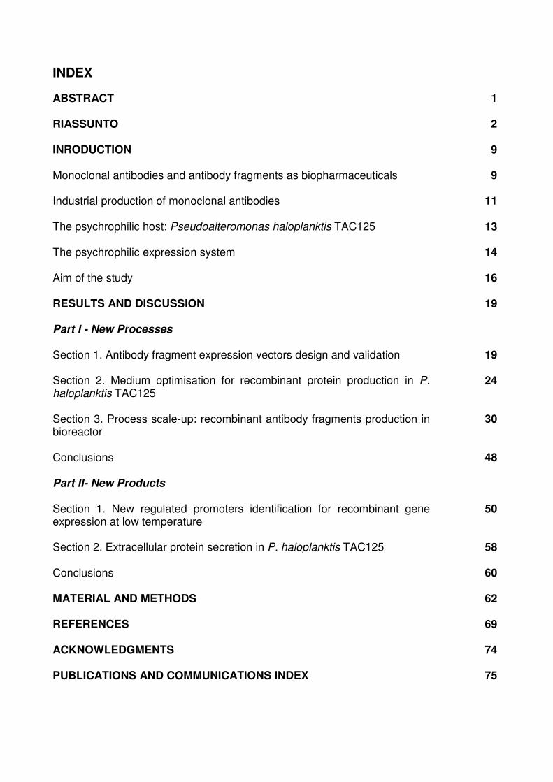
INDEX
ABSTRACT
1
RIASSUNTO
2
INRODUCTION
9
Monoclonal antibodies and antibody fragments as biopharmaceuticals
9
Industrial production of monoclonal antibodies
11
The psychrophilic host: Pseudoalteromonas haloplanktis TAC125
13
The psychrophilic expression system
14
Aim of the study
16
RESULTS AND DISCUSSION
19
Part I - New Processes
Section 1. Antibody fragment expression vectors design and validation
19
Section 2. Medium optimisation for recombinant protein production in P. haloplanktis TAC125
24
Section 3. Process scale-up: recombinant antibody fragments production in bioreactor
30
Conclusions
48
Part II- New Products
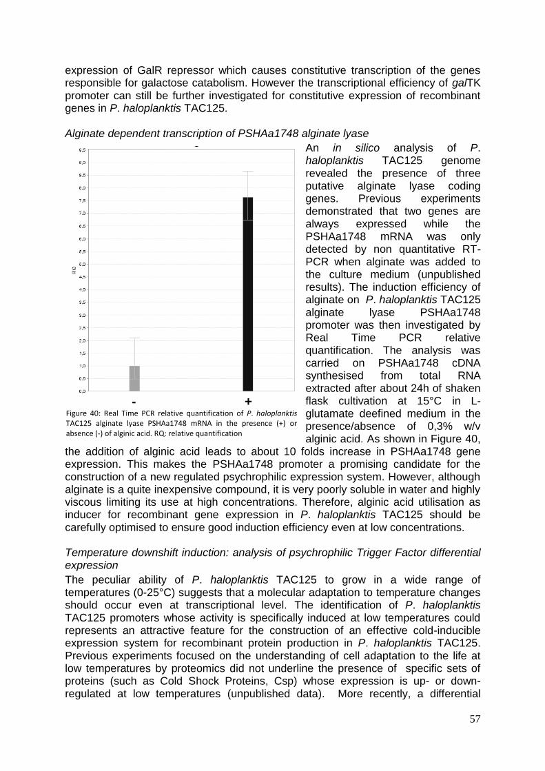
Section 1. New regulated promoters identification for recombinant gene expression at low temperature
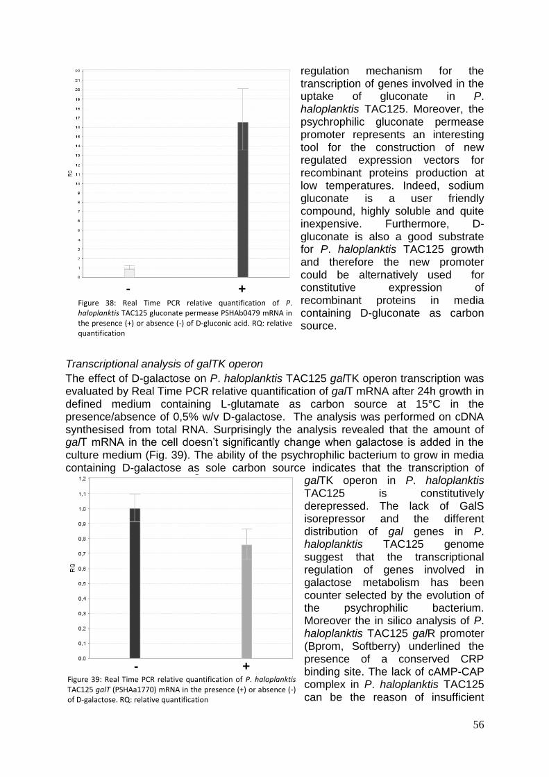
50
Section 2. Extracellular protein secretion in P. haloplanktis TAC125
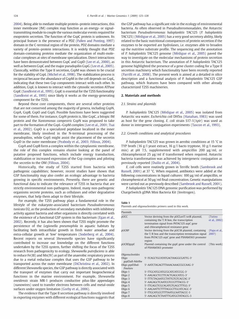
58
Conclusions
60
MATERIAL AND METHODS
62
REFERENCES
69
ACKNOWLEDGMENTS
74
PUBLICATIONS AND COMMUNICATIONS INDEX 75
1
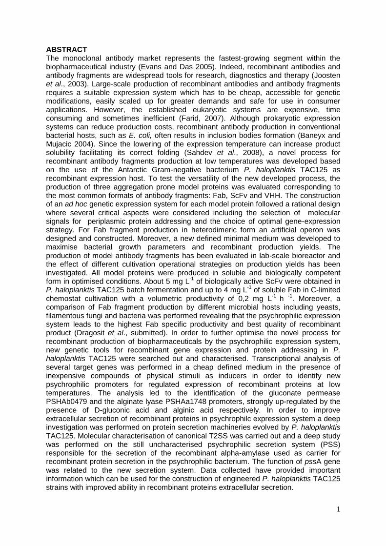
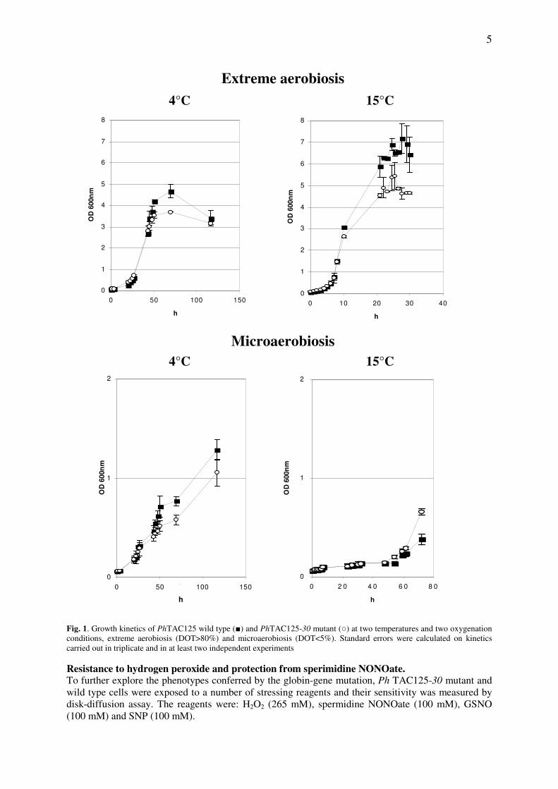
ABSTRACT The monoclonal antibody market represents the fastest-growing segment within the biopharmaceutical industry (Evans and Das 2005). Indeed, recombinant antibodies and antibody fragments are widespread tools for research, diagnostics and therapy (Joosten et al., 2003). Large-scale production of recombinant antibodies and antibody fragments requires a suitable expression system which has to be cheap, accessible for genetic modifications, easily scaled up for greater demands and safe for use in consumer applications. However, the established eukaryotic systems are expensive, time consuming and sometimes inefficient (Farid, 2007). Although prokaryotic expression systems can reduce production costs, recombinant antibody production in conventional bacterial hosts, such as E. coli, often results in inclusion bodies formation (Baneyx and Mujacic 2004). Since the lowering of the expression temperature can increase product solubility facilitating its correct folding (Sahdev et al., 2008), a novel process for recombinant antibody fragments production at low temperatures was developed based on the use of the Antarctic Gram-negative bacterium P. haloplanktis TAC125 as recombinant expression host. To test the versatility of the new developed process, the production of three aggregation prone model proteins was evaluated corresponding to the most common formats of antibody fragments: Fab, ScFv and VHH. The construction of an ad hoc genetic expression system for each model protein followed a rational design where several critical aspects were considered including the selection of molecular signals for periplasmic protein addressing and the choice of optimal gene-expression strategy. For Fab fragment production in heterodimeric form an artificial operon was designed and constructed. Moreover, a new defined minimal medium was developed to maximise bacterial growth parameters and recombinant production yields. The production of model antibody fragments has been evaluated in lab-scale bioreactor and the effect of different cultivation operational strategies on production yields has been investigated. All model proteins were produced in soluble and biologically competent form in optimised conditions. About 5 mg L
-1 of biologically active ScFv were obtained in
P. haloplanktis TAC125 batch fermentation and up to 4 mg L-1
of soluble Fab in C-limited chemostat cultivation with a volumetric productivity of 0,2 mg L
-1 h
-1. Moreover, a
comparison of Fab fragment production by different microbial hosts including yeasts, filamentous fungi and bacteria was performed revealing that the psychrophilic expression system leads to the highest Fab specific productivity and best quality of recombinant product (Dragosit et al., submitted). In order to further optimise the novel process for recombinant production of biopharmaceuticals by the psychrophilic expression system, new genetic tools for recombinant gene expression and protein addressing in P. haloplanktis TAC125 were searched out and characterised. Transcriptional analysis of several target genes was performed in a cheap defined medium in the presence of inexpensive compounds of physical stimuli as inducers in order to identify new psychrophilic promoters for regulated expression of recombinant proteins at low temperatures. The analysis led to the identification of the gluconate permease PSHAb0479 and the alginate lyase PSHAa1748 promoters, strongly up-regulated by the presence of D-gluconic acid and alginic acid respectively. In order to improve extracellular secretion of recombinant proteins in psychrophilc expression system a deep investigation was performed on protein secretion machineries evolved by P. haloplanktis TAC125. Molecular characterisation of canonical T2SS was carried out and a deep study was performed on the still uncharacterised psychrophilic secretion system (PSS) responsible for the secretion of the recombinant alpha-amylase used as carrier for recombinant protein secretion in the psychrophilic bacterium. The function of pssA gene was related to the new secretion system. Data collected have provided important information which can be used for the construction of engineered P. haloplanktis TAC125 strains with improved ability in recombinant proteins extracellular secretion.
2
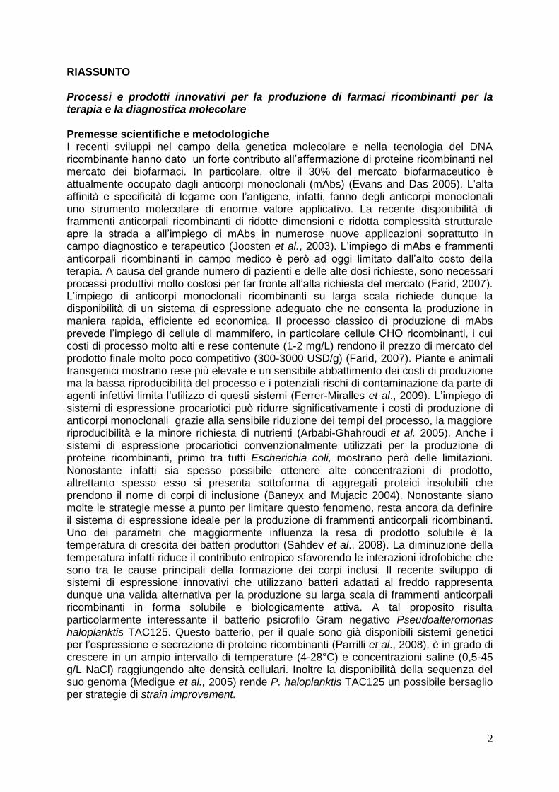
RIASSUNTO Processi e prodotti innovativi per la produzione di farmaci ricombinanti per la terapia e la diagnostica molecolare Premesse scientifiche e metodologiche I recenti sviluppi nel campo della genetica molecolare e nella tecnologia del DNA ricombinante hanno dato un forte contributo all’affermazione di proteine ricombinanti nel mercato dei biofarmaci. In particolare, oltre il 30% del mercato biofarmaceutico è attualmente occupato dagli anticorpi monoclonali (mAbs) (Evans and Das 2005). L’alta affinità e specificità di legame con l’antigene, infatti, fanno degli anticorpi monoclonali uno strumento molecolare di enorme valore applicativo. La recente disponibilità di frammenti anticorpali ricombinanti di ridotte dimensioni e ridotta complessità strutturale apre la strada a all’impiego di mAbs in numerose nuove applicazioni soprattutto in campo diagnostico e terapeutico (Joosten et al., 2003). L’impiego di mAbs e frammenti anticorpali ricombinanti in campo medico è però ad oggi limitato dall’alto costo della terapia. A causa del grande numero di pazienti e delle alte dosi richieste, sono necessari processi produttivi molto costosi per far fronte all’alta richiesta del mercato (Farid, 2007). L’impiego di anticorpi monoclonali ricombinanti su larga scala richiede dunque la disponibilità di un sistema di espressione adeguato che ne consenta la produzione in maniera rapida, efficiente ed economica. Il processo classico di produzione di mAbs prevede l’impiego di cellule di mammifero, in particolare cellule CHO ricombinanti, i cui costi di processo molto alti e rese contenute (1-2 mg/L) rendono il prezzo di mercato del prodotto finale molto poco competitivo (300-3000 USD/g) (Farid, 2007). Piante e animali transgenici mostrano rese più elevate e un sensibile abbattimento dei costi di produzione ma la bassa riproducibilità del processo e i potenziali rischi di contaminazione da parte di agenti infettivi limita l’utilizzo di questi sistemi (Ferrer-Miralles et al., 2009). L’impiego di sistemi di espressione procariotici può ridurre significativamente i costi di produzione di anticorpi monoclonali grazie alla sensibile riduzione dei tempi del processo, la maggiore riproducibilità e la minore richiesta di nutrienti (Arbabi-Ghahroudi et al. 2005). Anche i sistemi di espressione procariotici convenzionalmente utilizzati per la produzione di proteine ricombinanti, primo tra tutti Escherichia coli, mostrano però delle limitazioni. Nonostante infatti sia spesso possibile ottenere alte concentrazioni di prodotto, altrettanto spesso esso si presenta sottoforma di aggregati proteici insolubili che prendono il nome di corpi di inclusione (Baneyx and Mujacic 2004). Nonostante siano molte le strategie messe a punto per limitare questo fenomeno, resta ancora da definire il sistema di espressione ideale per la produzione di frammenti anticorpali ricombinanti. Uno dei parametri che maggiormente influenza la resa di prodotto solubile è la temperatura di crescita dei batteri produttori (Sahdev et al., 2008). La diminuzione della temperatura infatti riduce il contributo entropico sfavorendo le interazioni idrofobiche che sono tra le cause principali della formazione dei corpi inclusi. Il recente sviluppo di sistemi di espressione innovativi che utilizzano batteri adattati al freddo rappresenta dunque una valida alternativa per la produzione su larga scala di frammenti anticorpali ricombinanti in forma solubile e biologicamente attiva. A tal proposito risulta particolarmente interessante il batterio psicrofilo Gram negativo Pseudoalteromonas haloplanktis TAC125. Questo batterio, per il quale sono già disponibili sistemi genetici per l’espressione e secrezione di proteine ricombinanti (Parrilli et al., 2008), è in grado di crescere in un ampio intervallo di temperature (4-28°C) e concentrazioni saline (0,5-45 g/L NaCl) raggiungendo alte densità cellulari. Inoltre la disponibilità della sequenza del suo genoma (Medigue et al., 2005) rende P. haloplanktis TAC125 un possibile bersaglio per strategie di strain improvement.
3
Obiettivi L’obiettivo principale di questo progetto di ricerca è lo sviluppo e la messa a punto di un processo per la produzione di frammenti anticorpali ricombinanti nel batterio psicrofilo Pseudoalteromonas haloplanktis TAC125. L’efficienza del nuovo processo sarà valutata attraverso la produzione di tre proteine modello corrispondenti ai più comuni formati di anticorpi monoclonali, in particolare: -Fab (fragment antigen binding): nell’ambito del progetto europeo GENOPHYS il frammento Fab anti-idiotipo Ab2/2H5 Fab3H6 (Kunert et al., 2002) è stato scelto come proteina modello per l’analisi comparativa della produzione ricombinante di una proteina complessa in diversi organismi ospiti (lieviti, funghi filamentosi e batteri). Inoltre, il Fab3H6 è una molecola di grande interesse biofarmaceutico in quanto costituisce un elemento fondamentale nella formulazione di un vaccino contro un ampio spettro di ceppi del virus dell’immunodeficienza umana di tipo 1 (HIV-I) (Kunert et al., 2002). Il frammento Fab è un eterodimero di circa 50 kDa costituito da una catena leggera completa (CL + VL) legata mediante un ponte disolfurico ad un frammento della catena pesante che comprende il dominio variabile VH e il dominio costante CH1. -ScFv (Single chain variable Fragment): l’anticorpo a singola catena anti 2-fenil-5-ossazolone ScFvOx (Fiedler and Conrad, 1995) è stato scelto come modello di questa classe di frammenti anticorpali in quanto si presenta come una proteina molto insolubile ed è utilizzata come modello per l’ottimizzazione di protocolli di refolding di corpi di inclusione (Patil et al., 2008). L’anticorpo a singola catena è un frammento contenente le sole regioni variabili della catena pesante e leggera di un anticorpo monoclonale legate da un linker flessibile in modo da costituire un’unica catena polipeptidica di circa 30 kDa. -VHH (heavy chain antibody fragment): come modello per questa classe di frammenti anticopali che consiste nella sola regione variabile di anticorpi di camelidi caratterizzati dalla naturale mancanza della catena leggera, è stato scelto il VHH D6.1 diretto contro il fattore di crescita dei fibroblasti umano 1 (FGFR1). Questo anticorpo ricombinante è stato isolato da una library naive di lama (Monegal et al., 2009) ma la sua produzione su larga scala in E. coli è limitata a causa della formazione dei corpi di inclusione. Il frammento anticorpale VHH consiste di una singola catena polipeptidica di circa 15 kDa con un solo ponte disolfurico. Allo scopo di realizzare un efficiente sistema per la produzione di farmaci ricombinanti in forma solubile e biologicamente attiva nel nuovo sistema di espressione psicrofilo, altri obiettivi di questo lavoro saranno lo sviluppo di nuovi prodotti per la produzione di proteine ricombinanti alle basse temperature. In primo luogo saranno identificati nuovi e più efficienti sistemi genetici (promotori regolati e costitutivi) per l’espressione ricombinante alle basse temperature. Inoltre sarà effettuato lo studio dei meccanismi di secrezione proteica extracellulare evoluti dal batterio psicrofilo al fine di ottimizzare i sistemi per l’espressione e secrezione di proteine ricombinanti a freddo. Risultati e discussione Parte 1. Nuovi processi per la produzione di frammenti anticorpali ricombinanti in P. haloplanktis TAC125 Allestimento e validazione del sistema genetico di espressione: il primo passo necessario allo sviluppo di un processo per la produzione di frammenti anticorpali ricombinanti alle basse temperature è stato la messa a punto di opportune cassette di espressione utilizzando i sistemi genetici per l’espressione ricombinante a freddo già disponibili (Parrilli et al., 2008). In primo luogo, dal momento che per tutti i frammenti in esame la formazione di ponti disolfurici intramolecolari è necessaria per la corretta strutturazione dei domini immunoglobulinici e dunque per l’assunzione della struttura biologicamente attiva, si è reso necessario l’indirizzamento dei prodotti ricombinanti nello
4
spazio periplasmatico. Essendo disponibili due diversi peptidi segnale per la traslocazione periplasmatica, uno isolato dalla proteina periplasmatica endogena DsbA
(Madonna et al., 2005) e l’altro dalla proteina psicrofila eterologa -amilasi da PhTAB23 (Feller et al.,1998), la proteina modello ScFvOx è stata utilizzata per valutare l’efficienza di tali segnali nel promuoverne la secrezione nel periplasma. Il ScFvOx è stato scelto in quanto risulta avere una particolare tendenza ad aggregare se non messo in condizione di strutturarsi correttamente. Inoltre è stata valutata a possibilità di indirizzare il prodotto
ricombinante nel mezzo extracellulare mediante la fusione con la proteina carrier -amilasi, già dimostratasi in grado di promuovere la secrezione extracellulare di diverse proteine ricombinanti (Cusano et al., 2006). Il gene codificante l’anticorpo scFvOx è stato dunque fuso a valle delle due diverse sequenze segnale per la traslocazione
periplasmatica e al gene codificante l’ -amilasi e posta sotto il controllo di un promotore costitutivo forte. L’analisi della localizzazione cellulare del prodotto ricombinante è stata condotta in terreno ricco attraverso esperimenti di Western blotting su estratti proteici citoplasmatici, periplasmatici e sul mezzo di coltura. L’analisi ha rivelato che il ScFvOx viene prodotto e correttamente traslocato nel compartimento periplasmatico solo quando fuso al peptide segnale isolato dalla DsbA. Non si osserva invece alcuna produzione
quando la proteina è dotata del peptide segnale isolato dalla -amilasi. Inoltre, l’analisi della produzione del prodotto di fusione con la proteina carrier per la secrezione extracellulare ha rivelato un accumulo intracellulare e una conseguente degradazione proteolitica del prodotto ricombinante. Le ragioni di quanto osservato possono risiedere nel diverso meccanismo per la traslocazione periplasmatica guidato dai due diversi
peptidi segnale. L’ -amilasi infatti viene traslocata nel periplasma attraverso il sistema post-traduzionale Sec (Wickner et al., 1996). Il peptide segnale della DsbA invece presenta caratteristiche comuni a proteine riconosciute dal sistema di traslocazione SRP-like che invece segue un meccanismo co-traduzionale (Schierle et al., 2003). Probabilmente, la cinetica di aggregazione del ScFvOx nel citoplasma è maggiore di quella di secrezione periplasmatica guidata dal sistema Sec da cui consegue l’accumulo e la degradazione proteolitica del prodotto nel citoplasma. L’utilizzo di un sistema di secrezione co-traduzionale impedisce invece alla proteina di strutturarsi prima di raggiungere il compartimento periplasmatico. Per questi motivi si è scelto di utilizzare lo stesso segnale molecolare, PsDsbA, per la traslocazione periplasmatica di tutti i frammenti anticorpali in esame. Inoltre tutti i prodotti ricombinanti sono stati dotati di tag C-terminali (6xHis tag e c-Myc tag) per l’immunorivelazione e la successiva purificazione. Per quanto riguarda il frammento Fab, la sua natura eterodimerica richiede che le due catene siano sintetizzate all’interno del batterio ospite in maniera stechiometrica. A questo scopo è stato costruito un operone sintetico capace di esprimere i geni codificanti la catena leggera e il frammento VH + CH1 della catena pesante in un unico trascritto bicistronico, in modo da realizzare il coupling traduzionale dei due geni. L’operone artificiale è stato disegnato in base all’analisi in silico dei cluster genici di PhTAC125, focalizzando l’attenzione sulle regioni di sovrapposizione tra geni potenzialmente in coupling traduzionale. La costruzione ha dunque previsto una sovrapposizione tra il codone di stop del gene a monte (LC) con l’atg del gene a valle (HC) e di conseguenza l’inserimento della sequenza Shine-Dalgarno (SD), necessaria alla traduzione della HC, all’interno del gene LC attraverso l’introduzione di una mutazione silente. Dal momento che la lunghezza, la composizione e la posizione della sequenza SD hanno effetti sull’efficienza di inizio della traduzione, si è scelto di utilizzare una sequenza identica a quella che garantisce la traduzione del primo gene ed alla medesima distanza dal codone di start. Allo scopo di validare l’operone artificiale costruito è stata condotta un’analisi della produzione costitutiva e della localizzazione cellulare del prodotto il terreno ricco. Attraverso esperimenti di RT-PCR è stata verificata la stabilità del trascritto in vivo mentre mediante esperimenti di immunorivelazione condotti con anticorpi in grado
5
di riconoscere selettivamente la catena pesante e leggera del Fab è stata verificata la produzione bilanciata delle due catene e la loro corretta localizzazione periplasmatica. Al fine di utilizzare i costrutti ottenuti per l’espressione ricombinante di ScFvOx, Fab 3H6 e VHH D6.1 in bioreattore, essi sono stati infine posti sotto il controllo del promotore psicrofilo inducibile da L-malato (Papa et al., 2007) in quanto esso risulta essere il promotore più efficiente tra quelli disponibili nel promuovere la trascrizione in terreno minimo. Sviluppo di un nuovo terreno di coltura sintetico per la produzione di rAbs a freddo: al fine di massimizzare le rese di produzione dei costrutti ricombinanti realizzati, si è scelto di agire sulla composizione del mezzo di coltura. Inoltre, la disponibilità di un terreno minimo e definito, ottimale per la crescita del microrganismo ospite, rende possibile lo sviluppo di processi in chemostato. Dati pregressi hanno rivelato una preferenza da parte di PhTAC125 per substrati di natura peptidica o amminoacidica (Medigue et al., 2005) e sono state ottenute buone rese di produzione di proteine ricombinanti, alla temperatura di 15°C, in terreno minimo contenente una base salina SCHATZ e casaminoacidi come fonte di carbonio (Papa et al., 2007). Per questi motivi si è scelto di valutare diversi terreni sintetici contenenti singoli amminoacidi come unica fonte di carbonio le cui quantità sono state bilanciate sulla composizione del terreno di riferimento. Dall’analisi preliminare sono stati selezionati quattro terreni contenenti rispettivamente gli amminoacidi L-alanina, L-aspartato, L-glutammato e L-leucina come fonte di carbonio. Su questi terreni sono state allestite delle colture cellulari in beuta di
ceppi di PhTAC125 esprimenti una -galattosidasi psicrofila sotto il controllo dello stesso promotore, inducibile da L-malato, scelto per la produzione dei frammenti anticorpali. La scelta è ricaduta su questo ceppo in quanto la produzione dell’enzima psicrofilo permette di quantizzare più rapidamente la produzione ricombinante e avere così indicazioni importanti sull’efficienza del promotore nei terreni formulati. Dall’analisi dei dati ottenuti è
emerso che la produzione della -galattosidasi nel terreno contenente L-Leu risulta aumentata di circa 5 volte rispetto al riferimento (casaminoacidi). Le rese di produzione ottenute con gli altri terreni analizzati risultano invece poco soddisfacenti. L’analisi ha inoltre permesso di valutare e comparare alcuni parametri di crescita del microrganismo sui terreni formulati. In particolare è stata osservata una maggiore velocità specifica di crescita nei terreni contenenti L-Glu e L-Asp rispetto al riferimento. Dal momento che il terreno sintetico contenente L-Leu come unica fonte di carbonio ha mostrato le maggiori rese di biomassa e di prodotto ricombinante si è scelto di utilizzare tale substrato nella formulazione del terreno per l’allestimento della colture di PhTAC125 in bioreattore. In un fermentatore STR sono state allestite colture cellulari in terreni contenenti oltre che la sola L-Leu anche la stessa in combinazione con gli amminoacidi con i quali è stata osservata una elevata velocità di crescita (L-Glu, L-Asp, L-Glu+L-Asp, L-Glu+L-Ala), alla temperatura di esercizio di 15°C. Ancora una volta è stata osservata una resa di prodotto ricombinante molto più elevata nel terreno contenente la sola L-Leu. La persistenza di una lunga fase di latenza e i lunghi tempi di duplicazione che caratterizzano la crescita del microrganismo rende però questo terreno inadeguato per un utilizzo in un processo industriale. Inoltre l’aumento della concentrazione di leucina nel mezzo di coltura causa un’inibizione della crescita del batterio psicrofilo. Dati di letteratura (Quay et al.,1977) hanno evidenziato in E. coli la dipendenza della velocità di uptake della L-Leu dalla presenza degli altri amminoacidi ramificati (L-Ile e L-Val) nel mezzo di coltura. È stato dunque formulato un nuovo terreno di coltura contenente i tre amminoacidi ramificati L-Ile, L-Leu e L-Val (LIV) in un rapporto molare 1:1:2. In tale terreno è stato possibile osservare una drastica riduzione della fase di latenza della crescita cellulare e un aumento della velocità specifica di crescita a valori paragonabili a quelli ottenuti nel terreno di riferimento. Sono inoltre state condotte analisi del consumo dei substrati nel terreno di coltura per valutare la cinetica di consumo dei singoli componenti durante la
6
fase di crescita esponenziale e la loro concentrazione residua al termine della crescita. La curva del consumo dei substrati evidenzia che i tre amminoacidi sono consumati velocemente all’inizio della fase di crescita esponenziale mentre la concentrazione di acido L-malico resta pressoché invariata. Dopo 24 ore di crescita la velocità di consumo degli amminoacidi diminuisce sensibilmente mentre si ha un rapido consumo del malato la cui concentrazione si riduce a valori prossimi allo zero. Alla fine della fase di batch la concentrazione degli amminoacidi residua nel mezzo di coltura è ancora non trascurabile. Alla luce di quanto osservato, la insufficiente conversione dei substrati in biomassa (Yx/s=30%) potrebbe essere causata da uno squilibrio di nutrienti nel mezzo di coltura e potrebbe essere aumentata riformulando il brodo di coltura in base alla composizione elementare del microrganismo. È stata infine valutata la produzione di ammonio nel mezzo di coltura ed è stata osservata una cinetica di accumulo sovrapponibile a quella osservata per la curva di crescita. Questo dato concorda con l’osservazione di una forte tendenza del batterio a rendere più basico il pH del mezzo di crescita e potrebbe essere dovuto ad un eccesso di azoto nel terreno di coltura. Sulla base di quanto osservato, è stato inoltre allestito un processo per la crescita di PhTAC125 in continuo. Dopo una fase batch in terreno LIV è stato mantenuto lo steady state per 5 tempi di residenza addizionando L-Leu con una velocità di diluizione pari a 0,05 h
-1.
Scale up del processo di produzione dei frammenti anticorpali in bireattore: la produzione del frammento anticorpale Fab3H6 è stata effettuata in primo luogo in modalità batch in un bioreattore STR nel terreno di crescita precedentemente ottimizzato e in presenza dell’induttore L-malato. L’analisi della produzione, effettuata mediante saggio ELISA su campioni prelevati a diversi tempi di crescita, ha rivelato una accumulo del Fab3H6 fino a una resa di circa 4 mg L
-1. Attraverso esperimenti di Western blotting è stata inoltre
dimostrata la completa traslocazione del prodotto ricombinante nel periplasma. Nell’ambito del progetto europeo GENOPHYS, inoltre, la produzione del frammento Fab3H6 è stata valutata in chemostato utilizzando terreno LIV sia per la fase di crescita in batch che per l’alimentazione. Nelle condizioni scelte è stato possibile stabilizzare una
produttività volumetrica di Fab3H6 periplasmatico di circa 200 g L-1
h1. Inoltre
dall’analisi comparativa della produzione del Fab3H6 in diversi microrganismi ospite è emerso che PhTAC125 mostra la più alta resa di prodotto per grammo di biomassa e rese di produzione paragonabili a quelle ottenute nel consolidato Pichia pastoris. Il processo di produzione del Fab3H6 in continuo è stato ulteriormente ottimizzato attraverso l’utilizzo della sola L-Leu nel terreno di feeding. Le rese di prodotto ottenute con il nuovo processo sono paragonabili a quelle in terreno LIV ma i costi di processo risultano sensibilmente ridotti. Il processo per la produzione del frammento anticorpale ricombinante ScFvOx è stato esercito in modalità batch nelle condizioni precedentemente descritte per il Fab3H6. L’analisi della produzione, effettuata attraverso esperimenti di ELISA su estratti cellulari solubili prelevati a diversi tempi di crescita, ha rivelato un accumulo del prodotto ricombinate con una resa di produzione massima in tarda fase esponenziale (48h) di circa 4,7 mg L
-1. La verifica della corretta localizzazione cellulare è stata effettuata
mediante SDS-PAGE su estratti periplasmatici, citoplasmatici e totali di aliquote di coltura cellulare prelevate dopo 48 ore di crescita. Dall’analisi è emerso che l’anticorpo ScFvOx è prodotto in forma solubile e correttamente localizzato nel periplasma nelle condizioni analizzate. Il prodotto ricombinante è stato dunque purificato su piccola scala mediante cromatografia di pseudo affinità sfruttando la coda di poli-istidine presenta all’estremità C-terminale del prodotto di interesse. È stata infine condotta un’analisi dell’attività biologica del prodotto ricombinante ottenuto mediante esperimenti di ELISA in presenza dell’aptene 2-fenil-ossazolone. L’analisi ha rivelato una resa di proteina pura e biologicamente attiva di circa 4 mg per litro di coltura. L’analisi della produzione del
7
frammento ScFvOx è stata effettuata inoltre in colture cellulari in continuo. La stabilizzazione del processo in continuo è stata ottenuta in terreno LIV con D=0,05
-1 e la
produttività volumetrica di ScFvOx osservata è stata pari a 230 g L-1
h-1
. La produzione del frammento anticorpale VHH D6.1, infine, è stata effettuata in un processo in batch nelle condizioni già ottimizzate per gli altri frammenti. La verifica della produzione, effettuata attraverso esperimenti di immunorivelazione ha rivelato la presenza di una banda specifica di peso molecolare apparente di circa 30kDa, superiore alla massa attesa (≈15KDa), in corrispondenza degli estratti periplasmatici ottenuti da aliquote di coltura cellulare prelevati a diversi tempi di crescita. Il prodotto ottenuto è stato parzialmente purificato mediante cromatografia di pseudo affinità sfruttando la coda di poli-istidine presenta all’estremità C-terminale ma non è stato possibile effettuare un saggio di attività biologica in quanto il prodotto purificato è risultato poco stabile in soluzione. Parte 2. Nuovi prodotti per la produzione di farmaci ricombinanti in P. haloplanktis TAC125 Identificazione di nuovi promotori regolati e costitutivi per l’espressione genica ricombinante in PhTAC125 Allo scopo di migliorare l’efficienza del nuovo sistema di espressione a freddo e di rendere i processi di produzione di farmaci ricombinanti in PhTAC125 più competitivi in campo industriale si è scelto di sfruttare la conoscenza della sequenza del genoma del batterio psicrofilo per l’identificazione di nuovi e più efficienti sistemi per l’espressione regolata di geni ricombinanti. Il processo per la produzione di frammenti anticorpali ricombinanti precedentemente ottimizzato, basato sull’utilizzo del promotore indotto dalla presenza di L-malato nel mezzo di coltura, può risultare infatti economicamente vantaggioso solo se il prodotto presenta un elevato valore aggiunto, considerati l’alto costo dei substrati richiesti per massimizzare l’efficienza trascrizionale e quello dell’induttore stesso. Per questo motivo, mediante analisi in silico del genoma batterico, sono stati identificati alcuni promotori potenzialmente regolati e ne è stata valutata l’efficienza trascrizionale in vivo, in presenza ed in assenza di induzione, in un terreno di crescita minimo, molto economico, contenente L-glutammato come unica fonte di carbonio. In questo terreno infatti PhTAC125 mostra un’alta velocità specifica di crescita e una discreta resa in biomassa (Yx/s≥40%). In particolare, attraverso esperimenti di Real Time PCR, è stata valutata la variazione del numero di copie dei trascritti in esame nelle condizioni di induzione rispetto alla condizione standard (terreno con L-Glu, T=15°C). E’ stata dunque valutata la variazione del numero di copie dei seguenti trascritti: il gene galT appartenente all’operone del galattosio, in assenza e i presenza di galattosio; la CDS PSHAa1748 codificante una putativa alginato liasi, in presenza e in assenza di alginato; la CDS PSHAb0479 codificante una gluconato permeasi, in presenza e assenza di gluconato. Tutti i potenziali induttori sono stati addizionati al terreno standard a t=0 alla concentrazione di 4,0 g L
-1. E’ stata inoltre valutata la
variazione dell’efficienza trascrizionale del promotore del gene PSHAa2063 codificante il Trigger Factor (TF) alle temperature di 15°C e 4°C. Il TF è stato infatti precedentemente identificato, mediante analisi di proteomica differenziale, tra le proteine di PhTAC125 sovraespresse a basse temperature. L’analisi ha rivelato un effetto di induzione solo da parte dell’alginato (circa 10 volte rispetto alle condizioni standard) e del gluconato (circa 20 volte) sui rispettivi promotori che potranno dunque essere utilizzati per la costruzione di nuovi vettori per l’espressione regolata di geni ricombinanti a freddo. Studio dei sistemi di secrezione extracellulare nel batterio PhTAC125 La comprensione dei meccanismi di secrezione sfruttati dal batterio antartico PhTAC125 è un presupposto indispensabile per poter ottimizzare i processi di produzione di farmaci
8
ricombinanti a basse temperature. La collocazione extracellulare del prodotto di interesse consente infatti di agevolare notevolmente i processi di downstream data la minore concentrazione e complessità del contenuto proteico nel mezzo extracellulare. La recente disponibilità della sequenza completa del genoma di PhTAC125 ha facilitato lo studio della secrezione nel batterio antartico. Dall’analisi in silico del genoma è emerso che, nonostante il gran numero di proteine secrete dal batterio, PhTAC125 possiede solo uno dei sistemi di secrezione finora caratterizzati nei Gram negativi, il sistema di tipo II GSP (General Secretory Pathway). Attraverso la costruzione di un mutante genomico in cui il gene gspE, codificante l’ATPasi del sistema GSP, è stato inattivato, è stato dimostrato che il T2SS è responsabile della secrezione della maggior parte delle esoproteasi in PhTAC125. Il dato più interessante emerso da questo studio è che, nonostante l’unico sistema di secrezione del batterio risulti inattivato, il mutante PhTAC125 (gspE
-) è ancora in grado di secernere proteine nel mezzo extracellulare
indicando la presenza di almeno un altro sistema di secrezione, non ancora caratterizzato, evolutosi nel batterio psicrofilo. Inoltre, il sistema GSP non risulta essere
in alcun modo responsabile della secrezione della -amilasi eterologa, utilizzata come carrier per la secrezione di proteine ricombinanti nel batterio psicrofilo (Parrilli et al., 2008). Allo scopo di identificare il nuovo sistema di secrezione responsabile della
localizzazione extracellulare dell’ -amilasi nel batterio psicrofilo, il sistema PSS (Psychrophilic Secretion System), è stata realizzata una strategia di complementazione che ha previsto la costruzione di una genoteca cosmidica di PhTAC125 e il suo
trasferimento in cellule di E. coli ricombinanti in grado di produrre l’ -amilasi psicrofila ma non di secernerla nel mezzo di coltura. Tale strategia ha permesso di isolare un solo clone cosmidico, contenente una regione di circa 37Kb del cromosoma b di PhTAC125,
in grado di conferire al batterio mesofilo la capacità di secernere l’ -amilasi nel mezzo extracellulare. Dall’analisi della sintenia della porzione genica di PhTAC125 contenuta nel cosmide selezionato con genomi di organismi ad esso vicini filogeneticamente è emerso che la regione comprendente le CDS dalla PSHAb0134 alla PSHAb0142 è molto conservata nei microrganismi selezionati tranne per la CDS PSHAb0140. Inoltre non è presente un omologo di tale gene in E. coli. Per comprendere il ruolo della funzione
codificata dal gene PSHAb0140 nella secrezione dell’ -amilasi è stato costruito un mutante genomico di PhTAC125 in cui tale gene è stato deleto. Tale mutante non è in
grado di secernere l’ -amilasi nel mezzo di coltura dimostrando che il prodotto della CDS PSHAb0140, d’ora in avanti chiamato PssA, è necessario per il corretto funzionamento del nuovo sistema di secrezione PSS. Dall’analisi in silico si evince che PssA è un’ipotetica lipoproteina costituita da un peptide leader caratteristico di questa famiglia di proteine che la colloca nello spazio periplasmatico ancorata alla membrana esterna, da tre domini TPR (Tetratricopeptide Reapeat Domain) presenti all’estremità N-terminale e da due domini LysM di legame al peptidoglicano all’estremità C-terminale. Queste caratteristiche insieme ai risultati sperimentali ottenuti suggeriscono per la proteina PssA un ruolo di adattatore molecolare incaricato di reclutare altri gli componenti cellulari
necessari alla secrezione dell’ -amilasi e degli altri substrati del sistema PSS nel mezzo di coltura.
9
INTRODUCTION Monoclonal antibodies and antibody fragments as biopharmaceuticals
According to recent reports, it is clear that recombinant antibodies have come of age as biopharmaceuticals. The global sales of monoclonal antibodies were $33 billion in 2008 as compared to $27 billion in 2007 and total revenues are predicted to increase in the next years (Canadian Corporate News Report, 2008).
Antibodies are glycoproteins which specifically recognise foreign molecules (antigens). IgG antibodies are large molecules of about 150 kDa composed of 4 peptide chains. They contain two identical heavy chains of about 50 kDa and two identical light chains of about 25 kDa, thus tetrameric quaternary structure. The two heavy chains are linked to each other and to a light chain each by disulfide bonds.
The resulting tetramer has two identical halves which together form the Y-like shape. Each end of the fork contains an identical antigen binding site (Fig. 1). The unique ability of antibodies to specifically recognise and bind with high affinity to virtually any type of antigen, made them interesting molecules for medical and scientific research. In 1975 Köhler and Milstein developed the monoclonal antibody technology (Köhler et al. 1975) by immortalising mouse cell lines that secreted only one single type of antibody with unique antigen specificity, called monoclonal antibodies (mAbs). With this technology, isolation and production of mAbs against protein,
carbohydrate, nucleic acids and hapten antigens was achieved resulting in a rapid development of the use of antibodies in diagnostics, human therapeutics and as fundamental research tools. The development and applications of recombinant DNA technology led to the design of several formats of recombinant antibody fragments (Fig.2). Smaller recombinant antibody fragments (for example, classic monovalent antibody fragments (Fab, scFv) and engineered variants (diabodies, triabodies, minibodies and single-domain antibodies) retain the targeting specificity of whole mAbs and possess other unique and superior properties for a range of diagnostic and therapeutic applications. Indeed, for some clinical applications small antibody fragments have advantages over whole antibodies. Firstly, the lack of Fc regions reduces the risks of immune response. Secondly, the small size permits them to penetrate tissues and solid tumours more rapidly than whole antibodies (Yokota et
al., 1992). Furthermore smaller antibody fragments have a much faster clearance rate in the blood circulation, which leads to differences of selectivity (Yokota et al., 1992). By recombinant DNA technology, antibody fragments have been forged into multivalent and multispecific reagents and engineered for enhanced therapeutic efficacy. A new use of the binding capacity of antibody fragments is the design of a fusion approach, in which an antigen recognising antibody fragment is coupled to a
range of effector molecules (Fig.3) including enzymes for prodrug therapy, toxins for cancer treatment (Schrama et al., 2006), viruses for gene therapy, cationic tails for DNA delivery, liposomes for improved drug delivery and biosensors for real-time detection of target molecules (Spooner et al., 1994). The use of bi-functional antibodies in medicine is aimed at delivery of an effecter which is only active where it is required. It thereby limits the dose of the drug, resulting in less side effects of the drug towards healthy tissue and/or less immunogenic response to the drug itself. Also the physical interaction between the target and the effector molecule increases the potency of the effecter. More applications outside research and medicine can be considered, such as consumer applications. Single-domain antibodies are anticipated to significantly expand the repertoire of antibody-based reagents against the vast range of novel
biomarkers being discovered through proteomics. Examples are the use in biosensors, treatment of wastewater (Graham et al., 1995), industrial scale separation processes such as separation of chiral molecules (Got et al., 1997), purification of specific components like proteins from biological materials or the use as abzymes (Wade et al., 1997). They have also been considered as components of novel consumer goods with new improved functionalities like
the use of antibodies in shampoos to prevent the formation of dandruff or in toothpaste to protect against tooth decay caused by caries (Frenken et al., 1998). Antibody therapeutics are already a multi-billion dollar a year market and a large number of monoclonal antibodies and antibody fragments are at various stages of clinical trials (Evans and Das 2005). However, they are amongst the most expensive of all drugs where the annual cost per patient can reach $35,000 for treating cancer
Figure 3: Multispecific antibody fragments (Schrama et al., 2006).
11
(Farid, 2007). Indeed antibody therapies involve most often high doses (>1 g per patient per year) for a large number of patients and this comes up to a total production demand in the range of multi-tons per year (Farid, 2007). Consequently, expensive large-scale production capacity is required to fulfill market demand. Industrial production of monoclonal antibodies
To be able to use monoclonal antibodies, antibody fragments and antibody fusion proteins in large scale applications, a suitable expression system has to be chosen. The possibility of large-scale production of antibodies and fragments requires that the production system is cheap, accessible for genetic modifications, easily scaled up for greater demands and safe for use in consumer applications. Several expression systems are available, both from eukaryotic (Table 1) and prokaryotic (Table 2) origin.
Eukaryotic expression systems
Several eukaryotic systems can be envisaged for large-scale production of monoclonal antibodies and fragments like mammalian cells, insect cells, plants, transgenic animals and lower eukaryotes. Most of the approved monoclonal antibodies are manufactured in mammalian cells. The majority of them use batch/fed batch cultures followed by purification steps that rely primarily on chromatography with intermediate filtration and viral clearance operations. However, large-scale production is expensive and time-consuming (Farid, 2007). Cultured insect cells are used as hosts for recombinant baculovirus infections. The production of a recombinant viral vector for gene expression is time-consuming, the cell growth is slow when compared with former expression systems, the cost of growth medium is high and each protein batch preparation has to be obtained from fresh cells since viral infection is lethal. (Ferrer-Miralles 2009). Plants show several advantages as large-scale antibody production systems, like the ease and low costs of growing plants, even in large quantities. However, the generation of transgenic plants that express antibodies is a time consuming process and the downstream processing to isolate the recombinant antibodies from the plant parts is relatively expensive and laborious (Joosten et al., 2003). An attractive possibility for the cost-effective large-scale production of antibody fragments and antibody fusion proteins are yeast or fungal fermentations. Large-scale fermentation of these organisms is an established
+++ = excellent, ++ = good, + = sufficient, +/- = poor. * With transgenic animals in this context is mentioned the production of antibodies or antibody fragments in the milk of transgenic animals, for example rabbits, sheep, goats or cows
1 With
economical feasibility is mentioned the time and cost of molecular cloning, upscaling and downstream processing (purification).
2 Pathogenic contaminants like viruses or pyrogens. Modified from: Joosten et al. 2003
Table 1
Eukaryotic expression systems for heterologous protein production and possible advantages and disadvantages of the expression system.
12
technology already used for bulk production of several other recombinant proteins and extensive knowledge is available on downstream processes. Besides that, yeasts and filamentous fungi are accessible for genetic modifications and the protein of interest may be secreted into the culture medium. The main pitfall of this expression system is related to N-linked glycosylation patterns which differ from higher eukaryotes, in which sugar side chains of high mannose content affect the serum half-life and immunogenicity of the final product (Ferrer-Mirales et al., 2009). Furthermore, although less studied than in bacteria, the production of recombinant proteins in yeasts also triggers conformational stress responses and produced proteins fail sometimes to reach their native conformation (Gasser et al., 2008).
Prokaryotic expression systems
Bacterial expression systems have also been investigated for their potential to produce mAbs and different mAb fragments. While many bacterial strains, either Gram positive and Gram negative, have been tested for recombinant antibodies production with different extents (Tab.2), most experience has been gathered with Escherichia coli (Ferrer-Mirales et al., 2009). Incentives for the use of E. coli expression systems include simple fermentation conditions, ease of genetic manipulation, ease of scale-up, relatively short duration between transformation and protein purification, no concerns about viruses that are harmful to humans and relatively low capital costs for fermentation (Arbabi-Ghahroudi et al. 2005). However, several obstacles to the production of quality proteins limit its application as a factory for recombinant pharmaceuticals. Indeed, heterologous proteins over-expression in E. coli often results in insoluble aggregates production as cytoplasmic or periplasmic inclusion bodies (Baneyx and Mujacic 2004). Inclusion body (IB) proteins need elaborate and cost-intensive solubilisation, refolding and purification procedures to recover functionally active product (Vallejo et al., 2004). Hence, the final yield of fragments is only a small percentage of the protein that was initially present in the inclusion bodies and this causes a huge increase of process costs. In order to face the challenge to achieve a finer balance between the quality and the yield of recombinant proteins many strategies have been explored including expression of chimerical proteins (Park et al., 2008) and co-expression with chaperones (Kolaj et al., 2009). Expression of “difficult” proteins has also been carried out by lowering the temperature at the physiological limit allowed for the growth of mesophilic host organism (between 15 and 18°C). Lowering the temperature, in fact, has a pleiotropic effect on the folding process, destabilizing the hydrophobic interactions needed for intermediates aggregation (Sahdev et al., 2008). Although in some cases this approach has been reported to increase yields of soluble and active recombinant protein products, the major drawback in E. coli cultivation at sub-optimal temperatures is the decrease in biomass production which reduces the global process productivity. Therefore the exploitation of an industrial process performed in this conditions might hardly be considered. A rational alternative to improve the quality or recombinant products is to explore the potential of naturally cold-adapted bacteria as hosts for protein production at low temperature (even at around 0°C). In this context, a few cold adapted species are under early but intense exploration as cold cell factories, among them, Pseudoalteromonas haloplanktis being a representative example. The development of a cold expression system for recombinant proteins production in P. haloplanktis TAC125 was already described (Parrilli et al., 2008) and examples of fully soluble and biologically competent production of several thermal-labile and aggregation-prone proteins were extensively
13
reported (Vigentini et al., 2006; Papa et al., 2007). Furthermore, with respect to E. coli, P. haloplanktis TAC125 is extremely efficient in secreting proteins in the culture medium. By the use of a psychrophilic α-amylase as secretion carrier for the extra-cellular targeting of recombinant proteins an efficient gene-expression system was set up (Cusano et al., 2006b). Observed efficiency of the cold-adapted system (secretion yield was always above 80%) placed it amongst the best heterologous secretion systems in Gram-negative bacteria reported so far.
Therefore, the use of psychrophilic bacteria as alternative expression hosts is the compelling choice towards the exploitation of industrial processes for the production of soluble and biologically competent recombinant antibody fragments at low temperatures. Table 2
Antibody fragments production in prokaryotic expression systems
*ND = not determined. Modified from: Joosten et al., 2003
The psychrophilic host: Pseudoalteromonas haloplanktis TAC125
P. haloplanktis TAC125 is a Gram-negative bacterium isolated from an Antarctic coastal seawater sample collected in the vicinity of the French Antarctic station Dumont D’Urville, Terre Adélie. It can be classified as a Eurypsychrophile (i.e. a bacterium growing in a wide range of low temperatures; Atlas and Bartha, 1993) and was the first Antarctic Gram-negative bacterium of which the genome was fully sequenced and carefully annotated (Médigue et al., 2005). Genomic and metabolic features of this bacterium, accounting for its remarkable versatility and fast growth compared with other bacteria from aqueous environments, were discovered by combining genome sequencing and further in silico and in vivo analyses. P. haloplanktis TAC125 is able to duplicate in a wide range of temperatures (0-30°C), with an apparent optimal growth temperature a t 20°C, where the observed duplication time in rich medium is 31 minutes (Tutino et al., 1999). However, the bacterium still duplicates at fast speed even at lower temperatures (at 4°C, one cell division is completed in about 100 min; unpublished results from this laboratory) and, when provided with sufficient nutrients and aeration, it grows to very high density (up to A600=20) under laboratory settings, even at 0°C. This growth performance makes it one of the faster growing psychrophiles characterised so far. Fast growth rates, combined with the ability of P. haloplanktis TAC125 to reach very high cell densities even under laboratory growth conditions and to be easily transformed by intergeneric conjugation (Duilio et al., 2004), made this bacterium an attractive host for the development of an efficient gene expression system at low temperatures.
14
The psychrophilic expression system The psychrophilic expression vector
A few other reported examples of recombinant protein production in psychrophiles made use of molecular signals (such as the origin of replication and the transcriptional promoter) derived from mesophiles. A different philosophy inspired the construction of our gene-expression systems, which derived from the proper assembly of true psychrophilic molecular signals into a modified E. coli cloning vector. By combining mesophilic and psychrophilic genetic signals a collection of
gene-expression vectors was set up to produce recombinant proteins in P. haloplanktis TAC125 (Fig. 4). The mesophilic signals consist of the pUC18-derived origin of replication (OriC) and a
selection marker gene (a -lactamase encoding gene), allowing the plasmid to replicate either in E. coli or in the psychrophilic host. Another crucial mesophilic signal is represented by the OriT sequence, the conjugational DNA transfer origin from the
broad host range plasmid pJB3 (Blatny at al., 1997). Structural and functional studies led to the isolation of the psychrophilic origin of replication (OriR) from the P. haloplanktis TAC125 endogenous plasmid pMtBL (Tutino et al., 2001).
Psychrophilic promoters
○ Constitutive expression The structural/functional characterisation of P. haloplanktis TAC125 promoters was carried out by random cloning of genomic DNA fragments and identification of promoter sequences by evaluating their capability to express a promoter-less reporter gene (Duilio et al., 2004). By this promoter-trap strategy, a collection of constitutive psychrophilic promoters showing different strengths at different temperatures was identified. The implementation of the above described psychrophilic promoters in the pMtBL-derived shuttle vectors resulted in the set up of cold-adapted gene-expression systems, characterised by the constitutive production of the recombinant protein.
○ Regulated expression Sometimes efficient production can only be achieved by fine tuning the recombinant gene expression. This goal can be reached by using regulated promoters and efficient induction strategies. Recently, by using a differential proteomic approach, the isolation and characterisation of a two-component system has been carried out. This regulatory system is responsible for the transcriptional regulation of the gene coding for an outer protein porine, and it is strongly induced by the presence of L-malate in the medium (Papa et al., 2006). The regulative region of the porine gene
Figure 4: The psychrophilic expression vector
15
was used for the construction of an inducible cold expression vector, where the recombinant protein expression is under L-malate control (Papa et al., 2007). Molecular signals for protein addressing
○ Periplasmic secretion Although the production of recombinant protein in the host cytoplasm is the preferred strategy many processes due to higher production yields, this approach cannot be pursued when the wanted product requires the correct formation of disulphide bonds to attain its catalytic competent conformation. Indeed as for all Gram-negative bacteria, P. haloplanktis TAC125 cytoplasm is a reducing environment and the formation of disulphide bridges is confined in the periplasmic space. From the genome analysis, we know that P. haloplanktis TAC125 contains all the canonical periplasmic export machineries (Medigue et al., 2005). Therefore, gene fragments encoding two signal peptides from psychrophilic secreted proteins following different translocation mechanisms have been cloned in the psychrophilic expression vectors, under the control of different promoters in order to allow the signal peptides N-terminal fusion for periplasmic addressing of recombinant proteins.
○ Extra-cellular secretion In order to combine the effects of low temperatures on the recombinant product solubility with the advantages linked to extra-cellular protein targeting, a gene expression system for the production and extra-cellular secretion of recombinant proteins in psychrophilic bacteria was set up. The novel system makes use of the
psychrophilic -amylase from P. haloplanktis TAB23 (Feller et al., 1992) as a secretion carrier. This exo-protein is produced and secreted as a larger precursor
with a long C-terminal pro-peptide that is not mandatory for the -amylase secretion when it is produced by recombinant cold-adapted bacteria the propeptide (Tutino et al., 2002; Cusano et al., 2006a). Starting from the latter observation, the secretion of
chimeric proteins obtained by the replacement of -amylase C-terminal propeptide with a passenger protein was studied (Cusano et al., 2006b). The novel genetic system (Fig. 5) allows the easy in-frame cloning of any gene downstream of the
mature psychrophilic -amylase encoding region. The spacer between the carrier and passenger proteins contains the motif -Ala-Ser-Ser-Thr- recognised and cleaved by a P. haloplanktis TAC125 secreted protease that allows the separation of the protein of interest from the secretion carrier when it reaches the extra-cellular medium.
Figure 5: Schematic representation of recombinant proteins fusion to the secretion carrier
SP mature -amylase passenger protein
16
Aim of the study The aim of this study is the development of novel processes, involving P. haloplanktis TAC125 as expression host, for large scale production of recombinant antibody fragments in soluble and biologically competent form and new products for recombinant production of biopharmaceuticals at low temperatures. New processes Based on the previously described genetic tools for recombinant protein production and cellular addressing in P. haloplanktis TAC125, an ad hoc expression system for recombinant antibody fragments soluble production will be constructed and validated. By a rational approach, a new defined medium will be developed in order to maximise the expression system efficiency and biomass productivity. Antibody fragments production processes will be scaled up to lab scale bioreactors and tested in batch and continuous fermentation modes. To test the versatility of the psychrophilic expression system three different formats of recombinant antibody fragments will be considered:
Fab (fragment antigen binding)
Fab fragments (fragment antigen binding) are the antigen binding domains of an antibody molecule, containing the light chain (CL + VL) and the portion VH + CH1 of antibody heavy chain (Fig.5). Besides the four intramolecular disulfide bounds, one in each immunoglobulin domains, an additional interchain disulfide bridge is present between CL and CH1. Due to the heterodimeric structure, Fab fragments are quite difficult to produce in biologically active form in microbial expression systems. Indeed, the correct assembly of recombinant light and heavy chain in functional Fab fragments can only be achieved if the two subunits are synthesized by the host cell in stoichiometric ratio. Unbalanced production can lead to formation of soluble homodimers (Hotta et al., 2004), very difficult to separate from the desired product without an antigen-affinity purification, or misfolded insoluble aggregates (Wu et al., 1998). Therapeutic Fab fragments are mostly produced in mammalian expression systems or in yeasts while just a few examples are reported of Fab expression in bacteria (Peterson et al., 2006).
Figure 5: Schematic representation of the structure of a conventional IgG and fragments that can be generated thereof and used in this work. The constant heavy-chain domains CH1, CH2 and CH3 are shown in yellow, the constant light-chain domain (CL) in green and the variable heavy-chain (VH) or light-chain (VL) domains in red and orange, respectively. Modified from Joosten et al., 2003
17
In the framework of the European project GENOPHYS the anti-idiotypic antibody Ab2/2H5 Fab 3H6 fragment (Fig. 6) was chosen as a protein model for genome-wide comparison of physiological bottlenecks in multi-subunit protein production in prokaryotic and eukaryotic microbial hosts. The mAb 2F5 is one of the very few antibodies with the ability to neutralize a wide spectrum of type 1 human immunodeficiency virus (HIV-1) strains and primary isolates. However, neutralizing 2F5 like antibodies are rarely detected in patients sera. The anti-idiotypic antibody Ab2/3H6 Fab is able to recognize the paratope of mAb 2H5 bearing a structural miming of ELDKWA , the core epitope of gp-41, which is only exposed during the fusion of
the virus to cells. It can therefore provide the means to induce a broadly neutralizing anti HIV-1 antibody response and could represent a key component in anti HIV vaccines formulation (Kunert et al., 2002). The Ab2/2H5 Fab 3H6 represents an interesting model of complex therapeutic protein to validate the new developed processes for recombinant biopharmaceuticals production by the psychrophilic expression system. Furthermore, in the framework of GENOPHYS European project, a comparison of Fab 3H6 production in different microbial hosts (bacteria, yeasts and filamentous fungi) will be performed in chemostat cultivations. ScFv (Single chain variable Fragment)
ScFv (Single chain variable Fragment) is the minimal fragment (~30 kDa) that still contains the whole antigen-binding site of a IgG antibody, composed of both the variable heavy chain (VH) and variable light chain (VL) joined by a hydrophilic and flexible linker peptide (Fig.4). The length of the linker (15–25 amino acids) is
determinant for protein stability (Bird et al., 1988). Single chain antibodies contain two immunoglobulinic domains each with one disulfide bond. ScFvs have the tendency to form aggregates especially when over-produced in E. coli expression system (Baneyx and Mujacic 2004). To test the ability of psychrophilic expression system in recombinant biopharmaceuticals production in soluble form, the anti-2-phenyl-5-oxazolone single chain variable fragment (ScFvOx) was chosen as model (Fiedler and Conrad, 1995). ScFvOx (Fig. 7) is a typical example of aggregation-prone ScFv and it has been used for years as model for IB refolding protocol development (Patil et al., 2008).
Figure 6: Crystal structure of the 2F5 Fab'-3H6 Fab complex. S.Bryson et al. 2008)
Figure 7: Crystal structure of scFvOx in complex with its ligand (Scotti et al., 2006)
18
VHH (heavy chain antibody fragment)
In 1993 Hamers-Casterman et al. discovered a novel class of IgG antibodies in Camelidae (camels, dromedaries and llamas). These antibodies are devoid of light chains and therefore called 'heavy-chain antibody” or HCAb (Fig. 8). HCAbs have a molecular weight of ~95 kDa instead of the ~150 kDa for conventional IgG antibodies. Their binding domains consist only of the heavy-chain variable domains, referred to as VHHs (Muyldermans et al., 1999) to distinguish it from conventional VHs. VHH is the smallest available intact antigen-binding fragment (~15 kDa) and it has a great potential in therapeutic and diagnostic application as multispecific fusion
product (Joosten et al., 2003). To validate the new optimised process for recombinant antibody fragments production at low temperatures, it will be also tested for VHH format production. An anti- human fibroblast growth factor receptor 1 (FGFR1) VHH D6.1 was chosen as model protein. It has been selected by phage display from a pre-immune llama library (Monegal et al., 2009) but its large scale production in conventional E. coli expression systems was unsatisfactory due to inclusion bodies formation (De Marco A., personal communication). A new production process leading to improve soluble production of VHH D6.1 is therefore required for its further characterization.
New products In order to further optimise the novel processes for recombinant production of biopharmaceuticals by the psychrophilic expression system, new genetic tools for recombinant gene expression and protein addressing in P. haloplanktis TAC125 will be searched out and characterised. By in silico analysis of P. haloplanktis TAC125 genome several potentially regulated promoters will be identified and their transcriptional efficiency will be tested in minimal defined media in different conditions. The analysis will lead to the construction new expression vectors for regulated expression of recombinant proteins at low temperatures. In order to improve extracellular secretion of recombinant proteins in psychrophilc expression system and to identify new molecular signals for recombinant proteins addressing in culture medium, a deep investigation will be performed on protein secretion machineries evolved by P. haloplanktis TAC125. Molecular characterisation of canonical type II secretion system (T2SS) GSP (General Secretory Pathway) will be carried out and a deep study will be performed on the still uncharacterised psychrophilic secretion system (PSS) responsible for the secretion of the recombinant alpha-amylase used as carrier for recombinant protein secretion.
Figure 8 : Schematic representation of a heavy-chain IgG antibody and the variable heavy-chain antibody fragment (VHH) that can be generated of the latter. The constant heavy-chain domains CH1, CH2 and CH3 are shown in yellow, the constant light-chain domain (CL) in green and the variable heavy-chain (VH or VHH) or light-chain (VL) domains in red and orange, respectively. Modified from Joosten et al., 2003)
19
RESULTS AND DISCUSSION Part I: New Processes
Section 1: Antibody fragments expression vector design and validation The first step for antibody fragments production in P. haloplanktis TAC125 was the construction of a suitable expression vector based on the available genetic tools for recombinant gene expression at low temperatures. The construction of an ad hoc genetic expression system for each model protein followed a rational design where several critical aspects were considered including the selection of molecular signals for recombinant products cellular addressing, the choice of optimal gene-expression strategy and the addition of molecular tags for subsequent protein immunodetection and purification.
Choice of molecular signals for antibody fragments secretion
Antibody fragments, as well as all antibody molecules, contain disulfide bonds in their tertiary structure each in every immunoglobulin domain. Moreover, Fab fragments contain an additional disulfide bridge which joins together light and heavy chains thus forming heterodimeric quaternary structure. To achieve soluble and biologically competent production of recombinant antibody fragments in P. haloplanktis TAC125 a useful option is to address the recombinant proteins into the periplasmic compartment where the oxidising environment and the enzymatic repertoire facilitate disulfide bonds formation. Two different psychrophilic signal peptides were tested far for periplasmic secretion of recombinant proteins in P. haloplanktis TAC125: one (PsA) isolated from a
psychrophilic P. haloplanktis TAB23 -amilase and the other (PsD) from the endogenous periplasmic protein DsbA (Disulfide bond oxidoreductase I). In order to assess the ability of the available signal peptides to promote recombinant antibody fragments translocation across bacterial inner membrane, the sequences encoding the two different leader peptides were fused to the scFvOx gene. The protein ScFvOx was chosen because it shows the highest tendency to aggregate in non-native conditions and therefore represents a valid model to test the secretion systems efficiency. The PCR amplified gene was cloned into the cold adapted periplasmic gene expression vectors pPM13psA and pPM13psD respectively (unpublished results) under the control of a strong constitutive psychrophilic promoter (Duilio et al., 2004). The resulting fusion proteins, PsA-ScFvOx and PsD-ScFvOx, both contained the c-Myc tag at their C-terminal end to allow the product immunodetection. In addition, extracellular secretion of recombinant ScFvOx was
evaluated by a fusion approach involving the psychrophilic -amylase as secretion carrier (Cusano et al., 2006b). Hence, the scFvOx gene was cloned into pFFamy*
vector for constitutive expression of -amylase-passenger protein chimeras. Recombinant P. haloplanktis TAC125 strains were grown in rich medium at 15°C in shaken flasks. Protein patterns of the soluble cell extracts were analysed by SDS-PAGE to evaluate the production of the ScFvOx by the psychrophilic expression host. Interestingly, the analysis revealed that no ScFvOx production is obtained when
its periplasmic translocation is driven by the -amylase signal peptide PsA (data not shown). On the contrary, the periplasmic protein PsD-ScFvOx was produced in soluble form by P. haloplanktis TAC125 recombinant cells (Fig. 9A). Cellular localisation of recombinant ScFvOx was verified by cellular fractionation (cytoplasm and periplasm). The periplasmic extraction efficiency was evaluated looking at the distribution of the periplasmic alkaline phosphatase activity between
20
cytoplasmic and periplasmic fractions. 98% of total alkaline phosphatase was found in periplasmic fraction. Total cellular soluble extracts and corresponding periplasmic and cytoplasmic fractions of the recombinant and wild type P. haloplanktis TAC125 strains were analysed by Western blotting using specific anti c-Myc monoclonal antibodies. As shown in figure 9 panel B, a specific signal is present in total extract of recombinant cells showing an apparent molecular weight corresponding to the expected one for recombinant ScFvOx-c-Myc fusion protein (~35kDa). The same specific signal is present in the recombinant periplasmic fraction and completely absent in the corresponding cytoplasm indicating that PsD-ScFvOx-c-Myc protein is not only successfully produced in soluble form but also totally translocated in the bacterial periplasmic space.
The analysis of -amylase-ScFvOx chimera cellular localisation was performed on cytoplasmic soluble protein extracts, periplasmic proteic fraction and extracellular
medium by Western blotting experiments using specific polyclonal anti -amylase antibodies (Fig. 9C). Surprisingly, the analysis revealed specific signals, probably corresponding to proteolysis products, only in cytoplasmic fraction, indicating that the chimera is not able to reach the extracellular medium neither the periplasmic space but totally accumulates into bacterial cytoplasm. Due to the observed results, the correct ScFvOx translocation across the inner membrane seems to occur only when the PsD signal peptide is used as secretion
leader. Either if fused to the carrier protein -amylase or to its signal peptide PsA alone, the recombinant antibody fragment accumulates into bacterial cytoplasm where it cannot fold properly and it is subjected to proteolytic degradation which results in complete degradation in the case of PsA-ScFvOx. This can be due to the different translocation mechanism driven by the two tested signal peptides. The PsA leader peptide is a canonical molecular determinant
Mk A B C NC
119 kDa
79 kDa
47 kDa
33 kDa
25 kDa
T P C NC
ScFvOx
~35 kDa
NC Ec P C -amy-ScFvOx
~85 kDa
A B
C
Figure 9: Panel A: SDS-PAGE analysis of ScFvOx production. A, B and C are total soluble protein extracts from recombinant PhTAC125 (pPM13psDscFvOx) strain at respectively 24, 36, 48 hours cultivation. Panel B: anti c-Myc Western blotting analysis of ScFvOx cellular localization on total soluble proteins (T), periplasmic (P) and cytoplasmic fraction (C). Panel C:
anti -amilase Western blotting analysis of -amilase-ScFvOx chimera cellular localization on extracellular medium (Ec), periplasmic (P) and cytoplasmic (C) extracts. Total soluble protein extract of P. haloplanktis TAC125 wild type strain was used as negative control (NC). Mk: molecular weight protein ladder.
21
recognised by Sec secretion machinery which promotes protein secretion across the inner membrane by a post-translational mechanism (Wickner et al., 1996). To allow the recognition of the preprotein by the Sec machinery the unfolded state of protein is required and therefore some translocation problems could be experienced by those proteins whose folding kinetic is faster than recognition event by the export system. Indeed, if the protein acquires any three-dimensional structure, it becomes an inadequate substrate for the translocation machinery and it is retained in the cytoplasm. On the other hand, the PsD leader peptide is predicted to be a SRP-dependent molecular signal for periplasmic secretion which follows a co-translational mechanism. The SRP system (Schierle et al., 2003) recognises the nascent polypeptide during its synthesis and its translation and translocation result to be simultaneous. Data reported demonstrate that only the co-translational molecular signal (PsD) allows the ScFvOx production, its periplasmic translocation and accumulation in soluble form. The total absence of protein in the cytoplasmic fraction indicates a proper recognition and an efficient translocation mechanism. This result is suggestive that the choice of a translocation system that drives periplasmic export of the protein during its synthesis allows to overcome the incorrect folding problems and the physiological barrier due to a fast folding kinetic and therefore the PsD signal peptide will be used for the periplasmic addressing of all the model antibody fragments used in this work.
Construction of an artificial operon for Fab 3H6 expression
The correct assembly of recombinant light and heavy chain in functional Fab fragments can only be achieved if the two subunits are synthesized by the host cell in stoichiometric ratio. In order to achieve a balanced soluble production of Fab 3H6 in P. haloplanktis TAC125 an artificial operon was constructed for the co-expression of Fab 3H6 light and heavy chains coding genes (Fig.10).
Each gene was fused to the sequence encoding the psychrophilic signal peptide PsD for periplasmic secretion and a 6xHis tag coding sequence was added downstream of the heavy chain encoding gene. Moreover, particular attention was paid on the Fab 3H6 operon intergenic region design. Indeed, bacterial genes are commonly
Figure 10: A: Schematic representation of Fab 3H6 artificial operon. LC, Light chain gene; HC, Heavy chain gene; PsD, PhDsbA signal peptide; SD, Shine Dalgarno sequence; Prom, generic psychrophilic promoter; term, psychrophilic terminator. Red arrows indicate the position of primers used for cDNA synthesis and amplification. B: Fab 3H6 operon intergenic region.
A
B
22
transcribed to form polycistronic mRNAs bearing reading frames whose respective translational efficiencies are controlled by a fine regulation. Normally, genes products which are associated with common cellular processes such as a metabolic pathway are synthesized in equimolar amounts from polycistronic messenger RNA molecules and the complete translation of the preceding gene is necessary for efficient translation of a distal gene. Therefore, translational coupling can be used to control the relative stoichiometry of proteins expression. Coupled genes are characterised by a particular structure at their intercistronic junction, identified for the first time in tryptophan operon of E. coli (Oppenheim and Yanofsky, 1980). In order to achieve balanced translation of the two Fab 3H6 chains through translational coupling, an in silico analysis of the structural organisation of naturally coupled operons was performed on P. haloplanktis TAC125 genome. As reported in Table 3, most of the analysed operons show the same organisation where the start codon of the second gene of the cluster overlaps to the previous gene stop codon, sharing one base pair. Such structure was then used for Fab 3H6 artificial operon construction (Fig.10B). Since a ribosome binding site sequence is necessary for efficient expression of the distal gene of a translationally-coupled gene pair, the same Shine Dalgarno sequence placed upstream to the lc gene was added for HC translation by silent mutagenesis of the lc gene 3’ region. Table 3: Intergenic regions comparison of natural coupled operons in P. haloplanktis TAC125
In order to assess the stabily of the bicistronic Fab 3H6 mRNA, the artificial operon was cloned into the psychrophilic expression vector pPM13 under the control of a strong constitutive psychrophilic promoter and reverse transcription experiments
were carried out on total RNA extracted from recombinant and wild type bacterial cells after 48h cultivation in rich media at 15°C. PCR amplification of the synthesized cDNA was then performed by using specific primers annealing to the mRNA intergenic region and to the 3’ of hc gene respectively (Fig.10A). The analysis revealed a specific amplification of a fragment of the expected size of about 770bp length (Fig.11) demonstrating that Fab 3H6 artificial operon is correctly transcribed and its corresponding mRNA is stable within bacterial cell.
Figure 11: Amplification of cDNA synthesized from PhTAC125 (pPM13-fab) (R) and wild type (wt) total RNA after growth in rich medium up to stationary phase. Plasmidic DNA (pPM13-Fab) was used as template in positive control (PC), and total RNA in negative control (NC).
In blue are indicated the Shine Dalgarno sequences. Red circles: stop codons, Green circles: start codons.
23
A qualitative estimation of Fab 3H6 light and heavy chain relative production was also performed in order to evaluate the expression system efficiency at translational level. Recombinant pPM13-fab and wild type P. haloplanktis TAC125 strains were grown in rich media at 15°C and total soluble proteins were extracted from samples collected after about 48h cultivation and subjected to Western blotting analysis using specific antibodies directed against either the Fab light chain and the His-tag (present at the C-terminus of Fab heavy chain) in mild reducing condition (Fig.12 A-B). In these conditions, both heterodimeric Fab and free Fab chains should be detected in the analysed samples. Immunodetection performed with antibodies anti light chain revealed the presence of two specific signals, one showing an apparent molecular weight of about 50kDa which is the expected mass for Fab 3H6 heterodimer, and the
other corresponding to free light chain of about 25kDa (Fig.12 A). The same experiment carried by anti His tag antibodies (Fig. 12B) revealed the same immunodetected signals indicating the presence of heavy chain either in monomeric and dimeric form. The collected data demonstrate
that both light and heavy Fab chains are synthesised in soluble form by the host cells and suggest their correct assembly in soluble and heterodimeric form.
Expression cassettes for antibody fragments production in P. haloplanktis TAC125
The psychrophilic expression vector pUCRP (Papa et al., 2007) (Fig. 13) was chosen for large scale expression of recombinant antibody fragments in P. haloplanktis TAC125. It contains a regulated psychrophilic promoter which is strongly induced by the presence of L-malate in the culture medium and it shows the highest efficiency in minimal media among the available psychrophilic promoters (unpublished results). The pUCRP vector was previously modified by inserting a C-terminal 6xHis tag (unpublished result). The psD-scFvOx-c-myc gene was inserted into the modified pUCRP vector in frame with the His tag coding sequence thus creating a recombinant fusion protein containing, besides the N-terminal leader peptide for
periplasmic secretion PsD, two consecutive C-terminal tags: the c-Myc, for protein immunodetection and the 6xHis tag for affinity purification of recombinant product (Fig. 14 A). The same modified expression vector was also used for VHH D6.1production by replacing the scFvOx gene with the PCR amplified vhhD6.1 one (Fig 14B). Fab 3H6 operon, already containing all the needed tags and signals for periplasmic addressing (Fig. 10) was instead directly digested from the previously constructed pPM13-fab and inserted into a non modified pUCRP vector.
Figure 12: Western blotting analysis of recombinant Fab 3H6 production on total proteins soluble extracts of recombinant (R) and wild type (NC) P. haloplanktis TAC125 cells. Immunodetection was
performed with anti-human- -light chain monoclonal antibodies (Panel A) and anti His tag monoclonal antibodies (Panel B). LC, Fab light chain; HC, Fab heavy chain.
Figure 13: pUCRP expression vector map. Modified from Papa et al., 2007
A
Fab 3H6
~50 kDa
LC
~25kDa
HC
~25kDa
R NC R NC
24
Section 2: Medium optimization for recombinant biopharmaceuticals production in P. haloplanktis TAC125
The new regulated expression system recently developed for recombinant protein production at low temperatures, strongly induced by the presence of L-malate in culture medium (Papa et al., 2006) was chosen for recombinant antibody fragments production in P. haloplanktis TAC125. By this system, in optimised conditions, yields
of 620 mg L-1 of a psychrophilic β-galactosidase and 27 mg L-1 of a mesophilic -glucosidase were obtained in a fully soluble and catalytically competent form (Papa et al., 2007). However, performances of the new inducible expression system are still unsatisfactory for large scale recombinant protein production due to the low biomass yield (Papa et al., 2007) and consequently low recombinant protein productivity. The understanding of the growth physiology and the optimisation of cultivation strategies are essential factors to achieve the goal of high protein production by recombinant organisms. Indeed, when the product is cell-associated, the productivity is correlated to biomass level. In this case, the optimisation of the protein production is strictly depending on media composition and, in turn, closely related to the choice of the more suitable cultivation strategy to be used. To set up a new process for recombinant antibody fragments production in P. haloplanktis TAC125, the influence of medium composition and cultivation operational strategies on final biomass concentration, growth rate and recombinant protein production yields was investigated. Analysis of P. haloplanktis TAC125 growth profiles in amino acid-containing synthetic media
According to the composition of the optimised medium for psychrophilic β-galactosidase production, a mineral medium supplemented with 0,5%w/v Casaminoacids (DifcoTM), different growth media were formulated containing the same mineral medium supplemented with each amino acid as sole carbon source.
In contrast to many -proteobacteria, P. haloplanktis TAC125 does not possess a phosphoenolpyruvate-dependent phosphotransferase system for the transport and first metabolic step of carbohydrate degradation (Medigue et al., 2005) but the genome analysis reveals the presence of all metabolic pathways for amino acids biosynthesis and degradation. This evidence makes theoretically possible P.
scFvOx
B
Figure 14: Schematic representation of ScFvOx (A) and VHH D6.1 (B) expression cassettes. SD, Shine Dalgarno sequence; Prom, psychrophilic L-malate inducible promoter; term, psychrophilic terminator; PsD, DsbA signal peptide; VL, variable light chain; VH, variable heavy chain.
A
B
VHH
25
haloplanktis TAC125 growth even in presence of a single amino acid as sole carbon source.
A total of 15 synthetic media were formulated according to Casaminoacids composition (BD Bionutrients™ Technical Manual). Indeed, due to acid hydrolysis of caseins, several amino acids are absent or present in concentrations lower than 0,5% w/w in Casaminoacids and were not tested for P. haloplanktis TAC125 growth. In order to avoid a carbon/nitrogen
unbalance, the concentration of each amino acid in synthetic media was calculated keeping the total nitrogen concentration at the same value contained in the Casaminoacids medium when supplemented at 0,5% w/v (0,038M) (Tab.4). P. haloplanktis TAC125 growth was tested in shaken flask cultures at the optimal growth temperature of 15°C. Surprisingly, no significant growth was observed in most of the analysed media (data not shown) and only 4 amino acids were selected for further analysis (Fig.15).
Compared to the growth parameters observed in Casaminoacids medium, media containing L-glutamate and L-aspartate showed about two folds increase of specific growth rate and biomass yield (Tab. 5). P. haloplanktis TAC125 fast growth on acidic amino acids is probably due to the presence of an efficient uptake system and their rapid metabolism through
transamination to TCA cycle intermediates. Growth parameters are also improved in L-alanine containing medium. L-alanine is rapidly metabolised through transamination to piruvate and several genes are annotated as putative Na+-alanine symporter in P. haloplanktis TAC125 genome. More interestingly, biomass yield in the medium containing L-leucine as carbon source was almost three folds increased
compared to that obtained in the same amount of Casaminacids while specific
growth rate are comparable. L-leucine catabolism is complex and genes predicted to encode for metabolic enzymes involved in different pathways for branched amino acids degradation are present in P. haloplanktis TAC125 genome.
Carbon source Mol/L Carbon source Mol/L
L-alanine 0,0385 L-Lysine 0,0192
L-arginine 0,0096 L-methionine 0,0385
L-aspartate 0,0385 L-phenylalanine 0,0385
L-glutamate 0,0304 L-proline 0,0385
Glycine 0,0385 L-serine 0,0385
L-histidine 0,0130 L-threonine 0,0385
L-isoleucine 0,0385 L-valine 0,0385
L-leucine 0,0385 Ref. CAA 0,0400
Carbon source max Final biomass g L-1
L-Ala 0,202 2,1 ± 0,3
L-Asp 0,252 3,6 ± 0,4
L-Glu 0,340 4,4 ± 0,2
L-Leu 0,126 5,6 ± 0,3
Ref. CAA 0,194 2,2 ± 0,2
Table 4: Sinthetic media composition
Figure 15: P. haloplanktis TAC125 growth profiles in selected synthetic media
Table 5: Growth parameters in selected media
26
Furthermore no clear information are available regarding branched amino acids uptake systems in P. haloplanktis TAC125.
Recombinant psychropilic -galactosidase production in synthetic media
The next step for a new defined medium development for recombinant proteins production in P. haloplanktis TAC125 was to assess the transcriptional efficiency of the psychrophilic expression system in selected synthetic media. For this purpose, a recombinant strain of P. haloplanktis TAC125 was used harbouring the plasmid
pUCRP- -gal in which the gene encoding a psychrophilic -galactosidase is under
the control of L-malate inducible promoter. -galactosidase was chosen as reporter enzyme because its production is easily detected and quantified by enzymatic assays. Recombinant cells were cultivated in shaken flasks at 15°C in different synthetic media supplemented with L-malate and samples were collected from each
culture at late exponential growth phase. Interestingly, specific -galactosidase activity detected in protein extracts of cells grown in L-leucine-containing medium was about five folds higher than that obtained in Casaminoacids medium while a
huge decrease in -galactosidase production was observed in all other tested media (Fig. 16).
The observed inhibition effect on recombinant protein production particularly in L-aspartate and L-glutamate containing media is likely to occur at transcriptional level. Indeed, L-malate induction involves a two component system (Papa et al., 2009) where a periplasmic binding protein is responsible for L-malate sensing and, as a consequence of ligand binding, an altered state of the cis-acting gene regulation element is directed to the appropriate gene(s) by the protein's DNA binding activity The structural analogy between the acidic amino acids and the L-malate can be the reason for the observed inhibition of L-malate activation on the outer membrane porine PSHAb0363 promoter. When acidic amino acids are used in combination with L-malate a cross regulation can occur where L-malate uptake results to be repressed. Further investigation has been carried out in order to clarify the transcriptional
regulation of PSHAb0363 promoter in L-glutamate containing media and it will be reported in the next sections. Transcription activation by L-malate is also inhibited when L-glutamate and/or L-aspartate are used in combination with L-leucine (Fig. 17) even if specific growth rate is increased compared to that observed in L-leucine containing medium (data not shown).
Figure 16: -galactosidase production yield in selected media
Figure 17: -galactosidase production yield in combined synthetic media
27
Effect of L-leucine concentration on P. haloplanktis TAC125 growth
According to previous results, L-leucine represents a key component for the development of a new synthetic medium optimised for recombinant protein production in P. haloplanktis TAC125. In order to improve the biomass yield, different media containing increasing amount of L-leucine as carbon source were tested for P. haloplanktis TAC125 growth in a lab-scale STR bioreactor. Surprisingly, as shown in Figure 18, a clear inhibition effect is observed on P. haloplanktis TAC125 growth when higher L-leucine concentration is added in the medium in the range tested (0,5-1,0% w/v). This effect is likely to occur at the level of substrate uptake system.
Different systems have been described for branched amino acids uptake in bacteria. Among them, the regulation mechanism of the ATP-dependent high affinity branched amino acids transport system LIV-I in Escherichia coli has been deeply investigated and it was found that the rate of transport responds only to changes in concentrations of leucine in the medium. In the presence of high concentrations of leucine the transport activity is repressed (Penrose et al., 1968;
Rahmaniam et al., 1973; Oxender and Quay 1976; Quay and Oxender 1976) by the indirect action of a leucine-responsive regulatory protein Lrp on livJ and livKHMGF operon encoding all the components of the high affinity branched amino acids transport system (Haney et al., 1992; Landgraf et al., 1999). Actually it is not possible to univocally identify the liv gene cluster in P. haloplanktis TAC125 genome by an in silico analysis. However, the presence of several genes encoding putative ATP Binding Cassette (ABC) transporters and two lrp homologues (PSHAa1717 and PSHAa2263) and the experimentally observed inhibition on P. haloplanktis TAC125 growth by L-leucine addition in culture media suggest the presence of a mechanism similar to LIV-I for branched amino acids uptake in the psychrophilic bacterium. If L-leucine concentration could not be increased in the new developed medium, other carbon sources should be tested in combination with L-Leu in order to improve the biomass yield and growth kinetic of P. haloplanktis TAC125. Moreover a very long lag phase is observed in P. haloplanktis TAC125 growth on L-leucine alone even if it is added in lowest concentration. Quay and co-workers in 1976 proposed an alternative explanation for E. coli growth inhibition in leucine-containing minimal medium. Based on the observation of a huge decrease of intracellular levels of L-valine and L-Isoleucine during growth starvation, they hypothesised that mechanism of leucine sensitivity involved transport-mediated exchange of intracellular isoleucine and valine for leucine (Quay et al., 1976). In this situation, when leucine is the only carbon source available, LIV-I or any other similar system functionally uptakes extracellular leucine in exchange for intracellular isoleucine and valine that can become limiting for cellular protein synthesis. This leads to a period of restricted growth, corresponding to the long lag phase observed in P. haloplanktis TAC125 growth in leucine-containing media. When leucine concentration in the medium further decreases, ilv operon, also regulated by Lrp, can be completely derepressed
Figure 18: P. haloplanktis TAC125 growth profiles in synthetic media containing increasing L-leucine concentrations.
28
and the biosynthetic enzymes for isolecine and valine encoded by ilv genes are produced sufficiently to compensate the isoleucine and leucine consumption by protein synthesis and loss from the cell for solutes exchange and the growth can proceed normally. According to this model, a new synthetic medium was developed containing L-leucine in combination with L-isoleucine and L-valine in stoichiometric ratio.
Analysis of P. haloplanktis TAC125 growth and recombinant -galactosidase production in branched amino acids (LIV) synthetic medium
A new medium containing L-leucine, L-isoleucine and L-valine in 1:1:2 ratio, hereafter called LIV medium, was developed and it was tested for P. haloplanktis TAC125 growth in lab-scale STR bioreactor. As shown in Fig. 19, the new medium leads to a very short lag phase and a slight increase of specific growth rate and biomass yield compared to the previously optimised L-leucine containing medium. Furthermore,
recombinant -galactosidase production results to be above two folds increased in the new growth conditions. The observed results confirm the hypothesis that major factor in leucine sensitivity is ability of the transport system to carry out exchange of pool amino acids for extracellular leucine. The presence of all three branched amino acids in culture medium in stoichiometric ratio overcomes the loss of intracellular isoleucine and valine that was limiting for bacterial growth and for recombinant protein synthesis. When a substrate consumption analysis was performed (Fig. 20) it revealed a significant residual amino acid concentration in the spent medium at the end of exponential growth leading to a low Yx/s (≤0,30). The amino acidic substrates are rapidly consumed during the first 20 hours of cultivation, corresponding to exponential bacterial growth at maximum specific growth rate, while L-malic acid concentration remains almost constant. From 20 to 48 hours of cultivation the amino acids concentration in the extracellular medium slightly decreases while a rapid consumption of L-malic acid is observed which results to be completely depleted from the culture medium at about 24 hours of cultivation. This doesn’t seem to affect bacterial growth whose specific growth rate follows the rate of amino acids consumption. Furthermore a strong ammonia accumulation was observed at same rate of biomass production. This is not surprising considering that amino acids are
Figure 19: P. haloplanktis TAC125 growth profiles, kinetics, biomass and recombinant protein production yield in L-leucine
and LIV synthetic media. -galactosidase induction was performed by L-malate 0,4%addition at t=0.
29
also nitrogen sources and the LIV medium is unbalanced containing an excess of organic nitrogen. The role of ammonia accumulation in P. haloplanktis TAC125 growth inhibition was experimentally investigated (data not shown) and it was demonstrated that up to 150 mM ammonia does not affect P. haloplanktis TAC125 growth. According to the data collected, an explanation for the lower Yx/s in the new optimised medium must be searched out more deeply in bacterial physiology in this behaviour. Set-up of chemostat cultivation process
The obtained results paved the way to the possibility to set up a chemostat process where a feeding medium containing L-leucine as sole carbone source will be used after a batch cultivation in LIV medium. Due to the residual concentration of substrates observed at the end of the exponential growth of P. haloplanktis TAC125 in LIV medium and to the hypothesis of a total derepression of ilv operon in those conditions, the addition of leucine only after exponential growth in LIV medium should lead to bacterial growth at steady state. P. haloplanktis TAC125 was grown in LIV medium at 15°C in STR bioreactor until the highest biomass yield was reached (about 48 h of cultivation). The L-leucine
containing medium was fed to the culture at a dilution rate of 0,05 h-1, calculated on the base of the maximum specific growth rate observed in batch cultivation in LIV medium. As shown in Figure 21, in selected conditions a steady state is established in which the cell density remains constant for at least five resident times.
Figure 20: Substrate consumption analysis, biomass and ammonia accumulation during P. haloplanktis TAC125 batch cultivation in bioreactor.
Figure 21: Growth and oxygen consumption profiles of P. haloplanktis TAC 125 in chemostat cultivation at 15°C. The black arrow indicates the end of batch phase in LIV medium and the beginning of the feeding with L-Leu. Resident times are indicated on the graph.
Feeding
1 3 5
30
Section 3: Process scale up: recombinant antibody fragments production in bioreactor
After the construction of an ad hoc genetic expression system for each model protein and the development of a fermentation process optimised for recombinant proteins production by the selected psychrophilic expression system, the recombinant production of antibody fragments by P. haloplanktis TAC125 has been evaluated in bioreactor. The production of the three model proteins Fab 3H6, ScFvOx and VHH D6.1 has been performed at 15°C and pH 7 in LIV medium in presence of L-malate 0,4% as inducer and dissolved oxygen concentration was kept always above 30% by adjusting air flow inlet and stirring speed. Different cultivation operational strategies have been screened for each production process in order to maximize recombinant proteins yield. Fab fragment recombinant production
○ Batch cultivation:
The analysis of Fab 3H6 production was performed by ELISA experiments on total soluble protein extracts from P. haloplanktis TAC125-pUCRP-fab samples collected at different times of cultivation. As shown in Figure 22, recombinant Fab 3H6 nicely accumulates during fermentation reaching the highest production yield of 3,99 ± 0,11 mg L-1 in late exponential phase corresponding to about 48 h of cultivation thus demonstrating that our model protein Fab 3H6 is produced in soluble form by the psychrophilic expression system in the optimised conditions. The highest rate of Fab 3H6 accumulation is observed between 20 and 25 hours of cultivation corresponding to the range in which the inducer L-malate is rapidly consumed (Fig. 22).The
observed yield of soluble Fab 3H6 is quite high compared to that obtained by other conventional microbial recombinant hosts (Gach et al., 2007). Furthermore, a high YP/X of 0,89 ± 0,15 mg gbiomass
-1 was also observed suggesting that a further optimisation of culture media and/or cultivation strategies could represent a valid approach to increase the yields of recombinant Fab fragments production.
Figure 22: Fab 3H6 titer, biomass yield, oxygen and L-malate consumption profiles in P. haloplanktis TAC125 pUCRP-fab batch cultivation in LIV medium at 15°C.
31
Analysis of Fab 3H6 cellular localisation and quaternary structure was performed by Western blotting experiments on periplasmic and cytoplasmic extraxts from samples collected at different times of cultivation, using specific anti-light chain antibodies in non-reducing conditions (Fig. 23). The analysis revealed a specific signal
corresponding to Fab 3H6 in heterodimeric form (≈50kDa) exclusively in periplasmic fractions, demonstrating that soluble Fab 3H6 is not only effectively produced but also correctly and totally translocated into the periplasmic space. Moreover, the apparent molecular weight of the detected signal (≈50 kDa) and the complete absence of signals corresponding to free light chains (≈25 kDa) suggest that the recombinant product is fully assembled in heterodimeric quaternary structure.
○ Continuous cultivation:
Fab 3H6 recombinant production was also performed by a chemostat cultivation process. After 48 hours of batch growth in LIV medium the culture was fed with a medium containing L-leucine as sole carbon source at a dilution rate of 0,05 h-1. As shown in Figure 24, in the selected conditions, the steady state was achieved for at least 5 resident times in which both the cell density and the product titer remain constant. By the developed process a specific Fab 3H6 productivity of 0,19±0,02 mg L-1 h-1 was achieved with a constant production yield of about 3,8 mg L-1. Data collected demonstrated that a continuous fermentation process can be suitable for soluble production of recombinant Fab fragments at low temperatures.
Fab 3H6
~50kDa
Ref. P C P C P C P C
Figure 23: anti human light chain immunodetection on periplasmic (P) and cytoplasmic (C) fractions of bacterial samples collected at different times of cultivation. The black arrow indicates the increasing time of sampling. Ref, positive control, pure Fab 3H6 100ng.
Figure 23: Growth, Fab titer and oxygen consumption profiles during P. haloplanktis TAC125 pUCRP-fab chemostat cultivation. Resident times are indicated on the graph.
1 3 5
32
Comparative analysis of recombinant antibody fragment production in diverse microbial host cells
Martin Dragosits1§, Gianni Frascotti2§, Lise Bernard-Granger3§, Felícitas Vázquez4,5§, Maria Giuliani6§, Kristin Baumann7§, Escarlata Rodríguez-Carmona4,5, Jaana Tokkanen3, Ermenegilda Parrilli6, Marilyn G. Wiebe3, Renate Kunert1, Michael Maurer1,8, Brigitte Gasser1, Michael Sauer1,8, Paola Branduradi2, Tiina Pakula3, Markku Saloheimo3, Merja Penttilä3, Pau Ferrer7, Maria Luisa Tutino6, Antonio Villaverde4,5, Danilo Porro2 and Diethard Mattanovich1,8* 1 Department of Biotechnology, University of Natural Resources and Applied Life Sciences, Vienna,
Austria 2 Department of Biotechnology and Biosciences, University of Milano-Bicocca, Milan, Italy
3 VTT Technical Research Centre, Espoo, Finland
4 Institute of Biotechnology and Biomedicine, Universitat Autònoma de Barcelona, Spain
5 CIBER de Bioingeniería, Biomateriales y Nanomedicina (CIBER-BBN), Bellaterra, 08193 Barcelona
Spain. 6 Department of Organic Chemistry and Biochemistry, Università di Napoli Federico II, Naples, Italy.
7 Department of Chemical Engineering, Escola Tècnica Superior d’Enginyeria, Universitat Autònoma
de Barcelona, Spain 8 School of Bioengineering, University of Applied Sciences FH-Campus Vienna, Austria
§ Equal contributors
* To whom correspondence should be addressed. University of Natural Resources and Applied Life Sciences, Department of Biotechnology, Muthgasse 19, Vienna, Austria. Tel: ( 43)1/36006-6569. Fax: ( 43) 1 3697615. E-mail: [email protected]
ABSTRACT While efficient heterologous protein production has proven to be feasible in general terms, the production of complex proteins has turned out to have a low success rate. A lot of molecular, biochemical and biological information about different host cells has been progressively accumulated, but such data has been gained through a variety of non-comparable approaches, leading to a poor base for generalisation. Therefore, this study aimed at a comparative analysis of five different host species with proven capacities for protein expression to adress functional, structural and regulatory processes involved in expression of complex proteins. Another aim was to investigate the efficiency of folding, solubility and secretion and the physiological response in different host organisms at different critical environmental conditions. Expression of a heterodimeric antibody Fab fragment was assessed in five microbial host systems, namely Saccharomyces cerevisiae, Pichia pastoris, Trichoderma reesei, Escherichia coli and Pseudoalteromonas haloplanktis. The comparative analysis of the specific production and the effect of cultivation temperature and oxygenation as examples for production-relevant environmental parameters highlight fundamental differences but also common features of these protein production platforms. The current study highlights P. haloplanktis as a high potential candidate for the expression of recombinant proteins. Furthermore it could be shown that lowering temperature had a positive effect on heterologous protein secretion in 3 out of 4 analyzed microorganisms. Keywords Saccharomyces cerevisiae, Pichia pastoris, Trichoderma reesei, Escherichia coli, Pseudoalteromonas haloplanktis, comparison, antibody Fab fragment, 3H6, heterologous protein production, environmental conditions
Introduction Microbial host cells, most notably Escherichia coli and Saccharomyces cerevisiae, represent the most common model and host organisms for basic research and recombinant protein production. Of the few protein based biopharmaceuticals on the market, the majority is still produced in these two host organisms (10). Apart from these two working horses of
Submitted to Applied Enviromental Microbiology-2009
biotechnological research, many different organisms are currently used or tested for the production of technically and therapeutically relevant proteins, including bacterial and fungal host cells, insect and mammalian cell lines and transgenic plants. Since post-translational modifications (PTMs), such as glycosylation (6), play a major role for product quality and suitability for human administration, higher eukaryotic expression systems are commonly applied, because they can often produce human or human-like PTMs and generally achieve high product titers. Furthermore, the production of large proteins is often difficult in microbial, especially bacterial hosts (10). Nevertheless, detailed research has enabled substantial progress in producing PTMs such as glycosylation in bacterial cells (54) and fungal hosts such as Pichia pastoris (18, 20, 29, 41). Large proteins such as full length antibodies have also already been successfully produced in microbial hosts (35, 42, 48). In addition to these advances and the strong molecular biology knowledge background of microbial hosts, there are also the benefits of simple growth requirements on defined mineral growth media, high growth rates and consequently short fermentation and process times. Thus there is motivation to further develop and improve microbial host systems. In the current study, different microbial host systems for recombinant protein production were compared. A complex model protein, the Fab 3H6 (12, 26), was expressed and secreted either into the periplasm or into the culture broth in S. cerevisiae. P. pastoris, Trichoderma reesei, E. coli and Pseudoalteromonas haloplanktis. These organisms represent well established platforms for heterologous protein production in academia and industry or highly prospective new host systems, as in the case of P. haloplanktis (39). The general characteristics of these organisms are summarized in Table. 1. There are big differences in the capability to express specific
proteins even among yeast species, indicating the importance to identify differences as well as common features even in more closely related species. For each species a commonly used vector system with an inducible or a constitutive promoter for recombinant protein production in combination with chemostat cultivation was applied. Generally, secretion of the recombinant product into the periplasm or culture supernatant results in relatively pure protein and facilitates downstream processing, although periplasmic secretion of
recombinant polypeptides in bacteria such as E. coli may result in relatively low protein concentrations and filamentous fungi such as T. reesei secrete a vast amount of host protein into the culture supernatant (33). Codon optimization for the specific host organism may also
host organism cytoplasmic production
secretory production
periplasmic production
disulfide bond formation
glycosylation
S. cerevisiae Yes – may result in poor product titers
Yes - generally low titers
Na Yes Yes – protein often hyperglycosylated
P. pastoris Yes
Yes – generally lower titers than for intracellulary production
Na Yes Yes – smaller glycan-structures than S. cerevisiae
T. reesei Yes Yes Na Yes Yes
E. coli
Yes – often highly pure protein in the form of inclusion bodies (IBs)
Yes – but inefficient and the underlying mechanisms are poorly understood
Yes
Not for intracellulary produced proteins, but can occur in the periplasm
No
P. haloplanktis Yes Yes Yes Can occur in the periplasm
No
Table 1. Host organisms used in this study and their general protein production characteristics. na - not applicable
34
be applied to increase the product yield (7). In the current study non-codon optimized 3H6 Fab was used to facilitate comparison of the organisms and because codon optimization would not have been applicable to all organisms. It is often still unpredictable which host systems will be the most suitable for the production of a particular protein, thus a trial and error approach is necessary to find the best suited host cell. The results of this study confirm that there is a great variability in the production of Fab 3H6 among different host organisms. Furthermore microorganisms encounter diverse stress conditions during fermentation processes, which may interfere with process performance (34). These environmental stresses and the consequently triggered physiological reactions, including changes of the protein folding/secretion machinery, are highly interrelated (16), thus strongly supporting the investigation of environmental factors influencing protein expression and secretion. Of all the possible stresses, temperature, oxidative stress, osmolarity and pH appear particularly important. Therefore the impact of temperature and oxygenation on recombinant protein production was analysed at steady-state using chemostat cultivation. As microorganisms encounter rather long-term suboptimal conditions during production processes rather than short-term perturbations, the analysis of environmental factors at steady-state conditions was the method of choice. Furthermore growth rate related effects can be avoided by applying chemostat cultivation (45). The results obtained in the current study highlight common features and differences in the response to key fermentation parameters and the resulting product yields in different microbial species. Materials and Methods Strain construction Saccharomyces cerevisiae – The 3H6 heavy and light chain (12) coding sequences were both integrated on a pYX integrative expression vector. For constitutive expression the S. cerevisiae TPI (triose phosphate isomerase) promoter was used and the S. cerevisiae α-factor leader sequence served as secretion signal (4). A polyA sequence served as terminator. HIS3 and URA3 were used as selection markers and expression vectors were integrated into the genome of the reference S. cerevisiae CEN.PK strain (52).
Pichia pastoris – Heavy and light chain coding sequences were both inserted into a single pGAPzαA expression vector. Both chains were expressed under the control of the GAP (glyceraldehyde-3-phosphate dehydrogenase) -promoter and the AOX terminator (2). The S. cerevisiae α-factor secretion signal was used to target the protein into the supernatant. The plasmid was linearized by restriction digest and integrated into the genomic GAP promoter locus of P. pastoris X-33.
Trichoderma reesei – Two expression cassettes were constructed, where the 3H6 light and heavy chain fragments were between the cbh1 promoter and terminator. The cbh1 signal sequence was fused with the antibody chain sequences. The expression cassettes were subsequently cloned to a final expression plasmid where the light and heavy chain cassettes were in tandem, followed by the acetamidase selection marker gene and a cbh1 3´flanking region fragment. The fragment carrying these elements was transformed into the T. reesei strain RutC-30 (36). The Fab production strain studied in this work had the Fab fragment expression cassettes and the acetamidase gene integrated into the cbh1 locus.
Escherichia coli – 3H6 Fab heavy and light chain expression cassettes were present on the pET27b vector, using T7 promotor and terminator sequences and the pelB secretion signal for secretion into the periplasm. E. coli HMS174(DE3) was transformed with the obtained plasmid.
Pseudoalteromonas haloplanktis – An artificial operon was constructed for 3H6 heavy and light chain recombinant expression in P. haloplanktis strain TAC125. The operon was cloned in the psychrophilic vector pUCRP containing a strong L-malate inducible promoter (38) and each gene was fused to a psychrophilic signal peptide encoding sequence for periplasmic secretion. 6xHis tag was fused at the C-terminus of recombinant 3H6 heavy chain.
35
Secretion / Periplasmic expression of the 3H6 Fab fragment was verified by Western Blotting and quantified by ELISA (see section Analytical procedures for details) in all the hosts.
Growth conditions Generally, cultivations were all performed in triplicate and for temperature and oxygenation experiments, the combinations for the temperature and oxygenation regimes were different for each chemostat to avoid the effect of evolutionary adaption. Samples were taken at steady state, indicated either by constant RQ and biomass yield or as in the case of T. reesei also by monitoring the expression of 13 genes(rpl16a, hen6, hsp70, rps16b, gcn4, chs1, acs1, bgl2, bga1, egl1, vpa1, antibody light chain, antibody heavy chain, and nth1) using TRAC analysis (44). Detailed media recipes for all model-organisms of this study can be found in Supplemental data 1. Saccharomyces cerevisiae – Shake flask cultivations of S. cerevisiae were performed in mineral medium with 2% (w/v) glucose as carbon source (batch medium, Supplemental data 1). Fermentation was performed in a BIOSTAT B (B-Braun) bioreactor as follows: after a batch phase of approximately 24 hours (initial OD660 was 20), chemostat cultivation was performed on 2% (w/w) glucose and 0.05% ethanol (w/w) as carbon sources under fully aerobic conditions at pH 5.0 and at a dilution rate of D = 0.1h-1 and a working volume of 1.25L with an expected yeast dry mass of 10g L-1. Ethanol was added to avoid culture oscillation (40). The total gas flow was kept constant for all experiments at 1 vvm (volume gas per volume batch medium and minute) and the agitation was set at 1000 rpm. Chemostat cultivation was performed at 3 different temperatures: 23, 26 and 30°C. For cultivation at different oxygenation conditions, chemostat medium was used, with 2% (w/w) glucose, at a dilution rate of D = 0.1h-1. No ethanol was added in the oxygen experiment because no culture oscillations were observed without ethanol in the growth medium. The temperature was set at 26°C and the air concentration in the inlet gas stream was changed in a stepwise manner by partial replacement with an equivalent volume of nitrogen. The corresponding oxygen concentrations in the inlet air were 20.97 %, 5% and 2 %, resulting in normoxic, oxygen limiting and respiro-fermentative (hypoxic) conditions, respectively. The dissolved oxygen tension (DOT) in oxygen limiting condition was about 2-5%, in hypoxic conditions it was zero, whereas in fully aerobic conditions it was about 80%.
Pichia pastoris – Shake flask cultivations of P. pastoris were performed in YPD medium (2% (w/v) peptone, 1% (w/v) yeast extract, 2% (w/v) glucose). Bioreactor cultures were performed in a MBR 3L Bioreactor at a working volume of 1.5L as follows. After a batch phase of approximately 24 hours on mineral medium with glycerol as carbon source, chemostat cultivation was performed on mineral medium and glucose as carbon source under fully aerobic conditions (20% dissolved oxygen, DO), pH 5.0 and at a dilution rate of D = 0.1h-1 with an expected yeast dry mass of 25g L-1. Chemostat cultivation was performed at 20, 25 and 30°C, respectively. For cultivation at different oxygenation conditions, the chemostat medium was used, the temperature was set to 25 °C and the oxygen concentrations in the inlet air were: 20.97% (normoxic condition), 10.91% (oxygen limitation) and 8.39% (hypoxic condition). The total gas flow was kept constant for all experiments at 1.5 vvm with a
headspace overpressure of 0.2 bar, 700 rpm and 25 C. In both limited and hypoxic conditions the dissolved oxygen tension (DOT) was zero, whereas in fully aerobic conditions DOT was about 45%.
Trichoderma reesei – T. reesei was grown in Braun Biostat CT2 bioreactors (B. Braun Biotech International GmbH) with a working volume of 2 L, with 0.5 vvm aeration at 800 rpm and pH 4.8 ± 0.1. Cultures were inoculated with 200 ml (10% final culture volume) filamentous pre-cultures inoculated with 1 x 10⁶ conidia ml-1 and grown for approximately 72 h at 28°C in 50 ml volumes in 250 ml flasks which were shaken at 200 rpm. Nitrogen, oxygen and carbon dioxide were monitored online with an OmniStar mass spectrometer (Pfeiffer Vacuum, Germany). Chemostats were maintained at D = 0.03 h-1. Cultivations were carried out at 28, 24 and 21.5°C.
36
Escherichia coli – Chemostat cultivations were performed in a 2L bench-top bioreactor (Biostat B, Braun Biotech, Melsungen, Germany) as follows. During the batch phase, the temperature was set to 33ºC and the dissolved oxygen concentration was maintained above 20% saturation by cascade automatic control of the stirrer speed between 700 and 1000 rpm and the air-flow between 1.5 and 4 L min-1. pH was controlled at 7.0. After approximately 10 h of batch growth, continuous medium flow was started at a dilution rate of D = 0.10 h-1. Induction was performed by addition of IPTG after the continuous process began. IPTG concentration was maintained constant at 25 μM throughout the process. Chemostat
cultivations were carried out in glucose limited cultures at 37, 33 and 30 C. Fully aerobic conditions were maintained by means of constant aeration and stirring.
Pseudoalteromonas haloplanktis – Shake flask cultivations of P. haloplanktis were performed in complex TYP medium (1.6% (w/v) bacto-triptone, 1.6% (w/v) yeast extract, 1% (w/v) NaCl pH 7.5). Fermentation was performed in a STR 3L Bioreactor (Applikon) connected to an ADI 1030 Bio Controller (Applikon) in SCHATZ mineral medium (38) containing L-leucine 0.5% (w/v), L-isoleucine 0.5% (w/v) and L-valine 1% (w/v) as carbon sources and supplemented
with ampicillin 100 g ml-1 for plasmid selection and L-malate 0.4% (w/v) for promoter induction. After a batch phase of approximately 48 hours, chemostat cultivation was
performed on same medium under fully aerobic conditions ( 30% DOT) at a dilution rate of D = 0.05h-1 and a working volume of 1.0L. Chemostat cultivation was performed at 15°C (µmax=100%).
Analytical procedures Biomass determination - For biomass determination adequate sample volumes were washed in demineralised water and either dried to constant weight in pre-weight beakers at 105°C until constant weight (2) or collected and dried on pre-weighed filter discs and dried at 110°C until constant weight (43).
Periplasmic protein preparation for bacterial hosts - Bacterial pellets were resuspended in 1/20 of culture volume of borate buffer (Na2B4O7 200mM, NaCl 130mM, EDTA 5mM, pH 8) and incubated 18h at 4°C. The suspension was centrifuged at 8000rpm for 15min at 4°C and the supernatant used for ELISA.
Product quantification by ELISA – A sandwich ELISA was performed as described in previous studies (12, 14).
Western Blot Analysis – For Western Blot analysis, samples were subjected to standard polyacrylamide gel electrophoresis (SDS-PAGE) under non-reducing conditions to avoid reduction of disulfide-bonds and blotted onto a nitrocellulose membrane (Biorad). After blocking the membrane in PBS 2% (w/v) dry milk for at least 2 hours, 3H6 Fab was detected using either anti-human IgG (light chains) or anti-human Fab specific alkaline phosphatase conjugate (Sigma). Blots were developed using the AP-substrate kit (Biorad). Results Fab 3H6 could be successfully produced and in all bacterial and fungal host cells. A band of approximately 50 kDa in size was obtained in all host organisms and indicated full length Fab 3H6 (Figure 1). Additionally, it can be seen that besides the correctly assembled heterodimeric Fab 3H6, free unassembled light chains, of approximately 25 kDa in size, were released in nearly all host organisms, to a different extent with exception of P. haloplanktis. Furhermore a small fraction of degradation products was detectable in P. pastoris and E. coli cultures. Moreover, during E. coli chemostat cultivation, presence of the 3H6 Fab was not only observed in the periplasmic space but also in the culture broth (Figure 1).
Productivities at temperatures optimal for growth For comparative purposes, the standard operating conditions for each microorganism were defined as an aerobic carbon limited continuous culture with a temperature setpoint at which µmax was 100%. However, it should be noted that chemostat cultivations were not performed at a dilution rate corresponding to
37
µmax but at a dilution rate that corresponds to ≤ 60 % µmax depending on the host organism. Cultivations were not performed at µmax to allow subsequent cultivation at different temperatures that would not support growth at µmax and to enable the analysis of the impact of growth temperature on specific production in a growth rate independent manner. Final product titers differed between different host organisms, but also specific production varied between the expression hosts (Table 2). P. haloplanktis and P. pastoris were able to produce the 3H6 Fab very effectively and showed the highest specific production, followed by S. cerevisiae and E. coli. For T. reesei product yields were low compared to the other 4 organisms.
Table 2. Final product titers and specific Fab production (Y P/X) at optimal growth temperature, grown in carbon limited chemostat cultures at constant dilution rates (D). * For E. coli the combined (periplasmic + extracellular product) values are shown. +/- represents the standard error of the mean.
The Effect of Temperature on biomass yield and specific production Temperatures below the optimal growth value have already been reported to have a beneficial effect on heterologous protein production, and specifically on the Fab 3H6 secretion in P. pastoris (3, 9, 15, 27, 47). Therefore we investigated if decreased temperature had an impact on 3H6 Fab production in the other host species as well. Therefore, continuous cultivations were performed at 3 different temperatures and data were collected after steady-state was reached. For each host organism temperature setpoints, referring to 60, 80 and 100% of µmax were chosen. The impact of temperature was analysed for S. cerevisiae, P. pastoris, T. reesei and E. coli, whereas no data are available for P. haloplanktis.Interestingly the biomass yield during chemostat cultivation was affected by growth temperature in some of the host organisms
(Table 3). T. reesei and P. pastoris had a higher biomass yield at lower growth temperature, whereas S. cerevisiae had a lower biomass yield at the lower temperature. No significant differences were observed for E. coli. Furthermore, growth temperature had a significant effect on specific production during
host organism growth temperature [°C]
D[h-1] product titer
[mg L-1]
Y P/X [mg g biomass
-1]
S. cerevisiae 30 0.10 0.22 +/- 0.05 0.02 +/- 0.005
P. pastoris 30 0.10 5.4 +/- 0.2 0.21 +-/ 0.01
T. reesei 28 0.03 3.3 10-3
+/- 2 10-4
9.4 10-4
+/- 7 10-4
E. coli * 37 0.10 0.21 +/-0.043 0.017+/- 0.003
P. haloplanktis 15 0.05 4.14 +/- 0.16 0.93 +/- 0.05
Y x/s
Host organism T = 100% µmax T = 80% µmax T = 60% µmax
E. coli 0.37 +/- 0.004 0.38 +/- 0.012 0.37 +/-0.015
P. haloplanktis 0.22 +/- 0.00 nd nd
Fig.1 : Representative, non - quantitative Western Blot images to verify the successful production of antibody 3H6 Fab fragment. Molecular weight marker (M), S. cerevisiae culture supernatant (A), P. pastoris culture supernatant (B), T. reesei culture supernatant (C), E. coli periplasma preparation (D), E. coli culture supernatant (E) and P. haloplanktis periplasma preparation (F).
Table 3: Biomass yield coefficient Y X/S [g g-1
biomass per carbon source] in carbon limited chemostat cultures at constant dilution rate at different growth temperatures. T represents the temperature at which the indicated percentage of µmax could be reached in batch culture. +/- represents the standard error of the mean. nd no data available.
38
chemostat cultivation in 3 out of 4 host organisms. As can be seen in Figure 2, E. coli showed a 2.4-fold increase while P. pastoris and T. reesei a showed a 2-fold increase in specific production when the growth temperature was decreased to a setpoint equivalent to 80% of µmax in batch culture. At the temperature setpoint, which referred to 60% of µmax in batch culture, these 3 organisms showed an even higher increase in specific production (3 fold for P. pastoris and E. coli and 2.5 fold for T. reesei compared to the maximum temperature setpoint, respectively). In contrast, S. cerevisiae showed no change of specific production with temperature. Additionally an interesting effect was observed in E. coli. As already mentioned, during E. coli cultivations
product leakage into the culture broth occurred. 3H6 Fab was found both in the periplasma and in the culture medium. It was observed that the amounts found in both fractions varied with temperature and hence with the amount of Fab produced (data not shown). At the lowest Fab 3H6 concentration, more protein was found in the periplasma but as the amount of Fab increased, it was mainly found in the culture broth. As mentioned above for P. pastoris and E. coli a small amount of apparently degraded Fab was detectable on Western Blots. However the amount of degraded Fab did not accumulate or decrease at growth at different temperatures (data not shown). The Effect of Oxygenation on biomass yield and specific productivity It has already been reported in a previous study that decreased oxygen supply during chemostat cultivation of P. pastoris can lead to an increased specific productivity (2). A 2.5 fold increase in specific productivity was achieved by shifting from normoxic to hypoxic growth conditions. Based on these data, an oxygen limited fed batch strategy was developed (2). This shift was also accompanied by a 2-fold decrease in biomass yield and increased ethanol formation. However, to check whether this is a general effect that also appears in other yeasts, the same experiment was performed with S. cerevisiae. The obtained data showed that no positive effect on specific production could be achieved by decreasing oxygen availability in S. cerevisiae (Table 4). As for P. pastoris a reduction of oxygen supply resulted in a nearly 2-
fold reduction of biomass yield and ethanol accumulation in S. cerevisiae, but the specific Fab production showed no changes at any of the three oxygenation set points.
organism oxygenation Y X/S
[g g carbon source -1
] Y P/X
[mg g biomass -1
] ethanol in culture broth [g L
-1]
S. cerevisiae N 0.46 +/- 0.01 0.027 +/- 0.0046 0.004 +/- 0.0004
L 0.45 +/- 0.00 0.029 +/- 0.0046 0.008 +/- 0.0004
H 0.31 +/- 0.01 0.023 +/- 0.0039 2.16 +/- 0.11
P. pastoris N 0.48 +/- 0.01 0.27 +/- 0.02 0.31 +/- 0.17
L 0.45 +/- 0.02 0.36 +/- 0.06 0.71 +/- 0.19
H 0.25 +/- 0.01 0.69 +/- 0.02 5.72 +/- 0.14
0
0.5
1
1.5
2
2.5
3
3.5
50 60 70 80 90 100 110T [% µmax]
rel. s
pecif
ic p
rod
uct
yie
ld
Fig.2: Relative specific production in carbon-limited chemostat cultures (D ≤ 60% µmax) at different temperatures for S. cerevisiae (●), T. reesei (▲), P. pastoris (■) and E. coli (♦).The relative temperature values indicate the temperatures at which µmax in batch culture would be reduced, relative to growth at the optimal temperature, by the percentages indicated.
Table 4. The effect of oxygen provision on biomass yield, Fab 3H6 yield and ethanol production of recombinant P. pastoris (2) and S. cerevisiae, grown in glucose - limited chemostat cultures at D = 0.1h
-1. N – normoxic conditions, L
– limited oxygenation, H – hypoxic conditions. +/- represents the standard error of the mean.
39
No data are available for E. coli and T. reesei. For E. coli no stable cultivations could be established at decreased oxygen concentration because of excessive foaming at oxygen-limited conditions. Hypoxic conditions could not be applied due to the fact that media such as the one used in this study, do not support anaerobic cultivations. Standard media for aerobic cultivation of E. coli lack the co-factors selenium, nickel and molybdenum which are necessary for the formate hydrogen lyase complex (FLC). The FLC itself is induced under anaerobic conditions and serves to remove formate, which acts toxic on cells at high concentrations (49). Discussion The complex protein, Fab 3H6 was produced in 5 microbial host organisms with different success. The results demonstrate variation in the specific production between different expression hosts and platforms (Table 2). P. haloplantkis showed the highest specific production, demonstrating its potential as novel high yield protein expression host. Nevertheless, further optimization is required to improve P. haloplanktis growth rate, biomass yield and also to reduce medium complexity. Without these optimizations elaborate effort might be necessary to perform large scale cultivations. The methylotrophic yeast P. pastoris, which showed the second highest specific production, has already been used for a long time as a well suited host for heterologous protein production. Apart from its advantages to grow on methanol and also to grow to extremely high cell densities (>100g L-1 yeast dry mass), many proteins have already been produced at high titers in this yeast (12, 13, 57). The pGAP expression system may have contributed to the high specific production of Fab 3H6 in P. pastoris. Integration into the host genome results in a stable expression phenotype and application of high antibiotic concentrations (e.g 100µg mL-1 Zeocin) enables the selection of multicopy clones with high gene copy number and consequently high recombinant protein mRNA levels (53). S. cerevisiae showed rather moderate product levels, which may be partially explained by the pYX integrative vector systems, which generally may result in lower gene copy number / mRNA level, than the P. pastoris pGAP vector. Furthermore, S. cerevisiae is generally known as a non high performance secretor. However, the 3H6 Fab is a glycosylated protein (12) and P. pastoris and S. cerevisiae differ in the extent to which they glycosylate proteins (Table 1). Extensive clone characterization at the level of transcript and gene copy number will be necessary to better understand the reasons for these differences between S. cerevisiae and P. pastoris. E. coli also showed low levels of heterologous Fab, similar to the amounts produced in S. cerevisiae, but it is well established that large proteins with an MW of >50kDa are difficult to express in bacterial host systems without extensive host/vector fine tuning. Specific production in E. coli might be increased by strictly controlling the ratio of heavy and light chain mRNA levels (48). In contrast, in the current study no strain engineering was applied and the heavy and light chain mRNA ratio was expected to be near 1:1 as they were both expressed under the control of the same promoter. Interestingly T. reesei showed the lowest specific production although other proteins could be produced at high yields in this filamentous fungus (24, 25). TRAC analysis for T. reesei revealed that mRNA levels for both chains were very low (data not shown). Therefore, a bottleneck at the transcript level might exist. Low levels of Fab secretion (< 1 mg L-1) in T. reesei have been reported previously. A more than a 50-fold increase in Fab production was achieved by fusing the heavy chain to T. reesei cellobiohydrolase CBHI, a strategy commonly applied for heterologous protein production in filamentous fungi (37). This fusion-strategy was not applied in the present study to enable the comparison with the other host organisms, but not producing a fusion protein most likely contributed to the low secretion levels of the recombinant Fab in T. reesei. Generally, codon optimization might also increase product yields. Codon optimization has already been successfully applied in various host organisms to increase production of heterologous proteins (5, 7, 11, 31, 51). Nevertheless, it has been shown previously that codon optimization had no effect on the production of Fab 3H6 in P. pastoris (12).
40
Furthermore, codon usage analysis revealed that there are no significant differences in the general codon preferences of P. pastoris and S. cerevisiae (Supplemental data 3), indicating that for S. cerevisiae no improvement can be expected by codon optimization. In contrast codon usage analysis for E. coli revealed that many codons of the 3H6 Fab heavy and light expression cassettes represent less frequent codons in this bacterial host (Supplemental data 3). A great effect of codon optimization on productivity have been observed for E. coli (7), including our own unpublished data on Fab 3H6 expression, which showed that the productivity was increased by a factor of >10 when codon optimized sequences were used. With no data at hand for T. reesei and P. haloplanktis it remains unclear whether codon optimization can have a positive effect on Fab 3H6 secretion in these hosts. Product formation and secretion correlates with cellular growth (19). Generally, higher growth rates may increase the specific production in all organisms due to higher ribosomal / translational activity. In S. cerevisiae and P. pastoris, systems which use glycolytic or TCA-cycle derived promoters, increased specific production may be also achieved at higher growth rate due to increased transcript levels of the heterologous protein. Reducing the cultivation temperature showed a positive effect on specific production in 3 out of 4 species analysed, namely E. coli, P. pastoris and T. reesei but had no significant effect on specific production in S. cerevisiae. An increase of cell viability and lower amount of proteases in the culture broth was considered to be responsible for the increased product yield in P. pastoris (21, 30). For example, a temperature controlled methanol fed-batch strategy was developed for P. pastoris to increase product yield by reducing cellular lysis and contamination with intracellular proteases (21). Along with the increased specific production at decreased temperature T. reesei and P. pastoris responded with an increase of biomass yield at temperatures below the optimal growth temperature, whereas no such increased biomass yield was observed in E. coli and S. cerevisiae. In S. cerevisiae a decrease of temperature resulted in a decrease of biomass yield, although a positive effect of a reduction of growth temperature below the so-called optimum growth temperature has already been reported in several studies for a wide variety of microorgansisms (23, 27, 32, 56). It was concluded, that reduced protein folding stress and therefore lower maintenance energy were responsible for the increase in biomass yield. This conclusion is in good agreement with transcript data and proteome data available for S.cerevisiae and P. pastoris. It has been reported that environmental stress response (ESR) gene transcription was reduced and ribosome biogenesis related gene transcription increased at low temperature steady state growth of S. cerevisiae (50). Proteome data for P. pastoris showed that transcript and protein levels for stress induced chaperones such as SSA4 were reduced during chemostat cultivation at 20°C, whereas no change of mRNA levels for recombinant Fab 3H6 was observed (8, 15). Taken together it seems plausible that physiological changes related to protein folding and energy demand lead to increased specific production and / or increased biomass yield in T. reesei, P. pastoris and E.coli. The fact that S. cerevisiae showed no temperature dependent change (in the tested range) of biomass yield, although it was observed in batch culture studies (32) and no change of specific production occurred, may highlight strain dependency of the physiological response to temperature. Furthermore, differences in protein folding and processing between S. cerevisiae compared to T. reesei (1) or P. pastoris (17) have been reported, which may also account for the different behaviour observed in this study. The behaviour of S. cerevisiae may also be simply dependent on the low production level of Fab 3H6 resulting in no protein folding / secretion bottleneck that can be relieved by decreased temperature like in the other species. E. coli additionally responded with increased leakage of recombinant Fab into the culture broth at lower temperature. This effect has already been described in previous studies (46), but it remains unclear, whether this effect was caused by increased recombinant protein amount itself or if low temperature triggered physiological changes that enhanced product leakage into the culture broth. Nevertheless, this work demonstrates that producing recombinant proteins at temperatures below the optimal growth temperature improves protein production in many if not all hosts,
41
although the reasons for this are not well understood and are beyond the scope of this investigation. Although oxygen availability and its strong effect on cellular physiology have been analysed for several species (22, 28, 43, 55), its potential impact on heterologous protein production and secretion had not been reported until recently (2). In oxygen-limited conditions metabolism is respirofermentative rather than purely respirative, as reflected in increased ethanol levels in S. cerevisiae and P. pastoris (Table 4). Increased transcript levels of the 3H6 Fab heavy and light chain during hypoxic cultivation conditions in P. pastoris (unpublished data) might contribute to the increased productivity as during respirofermentative growth the flux and transcript levels for the glycolytic machinery increased, also boosting heterologous protein mRNA levels under the control of the GAP promoter. Still, this has to be further investigated, especially as there occurred no improvement of specific production in S. cerevisiae, although similar physiological events (22), happen in this yeast during growth at depleted oxygenation conditions. As in P. pastoris, the flux through glycolysis increases as the amount of oxygen available for the cellular metabolism is reduced (unpublished data). However, in S. cerevisiae (22) the mRNAs of genes involved in glycolysis are not increased in a comparable experimental setup (55). The different response of these two yeast species may highlight substantial differences in cellular control regimes, indicating massive regulation at the transcript level in P. pastoris and largely post-transcriptional regulation in S. cerevisiae. Nevertheless, it is still unclear whether recombinant protein transcript levels can be considered the sole explanation for increased production of 3H6 Fab in P. pastoris. The presented data show that common features as well as differences exist in the well-established protein production factories used in this study. Although eukaryotic systems are thought to be better equipped for the production of complex, disulfide bonded proteins such as Fab fragments we could show that also prokaryotic systems such as P. haloplanktis have a great potential as host systems as high production could be achieved without elaborate strain and vector engineering. In contrast, the filamentous fungus T. reesei showed only a weak performance at the secretion of the 3H6 Fab, without applying the commonly used method of generating fusion proteins to enhance heterologous protein production. However, to analyse the host / platform dependencies of protein production more closely, other secreted model proteins, various vector systems and strains should be included in future studies. Additionally, with affordable high through-put transcript and proteome analysis available nowadays, proteome and transcript profiling will be applied to fully understand the influence of environmental factors on heterologous protein secretion and to shed more light onto the discrepancies that appeared among different host organisms. Acknowledgements This work has been supported by the European Science Foundation (ESF, program EuroSCOPE), the Austrian Science Fund (FWF), project no. I37, the Austrian Research Promotion Agency (Program FHplus), the Spanish program on Chemical Processes Technologies (project CTQ2007- 60347/PPQ), the Complementary Actions Plan supporting the ESF EuroSCOPE program (project BIO2005-23733-E), the integrated action HU2005-0001 of the Spanish Ministry of Science and Innovation, the Generalitat de Catalunya (2009SGR-108) and by the Academy of Finland (grant no. 112391). This work was partially supported by F.A.R. 2008, 12-1-5140000-27, to DP. AV has been granted with an ICREA ACADEMIA award (from ICREA, Catalonia, Spain). The authors thank Polymun GmbH (Vienna, Austria) for providing the genes for the 3H6 Fab fragment. Furthermore, the authors also thank Elena Vitale, Valerio Mezzasalma, Rosaria Varlese and Hanna Kuusinen for technical contributions.
Author’s contributions MD performed cloning procedures for P. pastoris and E. coli. MD and KB performed cultivations for P. pastoris. FV and ER planned and performed cultivations for E. coli. PB performed cloning procedures for the S. cerevisiae studies. GF planed and performed cultivations for S. cerevisiae. JT constructed the T. reesei production strains, and LB, MGW and TP carried out the T. reesei cultivations and their analysis. MM assisted in media design and fermentation for S. cerevisiae, P. pastoris and E. coli. MG performed cloning procedures and cultivations for P. haloplanktis. EP assisted in media design for P.
42
haloplanktis. RK developed Fab 3H6 and provided vectors, ELISA and initial data on P. pastoris fermentations. BG, MiS, TP, MaS, AV and PB participated in the design of the study. AV, PF, DP, MLT, MP and DM conceived of the study.
References 1. Arvas, M., T. Pakula, K. Lanthaler, M. Saloheimo, M. Valkonen, T. Suortti, G. Robson, and M. Penttilä. 2006. Common features and interesting differences in transcriptional responses to secretion stress in the fungi Trichoderma reesei and Saccharomyces cerevisiae. BMC Genomics 7:32. 2. Baumann, K., M. Maurer, M. Dragosits, O. Cos, P. Ferrer, and D. Mattanovich. 2008. Hypoxic fed-batch cultivation of Pichia pastoris increases specific and volumetric productivity of recombinant proteins. Biotechnol Bioeng 100:177-83. 3. Blackwell, J., and R. Horgan. 1991. A novel strategy for production of a highly expressed recombinant protein in an active form. FEBS Lett 295:10-2. 4. Brake, A., J. Merryweather, D. Coit, U. Heberlein, F. Masiarz, G. Mullenbach, M. Urdea, P. Valenzuela, and P. Barr. 1984. Alpha-factor-directed synthesis and secretion of mature foreign proteins in Saccharomyces cerevisiae. Proc Natl Acad Sci U S A 81:4642-6. 5. Brat, D., E. Boles, and B. Wiedemann. 2009. Functional expression of a bacterial xylose isomerase in Saccharomyces cerevisiae. Appl Environ Microbiol 75:2304-11. 6. Brooks, S. 2004. Appropriate glycosylation of recombinant proteins for human use: implications of choice of expression system. Mol Biotechnol 28:241-55. 7. Burgess-Brown, N., S. Sharma, F. Sobott, C. Loenarz, U. Oppermann, and O. Gileadi. 2008. Codon optimization can improve expression of human genes in Escherichia coli: A multi-gene study. Protein Expr Purif 59:94-102. 8. Dragosits, M., J. Stadlmann, J. Albiol, K. Baumann, M. Maurer, B. Gasser, M. Sauer, F. Altmann, P. Ferrer, and D. Mattanovich. 2008. The Effect of Temperature on the Proteome of Recombinant Pichia pastoris. Journal of Proteome Research submitted. 9. Dragosits, M., J. Stadlmann, J. Albiol, K. Baumann, M. Maurer, B. Gasser, M. Sauer, F. Altmann, P. Ferrer, and D. Mattanovich. 2009. The Effect of Temperature on the Proteome of Recombinant Pichia pastoris. J Proteome Res 8:1380-92. 10. Ferrer-Miralles, N., J. Domingo-Espín, J. Corchero, E. Vázquez, and A. Villaverde. 2009. Microbial factories for recombinant pharmaceuticals. Microb Cell Fact 8:17. 11. Fletcher, S., M. Muto, and S. Mayfield. 2007. Optimization of recombinant protein expression in the chloroplasts of green algae. Adv Exp Med Biol 616:90-8. 12. Gach, J., M. Maurer, R. Hahn, B. Gasser, D. Mattanovich, H. Katinger, and R. Kunert. 2007. High level expression of a promising anti-idiotypic antibody fragment vaccine against HIV-1 in Pichia pastoris. J Biotechnol 128:735-46. 13. Gasser, B., and D. Mattanovich. 2007. Antibody production with yeasts and filamentous fungi: on the road to large scale? Biotechnol Lett 29:201-12. 14. Gasser, B., M. Maurer, J. Gach, R. Kunert, and D. Mattanovich. 2006. Engineering of Pichia pastoris for improved production of antibody fragments. Biotechnol Bioeng 94:353-61. 15. Gasser, B., M. Maurer, J. Rautio, M. Sauer, A. Bhattacharyya, M. Saloheimo, M. Penttilä, and D. Mattanovich. 2007. Monitoring of transcriptional regulation in Pichia pastoris under protein production conditions. BMC Genomics 8:179. 16. Gasser, B., M. Saloheimo, U. Rinas, M. Dragosits, E. Rodríguez-Carmona, K. Baumann, M. Giuliani, E. Parrilli, P. Branduardi, C. Lang, D. Porro, P. Ferrer, M. Tutino, D. Mattanovich, and A. Villaverde. 2008. Protein folding and conformational stress in microbial cells producing recombinant proteins: a host comparative overview. Microb Cell Fact 7:11. 17. Graf, A., B. Gasser, M. Dragosits, M. Sauer, G. Leparc, T. Tüchler, D. Kreil, and D. Mattanovich. 2008. Novel insights into the unfolded protein response using Pichia pastoris specific DNA microarrays. BMC Genomics 9:390. 18. Hamilton, S., R. Davidson, N. Sethuraman, J. Nett, Y. Jiang, S. Rios, P. Bobrowicz, T. Stadheim, H. Li, B. Choi, D. Hopkins, H. Wischnewski, J. Roser, T. Mitchell, R. Strawbridge, J. Hoopes, S. Wildt, and T. Gerngross. 2006. Humanization of yeast to produce complex terminally sialylated glycoproteins. Science 313:1441-3. 19. Hensing, M., R. Rouwenhorst, J. Heijnen, J. van Dijken, and J. Pronk. 1995. Physiological and technological aspects of large-scale heterologous-protein production with yeasts. Antonie Van Leeuwenhoek 67:261-79. 20. Jacobs, P., S. Geysens, W. Vervecken, R. Contreras, and N. Callewaert. 2009. Engineering complex-type N-glycosylation in Pichia pastoris using GlycoSwitch technology. Nat Protoc 4:58-70.
43
21. Jahic, M., F. Wallberg, M. Bollok, P. Garcia, and S. Enfors. 2003. Temperature limited fed-batch technique for control of proteolysis in Pichia pastoris bioreactor cultures. Microb Cell Fact 2:6. 22. Jouhten, P., E. Rintala, A. Huuskonen, A. Tamminen, M. Toivari, M. Wiebe, L. Ruohonen, M. Penttilä, and H. Maaheimo. 2008. Oxygen dependence of metabolic fluxes and energy generation of Saccharomyces cerevisiae CEN.PK113-1A. BMC Syst Biol 2:60. 23. Kasemets, K., I. Nisamedtinov, T. Laht, K. Abner, and T. Paalme. 2007. Growth characteristics of Saccharomyces cerevisiae S288C in changing environmental conditions: auxo-accelerostat study. Antonie Van Leeuwenhoek 92:109-28. 24. Kiiskinen, L., K. Kruus, M. Bailey, E. Ylösmäki, M. Siika-Aho, and M. Saloheimo. 2004. Expression of Melanocarpus albomyces laccase in Trichoderma reesei and characterization of the purified enzyme. Microbiology 150:3065-74. 25. Kontkanen, H., A. Westerholm-Parvinen, M. Saloheimo, M. Bailey, M. Rättö, I. Mattila, M. Mohsina, N. Kalkkinen, T. Nakari-Setälä, and J. Buchert. 2009. Novel Coprinopsis cinerea polyesterase that hydrolyzes cutin and suberin. Appl Environ Microbiol 75:2148-57. 26. Kunert, R., R. Weik, B. Ferko, G. Stiegler, and H. Katinger. 2002. Anti-idiotypic antibody Ab2/3H6 mimics the epitope of the neutralizing anti-HIV-1 monoclonal antibody 2F5. AIDS 16:667-8. 27. Lee, J., Y. Jeoin, D. Kim, J. Seo, and B. Hur. 2006. Eicosapentaenoic acid (EPA) biosynthetic gene cluster of Shewanella oneidensis MR-1: Cloning, heterologous expression, and effects of temperature and glucose on the production of EPA in Escherichia coli. Biotechnol. Bioprocess Eng 11:510-515. 28. Levanon, S., K. San, and G. Bennett. 2005. Effect of oxygen on the Escherichia coli ArcA and FNR regulation systems and metabolic responses. Biotechnol Bioeng 89:556-64. 29. Li, H., N. Sethuraman, T. Stadheim, D. Zha, B. Prinz, N. Ballew, P. Bobrowicz, B. Choi, W. Cook, M. Cukan, N. Houston-Cummings, R. Davidson, B. Gong, S. Hamilton, J. Hoopes, Y. Jiang, N. Kim, R. Mansfield, J. Nett, S. Rios, R. Strawbridge, S. Wildt, and T. Gerngross. 2006. Optimization of humanized IgGs in glycoengineered Pichia pastoris. Nat Biotechnol 24:210-5. 30. Li, Z., F. Xiong, Q. Lin, M. d'Anjou, A. Daugulis, D. Yang, and C. Hew. 2001. Low-temperature increases the yield of biologically active herring antifreeze protein in Pichia pastoris. Protein Expr Purif 21:438-45. 31. Lim, L., H. Li, N. Cheong, B. Lee, and K. Chua. 2004. High-level expression of a codon optimized recombinant dust mite allergen, Blo t 5, in Chinese hamster ovary cells. Biochem Biophys Res Commun 316:991-6. 32. Margesin, R. 2009. Effect of temperature on growth parameters of psychrophilic bacteria and yeasts. Extremophiles 13:257-62. 33. Martinez, D., R. Berka, B. Henrissat, M. Saloheimo, M. Arvas, S. Baker, J. Chapman, O. Chertkov, P. Coutinho, D. Cullen, E. Danchin, I. Grigoriev, P. Harris, M. Jackson, C. Kubicek, C. Han, I. Ho, L. Larrondo, A. de Leon, J. Magnuson, S. Merino, M. Misra, B. Nelson, N. Putnam, B. Robbertse, A. Salamov, M. Schmoll, A. Terry, N. Thayer, A. Westerholm-Parvinen, C. Schoch, J. Yao, R. Barabote, R. Barbote, M. Nelson, C. Detter, D. Bruce, C. Kuske, G. Xie, P. Richardson, D. Rokhsar, S. Lucas, E. Rubin, N. Dunn-Coleman, M. Ward, and T. Brettin. 2008. Genome sequencing and analysis of the biomass-degrading fungus Trichoderma reesei (syn. Hypocrea jecorina). Nat Biotechnol 26:553-60. 34. Mattanovich, D., B. Gasser, H. Hohenblum, and M. Sauer. 2004. Stress in recombinant protein producing yeasts. J Biotechnol 113:121-35. 35. Mazor, Y., T. Van Blarcom, B. Iverson, and G. Georgiou. 2009. Isolation of full-length IgG antibodies from combinatorial libraries expressed in Escherichia coli. Methods Mol Biol 525:217-39, xiv. 36. Montenecourt , B., and D. Eveleigh. 1977. Selective screening methods for the isolation of high yielding cellulase mutants of Trichoderma reesei on various carbon sources, p. 289-301, Adv Chem Ser. 37. Nyyssönen, E., and S. Keränen. 1995. Multiple roles of the cellulase CBHI in enhancing production of fusion antibodies by the filamentous fungus Trichoderma reesei. Curr Genet 28:71-9. 38. Papa, R., V. Rippa, G. Sannia, G. Marino, and A. Duilio. 2007. An effective cold inducible expression system developed in Pseudoalteromonas haloplanktis TAC125. J Biotechnol 127:199-210. 39. Parrilli, E., A. Duilio, and M. Tutino. 2008. Heterologous protein expression in psychrophilic hosts. Springer-Verlag Berlin Heidelberg. 40. Porro, D., E. Martegani, B. Ranzi, and L. Alberghina. 1988. Oscillations in continuous cultures of budding yeast: A segregated parameter analysis.Biotechnol Bioeng 32:411-7. 41. Potgieter, T., M. Cukan, J. Drummond, N. Houston-Cummings, Y. Jiang, F. Li, H. Lynaugh, M. Mallem, T. McKelvey, T. Mitchell, A. Nylen, A. Rittenhour, T. Stadheim, D. Zha, and M. d'Anjou. 2009. Production of monoclonal antibodies by glycoengineered Pichia pastoris. J Biotechnol 139:318-25.
44
42. Rakestraw, J., S. Sazinsky, A. Piatesi, E. Antipov, Wittrup, and KD. 2009. Directed evolution of a secretory leader for the improved expression of heterologous proteins and full-length antibodies in Saccharomyces cerevisiae, Biotechnol Bioeng. 43. Rautio, J., M. Bailey, T. Kivioja, H. Söderlund, M. Penttilä, and M. Saloheimo. 2006. Transcriptional monitoring of steady state and effects of anaerobic phases in chemostat cultures of the filamentous fungus Trichoderma reesei, p. 247, BMC Genomics. 44. Rautio, J., K. Kataja, R. Satokari, M. Penttilä, H. Söderlund, and M. Saloheimo. 2006. Rapid and multiplexed transcript analysis of microbial cultures using capillary electophoresis-detectable oligonucleotide probe pools. J Microbiol Methods 65:404-16. 45. Regenberg, B., T. Grotkjaer, O. Winther, A. Fausbøll, M. Akesson, C. Bro, L. Hansen, S. Brunak, and J. Nielsen. 2006. Growth-rate regulated genes have profound impact on interpretation of transcriptome profiling in Saccharomyces cerevisiae. Genome Biol 7:R107. 46. Rinas, U., and F. Hoffmann. Selective leakage of host-cell proteins during high-cell-density cultivation of recombinant and non-recombinant Escherichia coli. Biotechnol Prog 20:679-87. 47. Shi, X., T. Karkut, M. Chamankhah, M. Alting-Mees, S. Hemmingsen, and D. Hegedus. 2003. Optimal conditions for the expression of a single-chain antibody (scFv) gene in Pichia pastoris. Protein Expr Purif 28:321-30. 48. Simmons, L., D. Reilly, L. Klimowski, T. Raju, G. Meng, P. Sims, K. Hong, R. Shields, L. Damico, P. Rancatore, and D. Yansura. 2002. Expression of full-length immunoglobulins in Escherichia coli: rapid and efficient production of aglycosylated antibodies. J Immunol Methods 263:133-47. 49. Soini, J., K. Ukkonen, and P. Neubauer. 2008. High cell density media for Escherichia coli are generally designed for aerobic cultivations - consequences for large-scale bioprocesses and shake flask cultures. Microb Cell Fact 7:26. 50. Tai, S., P. Daran-Lapujade, M. Walsh, J. Pronk, and J. Daran. 2007. Acclimation of Saccharomyces cerevisiae to low temperature: a chemostat-based transcriptome analysis. Mol Biol Cell 18:5100-12. 51. Tokuoka, M., M. Tanaka, K. Ono, S. Takagi, T. Shintani, and K. Gomi. 2008. Codon optimization increases steady-state mRNA levels in Aspergillus oryzae heterologous gene expression. Appl Environ Microbiol 74:6538-46. 52. van Dijken JP, J. Bauer, L. Brambilla, P. Duboc, J. Francois, C. Gancedo, M. Giuseppin, J. Heijnen, M. Hoare, H. Lange, E. Madden, P. Niederberger, J. Nielsen, J. Parrou, T. Petit, D. Porro, M. Reuss, van Riel N, M. Rizzi, H. Steensma, C. Verrips, J. Vindeløv, and J. Pronk. 2000. An interlaboratory comparison of physiological and genetic properties of four Saccharomyces cerevisiae strains. Enzyme Microb Technol 26:706-714. 53. Vassileva, A., D. Chugh, S. Swaminathan, and N. Khanna. 2001. Expression of hepatitis B surface antigen in the methylotrophic yeast Pichia pastoris using the GAP promoter. J Biotechnol 88:21-35. 54. Wacker, M., D. Linton, P. Hitchen, M. Nita-Lazar, S. Haslam, S. North, M. Panico, H. Morris, A. Dell, B. Wren, and M. Aebi. 2002. N-linked glycosylation in Campylobacter jejuni and its functional transfer into E. coli. Science 298:1790-3. 55. Wiebe, M., E. Rintala, A. Tamminen, H. Simolin, L. Salusjärvi, M. Toivari, J. Kokkonen, J. Kiuru, R. Ketola, P. Jouhten, A. Huuskonen, H. Maaheimo, L. Ruohonen, and M. Penttilä. 2008. Central carbon metabolism of Saccharomyces cerevisiae in anaerobic, oxygen-limited and fully aerobic steady-state conditions and following a shift to anaerobic conditions. FEMS Yeast Res 8:140-54. 56. Wittmann, C., J. Weber, E. Betiku, J. Krömer, D. Böhm, and U. Rinas. 2007. Response of fluxome and metabolome to temperature-induced recombinant protein synthesis in Escherichia coli. J Biotechnol 132:375-84. 57. Zhao, H., Q. He, C. Xue, B. Sun, X. Yao, and Z. Liu. 2009. Secretory expression of glycosylated and aglycosylated mutein of onconase from Pichia pastoris using different secretion signals and their purification and characterization. FEMS Yeast Res 9:591-9.
45
Single chain antibody fragment production
○ Batch cultivation:
The anti 2-phenyl-5-oxazolone single chain antibody fragment ScFvOx production was firstly carried out by batch cultivation in LIV medium in optimised conditions. The production yield was evaluated by ELISA assays in presence of the hapten 2-phenyl-5-oxazolone on total soluble extracts from samples collected at different times of cultivation. As show in Figure 24, growth profiles of P. haloplanktis TAC125 pUCRP-scfv strain show a longer lag phase compared to that observed for the wild type and the Fab 3H6 producing strains. During the first 25 hours bacterial growth proceeds at a lower rate while just a few amount of recombinant ScFvOx is detected. After 30 hours of cultivation a huge increase of bacterial growth rate and ScFvOx production titer are observed reaching the highest yield after about 48h.
By our optimised psychrophilic expression system, a maximum yield of of 4,69 ± 0,12 mg L-1 of soluble and biologically active ScFvOx was obtained which is the highest reported so far by conventional prokaryotic expression systems even after inclusion bodies refolding (Patil et al., 2008). Moreover, performances of our optimized
process led to a high specific productivity (YP/X= 0,94±0,03 mg gX-1) suggesting that a further increase in specific biomass yield would lead to higher ScFvOx production titers. Cellular localisation of recombinant ScFvOx was also investigated by cellular fractionation and SDS PAGE analysis of total soluble protein extracts, cytoplasmic and periplasmic fractions (Fig. 25). The analysis revealed that a band corresponding to recombinant ScFvOx-c-Myc-6xHis (about 35kDa) is present only in total and periplasmic extracts of recombinant P. haloplanktis TAC125 cells. The latter observation demonstrates that ScFvOx antibody fragment is not only nicely produced in soluble and active form but it is also efficiently and
Figure 24: ScFvOx titer, biomass yield and oxygen consumption profiles in P. haloplanktis TAC125 pUCRP-scfv batch cultivation in LIV medium at 15°C.
120
79
43
30
25
Figure 25: SDS-PAGE analysis of recombinant ScFvOx cellular localisation on cytoplasmic (C), periplasmic (P) and total (T) protein extracts of P. haloplanktis TAC125 pUCRP-scfv cells. Total soluble proteins of P. haloplanktis wild type strain was used as negative control (NC). Mk, molecular weight ladder.
46
totally translocated in the periplasmic compartment. Recombinant ScFvOx was affinity purified from total cellular extracts collected after 48h of batch cultivation in optimised conditions by using the C-terminal 6xHis tag (Fig. 26). The binding activity of purified ScFvOx was verified by ELISA assay in the presence of the hapten 2-phenyl-5-oxazolone. The analysis revealed a yield of 4,05 ± 0,18 mg L-1 of pure biologically active ScFvOx. Data collected indicate that, apart from a slight loss due to the purification step, all the recombinant product obtained by the optimised psychrophilic expression system is correctly folded and biologically competent.
○ Continuous cultivation:
ScFvOx production at low temperatures was also performed by a continuous cultivation strategy. In particular, a C-limited chemostat cultivation process was set-up by using the optimised LIV medium for either batch growth phase and feeding. By a dilution rate of 0,05 h-1, the steady state was achieved for at least 5 resident times in which both the cell density and the product titer remain constant (Fig. 27). By the developed process a specific active ScFvOx productivity of 0,23 ± 0,05 mg L-1 h-1 was achieved with a constant production yield of about 4,5 mg L-1.
VHH antibody fragment production
The anti- human fibroblast growth factor receptor 1 (FGFR1) VHH D6.1 production was performed by P. haloplanktis TAC125 pUCRP-vhh batch cultivation in LIV medium in optimised conditions. Registered growth profiles (Fig. 28) are comparable to those observed for P. haloplanktis TAC125 wild type strain. Analysis of production and cellular localisation of recombinant anti hFGFR1 VHHD6.1 was carried out by semi-quantitative Western blotting analysis on total soluble protein extracts and periplasmic fractions of samples collected at different
Figure 27: ScFvOx titer, biomass yield and oxygen consumption profiles in P. haloplanktis TAC125 pUCRP-scfv chemostat cultivation.
Figure 26: SDS-PAGE analysis of ScFvOx affinity purification. T, total soluble extract; U, unbound proteins fraction; W, wash fraction; E1, elution 250mM imidazole; E2, elution 500mM imidazole; mk, molecular weight protein ladder.
120
79
43
30
1 3 5
47
times of cultivation (Fig. 29). Immunodetection performed with anti c-Myc antibodies revealed VHHD6.1 soluble production during all fermentation and its correct periplasmic localisation during early (22h) and middle (29h) exponential growth phase. In contrast, no recombinant VHHD6.1 was found in periplasmic fraction extracted from samples collected at late
exponential growth phase (42h) and stationary phase (60h) while production titers seem to increase during exponential growth reaching the highest yield at late exponential phase (42h). Furthermore, another specific signal showing an apparent molecular weight of about 30 kDa was detected in total soluble protein extracts probably corresponding to VHHD6.1 dimers. It is not surprising since a strong tendency of multimerisation has been reported for this and other formats of antibody fragments in vivo when their local concentration in recombinant host cells reaches a critical value (Hollinger and Hudson, 2005).
It is worth to notice that as far as the high molecular weight signal relative intensity increases the secretion efficiency of recombinant product into periplasmic space seems to decrease. One explanation can be found in VHHs dimerisation kinetics that could be faster than the product recruitment by the periplasmic secretion system. On the other hand, VHH dimers
formation could be a consequence of its cytoplasmic localisation. If protein secretion does not occur, its correct folding can not be achieved and hydrophobic interactions can take place among partly folded intermediates thus causing protein molecules aggregation in soluble dimeric complexes. Although the co-translational SRP-mediated secretion system was successfully employed for Fab and ScFv formats model proteins, VHHD6.1 translocation across the inner membrane results to be somehow inhibited at high cell densities. Further investigation has to be carried out in order to find out the reason of this phenomenon and the best strategy to overcome it. Recombinant anti hFGFR1 VHHD6.1 was partially purified from total soluble cell extracts by small scale His tag affinity chromatography (Fig. 30). Surprisingly, the purified product showed an apparent molecular weight of about 30 kDa corresponding to dimeric VHHD6.1. The total absence of purified proteins of the expected mass of about 15 kDa confirms the huge tendency of aggregation shown by this antibody fragment format even in soluble form. Moreover, the purified product resulted to be very poorly stable in solution when stored either at 4°C and -20°C
Figure 29: Western blotting analysis of VHHD6.1 soluble production and cellular localisation on total protein extracts (T) and periplasmic fraction (P) of recombinant P. haloplanktis TAC125 pUCRP-vhh cells collected at different times of batch cultivation.
Figure 28: P. haloplanktis TAC125 pUCRP-vhh growth profiles in batch cultivation in LIV medium at 15°C
48
(data not shown). The low protein stability prevented the quantification of its production yield and the evaluation of its binding activity. Further experiments are still ongoing in collaboration with Dr. A. De Marco of Ifom-IEO campus in Milan in order to clarify the recombinant VHH structure and to stabilise the product in order to be able to get information regarding its binding activity.
Conclusions
The number of candidate proteins used as biopharmaceuticals or in industrial processes is rapidly increasing in recent years and, in particular, the biopharmaceutical market is currently dominated by monoclonal antibodies and antibody fragments (Evans and Das 2005). However, efficient expression of genes in homologous/heterologous expression systems and rapid purification steps are actually major bottlenecks for industrial production of recombinant antibody-based biopharmaceuticals. In fact, although many recombinant proteins have been successfully produced by common prokaryotic (Escherichia coli) and eukaryotic (yeasts and CHO cells) hosts, these conventional systems have often proved to be unproductive due to the peculiar properties of the protein to be produced. One of the main limitations experienced while producing proteins in conventional mesophilic expression systems is the need to operate at their optimal growth temperature (usually above 30 ºC) for the production process. Since temperature has a general negative impact on protein folding due to the strong temperature dependence of hydrophobic interactions that mainly drive their aggregation in form of inclusion bodies, the production of recombinant proteins at low temperatures represents an exciting model to improve the quality of the products. Recombinant protein production in psychrophilic bacteria, i.e. at temperature as low as 4°C, may minimise undesired hydrophobic interactions during protein folding, desirably resulting in enhancing the yield of soluble and correctly folded products. In this context , a few cold adapted species are under early but intense exploration as cold cell factories, among them, Pseudoalteromonas haloplanktis being a representative example. The efficiency of cold-adapted expression systems was tested by fully soluble and biologically competent production of several thermal-labile and aggregation-prone products (Parrilli et al., 2008). The aim of this work was the development of a new process for recombinant production of soluble antibody fragments by the psychrophilic bacterium P. haloplanktis TAC125. To test the versatility of the new developed process, the production of three aggregation prone model proteins was evaluated representing examples of different formats of antibody fragments: Fab, ScFv and VHH. The first part of this work was the development of an ad hoc expression system for antibody fragments genes based on the available genetic tools for recombinant genes expression at low temperatures. A rational approach was applied for the choice of promoter, molecular signal for periplasmic protein addressing and fusion tags for immunodetection and purification of products. Moreover an artificial operon was designed and constructed for Fab fragment production in heterodimeric form.
Figure 30: SDS-PAGE analysis of VHHD6.1 affinity purification. T, total soluble cell extract; U, unbound proteins fraction; E1, elution 250mM imidazole; E2, elution 500mM imidazole; mk, molecular weight protein ladder.
120 79
43
30
25
20
49
By an experimental approach, a new defined minimal medium was developed to maximise bacterial growth parameters like specific growth rate and biomass yield and promoter transcription efficiency. The production of model antibody fragments has been evaluated in lab-scale bioreactor and the effect of different cultivation operational strategies on production yields has been investigated. Up to 4,0 mg L-1 of soluble and heterodimeric Fab 3H6 production were obtained by recombinant P. haloplanktis TAC125 batch cultivation in optimised conditions. The production yield was also stabilised in C-limited chemostat cultivation reaching a constant volumetric productivity of about 0,2 mg L-1 h-1. Moreover Fab 3H6 production by continuous cultivation was also performed in comparison with other conventional microbial hosts including yeasts, filamentous fungi and bacteria in the framework of the European project GENOPHYS. The analysis revealed that Fab 3H6 is produced very effectively by the psychrophilic expression system in optimised conditions leading to highest production yields together with Pichia pastoris expression system and to the highest specific productivity (Dragosit et al., submitted). Similar results were obtained for single chain antibody fragment model protein ScFvOx. About 5 mg L-1 of soluble and biologically active recombinant protein were obtained by recombinant P. haloplanktis TAC125 batch cultivation in optimised conditions. A volumetric productivity of above 0,2 mg L-1 h-1 of soluble and biologically active ScFvOx was stabilised in C-limited chemostat cultivation in optimised conditions. The production of recombinant VHHD6.1 heavy chain antibody fragment was also investigated in the optimised psychrophilic expression system. By batch cultivation in optimised conditions soluble and periplasmic production of recombinant VHHD6.1 was achieved. Nevertheless, the low stability in solution of recombinant product has prevented the analysis of its binding activity. It is worth to mention that when a codon usage analysis was performed on a set of genes highly expressed in P. haloplanktis TAC125 (http://www1.unifi.it/scibio/bioinfo/caiap/results/cut/Pseudoalteromonas_haloplanktis_TAC125_
35%20set.cut) a number of rare codons were identified and most of them were found in antibody fragments coding genes (data not shown) reflecting the different origin of recombinant genes from the host strain. The presence of rare codons in recombinant genes, especially if consecutive, may lead to amino acid misincorporation and/or truncation of the polypeptide, thus affecting the recombinant protein expression levels and quality (Rosano and Ceccarelli, 2009). A codon optimisation of recombinant antibody fragments coding genes can therefore be a good choice to further improve products yields. In conclusion, all tested antibody fragments were successfully produced in soluble and catalytically competent form. Our results demonstrated that the production of recombinant proteins in psychrophilic bacteria is not only a mature and reliable technology but it is also a successful strategy to overcome the product solubility problems often occurring in conventional systems such as in E. coli. In this context, P. haloplanktis TAC125 and the gene expression systems set up have a valuable biotechnological potential as non-conventional systems for the production of “difficult” proteins.and biopharmaceuticals such as recombinant antibodies and antibody fragments.
50
Part II: New Products Section 1: New regulated promoters identification for recombinant gene expression at low temperatures.
Recombinant protein production at low temperatures is right now a mature and reliable technology. As previously described, there are already several tools available for recombinant genes expressions in the psychrophilic host P. haloplanktis TAC125. However, an effective cold expression system needs to be finely tuned possibly using ad hoc promoters. Indeed, physical separation between bacterial growth phase and expression of the desired proteins can not only improve the productivity of the entire system but can also play an important role in the production of proteins toxic for the host cells. These goals can only be achieved by using regulated promoters and efficient induction strategies. Recently, by a proteomic approach, a two-component system has been isolated and characterized. The regulatory system (PSHAb0361–PSHAb0362) is involved in the transcriptional regulation of the gene coding for an outer membrane porin (PSHAb0363) and it is strongly induced by the presence of L-malate in the medium (Papa et al., 2008). The regulative region including the two-component system located upstream the PSHAb0363 gene has been used to construct the first inducible psychrophilic expression vector, named pUCRP, that is under the control of L-malate (Papa et al., 2007). The L-malate inducible expression system was successfully used in this work for recombinant production of soluble antibody fragments in P. haloplanktis TAC125. Nevertheless, good performances of the regulated promoter can be only achieved when branched amino acids are used as sole carbon sources for bacterial growth. The use of such substrates is unprofitable for a large scale production process due to their high cost and their very poor solubility in water. Moreover, the inducer itself, the L-malate, is also expensive and, since it is also consumed as carbon source by the psychrophilic bacterium, it is required in significantly high amounts (up to 4 g L-1) to ensure sufficient induction of recombinant gene expression during all the cultivation. As described in Part I-Section 2, a screening for suitable carbon sources for P. haloplanktis TAC125 growth revealed that the psychrophilic bacterium fast duplicates in minimal media containing amino acids like L-glutamate, L-aspartate and L-alanine. In particular, the synthetic medium containing L-glutamate as sole carbon source led to the highest bacterial specific growth rate either at the optimal (15°C) growth temperature (Tab. 5) and at temperatures as low as 4°C (data not shown). Moreover, the price of sodium glutamate, which is commonly used as food additive and flavour enhancer, is much more affordable than that of branched amino acids and it has a very high solubility in water (up to 74% w/v at RT). Unfortunately, none of the psychrophilic promoters available so far shows high transcriptional efficiency in minimal media when L-glutamate is used as sole carbon source (data not shown). In this section the identification of P. haloplanktis TAC125 potentially regulated genes and the evaluation of their transcription rates in L-glutamate containing defined medium is described in the presence of inexpensive compounds or physical stimuli as inducers. The identification of new regulated psychrophilic promoters with high transcriptional efficiency in cheap minimal media could pave the way to the use of the novel psychrophilic expression system even at industrial level.
51
Analysis of L-malate inducible promoter activity in different minimal media
In order to verify if the low production yields obtained in L-glutamate containing medium by the L-malate inducible expression system was really due to a low promoter efficiency in those conditions a relative quantification of PSHAb0363 transcription level was performed by Real Time PCR experiments. P. haloplanktis TAC125 wild type cells were grown in L-glutamate and in branched amino acids (LIV) containing media with or without L-malate. The inducer was added at t=0 at the concentration optimised for recombinant antibody fragments production (0,4% w/v). The analysis was performed on cDNA synthesised from total RNA extracted after 24h of cultivation in shaken flasks at 15°C. As shown in Figure 31, L-malate addition to the L-glutamate containing medium does not affect PSHAb0363 transcription thus confirming negative interference of L-glutamate or one of its catabolic derivatives on PSHAb0363 promoter transcriptional activation. On the contrary, in LIV medium, a 75 folds increase in PSHAb0363 mRNA copy number is observed when L-malate is added to the culture. Data collected confirm that the L-malate regulated expression system for recombinant proteins production at low temperatures can be effectively used only when branched amino acids are used as carbon sources for bacterial cultivation and therefore the identification of new regulated promoters is needed in order to set up a feasible process for large scale recombinant proteins production in psychrophilic bacteria. In silico analysis of P. haloplanktis TAC125 regulons involved in primary metabolism
An in silico analysis of P. haloplanktis TAC125 genome was firstly carried out in order to identify genes and/or operons whose transcription is expected to be up-regulated in the presence of particular carbon sources. In bacteria, genes involved in primary metabolism processes are generally finely regulated in response to the nutrients availability. Marine bacteria are facing a medium generally unbalanced in terms of carbon, nitrogen, and phosphorous supply (Moran et al. 2004). Excess of several easily metabolised carbon sources present simultaneously is unlikely, making catabolite repression the exception rather than the rule. Indeed, P. haloplanktis TAC125 is lacking the cAMP-CAP complex that regulates carbon availability in related organisms such as Vibrio and Shewanella
- + - +
Figure 31: Real Time PCR relative quantification of P. haloplanktis TAC125 PSHAb0363 mRNA in L-glutamate and LIV media in the presence (+) or absence (-) of L-malate. RQ: relative quantification.
L-Glu LIV
52
(Medigue et al., 2005). Furthermore, in contrast to many γ-proteobacteria, it does not possess the glycolytic pathway and a PTS system for the transport and first metabolic step of carbohydrate degradation. This accounts for its lack of growth on glucose, likely to be phosphorylated by glucokinase (PSHAa1364). P. haloplanktis TAC125 main metabolic pathway for pyruvate supply in the cell is the Entner-Doudoroff pathway according to the use of gluconate as a preferred carbon source (Medigue et al., 2005). Furthermore P. haloplanktis TAC125 is also able to grow on galactose thus indicating the presence of a functional Leloir pathway. According to the latter observations, the regulation of genes involved in gluconate and galactose catabolism in P. haloplanktis TAC125 was investigated.
o Gluconate catabolism
Figure 32: The superpathway of gluconate degradation including the Entner-Doudoroff pathway I. (Klemm et al., 1996)
The initial steps of gluconate metabolism in E. coli are its entry into the cell and its subsequent phosphorylation to 6-phosphogluconate. Then, two enzymes of the Entner-Doudoroff pathway, 6-phosphogluconate dehydratase (EC 4.2.1 .12) and 6-phospho-2-dehydrogluconate aldolase (EC 4.1 .2.14), convert 6-phosphogluconate to pyruvate and glyceraldehyde 3-phosphate (Fig. 32). Two sets of genes are involved in transport and phosporylation of gluconate in E. coli. The main system, GntI, contains gntT and gntU, encoding high- and low-affinity gluconate transporters respectively, and gntK, a thermoresistant gluconokinase (Porco et al., 1998). The GntII system, which was discovered in a GntI deletion mutant, contains gntW and gntV, encoding another high-affinity gluconate transporter and a thermosensitive gluconokinase (Istúriz et al., 1986). Thus, in E. coli there are four known gluconate transporters, including GntP another gluconate permease
Entner-Doudorof Pathway I
53
(Klemm et al., 1996). Previous genetic studies indicated that the GntI system is, together with the EDP genes edd and eda, negatively regulated by the gntR gene product (Izu et al., 1997). GntR belongs to the GalR-LacI family of regulators and it is highly conserved between bacteria (Haydon and Guest, 1991). In Figure 33 is shown the schematic representation of gntKU regulon. The gntR gene product which is constitutively expressed, binds to the operator site on gntKU promoter when gluconate is not present.
Figure 33: E. coli gntKU gene local context. GntR and IdnR boxes indicate repressor binding sites and CRP box indicates cyclic AMP receptor protein activator binding site. Arrows and loops indicate promoters and terminators sites respectively. EcoCyc: Nucl. Acid. Res. 37:D464-70 2009
Gene orthologs for all enzymatic functions required in Entner-Doudoroff pathway are present in P. haloplanktis TAC125 genome. In particular, orthologs of gntR and gntU, PSHAb0478 and PSHAb0479 respectively, were found in P. haloplanktis TAC125 chromosome II (Fig. 34). However structural organisation of GntI locus is not conserved in P. haloplanktis TAC125. The gluconate permease encoding gene is indeed a separate transcriptional unit in the psychrophilic bacterium and the gntK gene is replaced by a idnK homologue encoding a putative D-gluconate kinase active at low temperature. When an in silico analysis on PSHAb0479 promoter was performed it didn’t revealed the presence of conserved consensus sequence for the operator site in the permease transcription unit. However the PSHAb0479 gene was selected for further investigation on its transcription regulation in the presence of D-gluconic acid.
Figure 34: Multi-genome alignment of gntU local gene context. Genes colour indicates orthologous groups.
o Galactose catabolism
D-galactose catabolism goes through Leloir pathway (Fig. 35). The enzymes of the Leloir pathway catalyze the conversion of D-galactose to the more metabolically useful version of the sugar, D-glucose-6-phosphate. In most organisms five enzymes are required to catalyze this conversion: a galactose-1-epimerase, a galactokinase, a galactose-1-phosphate uridylyltransferase, a UDP-glucose 4-epimerase, and a β-phosphoglucomutase. D-glucose-6-phosphate in E. coli goes to glycolytic pathway while in P. haloplanktis TAC125 it is converted in 6-phosphogluconate which is recruited by the Entner-Doudoroff pathway. In E. coli, the genes responsible for D-galactose metabolism (the galactose operon galETKM) and for high affinity transport of methylgalactosides, glucose, and
gntK gntU gntR yhhW
yhgN
54
galactose (the mglBAC operon) (Fig. 36 A and B) are controlled by a repressor, GalR, and an isorepressor, GalS (Weickert and Adhya, 1993). Both operons are induced by D-galactose or by its nonmetabolizable analog, D-fucose. For GalR, inducer binding results in a loss of repressor affinity for DNA (Majumdar and Adhya, 1987). The same mechanism is presumed to mediate mgl induction, which is under
Figure 35: The Leloir pathway. Frey et al., 1996
Figure 36: galETKM (A) ang mglBAC (B) gene local context. GalR and GalS boxes indicate repressor binding sites and green box indicates cyclic AMP receptor protein activator binding site. Arrows and loops indicate promoters and terminators sites respectively. EcoCyc: Nucl. Acid. Res. 37:D464-70 2009
A
galT galK galM galE
B
mglB mglA mglC galS
55
the control of GalS (Weickert and Adhya, 1992a). GalR and GalS have overlapping regulatory specificities, as indicated by ultrainduction of the gal operon. Both belong to the GalR-LacI family of regulators (Weickert and Adhya, 1992b). While galS is negatively autoregulated and positively regulated by cyclic AMP (cAMP) receptor protein (CRP), galR is constitutively expressed. In P. haloplanktis TAC125 genome either the mgl operon and the galS regulator are not present. On the other hand a galR orthologue was found showing 42,7% identity to EcgalR (Tab. 5). Moreover, P. haloplanktis TAC125 gal operon shows a different organisation compared to E. coli (Fig. 37). The galE gene (PSHAa0469) is not included in the cluster and a new gene appears between galK and galM, PSHAa1768. It is predicted to encode a putative sodium/hexose cotransport protein but it shows only 20% identity with EcgalP.
When the intergenic distances between P. haloplanktis TAC125 gal genes are considered, it appears that only galT and galK are included in gal operon. Those two genes are indeed overlapped (-1bp distance) showing the typical structure of P. haloplanktis TAC125 translationally coupled genes intergenic regions. A promoter scan was performed on galM and PSHAa1768 upstream regions (Bprom, Softberry) and it revealed the presence of consensus sequences for the constitutive transcription of the analysed genes (data not shown). Although the same analysis carried on galT upstream region did not reveal the presence of canonical consensus sequences for GalR binding, we considered worth to investigate on the transcriptional regulation of P. haloplanktis TAC125 galTK operon in the presence of D-galactose. Transcriptional analysis of gluconate permease PSHAb0479
The relative amount of gluconate permease PSHAb0479 mRNA in P. haloplanktis TAC125 cells was evaluated during growth in defined medium containing L-glutamate as carbon source in the presence/absence of 0,5% w/v D-gluconic acid. Real Time PCR analysis on cDNA synthesised from total RNA extracted after about 24h of shaken flask cultivation at 15°C revealed that the addition of D-gluconic acid in the culture medium induces up to 20 folds increase of PSHAb0479 transcription (Fig. 38). This finding confirms the presence of a functional D-gluconate dependent
Organism Gene Gene Product
E. coli K-12 substr.
MG1655
galR GalR transcriptional dual regulator
P. haloplanktis TAC125 PSHAa1771 putative GalR repressor of contiguous operon, likely to bind a
galactoside
Table 5: Cross-species comparison of galR orthologs (MetaCyc)
Figure 37: Multi-genome alignment of galT and galE local gene context. Genes colour indicates orthologous groups.
regulation mechanism for the transcription of genes involved in the uptake of gluconate in P. haloplanktis TAC125. Moreover, the psychrophilic gluconate permease promoter represents an interesting tool for the construction of new regulated expression vectors for recombinant proteins production at low temperatures. Indeed, sodium gluconate is a user friendly compound, highly soluble and quite inexpensive. Furthermore, D-gluconate is also a good substrate for P. haloplanktis TAC125 growth and therefore the new promoter could be alternatively used for constitutive expression of recombinant proteins in media containing D-gluconate as carbon source.
Transcriptional analysis of galTK operon
The effect of D-galactose on P. haloplanktis TAC125 galTK operon transcription was evaluated by Real Time PCR relative quantification of galT mRNA after 24h growth in defined medium containing L-glutamate as carbon source at 15°C in the presence/absence of 0,5% w/v D-galactose. The analysis was performed on cDNA synthesised from total RNA. Surprisingly the analysis revealed that the amount of galT mRNA in the cell doesn’t significantly change when galactose is added in the culture medium (Fig. 39). The ability of the psychrophilic bacterium to grow in media containing D-galactose as sole carbon source indicates that the transcription of
galTK operon in P. haloplanktis TAC125 is constitutively derepressed. The lack of GalS isorepressor and the different distribution of gal genes in P. haloplanktis TAC125 genome suggest that the transcriptional regulation of genes involved in galactose metabolism has been counter selected by the evolution of the psychrophilic bacterium. Moreover the in silico analysis of P. haloplanktis TAC125 galR promoter (Bprom, Softberry) underlined the presence of a conserved CRP binding site. The lack of cAMP-CAP complex in P. haloplanktis TAC125 can be the reason of insufficient
+ - Figure 38: Real Time PCR relative quantification of P. haloplanktis TAC125 gluconate permease PSHAb0479 mRNA in the presence (+) or absence (-) of D-gluconic acid. RQ: relative quantification
Figure 39: Real Time PCR relative quantification of P. haloplanktis TAC125 galT (PSHAa1770) mRNA in the presence (+) or absence (-) of D-galactose. RQ: relative quantification
- +
57
expression of GalR repressor which causes constitutive transcription of the genes responsible for galactose catabolism. However the transcriptional efficiency of galTK promoter can still be further investigated for constitutive expression of recombinant genes in P. haloplanktis TAC125. Alginate dependent transcription of PSHAa1748 alginate lyase
An in silico analysis of P. haloplanktis TAC125 genome revealed the presence of three putative alginate lyase coding genes. Previous experiments demonstrated that two genes are always expressed while the PSHAa1748 mRNA was only detected by non quantitative RT-PCR when alginate was added to the culture medium (unpublished results). The induction efficiency of alginate on P. haloplanktis TAC125 alginate lyase PSHAa1748 promoter was then investigated by Real Time PCR relative quantification. The analysis was carried on PSHAa1748 cDNA synthesised from total RNA extracted after about 24h of shaken flask cultivation at 15°C in L-glutamate deefined medium in the presence/absence of 0,3% w/v alginic acid. As shown in Figure 40,
the addition of alginic acid leads to about 10 folds increase in PSHAa1748 gene expression. This makes the PSHAa1748 promoter a promising candidate for the construction of a new regulated psychrophilic expression system. However, although alginate is a quite inexpensive compound, it is very poorly soluble in water and highly viscous limiting its use at high concentrations. Therefore, alginic acid utilisation as inducer for recombinant gene expression in P. haloplanktis TAC125 should be carefully optimised to ensure good induction efficiency even at low concentrations. Temperature downshift induction: analysis of psychrophilic Trigger Factor differential expression
The peculiar ability of P. haloplanktis TAC125 to grow in a wide range of temperatures (0-25°C) suggests that a molecular adaptation to temperature changes should occur even at transcriptional level. The identification of P. haloplanktis TAC125 promoters whose activity is specifically induced at low temperatures could represents an attractive feature for the construction of an effective cold-inducible expression system for recombinant protein production in P. haloplanktis TAC125. Previous experiments focused on the understanding of cell adaptation to the life at low temperatures by proteomics did not underline the presence of specific sets of proteins (such as Cold Shock Proteins, Csp) whose expression is up- or down-regulated at low temperatures (unpublished data). More recently, a differential
Figure 40: Real Time PCR relative quantification of P. haloplanktis TAC125 alginate lyase PSHAa1748 mRNA in the presence (+) or absence (-) of alginic acid. RQ: relative quantification
- +
58
proteomic analysis of P. haloplanktis TAC125 grown at 4 and 18°C revealed that a particular protein, identified as the Trigger Factor (TF), results to be up to 50 folds over-expressed in response to temperature downshift (D’Amico S., personal communication). TF is a peptidyl-prolyl cis/trans isomerase that acts as a molecular chaperone by maintaining the newly synthesized protein in an open conformation. In prokaryotes, the ribosome-associated Trigger Factor is the first chaperone that newly synthesized polypeptides encounter when they emerge from the ribosomal exit tunnel (Hoffmann et al., 2006). In E. coli, Trigger Factor is induced upon cold shock and enhances its viability at low temperatures (Kandror and Goldberg, 1997). We analysed differential expression of P. haloplanktis TAC125 tig gene, encoding the psychrophilic Trigger Factor, at different temperature (4 and 15°C) in order to identify a new potential cold-inducible psychrophilic promoter to be used for recombinant gene expression at low temperatures. The quantification of P. haloplanktis TAC125 TF relative expression at different
temperatures was carried out by Real Time PCR experiments on cDNA synthesised from total RNA extracted after about 24h of shaken flask cultivation in L-glutamate defined medium at 15°C and 4°C. Unfortunately, the analysis revealed that the amount of tig mRNA does not significantly change between the two tested temperatures (Fig. 41). The obtained result indicates that the observed up-regulation of TF protein expression at low temperatures probably occur at a translational or post-translational level and therefore other targets have to be searched out for the identification new psychrophilic regulated promoters induced by temperature downshift.
Section 2: Extracellular protein secretion in P. haloplanktis TAC125 Protein secretion into the extra-cellular environment is one of most desirable strategy to allow a rapid and not expensive recovery of recombinant proteins. Secretion to the culture medium has several advantages over intracellular recombinant protein production. These advantages include simplified downstream processing, enhanced biological activity, higher product stability and solubility, and N-terminal authenticity of the expressed peptide (Cornelis, 2000; Makrides, 1996; Mergulhaõ et al., 2004). If the product is secreted to the culture medium, cell disruption is not required for recovery. As bacteria usually do not secrete amounts of proteins higher then they have in the intracellular space, recovery of a recombinant gene product can be greatly simplified by a secretion strategy that minimises contamination from host proteins.
Figure 41: Real Time PCR relative quantification of P. haloplanktis TAC125 Trigger Factor PSHAa2063 mRNA at different temperatures. RQ: relative quantification
15°C 4°C
59
The realisation of a "cold" recombinant secretion system in the Antarctic Gram-negative bacterium P. haloplanktis TAC125 has been previously reported (Cusano et al., 2006b). This system efficiently conjugates the obvious advantages of extracellular protein targeting with the positive effect of low temperature on the recombinant product solubility. Indeed, low expression temperature can facilitate the correct folding of "difficult" products (Sahdev et al., 2008) and the use of P. haloplanktis TAC125 as expression system allowed the efficient production of some "intractable" proteins in soluble and active form at temperature as low as 4°C (Parrilli
et al., 2008). The cold-adapted secretion system makes use of the psychrophilic -amylase from P. haloplanktis TAB23 (Feller et al., 1992) as secretion carrier. Three
chimerical proteins, made of the psychrophilic -amylase fused to an intracellular protein, were translocated in the extracellular medium with a secretion yield always above 80% (Cusano et al., 2006b). The system also allowed the correct disulphide bond formation of chimera components, secreting a fully active passenger (Cusano et al., 2006b). A deep understanding of the molecular mechanism driving extracellular protein secretion in the psychrophilic bacterium can pave the way to strain engineering strategies aimed at the construction of a P. haloplanktis TAC125 stain with improved secretion ability and reduced extracellular proteases content which can affect the quality of recombinant products (Cusano et al., 2006b). By an in silico analysis of P. haloplanktis TAC125 genome it is possible to indentify the presence of only one canonical secretion system, a Type II secretion machinery (T2SS), homologous to the General Secretory Pathway (GSP) already described in many other Gram-negative bacteria (Sandkvist, 2001). Moreover, experimental evidences suggested
that in P. haloplanktis TAC125 the secretion of the cold -amylase depends on a still uncharacterised pathway. The last part of this work has been focused on the study of P. haloplanktis TAC125 secretion systems. Starting from the molecular and phenotypic characterisation of the canonical T2SS, we developed a P. haloplanktis TAC125 mutant strain with reduced extracellular proteolytic activity. Moreover the existence of a functional still
uncharacterised Psychrophilic Secretion System (PSS) responsible for the cold -amylase extracellular localisation was demonstrated and the first key component of this secretion machinery, the protein PssA, was identified. Results of this section are reported into the following published articles: General Secretory Pathway from marine Antarctic Pseudoalteromonas haloplanktis TAC125 E. Parrilli, M. Giuliani, M.L. Tutino. Marine Genomics 2008, 1(3-4): 123-128 Cell engineering of Pseudoalteromonas haloplanktis TAC125: construction of a mutant strain with reduced exo-proteolytic activity E. Parrilli, A.M. Cusano, M. Giuliani and M.L. Tutino. Microbial Cell Factories 2006, 5 (Suppl 1):P36 PhPssA is required for alpha-amylase secretion in Antarctic Pseudoalteromonas haloplanktis E. Parrilli, M. Giuliani, C. Pezzella, A. Danchin, G. Marino, M.L. Tutino. Microbiology 2010, DOI 10.1099/mic.0.032342-0- In press
Marine Genomics 1 (2009) 123–128
Contents lists available at ScienceDirect
Marine Genomics
j ourna l homepage: www.e lsev ie r.com/ locate /margen
General Secretory Pathway from marine Antarctic Pseudoalteromonashaloplanktis TAC125
Ermenegilda Parrilli a,b, Maria Giuliani a, Maria Luisa Tutino a,b,⁎a Dipartimento di Chimica Organica e Biochimica, Università di Napoli Federico II — Complesso Universitario M.S. Angelo via Cinthia 4, 80126, Napoli, Italyb Facoltà di Scienze Biotecnologiche Università di Napoli Federico II, Napoli, Italy
Abbreviations: T2SS, Type II secretion system; GSP, Gtwin arginine translocation; SDS-PAGE, sodium dodecyelectrophoresis.⁎ Corresponding author. Dipartimento di Chimica Org
di Napoli Federico II— Complesso Universitario M.S. AngItaly. Tel.: +39 081 674317; fax: +39 081 674313.
In Gram-negative bacteria Received 30 September 2008Received in revised form 14 January 2009Accepted 21 January 2009
Keywords:Pseudoalteromonas haloplanktis TAC125General Secretory PathwayType II secretion pathwayPsychrophilicProtein secretion
Type II secretion system (T2SS) is one of six protein secretion machineries thatpermit the export of proteins fromwithin the bacterial cell to the extra-cellular milieu and/or into target hostcells. The ability to secrete proteins in the extra-cellular medium is a key aspect of the physiology of Gram-negative bacteria. Indeed, secreted proteins provide the means, among others, by which microorganismsinteract and modify the surrounding environment. This is a crucial aspect for microorganisms capable tosurvive in hostile extreme habitats, such as Antarctica. The sequencing and the annotation of marineAntarctic Pseudoalteromonas haloplanktis TAC125 (P. haloplanktis TAC125) genome paved the way toinvestigate on the molecular mechanisms of Type II dependent protein secretion in this bacterium. Thepresent work reports a detailed in silico description and a functional analysis of P. haloplanktis TAC125 T2SSpathway, which features were compared with other already characterized T2SS machineries. The coldadapted Type II secretion cluster resulted to be characterized by the notably presence of a downstream andphysically associated gene (called gspXX) which was found only in two other Alteromonadales T2SS geneclusters. A transcriptional analysis of some P. haloplanktis TAC125 T2SS genes demonstrated that thepsychrophilic cluster was constitutively transcribed during the cell growth and that gspXX gene is co-transcribed with another gene of the cluster. These data support the inclusion of gspXX into the cold-adaptedType II secretion system cluster and point towards the existence of a novel and previously not describedcomponent of the T2SS machinery. This evidence opens new questions concerning the role played by GspXXprotein in the architecture/function of the P. haloplanktis TAC125 T2SS machinery.
The external layer of the cell envelope of Gram-negative bacteria isan essential barrier against undesirable compounds and a place ofintense trafficking of proteins and signals. This layer allows theexchange of various small nutrient molecules between the cells andthe environment. Nevertheless, it is a compartment of traffic for largeproteins such as enzymes and toxins.
Numerous studies dealing with the molecular mechanism ofprotein secretion have revealed that Gram-negative bacteria evolveddifferent strategies to achieve this process. Among them, the Type IIprotein secretion system (T2SS), also known as the General SecretoryPathway (GSP), is part of a two steps process in which proteins arefirst translocated across the inner membrane by the Sec (de Keyzer et
eneral Secretory Pathway; TAT,l sulphate polyacrylamide gel
anica e Biochimica, Universitàelo via Cinthia 4, 80126, Napoli,
l rights reserved.
al., 2003) or TAT (Voulhoux et al., 2001) pathway and then, aretransported from the periplasm to the exterior by an outer membranesecretin (Filloux, 2004; Cianciotto, 2005; Sandkvist, 2001).
Dependingon the species, between 12 and 15 genes (often called gspgenes) have now been identified as essential for Type II secretion, andthe homologous genes and gene products have been designated, formost species, by the letters A through O and S. Although the T2SS servesas an outer membrane translocase, essential components of this systemare found in four distinct sub cellular locations. Together, these proteinsforma trans-envelope complex that spans the entire length of periplasmand penetrates both the outer and the cytoplasmic membranes.
GspD (also known as secretin) is embedded in outer membranewhere it works as gated secretion pore (Marciano et al., 1999; Filloux,2004). A pilus-like structure comprising one major (GspG) and fourminor (GspH, -I, -J, -K) pseudopilins (Nunn and Lory, 1993; Bleves et al.,1998; Sauvonnet et al., 2000) extends between cytoplasmic and outermembrane (Hu et al., 2002). The presumably retractable assembly ofpseudopilins might facilitate protein secretion by acting as a piston(Sandkvist, 2001).
Four cytoplasmic membrane proteins GspL, GspM, GspF and GspCform a hierarchically structured complex (Tsai et al., 2002; Filloux,
Table 1Plasmids and oligonucleotides primers used in this work.
Plasmids
pUCC Vector deriving from the pUCLoriT/oriR plasmid,containing the T/R box, the transcriptiontermination signal from PhTAC125 aspC geneand chloramphenicol resistance gene
(Tutinoet al., 2002)
pUCRP Vector deriving from the pUC18 plasmid, containingthe T/R box and the transcription termination signalfrom PhTAC125 aspC gene and PSHAb0363 promoterregion
(Papa et al.,2007)
pUCRDgspE Plasmid containing the gspE gene under the controlof PSHAb0363 promoter
124 E. Parrilli et al. / Marine Genomics 1 (2009) 123–128
2004). Being able to mediate multiple protein–protein interactions, thisinner membrane (IM) complex may function as an energy- or signal-transmittingmodule to couple the variousmolecular events required forexoprotein secretion. The function of the GspC protein is unknown. Itsprincipal feature is the presence of a PDZ (Pallen and Ponting, 1997)domain in the C-terminal region of the protein. PDZ domains mediate avariety of protein–protein interactions. It is widely thought that PDZdomain-containing proteins mediate the organization of multi-mole-cular complexes at sites of membrane specialization. Direct interactionshave been demonstrated between GspC and GspD (Lee et al., 2000), aswell as betweenGspC and themajor pseudopilin GspG (Lee et al., 2005).Classically, within the Type II secretion, GspM was shown to be crucialfor the stability of GspL (Michel et al., 1998). The stabilization process isreciprocal because the abundance of GspM in the cell depends on GspL,indicating that these two Gsp components interact with each other. Inaddition, GspL is known to interact with the cytosolic secretion ATPaseGspE (Sandkvist et al.,1995). GspE is essential for the T2SS functionality(Sandkvist et al., 1995) since likely it works as the energy-generatingcomponent for the T2SS.
Beyond these core components, there are several other proteinsthat are not conserved among the majority of genera, including GspN,GspA, GspB, GspO and GspS. Possible functions have been suggestedfor some of them. For instance, GspN protein is, like GspC, a bitopic IMprotein and the Xantomonas campestris GspN was proposed to takepart to the formation of the GspL–GspM complex (Lee et al., 2001; Tsaiet al., 2002). GspO is a specialized peptidase localized in the innermembrane, likely involved in the N-terminal processing of thepseudopilins, while GspS might assist the placement of the secretinGspD in the outer membrane (Peabody et al., 2003; Filloux, 2004).
GspA and GspB form a complex within the cytoplasmicmembrane,the role of this complex remains elusive looking at the panel ofputative proposed functions, which include energy transduction,stabilization or increased expression of the Gsp complex and pilotingthe secretin to the OM (Filloux, 2004).
Historically, the study of T2SS started from bacteria withpathogenic capabilities; however, recent studies have shown thatGSP functionality may also confer an ecologic advantage to bacteriagrowing in specific environments. Presently, there are genetic andfunctional data to indicate the relevance of T2SS in bacteria that arestrictly environmental non-pathogens. Indeed, many non-pathogenicorganisms secrete proteins, such as cellulases and other degradativeenzymes, that help them adapt to their lifestyle.
For example, the T2SS pathway plays a fundamental role in thelifestyle of the eukaryote-associated bacterium Pseudoalteromonastunicata D2, as the production of secondary metabolites with inhibitoryactivity against bacteria and other organisms is directly correlated withthe existence of a functional GSP system in this bacterium (Egan et al.,2002). Recently, it has also been shown that T2SS might promote thepersistence of the Legionella pneumophila in aquatic habitats byfacilitating both intracellular growth in fresh water amoebae andextra-cellular growth at ‘low’ temperatures (Soderberg et al., 2004).Recent reports on several Shewanella species have significantlycontributed to increase our knowledge on the different functionsundertaken by the T2SS system, further shifting the focus of the T2SSresearch from pathogenicity to ecology. Shewanella putrefaciens is ableto reduce Fe(III) andMn(IV) as part of the anaerobic respiratory processdue to a metal reductase complex that uses the GSP pathway to betransported across the outer membrane (DiChristina et al., 2002). Indifferent Shewanella species, theGSP pathway is directly associatedwiththe transport of enzymes that carry out important biogeochemicalfunctions in the marine environment. For example, Shewanellaoneidensis strain MR-1 produces conductive pilus-like appendages(nanowires) used to transfer electrons between cells and metal-oxidesurfaces under oxygen limitation (Gorby et al., 2006).
The evidence that the Type II secretion pathway is directly involvedin exporting enzymes with different ecological functions suggests that
the GSP pathway has a significant role in the ecology of environmentalbacteria. As often observed in Pseudoalteromonadales, the Antarcticbacterium Pseudoalteromonas haloplanktis TAC125 (P. haloplanktisTAC125) (Médigue et al., 2005) has a very good secretory ability, likelyrelated to the basic nutritional consequences of protein secretion if theenzymes to be exported are hydrolases, i.e. enzymes able to broadenup the nutritive substrate profile. The sequencing and the annotationof P. haloplanktis TAC125 genome (Médigue et al., 2005) paved theway to investigate on the molecular mechanisms of protein secretionin this Antarctic bacterium. The annotation of P. haloplanktis TAC125genome highlighted the presence of a gene cluster coding for a Type IIsecretion machinery which functionality have been recently reported(Parrilli et al., 2008). The present work is aimed at a detailed in silicodescription and a functional analysis of P. haloplanktis TAC125 GSPpathway, which features have been compared with other alreadycharacterized T2SS machineries.
2. Materials and methods
2.1. Strains and plasmids
P. haloplanktis TAC125 (Médigue et al., 2005) was isolated fromAntarctic sea water. Escherichia coli DH5α (Hanahan, 1983) was usedas host for the gene cloning. E. coli strain S17-1(λpir) was used asdonor in intergeneric conjugation experiments (Tascon et al., 1993).
2.2. Growth conditions and analytical procedures
P. haloplanktis TAC125 was grown in aerobic conditions at 15 °C inTYP broth (16 g/l yeast extract, 16 g/l bacto tryptone, 10 g/l marinemix) at pH 7.5, supplemented with ampicillin 200 µg/ml, orchloramphenicol 25 µg/ml if transformed when required. Antarcticbacteria transformation was achieved by intergeneric conjugation aspreviously reported (Duilio et al., 2004).
E. coli cells were routinely grown in Terrific broth (Sambrook andRussell, 2001) at 37 °C. When required, antibiotics were added at thefollowing concentrations in liquid cultures: 100 µg/ml of ampicillin, orcloramphenicol at 50 µg/ml final concentration. Genetic manipulationswere carried out as previously described (Sambrook and Russell, 2001).
P. haloplanktis TAC125 DNA genomic purificationwas performed byChargeSwitch gDNA Mini Bacteria Kit (Invitrogen).
125E. Parrilli et al. / Marine Genomics 1 (2009) 123–128
Protein samples were analyzed by Polyacrylamide Gel Electro-phoresis (SDS-PAGE) (12% acrylamide, w/v) according to standardmethods (Sambrook and Russell, 2001).
2.3. Construction of vector pUCRDgspE
pUCRDgspE was constructed starting from pUCC vector, a plasmidderiving from the pUCLOriT/R plasmid (Tutino et al., 2002), contain-ing the T/R box, the transcription termination signal from PhTAC125aspC gene and chloramphenicol resistance gene. In this vector wascloned the gspE gene under the control of L-malate induciblepromoter. DNA fragment containing promoter region of PSHAb0363gene(P363) was extracted from pUCRP (Papa et al., 2007) vector by adouble HindIII and NdeI digestion. The gspE gene was amplified tointroduce NdeI and PmeI restriction sites (primers oligo gspESalI-NdeIfw and oligo gspEXbaI-PmeIrev see Table 1) and the PCR productwas subjected to a double NdeI and PmeI digestion. Promoter regionand gspE gene were introduced in pUCC vector digested using HindIIIand SmaI restriction enzymes.
2.4. RNA isolation and RT-PCR analysis
P. haloplanktis TAC125 cells were grown at 15 °C in TYPmedium andtotal RNA was extracted according to Tosco et al. (2003). RNA wasreverse transcribed using SuperScript II RNase H-Reverse Transcriptase(Invitrogen) according to the manufacturer's instructions. cDNAs wereamplified using as primers specific oligonucleotides (Table 1) designedon P. haloplanktis TAC125 genome sequence, and Taq polymerase(Promega, Madison, WI, USA) according to the manufacturer's instruc-tions. The reactionmixturewasamplified (denaturation at95 °C for45s;annealing at 58 °C for 45 s; extension at 72 °C for 1 min, 35 cycles).
2.5. Zymographic assay
P. haloplanktis TAC125wild type and gspEmutant strainswere grownin standard conditions and culture samples were collected. Sampleswere centrifuged at 10000 ×g for 5min at 4 °C and the upper phasewascollected for further analysis. The collected culture media were tenfoldconcentrated by Centricon (AMICON, exclusion size 5 kDa), and 12 µlwere loaded onto a non reducing sodium dodecyl sulfate-polyacryla-mide gel electrophoresis (SDS-PAGE) containing gelatin (1.5 mgml−1).After electrophoresis, gel was soaked twicewith 2.5% Triton X-100 (v/v)solution for a total of 60min to remove SDS. The gel was then incubatedin a developing buffer (50mMTris–HCl, pH 7.5, containing 5mMCaCl2)for 18 h at 15 °C, rinsed with water, and stained with Coomassie blueR250. Areas of gelatin digestion corresponding to proteolytic activieswere visualized as unstained regions in the gel.
Fig. 1. Schematic representation of Pseudoalteromonas haloplanktis TAC125, P. tunicata D2,Schewanella oneidensis Type II secretion clusters. Genes (from A to O and S) are representethe same color. The P. aeruginosa and E. chrysanthemi T2SS gene clusters have a differentintroduced to indicate that genes are not adjacent. (For interpretation of the references to c
3. Results and discussion
3.1. In silico analysis of gsp cluster from P. haloplanktis TAC125
P. haloplanktis TAC125 General Secretory Pathway is located on itslarger chromosome and it is made up of twelve genes organized into acluster including gspC-N (Fig. 1). Respect to Type II secretion pathwayspresent in other Gram-negative bacteria, the psychrophilic GSPcontains all genes codifying functions necessary for the secretionsystem activity i.e.: the gene encoding the secretion ATPase GspE andpseudopilin genes gspG, -H, -I, -J, -K. P. haloplanktis TAC125 genomealso contains the genes encoding the cytoplasmatic platformcomponents (gspC, -F, -L, -M) and the secretin (gspD), but it is devoidof genes encoding GspA, -B, -O, -S proteins.
As for the absence of gspO gene in Type II secretion cluster, in thosespecieswhere theOgene is not physicallyassociated to the rest of the TypeII secretiongenes, it is often foundclusteredwitha subsetof genes requiredfor Type IVpilus biogenesis. In these species theprepilinpeptidase exhibitsa dual function, i.e. it is required for processing andmethylation of both theType IV prepilin subunits as well as the prepilin-like components encodedby the Type II secretion genes gspG, -H, -I, -J, and -K (Strom et al., 1993;Pugsley and Dupuy, 1992). It is therefore likely that in P. haloplanktis therole of the prepilin peptidase is played by the protein codified byPSHAa0380genewhich is located ina conservedPhTAC125genomeregioncoding for a canonical Type IV pilus biogenesis system (Médigue et al.,2005).
A databank search looking at the distribution of the gspN gene inT2SS containing genomes highlighted that this gene is present onlyin a reduced number of GSP clusters, thus confirming the likelydispensability of its encoded protein, which function still remainsnot understood. Interestingly, as shown in Fig. 1, PhTAC125 contains agspN gene and 21 bp downstream of its stop codon, an open readingframe is found, in the same direction of the gsp cluster. This codingsequence, annotated as PSHAa0243 gene, is characterized by acanonical SD sequence (5′-AAAGAG) centered at −8 from the ATGstart codon and encodes a hypothetical protein of 195 aa (hereaftercalled GspXX). The position of this gene is suggestive of a potentialinvolvement of its encoded product in the cold-adapted GSPfunction, and it makes atypical the P. haloplanktis TAC125 gsp cluster.Other two Alteromonadales, P. tunicata D2 (NZ_AAOH00000000)and Alteromonadales bacterium TW-7 (NZ_AAVS00000000), presenta similar Type II gene cluster organization (Fig. 1), that includes agene homologue to PSHAa0243 (Evans et al., 2008). The GspXXprotein contains highly conserved regions, one of which is the YgfB-YecA domain (IPRO11978), so called because it was found for the firsttime in the E. coli proteins YgfB and YecA. This domain, whichfunction is still unknown, characterizes several other proteins and
Alteromonadales bacterium TW-7, Pseudomonas aeruginosa, Erwinia chrysanthemi, andd by arrowheads to indicate their orientation, and homologous functions are filled bydesignation, and have been called xcp and out gene clusters, respectively. Spaces areolour in this figure legend, the reader is referred to the web version of this article.)
Table 2Identity percentages between P. haloplanktis TAC125 GSP proteins and their homologuesin selected bacteria.
126 E. Parrilli et al. / Marine Genomics 1 (2009) 123–128
consists of two tightly associated 3-helical bundles with differenttwists (Galkin et al., 2004).
Fig. 1 also presented the genetic organization of T2SS gene clusterin S. oneidensis, Pseudomonas aeruginosa and Erwinia chrysanthemi.These micro-organisms have been selected due to the close phyloge-netic relationship with P. haloplanktis TAC125 (S. oneidensis) orbecause their T2SS systems have been extensively characterized.
Each P. haloplanktis TAC125 gsp gene product was pair wisecompared to thehomologousprotein (if present) from theother selectedmicro-organisms, and the resulting identity percentages are reported inTable 2. As a whole, the identity percentages reflect the phylogeneticrelatedness between P. haloplanktis TAC125 and the compared bacteria,where Tw-7 is the closest one. Some Gsp proteins (i.e. GspC, -L and -M)resulted to be less conserved, while GspE, -F, and -G turned out to beproportionally more invariant. These observations are in good agree-mentwith those previously reported in literature (Sandkvist et al.,1995).
3.2. P. haloplanktis TAC125 genome encodes a functional GSP proteinsecretion system
In a recent paper (Parrilli et al., 2008), we described the construc-tion of a P. haloplanktis TAC125 genomic mutant strain in which GSP-dependent secretion was completely abolished. This mutant wasobtained by insertional mutagenesis of gspE gene that encodes aspecialized ATPase whose inactivation in other Gram-negativebacteria resulted in the total loss of T2SS functionality (Sandkvist
Fig. 2.12% SDS-PAGE analysis of tenfold concentratedculture supernatants ofP. haloplanktisTAC125wild type (wt) and gspEmutant cells (gspEmutant) grown in richmedium at 15 °C.
et al., 1995). When grown at 4 °C, the P. haloplanktis TAC125 gspEmutant strain resulted to secrete a reduced number of proteases withrespect to the wild type strain (Parrilli et al., 2008), thus demonstrat-ing the cold-adapted GSP functionality at 4 °C.
In order to asses the GSP functionality in different environmentalconditions, the P. haloplanktis TAC125 gspEmutant strain grown at 15 °Cin richmedium, and concentrated culture supernatants of P. haloplanktisTAC125 wild type and gspE mutant were analyzed by SDS-PAGE (Fig. 2).The analysis of the extra-cellular protein content demonstrated that alsoin these growth conditions the gspE mutant actually secretes loweramounts of proteins with respect to the wild type strain (Fig. 2). Extra-cellular protein secretion is however not completely abolished in thegspE mutant strain, thus supporting the functionality of other secretionpathways different from T2SS in P. haloplanktis TAC125.
The extra-cellular protease secretion in P. haloplanktis TAC125 gspEmutant strain was investigated by gelatin zymography of concentratedculture supernatants. As shown in Fig. 3, the gspE mutant samplecontains a notably reduced number of proteolytic activities as comparedto the protease content of wild type sample.
To exclude that the inability of P. haloplanktis TAC125 gspE mutantcells to secrete proteases was due to a polar effect on expression ofgspE neighboring genes, a complementation experiment was carriedout. The complementing plasmid, pUCRDgspE, was constructed torestore the GspE function in P. haloplanktis TAC125-gspE cells. Thisplasmid contains the DNA sequence encoding gspE gene under thecontrol of a regulated promoter (Papa et al., 2007), which function isinduced by L-malate.
The mutant strain was transformed with pUCRDgspE plasmid andthe recombinant cells were grown at 15 °C in rich medium in thepresence of 0.1% malate. The extra-cellular protease secretion in P.haloplanktis TAC125 gspE (pUCRDgspE) complemented strain wasinvestigated by gelatin zymography of concentrated culture super-natants. As shown in Fig. 3, the secretion of the psychrophilic proteasesis partially restored in P. haloplanktis TAC125 gspE complemented cells.The partial recovery of protease secretion is likely a consequence of thealtered stoichiometry of Type II protein complex components.
3.3. Transcriptional analysis of GSP cluster from P. haloplanktis TAC125
As previously discussed, GSP system is one of the most widespreadprotein secretion systems in Gram-negative bacteria (Cianciotto, 2005).This observation may reflect an early appearance of this system duringbacteria evolution, associated to its efficiency in protein translocation.
Fig. 3. Zymography of ten fold concentrated supernatants withdrawn from P.haloplanktis TAC125 wild type, P. haloplanktis TAC125 gspE mutant, and P. haloplanktisTAC125 gspE mutant-(pUCRDgspE) cultures at middle exponential phase. In thisexperiment the zymographic developing time was 18 h, a condition that assures thedetection of all proteases contained in the sample.
Fig. 4. Non quantitative Reverse Transcriptase RT-PCR analysis of gspC, gspE, and gspN genes in P. haloplanktis TAC125 cells. 1, RT-PCR analysis of sample withdrawn at 15 h; 2, RT-PCRanalysis of sample withdrawn at 47 h; 3, positive control, PCR reaction carried out on P. haloplanktis TAC125 genomic DNA; 4, negative control, PCR reaction carried on P. haloplanktisTAC125 DNA-free total RNA; 5, PCR negative control; M, broad range DNA ladder. The Fig. contains schematic representations of the expected PCR products, where black arrowsindicate the annealing position of oligonucleotide primers (see Table 1) used for PCR experiments.
127E. Parrilli et al. / Marine Genomics 1 (2009) 123–128
Although extremely conserved in structure/genetic organization,T2SS functionality can be subjected to a wide array of regulatorynetworks, often associated to the specific role to be exerted by thesecreted passenger(s). Therefore, reports can be found in literatureeither on constitutively expressed T2SSmachineries or onGSP secretion
Fig. 5. Non quantitative Reverse Transcriptase RT-PCR analysis of gspXX gene. Panel gspXX: 1, RT-Ppositive control, PCR reaction carried out onP haloplanktisTAC125 genomicDNA;4, negative contrM, broad range DNA ladder. Panel gspXX–gspN: non quantitative Reverse Transcriptase RT-PCR anThefigure containsschematic representationsof expectedPCRproductswhereblackarrows indica
systems which function is actually regulated in a growth-phase-dependent manner as a result of quorum sensing or auto inductionmechanisms (Sandkvist, 2001).
In order to asses if in P. haloplanktis TAC125 the gsp genesexpression is growth-phase-dependent, a Reverse Transcriptase (RT)-
CR analysis of samplewithdrawn at 15 h; 2 RT-PCR analysis of samplewithdrawn at 47 h; 3,ol, PCR reaction carried onP haloplanktisTAC125DNA-free total RNA; 5 PCRnegative control;alysis of the region between gspN and gspXX genes. Lanes 1 toM, see legend of Panel gspXX.te theannealingpositionof oligonucleotidesprimers (seeTable1)used for PCRexperiments.
128 E. Parrilli et al. / Marine Genomics 1 (2009) 123–128
PCR analysis was performed. Total RNA was extracted from cellsamples withdrawn at exponential (15 h) and stationary (47 h)growth phases and was used to evaluate the transcription of gspC,gspE, and gspN. As shown in Fig. 4 (lanes 1 and 2), all the tested gspgenes resulted to be constitutively expressed in P. haloplanktis TAC125cells. These results suggest that GSP functionality may be requiredover the development of psychrophilic bacterial culture.
The same approach was used to investigate the expression of gspXXgene. TheReverse Transcriptase (RT)-PCR analysis shown in Fig. 5, panelgspXX, demonstrates that the gene is actually expressed in the testedconditions, and that its expression is growth-phase-independent.
As previously observed, the relative position of gspXX and gspNgenes suggests that they could be co-transcribed. A transcriptionalanalysis performed using oligonucleotides designed to amplify theregion between the two genes (Fig. 5, panel gspN–gspXX) was carriedout. The presence of a PCR product of the expected size and sequencedemonstrated that gspXX and gspN genes are co-transcribed in theseexperimental conditions. This evidence supports the inclusion ofgspXX into the cold-adapted gsp cluster, and opens new questionsconcerning the role played by GspXX protein in the architecture/function of the psychrophilic T2SS machinery.
4. Conclusion
The present paper reports a detailed in silico description of P.haloplanktis TAC125 T2SS gene cluster, the first one isolated from anAntarctic marine bacterium. This cluster is characterized by the notablypresence of a downstream and physically associated gene (called gspXX)which was found only in two other Alteromonadales gsp gene clusters.The cold-adaptedGSPmachinerywasdemonstrated to be responsible forthe extra-cellular addressing of a large number of P. haloplanktis TAC125proteins, proteases amongst others. Transcriptional regulation of T2SSgene expression was also investigated, and it turned out that the testedpsychrophilic gsp genes were constitutively transcribed during the cellgrowth. Furthermore, the gspXX transcription was verified, and theinclusionof this gene into the cold-adaptedgsp clusterwas demonstratedby the co-transcription of the gspN–gspXX genes. These data pointedtowards the existenceof anovel andpreviouslynotdescribed componentof theGSPmachinery,which role shouldbe investigated topossibly relateits presence with the adaptation to marine psychrophilic lifestyle.
Acknowledgements
This study isfinancially supported by the ItalianNational Programmefor Antarctic Research (PNRA) 2004–2007 and by of Ministerodell'Università e della Ricerca Scientifica (Progetti di Rilevante InteresseNazionale 2007).
References
Bleves, S., Voulhoux, R., Michel, G., Lazdunski, A., Tommassen, J., Filloux, A., 1998. Thesecretion apparatus of Pseudomonas aeruginosa: identification of a fifth pseudopi-lin, XcpX (GspK family). Mol. Microbiol. 27, 31–40.
Cianciotto, N., 2005. Type II secretion: a protein secretion system for all seasons. TrendsMicrobiol. 13, 581–588.
de Keyzer, J., van der Does, C., Driessen, A.J., 2003. The bacterial translocase: a dynamicprotein channel complex. Cell. Mol. Life. Sci. 60, 2034–2052.
DiChristina, T.J., Moore, C.M., Haller, C.A., 2002. Dissimilatory Fe(III) and Mn(IV)reduction by Shewanella putrefaciens requires ferE, a homolog of the pulE (gspE)type II protein secretion gene. J. Bacteriol. 184, 142–151.
Duilio, A., Tutino, M.L., Marino, G., 2004. Recombinant protein production in AntarcticGram-negative bacteria. Methods Mol. Biol. 267, 225–237.
Egan, S., James, S., Holmstrom, C., Kjelleberg, S., 2002. Correlation between pigmenta-tion and antifouling compounds produced by Pseudoalteromonas tunicata. Environ.Microbiol. 4, 433–442.
Evan, F.F., Egan, S., Kjelleberg, S., 2008. Ecology of type II secretion in marinegammaproteobacteria. Environ. Microbiol. 10, 1101–1107.
Filloux, A., 2004. The underlying mechanisms of Type-II protein secretion. Biochim.Biophys. Acta 1694, 163–179.
Galkin, A., Sarikaya, E., Lehmann, C., Howard, A., Herzberg, O., 2004. X-ray structure ofHI0817 from Haemophilus influenzae: protein of unknown function with a novelfold. Proteins 57, 874–877.
Gorby, Y.A., Yanina, S., McLean, J.S., Rosso, K.M., Moyles, D., Dohnalkova, A., 2006.Electrically conductive bacterial nanowires produced by Shewanella oneidensis strainMR-1 and other microorganisms. Proc. Natl. Acad. Sci. U. S. A. 103, 11358–11363.
Hanahan, D., 1983. Studies on transformation of Escherichia coli with plasmids. J. Mol.Biol. 166, 557–580.
Hu, N.T., Leu, W.M., Lee, M.S., Chen, A., Chen, S.C., Song, Y.L., Chen, L.Y., 2002. XpsG, themajor pseudopilin in Xanthomonas campestris pv. campestris, forms a pilus-likestructure between cytoplasmic and outer membranes. Biochem. J. 365, 205–211.
Lee, H.M., Tyan, S.W., Leu, W.M., Chen, L.Y., Chen, D.C., Hu, N.T., 2001. Involvement of theXpsN protein in formation of the XpsL–xpsM complex in Xanthomonas campestrispv. campestris type II secretion apparatus. J. Bacteriol. 183, 528–535.
Lee, H.M., Wang, K.C., Liu, Y.L., Yew, H.Y., Chen, L.Y., Leu, W.M., Chen, D.C., Hu, N.T., 2000.Association of the cytoplasmic membrane protein XpsN with the outer membraneprotein XpsD in the type II protein secretion apparatus of Xanthomonas campestrispv. campestris. J. Bacteriol. 182, 1549–1557.
Lee, M.S., Chen, L.Y., Leu, W.M., Shiau, R.J., Hu, N.T., 2005. Associations of the majorpseudopilin XpsG with XpsN (GspC) and secretin XpsD of Xanthomonas campestrispv. campestris type II secretion apparatus revealed by cross-linking analysis. J. Biol.Chem. 280, 4585–4591.
Marciano, D.K., Russel, M., Simon, S.M., 1999. An aqueous channel for filamentous phageexport. Science 284, 1516–1519.
Médigue, C., Krin, E., Pascal, G., Barbe, V., Bernsel, A., Bertin, P.N., Cheung, F., Cruveiller, S.,D'Amico, S., Duilio, A., Fang, G., Feller, G., Ho, C., Mangenot, S., Marino, G., Nilsson, J.,Parrilli, E., Rocha, E.P.C., Rouy, Z., Sekowska, A., Tutino, M.L., Vallenet, D., von Heijne, G.,Danchin, A., 2005. Coping with cold: the genome of the versatile marine Antarcticabacterium Pseudoalteromonas haloplanktis TAC125. Genome Res. 15, 1325–1335.
Michel, G., Bleves, S., Ball, G., Lazdunski, A., Filloux, A., 1998. Mutual stabilization of theXcpZ and XcpY components of the secretory apparatus in Pseudomonas aeruginosa.Microbiology 144, 3379–3386.
Nunn, D.N., Lory, S., 1993. Cleavage, methylation, and localization of the Pseudomonasaeruginosa export proteins XcpT, -U, -V, and -W. J. Bacteriol. 175, 4375–4382.
Papa, R., Rippa, V., Sannia, G., Marino, G., Duilio, A., 2007. An effective cold inducibleexpression systemdeveloped in Pseudoalteromonas haloplanktis TAC125. J. Biotechnol.127, 199–210.
Parrilli, E., De Vizio, D., Cirulli, C., Tutino, M.L., 2008. Development of an improvedPseudoalteromonas haloplanktis TAC125 strain for recombinant protein secretionat low temperature. Microbial Cell Factories, vol. 7, p. 2.
Peabody, C.R., Chung, Y.J., Yen, M.R., Vidal-Ingigliardi, D., Pugsley, A.P., Saier, M.H., 2003.type II protein secretion and its relationship to bacterial type IV pili and archaealflagella. Microbiology 149, 3051–3072.
Pugsley, A.P., Dupuy, B., 1992. An enzyme with type IV prepilin peptidase activity isrequired to process components of the general extra-cellular protein secretionpathway of Klebsiella oxytoca. Mol. Microbiol. 6, 751–760.
Sambrook, J., Russell, D.W., 2001. Molecular Cloning in A Laboratory Manual, 3rd Ed.Edited by Cold Spring Harbor Laboratory, Cold Spring Harbor, NY.
Sandkvist, M., 2001. Biology of type II secretion. Mol. Microbiol. 40, 271–283.Sandkvist, M., Bagdasarian, M., Howard, S.P., DiRita, V.J., 1995. Interaction between the
autokinase EpsE and EpsL in the cytoplasmicmembrane is required for extracellularsecretion in Vibrio cholerae. EMBO J. 14, 1664–1673.
Sauvonnet, N., Vignon, G., Pugsley, A.P., Gounon, P., 2000. Pilus formation and proteinsecretion by the same machinery in Escherichia coli. EMBO J. 19, 2221–2228.
Soderberg, M.A., Rossier, O., Cianciotto, N.P., 2004. The type II protein secretion systemof Legionella pneumophila promotes growth at low temperatures. J. Bacteriol. 186,3712–3720.
Strom, M.S., Nunn, D.N., Lory, S., 1993. A single bifunctional enzyme,PilD, catalyzescleavage and N-methylation of proteins belonging to the type IV pilin family. Proc.Natl. Acad. Sci. U. S. A. 90, 2404–2408.
Tascon, R.I., Rodriguez-Ferri, E.F., Gutierrez-Martin, C.B., Rodriguez-Barbosa, I., Berche,P., Va zquez-Boland, J.A., 1993. Transposon mutagenesis in Actinobacillus pleur-opneumoniae with a Tn10 derivative. J. Bacteriol. 175, 5717–5722.
Tosco, A., Birolo, L., Madonna, S., Lolli, G., Sannia, G., Marino, G., 2003. GroEL from thepsychrophilic bacterium Pseudoalteromonas haloplanktis TAC 125: molecularcharacterization and gene cloning. Extremophiles 7, 17–28.
Tsai, R.T., Leu, W.M., Chen, L.Y., Hu, N.T., 2002. A reversibly dissociable ternary complexformed by XpsL, XpsM and XpsN of the Xanthomonas campestris pv. campestris typeII secretion apparatus. Biochem. J. 367, 865–871.
Tutino, M.L., Parrilli, E., Giaquinto, L., Duilio, A., Sannia, G., Feller, G., Marino, G., 2002.Secretion of alpha-amylase from Pseudoalteromonas haloplanktis TAB23: two differentpathways in different hosts. J. Bacteriol. 184, 5814–5817.
Voulhoux, R., Ball, G., Ize, B., Vasil, M.L., Lazdunski, A., Wu, L.F., Filloux, A., 2001.Involvement of the twin-arginine translocation system in protein secretion via thetype II pathway. EMBO J. 20, 6735–6741.
BioMed CentralMicrobial Cell Factories
ss
Open AccePoster PresentationCell engineering of Pseudoalteromonas haloplanktis TAC125: construction of a mutant strain with reduced exo-proteolytic activityErmenegilda Parrilli, Angela Maria Cusano, Maria Giuliani and Maria Luisa Tutino*
Address: Dipartimento di Chimica Organica e Biochimica, Università di Napoli "Federico II", Napoli, Italia
* Corresponding author
BackgroundWe have already shown that using cold-adapted bacteriaas host vectors, some "intractable" proteins can be effi-ciently produced at temperature as low as 4°C [1,2]. Fur-thermore, we set up a "cold" gene-expression systemimplemented for the secretion of recombinant proteins inthe Antarctic Gram-negative bacterium Pseudoalteromonashaloplanktis TAC125 (PhTAC125). Such a system couldeffectively conjugate the positive effect of low temperatureon the recombinant product solubility with the obviousadvantages linked to extra-cellular protein targeting. Thisnovel system makes use of the psychrophilic α-amylasefrom PhTAB23 [3] as secretion carrier. Several chimericalproteins were produced and used to test the versatility andefficiency of the novel secretion system. All the chimericalproteins were efficiently produced and secreted (CusanoAM, Ph. D thesis 2005 Università di Napoli "Federico II").However, bacteria belonging to Pseudoalteromonas genusare reported to secrete a wide range of exo-proteins, espe-cially proteases. This feature could hamper both applica-bility and efficiency of the cold-adapted secretion system,due to the possible recombinant product degradation.
The PhTAC125 genome sequence [4] was recently deter-mined. The in silico genome analysis highlighted the pres-ence of a putative Type II secretion system (T2SS), whilethe extra-cellular targeting of the cold α-amylase dependson a still uncharacterized secretion pathway [4].
ResultsConstruction of the PhTAC125 suicide vectorFigure 1 shows the Vs suicide vector constructed to gener-ate PhTAC125 genomic mutants. It is characterised by thepresence of: i) the pJB3-derived oriT (1), a DNA fragment
from The 4th Recombinant Protein Production Meeting: a comparative view on host physiologyBarcelona, Spain. 21–23 September 2006
Published: 10 October 2006
Microbial Cell Factories 2006, 5(Suppl 1):P36 doi:10.1186/1475-2859-5-S1-P36<supplement> <title> <p>The 4th Recombinant Protein Production Meeting: a comparative view on host physiology</p> </title> <sponsor> <note>The organisers would like to thank Novozymes Delta Ltd who generously supported the meeting.</note> </sponsor> <note>Meeting abstracts – A single PDF containing all abstracts in this supplement is available <a href="http://www.biomedcentral.com/content/files/pdf/1475-2859-5-S1-full.pdf">here</a>.</note> <url>http://www.biomedcentral.com/content/pdf/1475-2859-5-S1-info.pdf</url> </supplement>
Schematic representation of the PhTAC125 Vs suicide vectorFigure 1Schematic representation of the PhTAC125 Vs suicide vec-tor. mcs, Multiple cloning site; oriT, origin of conjugative transfer [1]; oriC and AmpR, origin of replication and beta-lactamase encoding gene from the pUC18 vector; P13, syn-thetic psychrophilic promoter; pheSGly294, E. coli gene encod-ing a mutated version of the α subunit of Phe-tRNA synthase.
pheSGly294
P13
AmpR
oriT
oriC
mcs
Page 1 of 3(page number not for citation purposes)
responsible for the initiation of the conjugative transferbetween an Escherichia coli λpir strain (donor) and the psy-chrophilic cells (acceptor); ii) the E.coli blaM gene, encod-ing a mesophilic β-lactamase which is used as selectiongene to isolate the first site-specific integration event; iii)pheSGly294, which encodes a mutated version of the E. coliα subunit of Phe-tRNA synthase [5], which renders bacte-ria sensitive to p-chlorophenylalanine. This phenylalanine
analog is used as counterselective agent for the isolation ofthose strains in which a second recombination eventoccurred. To assure a proper level of pheSGly294 expression,its transcription was subjected to the control of a psy-chrophilic synthetic promoter (P13).
Construction of a PhTAC125 gsp- [ΔgspCN] strainTo inactivate the T2SS pathway in PhTAC125 (Figure 2), adeletion strategy was applied. Two genomic fragmentswere PCR amplified by using specific oligonucleotides asprimers. They correspond to the 5' 360 bp portion of gspCand 3' 300 bp portion of gspN respectively. The fragmentswere suitably digested and cloned into the Vs vector. Theresulting vector (VsCN) was mobilized by intergenericconjugation [1] into PhTAC125, and the cells were platedat 4°C on TYP solid medium containing 30 μg/ml carbe-nicellin to select those in which a single recombinationevent occurred. Second recombination event was inducedby repeated plating of mutant psychrophilic cells at 4°Con minimum solid medium containing 20 mM p-ClPhe.The occurrence of the correct deletion was checked bysequencing the specific PCR fragments.
Phenotypic characterization of PhTAC125 gsp- strainThe global exo-proteolytic activity of the PhTAC125 gsp-
strain was analyzed by in gel zymography and comparedto that of the wild type strain. As shown in figure 3, culturesupernatant of gsp- strain contains a reduced number ofexo-proteases.
ConclusionWe report here a cell engineering approach to the con-struction of a Ph TAC125 strain with reduced exo-proteaseactivity. By applying a gene-placements strategy, weobtained a mutant strain in which the gene cluster encod-ing the T2SS was almost totally deleted. While the growthbehavior and some physiological features of the gsp-
mutant are indistinguishable from the wild type ones, thedeleted strain displays a remarkable reduction in the pro-
Gelatin zymography of PhTAC125 wt and gsp- supernatantsFigure 3Gelatin zymography of PhTAC125 wt and gsp- supernatants. Psychrophilic cells were grown in TYP medium at 4°C till late exponential phase. Culture supernatants were recovered by culture centrifugation, 10 times concentrated and loaded onto a 10% SDS-PAGE containing bovine gelatin. After the electrophoresis run, the gel was washed to remove the Na-SDS and incubated in the development solution overnight at 15°C. Finally the gel is stained with Comassie blue and destained. Molecular weight markers were marked in kDa.
118.0
85.0
47.0
36.0
26.0
wt gsp-
Genetic organization of the PhTAC125 gsp cluster and gspCN deletionFigure 2Genetic organization of the PhTAC125 gsp cluster and gspCN deletion. The PhTAC125 gsp- mutant was generated by deleting a genomic region corresponding to that displayed into the dotted rectangle.
C D E F G H I J K L M N
Page 2 of 3(page number not for citation purposes)
Microbial Cell Factories 2006, 5(Suppl 1):P36
Publish with BioMed Central and every scientist can read your work free of charge
"BioMed Central will be the most significant development for disseminating the results of biomedical research in our lifetime."
Sir Paul Nurse, Cancer Research UK
Your research papers will be:
available free of charge to the entire biomedical community
peer reviewed and published immediately upon acceptance
cited in PubMed and archived on PubMed Central
yours — you keep the copyright
Submit your manuscript here:http://www.biomedcentral.com/info/publishing_adv.asp
BioMedcentral
tease content in the culture supernatant. This aspectmakes the PhTAC125gsp- mutant a promising host for therecombinant secretion into the host extra-cellularmedium of proteins with biotechnological potential.
AcknowledgementsThis work was supported by grants from Ministero dell'Università e della Ricerca Scientifica (Progetti di Rilevante Interesse Nazionale 2003; FIRB 2001), of Programma Nazionale di Ricerche in Antartide 2004 and of Regione Campania L.R. 05/03. Support from the National Center of Excel-lence in Molecular Medicine (MIUR – Rome) and from the Regional Center of Competence (CRdC ATIBB, Regione Campania – Naples) is gratefully acknowledged.
References1. Duilio A, Tutino ML, Marino G: Recombinant protein production
in Antarctic Gram negativebacteria. Methods Mol Biol 2004,267:225-237.
2. Duilio A, Marino G, Mele A, Sannia G, Tutino ML: Ufficio ItalianoBrevetti e Marchi n. RM2003/A000155. .
3. Tutino ML, Duilio A, Parrilli E, Remaut E, Sannia G, Marino G: Anovel replication element from an Antarctic plasmid as atool for the expression of proteins at low temperature. Extre-mophiles 2001, 5:257-264.
4. Médigue C, Krin E, Pascal G, Barbe V, et al.: Coping with cold: thegenome of the versatile marine Antarctica bacterium Pseu-doalteromonas haloplanktis TAC125. Genome Research 2005,15:1325-35.
5. Kast P: pKSS – a second-generation general purpose cloningvector for efficient positive selection of recombinant clones.Gene 1994, 138:109-14.
Page 3 of 3(page number not for citation purposes)
1Dipartimento di Chimica Organica e Biochimica, Universita di Napoli Federico II – ComplessoUniversitario M.S. Angelo via Cinthia 4, 80126 Napoli, Italy
2Facolta di Scienze Biotecnologiche Universita di Napoli Federico II – Complesso Universitario M.S.Angelo via Cinthia 4, 80126 Napoli, Italy
3Genetique des Genomes Bacteriens URA 2171 CNRS, Institut Pasteur, 28 rue du Docteur Roux,75724 Paris Cedex 15, France
Received 3 July 2009
Revised 18 September 2009
Accepted 21 September 2009
Extracellular protein secretion is an essential feature in bacterial physiology. The ability to
efficiently secrete diverse hydrolytic enzymes represents a key nutritional strategy for all bacteria,
including micro-organisms living in extreme and hostile habitats, such as cold environments.
However, little is known about protein secretion mechanisms in psychrophilic bacteria. In this
study, the recombinant secretion; of a cold-adapted a-amylase in the Antarctic Gram-negative
Pseudoalteromonas haloplanktis TAC125 was investigated. By a combination of several
molecular techniques, the function of the pssA gene was related to a-amylase secretion in this
psychrophilic bacterium. Deletion of the pssA gene completely abolished amylase secretion
without affecting the extracellular targeting of other substrates mediated by canonical secretion
systems. The pssA gene product, PssA, is a multidomain lipoprotein, predicted to be localized in
the bacterial outer membrane, and displaying three TPR (tetratricopeptide repeat) domains and
two LysM modules. Based on functional annotation of these domains, combined with the
experimental results reported herein, we suggest a role for PssA as a molecular adaptor, in charge
of recruiting other cellular components required for specific a-amylase secretion. To the best of
our knowledge, no proteins exhibiting the same domain organization have previously been linked
to protein secretion.
INTRODUCTION
Protein export systems are present in all living organisms.Bacteria have evolved several complex systems for proteinexport. Didermic< bacteria face a special challenge in thisregard, since secreted proteins must cross the innermembrane, the periplasmic space, and the outer mem-brane. Secretion of proteins takes place via a variety ofmechanisms, from simple one-component systems tocomplex multi-component pathways (Desvaux et al.,2009; Johnson et al., 2006).
This wide diversity in the structure of protein secretionsystems can be interpreted in the light of the central roleplayed by extracellular protein secretion in bacterialphysiology. Secreted proteins provide the means by whichmicro-organisms interact with and modify their surround-ing environment, also a crucial aspect necessary for survival
in hostile extreme habitats, such as Antarctica. Althoughseveral cold-adapted exoenzymes have been characterized,the small amount of information available on themechanisms responsible for their extracellular addressinghas come from our laboratory (Cusano et al., 2006a, b;Parrilli et al., 2008b, 2009; Tutino et al., 2001). Thedevelopment of a cold-adapted expression system (Parrilliet al., 2008a) paved the way to investigate the cellularmachineries devoted to protein secretion in Antarcticbacterial hosts. The a-amylase from Pseudoalteromonashaloplanktis TAB23 (Feller et al., 1992) =was chosen as amodel enzyme. This enzyme is synthesized as a preproen-zyme, composed of a signal peptide, the mature enzyme(49 kDa), and a C-terminal propeptide (21 kDa), whichconstitutes a structurally independent domain that neitherexhibits any foldase function nor affects the amylasecatalytic activity (Feller et al., 1998). The export of the a-amylase precursor through the inner membrane likelyoccurs via the Sec pathway (Cao & Saier, 2003; Feller et al.,1998). Once in the periplasmic space, a-amylase undergoesfolding into its catalytically competent form >(Feller et al.,
%paper no. mic032342 charlesworth ref: mic032342&
Cell and Molecular Biology of Microbes
Abbreviations: T2SS, type II secretion system; TPR, tetratricopeptiderepeat.
Supplementary material is available with the online version of this paper.
Microbiology (2010), 156, 000–000 DOI 10.1099/mic.0.032342-0
032342 G 2010 SGM Printed in Great Britain 1
1998). Subsequently, secretion machinery drives its extra-cellular translocation. The proenzyme is found in the P.haloplanktis TAB23 culture supernatant as a precursor untilthe late exponential growth phase, when the action of anextracellular protease removes the C-terminal domain(Feller et al., 1998). Although the C-terminal propeptidesequence is not homologous to that of b-autotransporters(Henderson et al., 2004), in a previous paper (Feller et al.,1998) it was suggested that it may function as a proteinsecretion helper when amylase is produced in Escherichiacoli. However, different results were obtained when thesecretion of recombinant a-amylase? and its truncatedversion, devoid of the C-terminal domain, was investigatedeither in the enzyme source strain, P. haloplanktis TAB23,or in another Antarctic bacterium, P. haloplanktis TAC125(Tutino et al., 2002). It was demonstrated that the C-terminal propeptide is not mandatory for a-amylasesecretion and that the presence of the C-terminal domaindoes not interfere either with the secretion kinetics or withthe maximal secretion and production yield (Tutino et al.,2002).
In the light of the above results, we decided to study thesecretion of the truncated a-amylase (hereafter, ‘a-amylase’indicates the protein without C-terminal domain). Thisapproach was chosen to simplify data analysis andinterpretation, since it allows ruling out the occurrenceof C-terminal-domain-mediated effects on protein secre-tion, if any. a-Amylase secretion was studied in P.haloplanktis TAC125. Several reasons prompted us to usethis bacterium as recombinant host: (i) it is the firstAntarctic micro-organism whose genome has beensequenced and annotated (Medigue et al., 2005); (ii) inthis bacterium, recombinant a-amylase secretion occurs ina way similar to that in the source strain (Tutino et al.,2001, 2002); (iii) P. haloplanktis TAC125 is devoid ofendogenous a-amylase activity (Tutino et al., 2001); and(iv) the cold-adapted a-amylase was successfully used assecretion carrier in a cold gene-expression system forsecretion of heterologous proteins in P. haloplanktisTAC125 (Cusano et al., 2006b; Parrilli et al., 2008b).
The in silico analysis of the P. haloplanktis TAC125 genome(Medigue et al., 2005) demonstrated that the bacteriumpossesses, besides specialized machineries for secretion oftype IV pili and curli components, only one other secretionsystem: a type II secretion system (T2SS) (Parrilli et al.,2009) homologous to already described T2SSs in manyother Gram-negative bacteria (Johnson et al., 2006). Thissecretion system is not involved in recombinant a-amylasesecretion in P. haloplanktis TAC125 (Parrilli et al., 2008b).Indeed, a P. haloplanktis TAC125 mutant strain in whichthe T2SS was knocked out was able to specifically secretethe cold-adapted a-amylase like the wild-type strain(Parrilli et al., 2008b), thus suggesting the occurrence ofan as yet uncharacterized secretion pathway.
In this paper we report the identification of a cold-adaptedprotein required for a-amylase secretion in P. haloplanktisTAC125.
METHODS
Bacterial strains and growth condition. P. haloplanktis TAC125(Medigue et al., 2005) was isolated from Antarctic sea water. E. coliDH5a (Hanahan, 1983) was used as host for the gene cloning. E. coliEPI100 @[F2 mcrA D(mrr-hsdRMS-mcrBC) w80dlacZDM15 DlacX74recA1 endA1 araD139 D(ara, leu)7697 galU galK l
2 rpsL nupG] wasused as host for the cosmid library screening and for in vitrocomplementation.
P. haloplanktis TAC125 was grown in aerobic conditions at 15 uC inTYP broth Aat pH 7.5, supplemented with ampicillin (200 mg ml21) orchloramphenicol (50 mg ml21) if transformed. E. coli was routinelygrown in Luria–Bertani broth (Sambrook & Russell, 2001) at 20 uC orat 37 uC supplemented with ampicillin (100 mg ml21), chlorampheni-col (50 mg ml21) or kanamycin (50 mg ml21) if transformed.Transformation of P. haloplanktis was achieved by intergenericconjugation B(Parrilli et al., 2008a).
Construction and screening of cosmid library. Construction of acosmid library of P. haloplanktis TAC125 genomic DNA wasperformed as described in the pWEB cosmid cloning kitEPICENTRE brochure. The number of cosmid clones required toensure that any given P. haloplanktis TAC125 DNA sequence iscontained within the library is 420; the calculated total number ofindependent clones contained within the constructed PhTAC125cosmid library was 616. The cosmid clones were transformed byelectroporation with pBBRamyDCt and plated on selective LB platescontaining 100 mg ampicillin ml21, 50 mg chloramphenicol ml21 and1% soluble starch. The plates were incubated at 20 uC and coloniesable to secrete amylase were detected by the amylase activity test(Filloux et al., 1985). The selected clones were grown in liquid cultureat 20 uC in LB medium with 100 mg ampicillin ml21 and 50 mgchloramphenicol ml21. The amylase activity was tested on the culturemedium of each clone. The amylase secretion yield Cof clonesdisplaying higher amylase activity was evaluated by assaying amylaseand b-lactamase activity in the extracellular medium and corres-ponding intracellular extracts.
General techniques for plasmid construction. EXStandard methodswere employed for DNA manipulation and isolation, amplification byPCR, and DNA sequencing (Sambrook & Russell, 2001; Ausubel et al.,1994).
Construction of pBBRamyDCt vector. To produce the P.haloplanktis TAB23 a-amylase lacking its C-terminal propeptide in E.coli EPI100 cells an expression vector, pBBRamyDCt, was synthesized.The DNA fragment containing the amyDCt gene under control of thelacZ promoter was excised by TfiI/XbaI double digestion of paH12wt*(Feller et al., 1998) followed by enzymic reaction to fill protruding ends.The DNA fragment was inserted into AccI-digested pBBR122 (Antoine& Locht, 1992) after fill-in of protruding ends.
Construction of cosmid subclones. Subclone 1. 12-26 cosmidDNA was digested with BglII and derived DNA fragments were loadedin a 0.7% agarose gel EOand separated by electrophoresis. The 9201 bpDNA fragment was purified from the gel and cloned into pUC18vector (Norrander et al., 1983) previously digested with BglII.
Subcone 2. To obtain a subclone containing the cosmid regionbetween the PSHAb0127 and PSHAb0133 genes, 12-26 cosmid DNAwas digested with Bst1107I and the protruding ends obtained weresubjected to intramolecular ligation.
Subclone 3. 12-26 cosmid DNA was digested with AvaI and the DNAfragments obtained were loaded in a 0.7% agarose gel and separatedby electrophoresis. The 9268 bp DNA fragment was purified from thegel and cloned into pGEM vector previously digested with AvaI.
%paper no. mic032342 charlesworth ref: mic032342&
E. Parrilli and others
2 Microbiology 156
Subclone 4. PSHAb0139 and PSHAb0140 were amplified from P.
haloplanktis TAC125 genomic DNA in two separate PCRs. Primers
used to amplify the first fragment of 1102 bp were designed to
introduce a SphI restriction site and to cover the natural PvuIIrestriction site (oligo 140PvuII forward and oligo 140SphI reverse; see
Supplementary Table S1, available with the online version of thispaper). Primers used to amplify the second fragment of 1646 bp were
designed to introduce an EcoRI restriction site and to cover thenatural PvuII restriction site (oligo 139EcoRI forward and oligo
140PvuII reverse; see Supplementary Table S1). The two fragmentswere digested with PvuII/SphI and EcoRI/PvuII respectively and
ligated into pUC18 (SphI/EcoRI).
Subclone 5. The 12-26 cosmid DNA was digested with BamHI and
the DNA fragments obtained were loaded in a 0.7% agarose gel andseparated by electrophoresis. The 13580 bp DNA fragment was
purified from the gel and cloned into pUC18 vector previously
digested with BamHI.
Construction of pFCamyDCt-pssA vector. To complement pssA
function in P. haloplanktis TAC125-DpssA mutant cells the expressionvector pFCamyDCt-pssA was constructed. The 2504 bp region
including the pssA gene and its upstream region was excised by FspIdigestion of subclone 4 and then ligated into the a-amylase
psychrophilic expression vector pFCamy (Cusano et al., 2006b)
digested by BglII after enzymic fill-in of protruding ends.
Amplification of the P. haloplanktis TAB23 pssA gene
homologue. Two internal primers 43F and 43R (SupplementaryTable S1), designed to amplify a 595 bp fragment of the pssA gene,
were used to perform a PCR amplification on the P. haloplanktis
TAB23 genome. The sequence of the amplified fragment was
determined and aligned in the BLAST nucleotide database.
Protein electrophoresis and immunoblotting. Protein sampleswere analysed by SDS-PAGE (12%, w/v, acrylamide) according to
standard methods (Ausubel et al., 1994). For immunoblotting, theproteins were transferred to a PVDF membrane (Millipore). For
immunodetection of proteins, P. haloplanktis TAB23 anti-a-amylase(Feller et al., 1998) or anti-b-lactamase antisera were diluted in
blocking buffer (phosphate-buffered saline, 5% skimmed milk).Peroxidase-conjugated anti-rabbit IgG (Sigma-Aldrich) was used as
secondary antibody. Proteins were detected by chemiluminescence(Pierce).
Zymographic assay. P. haloplanktis TAC125 wild-type and P.
haloplanktis TAC125-DpssA mutant strains were grown at 15 uC instandard conditions and the zymographic assay was performed as
previously reported (Parrilli et al., 2008b).
Enzymic assays. a-Amylase activity was assayed by using theBoehringer-Roche kit AMYL under the conditions previously
reported (Feller et al., 1998). b-Lactamase activity was assayedaccording to O’Callaghan et al. (1972).
RESULTS
Isolation of a cosmid clone containing the
P. haloplanktis TAC125 genomic fragment
responsible for the recombinant a-amylase
secretion
To identify the genes encoding the secretion machineryresponsible for the extracellular addressing of the cold-
adapted a-amylase in P. haloplanktis TAC125, an in vivoheterologous complementation assay was set up.
Since E. coli cannot secrete the cold-adapted a-amylasedevoid of its C-terminal propeptide (Feller et al., 1998),this bacterium was used as a host for screening the P.haloplanktis TAC125 genomic DNA cosmid library, look-ing for cold-adapted gene product(s) allowing secretion ofthe a-amylase in the mesophilic host. The use of a cosmidlibrary was appropriate because secretion pathway genesare usually organized in large gene clusters.
The cosmid library of randomly generated P. haloplanktisTAC125 genomic DNA fragments (~40 kb long) wasobtained using pWEB cosmid cloning kit and therecombinant cosmids were transferred into recipient cellsalready containing an expression vector for production ofthe a-amylase (pBBRamyDCt). The pBBRamyDCt plasmidwas synthesized by cloning the amyDCt gene in thepBBR122 vector. This broad-host-range vector was chosensince its replication is compatible with the replication ofthe pWEB vector, while it carries two selection genes,which confer resistance to chloramphenicol and kanamy-cin, respectively. E. coli cells co-transformed withpBBRamyDCt and pWEB cosmid library clones were platedat 20 uC on a selective solid medium containing starch. Inthese conditions, an amylolytic halo will only surroundcolonies that can secrete an amylase activity in theextracellular medium.
About 600 clones were screened on plates, evaluating thesize of the colonies versus the haloes, and 60 clonesdisplaying wider amylolytic haloes were grown in liquidculture at 20 uC. Amylase activity was tested in the culturemedium of each clone. Nine clones displaying higheramylase activity were subjected to further analysis. Theamylase and b-lactamase activities were assayed in theextracellular medium and corresponding intracellularextracts of the nine clones (data not shown) to determinethe amylase secretion yield (expressed as the percentage oftotal enzymic activity detected in the extracellular med-ium). The localization of the periplasmic b-lactamase wasmonitored to rule out that the extracellular targeting of therecombinant cold-adapted a-amylase was due to host outermembrane leakage. One clone (E. coli clone 12-26) passedthis last screening step, being able to secrete the a-amylaseinto the culture medium with a secretion yield of about35%, under conditions where we did not detect significantamounts of b-lactamase (Table 1). Therefore, the produc-tion of the heterologous cold-adapted proteins encoded bythe 12-26 cosmid insert did not alter the stability of E. colicell envelope, but conferred amylase secretion ability to theE. coli cells (Table 1).
The sequencing of the P. haloplanktis TAC125 genomicDNA insert contained in the 12-26 cosmid locates thisfragment as the region between bp positions 152464 and189746 of P. haloplanktis TAC125 chromosome b, contain-ing the CDSs from PSHAb0127 and PSHAb0152 (seeSupplementary Fig. S1A, Supplementary Table EPS2, and
The selected DNA portion does not code either for a T2SScomponent or for any proteins of the already characterizedsecretion pathways. It encodes a few proteins of knownfunction and several proteins of unknown function(Supplementary Table S2).
Construction of 12-26 cosmid subclones and test
for their ability to promote a-amylase secretion
To find out which of the genes were involved in a-amylasesecretion, several subclones of the 12-26 cosmid insert wereconstructed. Different portions of the P. haloplanktisTAC125 genomic DNA region between CDS PSHAb0127and PSHAb0152 were cloned using different strategies.These subclones are illustrated in Supplementary Fig. S1B.
Subclone 1 includes a 9201 bp fragment that contains aportion of CDS PSHAb0127, full PSHAb0128 andPSHAb0129 genes, and a large section of CDSPSHAb0130. The 9268 bp DNA fragment cloned insubclone 2 includes the region between CDS PSHAb129and CDS PSHAb134 in addition to a portion of CDSPSHAb135. In subclone 3, the region between thePSHAb0127 and PSHAb0133 genes is present. Subclone 4contains a genomic DNA fragment that includes CDSPSHAb0139 and CDS PSHAb0140. In Subclone 5, a13 580 bp DNA fragment includes the region betweenPSHAb0141 and PSHAb0152. Attempts to clone the DNAregion comprising PSHAb0135 to PSHAb0138 failed,although several cloning strategies were applied.
E. coli/pBBRamyDCt cells were transformed with eachsubclone, and recombinant cells were grown in liquidculture at 20 uC. Amylase activity was tested in the culturemedium of each clone. The a-amylase was detected only inthe extracellular medium of E. coli cells transformed withsubclone 4. Extracellular medium and correspondingcellular extract of E. coli/pBBRamyDCt+Subclone 4
recombinant cells were analysed by Western blotting usinganti-PhTAB23 a-amylase serum (Fig. 1a) and anti-b-lactamase serum (Fig. 1b). The a-amylase was present inboth extracellular and intracellular extract of E. coli/pBBRamyDCt+Subclone 4 cells, whereas the periplasmicb-lactamase remained confined to the intracellular extractof recombinant E. coli cells. These results indicate thatsubclone 4 confers to E. coli cells the ability to secrete the a-amylase.
Subclone 4 includes a DNA fragment that contains twoCDSs, PSHAb0139 and PSHAb0140 (hereafter called pssA)(Supplementary Fig. S1B). In silico analysis revealed thatPSHAb0139 codes for a putative transcriptional regulator,and this gene displays 28.27% identity with the homolog-ous E. coli gene yfgA (Supplementary Table S2). Theprotein encoded by CDS PSHAb0140 (hereafter calledPssA) is a 577 aa lipoprotein and the sequence analysis ofits leader peptide (LipoP prediction: http://www.cbs.dtu.dk/services/LipoP; Juncker et al., 2003) pre-dicted that it is transported to and inserted into the innerface of the outer membrane via the Lol lipoprotein sortingpathway (Tokuda & Matsuyama, 2004). Moreover, in silicoanalysis of the PssA amino acid sequence with the InterProsoftware (http://www.ebi.ac.uk/InterProScan; Zdobnov &Apweiler, 2001) predicted the presence of three tetra-tricopeptide repeat (TPR) domains (IPR001440), encom-passing amino acid positions 40 and 160 of the protein,and of two LysM peptidoglycan-binding domains(IPR002482) at the C-terminal side. No significantsimilarities were found between the central domain of theprotein and proteins present in international proteindatabases.
PssA protein, labelled as putative lytic cell-wall-bindinglipoprotein (Supplementary Table S2), has no homologuein the E. coli proteome. As shown in Supplementary Fig.S1C, the gene cluster surrounding pssA is conserved inbeta- and gamma-proteobacteria. Furthermore, in thegenomes of many Gram-negative bacteria the CDSsranging between PSHAb0134 and PSHAb0142 are insynteny (filled rectangles in Supplementary Fig. S1C), withthe sole exception of pssA.
%paper no. mic032342 charlesworth ref: mic032342&
Table 1. Extracellular a-amylase and b-lactamase activities in E. coli EPI100/pBBRamyDCt cells transformed with the 12-26 cosmid(E. coli/pBBRamyDCt+12-26) at different growth phases
Enzyme activities are expressed as percentages of the total activity recovered in cells and culture supernatants. As control, the same values referring
to E. coli cells co-transformed with pBBRamyDCt and a non-secreting cosmid clone (13-64 cosmid) are reported. Each experiment was carried out
in triplicate; means±SD are shown FO.
Growth
phase
E. coli/pBBRamyDCt+12-26 E. coli/pBBRamyDCt+13-64 E. coli/pBBRamyDCt+ pUC18
A PCR experiment (data not shown) carried out on P.haloplanktis TAB23 genomic DNA using internal primersdesigned on the pssA sequence led to the specificamplification of a 595 bp DNA fragment. The sequenceof the amplified fragment showed 99% identity with thepssA sequence, indicating the presence of a PssA-codinggene homologue in the a-amylase source strain.
The PssA sequence was used as template for a homologysearch in protein databases, and gene products having asimilar domain organization to PssA were found in threemarine bacteria only: Colwellia psychrerythraea (Metheet al., 2005), Pseudoalteromonas tunicata D2, (GenBankAAOH00000000) and an unclassified Alteromonadalesstrain named TW-7 (GenBank AAVS00000000).
Construction and phenotype analysis of the P.
haloplanktis TAC125-DpssA mutant
To assess the role of PssA protein in a-amylase secretion, aP. haloplanktis TAC125 mutant was constructed in whichthe pssA gene was knocked out.
Functional inactivation of the P. haloplanktis TAC125 pssAgene was achieved by two-step gene deletion mutagenesis.The mutation was obtained by using a suicide vector(pVS), suitably constructed for P. haloplanktis TAC125(Parrilli et al., 2006, 2008b). The DpssA mutated gene ischaracterized by a 607 bp deletion that results in asequence frame shift.
Total RNA was extracted from the P. haloplanktis TAC125-DpssA deletion mutant and subjected to reverse transcrip-tase RT-PCR analysis to evaluate transcription of the pssAand PSHAb0139 genes. The deletion mutagenesis carriedout in P. haloplanktis TAC125-DpssA abolished pssA genetranscription while it did not affect PSHAb0139 geneexpression (data not shown). The growth behaviour of P.haloplanktis TAC125-DpssA in standard conditions
remained comparable to that of the wild-type strain (datanot shown), suggesting that inactivation of the pssA genedoes not affect cell viability.
To evaluate whether inactivation of the pssA gene may alterthe general secretion ability of the bacterium in a non-specific way, for instance by interfering with a process thatcould indirectly impair all protein secretion mechanisms,the secretion of extracellular proteases by P. haloplanktisTAC125-DpssA was evaluated. This group of extracellularenzymes was analysed since it was previously demonstra-ted (Parrilli et al., 2008b, 2009) that the majority ofextracellular proteases in P. haloplanktis TAC125 aresecreted by the type II secretory pathway. Extracellularprotease secretion in P. haloplanktis TAC125-DpssA wasinvestigated by gelatin zymography of culture supernatant.As shown in Fig. 2, the extracellular medium of the P.haloplanktis TAC125-DpssA mutant contains a number ofproteolytic activities similar to those detected in the wild-type.
PssA is necessary for a-amylase secretion in P.
haloplanktis TAC125
Secretion of cold-adapted a-amylase by P. haloplanktisTAC125-DpssA was investigated. The mutant strain wastransformed with pFCamyDCt, a vector previously con-structed for the recombinant secretion of a-amylase in P.haloplanktis (Cusano et al., 2006b). The recombinantmutant strain was grown at 15 uC and the a-amylasesecretion was evaluated by Western blot analysis ofintracellular and extracellular protein samples withdrawnat different growth phases. As shown in Fig. 3(a), the a-amylase was present only in the intracellular extracts,demonstrating that the inactivation of the pssA genecompletely abolishes the secretion of this enzyme in P.haloplanktis TAC125-DpssA.
To exclude the possibility that the inability of P.haloplanktis TAC125-DpssA cells to secrete the recombin-ant a-amylase was due to a polar effect on expression ofpssA neighbouring genes, a complementation experiment
%paper no. mic032342 charlesworth ref: mic032342&
Fig. 1. Analysis of a-amylase localization in E.
coli/pBBRamyDCt cells transformed with sub-
clone 4. Samples from E. coli/pBBRamyDCt
transformed with pUC18 or 12-26 cosmid
were used as negative and positive controls
respectively. (a) Western blot analysis with
anti-P. haloplanktis a-amylase polyclonal anti-
serum of extracellular samples (S) and corres-
ponding cellular extract (P). (b) Western blot
analysis with anti-E. coli b-lactamase polyclo-
nal antiserum of extracellular supernatant (S)
and corresponding cellular extract (P).
PssA and a-amylase secretion in P. haloplanktis
http://mic.sgmjournals.org 5
was carried out. The complementing plasmid,pFCamyDCt-pssA, was constructed to restore PssA func-tion in P. haloplanktis TAC125-DpssA cells. This plasmidcontains both the amyDCt gene and the DNA sequenceencoding pssA and its upstream region (150 bp long), inwhich the presence of a putative promoter sequence waspredicted (SoftBerry BPROM software: http://linux1.soft-berry.com/berry.phtml)EQ . The mutant strain was trans-formed with pFCamyDCt-pssA and the recombinant cellswere grown in standard conditions. Intracellular andextracellular protein samples were withdrawn at differentgrowth phases, and subjected to Western blotting analysiswith anti-P. haloplanktis a-amylase (Fig. 3b) and anti-E.coli b-lactamase sera (Fig. 3c). As shown in Fig. 3(b, c), thea-amylase was detected in the extracellular samples, whilethe b-lactamase was always fully associated with theintracellular extracts, thus demonstrating that the secretionof the a-amylase is partially restored in P. haloplanktisTAC125-DpssA/pFCamy-pssA complemented cells.
DISCUSSION
Over the last several years our group has been involved inunravelling the molecular mechanism involved in thesecretion of the cold-adapted a-amylase from the marineAntarctic bacterium P. haloplanktis TAB23. This a-amylaseis still one of the most cold-tolerant proteins intensivelystudied to clarify the structure/function relationshipswhich underlie enzyme cold-adaptation (D’Amico et al.,2006). This cold-adapted enzyme is likely translocated by aSec system in the periplasmic space, where it attains itscatalytically competent folded stateER (Feller et al., 1998), and
then an as yet unidentified secretion machinery catalyses itsextracellular translocation.
The development of genetic tools for manipulation ofAntarctica bacteria (Parrilli et al., 2008a) allowed us todemonstrate that the C-terminal propeptide is notmandatory for a-amylase recombinant secretion either inthe source strain or in the related strain P. haloplanktisTAC125 (Tutino et al., 2002). Indeed, the propeptide canbe replaced by other protein domains without affecting thesecretion of the chimeric proteins (Cusano et al., 2006b;Parrilli et al., 2008b). Therefore it can be assumed that themature a-amylase (i.e. devoid of the C-terminal propep-tide) contains some molecular signals responsible for itsspecific secretion. To investigate the nature and structureof these secretion signals, mature a-amylase was subjectedto a deletion mutagenesis analysis (Cusano et al., 2006a).Features necessary for exoenzyme secretion were localizedin a a-amylase domain, and a likely 3D recognitionbetween a-amylase and the cognate cold-adapted secretionmachinery was suggested (Cusano et al., 2006a).
These results may point towards the involvement of a T2SSsecretion pathway (Johnson et al., 2006), which is the onlycanonical secretion system identified in silico in the P.haloplanktis TAC125 genome. However our recent resultsdemonstrated that the extracellular targeting of the cold-adapted a-amylase does not depend on the T2SS in P.haloplanktis TAC125 (Parrilli et al., 2008b).
To identify cellular components involved in the secretionof a-amylase, a P. haloplanktis TAC125 genomic DNAcosmid library was constructed and screened in E. coli. Bythis in vivo complementation experiment a P. haloplanktisTAC125 genomic fragment was selected, which did notencode any of the components of previously characterizedsecretion pathways. Data reported in this paper allowed usto identify a protein (PssA) required for the secretion of a-amylase in P. haloplanktis TAC125. The presence of a PssAhomologue was demonstrated only in P. haloplanktisTAB23, i.e. the a-amylase source strain, and in threemarine bacteria.
In silico analysis suggests that PssA is a lipoprotein thatmay be anchored into the inner face of the outermembrane. The PssA protein is predicted to contain threeTPR domains; the TPR is an imperfect 34 aa repeat that isfound as a protein–protein interaction module thatmediates the assembly of multiprotein complexes ( ESBlatch& Lassle, 1999; D’Andrea & Regan, 2003; Das et al., 1998).
Recently, two lipoproteins equipped with TPR domainshave been related to the functionality of the bacterial typeIV pilus biogenesis system, which closely resemble the T2SS(Peabody et al., 2003), and reported data (Carbonnelleet al., 2005; Nudleman et al., 2005) indicate that TPR-containing lipoproteins have the potential to interact withand/or form an outer-membrane multiprotein complex.
The PssA protein contains also two LysM domains in its C-terminal region ET. This protein module was originally
%paper no. mic032342 charlesworth ref: mic032342&
Fig. 2. Protease zymography of P. haloplanktis TAC125 wild-type
(wt) and P. haloplanktis TAC125-DpssA mutant (DpssA) culture
supernatants. Samples were withdrawn at the mid-exponential
phase and concentrated tenfold. The zymographic developing time
was 18 h, a condition that ensures the detection of all proteases
contained in the sample.
E. Parrilli and others
6 Microbiology 156
identified in enzymes that degrade bacterial cell walls but itis also present in many other bacterial proteins (Birkeland,1994). The available data suggest that the LysM domain is ageneral peptidoglycan-binding module (Bateman &Bycroft, 2000).
In summary, PssA is a putative multidomain lipoproteinpredicted to be localized in the inner leaflet of the outermembrane and displaying three TPR domains and twoLysM modules. To the best of our knowledge, no proteinsexhibiting the same domain organization have previouslybeen related to protein secretion systems. Based on thefunctional annotation of TPR and LysM domains,combined with experimental evidence reported in thepresent work, a model for the role of PssA protein issuggested (Fig. 4) in which PssA interacts with otherproteins and/or with itself while bound to the peptidogly-can, allowing the specific secretion of cold-adapted a-
amylase. According to the proposed model, the lack ofPssA completely abolishes a-amylase secretion.Symmetrically, PssA overproduction may alter the secre-tion complex stoichiometry, affecting amylase secretionefficiency. A similar effect was reported when someproteins of a multicomponent secretion system wereoverproduced (Ball et al., 1999). This may account forthe partial recovery of amylase secretion in the comple-mented strain (Fig. 3b), where pssA was supplied inmultiple copies.
Surprisingly, the production of the PssA protein permitsrecombinant E. coli/pBBRamyDCt+Subclone 4 cells tosecrete the cold-adapted a-amylase. The most plausibleexplanation of this result is that PssA can recruit some E.coli proteins, forming a protein complex able to recognizeand secrete the a-amylase. Data presented in this paper donot allow speculation on structural similarities and/or
%paper no. mic032342 charlesworth ref: mic032342&
Fig. 3. Western blot analysis of extracellular
samples (S) and corresponding cellular
extracts (P) collected at different growth
phases of P. haloplanktis TAC125-DpssA/
pFCamyDCt (a), P. haloplanktis TAC125-
DpssA/pFCamyDCt-pssA (b, c) and P. halo-
planktis TAC125/pFCamyDCt (positive con-
trols; d, e). In (a), (b) and (d), Western blot
analysis was carried out by using anti-P.
haloplanktis a-amylase polyclonal antiserum,
while in (c) and (e) anti-E. coli b-lactamase
polyclonal antiserum was used.
PssA and a-amylase secretion in P. haloplanktis
http://mic.sgmjournals.org 7
differences between the protein complexes responsible foramylase secretion in P. haloplanktis TAC125 and recom-binant E. coli/pBBRamyDCt+Subclone 4 cells. This aspectis currently under investigation and will be the subject of afollowing paper.
ACKNOWLEDGEMENTS
We are grateful to Dr Gerard Michel (LISM/IBSM CNRS 31 CheminJ. Aiguier, 13402 Marseille, France) for critical reading of themanuscript and very helpful discussions. We thank Rosaria Varlesefor her excellent technical assistance. This work was supported bygrants from the Ministero dell’Universita e della Ricerca Scientifica(Progetti di Rilevante Interesse Nazionale 2006) and the ProgrammaNazionale di Ricerche in Antartide 2004–2006.
REFERENCES
Antoine, R. & Locht, C. (1992). Isolation and molecular characteriza-tion of a novel broad-host-range plasmid from Bordetella bronchi-septica with sequence similarities to plasmids from gram-positiveorganisms. Mol Microbiol 6, 1785–1799.
Ausubel, F. M., Brent, R., Kingston, R. E., Moore, D. D., Seidman, J.
G., Smith, J. A. & Struhl, K. (1994). Current Protocols in Molecular
Biology. New York: Wiley.
Ball, G., Chapon-Herve, V., Bleves, S., Michel, G. & Bally, M. (1999).
Assembly of XcpR in the cytoplasmic membrane is required forextracellular protein secretion in Pseudomonas aeruginosa. J Bacteriol181, 382–388.
Bateman, A. & Bycroft, M. (2000). The structure of a LysM domainfrom E. coli membrane-bound lytic murein transglycosylase D(MltD). J Mol Biol 299, 1113–1119.
Birkeland, N. K. (1994). Cloning, molecular characterization andexpression of the genes encoding the lytic functions of lactococcalbacteriophage phi LC3:a dual lysis system of modular design. Can JMicrobiol 40, 658–665.
Blatch, G. L. & Lassle, M. (1999). The tetratricopeptide repeat: astructural motif mediating protein-protein interactions. Bioessays 21,932–939.
Cao, T. B. & Saier, M. H., Jr (2003). The general protein secretorypathway: phylogenetic analyses leading to evolutionary conclusions.Biochim Biophys Acta 1609, 115–125.
Carbonnelle, E., Helaine, S., Prouvensier, L., Nassif, X. & Pelicic, V.
(2005). Type IV pilus biogenesis in Neisseria meningitidis: PilW isinvolved in a step occurring after pilus assembly, essential for fibrestability and function. Mol Microbiol 55, 54–64.
Cusano, A. M., Parrilli, E., Duilio, A., Sannia, G., Marino, G. & Tutino,
M. L. (2006a). Secretion of psychrophilic alpha-amylase deletionmutants in Pseudoalteromonas haloplanktis TAC125. FEMS MicrobiolLett 258, 67–71.
Cusano, A. M., Parrilli, E., Marino, G. & Tutino, M. L. (2006b). A novelgenetic system for recombinant protein secretion in the AntarcticPseudoalteromonas haloplanktis TAC125. Microb Cell Fact 5, 40.
D’Amico, S., Collins, T., Marx, J. C., Feller, G. & Gerday, C. (2006).
Psychrophilic microorganisms: challenges for life. EMBO Rep 7, 385–389.
D’Andrea, L. D. & Regan, L. (2003). TPR proteins: the versatile helix.Trends Biochem Sci 28, 655–662.
Das, A. K., Cohen, P. W. & Barford, D. (1998). The structure of thetetratricopeptide repeats of protein phosphatase 5: implicationsfor TPR-mediated protein-protein interactions. EMBO J 17, 1192–1199.
Desvaux, M., Hebraud, M., Talon, R. & Henderson, I. R. (2009).
Secretion and subcellular localizations of bacterial proteins: asemantic awareness issue. Trends Microbiol 17, 139–145.
Feller, G., Lonhienne, C., Deroanne, C., Libioulle, J., Van Beeumen, J.
& Gerday, C. (1992). Purification, characterization, and nucleotidesequence of the thermolabile alpha-amylase from the Antarcticpsychrotroph Alteromonas haloplanctis A23. J Biol Chem 267, 5217–5221.
Feller, G., D’Amico, S., Benotmane, A. M., Joly, F., Van Beeumen, J. &
Gerday, C. (1998). Characterization of the C-terminal propeptideinvolved in bacterial wall spanning of alpha-amylase from thepsychrophile Alteromonas haloplanctis. J Biol Chem 273, 12109–12115.
Filloux, A., Joyet, P., Murgier, M. & Lazdunski, A. (1985). Cloning andexpression of a Bacillus licheniformis alpha-amylase gene inPseudomonas aeruginosa. FEMS Microbiol Lett 30, 203–207.
Hanahan, D. (1983). Studies on transformation of Escherichia coliwith plasmids. J Mol Biol 166, 557–580.
Henderson, I.R., Navarro-Garcia, F., Desvaux, M., Fernandez, R.C. &
Ala’Aldeen, D. (2004). Type V protein secretion pathway: theautotransporter story. Microbiol Mol Biol Rev 68, 692–744.
Johnson, T. L., Abendroth, J., Hol, W. G. & Sandkvist, M. (2006). TypeII secretion: from structure to function. FEMS Microbiol Lett 255,175–186.
Joris, B., Englebert, S., Chu, C. P., Kariyama, R., Daneo-Moore, L.,
Shockman, G. D. & Ghuysen, J. M. (1992). Modular design of theEnterococcus hirae muramidase-2 and Streptococcus faecalis autolysin.FEMS Microbiol Lett 70 EU257–264.
Juncker, A. S., Willenbrock, H., von Heijne, G., Nielsen, H., Brunak, S.
& Krogh, A. (2003). Prediction of lipoprotein signal peptides inGram-negative bacteria. Protein Sci 12, 1652–1662.
%paper no. mic032342 charlesworth ref: mic032342&
Fig. 4. Model for the role of PssA protein. The protein is proposed
to work as a molecular adaptor, able to interact with other proteins
(outer-membrane and/or periplasmic ones) while bound to the
peptidoglycan, allowing the specific secretion of the cold-adapted
a-amylase. OM, outer membrane; IM, inner membrane.
E. Parrilli and others
8 Microbiology 156
Kast, P. (1994). pKSS-a second-generation general purpose cloningvector for efficient positive selection of recombinant clones. Gene 138,109–114.
Medigue, C., Krin, E., Pascal, G., Barbe, V., Bernsel, A., Bertin, P. N.,
Cheung, F., Cruveiller, S., D’Amico, S. & other authors (2005).
Coping with cold: the genome of the versatile marine Antarcticabacterium Pseudoalteromonas haloplanktis TAC125. Genome Res 15,1325–1335.
Methe, B. A., Nelson, K. E., Deming, J. W., Momen, B., Melamud, E.,
Zhang, X., Moult, J., Madupu, R., Nelson, W. C. & other authors
(2005). The psychrophilic lifestyle as revealed by the genome sequenceof Colwellia psychrerythraea 34H through genomic and proteomicanalyses. Proc Natl Acad Sci U S A 102, 10913–10918.
Mol, O. & Oudega, B. (1996). Molecular and structural aspects offimbriae biosynthesis and assembly in Escherichia coli. FEMSMicrobiol Rev 19EV 25–52.
Norrander, J., Kempe, T. & Messing, J. (1983). Construction ofimproved M13 vectors using oligodeoxynucleotide-directed muta-genesis. Gene 26, 101–106.
Nudleman, E., Wall, D. & Kaiser, D. (2005). Cell-to-cell transfer ofbacterial outer membrane lipoproteins. Science 309, 125–127.
O’Callaghan, C. H., Morris, A., Kirby, S. M. & Shingler, A. H. (1972).
Novel method for detection of b-lactamase by using a chromogeniccephalosporin substrate. Antimicrob Agents Chemother 1, 283–288.
Papa, R., Rippa, V., Sannia, G., Marino, G. & Duilio, A. (2007). Aneffective cold inducible expression system developed inPseudoalteromonas haloplanktis TAC125. J Biotechnol 127, 199–210.
Parrilli, E., Cusano, A. M., Giuliani, M. & Tutino, M. L. (2006). Cellengineering of Pseudoalteromonas haloplanktis TAC125: constructionof a mutant strain with reduced exo-proteolytic activity. Microb CellFact 5EW 36.
Parrilli, E., Duilio, A. & Tutino, M. L. (2008a). Heterologous proteinexpression in psychrophilic hosts. In Psychrophiles: from Biodiversityto Biotechnology, pp. 365–379. Edited by R. Margesin, F. Schinner, J.C. Marx & C. Gerday. Berlin & Heidelberg: Springer.
Parrilli, E., De Vizio, D., Cirulli, C. & Tutino, M. L. (2008b).
Development of an improved Pseudoalteromonas haloplanktisTAC125 strain for recombinant protein secretion at low temperature.Microb Cell Fact 7, 2.
Parrilli, E., Giuliani, M. & Tutino, M. L. (2009). General secretorypathway from marine Antarctic Pseudoalteromonas haloplanktisTAC125. Marine Genomics 1, 123–128.
Peabody, C. R., Chung, Y.-J., Yen, M.-R., Vidal-Ingigliardi, D.,
Pugsley, A. P. & Saier, M. H., Jr (2003). Type II protein secretionand its relationship to bacterial type IV pili and archaeal flagella.Microbiology 149, 3051–3072.
Sambrook, J. & Russell, D. W. (2001). Molecular Cloning: aLaboratory Manual, 3rd edn. Cold Spring Harbor, NY: Cold SpringHarbor Laboratory.
Thanassi, D. G. & Hultgren, S. J. (2000). Assembly of complexorganelles: pilus biogenesis in gram-negative bacteria as a modelsystem. Methods 20 FX111–126.
Tokuda, H. & Matsuyama, S. (2004). Sorting of lipoproteins to theouter membrane in E. coli. Biochim Biophys Acta 1694, IN1–9.
Tutino, M. L., Duilio, A., Parrilli, E., Remaut, E., Sannia, G. & Marino, G.
(2001). A novel replication element from an Antarctic plasmid as a toolfor the expression of proteins at low temperature. Extremophiles 5, 257–264.
Tutino, M. L., Parrilli, E., Giaquinto, L., Duilio, A., Sannia, G., Feller, G.
& Marino, G. (2002). Secretion of a-amylase from Pseudoalteromonashaloplanktis TAB23: two different pathways in different hosts. JBacteriol 184, 5814–5817.
Yen, M. R., Peabody, C. R., Partovi, S. M., Zhai, Y., Tseng, Y. H. &
Saier, M. H., Jr (2002). Protein-translocating outer membrane porinsof gram-negative bacteria. Biochim Biophys Acta 1562, 6–31.
Zdobnov, E. M. & Apweiler, R. (2001). InterProScan – an integrationplatform for the signature recognition methods in InterProBioinformatics 17, 847–848.
Edited by: F. Sargent
%paper no. mic032342 charlesworth ref: mic032342&
PssA and a-amylase secretion in P. haloplanktis
http://mic.sgmjournals.org 9
60
Conclusions
The use of psychrophilic bacteria as hosts for recombinant proteins production is a promising strategy for improving the quality of recombinant products in terms of solubility and correct folding. By the use of the psychrophilic genetic expression systems available so far, many examples are reported of soluble production ―difficult‖ proteins from different origins in the Antarctic bacterium P. haloplanktis TAC125. However, to achieve the goal of the utilisation the cold expression system for recombinant proteins production at industrial scale, the development of new genetic tools for recombinant genes expression at low temperatures is required in order to improve the performances of the psychrophilic expression system and to make it competitive with the well established microbial cell factories such as E. coli. The identification of new regulated psychrophilic promoters, for example, can be a useful strategy to improve protein production yields. Indeed, physical separation between bacterial growth phase and recombinant protein expression by the use of a regulated promoter and an efficient induction strategy can improve the productivity of the entire system. By a rational strategy a set of potentially regulated genes were identified in P. haloplanktis TAC125 genome. Transcriptional analysis of target genes was performed in a cheap minimal medium in the presence of inexpensive compounds of physical stimuli as inducers. The analysis led to the identification of the gluconate permease PSHAb0479 which is up-regulated in the presence of D-gluconic acid in the culture medium resulting in about 20 folds increase in corresponding mRNA levels. Moreover the alginate lyase PSHAa1748 resulted to be up to 10 folds over-expressed when alginic acid is added to the culture. The two identified promoters represent promising candidates for the construction of new regulated psychrophilic expression systems for recombinant proteins production at low temperatures. Another useful strategy to improve the yields of soluble products and to facilitate the downstream processes is the recombinant protein addressing into extracellular medium. The reduced extracellular proteins content can indeed significantly simplify the purification process and the lower local concentration of recombinant products can further overcome the aggregation phenomena and inclusion bodies formation. An efficient system for recombinant proteins extracellular secretion in P. haloplanktis
TAC125 was already developed making use of a psychrophilic -amylase as secretion carrier. However only a few information are available so far about protein secretion mechanisms in psychrophilic bacteria. An in silico analysis of P. haloplanktis TAC125 genome reveals the presence of only one canonical secretion system, the T2SS GSP. Molecular and phenotypic characterisation of P. haloplanktis TAC125 GSP secretion system was performed and it revealed that GSP is responsible of the secretion of most of P. haloplanktis TAC125 extracellular proteases. This observation led to the construction of an engineered P. haloplanktis TAC125 strain where the GSP system was inactivated resulting in lower extracellular proteases content. Moreover, GSP system inactivation did not completely abolished proteins extracellular secretion in P. haloplanktis TAC125 indicating the presence of at least another still uncharacterised system also responsible for the secretion of the
cold -amylase carrier protein. In order to improve the efficiency of the system used for extracellular secretion of recombinant proteins in P. haloplanktis TAC125 the
mechanism involved in psychophilic -amylase secretion was investigated. By a combination of several molecular techniques, the function of pssA gene was related
to -amylase secretion in the psychrophilic bacterium. pssA gene deletion completely abolished the amylase secretion without affecting the extracellular targeting of other
61
substrates mediated by canonical secretion systems. The pssA gene product, PssA, is a multidomain lipoprotein, predicted to be localised into the bacterial outer membrane, and displaying three TPR (Tetratrico Peptide Repeat) domains and two LysM modules. Relying on functional annotation of these domains combined with the experimental results obtained, we have suggested a role for PssA as a molecular
adaptor in charge of recruiting other cellular components required for specific -amylase secretion. Data collected have provided important information which can be used for the construction of engineered P. haloplanktis TAC125 strains with improved ability in recombinant proteins extracellular secretion.
62
MATERIALS AND METHODS
Strains and plasmids Pseudoalteromonas haloplanktis TAC125 was isolated from Antarctic sea water (Medigue et al., 2005). Escherichia coli DH5α (Hanahan, 1983) was used as host for the gene cloning. E. coli strain S17-1(λpir) was used as donor in interspecific conjugation experiments (Tascon et al., 1993).
Growth conditions P. haloplanktis TAC125 was grown at 15°C in aerobic conditions in TYP broth (16 g L-1 yeast extract, 16 g L-1 bacto tryptone, 10 g L-1 marine mix) at pH 7.5, supplemented with ampicillin 100 μg/ml when required. P. haloplanktis TAC125 cultivation in synthetic media was performed in SCHATZ mineral medium (Papa et al., 2007) supplemented with different amino acidic carbon sources listed in table 4 and with the addition of 100 μg/ml ampicillin and 0,4%w/v L-malate when required. Antibody fragments production was performed at 15°C in SCHATZ mineral medium supplemented with 0,5% w/v L-leucine, 0,5% w/v L-isoleucine and 1% w/v L-valine (LIV medium), 100 μg/ml ampicillin with additional 0,4%w/v L-malate as inducer. Experimental conditions for antibody fragments production processes are as follows. P. haloplanktis TAC125 batch and continuous cultivations in bioreactor were performed in LIV medium at 15°C in a STR 3L fermenter (Applikon) connected to an ADI 1030 Bio Controller (Applikon) with a working volume of 1L. The culture was mantained in fully aerobic conditions (DOT ≥ 30%) by an airflow of 20 L h-1 and a stirring rate of 500 rpm. The culture pH was maintained at 7.00 by automatic addition of H2SO4 5% v/v. The cell biomass from a preinoculum, performed in shaken flask with the same medium and temperature used for the successive experiment in batch, was used to inoculate batch cultures. Chemostat cultivation was performed as described on SCHATZ medium supplemented with 1xLIV or 0,5% w/v L-leucine at a dilution rate of D = 0.05 h-1 and a working volume of 1.0L. Antarctic bacteria transformation was achieved by intergeneric conjugation as previously reported (Duilio et al., 2004b). E. coli cells were routinely grown in LB broth (Sambrook and Russell, 2001) at 37°C. When required, 100 μg/ml of ampicillin was added in liquid and solid cultures. Cell growth was monitored by measuring the optical density (OD) at 600 nm using a UVIKON 922 spectrophotometer (Kontron). Genetic manipulations were carried out following standard procedures (Sambrook and Russell, 2001). All media component were purchased from Sigma-Aldrich.
Plasmids construction DNA manipulation and analysis were performed according to Sambrook and Russell, 2001. Plasmidic DNA extraction and fragments purification was carried out with the QUIAEX II kit from Qiagen. Restriction enzymes, T4 DNA ligase, alkaline phosphatase, Phusion DNA polymerase were supplied by Boehringer-Roche, Promega, Fermentas or Finnzyme. Plasmids and oligonucleotide primers used in this work are listed in Table 6.
ScFvOx expression vectors construction – The scFvOx-c-myc gene was PCR amplified according to standard methods (Sambrook and Russell, 2001) from the source vector pScPelB, kindly provided by Dr B. Soehling, University of Halle (Germany) in order to introduce 5’ SmaI-SalI and 3’ EcoRI restriction sites by using primers Sc-SS-fw and Sc-tagE respectively (Tab 6). The amplified fragment was
63
cloned into pGEMTeasy (Promega) vector and its nucleotide sequence was checked by sequencing to rule out the occurrence of mutations during synthesis. For the construction of vectors pPM13psD-scFvOx-c-myc and pPM13psA-scFvOx-c-myc used for the screening of signal peptide for periplasmic secretion, the scFvOx-c-myc gene was double digested with SalI/EcoRI and inserted into the corresponding sites of the psychrophilic periplasmic vectors pPM13psD and pPM13psA (unpublished results). For the construction of pFFamy-scFvOx-c-myc vector for ScFvOx extracellular secretion the scFvOx-c-myc gene was double digested with SmaI/EcoRI and ligated into pFFamy* vector (Cusano et al., 2006) previously digested with the same restriction enzymes. For pUCRP-scFv expression vector construction the psD-scFvOx-c-myc gene was PCR amplified according to standard methods (Sambrook and Russell, 2001) from the previously constructed pPM13psD-scFvOx-c-myc vector in order to introduce the 5’ NdeI and 3’ XhoI restriction sites and to remove the stop codon by using the primers PsD-N-fw and the c-Myc-X-rv respectively (Tab. 6). The amplified fragment was cloned into pGEMTeasy (Promega) vector and its nucleotide sequences was checked by sequencing to rule out the occurrence of mutations during synthesis. The gene fragment was then digested with NdeI/XhoI and cloned into a modified pUCRP vector (unpublished results) in-frame to a C-terminal 6xHis tag (Fig. 14).
Fab 3H6 operon and expression vectors construction – Fab 3H6 lc and hc genes were fused to the sequence coding the psychrophilic leader peptide PsD separately as follows.The Fab lc gene was PCR amplified by standard methods from pGAPZalphaA-lc source vector, kindly provided by Prof. D. Mattanovich, University of Natural Resources and Applied Life Sciences, Vienna (BOKU). Lc-S-fw and Lc-BE-rv primers (Tab. 6) were used to insert a 5’ SalI and 3’ Bst98I-EcoRI restriction sites respectively. Moreover, the reverse primer was designed in order to introduce a Shine Dalgarno sequence (Fig. 10) into the 3’ lc gene by silent mutagenesis. The amplified fragment was cloned into pGEMTeasy (Promega) vector and its nucleotide sequence was checked by sequencing to rule out the occurrence of mutations during synthesis. The fragment was then digested with SalI/EcoRI and cloned into the corresponding sites of a previously digested pPM13psD vector thus obtaining a pPM13psD-lc plasmid. In parallel, Fab hc gene was PCR amplified from pGAPZalphaA-lc source vector, kindly provided by Prof. D. Mattanovich, University of Natural Resources and Applied Life Sciences, Vienna (BOKU), in order to add a 5’ Acc65I and 3’ XhoI/EcoRI restriction sites by using primers Hc-A-fw and Hc-XE-rv respectively (Tab. 6). The fragment encoding the PsD signal peptide was also PCR amplified from pPM13psD vector in order to insert 5’ BamHI/Bst98I and 3’ Acc65I restriction sites by using PsD-BB-fw and PsD-A-rv primers respectively (Tab. 6). The amplified hc and psD gene fragments were digested with BamHI/Acc65I and Acc65I/EcoRI and cloned into pUC18 plasmid (New England Biolabs) previously digested with BamHI/EcoRI vector and their nucleotide sequences was checked by sequencing. For Fab 3H6 operon construction (Fig. 10) the resulting gene fusion psD-hc was excised from pUC18psD-hc by Bst98I/XhoI double digestion. In parallel, the fusion gene psD-lc was excised from the previously constructed pPM13psD-lc vector by NdeI/Bst98I double digestion. The two obtained gene fragments: psD-lc (NdeI/Bst98I) and psD-hc (Bst98I/XhoI) were finally cloned into a modified pPM13 vector (unpublished results) previously digested with NdeI/XhoI in-frame to a C-terminal 6xHis tag coding sequence thus obtaining the pPM13-fab expression vector.
64
For pUCRP-fab expression vector construction the fab operon was excised from the previously constructed pPM13-fab plasmid by NdeI/EcoRI double digestion and cloned into the same restriction sites of pUCRP expression vector (Papa et al., 2007).
VHH D6.1 expression vector construction – The vhh D6.1 gene was PCR amplified according to standard methods (Sambrook and Russell, 2001) from pHEN-D6.1 source vector kindly provided by Dr. A. De Marco, IFOM-IEO campus Milan, in order to insert a 5’ SalI and a 3’ NotI restriction sites by using primers VH-S-fw and VH-N-rv respectively (Tab. 6). The amplified fragment was cloned into pGEMTeasy (Promega) vector and its nucleotide sequence was checked by sequencing to rule out the occurrence of mutations. The vhh D6.1 gene was then digested with SalI/NotI and cloned into the previously constructed pUCRP-scFv expression vector by replacing the scFvOx gene excised by a SalI/NotI double digestion. The resulting expression vector, pUCRP-vhh contains the vhh D6.1 gene in-frame to N-terminal PsD and C-terminal c-Myc and 6xHis tag coding sequences (Fig. 14) Table 6: Plasmids and oligonucleotides
Plasmid Description References
pPM13
Vector deriving from the pUC18 plasmid, containing the T/R box, the psychophilic promoter 13 and the transcription termination signal from PhTAC125 aspC gene
Duilio et al., 2004
pPM13psD pPM13 containing PhDsbA signal peptide coding sequence
Analytical procedures Biomass determination - For biomass determination suitable sample volumes were washed in demineralised water, collected and dried on pre-weighed filter discs and dried at 110°C until constant weight. The dry cell weight was correlated with OD at 600nm throughout the following equation: Dry cell weight (g L-1) = 0,74 x OD600nm
Cell lysis – For recombinant -galactosidase production analysis aliquots of bacterial pellet corresponding to 1 mL culture volume were resuspended in 1mL Lysis buffer (Na phosphate buffer 0,1M pH 7,8, EDTA 2 mM, DTT 1 mM, Triton X-100 1% v/v, lisozyme 5 mg mL-1, PMSF 1 mM) and incubated at 15°C for 30’. The suspension was then centrifuged at 10000rpm for 15 mins at 4°C and the supernatant used for activity assays. For analysis of recombinant antibody fragments production and purification, aliquots of bacterial pellet corresponding to 50 mL culture volume were resuspended in Na phosphate buffer 50 mM pH 8, NaCl 300 mM, PMSF 1 mM and subjected to 5 cycles of French Press (Sinstem, Limited Basic Z Model) at 1,8 kbar. The resulting suspension was subjected to ultracentrifugation (Beckman 50.2Ti) at 45000rpm for 2h at 4°C and the supernatant used for further analysis.
66
Periplasmic proteins preparation- Bacterial pellets were resuspended in 1/20 of culture volume of borate buffer (Na2B4O7 200 mM, NaCl 130 mM, EDTA 5 mM, pH 8) and incubated 18h at 4°C. The suspension was centrifuged at 8000 rpm for 15min at 4°C and the supernatant used for further analysis.
SDS-PAGE and Western blotting analysis – Protein samples (5 g protein extracts and 500ng pure proteins) were analyzed by Polyacrylamide Gel Electrophoresis (Sodium Dodecyl Sulphate-PAGE) (10% acrylamide, w/v) according to standard
methods (Sambrook and Russell, 2001). For Western blotting analysis, 1 g protein extracts were subjected to standard polyacrylamide gel electrophoresis (SDS-PAGE), under non-reducing conditions in the case of Fab 3H6, and transferred to a polyvinylidene difluoride membrane (PVDF) (Immobilon PSQ, Millipore).
Proteins immunodetection - After blocking the membrane 1h at RT in blocking (buffer phosphate buffer saline (PBS), 5% w/v skimmed milk, 0,05% v/v Triton X-100) immunodetection was performed as follows: for Fab 3H6 anti-light chain detection,
anti-human- -light chain mAb-alkaline phosphatase conjugate (Sigma) was diluted 1:2000 in blocking buffer and incubated 2h at RT. Blots were developed using the colorimetric AP-substrate kit (Biorad). For Fab 3H6 HC detection anti-His tag mAb (Sigma-Aldrich) was diluted 1:1000 in blocking buffer and incubated for 16h at 4°C. Peroxidase conjugate anti-mouse IgG (Calbiochem) was used as secondary antibody. Proteins were detected by chemiluminescence (Pierce). For ScFvOx and VHHD6.1 detection anti c-Myc mAb (Calbiochem) was diluted 1:5000 in blocking buffer and incubated for 1h at RT. Peroxidase conjugate anti-mouse IgG (Calbiochem) was used as secondary antibody. Proteins were detected by chemiluminescence (Pierce).
For cold -amylase detection P. haloplanktis TAB23 anti- -amylase antiserum was diluted 1:4000 in blocking buffer and incubated 1h at RT. Peroxidase conjugate anti-rabbit IgG (Sigma-Aldrich) was used as secondary antibody. Proteins were detected by chemiluminescence's (Pierce).
ELISA quantification – For Fab 3H6 quantification a sandwich ELISA was performed as described in previous studies (Gach et al., 2007). Pure Fab 3H6 used as standard was purchased from Rockland. Concentrations of active ScFvOx were determined by ELISA assay as previously described by Lange and co-workers (Lange et al., 2005). Refolded purified ScFvOx IB produced in E. coli (Lange et al., 2005) was used as standard for the assay.
Enzymatic assays - Protein concentration was determined with the Bio-Rad protein assay (Bradford, 1976), using bovine serum albumine as standard.
Recombinant cold-active -galactosidase was assayed spectrophotometrically at 25°C as previously reported (Hoyoux et al., 2001). Alkaline phoshatase activity assay was performed according to Jones et al., 1989. Enymatic kinetic were registered with a DU7500 spectrophotometer (Beckman).
Amino acids and total ammonium quantification – Residual amino acids concentation during P. haloplanktis TAC125 cultivation was evaluated by AccQ·Tag TM Amino Acid Analysis method as described by Cohen and Michaud, 1993. AccQ·FluorTM Reagent Kit was purchased from Waters. Sample derivatisation has been performed accordind to manufacturer’s instructions. For calibration standard 50 pmol of each amino acid derivative (L-Leu, L-Ile, L-Val) were used separately. For calibration curve, different dilutions of L-Leu, L-Ile and L-Val derivative were used separately at
67
concentrations ranging from 0,0125 to 3,1250 mM. For total ammonium quantification a calibration curve ranging from 0,32 to 10,00 mM NH4Cl derivative was used. A Waters Alliance 2695 HPLC system (Waters) was utilised for the HPLC analyses. AccQ·Tag TM amino acid column Nova-Pak C18, 4 μm (150×3.9 mm) from Waters was used. The column was thermostated at 37 °C and 10 μL was the injection volume (concentration of amino acids 5–200 pmol). A gradient mobile phase was used for chromatography. The mobile phase consisted of eluent A (140 mM NaOAc, 1,107 mg L-1 EDTA, 2,35 mL L-1 triethylamine pH 5,05 with H3PO4) and eluent B (60% v/v acetonitrile, HPLC grade). The best gradient separation program was the following: Time 0 min: A—100%, B—0% Time 0.5 min: A—98%, B—2% Time 15 min: A—93%, B—7% Time 19 min: A—90%, B—10% Time 29 min: A—67%, B—33% Time 37 min: A—0%, B—100% Time 38 min: A—100%, B—0% Time 50 min: A—100%, B—0%
L-malate quantification – Residual L-malate concentration during P. haloplanktis TAC125 cultivation was analysed by HPLC (Hewlett Packard 1050) analysis using an ionic exchange column Aminex HPX-87H (Bio Rad). The mobile phase was 15 mM sulphuric acid. For the calibration curve L-malate concentrations ranging from 0,93 up to 29,85 mM were used.
RNA preparation and RT - Total RNA was isolated from pellets corresponding to
500 l of P. haloplanktis TAC125 cell culture (RNasy Mini kit, Qiagen) and subjected to in-column DNase treatment (Rnase-Free Dnase Set, Qiagen). Quality of the RNA isolation was checked by gel electrophoresis followed by quantification in spectrophotometer according to Sambrook and Russell, 2001. Only samples showing a A260nm/A280nm ratio ≥ 0,8 were used for further experiments. Reverse transcription (RT) reactions were performed using SuperScript II RNase H- Reverse Transcriptase (Invitrogen) according to the manufacturer's instructions using 100 pmol of specific primers (Tab. 6) on approximately 5 μg of total purified RNA. In addition, RT negative control reactions were performed where water was added instead of template.
Analysis of mRNA by RT-PCR - For the evaluation of Fab 3H6 operon mRNA stability PCR amplifications (Sambrook and Russell 2001) were performed using 2 μl of RT reaction sample as a template, Taq DNA polymerase (Promega) using Fab-RT-fw and Hc-XE-rv specific primers (Tab. 6) An additional PCR reaction on DNA-free total RNA was carried as negative control. PCR products were visualised by agarose gel electrophoresis on a 1% agarose gel containing 1 μg mL-1 ethidium bromide.
Analysis of mRNA by qRT-PCR - The qRT-PCR method was used to determine the relative amount of specific transcriptional products in the presence/absence of different compounds or physical stimuli. The qRT-PCR was performed with cDNAs prepared from 2 separate cultures per treatment. A total of 12 data were obtained per point derived from two cDNA separate preparations from each culture. Each of the four cDNA samples obtained was amplified in triplicate experiments. Real-time PCR was carried out using a StepOne™ Real Time PCR System (Applied Biosystems) and the amplification of the target sequences was detected using SYBR
68
Green technology. The housekeeping gene ihfB was chosen as an internal control to correct for variations of mRNA amounts and cDNA synthesis efficiency. The primers for specific amplification were designed by Primer Express® Software Version 3.0 (Applied Biosystems) and are listed in Table 6. qRT-PCR amplification mixtures (20 μl) contained 2 μl template cDNA, 2x SYBR® Green I Master Mix (10 μl) (Applied Biosystems) and 300 nM forward and reverse primer. A non-template control reaction mixture was included for each gene. The PCR cycling programme was as follows: holding stage, 1 cycle of 95°C for 10 min; cycling stage, 40 cycles at 95°C for 15 s, 60°C for 60 s; melting curve stage 1 cycle at 95°C for 15 s, 60°C for 60 s, 95°C for 15 s with a temperature increment of +0,3°C. Specificity of the reaction was checked by analysis of the melting curve of the final amplified product. Experiments and data analysis were performed using StepOne™ Software v2.0 by ΔΔCt method (Applied Biosystems User Bulletin, 1997).
ScFvOx and VHHD6.1 affinity purification The 6xHis tagged recombinant antibody fragments ScFvOx and VHHD6.1 were
affinity purified on Ni2+-NTA resin in batch conditions. 200 l of HIS-Select Nickel Affinity Gel resin (Sigma-Aldrich), pre-equilibrated with binding buffer (Na-phosphate buffer 50 mM pH 8, NaCl 300 mM) were incubated with about 25 mg of crude protein extracts for 16h at 4°C while shaking. Five washing steps were performed with washing buffer (Na-phosphate 50 mM pH 8, NaCl 300 mM, imidazole 30 mM).
Protein elution was carried with 50 l of elution buffer (Na-phosphate 50 mM pH 8,
NaCl 300 mM, imidazole 250 mM). A second elution step was performed with 50 l of the same elution buffer supplemented with 500 mM imidazole.
69
REFERENCES Arbabi-Ghahroudi M, Tanha J, MacKenzie R: Prokaryotic expression of antibodies. Canc
Metast Rev. (2005), 24: 501–19
Baneyx F and Mujacic M: Recombinant protein folding and misfolding in Escherichia
coli.Nature Biotechnol. (2004), 22(11): 1399-408
Blatny JM, Brautaset T, Winther-Larsen HC, Haugan K, Valla S: Construction and use of a
versatile set of broad-host-range cloning and expression vectors based on the RK2
replicon. Appl Environ Microbiol. (1997), 63: 370-9
Bradford MM: A rapid and sensitive method for the quantitation of microgram quantities of
protein utilizing the principle of protein-dye binding. Anal Biochem. (1976), 72: 248-54
Bird RE, Hardman KD, Jacobsen JW, Johnson S, Kaufman BM, Lee SM, Lee T, Pope SH,
Tutino ML, Duilio A, Fontanella B, Moretti MA, Sannia G, Marino G: Plasmids from Antarctic
bacteria. In: R. Margesin and F. Schinner, Editors Cold-adapted organisms: ecology,
physiology, enzymology and molecular biology. Spriger, Berlin. (1999) pp.335-48
Tutino ML, Duilio A, Parrilli E, Remaut E, Sannia G, Marino G: A novel replication element
from an Antarctic plasmid as a tool for the expression of proteins at low temperature.
Extremophiles. (2001), 5: 257-64
Tutino ML, Parrilli E, Giaquinto L, Duilio A, Sannia G, Feller G, Marino G: Secretion of -
amylase from Pseudoalteromonas haloplanktis TAB23: two different pathways in
different hosts. J Bacteriol. (2002), 184: 5814-17
Ullers RS, Houben ENG, Raine A, ten Hagen-Jongman CM, Ehrenberg M, Brunner J,
Oudega B, Harms N, Luirink J: Interplay of signal recognition particle and trigger factor
at L23 near the nascent chain exit site on the Escherichia coli ribosome. J Cell Biol.
(2003), 161: 679-84
Vallejo LF and Rinas U: Strategy for recovery of active protein through refolding of bacterial
inclusion body proteins. Microb Cell Fact. (2004), 3, 2-12
Vigentini I, Merico A, Tutino ML, Compagno C, Marino G: Optimization of recombinant
Human Nerve Growth Factor production in the psychrophilic Pseudoalteromonas
haloplanktis. J Biotechnol. (2006), 127:141-50
Wade H, Scanlan TS: The structural and functional basis of antibody catalysis. Annu Rev
Biophys Biomol Struct. (1997), 26 :461-93
Weickert M, and Adhya S: Isorepressor of the gal regulon in Escherichia coli. J Mol Biol.
(1992a) 226: 69-83
Weickert M, and Adhya S: A family of bacterial regulators homologous to Gal and Lac
repressors. J Biol Chem. (1992b), 267: 15869-74
Weickert M, and Adhya S: Control of Transcription of Gal Repressor and Isorepressor Genes
in Escherichia coli. J Bacteriol. (1993), 175: 251-8
Wickner W and Leonard MR: Escherichia coli Preprotein Translocase. J Biol Chem. (1996),
271(47): 29514-16
Wu SC, Ye R, Wu XC, Ng SC, Wong SL: Enhanced Secretory Production of a Single-Chain
Antibody Fragment from Bacillus subtilis by Coproduction of Molecular Chaperones. J
Bacteriol. (1998), 180(11): 2830-35
Yokota T, Milenic DE, Whitlow M, Schlom J: Rapid tumor penetration of a single-chain Fv and
comparison with other immunoglobulin forms. Cancer Res. (1992), 52: 3402-8
74
ACKNOWLEDGMENTS: I would like to express my gratitude to my supervisor, Prof.ssa Maria Luisa Tutino, whose expertise, understanding, and patience, added considerably to my graduate experience. It was under her tutelage that I developed a focus and became interested in scientific research. She provided me with direction, technical support and became more of a mentor and friend, than a professor. I would like to thank Prof. Giovanni Sannia for his continuing support and his frequent rebukes which have made me more ―professional‖ inside and outside the laboratory. I am especially grateful to Prof. Gennaro Marino for his support and encouragement, his enthusiasm in sharing his vast knowledge, and the many conversations, as enlightening as always. A very special thanks goes out to Dr. Gilda Parrilli, for the amount of time and energy that invested in guiding me throughout this work, her support in the most critical moments, the many enriching scientific discussions. I am also grateful to Prof. Pau Ferrer for giving me the possibility to spend a part of my research activity in his lab and for his preciuous help in process development. I would like to thank Dr. Ario de Marco, Dr. Donatella de Pascale, all the members of GENOPHYS project and all the people who collaborated with me during this years. A special thank goes to all the students who spent their undergraduate research activity under my supervision: Ada Pascale, Gabriella De Angelis, Sergio Volpe, Fabio Borrelli and Cristina Faralla for their invaluable contribution to this work. Thanks to Valentina Rippa and Donata Iandolo for having shared with me the alternating fortunes of the PhD student life, for understanding that my daily tirades against everything and for your preciuous friendship. Moreover, I would like to extend my gratitude to all the numerous people who contribute in making the lab a so stimulating, lively and friendly place despite all the frequent troubles and stressful situations. Finally, I would like to thank my family for the support they provided me through my entire life and Danilo for his indispensable encouragement especially in the last period.
75
Publications inserted in this work:
- M. Dragosit, G. Frascotti, L. Bernard-Granger, F. Vázquez, M. Giuliani, K. Baumann, E. Rodríguez-Carmona, J. Tokkanen, E. Parrilli, M.G. Wiebe, R. Kunert, M. Maurer, B. Gasser, M. Sauer, P. Branduradi, T. Pakula, M. Saloheimo, M. Penttilä, P. Ferrer, M.L. Tutino, A. Villaverde, D. Porro, D. Mattanovich: ―Comparative analysis of recombinant antibody fragment production in diverse microbial host cells‖ Applied Environmental Microbiology 2009-Submitted
- E. Parrilli, M. Giuliani, M.L. Tutino: ―General Secretory Pathway from marine Antarctic Pseudoalteromonas haloplanktis TAC125‖ Marine Genomics 2008, 1(3-4): 123-128
- E. Parrilli, A.M. Cusano, M. Giuliani and M.L. Tutino: Cell engineering of Pseudoalteromonas haloplanktis TAC125: construction of a mutant strain with reduced exo-proteolytic activity‖ Microbial Cell Factories 2006, 5 (Suppl 1):P36
- E. Parrilli, M. Giuliani, C. Pezzella, A. Danchin, G. Marino, M.L. Tutino: ―PhPssA is required for alpha-amylase secretion in Antarctic Pseudoalteromonas haloplanktis‖ Microbiology 2010, DOI 10.1099/mic.0.032342-0- In press
Other publications:
- B. Gasser, M. Saloheimo, U. Rinas, M. Dragosits, E. Rodríguez-Carmona, K. Baumann, M. Giuliani, E. Parrilli, P. Branduardi, C. Lang, D. Porro, P. Ferrer, M.L. Tutino, D. Mattanovich, A. Villaverde: ―Protein folding and conformational stress in microbial cells producing recombinant proteins: a host comparative overview‖ Microbial Cell Factories 2008, 7:11 doi:10.1186/1475-2859-7-11
- M.L. Tutino, E. Parrilli, C. De Santi, M. Giuliani, G. Marino, D.de Pascale: ―Cold-adapted esterases and lipases: a biodiversity still under-exploited‖ Current Chemical Biology 2010 - In press
- E. Parrilli, M. Giuliani, G. Marino, M.L. Tutino: ―Influence of production process design on inclusion bodies protein: the case of an Antarctic flavohemoglobin‖ Microbial Cell Factories 2009 – Submitted
- E. Parrilli, M. Giuliani, D. Giordano, R. Russo, G. Marino, C. Verde, M.L. Tutino: ―The role of a 2-on-2 haemoglobin in oxidative and nitrosative stress resistance of Antarctic Pseudoalteromonas haloplanktis TAC125‖ Research in Microbiology 2009 - Submitted
- D. de Pascale, M. Giuliani, C. De Santi, N. Bergamasco, A. Amoresano, A. Carpentieri, E. Parrilli, M.L. Tutino: ―PhAP protease from Pseudoalteromonas haloplanktis TAC125: gene cloning, recombinant production in E. coli and enzyme characterization‖ Polar Science 2009 – Submitted
Oral communications:
- M. Giuliani, E. Parrilli, G. Marino, M.L. Tutino: ―A strategy for the construction of Pseudoalteromonas haloplanktis TAC125 mutant strains: a tool to elucidate the physiology of protein secretion in Antarctic bacteria‖ Cortona Procarioti 2007, Cortona (AR), Italy, April 2-4th, 2007
- M. Giuliani, E. Parrilli, M.L. Tutino: ―Antibody fragments production in a cold-adapted cell factory‖ Cortona Procarioti 2008 , Cortona (AR), Italy, March 14-15 th, 2008
76
- M. Giuliani, E. Parrilli, M.L. Tutino, G. Sannia, G. Marino: ―Novel expression systems for recombinant protein production at low temperatures‖ IBIC 2008, Industrial Biotechnology International Conference, Naples, June 8-11th, 2008
- M. Giuliani, E. Parrilli, G. Marino, M.L. Tutino: ―Recombinant production of therapeutic proteins in cold adapted bacteria: an alternative to conventional microbial systems‖ SIMGBM 2009, Spoleto (PG), Italy, June 10-13th, 2009
- M. Giuliani: ―Production of Recombinant Antibody Fragments in Cold Adapted Bacteria: An Alternative to Conventional Microbial Systems‖ PEGS Europe, Protein Engineering Summit, Hannover, Germany, October 6-8th, 2009
Poster communications:
- M. Giuliani, G. De Angelis, E. Parrilli and M.L. Tutino: ―Recombinant production of antibody fragments in a cold-adapted bacterium‖. Symbiosis, 13th European Congress on Biotechnology, Barcelona, Spain, September 16-19th, 2007.
- M. Giuliani, G. De Angelis, E. Parrilli and M.L. Tutino: ―Recombinant production of antibody fragments in a cold-adapted bacterium‖ Giornate Scientifiche Polo delle scienze e delle tecnologie per la vita, Università degli Studi di Napoli Federico II. Naples, Italy, September 20-21th, 2007
- M. Giuliani, F. Borrelli, S. Volpe, E. Parrilli, G. Marino, M.L. Tutino ―Sviluppo razionale di un terreno di coltura ottimale per la produzione di frammenti anticorpali ricombinanti in Pseudoalteromonas haloplanktis TAC125‖ Giornate Scientifiche Polo delle scienze e delle tecnologie per la vita, Università degli Studi di Napoli Federico II. Naples, Italy, December 10-12th, 2008
International Courses:
- OSSIBE 4, the 4th Oulu Summer School in Bioprocess Engineering ―Protein production- what can go wrong and haw can you improve it fast?" Oulu, Finland, June 11-15th 2007
- ADVANCED LARGE SCALE EXPRESSION PROFILING -FOCUS ON miRNA, ChIP-on-chip/seq., AND WHOLE GENOME SEQUENCING Mini symposium, Turku, Finland, May 19-22th, 2008
Research activity in international foreign laboratories:
From May 1st to July 31th 2008 my research activity was performed in the Yeast Biotechnology and Bioprocess Engineering laboratory, Department of Chemical Engineering, Universitat Autonoma de Barcelona, Bellaterra, Barcelona, Spain, under the supervision of Prof. Pau Ferrer. During this period I’ve been involved in the development of fermentation processes for P. haloplanktis TAC125 continuous cultivation.
Other publications
BioMed CentralMicrobial Cell Factories
ss
Open AcceReviewProtein folding and conformational stress in microbial cells producing recombinant proteins: a host comparative overviewBrigitte Gasser1, Markku Saloheimo2, Ursula Rinas3, Martin Dragosits1, Escarlata Rodríguez-Carmona4, Kristin Baumann5, Maria Giuliani6, Ermenegilda Parrilli6, Paola Branduardi7, Christine Lang8, Danilo Porro7, Pau Ferrer5, Maria Luisa Tutino6, Diethard Mattanovich*1 and Antonio Villaverde4
Address: 1University of Natural Resources and Applied Life Sciences Vienna, Department of Biotechnology, Vienna, Austria, 2VTT Technical Research Centre, Espoo, Finland, 3Helmholtz Center for Infection Research, Braunschweig, Germany, 4Autonomous University of Barcelona, Institute for Biotechnology and Biomedicine, Department of Genetics and Microbiology, and CIBER-BBN Network in Bioengineering, Biomaterials and Nanomedicine, Barcelona, Spain, 5Autonomous University of Barcelona, Department of Chemical Engineering, Barcelona, Spain, 6University of Naples Federico II, School of Biotechnological Sciences, Naples, Italy, 7University of Milano-Bicocca, Department of Biotechnology and Bioscience, Milan, Italy and 8Technical University Berlin, Faculty III, Institute for Microbiology and Genetics, Berlin, Germany
AbstractDifferent species of microorganisms including yeasts, filamentous fungi and bacteria have been usedin the past 25 years for the controlled production of foreign proteins of scientific, pharmacologicalor industrial interest. A major obstacle for protein production processes and a limit to overallsuccess has been the abundance of misfolded polypeptides, which fail to reach their nativeconformation. The presence of misfolded or folding-reluctant protein species causes considerablestress in host cells. The characterization of such adverse conditions and the elicited cell responseshave permitted to better understand the physiology and molecular biology of conformationalstress. Therefore, microbial cell factories for recombinant protein production are depicted here asa source of knowledge that has considerably helped to picture the extremely rich landscape of invivo protein folding, and the main cellular players of this complex process are described for themost important cell factories used for biotechnological purposes.
ReviewOne of the main bottlenecks in recombinant protein pro-duction is the inability of the foreign polypeptides to
reach their native conformation in heterologous hostcells, which usually results into their prevalence in theinsoluble cell fraction. The unusually high and non-phys-
iological rates of recombinant protein production and theoccurrence of significant amounts of misfolded proteinspecies drive the cells to a global conformational stresscondition. This situation is characterized by a series ofindividual physiological responses provoked in order tominimize any toxicity of misfolded protein species and torestore cellular folding homeostasis. The generalized useof microbial cell factories for biological synthesis of pro-teins and the growing interest in the physiological aspectsof conformational stress have converted recombinantcells into schools of protein folding, from which scientistsare learning about the cell-protein relationships duringthe complex process of in vivo protein folding.
The purpose of this review is to summarize the major con-cepts of the cell biology of protein folding. For that,eukaryotic cells, illustrated by yeasts and filamentousfungi are dissected regarding the mechanics and composi-tion of their folding machinery, misfolding stressresponses and strategies to cope with conformationalstress. The complexity of the folding, trafficking and secre-tion machineries of these cell factories is presented versusthe relatively simple folding scheme in bacterial cells suchas Escherichia coli that are also common hosts for recom-binant protein production. Despite the existing obviousdifferences, evolutionary conserved physiological traitsregarding folding stress can be identified when comparingeukaryotic and prokaryotic hosts. Furthermore, practicalimplications of all these findings to improve protein pro-duction processes are discussed in their biotechnologicalcontext.
Protein folding and conformational stress in eukaryotic cellsYeasts and filamentous fungi are among the most fre-quently used eukaryotic cell systems for recombinant pro-tein production, in part due to the performance of post-translational modifications that bacteria cannot perform,that are, in most cases, required for proper protein activ-ity. In eukaryotic cells, endoplasmatic reticulum (ER) res-ident proteins are responsible for correct protein folding.The list of such folding-assistant proteins includes cal-nexin, chaperones of the hsp70 and hsp90 families (e.g.BiP/Grp78, Grp94), the protein disulfide isomerases (Pdi)which catalyze the formation of disulfide bonds and thepeptidyl-prolyl-isomerases. Some of the post-transla-tional modifications such as N-glycosylation are initiatedin the ER lumen. Both natural and recombinant proteinsare only exported to the Golgi by vesicular transport whentheir correct conformation has been assured by a glucose-dependent surveillance mechanism of the ER. Unlessthere is a differing signal, proteins intended for secretionare directed from the Golgi to the outside of the plasmamembrane by specific transport vesicles [1,2]. A schematicoverview of the protein folding processes is presented in
Figure 1, while the responses to secretion stress are sum-marized in Figure 2.
The protein folding process and subsequent secretion is arather complex process involving many interacting partic-ipants. Due to this interdependence, genetically increas-ing the rate of one step can lead to rate-limitation ofanother one, which can then become the bottleneck of theexpression system. Moreover, in most cases the rate limit-ing step in the eukaryotic secretion pathway has beenidentified to be the exit of proteins from the ER [3].Linked to this control point is a mechanism called ER-associated protein degradation (ERAD), which is respon-sible for the retention of misfolded or unmodified non-functional proteins in the ER and their subsequentremoval. Protein degradation is executed by linking themisfolded protein to ubiquitin after it has been re-translo-cated into the cytosol through the same ER transloconpore where it had been imported. The ubiquitin-markedprotein is then recognized and degraded by the 26S pro-teasome in the cytosol (recently reviewed by [4,5].
Two quality control systems in the ER ensure that onlycorrectly folded, modified and assembled proteins travelfurther along the secretory pathway. The UDP-glu-cose:glycoprotein glucosyltransferase (UGT) is a centralplayer of glycoprotein quality control in the ER (reviewedamong others by [6]). After addition of the core glycan(GlcNac2-Man9-Glc3) to specific asparagine residues ofthe nascent polypeptide, the three terminal glucose resi-dues have to be clipped off before the protein can exit theER. Non-native polypeptides are tagged for reassociationwith the ER-lectin calnexin by readdition of the terminalglucose onto the N-glycan mediated by UGT. This enzymespecifically recognizes and binds to molten globule-likefolding intermediates, thereby acting as sensor of the pro-tein folding status. Re-glucosylation of erroneous glyco-proteins prevents their release from the calnexin cycle andsubsequent secretion. Upon persistent misfolding, N-gly-cosylated polypeptides are slowly released from calnexinand enter a second level of retention-based ER qualitycontrol by aggregating with the BiP chaperone complex[7]. This correlates with the loss in the ability to emendmisfolding. The BiP complex is involved in co-transla-tional translocation of the nascent polypeptide into theER lumen and preferentially binds to hydrophobicpatches. Prolonged binding to either calnexin or the BiPcomplex targets the polypeptides to the ERAD, however,the exact mechanisms remain elusive (reviewed by [6]).The fact that accumulation of proteins in the ER is able toinfluence the synthesis of foldases and chaperones such asBiP and Pdi by transcriptional activation in the nucleuslead to the conclusion early on that there must be an intra-cellular signalling pathway from the ER to the nucleus,
Page 2 of 18(page number not for citation purposes)
called the unfolded protein response (UPR) (for reviewssee [8,9]).
After having passed ER quality control successfully, pro-teins intended for secretion have to be transported to theGolgi network. Specialized cargo vesicles that selectivelyincorporate these proteins bud from the ER and are tar-geted to the Golgi membrane by the activity of the coat
protein complex II (COPII). In the Golgi network proteinsundergo additional post-translational modifications andare subjected to sorting mechanisms that finally targetthem to their final destination. Possible trafficking routesinclude direction to the plasma membrane, to the endo-somal compartments, to the vacuole, as well as retrogradetransport to the ER (review by [10]). Secretory proteins arethen delivered to the cell surface by specialized post-Golgi
Schematic representation of protein folding, quality control, degradation and secretion in yeast (as an example for lower eukaryotic cells)Figure 1Schematic representation of protein folding, quality control, degradation and secretion in yeast (as an exam-ple for lower eukaryotic cells). Secretory proteins are transported into the ER through the Sec61 translocon complex of the ER membrane either co-translationally or post-translationally. In the latter case, cytosolic chaperones (Ssa1-4, Ssb, Sse1/2) support solubility and prevent aggregation of the polypeptide chains. After translocation to the ER, nascent polypeptides are bound by BiP and mediated to mature folding in an ATP-dependent cyclic process of release of and binding to BiP. The forma-tion of correct disulfide bonds is mediated in a cycle of Pdi and Ero activity, which may lead to the formation of reactive oxygen species (ROS). Correctly folded protein is released to transport vesicles, while prolonged BiP binding, indicating misfolding, leads to retrograde translocation to the cytosol and proteasomal degradation (ERAD). Nascent glycoproteins are bound by calnexin and mediated to correct folding and processing of the N-glycans. Failed folding leads to binding by the BiP complex and targeting to ERAD, while correctly folded and processed glycoproteins are released to transport vesicles. Prolonged bind-ing of BiP to partially misfolded proteins leads to the induction of the unfolded protein response (UPR), mediated by Ire1 (see also figure 2).
BiP
Pdi
Ero
ROS
ERAD
BiPmediatedfolding
correctlyfoldedprotein
UPR
PP
Calnexin
correctlyfolded
glycoprotein
failedfolding
Ire1
NucleusER
Cytosol Ssa1-4SsbSse1/2
Page 3 of 18(page number not for citation purposes)
Page 4 of 18(page number not for citation purposes)
Schematic representation of secretion stress responses in eukaryotesFigure 2Schematic representation of secretion stress responses in eukaryotes Secretory proteins are translocated to the ER either during their translation or post-translationally. Folding of these proteins in the ER can be disturbed by environmental factors or it can be inhibited experimentally by agents inhibiting protein folding like dithiothreitol (DTT) and Ca-ionophores or agents inhibiting glycosylation like tunicamycin. It has been observed that foreign proteins often do not fold well and cause con-formational stress. Several responses of the cell to impaired protein folding in the ER have been discovered: 1.) Unfolded pro-tein response (UPR). Genes encoding folding helpers like the chaperone Bip and the foldase protein disulfide isomerase Pdi, and a large number of other genes involved in other functions of the secretory pathway are induced. The proteins Ire1 and Hac1 involved in this signal transduction pathway are shown in the figure. 2.) Translation attenuation. The translation initiation factor eIF2 alpha is phosphorylated, and subsequently translation initiation is inhibited. This reduces the influx of proteins into the ER. This response is only known from mammalian cells. 3.) Repression under secretion stress (RESS). The mRNA levels of genes encoding secreted proteins are down-regulated during ER stress. This response has been discovered in filamentous fungi, but there is evidence for its occurrence in plants.
secretory vesicles that dock to and fuse with the plasmamembrane. The process called exocytosis includes target-ing of the secretory vesicles to the appropriate membranemediated by the Exocyst, a multiprotein complex, and byinteraction of the v-SNAREs (vesicle, in yeast: Snc1/2 pro-teins) and t-SNAREs (target membrane; Sso1/2p andSec9p) and release of the cargo proteins outside the cellafter fusion of the secretory vesicle with the plasma mem-brane.
Impact of the environment on folding and folding stressDuring the recent years, it has become evident that a vari-ety of metabolic and environmental stresses may have astrong impact on recombinant protein production. Bothtypes of stress factors occurring during industrial produc-tion processes in yeasts, along with potential metabolicand cell engineering approaches to overcome productionconstraints, were reviewed in Mattanovich et al. [11].Among environmental factors that affect protein foldingand secretion, especially temperature, low pH, high osmo-larity and oxidative stress may play an important role.
While many studies have been performed on optimizingfermentation conditions for maximum specific productiv-ity in yeasts, data correlating increased product yields toimproved protein folding and secretion mechanisms arestill missing. Similar reports regarding the impact of culti-vation conditions on protein production in filamentousfungi remain scarce and usually limited to case studies[12-14]. Wang et al. [15] reviewed the impacts of bioproc-ess strategies on recombinant protein production in fila-mentous fungi, and concluded that the major effect of theenvironmental changes correlates to varying morphologi-cal forms, which exhibit different secretory capacities.
TemperatureTemperature has a profound impact on cell metabolismand abundance/regulation of folding-related genes/pro-teins (hsp70 family, ER-membrane proteins, etc.). Lower-ing the cultivation temperature from 30 to 20–25°C hasbeen reported to increase product titers in yeasts in severalcases [16-19]. While it may be speculated that a lowergrowth temperature is leading to lower specific growthrates, thus enabling folding of the recombinant proteinsat a lower rate, it was shown recently in chemostat culturesthat actually gene regulatory events take place. In contin-uous cultures of Pichia pastoris expressing a human anti-body Fab fragment specific productivity of theheterologous protein was significantly increased duringthe chemostat process at lower temperature (1.4-fold onaverage). Several genes related to protein targeting to theER and folding (SSA4, SEC53, KAR2, ERO1) and coremetabolism genes were found among the genes down-reg-ulated at 20°C, as were the product genes [20]. Transcrip-tion of genes involved in the regulation of vesicular
transport, exocytosis, ER-associated protein degradationas well as markers for response to oxidative and hyperos-motic stress was enhanced in comparison to 25°C steadystate. The reduction in transcriptional activity of the coremetabolism is a likely explanation for the reduced mRNAlevels of the product genes (LC and HC), which wereunder control of the glycolytic GAP (glyceraldehyde 3-phosphate dehydrogenase) promoter. The authorshypothesized that at lower temperature a reduced amountof folding stress is imposed on the cells, consequentlyleading to a higher rate of correctly folded product.Although lower temperature has been shown to improveprotein secretion rates, this still depends on the nature ofthe heterologous protein. Production of a hyperther-mophilic enzyme was improved by cultivation at highertemperature (40°C) in Saccharomyces cerevisiae, therebyreducing ER folding stress [21].
Additionally to regulatory events, many positive effects oftemperature shifts on protein production might be linkedto cell wall composition (porosity) and cell cycle. Indeed,increased levels of chitin and cell wall linking beta-glu-cans have been determined in yeast cells grown at 37°Ccompared to 22°C in batch cultures [22].
Generally, it turns out that cultivation at an optimizedtemperature is one of the crucial parameters for improvedspecific productivity, as it is likely to direct carbon fluxestowards heterologous protein production, and maintainsthe cells in the more secretion competent phases of thecell cycle.
OxygenationRedox processes play a major role in heterologous proteinproduction, both related to the oxidation of the productto form disulfide bonds, and to oxidative stress of the hostcell during cultivation. Cultivation of methylotrophicyeasts like P. pastoris on methanol leads to significant oxi-dative stress, which may be relieved by the addition ofantioxidants like ascorbic acid [23]. Similarly, the expres-sion of antioxidant enzymes like superoxide dismutasewas reported to relieve oxidative stress [24].
Apart from environmental stressors, oxidative stress canbe imposed on the host cells by intrinsic factors such asleakage in the respiratory pathway, beta-oxidation of lip-ids, or accumulation of misfolded protein in the ER. Thereis strong evidence that oxidative stress is connected togrowth temperature. While in most cases lower growthtemperature results in lower oxidative stress, Gasser et al.[20] showed that the genes coding for the key regulatoryenzymes of both the cell redox homeostasis (thioredoxinreductase TRR1, thioredoxin peroxidase TSA1, glutath-ione oxidoreductase GLR1) and osmoregulation(mitogen-activated protein (MAP) kinase HOG1) were
Page 5 of 18(page number not for citation purposes)
induced at the lower temperature where higher secretionrates occur. Generally, the secretory pathway compart-ments maintain a higher oxidized status compared to thecytosol in order to enable disulfide-bond formation.Finally the electrons generated during the oxidative fold-ing cycles are transferred to molecular oxygen and maylead to the formation of reactive oxygen species [25].
Interestingly, it was shown recently that very low oxygensupply enhances the secretion rate of heterologous pro-teins in P. pastoris significantly, which led to the develop-ment of a hypoxic fed batch strategy with over 2-foldincreased productivity [26].
OsmolaritySo far no clear connection between medium osmolarityand protein folding has been established. Previous dataindicate that the response is extremely transient [27]; andeven less is known of the effect of osmolarity on heterolo-gous protein production. Mager and Siderius [28]describe temporary cell growth arrest (either at G1 or G2/M) upon hyperosmotic stress conditions accompanied bythe induction of the high osmolarity glycerol (HOG)kinase pathway in S. cerevisiae. Intracellular glycerol levelsare increased in order to adjust osmo-balance through themodification of cell wall integrity. Unlike in animal cellswhere an osmotic shock leads to increased exocytosis[29], and hyperosmotic GS-NS0 mammalian cells thatexhibit an increased specific production rate (albeitdecreased growth rate) as compared with iso-osmotic cul-tures [30], osmo-regulated secretion behaviour in fungiremains unproven. In methanol grown P. pastoris cells,salt stress prior to induction was shown to have a positiveeffect on single chain antibody scFv titers [19], while Linet al. [18] reported a negative effect of salt supplementa-tion on the secretion of an Fc fusion protein.
pHOsmolarity and pH seem to trigger highly interrelatedresponses. From an industrial point of view the maindesired effect of low pH is to reduce the activity of hostproteases which can lead to severe protein degradation(reviewed among others by [31]), but no uniform picturehas been assigned to the correlation of pH and proteaseactivity in the culture broth. Both in yeasts and filamen-tous fungi changing the pH of the culture medium can sig-nificantly improve protein yields, however, this effect ismost probably not directly associated with improved pro-tein folding mechanisms. On the other hand, lower extra-cellular pH requires higher energy to maintainintracellular pH values constant/physiological, therebydelaying cell growth and enforcing the cell wall barrier[22,32,33]. Subsequently this more rigid cell wall maydiminish secretion efficiency of the pH stressed cells. Linat al. [18] tested different pH values (ranging from 3.0 to
7.2) during fed batch production of a Fc fusion protein inP. pastoris and reported detection of the heterologous pro-tein only at the highest pH of 7.2, however, the authorsconclude that the pH optimum is strongly protein andstrain dependent.
Folding stress and heterologous protein productionThe ER-resident chaperone BiP (binding protein, in yeastencoded by KAR2) belongs to Hsp70 family of heat shockproteins and it is present in the lumen of the endoplas-matic reticulum of all eukaryotes. The yeast homologue issometimes referred to as Grp78. Binding to BiP preventsthe nascent part of secretory or transmembrane proteinsfrom misfolding, until synthesis of the protein is finished.It has been suggested that BiP is not only involved in thetranslocation of the nascent polypeptides across the ERmembrane into the ER lumen, but that it is a key elementof an ER-resident quality control mechanism that preventsunfolded proteins from leaving the ER [34]. Other func-tions associated to BiP are the solubilisation of foldingprecursors, stabilization of unassembled protein subunitsand redirecting misfolded polypeptide chains to thecytosol for ubiquitin-labeling and subsequent degrada-tion by the proteasome (ERAD, ER-associated protein deg-radation, [35]). Besides a basal constitutive expressionlevel, BiP transcription is induced by the presence ofmutant and misfolded proteins in the ER lumen and bystress effects that result in the accumulation of unfoldedproteins [36], presumably including the high level expres-sion of heterologous proteins. A saturation of the secre-tory pathway seems possible, as extractable levels of freefolding assistants BiP and Pdi1 decrease when heterolo-gous proteins are overexpressed in S. cerevisiae [37]. Kauff-man et al. [38] observed an induction of BiP during theexpression of a scFv fragment in this yeast species, andHohenblum et al. [39] have reported increased levels ofBiP upon expression of recombinant human trypsinogenin P. pastoris. Likewise, biPA and pdiA transcript levelswere increased due to heterologous protein overexpres-sion, as well as upon high level secretion of homologousenzymes in filamentous fungi [40-42].
ER-associated protein degradation is a complex process inwhich misfolded proteins in the ER are redirected to thetranslocon for retranslocation to the cytosol, where theyare subjected to proteasomal degradation. Additionally,excess subunits of multimeric proteins that are unable toassemble are degraded through the ERAD mechanism.According to Plemper et al. [43], the malfolded proteinsare retro-translocated through the Sec61-complex translo-con pore, through which they had entered the lumen ofthe ER before, accompanied by ubiquitination at thecytosolic side of the ER membrane. The labeling of sub-strates destined for degradation by the cytosolic 26S pro-teasome requires an Ub (ubiquitin) activating enzyme, an
Page 6 of 18(page number not for citation purposes)
Ub conjugating enzyme and an Ub ligase besides ubiqui-tin itself. In P. pastoris three essential components of theERAD pathway have been shown to be up-regulated uponproduction of an antibody Fab fragment in correlation tohigher protein secretion rates: HRD1, coding for an Ubprotein ligase, that is able to recruit Ub conjugatingenzymes (such as the gene product of UBC1) next to theSec61 translocon pore complex [20].
Prolonged ER retention of misfolded proteins entailsrepetitive rounds of oxidative protein folding attempts byfoldases such as Pdi and consequently results in the gen-eration of reactive oxygen species (ROS). Alleviation ofthe ER stress is accomplished by the upregulation of theUPR and subsequent induction of the ERAD, however,prolonged UPR induction can also contribute to the stresssituation by the accumulation of ROS. In this context,both oxidative stress and ERAD occur in addition to UPRinduction when hydrophobic cutinase accumulates in theER of S. cerevisiae [44], while hirudin production in P. pas-toris lead to increased levels of ROS [23]. Recently it hasbeen shown that overstraining or failure of the ERADcomponents leads to persistent ER stress conditions andsubsequent cell death in both yeasts and higher eukaryoticcells [45,46].
The unfolded protein response pathway is activated by aunique mechanism not known in any other signal trans-duction pathway (for a recent review see [47]). The sensorprotein Ire1p [48] resides in the ER membrane and pos-sesses both kinase and endonuclease activities. Whenunfolded proteins accumulate in the ER, Ire1p undergoesautophosphorylation and oligomerisation, and catalysesthe cleavage of the mRNA encoding the UPR transcriptionfactor, called Hac1/hacA in yeasts and filamentous fungi[49,50] or Xbp1 in mammalian cells [51]. In this wayIre1p initiates an unconventional intron splicing eventthat has been shown in S. cerevisiae to be completed bytRNA ligase [52]. Splicing of yeast HAC1 mRNA removesa translation block mediated by the intron [53] and ena-bles formation of the activator protein. For mammalianXbp1 it has been shown that the unspliced mRNA pro-duces an unstable protein that represses the UPR targetgenes, whereas the spliced mRNA is translated to a potent,stable activator protein [51]. In the filamentous fungi Tri-choderma reesei, Aspergillus nidulans [50] and Aspergillusniger [54], the hac1/hacA mRNA is truncated at the 5'flanking region during UPR induction, in addition to theunconventional intron splicing. This truncation removesupstream open reading frames from the mRNAs, mostprobably increasing translation initiation at the startcodon of the HAC1/HACA open reading frame. Kincaidand Cooper [46] identified a novel function of Ire1p inthe degradation of mRNAs encoding selected secretory
proteins thus avoiding potential overload of the ER andthe translocon complex a priori.
ER-associated stress responses such as UPR and ERADwere reported to be induced upon overexpression of sev-eral heterologous proteins, e.g., human tissue plasmino-gen activator (t-PA) in T. reesei [55] and A. niger [56], andbovine chymosin in A. nidulans [57]. Similarily, overex-pression of Fab fragments [20] and Rhizopus oryzae lipase[58] revealed UPR induction in P. pastoris.
In another layer of ER stress regulation, mammalian cellscan attenuate translation initiation during unfolded pro-tein accumulation into the ER, in order to reduce theinflux of proteins to the ER. This regulation pathway is ini-tiated by the ER membrane kinase PERK that has somesimilarity with Ire1 [59]. PERK phosphorylates the trans-lation initiation factor eIF2alpha, resulting in drasticreduction in translation. This mechanism is not known inyeasts or filamentous fungi, and PERK orthologues cannot be found in the genomes of the lower eukaryotes.Interestingly, the filamentous fungi T. reesei [60] and A.niger [61] have an alternative mechanism for controllingthe protein influx to the ER. In conditions of ER stress themRNAs encoding secreted proteins are rapidly down-reg-ulated. This mechanism called RESS (repression undersecretion stress) was shown to be dependent on the pro-moters of the genes encoding secreted proteins, and thusit probably functions at the level of transcription [60]. Ithas been observed that in Arabidopsis thaliana a largenumber of genes encoding secreted proteins are down-regulated when cells are exposed to ER stress [62], imply-ing the possibility that RESS might also exist in plants.
Overcoming folding stress for improved protein productionAlthough promising expectations emerged that increasedBiP levels would result in increased folding capacity in theER, and thus improved secretion rates, the findings wererather inconsistent and unpredictable. Some studiesemphasize that overproduction of BiP stimulates proteinsecretion in S. cerevisiae (5-fold increase in secretion ofhuman erythropoietin [63], 26-fold increase in bovineprochymosin [64], 2.5-fold increase in the titer of anti-thrombotic hirudin due to 2.5 times higher biomassyields [65]). While the secretion level of plant thaumatinin Aspergillus awamori was increased up to 2.5-fold com-pared to a wild type strain due to bipA overexpression[66], the secretory behaviour of the same protein was notaffected by overexpression of KAR2 in S. cerevisiae [64].According to Wittrup and coworkers, a reduction of BiPlevels leads to decreased secretion of foreign proteins,however, no effect was observed upon a 5-fold overexpres-sion of BiP on secretion levels of three different recom-binant proteins in S. cerevisiae [67], and neither forcutinase in A. awamori [68]. Other reports even suggest a
Page 7 of 18(page number not for citation purposes)
negative impact of BiP overexpression, as extracellular lev-els of A. niger glucose oxidase (GOX) decreased 10-foldupon BiP overexpression in Hansenula polymorpha [69]. Asprolonged binding to BiP seems to direct proteins ratherto degradation than to the secretory pathway, it becomesmore obvious why the overexpression of this chaperonealone does not result in higher levels of secreted foreignproteins, but can negatively influence expression levels, asreported by Kauffman et al. [38] and van der Heide et al.[69]. Interestingly, Pyrococcus furiosus beta-glucosidasesecretion in S. cerevisiae is diminished with increased BiPlevels, but benefited from higher protein disulfide isomer-ase (Pdi) levels, although the protein did not contain anydisulfide bonds [70], pointing at the chaperone activity ofPdi, as discussed below.
Conesa et al. [71] examined the impact of overexpressionof two ER quality control factors, BiP and calnexin, on thesecretion of glycosylated Phanerochaete chrysosporium man-ganese peroxidase (MnP) in A. niger, as the expression lev-els of these genes were induced upon recombinantprotein production. While BiP overproduction dimin-ished manganese peroxidase secretion levels severely,overexpression of calnexin resulted in a four- to fivefoldincrease in the extracellular MnP levels. Higher levels ofcalnexin also showed beneficial effects in mammalianand baculo virus expression systems [72,73]. Recently, theco-overexpression of calnexin was shown to stimulate thesecretion of three glycoproteins and one unglycosylatedproduct (HSA) in H. polymorpha (2–3 fold on average;[74]). On the other hand, secretion of human serum albu-min (HSA) remained unaffected by raising calnexin levelsin Schizosaccharomyces pombe [75], while in S. cerevisiaedeletion of the calnexin gene CNE1 was reported toenhance secretion of both antitrypsin [76] and unstablelysozymes [77,78].
Protein disulfide isomerase (Pdi) is a multifunctional pro-tein resident in the ER lumen that is responsible for thecorrect formation of disulfide bonds during oxidativefolding and the isomerisation of uncorrectly foldeddisulfides. Apart from this foldase activity, Pdi also acts asa chaperone. An additional PDI gene copy in S. cerevisiaesuccessfully improved secretion of human growth factorby 10-fold, of S. pombe acid phosphatase by 4-fold [63]and of human lysozyme by around 30–60% [79]. Humanlysozyme as well as HSA production could also beenhanced by the same strategy in Kluyveromyces lactis (1.8fold and 15 fold, respectively; [80,81]). Both S. cerevisiaePDI1 and the P. pastoris own homolog were proven to befunctional in P. pastoris by facilitating secretion of thehuman parathyroid hormone (hPTH, [82]), human antiHIV1 2F5 Fab [83], and Necator americanus secretory pro-tein Na-ASP1 [84], the latter reporting a correlationbetween the secretory enhancement and the PDI copy
number. Generally, no clear picture emerged from the co-overexpression of the two folding helpers, BiP and Pdi.Whereas synergistic action of BiP and Pdi was suggestedregarding the improvement of the secretion of various sin-gle chain fragments (scFv) in S. cerevisiae [85], a 2-foldincrease in secretion of the A33scFv in P. pastoris was onlyachieved by additional copies of KAR2, but not PDI, andnot by the combination of both [86], in analogy to theantagonistic effect observed in CHO cells [87]. Coexpres-sion of KAR2, PDI1 or SSO2 exhibited no effect on secre-tion of gamma-Interferon (IFNgamma) in H. polymorpha[88]. Moreover, coexpression of cypB, which encodes a fol-dase of the ER secretory pathway [89], did no increase pro-duction of tissue plasminogen activator (t-PA) in A. niger,although t-PA production elicited an UPR responsedetectable through elevated transcript levels of bip, pdi andcypB [90]. Thus, it seems that the effect of coexpression ofchaperone and foldase genes strongly depends on theproperties of the target protein and, moreover, it seemsthat fine-tuned overexpression of these genes are requiredto generate a functional secretory network to improve for-eign protein overproduction. For example, in A. niger,overexpression of bip to a certain threshold was beneficialfor plant sweet protein thaumatin production, but abovethis threshold level thaumatin production decreased [66].Similarly, defined levels of Pdi were required for optimumthaumatin secretion in A. niger [91].
The flavoenzyme Ero1 is required for oxidation of proteindithiols in the ER. It is oxidized by molecular oxygen andacts as a specific oxidant of protein disulfide isomerase(Pdi). Disulfides generated de novo within Ero1 are trans-ferred to Pdi and then to substrate proteins by dithiol-disulfide exchange reactions [92]. Duplication of eitherKlPDI1 or KlERO1 genes led to a similar increase in HSAyield in K. lactis, while duplication of both genes acceler-ated the secretion of HSA and improved cell growth rateand yield. Increasing the dosage of KlERO1 did not affectthe production of human interleukin 1beta, a protein thathas no disulfide bridges [93].
Finally, another approach to stimulate the secretory path-way concertedly is to overexpress the unfolded proteinresponse (UPR) activating transcription factor Hac1. Tran-scriptional analyses in S. cerevisiae revealed that up to 330genes are regulated by Hac1, most of them belonging tothe functional groups of secretion or the biogenesis ofsecretory organelles (e.g. ER-resident chaperones, fol-dases, components of the translocon). Interestingly, genesencoding proteins involved in protein degradation, vesic-ular trafficking, lipid biogenesis and vacuolar sorting arealso induced by Hac1 [94]. In this context, Higashio andKohno [95] describe the stimulation of ER-to-Golgi trans-port through the UPR by inducing COPII vesicle forma-tion. The homologs of S. cerevisiae HAC1 in T. reesei
Page 8 of 18(page number not for citation purposes)
(hac1) and A. nidulans (hacA) have been identified [50]and the effects of UPR induction by constitutive overex-pression of these genes have been evaluated. The heterol-ogous overexpression of T. reesei hac1 in S. cerevisiaeyielded a 2.4-fold improvement in Bacillus α-amylasesecretion, and a slight increase in the yeast endogenousinvertase as well as in total protein secretion. S. cerevisiaeHAC1 overexpression was shown to enhance secretion ofthe endogenous invertase (2-fold), and recombinant α-amylase (70% increase), but did not effect secretion of T.reesei EGI, a protein supposed to accumulate in the ER.Disruption of HAC1 in S. cerevisiae led to a reduced secre-tion of the two recombinant proteins (α-amylase -75%,EGI -50%), but not of the endogenous invertase [96].Similar results could also be seen in A. awamori, whereoverproduction of A. awamori hacA ameliorated secretionof Trametes versicolor laccase and bovine preprochymosin7-fold and 2.8 fold, respectively [97], and in P. pastoris,where heterologous expression of S. cerevisiae HAC1increased the secretion rate of a Fab antibody fragment[83].
Novel strategies: genome wide-screeningAll these approaches are limited to the existing knowledgebase. Novel processes might be identified and targeted toimprove secretion (including non-UPR regulated genes)through different approaches. In this regard, highthroughput flow cytometry and cell sorting are valuabletools to isolate overproducing clones [98]. One approachis to screen overexpression libraries for improved secre-tion of heterologous protein, which is anchored to the cellsurface via agglutinin (Aga2p) and detected by immun-ofluorescent staining. Shusta et al. [99] showed that thelevels of surface-displayed single chain T-cell receptorscorrelated strongly with the soluble expression of therespective proteins. A 3-fold higher secreting clone couldbe isolated out of a library potentially as large as 108 in acouple of weeks [100]. Screening of a yeast cDNA libraryin S. cerevisiae surface display strains identified cell wallproteins, translational components and the folding assist-ant Ero1 as beneficial for the secretion of various antibodyfragments [101]. However, one potential drawback of thishigh throughput method is that the display efficiency ofthe protein of interest can be dominated by its fusion part-ner Aga2p, as BiP and PDI overexpression had no effect onsurface display levels of the scFvs although they increasedsoluble expression levels [85].
Furthermore, genome-wide analytical tools like DNAmicroarrays are regarded as data mining source for physi-ological effects, stress regulation and host engineering.Sauer et al. [102] have analysed the differential transcrip-tome of a P. pastoris strain overexpressing humantrypsinogen versus a non-expressing strain. 13 out of the524 significantly regulated genes were selected, and their
S. cerevisiae homologs were overexpressed in a P. pastorisstrain producing a human antibody Fab fragment [103].Five previously characterized secretion helpers (PDI1,ERO1, SSO2, KAR2/BiP and HAC1), as well as 6 novel,hitherto unidentified, factors, more precisely Bfr2 andBmh2 involved in protein transport, the chaperones Ssa4and Sse1, the vacuolar ATPase subunit Cup5 and Kin2, aprotein kinase connected to exocytosis, proved their ben-efits for practical application in lab scale production proc-esses by increasing both specific production rates as wellas volumetric productivity of an antibody fragment up to2.5-fold in fed batch fermentations of P. pastoris [103].
Protein folding and conformational stress in prokaryotic cellsSince early recombinant DNA times, bacteria (especiallyE. coli) have been the most widely used microorganismsfor recombinant protein production due to genetic sim-plicity, fast growth rate, high cell density production andavailability of a spectrum of genetic systems, among oth-ers. For production processes being efficient, foreign genesare expressed from plasmids and under the control ofinducible promoters, what results into non physiologicaland unusually high transcription rates. Strong productionof recombinant proteins can lead to a stressful situationfor the host cell, with the extent of the bacterial stressresponse being determined by the specific properties ofthe recombinant protein, and by the rates of transcriptionand translation [104]. This fact has a clear and profoundlynegative impact on productivity and probably proteinquality. In addition, recombinant proteins fail, very often,to reach their native conformation when produced in bac-teria [105]. This is caused by a coincidence of diverseevents impairing protein folding including bottlenecks intranscription and translation, undertitration of chaper-ones and proteases, improper codon usage and inabilityof disulfide-bond formation [106,107]. Misfolded pro-tein species usually deposit as amorphous masses of insol-uble material called inclusion bodies [108], recorded asby-products of bacterial protein production processes.Inclusion bodies are mainly formed by the deposition ofunfolded or partially misfolded protein species that inter-act through hydrophobic patches unusually exposed tothe solvent and with high amino acid sequence homology[109,110]. The specificity in protein aggregation makesinclusion bodies highly pure in composition and there-fore enriched in the recombinant protein itself. However,also truncated versions of the recombinant product, otherplasmid-encoded proteins, but also defined host cell pro-teins can get entrapped within bacterial inclusion bodies[111-116]. Moreover, the presence of folding assistantproteins in inclusion bodies [117-119] confirm that spe-cific interactions lead to entrappment of cellular proteinsin these aggregates. The high purity of inclusion bodiesmakes them a convenient source of easily extractable pro-
Page 9 of 18(page number not for citation purposes)
tein that must be refolded in vitro by denaturing proce-dures, a fact that has been largely exploited forbiotechnology purposes [120]. The potential routes of anewly synthesized protein in the bacterial cytosol areillustrated in Figure 3.
Although inclusion bodies are mainly found in the cyto-plasm, they occur also in the bacterial periplasm if pro-teins have been engineered to present a leader peptide forsecretion [121]. In fact, a control quality system mostlyseparated from that acting in the cytoplasm assist foldingof secreted proteins in the periplasmic space of gram neg-ative bacteria. This is regulated through the combinedactivity of two partially overlapping systems, regulated by
the alternate σ factor σE and by the Cpx envelope stress sig-nalling system, that intricately combine the activity ofchaperones and proteases [122,123]. However, the simul-taneous activation of stress signals in both bacterial com-partments upon the production of misfolding proneproteins strongly suggest a close physiological and geneticconnection between cytoplasmic and extracytoplasmicsystems [124]. The quality control and conformationalstress in the periplasmic space has been extensivelyrevised elsewhere [121,125].
Different to the unfolded protein response (UPR)described in eukaryotic cells, the physiological reaction toconformational stress in the bacterial cytoplasm has not
Schematic representation of protein folding and aggregation in recombinant E. coliFigure 3Schematic representation of protein folding and aggregation in recombinant E. coli. After de novo synthesis, a fraction of recombinant proteins (especially heterologous proteins with conformationally complex disulfide bridges) do not reach their native conformation and aggregate as insoluble deposits named inclusion bodies. Protein aggregates already exist in the soluble cell fraction, and can involve native or quasi-native protein species. The main cytoplasmic chaperones involved in the protein folding process (red arrows) include the trigger factor, DnaK, DnaJ, GrpE, GroEL and GroES. Both soluble aggre-gates and individual protein species can enter the virtual insoluble cell fraction (indicated by a dashed line) and deposit as inclu-sion bodies, in a fully reversible process (green arrows). Protein release from inclusion bodies is mainly controlled by DnaK, ClpB and IbpA,B. Proteases (lon, ClpP and others) attack both soluble and insoluble species with folding defects. In particular, proteases degrade inclusion body proteins in situ, or through a more complex process intimately related to the protein release process, and therefore, strongly dependent on DnaK.
De novo proteinsynthesis
Folding intermediates Native or quasi-native species
Misfolded species
Polypeptidesdeposited
as insoluble inclusion bodies
Degradation
Degradation
Soluble aggregates
Page 10 of 18(page number not for citation purposes)
received any similar precise name. Transcriptome analysisof recombinant E. coli has resulted in a catalogue of genesup-regulated during protein production [126,127].Among them several heat shock genes have been identi-fied (including those encoding the proteases Lon, ClpP,HslV and HslU, and the chaperones IbpA, IbpB, DnaK,DnaJ, ClpB, HtpG, MopA and MopB among others) butalso other ones not directly involved in protein quality(such as YagU, YojH, YbeD and others) and whose preciserole remains to be identified. This fact indicates that theconformational stress imposed by protein production ismore complex and physiologically distinguishable fromthat caused by thermal denaturation, namely the heatshock response [128,129], and includes several overlap-ping stress responses [104] Well characterized stressevents have been observed during recombinant proteinproduction such as SOS DNA repair [130] and stringentresponses [131], although it is still be solved whether suchreactions are directly associated to the prevalence ofunfolded or misfolded protein species and the eventualconnection with the σ32-regulated heat shock response.The expression of some of these stress genes is being usedas a convenient marker of conformational stress in recom-binant cells [132].
The bacterial conformational stress itself has been poorlycharacterized from its physiological side. Instead, manyefforts have been addressed to a rather practical issue suchas minimizing aggregation, what in turn has resulted in abetter comprehension of in vivo protein folding proc-esses. Since solubility has been considered for a long timebeing synonymous with protein quality, increasing therelative yield of soluble protein has been targeted by phys-icochemical approaches. From already classical studies, itis well known that high temperatures impair protein fold-ing and favour aggregation of the recombinant proteins asinclusion bodies [133,134]. Therefore, reducing thegrowth temperature has been a general strategy used tominimize inclusion body formation [135-137] that, likeother strategies, has rendered moderately positive, butunpredictable and product-dependent results [107].Fusion of folding-reluctant species to highly solublehomologous or thermostable proteins has in some cases,resulted in moderate enhancement of the passenger pro-tein solubility [106,138,139].
Chaperones and protein degradationFolding failures of recombinant proteins produced in E.coli is generally attributed to a limitation in the cell con-centration of folding assistant elements, which cannotprocess the newly synthesized aggregation prone polypep-tides. This assumption is physiologically supported by theoverexpression of chaperone genes, in particular of chap-erone genes from the heat-shock protein family, inresponse to recombinant protein overproduction
[126,127,133]. Thus, coproduction of the main heatshock chaperones (specially GroEL and DnaK) togetherwith the target protein has been largely explored as a wayto minimize aggregation and to enhance the solubility ofthe recombinant protein product (reviewed in [140-143]). In many cases, solubility has been significantlyenhanced by coexpression of individual chaperone genes,while in others an even negative effect on product stabilityand host viability has been observed. Selection of the suit-able chaperone(s) is still a trial-and-error process. How-ever, more recent results indicate that completechaperones gene sets rather than individual chaperonegenes with synergistic and/or cooperative activities (suchas DnaK-DnaJ-GrpE and GroEL-GroES sets) will lead to amore predictable improvement of target protein solubil-ity. [144-147].
Interestingly, when producing enzymes or fluorescentproteins in DnaK- cells, the biological activities and there-fore the conformational quality of aggregated polypep-tides is much more close to that of soluble versions,compared to wild type cells [148-150]. Furthermore, theoverexpression of the dnaK gene along with a model GFPrecombinant protein dramatically reduces the specific flu-orescence of a GFP fusion in both soluble and insolubleversions [151]. This indicates that DnaK directly or indi-rectly impairs the folding state of the aggregated proteins.In this regard, the production of GFP variants in absenceof DnaK results in highly fluorescent inclusion bodies[152]. In these cells, both the protein yield and qualitywere dramatically enhanced although the solubility islower than in the wild type, as expected. This occurs by theinhibition of GFP proteolysis mediated by the proteasesLon and ClpP, which participate in the in vivo disintegra-tion of inclusion bodies in absence of protein synthesis[153,154]. Probably, such proteases act coordinately in adisaggregation complex [155-157] in which DnaK, ClpBand IbpAB remove aggregated polypeptides for proteo-lytic digestion. Therefore, although solubility can beindeed enhanced by high levels of DnaK, GroEL and otherchaperones it occurs at expenses of quality and yield,probably by generally stimulating proteolysis [116]. Infact, solubility and conformational quality are not onlynon coincident parameters [158] but highly divergentprotein features [152].
Disulfide-bond formation in recombinant E. coliUsually, the cytoplasmic space of E. coli is a reducing envi-ronment. Therefore, disulfide-bonds within proteins arenot formed, a fact that represents an additional obstaclefor proper folding of many recombinant proteins. Thereare two approaches to produce disulfide-bonded proteinsin E. coli expression, namely in vitro refolding of inclusionbody proteins under appropriate redox conditions [120]or manipulating in vivo conditions by either converting
Page 11 of 18(page number not for citation purposes)
the cytoplasm into an oxidizing environment or secretingthe protein into the periplasmic space or even further intothe culture medium (less reducing environments). Cor-rect disulfide bond formation in the periplasm of E. coli isa catalyzed process, where the oxidation of cysteine pairsoccurs through the transfer of disulfides from the highlyoxidizing DsbA/DsbB proteins to the proof-reading pro-teins DsbC/DsbD which are able to rearrange non-nativedisulfides to their native configuration [159]. In particu-lar, overexpression of DsbC has been shown to increasethe yield of correctly disulfide-bonded proteins in theperiplasm of E. coli [160-162]. The co-expression ofeukaryotic protein disulfide isomerases in E. coli can alsofavour the formation of disulfide bonds in the periplas-mic space [163,164].
Disulfide bond formation in the cytoplasm of E. coli canoccur when the genes encoding thioredoxin reductase(trxB) and glutathione oxido-reductase (gor) are inacti-vated [165,166]. A double-mutant strain containingappropriate mutations, known as Origami, has been used,for example, to generate active variants of tissue-type plas-minogen activator [165] and functional antibody frag-ments in the E. coli cytoplasm [167,168]. In some cases,recovery of functional cytoplasmic disulfide-bonded pro-teins can be further enhanced by coexpressing signalsequence deficient periplasmic chaperones and/ordisulfide-bond isomerases such as DsbC[165,167,169,170]. Unfortunately, trxB gor mutantsexhibit impaired growth characteristics [112,165], but, atleast for antibody fragments it has been shown thatexpression yields of correctly disulfide-bonded proteins inthe cytoplasm can be similar to those obtained by secre-tion into the periplasmic space [171].
Protein folding and secretion in non-conventional bacterial expression systemsAlthough E. coli is still the most commonly used prokary-otic organism for heterologous protein production, otherbacterial hosts are becoming more and more attractive.
Gram-positive Bacilli strains are also frequently employedat industrial level. In contrast to E. coli, their outer enve-lope has no lipopolysaccharides, also called "endotoxins"since they exert a pyrogenic activity in humans or othermammals. Therefore, many pharmaceutically relevantproducts have been obtained in several strains [172]. Inaddition, the Bacilli strains are attractive hosts becausethey have a naturally high secretion capacity, as theyexport proteins directly into the extracellular medium.Amongst Bacilli species, the protein secretion pathway inB. subtilis have been deeply investigated at molecular leveland a comprehensive literature survey is reported in[173]. Several bottlenecks in the expression and secretionof heterologous proteins have been highlighted [174].
The secretory pathway of proteins can be divided intothree functional stages: the early stage, involving the syn-thesis of secretory pre-proteins, their interaction withchaperones and binding to the secretory translocase com-plex; the second stage, consisting in translocation acrossthe cytoplasmic membrane; and the last stage, includingremoval of the N-terminal signal peptide, protein refold-ing and passage through the cell wall. A pivotal role in thewhole secretion process is played by molecular chaper-ones [175]. B. subtilis has two types of molecular chaper-ones, intracellular and extra-cytoplasmic molecularchaperones. GroES, GroEL, DnaK, DnaJ and GrpE areintracellular molecular chaperones. Besides beinginvolved in and largely responsible for protein foldingand minimizing aggregation, these chaperones maintainpre-proteins (the products to be secreted) in transloca-tion-competent conformations [176]. PrsA is the onlyknown extracytoplasmic folding factor in B. subtilis. PrsAis a lipoprotein that consists of a 33 kDa lysine-rich pro-tein part and the N-terminal cysteine with a thiol-linkeddiacylglycerol anchoring the protein to the outer leaflet ofthe cytoplasmic membrane [177]. Subsequent folding ofa secreted mature protein into a stable and active confor-mation usually requires PrsA protein. In prsA mutants, thesecretion and stability of some model proteins isdecreased, if not abolished, while overproduction of PrsAenhances the secretion of exoproteins engineered to beexpressed at a high level [178].
There is, however, a physiologic limit to the overloadingof B. subtilis secretory pathway. The massive production ofhomologous and heterologous exoproteins has beenreported to induce a phenomenon called "protein secre-tion stress response" [179]. The CssRS two-componentregulatory system is able to detect the presence of partiallyfolded or unfolded exo-protein intermediates and acti-vates the transcription of several genes, among which akey role is played by htrAB. These genes encode two mem-brane localised serine proteases involved in the proteoly-sis of aberrant products [180].
Several gene expression systems using non-conventionalprokaryotic organisms as host cells have been developedover the last decades. Each bacterial host was generallyimplemented to overcome defined problems/bottlenecksobserved during the recombinant production of specificprotein classes in conventional systems, such as E. coli andB. subtilis. The use of such non-conventional systems isstill very limited and largely suffers from the lack ofmolecular details concerning host physiology and anyother phenomenon related to massive recombinant pro-tein production. Notwithstanding, some of them may rep-resent useful model systems to further investigate on theoptimization of recombinant protein folding and quality.
Page 12 of 18(page number not for citation purposes)
In this context, some interest has been raised by the imple-mentation of an Antarctic Gram negative bacterium asproduction host. Pseudoalteromonas haloplanktis TAC125was isolated from a sea water sample collected in thevicinity of the Dumont d'Urville Antarctic station, in TerreAdélie. It is characterised by fast growth rates, combinedwith the ability to reach very high cell densities even inuncontrolled laboratory growth conditions and to be eas-ily transformed by intergeneric conjugation [181]. Therefeatures made P. haloplanktis TAC125 an attracting host forthe development of an efficient gene-expression systemfor the recombinant protein production at low tempera-tures of thermal-labile and aggregation-prone proteins[182]. Furthermore, it was the first Antarctic Gram-nega-tive bacterium which genome was fully determined andcarefully annotated [183].
Since high temperatures have a general negative impacton protein folding due to the strong temperature depend-ence of hydrophobic interactions that mainly drive theaggregation reaction [184], and favour conformationalstress, the production of recombinant proteins at low tem-peratures represents an exciting model to study thedynamics of protein folding and misfolding and toimprove the quality of the products. The growth of E. colibelow 37°C has been often explored to minimize aggre-gation but without consistent, protein-irrespective results.Also, the use of suboptimal growth temperatures mightnegatively affect the biology of the host cell and the per-formance of the process in undesirable and non predicta-ble ways. Recombinant protein production inpsychrophilic bacteria, i.e. at temperature as low as 4°C,may minimize undesired hydrophobic interactions dur-ing protein folding, desirably resulting in enhancing theyield of soluble and correctly folded products while oper-ating close to the optimal growth range. Furthermore,with respect to mesophilic cells growing at suboptimaltemperatures, psychrophiles contain a full set of foldingfactors already adapted to assist optimally, when required,protein folding at freezing temperatures.
The efficiency of the cold-adapted expression system wastested by producing several aggregation-prone products inP. haloplanktis TAC125, such as a yeast α-glucosidase[182], the mature human nerve growth factor [182], anda cold adapted lipase [185]. All the recombinant productsresulted to be fully soluble and biologically competent.
Concluding remarksIn vivo protein folding is a very complex issue thatinvolves many cellular proteins and physiologicalresponses. During recombinant protein production, con-formational stress conditions elicited by the synthesis ofaggregation prone polypeptides profoundly alter thephysiology of the host cell, triggering mechanisms
addressed to manage potentially toxic misfolding proteinspecies and to recover the cell folding homeostasis. Theuse of different microorganims as factories for recom-binant protein production, including yeast, filamentousfungi and bacteria has resulted in dramatic gains of infor-mation about the biology of such stress responses, andhas provided valuable information to better understandthe mechanics of in vivo protein folding and aggregation.
However, so far it has not been possible to create the "per-fect folding environment". Especially with respect toindustrial protein production processes, the direct impactof altered process conditions on recombinant proteinfolding remains unclear. Ongoing research in the authors'labs is targeted to elucidate the physiological responses ofdifferent eukaryotic and prokaryotic microbial hosts on agenome wide level in order to interrelate environmentalstresses to protein folding/aggregation mechanisms andeliminate bottlenecks.
Competing interestsThe author(s) declare that they have no competing inter-ests.
Authors' contributionsAll authors contributed equally to this manuscript, andread and approved the final version.
AcknowledgementsThe authors gratefully acknowledge the financial support provided by MEC, Spain and Austria Science Fund through the EUROCORES Programmes EuroSCOPE of the European Science Foundation, which is also funded by the European Commission, Contract no. ERAS-CT-2003-980409 of the Sixth Framework Programme. References of received grants are:
to AV: BIO2005-23732-E and BIO2007-61194 MEC, and 2005SGR-00956 AGAUR, Spain
to PF: Complementary Actions Plan from the Spanish MEC, Project No. BIO2005-23733-E. AGAUR (Generalitat de Catalunya, Spain), Grant 2005SGR 00698 and Reference Network in Biotechnology (XerBa).
to DM: Austrian Science Fund, project I37.
References1. Gething MJ, Sambrook J: Protein folding in the cell. Nature 1992,
355(6355):33-45.2. Stryer , Lubert : Biochemie. Spektrum der Wissenschaft VerlagsGmbH
1995.3. Shuster JR: Gene expression in yeast: protein secretion. Curr
Opin Biotechnol 1991, 2(5):685-690.4. Brodsky JL, Scott CM: Tipping the delicate balance: defining
how proteasome maturation affects the degradation of asubstrate for autophagy and endoplasmic reticulum associ-ated degradation (ERAD). Autophagy 2007, 3(6):623-625.
5. Meusser B, Hirsch C, Jarosch E, Sommer T: ERAD: the long roadto destruction. Nat Cell Biol 2005, 7(8):766-772.
6. Kleizen B, Braakman I: Protein folding and quality control in theendoplasmic reticulum. Curr Opin Cell Biol 2004, 16(4):343-349.
7. Molinari M, Galli C, Vanoni O, Arnold SM, Kaufman RJ: Persistentglycoprotein misfolding activates the glucosidase II/UGT1-
Page 13 of 18(page number not for citation purposes)
driven calnexin cycle to delay aggregation and loss of foldingcompetence. Mol Cell 2005, 20(4):503-512.
8. Ma Y, Hendershot LM: The unfolding tale of the unfolded pro-tein response. Cell 2001, 107(7):827-830.
9. Patil C, Walter P: Intracellular signaling from the endoplasmicreticulum to the nucleus: the unfolded protein response inyeast and mammals. Curr Opin Cell Biol 2001, 13(3):349-355.
10. Nakano A: Yeast Golgi apparatus – dynamics and sorting. CellMol Life Sci 2004, 61(2):186-191.
11. Mattanovich D, Gasser B, Hohenblum H, Sauer M: Stress in recom-binant protein producing yeasts. J Biotechnol 2004,113(1–3):121-135.
12. Garcia-Kirchner O, Segura-Granados M, Rodriguez-Pascual P: Effectof media composition and growth conditions on productionof beta-glucosidase by Aspergillus niger C-6. Appl Biochem Bio-technol 2005, 121–124:347-359.
13. Liu CQ, Chen QH, Cheng QJ, Wang JL, He GQ: Effect of cultivat-ing conditions on alpha-galactosidase production by a novelAspergillus foetidus ZU-G1 strain in solid-state fermenta-tion. J Zhejiang Univ Sci B 2007, 8(5):371-376.
14. Wang L, Ridgway D, Gu T, Moo-Young M: Effects of processparameters on heterologous protein production inAspergilus niger fermentation. Journal of Chemical Technology andBiotechnology 2003, 78(12):1259-1266.
15. Wang L, Ridgway D, Gu T, Moo-Young M: Bioprocessing strate-gies to improve heterologous protein production in filamen-tous fungal fermentations. Biotechnol Adv 2005, 23(2):115-129.
16. Hackel BJ, Huang D, Bubolz JC, Wang XX, Shusta EV: Production ofsoluble and active transferrin receptor-targeting single-chain antibody using Saccharomyces cerevisiae. Pharm Res2006, 23(4):790-797.
17. Li Z, Xiong F, Lin Q, d'Anjou M, Daugulis AJ, Yang DS, Hew CL: Low-temperature increases the yield of biologically active herringantifreeze protein in Pichia pastoris. Protein Expr Purif 2001,21(3):438-445.
18. Lin H, Kim T, Xiong F, Yang X: Enhancing the production of Fcfusion protein in fed-batch fermentation of Pichia pastoris bydesign of experiments. Biotechnol Prog 2007, 23(3):621-625.
19. Shi X, Karkut T, Chamankhah M, Alting-Mees M, Hemmingsen SM,Hegedus D: Optimal conditions for the expression of a single-chain antibody (scFv) gene in Pichia pastoris. Protein Expr Purif2003, 28(2):321-330.
20. Gasser B, Maurer M, Rautio J, Sauer M, Bhattacharyya A, SaloheimoM, Penttilä M, Mattanovich D: Monitoring of transcriptional reg-ulation in Pichia pastoris under protein production condi-tions. BMC Genomics 2007, 8:179.
21. Smith JD, Richardson NE, Robinson AS: Elevated expression tem-perature in a mesophilic host results in increased secretionof a hyperthermophilic enzyme and decreased cell stress.Biochim Biophys Acta 2005, 1752(1):18-25.
22. Aguilar-Uscanga B, Francois JM: A study of the yeast cell wallcomposition and structure in response to growth conditionsand mode of cultivation. Lett Appl Microbiol 2003, 37(3):268-274.
23. Xiao A, Zhou X, Zhou L, Zhang Y: Improvement of cell viabilityand hirudin production by ascorbic acid in Pichia pastoris fer-mentation. Appl Microbiol Biotechnol 2006, 72(4):837-844.
24. Li JR, Yu P: Expression of Cu, Zn-superoxide dismutase genefrom Saccharomyces cerevisiae in Pichia pastoris and itsresistance to oxidative stress. Appl Biochem Biotechnol 2007,136(1):127-139.
25. Tu BP, Weissman JS: Oxidative protein folding in eukaryotes:mechanisms and consequences. J Cell Biol 2004, 164(3):341-346.
26. Baumann K, Maurer M, Dragosits M, Cos O, Ferrer P, Mattanovich D:Hypoxic fed batch cultivation of Pichia pastoris increasesspecific and volumetric productivity of recombinant pro-teins. Biotechnol Bioeng 2008, 100(1):177-83.
27. Gasch AP, Spellman PT, Kao CM, Carmel-Harel O, Eisen MB, StorzG, Botstein D, Brown PO: Genomic expression programs in theresponse of yeast cells to environmental changes. Mol Biol Cell2000, 11(12):4241-4257.
28. Mager WH, Siderius M: Novel insights into the osmotic stressresponse of yeast. FEMS Yeast Res 2002, 2(3):251-257.
29. Ollivier H, Pichavant-Rafini K, Puill-Stephan E, Calves P, Nonnotte L,Nonnotte G: Effects of hyposmotic stress on exocytosis in iso-lated turbot, Scophthalmus maximus, hepatocytes. J CompPhysiol [B] 2006, 176(7):643-652.
30. Wu MH, Dimopoulos G, Mantalaris A, Varley J: The effect ofhyperosmotic pressure on antibody production and geneexpression in the GS-NS0 cell line. Biotechnol Appl Biochem 2004,40(Pt 1):41-46.
31. Jahic M, Veide A, Charoenrat T, Teeri T, Enfors SO: Process tech-nology for production and recovery of heterologous proteinswith Pichia pastoris. Biotechnol Prog 2006, 22(6):1465-1473.
32. Fernandes AR, Mira NP, Vargas RC, Canelhas I, Sa-Correia I: Saccha-romyces cerevisiae adaptation to weak acids involves thetranscription factor Haa1p and Haa1p-regulated genes. Bio-chem Biophys Res Commun 2005, 337(1):95-103.
33. Kapteyn JC, ter Riet B, Vink E, Blad S, De Nobel H, Van Den Ende H,Klis FM: Low external pH induces HOG1-dependent changesin the organization of the Saccharomyces cerevisiae cellwall. Mol Microbiol 2001, 39(2):469-479.
34. Dorner A, Kaufman R: The levels of endoplasmic reticulumproteins and ATP affect folding and secretion of selectiveproteins. Biologicals 1994, 22(2):103-112.
35. Nishikawa S, Fewell S, Kato Y, Brodsky J, Endo T: Molecular chap-erones in the yeast endoplasmic reticulum maintain the sol-ubility of proteins for retrotranslocation and degradation. JCell Biol 2001, 153(5):1061-1070.
36. Kozutsumi Y, Segal M, Normington K, Gething M, Sambrook J: Thepresence of malfolded proteins in the endoplasmic reticulumsignals the induction of glucose-regulated proteins. Nature1988, 332(6163):462-464.
37. Robinson A, Wittrup K: Constitutive overexpression ofsecreted heterologous proteins decreases extractable BiPand protein disulfide isomerase levels in Saccharomyces cer-evisiae. Biotechnol Prog 1995, 11(2):171-177.
38. Kauffman K, Pridgen E, Doyle Fr, Dhurjati P, Robinson A: Decreasedprotein expression and intermittent recoveries in BiP levelsresult from cellular stress during heterologous proteinexpression in Saccharomyces cerevisiae. Biotechnol Prog 2002,18(5):942-950.
39. Hohenblum H, Gasser B, Maurer M, Borth N, Mattanovich D: Effectsof gene dosage, promoters, and substrates on unfolded pro-tein stress of recombinant Pichia pastoris. Biotechnol Bioeng2004, 85(4):367-375.
40. Nyyssönen E, Keränen S: Multiple roles of the cellulase CBHI inenhancing production of fusion antibodies by the filamen-tous fungus Trichoderma reesei. Curr Genet 1995, 28(1):71-79.
41. Pakula T, Salonen K, Uusitalo J, Penttilä M: The effect of specificgrowth rate on protein synthesis and secretion in the fila-mentous fungus Trichoderma reesei. Microbiology 2005, 151(Pt1):135-143.
42. Punt P, van Gemeren I, Drint-Kuijvenhoven J, Hessing J, van Muijlwijk-Harteveld G, Beijersbergen A, Verrips C, van den Hondel C: Analy-sis of the role of the gene bipA, encoding the major endo-plasmic reticulum chaperone protein in the secretion ofhomologous and heterologous proteins in black Aspergilli.Appl Microbiol Biotechnol 1998, 50(4):447-454.
43. Plemper RK, Bordallo J, Deak PM, Taxis C, Hitt R, Wolf DH: Geneticinteractions of Hrd3p and Der3p/Hrd1p with Sec61p suggesta retro-translocation complex mediating protein transportfor ER degradation. J Cell Sci 1999, 112(Pt 22):4123-4134.
44. Sagt C, Müller W, van der Heide L, Boonstra J, Verkleij A, Verrips C:Impaired cutinase secretion in Saccharomyces cerevisiaeinduces irregular endoplasmic reticulum (ER) membraneproliferation, oxidative stress, and ER-associated degrada-tion. Appl Environ Microbiol 2002, 68(5):2155-2160.
45. Haynes CM, Titus EA, Cooper AA: Degradation of misfoldedproteins prevents ER-derived oxidative stress and cell death.Mol Cell 2004, 15(5):767-776.
46. Kincaid MM, Cooper AA: ERADicate ER stress or die trying.Antioxid Redox Signal 2007, 9(12):2373-2387.
47. Ron D, Walter P: Signal integration in the endoplasmic reticu-lum unfolded protein response. Nat Rev Mol Cell Biol 2007,8(7):519-529.
48. Cox JS, Shamu CE, Walter P: Transcriptional induction of genesencoding endoplasmic reticulum resident proteins requiresa transmembrane protein kinase. Cell 1993, 73(6):1197-1206.
49. Cox JS, Walter P: A novel mechanism for regulating activity ofa transcription factor that controls the unfolded proteinresponse. Cell 1996, 87(3):391-404.
Page 14 of 18(page number not for citation purposes)
50. Saloheimo M, Valkonen M, Penttila M: Activation mechanisms ofthe HAC1-mediated unfolded protein response in filamen-tous fungi. Mol Microbiol 2003, 47(4):1149-1161.
51. Yoshida H, Matsui T, Yamamoto A, Okada T, Mori K: XBP1 mRNAis induced by ATF6 and spliced by IRE1 in response to ERstress to produce a highly active transcription factor. Cell2001, 107(7):881-891.
52. Sidrauski C, Cox JS, Walter P: tRNA ligase is required for regu-lated mRNA splicing in the unfolded protein response. Cell1996, 87(3):405-413.
53. Ruegsegger U, Leber JH, Walter P: Block of HAC1 mRNA trans-lation by long-range base pairing is released by cytoplasmicsplicing upon induction of the unfolded protein response. Cell2001, 107(1):103-114.
54. Mulder HJ, Saloheimo M, Penttila M, Madrid SM: The transcriptionfactor HACA mediates the unfolded protein response inAspergillus niger, and up-regulates its own transcription. MolGenet Genomics 2004, 271(2):130-140.
55. Arvas M, Pakula T, Lanthaler K, Saloheimo M, Valkonen M, Suortti T,Robson G, Penttila M: Common features and interesting differ-ences in transcriptional responses to secretion stress in thefungi Trichoderma reesei and Saccharomyces cerevisiae.BMC Genomics 2006, 7:32.
56. Guillemette T, van Peij N, Goosen T, Lanthaler K, Robson G, van denHondel C, Stam H, Archer D: Genomic analysis of the secretionstress response in the enzyme-producing cell factoryAspergillus niger. BMC Genomics 2007, 8:158.
57. Sims AH, Gent ME, Lanthaler K, Dunn-Coleman NS, Oliver SG, Rob-son GD: Transcriptome analysis of recombinant proteinsecretion by Aspergillus nidulans and the unfolded-proteinresponse in vivo. Appl Environ Microbiol 2005, 71(5):2737-2747.
58. Resina D, Bollók M, Khatri N, Valero F, Neubauer P, Ferrer P: Tran-scriptional response of P. pastoris in fed-batch cultivations toRhizopus oryzae lipase production reveals UPR induction.Microb Cell Fact 2007, 6:21.
59. Harding HP, Zhang Y, Ron D: Protein translation and folding arecoupled by an endoplasmic-reticulum-resident kinase. Nature1999, 397(6716):271-274.
60. Pakula TM, Laxell M, Huuskonen A, Uusitalo J, Saloheimo M, PenttilaM: The effects of drugs inhibiting protein secretion in the fil-amentous fungus Trichoderma reesei. Evidence for down-regulation of genes that encode secreted proteins in thestressed cells. J Biol Chem 2003, 278(45):45011-45020.
61. Al-Sheikh H, Watson AJ, Lacey GA, Punt PJ, MacKenzie DA, JeenesDJ, Pakula T, Penttila M, Alcocer MJ, Archer DB: Endoplasmicreticulum stress leads to the selective transcriptional down-regulation of the glucoamylase gene in Aspergillus niger. MolMicrobiol 2004, 53(6):1731-1742.
62. Martinez IM, Chrispeels MJ: Genomic analysis of the unfoldedprotein response in Arabidopsis shows its connection toimportant cellular processes. Plant Cell 2003, 15(2):561-576.
63. Robinson A, Hines V, Wittrup K: Protein disulfide isomeraseoverexpression increases secretion of foreign proteins inSaccharomyces cerevisiae. Biotechnology (NY) 1994,12(4):381-384.
64. Harmsen M, Bruyne M, Raué H, Maat J: Overexpression of bindingprotein and disruption of the PMR1 gene synergisticallystimulate secretion of bovine prochymosin but not plantthaumatin in yeast. Appl Microbiol Biotechnol 1996, 46(4):365-370.
65. Kim M, Han K, Kang H, Rhee S, Seo J: Coexpression of BiPincreased antithrombotic hirudin production in recom-binant Saccharomyces cerevisiae. J Biotechnol 2003,101(1):81-87.
66. Lombraña M, Moralejo F, Pinto R, Martín J: Modulation ofAspergillus awamori thaumatin secretion by modification ofbipA gene expression. Appl Environ Microbiol 2004,70(9):5145-5152.
67. Robinson A, Bockhaus J, Voegler A, Wittrup K: Reduction of BiPlevels decreases heterologous protein secretion in Saccharo-myces cerevisiae. J Biol Chem 1996, 271(17):10017-10022.
68. van Gemeren I, Beijersbergen A, van den Hondel C, Verrips C:Expression and secretion of defined cutinase variants byAspergillus awamori. Appl Environ Microbiol 1998,64(8):2794-2799.
69. van der Heide M, Hollenberg C, van der Klei I, Veenhuis M: Over-production of BiP negatively affects the secretion of
Aspergillus niger glucose oxidase by the yeast Hansenulapolymorpha. Appl Microbiol Biotechnol 2002, 58(4):487-494.
70. Smith J, Tang B, Robinson A: Protein disulfide isomerase, but notbinding protein, overexpression enhances secretion of a non-disulfide-bonded protein in yeast. Biotechnol Bioeng 2004,85(3):340-350.
71. Conesa A, Jeenes D, Archer DB, van den Hondel CA, Punt PJ: Cal-nexin overexpression increases manganese peroxidase pro-duction in Aspergillus niger. Appl Environ Microbiol 2002,68(2):846-851.
72. Chung JY, Lim SW, Hong YJ, Hwang SO, Lee GM: Effect of doxycy-cline-regulated calnexin and calreticulin expression on spe-cific thrombopoietin productivity of recombinant Chinesehamster ovary cells. Biotechnol Bioeng 2004, 85(5):539-546.
73. Higgins MK, Demir M, Tate CG: Calnexin co-expression and theuse of weaker promoters increase the expression of cor-rectly assembled Shaker potassium channel in insect cells.Biochim Biophys Acta 2003, 1610(1):124-132.
74. Klabunde J, Kleebank S, Piontek M, Hollenberg CP, Hellwig S, Degel-mann A: Increase of calnexin gene dosage boosts the secre-tion of heterologous proteins by Hansenula polymorpha.FEMS Yeast Res 2007, 7(7):1168-1180.
75. Marechal A, Tanguay PL, Callejo M, Guerin R, Boileau G, Rokeach LA:Cell viability and secretion of active proteins in Schizosac-charomyces pombe do not require the chaperone functionof calnexin. Biochem J 2004, 380(Pt 2):441-448.
76. Parlati F, Dominguez M, Bergeron JJ, Thomas DY: Saccharomycescerevisiae CNE1 encodes an endoplasmic reticulum (ER)membrane protein with sequence similarity to calnexin andcalreticulin and functions as a constituent of the ER qualitycontrol apparatus. J Biol Chem 1995, 270(1):244-253.
77. Arima H, Kinoshita T, Ibrahim HR, Azakami H, Kato A: Enhancedsecretion of hydrophobic peptide fused lysozyme by theintroduction of N-glycosylation signal and the disruption ofcalnexin gene in Saccharomyces cerevisiae. FEBS Lett 1998,440(1–2):89-92.
78. Song Y, Sata J, Saito A, Usui M, Azakami H, Kato A: Effects of cal-nexin deletion in Saccharomyces cerevisiae on the secretionof glycosylated lysozymes. J Biochem (Tokyo) 2001,130(6):757-764.
79. Hayano T, Hirose M, Kikuchi M: Protein disulfide isomerasemutant lacking its isomerase activity accelerates proteinfolding in the cell. FEBS Lett 1995, 377(3):505-511.
80. Bao W, Fukuhara H: Secretion of human proteins from yeast:stimulation by duplication of polyubiquitin and proteindisulfide isomerase genes in Kluyveromyces lactis. Gene 2001,272(1–2):103-110.
81. Iwata T, Tanaka R, Suetsugu M, Ishibashi M, Tokunaga H, Kikuchi M,Tokunaga M: Efficient secretion of human lysozyme from theyeast, Kluyveromyces lactis. Biotechnol Lett 2004,26(23):1803-1808.
82. Vad R, Nafstad E, Dahl L, Gabrielsen O: Engineering of a Pichiapastoris expression system for secretion of high amounts ofintact human parathyroid hormone. J Biotechnol 2005,116(3):251-260.
83. Gasser B, Maurer M, Gach J, Kunert R, Mattanovich D: Engineeringof Pichia pastoris for improved production of antibody frag-ments. Biotechnol Bioeng 2006, 94(2):353-361.
84. Inan M, Aryasomayajula D, Sinha J, Meagher M: Enhancement ofprotein secretion in Pichia pastoris by overexpression of pro-tein disulfide isomerase. Biotechnol Bioeng 2006, 93(4):771-778.
85. Shusta E, Raines R, Plückthun A, Wittrup K: Increasing the secre-tory capacity of Saccharomyces cerevisiae for production ofsingle-chain antibody fragments. Nat Biotechnol 1998,16(8):773-777.
86. Damasceno L, Anderson K, Ritter G, Cregg J, Old L, Batt C: Coo-verexpression of chaperones for enhanced secretion of a sin-gle-chain antibody fragment in Pichia pastoris. Appl MicrobiolBiotechnol 2006.
87. Borth N, Mattanovich D, Kunert R, Katinger H: Effect of increasedexpression of protein disulfide isomerase and heavy chainbinding protein on antibody secretion in a recombinantCHO cell line. Biotechnol Prog 2005, 21(1):106-111.
88. Klabunde J, Hollenberg C, Gellissen G: Impact of overexpressedsecretory-pathway genes on the secretion of IFNgamma in
Page 15 of 18(page number not for citation purposes)
Hansenula polymorpha applying an rDNA-cointegrationapproach for assessment. FEMS Yeast Res 2005.
89. Derkx P, Madrid S: The foldase CYPB is a component of thesecretory pathway of Aspergillus niger and contains theendoplasmic reticulum retention signal HEEL. Mol GenetGenomics 2001, 266(4):537-545.
90. Wiebe M, Karandikar A, Robson G, Trinci A, Candia J, Trappe S, Wal-lis G, Rinas U, Derkx P, Madrid S, et al.: Production of tissue plas-minogen activator (t-PA) in Aspergillus niger. Biotechnol Bioeng2001, 76(2):164-174.
91. Moralejo F, Watson A, Jeenes D, Archer D, Martín J: A defined levelof protein disulfide isomerase expression is required for opti-mal secretion of thaumatin by Aspegillus awamori. Mol GenetGenomics 2001, 266(2):246-253.
92. Gross E, Kastner D, Kaiser C, Fass D: Structure of Ero1p, sourceof disulfide bonds for oxidative protein folding in the cell. Cell2004, 117(5):601-610.
93. Lodi T, Neglia B, Donnini C: Secretion of human serum albuminby Kluyveromyces lactis overexpressing KlPDI1 andKlERO1. Appl Environ Microbiol 2005, 71(8):4359-4363.
94. Travers K, Patil C, Wodicka L, Lockhart D, Weissman J, Walter P:Functional and genomic analyses reveal an essential coordi-nation between the unfolded protein response and ER-asso-ciated degradation. Cell 2000, 101(3):249-258.
95. Higashio H, Kohno K: A genetic link between the unfolded pro-tein response and vesicle formation from the endoplasmicreticulum. Biochem Biophys Res Commun 2002, 296(3):568-574.
96. Valkonen M, Penttilä M, Saloheimo M: Effects of inactivation andconstitutive expression of the unfolded- protein responsepathway on protein production in the yeast Saccharomycescerevisiae. Appl Environ Microbiol 2003, 69(4):2065-2072.
97. Valkonen M, Ward M, Wang H, Penttilä M, Saloheimo M: Improve-ment of foreign-protein production in Aspergillus niger var.awamori by constitutive induction of the unfolded-proteinresponse. Appl Environ Microbiol 2003, 69(12):6979-6986.
98. Mattanovich D, Borth N: Applications of cell sorting in biotech-nology. Microb Cell Fact 2006, 5:12.
99. Shusta EV, Kieke MC, Parke E, Kranz DM, Wittrup KD: Yeastpolypeptide fusion surface display levels predict thermal sta-bility and soluble secretion efficiency. J Mol Biol 1999,292(5):949-956.
101. Wentz AE, Shusta EV: A novel high-throughput screen revealsyeast genes that increase secretion of heterologous proteins.Appl Environ Microbiol 2007, 73(4):1189-1198.
102. Sauer M, Branduardi P, Gasser B, Valli M, Maurer M, Porro D, Mat-tanovich D: Differential gene expression in recombinantPichia pastoris analysed by heterologous DNA microarrayhybridisation. Microb Cell Fact 2004, 3(1):17.
103. Gasser B, Sauer M, Maurer M, Stadlmayr G, Mattanovich D: Tran-scriptomics-based identification of novel factors enhancingheterologous protein secretion in yeasts. Appl Environ Microbiol2007, 73(20):6499-6507.
104. Hoffmann F, Rinas U: Stress induced by recombinant proteinproduction in Escherichia coli. Adv Biochem Eng Biotechnol 2004,89:73-92.
105. Baneyx F, Mujacic M: Recombinant protein folding and misfold-ing in Escherichia coli. Nat Biotechnol 2004, 22(11):1399-1408.
106. Sorensen HP, Mortensen KK: Advanced genetic strategies forrecombinant protein expression in Escherichia coli. J Biotech-nol 2005, 115(2):113-128.
107. Sorensen HP, Mortensen KK: Soluble expression of recom-binant proteins in the cytoplasm of Escherichia coli. Microb-Cell Fact 2005, 4(1):1.
108. Villaverde A, Carrio MM: Protein aggregation in recombinantbacteria: biological role of inclusion bodies. Biotechnol Lett2003, 25(17):1385-1395.
109. Carrio M, Gonzalez-Montalban N, Vera A, Villaverde A, Ventura S:Amyloid-like properties of bacterial inclusion bodies. J MolBiol 2005, 347(5):1025-1037.
110. Speed MA, Wang DI, King J: Specific aggregation of partiallyfolded polypeptide chains: the molecular basis of inclusionbody composition. Nat Biotechnol 1996, 14(10):1283-1287.
111. Hart R, Rinas U, Bailey J: Protein composition of Vitreoscillahemoglobin inclusion bodies produced in Escherichia coli. JBiol Chem 1990, 265(21):12728-12733.
112. Neubauer A, Soini J, Bollok M, Zenker M, Sandqvist J, Myllyharju J,Neubauer P: Fermentation process for tetrameric human col-lagen prolyl 4-hydroxylase in Escherichia coli: Improvementby gene optimisation of the PDI/beta subunit and repeatedaddition of the inducer anhydrotetracycline. J Biotechnol 2007,128:308-321.
113. Rinas U, Bailey JE: Protein compositional analysis of inclusionbodies produced in recombinant Escherichia coli. Appl Micro-biol Biotechnol 1992, 37(5):609-614.
114. Rinas U, Boone TC, Bailey JE: Characterization of inclusion bod-ies in recombinant Escherichia coli producing high levels ofporcine somatotropin. J Biotechnol 1993, 28(2–3):313-320.
115. Rinas U, Bailey JE: Overexpression of bacterial hemoglobincauses incorporation of pre-beta-lactamase into cytoplasmicinclusion bodies. Appl Environ Microbiol 1993, 59(2):561-566.
116. Rinas U, Hoffmann F, Betiku E, Estape D, Marten S: Inclusion bodyanatomy and functioning of chaperone-mediated in vivoinclusion body disassembly during high-level recombinantprotein production in Escherichia coli. J Biotechnol 2007,127(2):244-257.
117. Allen SP, Polazzi JO, Gierse JK, Easton AM: Two novel heat shockgenes encoding proteins produced in response to heterolo-gous protein expression in Escherichia coli. J Bacteriol 1992,174(21):6938-6947.
118. Carrio MM, Villaverde A: Construction and deconstruction ofbacterial inclusion bodies. J Biotechnol 2002, 96(1):3-12.
119. Carrio MM, Villaverde A: Localization of chaperones DnaK andGroEL in bacterial inclusion bodies. J Bacteriol 2005,187(10):3599-3601.
120. Vallejo LF, Rinas U: Strategies for the recovery of active pro-teins through refolding of bacterial inclusion body proteins.MicrobCell Fact 2004, 3(1):11.
121. Miot M, Betton JM: Protein quality control in the bacterial peri-plasm. MicrobCell Fact 2004, 3(1):4.
122. Connolly L, De Las Penas A, Alba B, Gross C: The response toextracytoplasmic stress in Escherichia coli is controlled bypartially overlapping pathways. Genes Dev 1997,11(15):2012-2021.
123. Hunke S, Betton JM: Temperature effect on inclusion body for-mation and stress response in the periplasm of Escherichiacoli. Mol Microbiol 2003, 50(5):1579-1589.
124. Zahrl D, Wagner M, Bischof K, Koraimann G: Expression andassembly of a functional type IV secretion system elicitextracytoplasmic and cytoplasmic stress responses inEscherichia coli. J Bacteriol 2006, 188(18):6611-6621.
125. Duguay A, Silhavy T: Quality control in the bacterial periplasm.Biochim Biophys Acta 2004, 1694(1–3):121-134.
126. Jurgen B, Lin HY, Riemschneider S, Scharf C, Neubauer P, Schmid R,Hecker M, Schweder T: Monitoring of genes that respond tooverproduction of an insoluble recombinant protein inEscherichia coli glucose-limited fed-batch fermentations.Biotechnol Bioeng 2000, 70(2):217-224.
127. Lesley SA, Graziano J, Cho CY, Knuth MW, Klock HE: Gene expres-sion response to misfolded protein as a screen for solublerecombinant protein. Protein Eng 2002, 15(2):153-160.
128. Arsene F, Tomoyasu T, Bukau B: The heat shock response ofEscherichia coli. Int J Food Microbiol 2000, 55(1–3):3-9.
129. Bukau B: Regulation of the Escherichia coli heat-shockresponse. Mol Microbiol 1993, 9(4):671-680.
130. Aris A, Corchero JL, Benito A, Carbonell X, Viaplana E, Villaverde A:The expression of recombinant genes from bacteriophagelambda strong promoters triggers the SOS response inEscherichia coli. Biotechnol Bioeng 1998, 60(5):551-559.
131. Harcum SW, Bentley WE: Heat-shock and stringent responseshave overlapping protease activity in Escherichia coli. Impli-cations for heterologous protein yield. Appl Biochem Biotechnol1999, 80(1):23-37.
132. Schultz T, Martinez L, de MA: The evaluation of the factors thatcause aggregation during recombinant expression in E. coliis simplified by the employment of an aggregation-sensitivereporter. MicrobCell Fact 2006, 5:28.
133. Hoffmann F, Rinas U: Kinetics of heat-shock response and inclu-sion body formation during temperature-induced produc-
Page 16 of 18(page number not for citation purposes)
tion of basic fibroblast growth factor in high-cell-densitycultures of recombinant Escherichia coli. Biotechnol Prog 2000,16(6):1000-1007.
134. Strandberg L, Enfors SO: Factors influencing inclusion body for-mation in the production of a fused protein in Escherichiacoli. Appl Environ Microbiol 1991, 57(6):1669-1674.
135. Vasina JA, Baneyx F: Recombinant protein expression at lowtemperatures under the transcriptional control of the majorEscherichia coli cold shock promoter cspA. Appl Environ Micro-biol 1996, 62(4):1444-1447.
136. Vasina JA, Baneyx F: Expression of aggregation-prone recom-binant proteins at low temperatures: a comparative study ofthe Escherichia coli cspA and tac promoter systems. ProteinExpr Purif 1997, 9(2):211-218.
138. De Marco V, Stier G, Blandin S, de Marco A: The solubility and sta-bility of recombinant proteins are increased by their fusionto NusA. Biochem Biophys Res Commun 2004, 322(3):766-771.
139. Huang H, Liu J, de MA: Induced fit of passenger proteins fusedto Archaea maltose binding proteins. Biochem Biophys Res Com-mun 2006, 344(1):25-29.
140. Baneyx F, Palumbo JL: Improving heterologous protein foldingvia molecular chaperone and foldase co-expression. MethodsMol Biol 2003, 205:171-197.
141. Hoffmann F, Rinas U: Roles of heat-shock chaperones in theproduction of recombinant proteins in Escherichia coli. AdvBiochem Eng Biotechnol 2004, 89:143-161.
142. Schlieker C, Bukau B, Mogk A: Prevention and reversion of pro-tein aggregation by molecular chaperones in the E. colicytosol: implications for their applicability in biotechnology.J Biotechnol 2002, 96(1):13-21.
143. Thomas JG, Ayling A, Baneyx F: Molecular chaperones, foldingcatalysts, and the recovery of active recombinant proteinsfrom E. coli. To fold or to refold. Appl Biochem Biotechnol 1997,66(3):197-238.
144. de Marco A, de Marco V: Bacteria co-transformed with recom-binant proteins and chaperones cloned in independent plas-mids are suitable for expression tuning. J Biotechnol 2004,109(1–2):45-52.
145. de Marco A, Deuerling E, Mogk A, Tomoyasu T, Bukau B: Chaper-one-based procedure to increase yields of soluble recom-binant proteins produced in E. coli. BMC Biotechnol 2007,7(1):32.
146. de Marco A: Protocol for preparing proteins with improvedsolubility by co-expressing with molecular chaperones inEscherichia coli. Nat Protoc 2007, 2(10):2632-2639.
147. Nishihara K, Kanemori M, Kitagawa M, Yanagi H, Yura T: Chaper-one coexpression plasmids: differential and synergistic rolesof DnaK-DnaJ-GrpE and GroEL-GroES in assisting folding ofan allergen of Japanese cedar pollen, Cryj2, in Escherichiacoli. Appl Environ Microbiol 1998, 64(5):1694-1699.
148. Garcia-Fruitos E, Gonzalez-Montalban N, Morell M, Vera A, FerrazRM, Aris A, Ventura S, Villaverde A: Aggregation as bacterialinclusion bodies does not imply inactivation of enzymes andfluorescent proteins. MicrobCell Fact 2005, 4:27.
149. Garcia-Fruitos E, Carrio MM, Aris A, Villaverde A: Folding of a mis-folding-prone beta-galactosidase in absence of DnaK. Biotech-nol Bioeng 2005, 90(7):869-875.
150. Gonzalez-Montalban N, Garcia-Fruitos E, Ventura S, Aris A, Villav-erde A: The chaperone DnaK controls the fractioning of func-tional protein between soluble and insoluble cell fractions ininclusion body-forming cells. MicrobCell Fact 2006, 5:26.
151. Martinez-Alonso M, Vera A, Villaverde A: Role of the chaperoneDnaK in protein solubility and conformational quality ininclusion body-forming Escherichia coli cells. FEMS Microbiol-Lett 2007, 273(2):187-195.
152. Garcia-Fruitos E, Martinez-Alonso M, Gonzalez-Montalban N, Valli M,Mattanovich D, Villaverde A: Divergent Genetic Control of Pro-tein Solubility and Conformational Quality in Escherichiacoli. J Mol Biol 2007, 374:195-205.
153. Carrio MM, Villaverde A: Role of molecular chaperones in inclu-sion body formation. FEBS Lett 2003, 537(1–3):215-221.
154. Vera A, Aris A, Carrio M, Gonzalez-Montalban N, Villaverde A: Lonand ClpP proteases participate in the physiological disinte-
gration of bacterial inclusion bodies. J Biotechnol 2005,119(2):163-171.
155. Mogk A, Schlieker C, Friedrich KL, Schonfeld HJ, Vierling E, Bukau B:Refolding of substrates bound to small Hsps relies on a disag-gregation reaction mediated most efficiently by ClpB/DnaK.J Biol Chem 2003, 278(33):31033-31042.
156. Mogk A, Deuerling E, Vorderwulbecke S, Vierling E, Bukau B: Smallheat shock proteins, ClpB and the DnaK system form a func-tional triade in reversing protein aggregation. Mol Microbiol2003, 50(2):585-595.
157. Mogk A, Bukau B: Molecular chaperones: structure of a proteindisaggregase. Curr Biol 2004, 14(2):R78-R80.
158. Gonzalez-Montalban N, Garcia-Fruitos E, Villaverde A: Recom-binant protein solubility-does more mean better? Nat Biotech-nol 2007, 25(7):718-720.
159. Nakamoto H, Bardwell JC: Catalysis of disulfide bond formationand isomerization in the Escherichia coli periplasm. BiochimBiophys Acta 2004, 1694:111-119.
160. Hu X, O'Dwyer R, Wall JG: Cloning, expression and character-isation of a single-chain Fv antibody fragment againstdomoic acid in Escherichia coli. J Biotechnol 2005, 120:38-45.
161. Maskos K, Huber-Wunderlich M, Glockshuber R: DsbA and DsbC-catalyzed oxidative folding of proteins with complexdisulfide bridge patterns in vitro and in vivo. J Mol Biol 2003,325:495-513.
162. Qiu J, Swartz JR, Georgiou G: Expression of active human tissue-type plasminogen activator in Escherichia coli. Appl EnvironMicrobiol 1998, 64:4891-4896.
163. Ostermeier M, De Sutter K, Georgiou G: Eukaryotic proteindisulfide isomerase complements Escherichia coli dsbAmutants and increases the yield of a heterologous secretedprotein with disulfide bonds. J Biol Chem 1996, 271:10616-10622.
164. Zhan X, Schwaller M, Gilbert HF, Georgiou G: Facilitating the for-mation of disulfide bonds in the Escherichia coli periplasmvia coexpression of yeast protein disulfide isomerase. Biotech-nol Prog 1999, 15:1033-1038.
165. Bessette PH, Aslund F, Beckwith J, Georgiou G: Efficient folding ofproteins with multiple disulfide bonds in the Escherichia colicytoplasm. Proc Natl Acad Sci USA 1999, 96:13703-13708.
166. Stewart EJ, Aslund F, Beckwith J: Disulfide bond formation in theEscherichia coli cytoplasm: an in vivo role reversal for thethioredoxins. EMBO J 1998, 17:5543-5550.
167. Jurado P, Ritz D, Beckwith J, de LV, Fernandez LA: Production offunctional single-chain Fv antibodies in the cytoplasm ofEscherichia coli. J Mol Biol 2002, 320:1-10.
168. Santala V, Lamminmaki U: Production of a biotinylated single-chain antibody fragment in the cytoplasm of Escherichia coli.J Immunol Methods 2004, 284:165-175.
169. Heo MA, Kim SH, Kim SY, Kim YJ, Chung J, Oh MK, Lee SG: Func-tional expression of single-chain variable fragment antibodyagainst c-Met in the cytoplasm of Escherichia coli. Protein ExprPurif 2006, 47:203-209.
170. Levy R, Weiss R, Chen G, Iverson BL, Georgiou G: Production ofcorrectly folded Fab antibody fragment in the cytoplasm ofEscherichia coli trxB gor mutants via the coexpression ofmolecular chaperones. Protein Expr Purif 2001, 23(2):338-347.
171. Fernandez LA: Prokaryotic expression of antibodies and affi-bodies. Curr Opin Biotechnol 2004, 15:364-373.
172. Terpe K: Overview of bacterial expression systems for heter-ologous protein production: from molecular and biochemi-cal fundamentals to commercial systems. Appl MicrobiolBiotechnol 2006, 72(2):211-222.
173. van Wely K, Swaving J, Freudl R, Driessen A: Translocation of pro-teins across the cell envelope of Gram-positive bacteria.FEMS Microbiol Rev 2001, 25(4):437-454.
174. Li W, Zhou X, Lu P: Bottlenecks in the expression and secre-tion of heterologous proteins in Bacillus subtilis. Res Microbiol2004, 155(8):605-610.
175. Bolhuis A, Tjalsma H, Smith HE, de Jong A, Meima R, Venema G, BronS, van Dijl JM: Evaluation of Bottlenecks in the Late Stages ofProtein Secretion in Bacillus subtilis. Appl Environ Microbiol1999, 65(7):2934-2941.
176. Yuan G, Wong S: Isolation and characterization of Bacillus sub-tilis groE regulatory mutants: evidence for orf39 in the dnaKoperon as a repressor gene in regulating the expression ofboth groE and dnaK. J Bacteriol 1995, 177(22):6462-6468.
Page 17 of 18(page number not for citation purposes)
Publish with BioMed Central and every scientist can read your work free of charge
"BioMed Central will be the most significant development for disseminating the results of biomedical research in our lifetime."
Sir Paul Nurse, Cancer Research UK
Your research papers will be:
available free of charge to the entire biomedical community
peer reviewed and published immediately upon acceptance
cited in PubMed and archived on PubMed Central
yours — you keep the copyright
Submit your manuscript here:http://www.biomedcentral.com/info/publishing_adv.asp
BioMedcentral
177. Leskelä S, Wahlström E, Kontinen V, Sarvas M: Lipid modificationof prelipoproteins is dispensable for growth but essential forefficient protein secretion in Bacillus subtilis: characteriza-tion of the Lgt gene. Mol Microbiol 1999, 31(4):1075-1085.
178. Kontinen V, Sarvas M: The PrsA lipoprotein is essential for pro-tein secretion in Bacillus subtilis and sets a limit for high-level secretion. Mol Microbiol 1993, 8(4):727-737.
179. Hyyryläinen H, Bolhuis A, Darmon E, Muukkonen L, Koski P, Vitikai-nen M, Sarvas M, Prágai Z, Bron S, van Dijl J, et al.: A novel two-component regulatory system in Bacillus subtilis for the sur-vival of severe secretion stress. Mol Microbiol 2001,41(5):1159-1172.
180. Westers H, Westers L, Darmon E, van Dijl J, Quax W, Zanen G: TheCssRS two-component regulatory system controls a generalsecretion stress response in Bacillus subtilis. FEBS J 2006,273(16):3816-3827.
181. Duilio A, Tutino M, Marino G: Recombinant protein productionin Antarctic Gram-negative bacteria. Methods Mol Biol 2004,267:225-237.
182. Parrilli E, Duilio A, Tutino ML: Heterologous protein expressionin psychrophilic hosts. In Psychrophiles: from Biodiversity to Biotech-nology Edited by: RMea. Berlin Heidelberg: Springer-Verlag;2008:365-379.
183. Médigue C, Krin E, Pascal G, Barbe V, Bernsel A, Bertin P, Cheung F,Cruveiller S, D'Amico S, Duilio A, et al.: Coping with cold: thegenome of the versatile marine Antarctica bacterium Pseu-doalteromonas haloplanktis TAC125. Genome Res 2005,15(10):1325-1335.
184. Kiefhaber T, Rudolph R, Kohler H, Buchner J: Protein aggregationin vitro and in vivo: a quantitative model of the kinetic com-petition between folding and aggregation. Biotechnology (NY)1991, 9(9):825-829.
185. de Pascale D, Cusano AM, Autore F, dP Parrilli E, Marino G, TutinoML: The cold-active Lip1 lipase from the Antarctic bacteriumPseudoalteromonas haloplanktis TAC125 is a member of anew bacterial lipolytic enzyme family. Extremophiles 2008.
Page 18 of 18(page number not for citation purposes)
Cold-Adapted Esterases and Lipases: A Biodiversity Still Under-Exploited
M.L. Tutino1,2
, E. Parrilli1,2
, C. De Santi3, Giuliani M
1,2, G. Marino
1,2 and D. de Pascale
*,3
1Department of Organic Chemistry and Biochemistry, University of Naples Federico II, Complesso Universitario Monte Sant’Angelo,
Via Cinthia 4, I-80126 Naples, Italy; 2School of Biotechnological Sciences, University of Naples Federico II, Complesso Universi-
tario Monte Sant’Angelo, Via Cinthia 4, I-80126, Naples, Italy; 3Institute of Protein Biochemistry, CNR, Via Pietro Castellino 111, I-
80131 Naples, Italy
Abstract: Micro-organisms that thrive at low temperatures produce cold-adapted enzymes which generally display high catalytic effi-
ciency making these biocatalysts particularly interesting either for investigating stability/flexibility relationships, or for their quite wide applications.
Psychrophilic lipases and esterases have attracted attention because of their increasing use in the organic synthesis of chiral intermediates due to their low optimum temperature and high activity in cold conditions, which are favourable properties for the production of rela-
tively frail compounds. In addition, these enzymes have an advantage under low water conditions due to their inherent greater flexibility, wherein the activity of mesophilic and thermophilic enzymes is severely impaired by an excess of rigidity.
In this review we present an up to date overview on some psychrophilic esterases and lipases from microbial sources. The different ex-perimental strategies available for the search of psychrophilic biocatalysts and their application to discover novel cold-adapted lipolytic
enzymes will be outlined. Some structural features that justify the unusually high enzymatic activity at low temperature will be discussed, in view of the recent achievements concerning the use of cold-adapted lipases and esterases in the synthesis of fine chemicals.
Life at low temperatures is abundant, diverse and widespread, with organisms from all three domains of life being represented. These cold-adapted, or psychrophilic, micro-organisms have been found to not only endure, but to flourish under the harsh conditions of permanently low-temperatures. In fact, for some, this environ-ment is not only optimal, but mandatory for sustained cell prolifera-tion, with moderate to high temperatures (e.g., >12°C) being delete-rious [1]. Clearly, adaptations at level of structural and physiologi-cal organisation have occurred, which allow these psychrophilic micro-organisms to overcome key obstacles inherent to life at low temperatures [2].
An up-to-date survey of the adaptive features characteristic to psychrophilic organisms can be found in a recent book [3]. It is out of doubt that one of the strongest effect of low temperatures on biological systems is the slowdown of reaction rates, a simple ob-servation that can result as the sum of several phenomena, such as an alterations in the strength of enzyme-substrate interactions, an increased viscosity of the solvent and an altered solubility of pro-teins, salts and gases and ultimately leading to protein cold-denaturation [4-5].
As a result of these diverse adverse effects, most biological re-actions display an approximately 16- to 80-fold drop in activity on reducing the temperature from 37°C to 0°C. In contrast, psychro-philic micro-organisms have been found to maintain relatively high metabolic fluxes at low temperatures, indicating that adaptation of the enzymatic repertoire has taken place to allow for appropriate reaction rates.
Focusing only on microbial enzymes isolated from organisms inhabiting permanently cold environments, about 200 biocatalysts have been characterized so far (February 2009) [3]. The majority of them acquired specific structural features correlating with enzyme cold adaptation, but it is now widely accepted that no structural *Address correspondence to this author at the Institute of Protein Biochem-
istry, CNR, Via Pietro Castellino 111, I-80131 Naples, Italy;
feature is present in all cold-adapted enzymes, and no structural features always correlate with cold adaptation [6]. It is frequently observed that proteins from psychrophiles are thermo labile and more flexible than their counterparts from thermophiles. Many cold-adapted proteins have regions of local flexibility, particularly around the active site. The high local flexibility is translated into a reduction in H# (change in activation enthalpy between the ES ground state and the corresponding activated complex), high kcat, and, in the majority of cases, high Km. Therefore, the often associ-ated high activity and low stability of cold-adapted enzymes under-lie a general principle of activity-stability trade-off [6].
The two above mentioned properties of cold adapted biocata-lysts make these enzymes of great interest in many industrial appli-cations, where a high catalytic efficiency in low temperatures proc-esses is required and the easy inactivation of the added catalysts by moderate heating is an added value [7].
Esterases and lipases are the most applied biocatalysts in indus-trial applications. The reasons of this success are numerous and related to the wide diversity in the substrates recognised, combined to the exquisite chemoselectivity, regioselectivity and stereoselec-tivity frequently displayed by this class of enzymes. Furthermore, they are readily available in large quantities because many of them can be produced in high yields from microbial organisms and the crystal structures of many lipolytic enzymes have been solved, making possible the design of rational engineering strategies. Fi-nally, they do not usually require cofactors nor do they catalyse side reactions.
Despite the lack of significant sequence similarity between li-pases and esterases of different families, they do share a common three-dimensional fold, known as the / hydrolase fold. This fold is also found in proteins of other functional families, including Ser proteases, haloperoxidases, haloalkane-dehalogenases and chlorine-esterase, which highlights the value of the fold as a template for various enzymes.
It was discovered that lipases and esterases carry out their func-tion as carboxyl ester hydrolases by virtue of a catalytic triad, which is a mirror image of that found in serine proteases such as chymotrypsin. The triad is composed of a serine, a carboxylic acid residue (Asp or Glu) and a histidine, in this order in the sequence,
2 Current Chemical Biology, 2010, Vol. 4, No. 1 Tutino et al.
and the mechanism of action proceeds through four consecutive steps. Fig. (1) summarizes this process as described for the Candida antarctica lipase B [8]. When substrates are hydrolysed, the car-boxyl carbon of the leaving fatty acid is hydrogen-bound to the serine. The histidine (stabilized by the acidic residue) can donate a hydrogen to the leaving alcohol, after which a water molecule is added to the carbonyl group and the serine regains its hydroxyl group. As in the case of proteases there is an anion intermediate, stabilized in a so-called oxyanion hole by two or three hydrogen bonds [9].
Due to these structural/functional similarities, it is still difficult to predict a priori if a lipolytic enzyme is an esterase or a lipase. Although the presence of a so called “lid” in lipases has been some-times claimed to discriminate between the two types of enzymes, the number of true lipases not displaying this structural feature is growing. Therefore, the most reliable tool to classify the lipolytic enzymes is the most classical one: testing the substrate specificity. Water-insoluble long-chain acylglycerols ( C10) are very good substrates for lipases only, while esters with short-chain fatty acids ( C10), at least partially soluble in water, are hydrolysed by es-terases.
Is there a need for cold-adapted esterases and lipases? The in-terest on cold-adapted lipolytic enzymes is related to several as-pects: they are interesting objects of study to highlight their physio-logical and/or structural adaptive mechanisms; many industrial and commercial applications would benefit from the use of psychro-philic enzymes; lipases are potentially useful in the production of secondary chemical compounds, of "domestic" compounds such as soap, and in the degradation of organic matter, i.e. "bioremedia-tion". For instance, the most commercially important field of appli-cation for hydrolytic lipases is their addition to detergents, which are used mainly in household and industrial laundry and in house-hold dishwashers. Godfrey and West (1996) reported that about 1000 tons of lipases are sold every year in the area of detergents. The use of cold-active lipase in the formulation of detergents is of great advantage in cold washing that mean to reduce the energy consumption and wear and tear of textile fibres [10].
In the next sections, we summarized recent results on some psychrophilic esterases and lipases from microbial sources. The different experimental strategies available for the search of psy-
chrophilic biocatalysts and their application to discover novel cold-adapted lipolytic enzymes have been summarised. A brief discus-sion on several structural features that justify the unusually high enzymatic activity at low temperature of these enzymes have been reported, in view of the recent achievements concerning the use of cold-adapted lipases and esterases in the synthesis of fine chemi-cals.
1. STRATEGIES FOR THE SEARCH OF NOVEL COLD-
ADAPTED LIPOLYTIC ENZYMES
The isolation of novel enzymes with specific properties is the result of several experimental strategies that could be distinguished in: classical, genomic and metagenomic approach. In the following paragraphs each strategy will be outlined together with some se-lected examples of cold-active lipolytic enzymes isolation. It is worth mentioning that the three approaches differ significantly in the expected size of the enzymatic repertoire potentially accessible to the screening procedures. In fact classical and genomic ap-proaches require the microbial source isolation and its cultivation in laboratory conditions. This prerequisite makes it impossible to ana-lyse the content of all the uncultivable microbes, a large fraction that can reach up to 99.8% of the micro-organisms present in some environments. To overcome this overwhelming limit, the metage-nomic approach has been developed. This strategy allows the study of collective genomes of all micro-organisms thriving in a given habitat without the need of their culturing.
1.1. The Classical Approach
Classical approach consists in the isolation of the enzymatic ac-tivity of interest from a given bacterial strain by screening it with classical molecular techniques. As previously mentioned, cold-adapted microbial strains can be isolated from a wide array of cold environments either natural (i.e. polar and alpine ecosystems) or artificial (refrigerators or cold-rooms, for instance). These environ-ments may be also characterized by other dominant parameters, such as unusually high values of pH or salinity, and these properties may be used as pre-selection criterion when a lypolitic enzyme with peculiar properties is searched. A rapid and cheap method to dis-criminate which strains possess lipolytic activity is used as first step in the screening programme. A convenient assay is the cultivation of the strains on agar plates containing 1% tributyrin. The strain
Fig. (1). Proposed CAL B reaction mechanism. In step 1 of the reaction, an ester substrate enters the active site and is subjected to nucleophilic attack by
Ser177, activated by proton abstraction by the His346/Asp280 pair. The resulting tetrahedral intermediate is stabilised by interactions with the backbone nitro-
gen of Gly, and the protonated side chain of Asp. In step 2, His346 releases its proton to the alcohol product as the tetrahedral intermediate collapses. The
alcohol leaves the site, and a water molecule enters (step 3). After being activated by the His/Asp pair, the water molecule attacks the acyl enzyme to generate
a second tetrahedral intermediate, which then collapses to release the carboxylate product (step 4). Taken from [8].
OH
Asp 95O
N-HGly 185
O-H
Ser 184
NHN
His 366
O
O
Asp 334-
O
O-R'
R
N-HGly 185
NHHN
His 366
O
O
Asp 334-1
OH
Asp 95O Ser 184
O
O
R
O-R'
+
HO-R'
NHN
His 366
O
O
Asp 334-
2
N-HGly 185
OH
Asp 95 O
O
O
R
Ser 184
H2O
3
N-HGly 185
NHH-N
His 366
O
O
Asp 334-
OH
Asp 95O Ser 184
O
O
R
OH
+
4O
O-H
R
Free enzyme Tetrahedral intermediate 1
Tetrahedral intermediate 2 Acyl enzyme
-
-
Cold-Adapted Esterases and Lipases Current Chemical Biology, 2010, Vol. 4, No. 1 3
showing the highest lipolytic activity is then picked out and a suit-able library of its genomic DNA is constructed. The chromosomal DNA is extracted and enzymatically digested in order to obtain fragments with sizes between 1 to 5 kb. The purified gene frag-ments are then ligated with commercial cloning vectors and the library is constructed by transforming the recombinant plasmids into competent Escherichia coli cells. A second screening step on agar plates is required for the identification of the library clones containing lipase encoding genes. Clones showing lipase activity by forming a clear halo on the agar plates containing tributyrin and supplemented with the appropriate antibiotic are finally selected. The new identified cold-active lipase is then over-expressed in the recombinant host and purified for further characterization.
At the moment, several microbial sources of cold-active lipases are available. As shown in Table 1, the most of cold-active li-pases/esterases producing micro-organisms are cold-adapted bacte-ria, mostly isolated from Antarctic and polar regions. Other power-ful sources of cold-active lipolytic enzymes are bacterial genera isolated from deep-sea water and sediments or refrigerated milk and food samples. The investigations on cold-active lipases are also extended to mycotic sources (Table 1). Even though only a few lipolytic fungi were reported to produce cold-active lipases, an extensive research has been carried out on the cold-active lipase B of Candida antarctica (CAL-B), which is widely used in industrial applications.
Over the last few years many examples of cold-active bacterial lipolytic enzymes isolated by classical approach were reported. The first example of isolation of cold-active lipolytic enzymes from a psychrophilic bacterium was from the Antarctic strain Psychrobac-ter sp. TAA144 (previously called Moraxella TAA144) [11]. Re-cently, the isolation of a cold-active lipase from the Antarctic deep-sea psychrotrophic bacterium Psychrobacter sp. 7195 and its char-acterization has been described [12]. By screening a genomic DNA library, an open reading frame of 954 bp coding for a lipase gene, lipA1, was identified, cloned, and expressed in Escherichia coli BL21 (DE3) cells. The purified recombinant LipA1 enzyme showed highest activity at pH 9.0 and low stability at temperatures higher than 30°C, indicating that it is a typical cold-adapted alka-line lipase.
Another recent paper reports the isolation of cold-active LipP from Moritella sp.2-5-10-1 by classical approach [13]. The recom-binant LipP enzyme, expressed in E. coli BL21 (DE3), showed a relatively low activity maybe due to incorrect protein folding and its tendency to aggregate into inclusion bodies. It is interesting to note that several papers reports the limits highlighted by E. coli to
correctly produce and process recombinant lipases and esterases [14]. One of the main limitations experienced while producing pro-teins in conventional bacterial mesophilic systems is the need to operate at their optimal growth temperature (usually 37ºC) for the production process. Since temperature has a general negative im-pact on protein folding, the production of recombinant proteins at low temperatures represents an exciting model to improve the qual-ity of the products. Recombinant protein production in psychro-philic bacteria, i.e. at temperature as low as 4°C, may minimize undesired hydrophobic interactions during protein folding, possibly enhancing the yield of soluble and correctly folded products. There-fore, overexpression of cold-active lipase could be achieved in non conventional hosts naturally adapted to the growth at very low tem-peratures [15].
1.2. The Genomic Approach
The isolation of novel enzymes can also be achieved by com-bining in silico genome analysis with in vivo functional characteri-zation. Genomic approach requires the knowledge of the whole genome sequence of the microbial source. Nowadays, several ge-nomes of psychrophilic micro-organisms are available and their number in steadily increasing (Table 2). By using bioinformatic tools is possible to identify genes potentially encoding lipolytic enzymes and to predict their properties or some new features by comparing in a multiple alignment protein coding regions highly conserved at the nucleotide level. After the in silico selection, the genes of interest are PCR amplified from the microbial source and then over-expressed in an appropriate recombinant host for further recombinant protein purification and characterization.
To illustrate this strategy, the Gram-negative Antarctic bacte-rium Pseudoalteromonas haloplanktis TAC125 (P. haloplanktis TAC125), reports two examples of cold-active lipolytic enzyme characterization. The genome of the Antarctic organism contains several genes annotated as putative esterase/lipase encoding se-quences. Recently, a gene coding for a putative esterase activity corresponding to the CDS PSHAa1385 has been functionally char-acterized [16]. The 843 bp gene was cloned, recombinantly ex-pressed in E. coli and the encoded product was identified as a cyto-plasmic, dimeric feruloyl esterase. Another P. haloplanktis TAC125 gene, PSHAa0051, selected on the bases of the peculiar features of the encoded product, which was annotated as “putative secreted esterase”, was also recombinantly expressed in the same cold adapted bacterium [15]. The recombinant protein, named Lip1, found associated to the bacterial outer membrane although contain-ing none of the already described anchoring domains, was purified and functionally characterized [17].
4 Current Chemical Biology, 2010, Vol. 4, No. 1 Tutino et al.
1.3. The Metagenomic Approach
The major limitation of the two described methodologies, clas-sical gene cloning and genomic approach, is the need to cultivate the lipase-producing micro-organisms in laboratory conditions. In spite of the great variety of potential sources of novel enzymes, it is estimated that more than 99% of the prokaryotic organisms are so far uncultivable [18] and therefore not accessible for biotechnology and basic research. However, valuable resources from uncultivable microbial communities can be exploited by using metagenomic approach. This approach is based on the culture-independent re-trieval of genomic DNA from microbial communities living in par-ticular environments [19]. Metagenomic libraries construction in-volves the same methods as the cloning of genomic DNA of indi-vidual micro-organisms; that is, DNA fragmentation by restriction-enzyme digestion or mechanical shearing, insertion of DNA frag-ments into an appropriate vector system, and transformation of the recombinant vectors into a suitable host. Once established, metage-nomic libraries can be accessed and tested by multiple screening methodologies to discover novel biocatalysts. The principal testing criteria include the functional screening [20-22], the identification of interesting genes based on sequence homology through PCR or random sequencing [19] and the large-scale shotgun sequencing [23, 24], the hybridization [25] or the detection of substrate induced gene expression [26].
Several cold-active lipases have been identified by the metage-nomic approach during the last years.
A novel low temperature active lipase was isolated from a me-tagenomic library of Baltic Sea sediment bacteria [27]. A fosmid library with inserts of 24-39 kb was generated and screened for clones producing lipolytic activity. A 978 bp open reading frame was selected encoding a putative lipase/esterase, called h1Lip1. Further characterization of the recombinant enzyme confirmed that it was a lipase.
The same strategy was adopted to search another cold-active li-pase from the sea sediment samples at Edison Seamount, South West Pacific [28]. The sequence analysis of the selected fosmid clone revealed the presence of an open reading frame (EML1) showing similarities to lipases. EML1 enzyme was then recombi-nantly produced in E. coli BL21, and purified for biochemical char-acterization. Based on the data of optimum conditions for the re-combinant enzyme activity and the calculated activation energy, it was suggested that EML1 is cold-active, consistent with the origin of the sample.
The reported examples show that metagenomic approach, using extreme environment samples, can provide an opportunity to isolate enzymes perfectly adapted to extreme working conditions which display unique primary sequence. On the other hand, the method suffers the general limitations of the heterologous gene-expression screening in E. coli which have been discussed in full details else-where [29].
2. STRUCTURAL/FUNCTIONAL RELATIONSHIP IN COLD-ADAPTED LIPOLYTIC ENZYMES
2.1. General Structural Features of Lipases and Esterases
The superfamily of carboxyl ester hydrolases (which comprises also lipases and esterases) includes ubiquitous enzymes of great physiological function with a wide range of substrate specificities; they are member of the / hydrolase family.
Since its original discovery [30] the / hydrolase fold has grown into a large superfamily of proteins that appear to be related by divergent evolution. The fold has proved remarkably adaptable, allowing it to carry out a truly impressive range of functions. The members vary in subunit size and oligomeric structure so that it is difficult to give a concise definition of the fold – they are predomi-nantly simple hydrolytic enzymes with a conserved / structural core. Enzymatic catalysis utilises a 'catalytic triad' of residues that is usually comprised of a serine (nucleophile), a histidine and an aspartic acid that occur after the same secondary structure elements in the / structure – as shown in Fig. (2). The term catalytic triad was first applied to the active site residues of a number of families of proteolytic enzymes. These families include the well-known eukaryotic serine proteases, cysteine proteases, and subtilisin-like enzymes. The catalytic triad is a particularly useful arrangement of amino acids and has probably arisen several times during the course of evolution – the overall structures of the serine proteases and the / hydrolase fold enzymes are quite different and their triads are
clearly related by convergent evolution.
What are the features of a typical / hydrolase fold protein? This question has been raised and addressed on several occasions [31], and it will be only briefly dealt with here. The basic canonical fold was originally defined as having eight strands, the second strand being antiparallel to the others. The strand order is 12435678 with helices that connect all. The strands have a super-helical twist such that the first and last strands are orthogonal to each other. The most conserved feature of the fold and its key signature is the so-called 'nucleophilic elbow' – a turn with the nucleophile at its apex. The sharpness of this turn requires that it posses a pair of non-Ramachandran backbone dihedral angles and small side chains, often glycine, on the residues at positions two before and two after the nucleophile (Nu-2, Nu+2). The residue at Nu+3 also needs to be small to avoid a steric clash with strand 4. This conservation of amino acids is frequently used to identify the nucleophile. The ge-ometry of the nucleophilic elbow allows it to make a close approach to the substrate and also allows it to form part of the “oxy-anion binding hole” that stabilizes the negatively charged transition state formed during hydrolysis. This oxy-anion hole is usually formed by two backbone nitrogen atoms; the first always from the residue immediately following the nucleophile, the second usually between strand 3 and helix A. In most cases the oxy-anion hole is evident in the resting state of the enzyme, but in some cases substrate binding is required for activation [32]. The Asp/Glu and His residues of the
Table 2. Sequenced Genomes of Cold-Adapted Bacteria
Cold-Adapted Esterases and Lipases Current Chemical Biology, 2010, Vol. 4, No. 1 5
catalytic triad are usually located in a loop containing two reverse turns at the end of strands 7 and 8 respectively. Although the cata-lytic machinery of / hydrolase enzymes is very similar, the way by which they bind substrates varies from protein to protein. In most cases, substrate binding occurs in a “cap” domain that sits over the catalytic triad. The cap domains are built from one or more peptides that emanate from the C-terminal ends of strands 4, 6, 7 or 8. The differing sizes of the cap domains give rise to subunit molecular weights that vary considerably.
Although most of the / hydrolase fold enzymes can be easily classified as such, there are other members that have some, but not all of the fold characteristics. Perhaps the most profound difference in the family members is in their function. Most of the proteins are simple esterases, lipases or peptidases. The mechanisms of these proteins have been studied and in many cases appears to be very similar to that of the serine proteases.
Lipases and esterases are collectively known as lipolytic en-zymes, which hydrolyze hydrophobic long- and short-chain car-boxylic acid esters. Due to their respective solubility, esterase activ-ity occurs in aqueous solution, while lipolytic reactions occur at the lipid–water interface, implying that the kinetics cannot be described by Michaelis–Menten equations, as these are valid only if the cata-lytic reaction takes place in one homogenous phase [33]. Lipolytic substrates usually form equilibrium between monomer, micellar and emulsified states, resulting in the need for a suitable model
system to study lipase kinetics. The best-known phenomenon emerging from early kinetic studies of lipolytic reactions became known as ‘interfacial activation’, describing the fact that the activ-ity of lipases is enhanced towards insoluble substrates that form an emulsion [9].
Lipases, in contrast to esterases, were therefore defined as car-boxylesterases acting on emulsified substrates. The determination of their 3D structures seemed to provide an elegant explanation for interfacial activation: the active site of lipases was found to be cov-ered by a surface loop, which was called the lid (or flap). Upon binding to the interface, this lid moves away, turning the ‘closed’ form of the enzyme into an ‘open’ form, with the active site now accessible to the solvent; at the same time, a large hydrophobic surface is exposed, which is thought to facilitate binding of the lipase to the interface. More recently, it turned out that the presence of a lid-like structure is not necessarily correlated with interfacial activation: lipases from Pseudomonas aeruginosa [34], P. glumae [35], and Candida antarctica CalA and CalB [36], and LipA lipase from Serratia marcescens [37], do not show interfacial activation but nevertheless have an amphiphilic lid covering their active sites. This observation led the conclusion that the presence of a lid do-main and interfacial activation are unsuitable criteria to classify an enzyme as a lipase. Therefore, the current definition is rather sim-ple: a lipase is a carboxylesterase that catalyses the hydrolysis of long-chain acylglycerols [33].
Fig. (2A,B): (A) Schematic representation of / idrolase canonical fold. * represent the catalytic residues. (B) The 3D overall fold of Archeoglobus fulgidus
carboxylesterase: -strands and helices belonging to the canonical / hydrolase fold are shown. The residues of the catalytic triad are shown in ball-and-stick
representation: Ser160, His285 and Asp255. Taken from [89].
6 Current Chemical Biology, 2010, Vol. 4, No. 1 Tutino et al.
2.2. Lipolytic Enzymes Adaptation to Cold Conditions
Psychrophilic enzymes are of great interest in the scientific community, and are currently under study to characterize their physical and chemical properties in an attempt to understand the molecular basis of cold adaptation. Low temperatures have a nega-tive effect on enzyme kinetics: any drop off in temperature results in an exponential decrease in reaction rate. For example, lowering the temperature by 10 C° causes a two-fold to four-fold decrease in enzyme activity [10, 38]. Therefore, enzymes from psychrophiles show high catalytic efficiency in the 0–20 °C temperature range, temperatures at which counterparts from mesophilic or thermophilic organisms do not allow adequate metabolic rates to support life or cellular growth. Such high activity balances the cold-induced inhi-bition of reaction rates. However, the structure of cold-adapted enzymes is also heat-labile. Indeed, low stability at moderate tem-peratures (usually >40 °C) is the other peculiar characteristic of psychrophilic enzymes [39-40]. This trend was revealed by calo-rimetric analysis of residual enzyme activities after incubation at increasing temperatures (it should be pointed out, however, that the loss of activity at moderate temperatures might not be always di-rectly related to the loss of enzyme structure). It is generally be-lieved that cold adaptation results from a combination of lack of selective pressure for thermostability and strong selection for high activity at low temperatures [41]. Psychrophilic enzymes are often characterized by high flexibility, which allows better interaction with substrates, and by lower activation energy requirements in comparison with their mesophilic and thermophilic counterparts. Hence, the presence of high flexibility could explain both thermo-lability and high catalytic efficiency at low temperatures [42]. The higher structural flexibility of psychrophilic enzymes, as compared to their mesophilic and thermophilic counterparts, could be the result of a combination of several features: weakening of in-tramolecular bonds (fewer hydrogen bonds and salt bridges as com-pared to mesophilic and thermophilic homologs have been shown); a decrease in compactness of the hydrophobic core; an increase in the number of hydrophobic side chains that are exposed to the sol-vent; longer and more hydrophilic loops; a reduced number of proline and arginine residues; and a higher number of glycine resi-dues [43]. However, each protein family adopts its own strategy to increase its overall or local structural flexibility by using one or a combination of these structural modifications.
Earlier studies on the structural adaptation of extremophilic en-zymes [44-47], were based on comparative analysis, also using homology modeling in cases where no experimental three-dimensional structures were available. These approaches could give valuable information on rules to be followed by protein engineers to produce modified enzymes with suitable features for biotechnologi-cal applications [48].
A clear example of this study was the model construction of the PhTAC125 Lip1, using the crystal structure of the Geobacillus stearothermophilus carboxylesterase Est30 as template [17]. The most evident difference consisted in the presence of a large extra-loop (32 amino acids long), placed between a -helix ( E) and the following -sheet ( 8) with respect to the Geost Est30 structure. As far the size of the active site pocket is concerned, PhTAC125 Lip1 displayed a wider groove with respect to the Geost Est30 one and the PhTAC125 Lip1 surface resulted to be more hydrophobic than that of Geost Est30. The above tendency to be more hydrophobic than the thermophilic counterpart is also observed in PhTAC125 Lip1 active site pocket. Geost Est30 active site groove was charac-terised by the presence of a positively charged surface, which has no counterparts in the model of the psychrophilic enzyme.
Furthermore, because of their high catalytic efficiency at low temperatures, psychrophilic enzymes are investigated for their high potential economic benefit: in particular, they could be utilized in novel industrial processes requiring less energy.
3. USE OF THE COLD-ADAPTED ESTERASES AND LI-
PASES IN FINE CHEMICAL SYNTHESIS: ESTER SYN-
THESIS
Novel catalytic synthetic methods in organic chemistry that have to satisfy increasingly stringent environmental constraints are in great demand by chemical industry. Moreover novel catalytic procedures are essential to produce new classes of organic com-pounds that are becoming targets of biomedical research Enzyme-catalysed chemical transformations are now widely recognized as a good alternative to traditional organic synthesis, and as convenient solutions to same intractable synthetic problems. In organic synthe-sis these biocatalysts can be used as the sole catalyst in reaction, in combination with other enzymes or with non biological reagents. In addition many enzymes can accept unnatural substrates and can be engineered to modify their stability substrate specificity and spe-cific activity.
Lipase, due to their high catalytic efficiency, selectivity, and the “far than harsh” conditions in which they operate, are used to pro-duce some of industrially important chemicals, traditionally manu-factured from fats and oils by chemical processes, with greater ra-pidity and better specificity under mild conditions [49]. The in-creasing interest in bacterial lipases is based on their potential to catalyze the hydrolysis of fats (their natural function), trans-esterification, alcoholysis and esterification among other reactions. Indeed lipases, which cleave ester bonds of triacylglycerols with the subsequent release of free fatty acids, diacylglycerols, monoacyl-glycerols and glycerol, are also able to catalyze the reverse reac-tions (esterification, inter-esterification and trans-esterification) provided that the aqueous medium is replaced by an organic or a biphasic aqueous/organic medium. Indeed, various esters and lac-tones are among the substances synthesized using bacterial lipases [14], moreover the high selectivity of these enzymes is a key fea-ture of these biocatalysts which are used in the resolution of race-mic mixtures for preparation of optically pure compounds.
From the biotechnological standpoint there are numerous ad-vantages of conducting enzymatic conversions in organic solvents as opposed to water: (i) high solubility of most organic compounds in nonaqueous media; (ii) ability to carry out new reactions impos-sible in water because of kinetic or thermodynamic restrictions; (iii) relative ease of product recovery from organic solvents as com-pared to water; and (iv) the insolubility of enzymes in organic me-dia, which permits their easy recovery and reuse and thus eliminat-ing the need for immobilization. It was demonstrated that some lipases act as a catalyst in the 99.98% organic medium; in addition, upon dehydration the enzymes acquire some remarkable new prop-erties, e.g. they become more thermostable and more selective to trans-esterification and able to catalyze several processes including esterification, aminolysis, acyl exchange, thiotrans-esterification, and oximolysis; some of these reactions proceed to a substantial extent only in nonaqueous solvents [50].
Esterification is generally a water limited reaction because the equilibria catalyzed by hydrolytic enzymes is in favour of hydroly-sis [51]. Water plays multiple roles on lipase-catalyzed esterifica-tions performed in nonconventional media. It is widely known that water is necessary for the catalytic function of enzymes because it participates, directly or indirectly, in all non-covalent interactions that maintain the conformation of the catalytic site of enzymes. On the other hand, in esterification/hydrolysis reactions it is known that the water content affects the equilibrium conversion of the reactions as well as the distribution of products in the media [52]. Particularly for esterification, as the water content increases, lower equilibrium conversions are achieved. So, there is therefore a conflicting situa-tion because under very low hydration conditions, enzyme effi-ciency is generally poor and hence reaction kinetics is too slow. Although the role of water in promoting enzyme action is not fully established, it is thought that when the associated water falls below a certain level, the enzymes become more rigid and then perhaps
Cold-Adapted Esterases and Lipases Current Chemical Biology, 2010, Vol. 4, No. 1 7
less efficient [53]. Psychrophilic enzymes might therefore have a potential advantage for applications under low water conditions as a result of their inherent greater flexibility, which will be particularly useful in conditions wherein the activity of mesophilic and thermo-philic enzymes is severely impaired by an excess of rigidity. This will allow the use of lower water content with the consequence of increasing yield [54].
Several ester synthesis reaction using cold-active lipases have been reported [14]. In particular, the lipase from Candida antarc-tica fraction B (CAL-B) [36] is a robust lipase in organic synthesis, showing high catalytic efficiency in several ester synthesis reac-tions [14].
3.1. Synthesis of Short-Chain Flavour Esters
Short-chain esters are compounds with wide applications mainly in food and cosmetic industries due to their characteristic fragrance and flavour. Moreover, some of these esters are also be-ing used in the pharmaceutical industry. Most of the commercial esters can be directly obtained by extraction from plant materials, but the high cost and low quantities of product achieved make this technique inadequate for industrial applications. Then, the industrial production of this kind of compounds has been traditionally carried out by chemical synthesis. In the last decade, biotechnology has been considered for the production of esters used in food industry, since the obtained flavour can be labelled as ‘natural’ [55]. Thus, the enzymatic synthesis using lipases seems to be a competitive alternative to traditional chemical synthesis.
CAL-B was employed as a biocatalyst for esterification reac-tions leading to the synthesis of short-chain flavour esters in an organic solvent [56]. Lairos and co-workers demonstrated that Candida antarctica lipase CAL-B showed substrate specificity in the synthesis of esters in hexane involving reactions of short-chain acids having linear (acetic and butyric acids) and branched chain (isovaleric acid) structures, an unsaturated (tiglic acid) fatty acid, and phenylacetic acid with n-butanol and geraniol. Moreover they observed the alcohol specificity of the enzyme for esterification of acetic and butyric acids with four alcohols, such as n-butanol, isopentanol, 2-phenylethanol, and geraniol.
3.2. Regioselective and Enatioselective Ester Synthesis
Chiral compounds constitute a significant part of the fine chemical market, and biocatalysis has proven to be a valuable tool in the production of enantiomerically pure compounds. Therefore one of the main application of lipase lies clearly in the exploitation of the outstanding properties of biocatalysts with respect to che-moselectivity, regioselectivity and, particularly, stereoselectivity for the production of enantiomerically pure compounds.
C. antarctica lipase B is an excellent biocatalyst which provides some specific examples of stereoselective biotransformations. In a recent paper [57], the use of C. antarctica lipase B in the synthesis of monoester of pyridoxine (vitamin B6) was investigated. Pyridox-ine (PN) is one of the three members of the vitamin B6 group, and it is the most important form of commercial vitamin B6, because its ester derivatives have broad applications in food industry, cosmet-ics and medical supplies [58]. Regioselective esterification of pyri-doxine is a difficult task since the pyridine-ring possesses three hydroxyl groups (including one primary -OH and two secondary –OH) of similar reactivity; as a result, it is very difficult to discrimi-nate among these three groups from a chemical point of view. Fur-thermore, the chemical methods leading to such regioselective ana-logues involve multi-step protection and deprotection procedures owing to its multiple hydroxyl groups. So the lipase enzymatic reactions were used to provide a helpful option to achieve regioi-somer of pyridoxine ester. C. antarctica lipase B catalyzed esterifi-cation of pyridoxine [57] provides not only an effective selection of one out of the three hydroxyl groups thus leading to synthesis of regioselective monoester.
Ong and co-workers [59] proposed an alternative enzymatic route in the preparation of enantiopure (S)-ketoprofen using CAL-B biocatalyst making use of its remarkable properties e. g. regio, ste-reo, and substrate specificity. Ketoprofen, a non-steroidal anti-inflammatory drug (NSAID), is still marketed and administered as racemic mixture of “R” and “S” enantiomers, which are equivalent on an unit weight basis. However, (S)-ketoprofen and (R)-ketoprofen display significantly different pharmacological activities and benefits [60]. The paper investigates [59] the performance of free Candida antarctica lipase B (CALB) in a mixed solvent sys-tem for enantioselective esterification of (R)-ketoprofen, leaving the target product (S)-ketoprofen in unreacted form (Fig. 3). The effects of enzyme loading, substrate concentration, alcohol to acid molar ratio, type of solvents and reaction temperature were studied and, in the optimum reaction conditions, a conversion of (R)-ketoprofen of 81% was obtained.
3.4. Polyester Synthesis
Usually, condensation poly-esterifications are performed by es-ter-interchange reactions or by direct esterification of hydroxyacids or diacid/diol combinations [61], using chemical catalysts. These reactions need harsh conditions (e.g., temperatures > 200°C) and metal catalysts are potentially problematic for certain product end uses. Indeed, these reaction conditions can limit product molecular weight and prevent the possibility of using building blocks that are not stable at such temperature-catalyst conditions.
The use of enzymes for polymer synthesis allows carrying out these reactions in the absence of heavy metals, at lower tempera-tures, and with increased selectivity. Biocompatibility, biodegrad-ability, and environmental acceptability of biotechnologically pro-duced polyesters are desired properties in agricultural and medical applications [62]. The discovery that lipases can also catalyze ester syntheses and trans-esterification reactions in organic solvent sys-tems paved the way of enzyme catalyzed synthesis of biodegradable polyesters [63-65]. Indeed, lipases as catalysts can boast about (i) a promising substrate-conversion efficiencies for non-natural sub-strates, (ii) high enantio- and regioselectivity, (iii) catalyst recy-clability.
Previous studies on lipase-catalyzed poly-esterifications have focused on reactions between diols and activated diacids. For ex-ample, Russell and co-workers [66] showed that by using CALB, the solventless co-polymerization of divinyl adipate and 1,4-butanediol gave the corresponding polyester with an average mo-lecular weight (Mw) of about 23200. However, activation of diacids is expensive and limits the potential technological impact of these methods. Important progress has been made in lipase-catalyzed co-polymerizations of acid and alcohol monomers using nonactivated
Fig. (3): Application of Candida Antarctica lipase B in the enantioselective
esterification of (R,S)-ketoprofen. Taken from [59].
8 Current Chemical Biology, 2010, Vol. 4, No. 1 Tutino et al.
free acids [67-68]. The polymerization of aliphatic diols with isopthalic acid was described [69] using CALB as the catalyst. Uyama et al. [70] reported that Candida antarctica lipase catalyzed condensation reactions between sebacic acid and 1,5-pentanediol in bulk to give aliphatic polyesters with Mn of 14 000. Binns et al [67] investigated reactions of adipic acid and 1,4-butanediol using CALB. The reactions were performed under solvent-free conditions and the product obtained was a polyesters with Mw of about 2200. Mahapatro and co-workers reported the catalysis of condensation polymerizations between adipic acid and 1,8-octanediol by CALB [71]. Furthermore they extended the study to different substrates: the diols studied include 1,4-butanediol, 1,6-hexanediol, and 1,8-octanediol (HO-(CH2)nOH, where n ) 4, 6, and 8, respectively), the diacids studied include succinic, glutaric, adipic, and sebacic acids demonstrating that reactions with longer chain length diacids (seba-cic and adipic acid) and diols (1,8-octane and 1,6-hexane diol) pro-vided higher reactivity than systems with shorter chain-length diacids (succinic and glutaric) and 1,4-butanediol [72].
CALB was applied also for bulk poly-esterifications of linear aliphatic hydroxyacids like 6-hydroxyhexanoic acid, 10-hydroxydecanoic acid, 12-hydroxydodecanoic acid, and l6-hydroxyhexadecanoic acid [73]. The above references strongly established the possibility of performing polymerization reactions between diacids and diols in the presence and absence of solvents.
CONCLUSIONS AND PERSPECTIVES
In this review we reported an overview on cold adapted es-terases and lipases from microbial sources, describing some struc-tural features that justify the unusually high enzymatic activity at low temperature of these enzymes. Moreover our attention was focused on the different strategies available to discover novel cold-adapted lipolytic enzymes and on the recent use of cold-adapted lipases and esterases in the synthesis of fine chemicals.
Cold active lipases have proven to be promising enzymes to re-place the conventional processes of the biotechnological industries. Moreover, these enzymes represent an extremely versatile group of enzymes that are capable of performing a variety of reactions thereby presenting a fascinating field for future research. However, several studies are necessary to overcome several bottlenecks con-cerning the high enzyme cost, its low activity and/or stability in some conditions and the low biodiversity of cold-adapted microbes explored so far. The development of novel recombinant DNA tech-nologies such as, site-directed mutagenesis and metagenomics have a positive effect on the expression and production of greater amounts of recombinant proteins, which means more competitive prices, by introducing new or improved catalytic activities of these proteins at low temperature. Therefore, several efforts have to be made in order to attain economical overproduction of cold active lipase and to obtain modified lipase more stable and active.
ACKNOWLEDGEMENTS
This work was supported by grants of Ministero dell'Università e della Ricerca Scientifica (Progetti di Rilevante Interesse Nazion-ale 2008) and P.N.R.A. (Programma Nazionale Ricerche in Antar-
tide 2004).
REFERENCES
[1] Xu Y, Nogi Y, Kato C, et al. Moritella profonda sp. nov. and Mo-
ritella abyssi sp. nov., two psychropiezophilic organisms isolated
from deep Atlantic sediments. Int J Syst Evol Microbiol 2003; 53:
Influence of production process design on inclusion bodies protein: the case of an Antarctic flavohemoglobin
Ermenegilda Parrilli1,2, Maria Giuliani1, Gennaro Marino1,2, and Maria Luisa Tutino1,2, §
1Dipartimento di Chimica Organica e Biochimica, Università degli studi di Napoli Federico II –
Complesso Universitario M.S. Angelo via Cinthia 4, 80126, Napoli Italia. 2Facoltà di Scienze Biotecnologiche, Università degli studi di Napoli Federico II, Napoli Italia.
Keywords: Flavohemoglobin, Pseudoalteromonas haloplanktis TAC125, inclusion bodies, protein quality.
Abstract
Background Protein over-production in Escherichia coli often results in formation of inclusion bodies (IBs). Some recent reports have shown that the aggregation into IBs does not necessarily mean that the target protein is inactivated and that IBs contain a high proportion of correctly folded protein. This proportion is variable depending on the protein itself, the genetic background of the producing cells and the expression temperature. In this paper we have evaluated the influence of the production process design on an inclusion bodies protein quality.
Results The present paper describes the recombinant production in Escherichia coli of the flavohemoglobin from the Antarctic bacterium Pseudoalteromonas haloplanktis TAC125. Flavohemoglobins are multidomain proteins requiring FAD and heme cofactors. The production was carried out in several different experimental setups differing in bioreactor geometry, oxygen supply and the presence of a nitrosating compound. Inall production processes, the recombinant protein accumulates in IBs, from which it was solubilized in non-denaturing conditions. Comparing structural properties of the solubilized flavohemoglobins, i.e. deriving from thedifferent process designs, our data demonstrated that the protein preparations differ significantly in the presence of cofactors (heme and FAD) and as far as their secondary and tertiary structure content is concerned.
Conclusions Data reported in this paper demonstrate that other production process parameters, besides growth temperature, can influence the structure of a recombinant product even if it accumulates in IBs. To the best of our knowledge, this is the first reported example in which the structural properties of a protein solubilized from inclusion bodies have been correlated to the production process design. Background Protein over-production in Escherichia coli (E. coli) often results in formation of inclusion bodies (IBs). Aggregation most probably occurs as a consequence of interactions among the newly-formed folding intermediates which expose hydrophobic residues at their surface [1]. For a long time it was believed that IBs were compact, insoluble aggregates of misfolded proteins [2], remaining in the cell as biologically inactive deposits. However, some recent reports have shown that the aggregation into IBs does not necessarily mean that the target protein is inactivated [3, 4]. Structural data collected from many model proteins revealed the presence of significant proportions of native-like secondary structure in IBs proteins [5, 6]. Consequently, it is not surprising that the analysis of the biological properties of IBs formed by enzymes demonstrated in some cases the occurrence of enzymatic activity inside the IBs [3, 7]. These evidences introduced the concept that IBs are
composed, at least partially, by functional polypeptides, whose deposition is necessarily driven by discrete aggregation determinants, that act irrespective of the global folding state of the protein [8]. It has been observed that IBs containing a high proportion of correctly folded protein can be easily solubilized under non-denaturing conditions [9] by using mild detergents or polar solvents, widely preserving the target protein folding. The prevalence and extent of native structure and biological activity of IB proteins is variable depending on the protein itself, the genetic background of the producing cells and the expression temperature [6, 10]. Flavohemoglobins (flavoHbs) have been identified in a number of bacteria and yeasts [11]. These proteins are characterized by a modular structure, where a N-terminal hemoglobin
domain, displaying a classical three-over-three -helical sandwich motif around a single heme b [12], is linked to a C-terminal FAD-containing reductase domain which resembles ferredoxin reductase [13]. The flavoHbs C-terminal domain binds NAD(P)H and transfers electrons to the heme in the globin domain via FAD [14, 15]. It is generally believed that flavohemoglobins provide protection against NO and related reactive nitrogen species although the exact mechanisms of action is still under debate [16, 17, 18, 19, 20]. A flavoHb encoding gene (PSHAa2880) was identified by in silico genome analysis of the Antarctic Gram-negative marine eubacterium Pseudoalteromonas haloplanktis TAC125 (P. haloplanktis TAC125) [22]. In the present paper, the recombinant production of the psychrophilic flavoHb (hereinafter called PhflavoHb) in E. coli cells was carried out in several different experimental setups in order to identify the best production condition . Indeed, previously reported results, on E. coli flavoHb, demonstrated that the heterologous over-production of flavoHb may lead to host cell damage [29] due to the action of flavoHb. as a potent generator of products of oxygen radical partial reduction (i.e., superoxide and peroxide) [27, 28]. Due to the expected toxicity of the recombinant product, flavoHb recombinant productions were carried out exploring several expression systems and/or microbial cell factories, with different results [29]. Amongst many other examples reported in literature, recombinant production of E. coli flavoHb in E. coli was obtained in absence of oxygen and in presence of nitrosating compound, an experimental setup in which the hmp gene expression is physiologic and the flavoHb activity is required [30]. Starting from the above information, in the present work, the recombinant production of the flavoHb from the Antarctic Gram-negative bacterium P. haloplanktis TAC125 [22] was performed in E. coli cells condition differing in presence of nitrosating compound and in O2 viability. All production processes resulted in the accumulation of the recombinant protein in IBs, from which it was solubilized in non-denaturing conditions. Comparing structural properties of the solubilized PhflavoHbs, i.e. deriving from the different production processes, our data demonstrated that the protein preparations differ significantly in the presence of cofactors (heme and FAD) and in their secondary and tertiary structure, demonstrating the impact of the specific production process design on the quality of inclusion bodies protein. Results
Recombinant production of P. haloplanktis TAC125 flavohemoglobin in E. coli cells resulted in full deposition of the protein in the inclusion bodies. The PSHAa2880 gene was PCR amplified to suitably introduce NdeI and SalI restriction sites, and cloned into pET22b vector corresponding sites, thus generating the recombinant pET22b-2880 plasmid. E. coli BL21(DE3) cells were transformed with the recombinant vector and, keeping in mind that proteins coming from psychrophilic micro-organisms are often characterized by a moderate to extreme thermal-lability [23], the production of the PhflavoHb was carried out at 20°C. However, two different production process setups were explored. First, E. coli BL21(DE3)(pET22b-2880) recombinant cells were grown in a 7.5 L automatic fermenter, in which the recombinant mesophilic cells were grown aerobically at 20°C till the culture density reached the value of 0.6 OD at 600 nm. Induction was then performed by IPTG, in the
- 3 -
following conditions: i) addition of heme and FAD precursors (i.e. D-aminolevulinic acid, FeCl3, and riboflavin); ii) addition of the nitrosating compound sodium nitroprusside (SNP), and iii) in microaerophilic conditions (dissolved oxygen tension always below 5% of saturation). Microareophilic conditions were achieved by stopping air supply during the next 16-18 hr of fermentation. The second experimental condition consisted in growing recombinant cells in shake flask at 20°C until the culture absorbance at 600 nm reached 0.6 OD, when the protein production was induced in the same conditions as in automatic fermenter but without SNP and in aerobic conditions. Then, cells coming from the above production processes were analyzed looking for production and soluble/insoluble distribution of the recombinant protein by cell fractionation followed by SDS-PAGE analysis. Both production processes resulted in the total deposition of recombinant flavohemoglobin as cytosolic inclusion bodies (IBs), that were called flask-flavoHb IBs and ferm-flavoHb IBs if derived from cells grown in shake flasks or in fermenter, respectively.
Recombinant P. haloplanktis TAC125 flavohemoglobin is solubilized from inclusion bodies by non-denaturing solutions. Treatment of P. haloplanktis TAC125 flavo-Hb IBs with different non-denaturing solvents such as low concentration of mild detergents or polar solvents was applied to the recovery of the recombinant protein in solution. Identical aliquots of flask-flavoHb IBs and ferm-flavoHb IBs were incubated overnight at 4°C with different non-denaturing solutions (i.e. buffered solutions containing 0.2% N-lauroyl sarcosine, or 5% DMSO, or 5% n-propanol, or 0.5% Triton X-100, or 1% Na-deoxycholate). Solubilized proteins were then separated from the insoluble matter by a centrifugation step and subjected to SDS-PAGE analysis. As shown in figure 1, both ferm-flavoHb IBs (panel A) and flask-flavoHb IBs (panel B) are partially solubilized in all tested conditions, although the solubilization yields (defined as the percentage of solubilized proteins relative to the total amount contained into the IBs sample) result to be quite different (Table 1).Percentage of solubilization of IBs in different solvents
N-l
auro
yl
sarc
osi
ne
118.0 kDa
85.0 kDa
47.0 kDa
36.0 kDa
26.0 kDa
20.0 kDa
Ph-flavoHb
(44 kDa)
A
Tri
ton
X-1
00
n-
pro
pan
ol
DM
SO
Na-
deo
xy
cho
late
B
Tri
ton
X-1
00
n-
pro
pan
ol
DM
SO
N-l
auro
yl
sarc
osi
ne
Na-
deo
xy
cho
late
Figure 1 - Solubilization of flavoHb inclusion bodies produced in automatic fermenter (panel A) and shake flask (panel B) Same amounts of IBs were re-suspended in 40 mM Tris/HCl, pH 8.0 buffer containing different non denaturing agents. Same volumes of solubilized proteins were analysed by SDS-PAGE. Black star indicates that the loaded sample corresponds to one tenth of the other samples.
- 4 -
% solubilized protein
Solubilization solvent ferm-flavoHb
flask-flavoHb 5 % di n- propanol in 40 mM Tris/HCl, pH 8.0 6 ± 0.7 5 ± 0.1
00.5 % Triton X-100 in 40 mM Tris/HCl, pH 8.0 8 ± 0.6 10 ± 0.9
0.5 % DMSO in 40 mM Tris/HCl, pH 8.0 9 ± 0.9 5 ± 0.4
0.1 % Na-deoxycholate in 40 mM Tris/HCl, pH 8.0 22 ± 1.5 12 ± 0.3
0.2 % N-lauroyl sarcosine in 40 mM Tris/HCl, pH 8.0 95 ±1.0 57±0.8
Table 1 Percentage of solubilization of IBs in different solvents
was calculated using as 100% the protein concentration obtained dissolving IBs in urea 8 M and comparing this value with the protein concentration of samples obtained by treatment with different solvents. Evaluation of protein concentration was obtained by measuring the absorbance at 280 nm (Abs280). Indeed, best recovery in solution was obtained in N-lauroyl sarcosine either for ferm-flavoHb IBs or flask-flavoHb IBs (Table 1), but the corresponding solubilization yields exceed 95% in case of ferm-flavoHb IBs, while only about fifty percent of total proteins contained into IBs from flask culture went in solution. It is worth mentioning that in figure 1 the loaded amount of N-lauroyl sarcosine solubilized samples correspond to one tenth of the other samples. The two solubilized IBs preparations also differ in their respective protein composition. As shown in figure 1, flask-flavoHb IBs seems to contain several proteins other than flavoHb (panel B), while IBs produced in fermenter contains almost only the psychrophilic recombinant protein (panel A).
Structural comparison of recombinant P. haloplanktis TAC125 ferm- and flask-flavoHb. Flavohemoglobins extracted from either flask-flavoHb IBs (flask-flavoHb) or ferm-flavoHb IBs (ferm-flavoHb) were subjected to further analyses to investigate the presence of the two protein cofactors, i.e. heme and FAD. Absorption spectra of heme-containing proteins are characterized by the presence of Soret
signal, a peak centered at about 413 nm. Therefore, UV/VIS absorption spectra of each solubilized flavoHb were recorded, and their respective spectra regions between 380 and 450 nm are shown in figure 2. Only the ferm-flavoHb spectrum is characterized by the presence of a typical Soret signal, centered at about 413 nm (Figure 2). Then, the presence of the FAD cofactor in flask-flavoHb and ferm-flavoHb proteins was investigated by fluorescence measurements. In detail, emission spectra between 500 nm and 600 nm, exciting at 450 nm, were recorded and are shown in figure 3. An emission signal at 520 nm, which is indicative of the presence of the FAD cofactor, was only detected in the ferm-flavoHb fluorescence spectrum (Figure 3). To explore the secondary structure of flask-flavoHb and ferm-flavoHb proteins,
flask-flavoHb
0
0,05
0,1
0,15
0,2
0,25
0,3
0,35
380 390 400 410 420 430 440 450
Wavelength (nm)
Ab
so
rban
ce
flask-flavoHb
ferm-flavoHb
Figure 2 - Absorption spectra of ferm-flavoHb and flask-flavoHb Absorption spectra of cold-adapted flavohemoglobin extracted from IBs produced in fermenter (ferm-flavoHb) and in flask (flask-flavoHb). The spectra were recorded in 0.2% N-lauroyl sarcosine, 40 mM Tris/HCl pH 8.0, and the proteins concentration was 2 µM.
- 5 -
Figure 3 - Fluorescence spectra of ferm-flavoHb and flask-flavoHb Fluorescence spectra of cold-adapted flavohemoglobin extracted from IBs produced in fermenter (ferm-flavoHb) and in flask (flask-flavoHb). The spectra were obtained exciting at 450 nm and recording emission between 500 nm and 600 nm. The proteins were in 0.2% N- lauroyl sarcosine, 40 mM Tris/HCl pH 8.0, and their concentration was 2 µg/µl
circular dichroism measurements were performed. As shown in figure 4, both proteins display secondary structure, although not identical since the two recorded CD spectra are clearly not superimposable. The collected CD data were used to calculate the percentage of
-helix, -sheets and random coil for each protein (by using the software K2d, accessible through the site www.embl-heidelberg.de/~andrade/k2d [24, 25]). As shown in table 2, the two proteins differ significantly in their secondary structure content, and the ferm-flavoHb
protein is predicted to have a higher content of either -sheets or -helix.
Table 2 - Percentage of calculated secondary structures of ferm- flavoHb and flask-flavoHb Percentage of secondary structures was calculated from CD data spectra by K2d software (Merelo, Andrade). K2d offer an algorithm for the estimation of the percentages of protein secondary structure from UV circular dichroism spectra using a Kohonen neural network
The structural comparison between flask-flavoHb and ferm-flavoHb proteins was then extended to the study of fluorescence emission spectra of tryptophan residues (3 Trp residues are present in the PhflavoHb sequence). Emission spectra in the range between 310 nm and 500 nm, exciting at 295 nm, were recorded and are presented in figure 5. Both proteins display an emission spectrum λmax close to 338 nm, indicating that Trp residues are not solvent exposed. Both protein preparations were fully denatured by addition of guanidinium chloride (at a final concentration of 6 M) and the exposure of the tryptophan residues to a more polar environment was mirrored by the shift of the emission maximum to 366 nm (data not shown). Spectra presented in figure 5 differ in the intensity of λmax signal, where flask-flavoHb protein has a fluorescence emission at 338 nm about three times higher than that of ferm-flavoHb. To investigate if the FAD cofactor, which is present only in ferm-flavoHb protein, is responsible for the observed quenching of the signal at 338 nm,
tryptophan fluorescence spectra of flask-flavoHb were recorded in the presence of two molar ratio of exogenous FAD. As shown in figure 6, the intensity of λmax signal is not quenched by the addition FAD molecule, neither in the presence of the higher 1:1 FAD:protein molar ratio.
Figure 5 - Tryptophan fluorescence spectra of ferm-flavoHb and flask-flavoHb
Fluorescence spectra of ferm flavoHb (A) and flask-flavoHb (B) obtained recording an emission spectra between 200 nm and 500 nm exciting at 295 nm. Fluorescence spectra were recorded in 0. % N lauroyl sarcosine, 40 mM Tris/HCl pH 8.0, protein concentration was 2 µg/µl.
Analysis of different parameters influence on inclusion bodies protein In order to understand what parameter is relevant in flavohemoglobin production, i. e. if the observed differences between flask-flavoHb ferm-flavoHb depend on the SNP presence or on oxygen availability, three different production process setups were explored. The production of the PhflavoHb was carried out at 20°C in 7.5 L automatic fermenter, in microaerophilic conditions without SNP, in aerobic condition with and without SNP (in previously described induction conditions). Then, cells coming from the above production processes were analyzed and flavohemoglobin resulted to accumulate in inclusion bodies in all tested condition. P. haloplanktis TAC125 flavo-Hb IBs extracted from the different production processes were solubilized in presence of 0.2% N-lauroyl sarcosine (Figure 7). As shown in Figure 7, the solubilized IBs produced in fermenter contains almost only the
-20
20
-10
0
10
AU
300
1100
400
600
800
1000
190 250200 220 240
AU
Wavelength [nm]
flask-flavoHb
ferm-flavoHb
mo
lar
elli
pticity (
[])
degre
es c
m²/
dm
ol
Wavelength (nm)
190 200 220 250 240
Figure 4 - CD analysis of ferm-flavoHb and flask-flavoHb CD analysis of ferm-flavoHb (A) and flask-flavoHb (B). The CD spectra were recorded in phosphate buffer 40 mM pH 8.0 at 25°C, the proteins concentration was 100 ng/µl. In case of ferm-flavoHb secondary structure prediction, the program has given a maximum error of 0.080. The maximum error obtained for flask-flavoHb secondary structure prediction is 0.085. This means that the sum of the errors in the prediction of the alpha, beta and random percentage values divided by three is expected to be less then 0.085. In both cases the error values are below the threshold maximal error 0.227. Maximal errors above this value indicate that the result given by the network prediction is not reliable.
flask-flavoHb
ferm-flavoHb
Wavelength (nm)
- 7 -
psychrophilic recombinant protein indicating that the different protein composition of .solubilized IBs of flavoHb and fermHb (Figure 1) was due to the (fermentation device) system used to grow the cells, i.e flask or bioreactor. The analyses aimed to investigate the presence of the heme and FAD (data not shown) demonstrated that proteins obtained in microaerophilic conditions without SNP and in aerobic condition with and without SNP lack of both cofactors. Discussion Several recent reports have shown that IBs could contain proteins that posses a native-like secondary structure and an enzymatic activity [3, 5, 6, 7]. Moreover, it has been reported that IBs containing a high proportion of correctly folded protein can be easily solubilized under non-denaturing conditions [9] In this paper we report a case in which
using different process design we obtained always recombinant proteins in IBs easily solubilized under non-denaturing conditions t, although their common attitude to be solubilzed recombinant proteins derived from the different production prosses differ in presence of cofactors and in their secondary and tertiary structure. In detail, we produced a flavohemoglobins from the Antarctic Gram-negative bacterium P. haloplanktis TAC125 [22] in E.coli cells in fermenter in the presence of a nitrosating compound and in microaerobiosis. In
parallel, PhflavoHb was produced in E. coli recombinant cells in standard conditions, i.e. grown in shake flasks by an aerobic production scheme and in absence of SNP. Both processes, carried out at 20°C, resulted in the accumulation of the recombinant protein in cytoplasmic inclusion
bodies. The treatment of ferm-flavoHb IBs and flask-flavoHb IBs with a N-lauroyl sarcosine solution
highlighted unexpected differences in i) the solubilization yield, and ii) the composition of the solubilized matter. This evidence prompted us to carry out a structural comparison of the two recombinant PhflavoHbs, in order to assess if the production setup could influence the main structural features of IBs proteins. Data reported in the present paper demonstrate that the two proteins differ significantly, mainly in the presence of FAD and heme cofactors. Indeed, when subjected to suitable spectroscopic analyses, proofs of the presence of heme and FAD were collected only in the case of ferm-flavoHb. Furthermore, CD spectra demonstrate
FAD: flask-flavoHb (1:10)
FAD: flask-flavoHb (1:1)
Wavelength (nm)
Figure 6 - Tryptophan fluorescence spectra of flask-flavoHb in presence of FAD Tryptophan fluorescence spectra of flask-flavoHb in presence of FAD cofactor. FAD was added to flask-flavoHb protein at molar ratio of 1:10 and 1:1. Fluorescence spectra were recorded in 0.2% N-lauroyl sarcosine, 40 mM Tris/HCl pH 8.0, proteins concentration was 2 µg/µl.
1 2 3
118.0 kDa 85.0 kDa
47.0 kDa
36.0 kDa
26.0 kDa
20.0 kDa
Ph-flavoHb (44 kDa)
Figure 7 - FlavoHb inclusion bodies produced in automatic fermenter in different conditions SDS-PAGE of flavoHb IBs extracted from culture obtained in microaerophilic conditions without SNP (1), in aerobic condition with (2)and without SNP(3). IBs were-suspended in 0.2% N-lauroyl sarcosine 40 mM Tris/HCl, pH 8.0.
- 8 -
that both proteins possess a secondary structure, but the ferm-flavoHb content of alpha-helix and beta-sheets is higher than that observed in the protein produced in flask. Taking advantage from the presence of three tryptophan residues along the PhflavoHb protein sequence, fluorescence emission spectra of tryptophan residues were recorded. Both spectra are characterized by a λmax emission signal around 338 nm, a result indicative that the Trp residues are not exposed to the solvent. Indeed, in these experimental conditions, unfolded proteins usually present a shift of λmax towards 350 nm. As expected, when the proteins were chemically denatured by guanidinium chloride a shift of the λmax emission signal was observed (new maximum at 366 nm, data not shown). These data are highly suggestive that both proteins display a 3D structure, although likely not identical. In fact, fluorescence intensity at λmax of flask-flavoHb is about three times higher than that of protein produced in fermenter. This different spectroscopic behavior is not justified by the likely quenching effect of FAD cofactor associated only to the ferm-flavoHb. Therefore, the observed difference in intensity of λmax signal could be due to some amino acids residues close to tryptophan residues that work as fluorescence quencher in ferm-flavoHb. These results are suggestive of a different chemical surrounding around the tryptophan residues in flask-flavoHb and ferm-flavoHb proteins, indicating that the two proteins differ in 3D structure too. Moreover reported results demonstrated that observed differences between flask-flavoHb ferm-flavoHb depend on a synergic effect of SNP presence and microaerophilic conditionsindee only in this condition we obtained a flavoHb endowed with FAD and heme cofactors Lacking a structural/functional characterization of native PhflavoHb, a final assessment on the quality of the recombinant proteins produced in this work cannot be formulated. However, the presence of FAD and heme cofactors, together with the collected indirect evidences of a different secondary and, eventually, tertiary structures, looks very promising of a better quality of flavoHb obtained in the presence of a nitrosating compound and in microaerobiosis. . Conclusions The present paper describes the recombinant production of a flavohemoglobin, a multidomain protein requiring FAD and heme cofactors for its activity. Two significantly different production process designs were exploredboth resulting in the full product accumulation in IBs. Data reported here demonstrate that other process parameters, besides growth temperature, influence the quality of a recombinant product even if it accumulates in IBs. To the best of our knowledge, this is the first reported example in which the quality of protein solubilized from inclusion bodies has been correlated to the production process design. Methods Bacterial strains, plasmid and culture condition The E. coli BL21(DE3) (Novagen) strain was routinely used for cloning and expressing recombinant gene. Cells were grown in Luria-Bertani (LB) medium at 20°C. When required, Ampicillin (Sigma) was added at 100 μg/ml. Plasmid pET-22b (Novagen) was utilized for cloning and expression. Restriction and modifying enzymes were obtained from Promega. The oligonucleotides were custom synthesized from PRIMM.
Cloning of the PSHAa2880gene The primer pairs for the PSHAa2880 gene (Oligo 2880 fw 5’ TTCATATGTTATCTGATAAAACTATTGAAA 3’, Oligo 2880 rv 5’ AAGTCGACTTATAGATCTTGATGCGG 3’) were designed on the basis of the P. haloplanktis TAC125-genome sequence [31]. Sequences corresponding to the NdeI site and a SalI site were introduced in the forward and reverse primers, respectively. The amplifications were performed in a mixture containing 80 ng of P. haloplanktis TAC125-genomic DNA as template, 50 pmol of each oligonucleotide primer, 1.8 mM MgCl2, 50 mM KCl, 20 mM Tris-HCl pH 8.3, 0.1% gelatine, 200 μM dNTP in a final volume of 50 μl. The mixtures were incubated at 95°C for 10 min, then 1.25 units of Taq DNA polymerase were added. Twenty cycles of amplification (consisting of 1 min at 95°C, 1.5 min
- 9 -
at 60°C and 1 min plus 5sec/cycle at 72°C) were carried out and followed by a cycle in which the extension reaction at 72°C was prolonged for 15 min in order to complete DNA synthesis. The amplified fragment was cloned and its nucleotide sequence checked to rule out the occurrence of any mutation during synthesis. The NdeI-SalI-digested fragment of the PSHAa2880 gene was then subcloned into the corresponding sites of the expression vector pET-22b, obtaining the plasmid pET22b-2880. The recombinant vector was used to transform E. coli BL21(DE3) cells, that were used for the following production processes. All DNA manipulation were performed as previously described [32]
Shake Flask Culture For the over-expression of cold-adapted flavoHb in flask, a single colony of recombinant E. coli BL21(DE3) (pET22b-2880) was inoculated in LB medium supplemented with 4 g/L glucose and ampicillin (100 μg/ml) and allowed to grow at 20°C, in the rotary shaker, until absorbance at 600 nm reached ~0.6 OD. The culture was then induced with 1 mM isopropyl-β-D-thiogalactopyranoside and
further incubated for another 16-18 h at 20°C in the presence of 50 M of D-aminolevulinic acid,
FeCl3, 100 riboflavin. After production, the cell culture was aliquoted, centrifuged, and the supernatant was discarded. The bacterial pellet was stored for further analysis.
Laboratory Fermentation A proper preinoculum of overnight grown recombinant E. coli BL21(DE3) (pET22b-2880) was diluted in 4.5 L of LB medium supplemented with 4 g/L glucose and ampicillin (100 μg/ml) in a 7.5-L Techfors S (Infors, HT Switzerland) automatic fermenter. Cells were grown aerobically at 20°C till the culture density reached the value of 0.6 OD at 600 nm. Induction was then performed by 1 mM IPTG, in the
presence of 50 M mM D-aminolevulinic acid, FeCl3, 100 riboflavin and 0.4 mM sodium nitroprusside (SNP). Then, air supply was stopped during the next 16-18 hr of fermentation keeping a microaerophilic conditions (dissolved oxygen tension always below 5% of saturation). In case of production of the PhflavoHb in aerobic condition with and without SNP the preinoculum of overnight grown recombinant E. coli BL21(DE3) (pET22b-2880) was diluted in 2.5 L of medium and dissolved oxygen tension was maintained always above 30% of saturation. After production, the cell culture was collected, centrifuged, and the supernatant was discarded. The bacterial pellet was stored for further analysis.
Protein inclusion bodies extraction Biomass was harvested at the end of the production process by centrifugation, and the wet bacterial pellet was resuspended in 10 mM Tris/HCl, pH 8.0. Samples were kept on ice and disrupted by sonication using an Branson sonicator (Model B-15), using a program consisting of 20 cycles (30’’ on, 60’’ off, intensity 4.5). After disruption of the cells, samples were centrifuged at 5000 rpm for 30 min at a constant temperature of 4°C. The supernatant was discarded, and the inclusion bodies fraction was washed twice with chilled water, divided in several aliquots and incubated overnight for the solubilization at 4°C in 40 mM Tris/HCl, pH 8.0 buffer containing alternatively: • 5% di n-propanol; • 0.5% Triton X-100 • 5% DMSO • 1% Na-deoxycholate.; • 0.2% N-lauroyl sarcosine As negative control the insoluble matter was treated with water. After the incubation, the suspensions were centrifuged at 4400xg for 15 minutes at 4°C. The supernatants were analyzed by SDS-PAGE.
Protein concentration measurements The Bradford method [33] was applied to determine protein concentration. In case of protein solubilized from IBs with different solvents a qualitative measure of protein content was determined by measuring the amount of light absorbed at 280 nm (Abs280).
Spectroscopic Measurements UV–Vis absorption spectra were recorded in UNIKON 930spectrophotometer. Fluorescence measurements were carried out in a PERKIN ELMER LS 50B fluorospectrometer . Circular dichroism (CD) spectra were obtained in a Jasco spectropolarimeter (model J-715) equipped with a thermostatically controlled cell holder.
Authors' contributions EP and MG performed the experiments and helped to draft the manuscript. EP and MLT drafted the manuscript and designed and coordinated the study. GM has been involved in manuscript preparation and critical reading. All authors read and approved the manuscript
- 10 -
Acknowledgements We are grateful to Prof.Leila Birolo (Dipartimento di Chimica Organica e Biochimica, Università degli studi di Napoli Federico II Napoli Italia) for critical reading of the manuscript and very helpful discussions. Thank to Rosaria Varlese for technical support.
References 1. Schlieker C, Bukau B, Mogk, A: Prevention and reversion of protein aggregation
by molecular chaperones in the E. coli cytosol: implications for their applicability in biotechnology. J Biotechnol. 2002, 96:13-21.
2. Marston FA: The purification of eukaryotic polypeptides synthesized in Escherichia coli. Biochem J. 1986, 240:1-12.
3. Garcia-Fruitos E, Gonzalez-Montalban N, Morell M, Vera A, Ferraz RM, Aris A, Ventura S, Villaverde A: Aggregation as bacterial inclusion bodies does not imply inactivation of enzymes and fluorescent proteins. Microb Cell Fact. 2005, 4:27.
4. Ventura S, Villaverde A: Protein quality in bacterial inclusion bodies. Trends Biotechnol. 2006, 24:179-185.
5. Ami D, Natalello A, Taylor G, Tonon G, Doglia SM: Structural analysis of protein inclusion bodies by Fourier transform infrared microspectroscopy. Biochim Biophys Acta. 2006, 1764:793–799.
6. Gonzalez-Montalban N, Garcia-Fruitos E, Ventura S, Aris A, Villaverde A: The chaperone DnaK controls the fractioning of functional protein between soluble and insoluble cell fractions in inclusion body-forming cells. Microb Cell Fact. 2006, 5:26.
7. Arie JP, Miot M, Sassoon N, Betton JM: Formation of active inclusion bodies in the periplasm of Escherichia coli. Mol Microbiol. 2006, 62:427–437.
8. Gonzalez-Montalban N, Garcia-Fruitos E, Villaverde A: Recombinant protein solubility-does more mean better? Nat Biotechnol. 2007, 25:718–720.
9. Jevsevar S, Gaberc-Porekar V, Fonda I, Podobnik B, Grdadolnik J, Menart V: Production of nonclassical inclusion bodies from which correctly folded protein can be extracted. Biotechnol.Prog. 2005, 21:632-639.
10. Garcia-Fruitos E, Carrio MM, Aris A, Villaverde A: Folding of misfolding-prone beta-galactosidase in absence of DnaK. Biotechnol Bioeng. 2005, 90:869–875.
11. Ermler U, Siddiqui RA, Cramm R, Friedrich B: Crystal structure of the flavohemoglobin from Alcaligenes eutrophus at 1.75 A resolution. EMBO J. 1995, 14:6067–6077.
12. Liu L, Zeng M, Hausladen A, Heitman J, Stamler JS: Protection from nitrosative stress by yeast flavohemoglobin. Proc Natl Acad Sci. 2000, 97:4672–4676.
13. Tarricone C, Galizzi A, Coda A, Ascenzi P, Bolognesi M: Unusual structure of the oxygen-binding site in the dimeric bacterial hemoglobin from Vitreoscilla sp. Structure. 1997, 975:497–507.
14. Poole RK, Ioannidis N, Orii Y: Reactions of the Escherichia coli flavohaemoglobin (Hmp) with oxygen and reduced nicotinamide adenine dinucleotide: evidence for oxygen switching of flavin oxidoreduction and a mechanism for oxygen sensing. Proc Biol Sci. 1994, 255:251-258.
15. Andrews SC, Shipley D, Keen JN, Findlay JB, Harrison PM, Guest JR: The haemoglobin-like protein (HMP) of Escherichia coli has ferrisiderophore reductase activity and its C-terminal domain shares homology with ferredoxin NADP+ reductases. FEBS Lett. 1992, 302:247–252.
16. Gardner PR, Gardner AM, Martin LA, Salzman AL: Nitric oxide dioxygenase: an enzymic function for flavohemoglobin Proc Natl Acad Sci. 1998, 95:10378–10383.
17. Hausladen A, Gow AJ, Stamler JS: Nitrosative stress: metabolic pathway involving the flavohemoglobin. Pro. Nat. Acad Sci. 1998, 95:14100–14105.
- 11 -
18. Crawford MJ, Goldberg DE: Regulation of the Salmonella typhimurium flavohemoglobin gene. A new pathway for bacterial gene expression in response to nitric oxide. J Biol Chem. 1998, 273:34028-32.
19. Membrillo-Hernández J, Coopamah MD, Anjum MF, Stevanin TM, Kelly A, Hughes MN, Poole RK: The flavohemoglobin of Escherichia coli confers resistance to a nitrosating agent, a "Nitric oxide Releaser," and paraquat and is essential for transcriptional responses to oxidative stress. J Biol Chem. 1999, 274:748–754.
20. Hausladen A, Gow A, Stamler JS: Flavohemoglobin denitrosylase catalyzes the reaction of a nitroxyl equivalent with molecular oxygen. Proc Natl Acad Sci. 2001, 98:10108–10112.
21. Kim SO, Orii Y, Lloyd D, Hughes MN, Poole RK: Anoxic function for the Escherichia coli flavohaemoglobin (Hmp): reversible binding of nitric oxide and reduction to nitrousoxide. FEBS Lett. 1999, 445:389–394.
22. Giordano D, Parrilli E, Dettaï A, Russo R, Barbiero G, Marino G, Lecointre G, di Prisco G, Tutino ML, Verde C: The truncated hemoglobins in the Antarctic psychrophilic bacterium Pseudoalteromonas haloplanktis TAC125. Gene. 2007, 398:69-77.
24. Andrade A, Chacón P, Merelo JJ, Morán F: Evaluation of secondary structure of proteins from UV circular dichroism using an unsupervised learning neural network. Prot. Eng. 1993, 6:383-390.
26. Spiro S: Regulators of bacterial responses to nitric oxide. FEMS Microbial rev. 2007, 31:193-211.
27. Wu G, Corker H, Orii Y, Poole RK: Escherichia coli Hmp, an "oxygen-binding flavohaemoprotein", produces superoxide anion and self-destructs. Arch Microbiol. 2004, 182:193-203.
28. Membrillo-Hernández J, Ioannidis N, Poole RK: The flavohaemoglobin (HMP) of Escherichia coli generates superoxide in vitro and causes oxidative stress in vivo. FEBS Lett. 1996, 382:141-4.
29. Lewis ME, Corker HA, Gollan B, Poole RK: A survey of methods for the purification of microbial flavohemoglobins. Methods Enzymol. 2008, 436:169-86.
30. Gardner AM, Martin LA, Gardner PR, Dou Y, Olson JS: Steady-state and transient kinetics of Escherichia coli nitric-oxide dioxygenase (flavohemoglobin). The B10 tyrosine hydroxyl is essential for dioxygen binding and catalysis. J Biol Chem. 2000, 275:12581-9.
31. Médigue C, Krin E, Pascal G, Barbe V, Bernsel A, Bertin PN, Cheung F, Cruveiller S, D'Amico S, Duilio A, Fang G, Feller G, Ho C, Mangenot S, Marino G, Nilsson J, Parrilli E, Rocha EP, Rouy Z, Sekowska A, Tutino ML, Vallenet D, von Heijne G, Danchin A: Coping with cold: the genome of the versatile marine Antarctica bacterium Pseudoalteromonas haloplanktis TAC125.Genome Res. 2005, 15:1325-35.
32. Sambrook J, Russell DW: Molecular Cloning in A Laboratory Manual, 3rd Ed. 2001 Edited by Cold Spring Harbor Laboratory, Cold Spring Harbor, NY.
33. Bradford M: A Rapid and Sensitive Method for the Quantitation of Microgram Quantities of Protein Utilizing the Principle of Protein-Dye Binding. Anal. Biochem. 1976, 72:248-254.
OD600 max 4.3±0.3 3.5±0.2 7.2±0.3 5.4±0.3 1.25±0.01 1.01±0.01 0.38±0.03 0.6±0.03
Extreme aerobiosis Microaerobiosis
4°C 15°C 4°C 15°C
Wild type Mutant Wild type Mutant Wild type Mutant Wild type Mutant
PSHAa0030 + - + - + - + -
PSHAa0458 + + + + + + + +
PSHAa2217 + + + + + + + +
PSHAa2880 - - - - - +a - -
5
Fig. 1. Growth kinetics of PhTAC125 wild type (■) and PhTAC125-30 mutant (○) at two temperatures and two oxygenation
conditions, extreme aerobiosis (DOT>80%) and microaerobiosis (DOT<5%). Standard errors were calculated on kinetics
carried out in triplicate and in at least two independent experiments
Resistance to hydrogen peroxide and protection from sperimidine NONOate. To further explore the phenotypes conferred by the globin-gene mutation, Ph TAC125-30 mutant and
wild type cells were exposed to a number of stressing reagents and their sensitivity was measured by