STRATEGIES FOR USING MICROORGANISMS AND ENZYMATIC SYSTEMS IN BIOREMEDIATION Francesca Pennacchio Dottorato in Scienze Biotecnologiche – XXI ciclo Indirizzo Biotecnologie Industriale Università di Napoli Federico II
Transcript
STRATEGIES FOR USING
MICROORGANISMS AND ENZYMATIC
SYSTEMS IN BIOREMEDIATION
Francesca Pennacchio
Dottorato in Scienze Biotecnologiche – XXI cicloIndirizzo Biotecnologie Industriale
Università di Napoli Federico II
Dottorato in Scienze Biotecnologiche – XXI cicloIndirizzo Biotecnologie Industriale
Università di Napoli Federico II
STRATEGIES FOR USING
MICROORGANISMS AND ENZYMATIC
SYSTEMS IN BIOREMEDIATION
Francesca Pennacchio
Dottoranda: Francesca Pennacchio
Relatore: Prof. Alberto Di Donato
Coordinatore: Prof. Giovanni Sannia
INDEX
SUMMARY pag. 1
RIASSUNTO pag. 2
INTRODUCTION pag. 9
Protein engineering of the catechol 2,3-dioxygenase from Pseudomonas sp. OX1 pag. 10
Isolation of new strains from polluted environments pag. 14
MATERIALS AND METHODS pag. 16
RESULTS & DISCUSSION
Part 1: Protein engineering of the catechol 2,3-dioxygenasefrom Pseudomonas sp. OX1. pag. 22
1.1. Design of C2,3O mutants. pag. 22
1.2. Preparation of the C2,3O mutants. pag. 24
1.3. Expression, purification and characterization of C2,3O mutants. pag. 25
Part 2: Isolation of new strains from polluted environments pag. 30
2.1. Analysis of 16S rDNA gene and identification of strain PP1Y. pag. 31
2.2. Phenotypic characterization. pag. 32
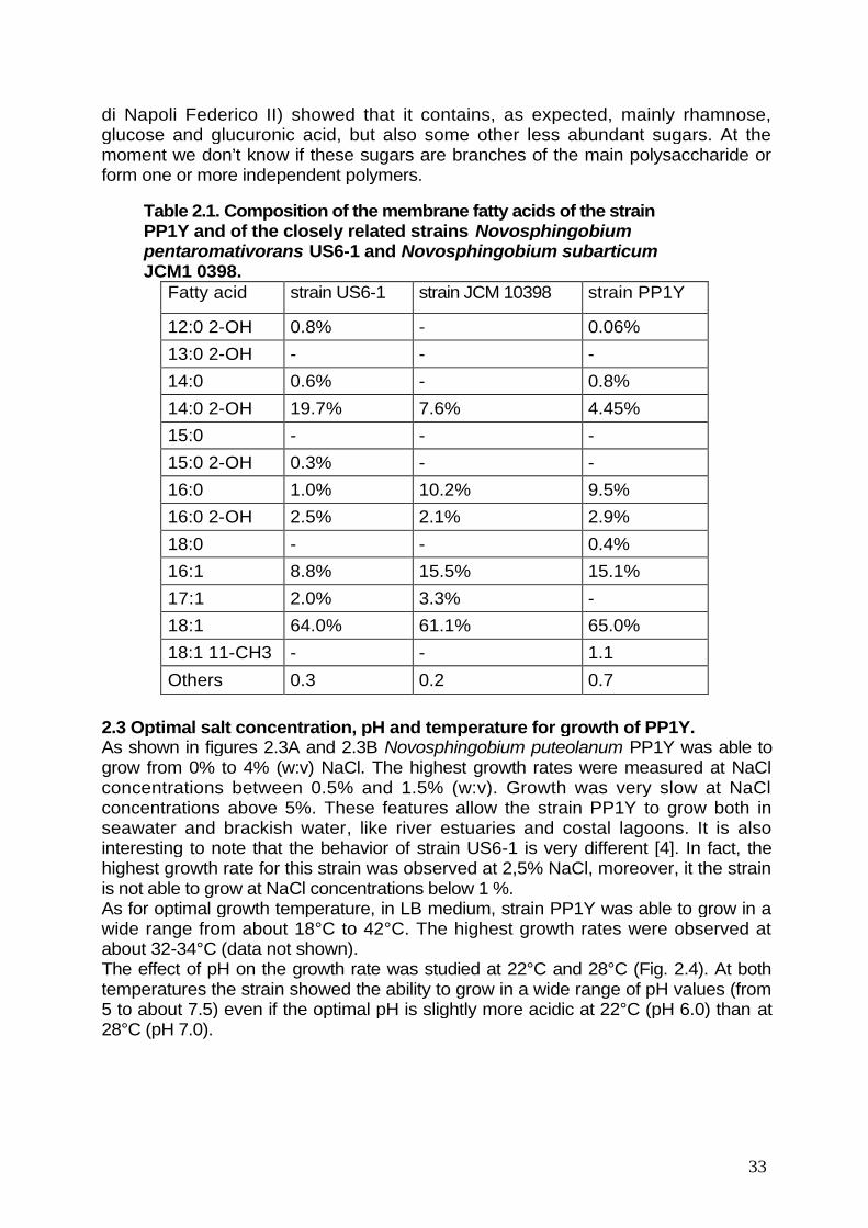
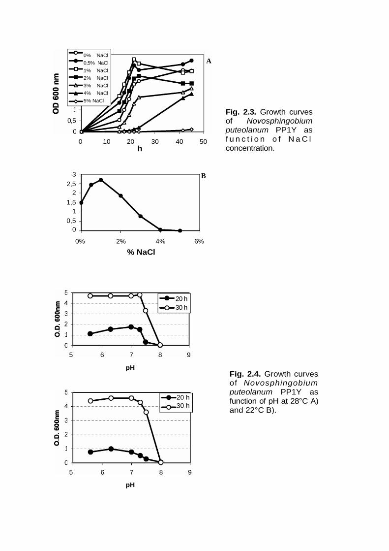
2.3. Optimal salt concentration, pH and temperature for growth of PP1Y. pag. 33
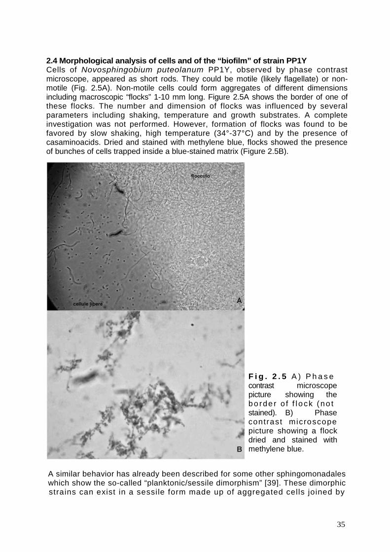
2.4. Morphological analysis of cells and of the “biofilm” of the strain PP1Y pag. 35
2.5. Growth on oil fuels. pag. 37
2.6. Analysis of the degradative potentialities of N. puteolanum PP1Y. pag. 40
2.7. Degradation of paraffin-dissolved aromatic hydrocarbons. pag. 45
CONCLUSIONS pag. 47
BIBLIOGRAPHY pag. 49
1
SUMMARY
Environmental pollution caused by the release of a wide range of xenobioticcompounds has assumed serious proportions. Bioremediation techniques, utilizingmicroorganisms to reduce the concentration and toxicity of various chemicalpollutants such as petroleum hydrocarbons, polycyclic aromatic hydrocarbons,polychlorinated biphenyls, industrial solvents and pesticides, are the most promisingstrategies for the restoration of polluted environments.
Several bacterial strains with promising degradative abilities have already beenisolated and characterized. However, the full exploitation of the potential ofbioremediation strategies requires not only the isolation of a large number of strainswith wide degradative abilities but also an accurate characterization of these strainsboth at the microbiological and biochemical/genetic level. This knowledge isnecessary to perform a rational planning of bioremediation interventions.
The present work describes two research lines which illustrate the two differentapproaches:1. Engineering of catechol 2,3-dioxygenase (C2,3O) a crucial enzyme in
degradative pathway for aromatic compounds of Pseudomonas sp. OX1, a well-studied microorganism.
2. Isolation of new strains able to degrade aromatic hydrocarbons directly frompolluted environments by means of appropriate selection procedures.
Ring cleavage dioxygenases catalyze the oxygen dependent cleavage of catecholiccompounds, a critical step in the aerobic degradation of aromatic compounds. Theirspecificity and regioselectivity control the range of molecules degraded through thecatabolic pathway of microorganisms able to use aromatic hydrocarbons as growthsubstrates. Catechol-2,3-dioxygenase from Pseudomonas sp. OX1 can cleaveeffectively only 3,4-dimethylcatechol (DMC), a catechol which derives from thehydroxylation of o-xylene, and not 3,5- and 3,6-DMC, the hydroxylation products ofm- and p-xilene, respectively. Thus, the restricted specificity of C2,3O is the primarymetabolic determinant which limits the ability of Pseudomonas sp. OX1 to efficientlygrow on xylene mixtures. Thus, understanding the molecular determinants thatcontrol substrate binding in Pseudomonas sp. OX1 C2,3O could allow to developmutated enzymes with a wider substrate specificity and eventually engineeredmicroorganisms with enhanced ability to grow on substituted aromatic compounds. Inthis thesis we describe the preparation of several C2,3O variants with mutations atpositions 249, 270, 267 and/or 198 designed with the aim of obtaining a mutatedenzyme with both a good affinity and a high catalytic efficiency on 3,5-DMC, and 3,6-DMC.
In the second section we describe the isolation from surface seawater samplesand the characterization of a new Sphingomonadales, Novosphingobium sp. PP1 Y.The strain PP1Y is able to use a surprisingly large number of mono- and polycyclicaromatic compounds as the sole carbon and energy sources but shows also a veryinteresting and effective adaptation to grow on complex mixtures of aromaticcompounds dissolved in oil phases like gas-oil and gasoline.
2
RIASSUNTO
Per “bioremediation” si intende l’utilizzo di organismi viventi, in particolaremicrorganismi quali batteri, funghi e lieviti, per la degradazione di composti chimicitossici.
Alcune tipologie di composti che possono essere trattate con le tecniche dibioremediation sono gli idrocarburi alifatici, quelli aromatici e gli idrocarburi aromaticipoliciclici (IPA). Ancora, i composti policiclici aromatici alogenati (diossine, PCB,dibenzofurani, dibenzotiofeni, ecc.), i composti organici clorurati non aromatici(cloruro di vinile, policloroetileni, ecc.), fenoli e ammine aromatiche, compostiutilizzati in agricoltura (diserbanti, pesticidi, fungicidi, ecc.), e infine svariati refluiindustriali (solventi, sgrassanti, detergenti, reagenti per la produzione dei polimeri,ecc.).
A causa della loro tossicità, ed in alcuni casi della loro cancerogenicità, gliidrocarburi aromatici, i loro derivati e le molecole aromatiche eterocicliche destanoparticolare preoccupazione e sono quindi stati oggetto di numerosi studi.
L’eccezionale adattabilità dei microrganismi fa si che, dopo un temposufficientemente lungo, in un ambiente inquinato compaiano ceppi adattati capacinon solo di tollerare i composti inquinanti ma di utilizzarli come fonte di nutrimento.Questi ceppi possono essere utilizzati, singolarmente o in consorzi composti danumerosi ceppi, per ridurre o eliminare composti chimici presenti in acque dolci osalate, suoli, sabbia e fondali marini.
Sono stati già isolati e caratterizzati numerosissimi ceppi batterici capaci didegradare un’ampia varietà di composti xenobioticiquali ad esempio gliSfingomonadali (capaci di degradare idrocarburi alifatici e aromatici, IPA, diossine edibenzofurani), i Micobatteri (capaci di degradare gli IPA), gli Pseudomonadali(capaci di degradare idrocarburi alifatici e aromatici, IPA, fenoli e amminearomatiche, detergenti, solventi) e gli Attinomiceti (capaci di degradare idrocarburialifatici e aromatici, IPA, diossine e dibenzofurani).
Gli interventi di bioremediation possono svolgersi in situ o ex situ. Nel primo caso,se nell’ambiente da trattare si è già sviluppata una microflora con capacitàdegradative, l’attività della microflora indigena può essere stimolata mediante unaserie di interventi volti ad ottimizzare parametri ambientali quali ad esempio ladisponibilità di nutrienti (mediante fertilizzazione con azoto, fosforo, zolfo ecc.),l’aerazione (mediante aratura, pompaggio forzato di aria ecc.), l’umidità (medianteirrigazione) e la temperatura (mediante pompaggio forzato di aria calda). Nel caso incui nell’ambiente da trattare non si sia ancora sviluppata una microflora con capacitàdegradative, la microflora endogena può essere arricchita inoculando opportuni ceppicoltivati in laboratorio. Negli interventi ex situ il substrato inquinato (acqua, terreno,sabbia, limo di fondali, ecc.) viene asportato e trattato in opportuni impianti(bioreattori, vasche, ecc.). Tutti i parametri fondamentali (temperatura, pH, umidità,ossigenazione, salinità, ecc.) possono essere monitorati costantemente e modificatiall’occorrenza. Questo tipo di intervento, dal costo più elevato, è adatto alladecontaminazione di volumi relativamente piccoli di substrato inquinato.
I protocolli di trattamento vanno ottimizzati caso per caso. La programmazionedell’intervento di bonifica deve partire dall’analisi della tipologia degli inquinanti e deiparametri chimico-fisici del substrato da trattare (disponibilità di nutrienti minerali,
3
temperatura, pH, umidità, ossigenazione, salinità, ecc.) in modo da scegliere lacombinazione più opportuna di ceppi e trattamenti. Il successo degli interventi dibonifica dipende quindi in larga parte dalla disponibilità di un’ampia scelta di ceppimicrobici.
Molti ceppi particolarmente promettenti per impieghi nelle bioremediation sono giàdisponibili (molti sono coperti da brevetto), tuttavia, l’isolamento di nuovi ceppi utili èrelativamente semplice dal punto di vista tecnico.
Il presente lavoro di tesi si inserisce in un progetto più ampio il cui scopo èl’ottenimento di microrganismi e sistemi enzimatici da utilizzare per le strategie dibioremediation intese sia per interventi in situ ed ex situ che per l’impiego inbioreattori progettati per lo smaltimento di reflui industriali quali ad esempio acquereflue contenenti elevati livelli di idrocarburi aromatici e/o loro derivati. Perconseguire questo obiettivo sono in corso di svolgimento sia linee di ricercaindirizzate all’isolamento da ambienti inquinati di nuovi ceppi dotati di specifichecapacità degradative naturali e alla loro caratterizzazione che linee di ricerca il cuiscopo è studiare, al fine di poter manipolare, le capacità catalitiche degli enzimi deipathway degradativi e a più lungo termine progettare microrganismi ingegnerizzatidotati di più ampie capacità degradative o più adatti all’impiego in campo e neisistemi bioreattoristici.
Il lavoro di tesi persegue entrambe le linee di ricerca esposte e si articola in dueparti che illustrano i due differenti approcci.
La prima parte descrive la progettazione, la realizzazione e la caratterizzazione dimutanti della catecolo 2,3-diossigenasi (C2,3O) da Pseudomonas sp. OX1 dotati diuna più ampia specificità di substrato. Nella seconda parte viene descrittol’isolamento e la caratterizzazione di un nuovo microrganismo, Novosphingobiumputeolanum PP1Y, in grado di degradare una delle più ampie varietà di idrocarburiaromatici descritte in letteratura.
Le catecolo diossigenasi ed in generale le “ring cleavage dioxygenases” (RCD)svolgono un ruolo cruciale nella degradazione dei substrati aromatici essendoresponsabili del taglio dell’anello aromatico. Tuttavia la loro specificità di substrato,spesso ristretta, pone un limite alla varietà di composti aromatici che un ceppo puòdegradare. Pseudomonas sp. OX1 è un interessante esempio. Questo ceppo infatti,capace di utilizzare benzene, toluene, o-xilene e tutti i fenoli che da essi derivanocome unica fonte di carbonio ed energia, è stato impiegato con successo nellarealizzazione di un bioreattore per il trattamento di reflui contenenti idrocarburiaromatici realizzato grazie ad una collaborazione con il gruppo di ricerca deiProfessori Salatino e Marzocchella del Dipartimento di Ingegneria Chimicadell’Università di Napoli Federico II. Tuttavia il ceppo OX1 non è in grado didegradare m- e p-xilene spesso associati a benzene, toluene, o-xilene. Questaincapacità è stata attribuita al fatto che il 3,5- e il 3,6-dimetilcatecolo (DMC), chevengono prodotti dall’azione degli enzimi dell’upper pathway su m- e p-xilene, nonsono substrati per la C2,3O di questo ceppo. Da ciò nasce l’esigenza di ampliare lospettro di substrati che possono essere degradati dalla C2,3O per poi sviluppareceppi ingegnerizzati di Pseudomonas sp. OX1 capaci di degradare miscele di isomeridello xilene.
Lo studio del modello del sito attivo della C2,3O ha permesso di individuare nelloop G247-L248-T249 i determinanti molecolari che impediscono l’accesso al sitoattivo di 3,5- e 3,6-DMC.
4
La caratterizzazione dei mutanti della posizione T249, (T249S)-, (T249A)- e(T249G)-C2,3O, ha confermato che l’ingombro sterico, dato dalla catena lateraledella T249, è il principale responsabile dell’incapacità della C2,3O di legareefficientemente i dimetilcatecoli, ma ha anche mostrato che il gruppo ossidrilico dellacatena laterale di questo residuo svolge un ruolo essenziale nella catalisi.
Qualsiasi intervento di mutagenesi sito diretta per compensare la diminuzionedell’efficienza catalitica mostrata dai mutanti della posizione 249, richiedeovviamente la preliminare comprensione del ruolo svolto dal gruppo ossidrilico dellaT249 nella catalisi. L’analisi approfondita delle strutture cristallografiche disponibili,nonché la preparazione di modelli dei complessi C2,3O/substrati, ha permesso diipotizzare l’esistenza di un sito di legame per una molecola d’acqua, che sidisporrebbe a ponte fra il substrato e il gruppo ossidrilico della T249 creando unreticolo di legami a idrogeno. Questa ipotesi è estremamente interessante sia dalpunto di vista teorico che da quello applicativo. Infatti, finora, la possibilità che lamolecola d’acqua visibile nei siti attivi delle RCD, abbia un significato funzionale, nonè mai stata investigata ed avrebbe profonde implicazioni per quanto riguarda lacomprensione del meccanismo catalitico di questa famiglia di enzimi. Dal punto divista pratico, l’ipotesi dell’esistenza di una molecola d’acqua con funzione catalitica,suggerisce una possibile via per la preparazione di mutanti che non solo leghino adalta affinità il 3,5- ed il 3,6-DMC, ma che ne catalizzino efficientemente il taglio.Proprio sulla base di queste considerazioni sono stati selezionati quattro residui,A1 98, G251, F267 e G270, che circondano il sito ipotetico per la molecola d’acquacatalitica e quindi rappresentano bersagli per ulteriori interventi di ingegneriaproteica. Mediante mutagenesi in silico ed esperimenti di docking sono state sceltetre mutazioni, F267H, G270E e G270Q, che nel mutante (T249G)-C2,3O potrebberoripristinare il sito di legame della molecola d’acqua catalitica e quindi generare unmutante multiplo che combini la capacità di legame dei dimetilcatecoli di (T249G)-C2,3O con l’efficienza catalitica della C2,3O wt.
L’analisi informatica ha anche mostrato che il tipo di conformazione adottato dallecatene laterali di glutammina e glutammato in posizione 270 è determinato dallanatura dei due residui in posizione 198 e 267 adiacenti al residuo 270. Per questomotivo si è deciso di combinare le mutazioni già descritte con mutazioni dei residuiA1 98 e F267 per restringere o aumentare le possibilità di movimento delle catenelaterali di glutammato e glutammina. In particolare si è deciso di mutare il residuo dialanina in glicina, per ridurre l’ingombro sterico, o in serina, per aumentarlo. Il residuodi fenilalanina è stato invece sostituito con leucina.
La tecnica di mutagenesi scelta ha permesso di creare una library contenente lecombinazioni desiderate di mutazioni a partire dal vettore contenente la sequenzacodificante il mutante (T249G)-C2,3O, mentre lo “screening” mediante PCR hapermesso la rapida identificazione dei cloni di interesse.
I mutanti realizzati sono stati espressi in E. coli, purificati e successivamentecaratterizzati su quattro substrati catecolo, 3-metilcatecolo, 3,5- e 3,6-dimetilcatecolo.La caratterizzazione cinetica dei doppi mutanti (T249G, F267H)-, (T249G, G270E)- e(T249G, G270Q)-C2,3O ha mostrato che i due mutanti, (T249G, F267H)- e (T249G,G270E)-C2,3O mostrano un sensibile miglioramento dei valori di kcat per il 3,5- e il3,6-DMC, suggerendo che l’ipotesi iniziale sul coinvolgimento di una molecola diacqua nella catalisi e l’analisi informatica successiva siano valide.
L’analisi cinetica ha però anche rivelato effetti parzialmente inattesi delle duemutazioni. In particolare la mutazione G270E causa un aumento di 2-3 volte neivalori di KM per tutti i substrati utilizzati rispetto al mutante di riferimento (T249G)-
5
C2,3O. Questo effetto è probabilmente dovuto al fatto che la sostituzione di unaglicina con glutammato crea ingombro nel sito attivo ed in particolare con i residui delloop 197-199.
Al contrario della mutazione G270E, la mutazione F267H lascia invariato o riduceil valore di KM per il 3,5-DMC e per il 3,6-DMC e provoca contemporaneamente unaumento della kcat. Conseguentemente nel mutante (T249G, F267H)-C2,3O il valore diKS aumenta di un ordine di grandezza
Infine, è da sottolineare che la simultanea presenza dei residui E270 e H267provoca una riduzione del valore della KM per il 3,5 e il 3,6-DMC rispetto al mutante(T249G, G270E)-C2,3O, mentre i valori di kcat sono confrontabili ai valori osservatiper il mutante che presenta la sola mutazione in posizione 270. E’ interessantenotare che la mutazione fenilalanina → istidina riduce l’ingombro in posizione 267 equesto può in parte compensare l’aumento di ingombro causato dalla mutazioneglicina → glutammato a cui è stato attribuito l’aumento del valore di KM nel mutante(T249G, G270E)-C2,3O.
Ulteriori analisi hanno mostrato che mutazioni di due residui adiacenti allaposizione 270, A1 98 e F267, possono essere usate per modificare le possibilità dimovimento delle catene laterali dei residui di glutammato o glutammina in posizione270. Queste ultime, secondo i modelli molecolari, dovrebbero collocarsi proprio alcentro delle catene laterali dei residui in posizione 198 e 267. La mutazione A198G,ad esempio, riducendo l’ingombro in posizione 198 potrebbe favorire l’avvicinamentodella catena laterale di E270 a questa posizione mentre la mutazione A198Spotrebbe avere l’effetto opposto.
Al contrario la mutazione F267L riducendo l’ingombro della catena lateralepresente in posizione 267 potrebbe permettere alla catena laterale di E270 o Q270 diavvicinarsi a questo residuo allontanandosi dalla catena laterale in posizione 198.
Sono stati analizzati gli effetti delle mutazioni A198G e A198S inserite nellasequenza del doppio mutante (T249G, G270E)-C2,3O e gli effetti della mutazioneF267L inserita sia nella sequenza del doppio mutante (T249G, G270E)-C23O chedel doppio mutante (T249G, G270Q)-C2,3O. La mutazione A1 98G ha determinatoun peggioramento, talvolta anche significativo, di tutte le costanti catalitiche siaimpiegando il catecolo o il 3-MC che i dimetilcatecoli.
Al momento l’ipotesi più plausibile è che la mutazione A198G alteri laconformazione del loop K197-A198-H199 che contribuisce alla formazione del sitocatalitico diminuendo così sia l’affinità per i substrati che l’efficienza catalitica.
La mutazione A198S, invece, ha determinato lievi miglioramenti dei valori dellecostanti catalitiche per i substrati fisiologici (catecolo e 3-MC) ma non ha modificato iparametri catalitici misurati utilizzando i dimetilcatecoli. I risultati ottenuti mostranochiaramente che la sostituzione del residuo A198 non è adatta allo scopo dimigliorare le performance catalitiche della C2,3O sui dimetilcatecoli.
Decisamente più incoraggianti sono gli effetti della mutazione F267L.Contrariamente alle mutazioni della posizione 198, questa mutazione ha effettitrascurabili sui parametri catalitici misurati utilizzando catecolo e 3-MC ma miglioranotevolmente i parametri catalitici misurati utilizzando i dimetilcatecoli.
In particolare i valori di KS aumentano di 10-70 volte a seconda del substrato edella natura del residuo in posizione 270 (Glu o Gln). Considerando invece i valoriassoluti della costante KS riportati in tabella 2 si può notare che il mutante (T249G,F267L, G270E)-C2,3O è attualmente il miglior catalizzatore disponibile del taglio deidimetilcatecoli.
6
La seconda parte del lavoro di tesi descrive l’isolamento e la caratterizzazzionedel microrganismo Novosphingobium puteolanum PP1Y.
Il ceppo PP1Y è stato isolato mediante una procedura di arricchimento confenantrene da campioni di acqua superficiale prelevati in un bacino interno del portodi Pozzuoli. Dopo diversi passaggi in terreno minimo contenente cristalli difenantrene i campioni sono stati piastrati su piastre di M9G/agar contente fenantrenecome unica fonte di carbonio ed energia. Su alcune piastre sono state osservatecolonie di colore giallo circondate da aree in cui l’agar - reso lattescente daimicrocristalli di fenantrene - era tornato trasparente indicando l’avvenutadegradazione del fenantrene. Il ceppo PP1Y è stato isolato a partire da una di questecolon ie.
Il sequenziamento parziale del gene per l’RNA ribosomale 16S ha mostrato che ilceppo PP1Y è uno sfingomonadale strettamente imparentato (2 nucleotidi didifferenza) con Novosphingobium pentaromativorans US6-1, un ceppo isolato da unabaia in Korea capace di crescere su composti policiclici aromatici con un numero dianelli compreso tra 3 e 5, e con Novosphingobium sp. Phe-8 (5 nucleotidi didifferenza). Tuttavia la caratterizzazione microbiologica mostra che il ceppo PP1Ydifferisce dal ceppo US6-1 per alcune proprietà rilevanti quali condizioni ottimali dicrescita, composizione degli acidi grassi di membrana, caratteristiche metaboliche,spettro di composti aromatici degradati e morfologia di crescita; ciò fa supporre che ilceppo PP1Y sia probabilmente una nuova specie del genere Novosphingobiumaffine alle due specie citate.
Le cellule di Novosphingobium puteolanum PP1Y hanno la forma di cortibastoncel l i negat ivi a l la colorazione di Gram. Possono essere mobi l i(presumibilmente flagellate) o non-mobili. Le cellule non-mobili possono presentarsiin forma libera o formare aggregati cellulari che in alcuni casi divengonomacroscopici presentandosi sotto forma di “flocculi” ramificati. Queste caratteristichesono state riscontrate anche in altri sfingomonadali che mostrano quello che vienedefinito “di morfismo planctonico/sessile”. Tali sfingomonadali dimorfici possonoesistere in forma aggregata (forma “sessile”) e in forma libera (forma ”planctonica”).L’esistenza della forma sessile è dovuta alla presenza della capsula polisaccaridicache fa da collante fra le cellule.
I flocculi del ceppo PP1Y mostrano alcune caratteristiche che non sono statedescritte nel caso di altri sfingomonadali dimorfici. I f locculi aderisconospontaneamente alle superfici idrofobiche quali i polimeri plastici e adsorbono,concentrandoli, molecole idrofobiche quali coloranti idrofobici e IPA. Il ceppo PP1Yforma spontaneamente su superfici idrofobiche, sia solide che liquide, un “biofilm”che appare come una versione strutturata dei flocculi amorfi che si formano in colturaliquida.
Sono state analizzate le potenzialità degradative del ceppo PP1Y, effettuandodelle crescite del microrganismo e fornendo come substrato derivati del petrolio. Ilceppo PP1Y è in grado di crescere utilizzando benzina e gasolio come uniche fonti dicarbonio ed energia. In particolare il gasolio permette velocità di crescita quasiconfrontabili a quelle registrate in mezzo ricco con estratto di lievito e triptone. Ilgasolio è ben tollerato almeno fino ad un rapporto 2:1 = acqua:gasolio. La crescita ècirca 2-5 volte più lenta nel caso della benzina. In un sistema bifasico acqua/gasolioo acqua/(paraffina-benzina) il ceppo PP1Y è responsabile della formazione di unaemulsione stabile della fase oleosa che viene frammentata in goccioline condiametro inferiore a 1 mm. L’analisi al microscopio a contrasto di fase delle gocce digasolio ha mostrato che esse sono rivestite da uno strato di biofilm contente cellule
7
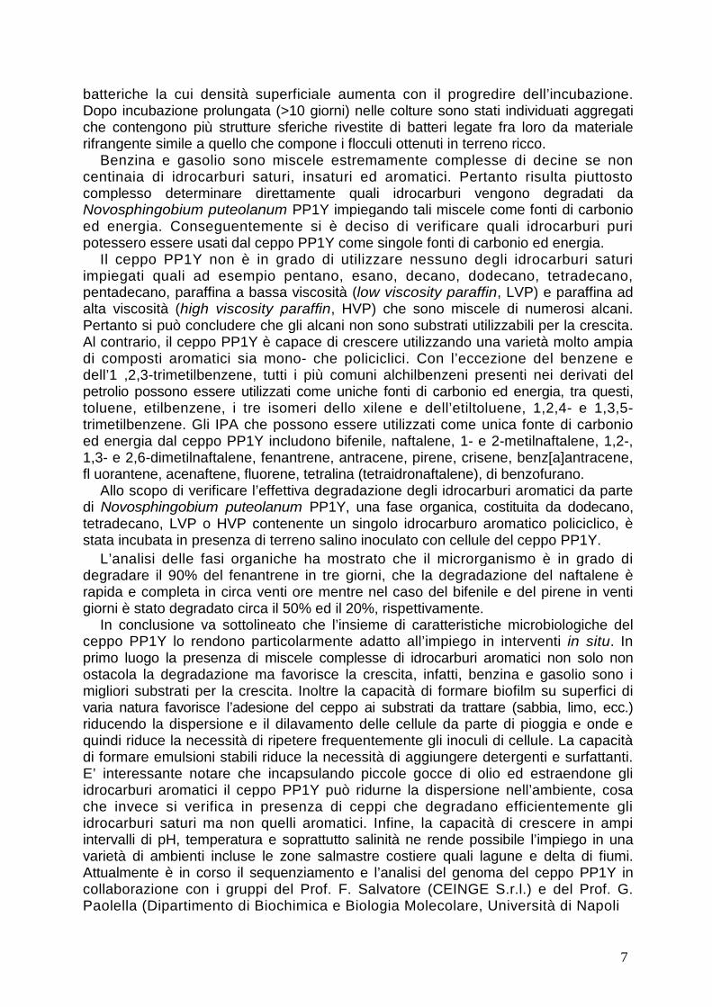
batteriche la cui densità superficiale aumenta con il progredire dell’incubazione.Dopo incubazione prolungata (>10 giorni) nelle colture sono stati individuati aggregatiche contengono più strutture sferiche rivestite di batteri legate fra loro da materialerifrangente simile a quello che compone i flocculi ottenuti in terreno ricco.
Benzina e gasolio sono miscele estremamente complesse di decine se noncentinaia di idrocarburi saturi, insaturi ed aromatici. Pertanto risulta piuttostocomplesso determinare direttamente quali idrocarburi vengono degradati daNovosphingobium puteolanum PP1Y impiegando tali miscele come fonti di carbonioed energia. Conseguentemente si è deciso di verificare quali idrocarburi puripotessero essere usati dal ceppo PP1Y come singole fonti di carbonio ed energia.
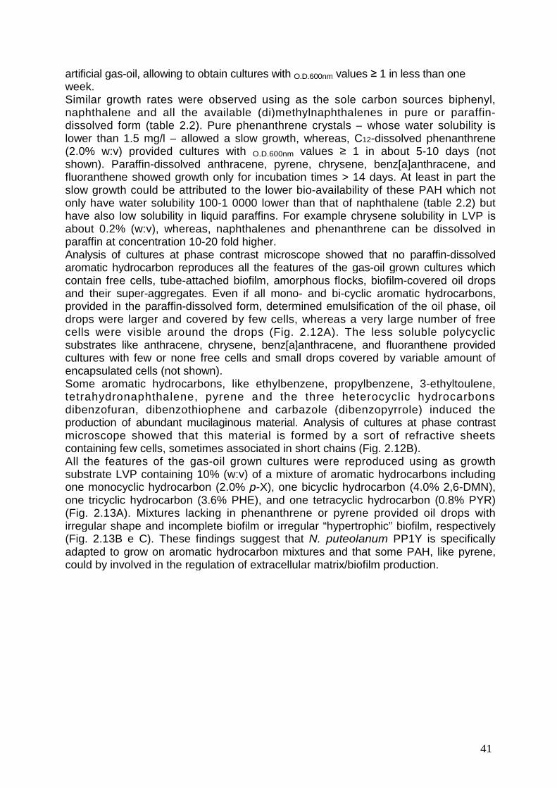
Il ceppo PP1Y non è in grado di utilizzare nessuno degli idrocarburi saturiimpiegati quali ad esempio pentano, esano, decano, dodecano, tetradecano,pentadecano, paraffina a bassa viscosità (low viscosity paraffin, LVP) e paraffina adalta viscosità (high viscosity paraffin, HVP) che sono miscele di numerosi alcani.Pertanto si può concludere che gli alcani non sono substrati utilizzabili per la crescita.Al contrario, il ceppo PP1Y è capace di crescere utilizzando una varietà molto ampiadi composti aromatici sia mono- che policiclici. Con l’eccezione del benzene edell’1 ,2,3-trimetilbenzene, tutti i più comuni alchilbenzeni presenti nei derivati delpetrolio possono essere utilizzati come uniche fonti di carbonio ed energia, tra questi,toluene, etilbenzene, i tre isomeri dello xilene e dell’etiltoluene, 1,2,4- e 1,3,5-trimetilbenzene. Gli IPA che possono essere utilizzati come unica fonte di carbonioed energia dal ceppo PP1Y includono bifenile, naftalene, 1- e 2-metilnaftalene, 1,2-,1,3- e 2,6-dimetilnaftalene, fenantrene, antracene, pirene, crisene, benz[a]antracene,fl uorantene, acenaftene, fluorene, tetralina (tetraidronaftalene), di benzofurano.
Allo scopo di verificare l’effettiva degradazione degli idrocarburi aromatici da partedi Novosphingobium puteolanum PP1Y, una fase organica, costituita da dodecano,tetradecano, LVP o HVP contenente un singolo idrocarburo aromatico policiclico, èstata incubata in presenza di terreno salino inoculato con cellule del ceppo PP1Y.
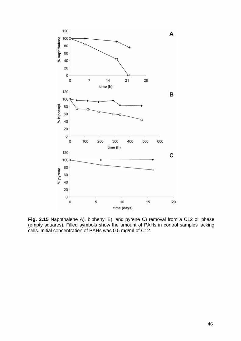
L’analisi delle fasi organiche ha mostrato che il microrganismo è in grado didegradare il 90% del fenantrene in tre giorni, che la degradazione del naftalene èrapida e completa in circa venti ore mentre nel caso del bifenile e del pirene in ventigiorni è stato degradato circa il 50% ed il 20%, rispettivamente.
In conclusione va sottolineato che l’insieme di caratteristiche microbiologiche delceppo PP1Y lo rendono particolarmente adatto all’impiego in interventi in situ. Inprimo luogo la presenza di miscele complesse di idrocarburi aromatici non solo nonostacola la degradazione ma favorisce la crescita, infatti, benzina e gasolio sono imigliori substrati per la crescita. Inoltre la capacità di formare biofilm su superfici divaria natura favorisce l’adesione del ceppo ai substrati da trattare (sabbia, limo, ecc.)riducendo la dispersione e il dilavamento delle cellule da parte di pioggia e onde equindi riduce la necessità di ripetere frequentemente gli inoculi di cellule. La capacitàdi formare emulsioni stabili riduce la necessità di aggiungere detergenti e surfattanti.E’ interessante notare che incapsulando piccole gocce di olio ed estraendone gliidrocarburi aromatici il ceppo PP1Y può ridurne la dispersione nell’ambiente, cosache invece si verifica in presenza di ceppi che degradano efficientemente gliidrocarburi saturi ma non quelli aromatici. Infine, la capacità di crescere in ampiintervalli di pH, temperatura e soprattutto salinità ne rende possibile l’impiego in unavarietà di ambienti incluse le zone salmastre costiere quali lagune e delta di fiumi.Attualmente è in corso il sequenziamento e l’analisi del genoma del ceppo PP1Y incollaborazione con i gruppi del Prof. F. Salvatore (CEINGE S.r.l.) e del Prof. G.Paolella (Dipartimento di Biochimica e Biologia Molecolare, Università di Napoli
8
Federico II). Lo studio del genoma completo non solo faciliterà la comprensione dellebasi molecolari delle peculiari proprietà del ceppo PP1Y ma consentirà anche diprogettare modifiche per potenziarne le abilità degradative ed ampliarneulteriormente i possibili campi di utilizzo.
9
INTRODUCTION
Bioremediation techniques exploit microorganisms, like bacteria, fungi, and yeasts,to degrade pollutants [1]. Environmental pollutants (mainly xenobiotics) which couldbe treated using bioremediation techniques include:a) aliphatic and aromatic hydrocarbons;b) polycyclic aromatic hydrocarbons (PAH);c) polycyclic aromatic halogenated compounds like polychlorobiphenils (PCB) and
polychlorodioxines;d) alogenated aliphatic hydrocarbons like vinyl chlorides;e) (poly)phenols and aromatic amines;f) industrial wastes containing organic solvents, degreasers, detergents, residual
reagents etc..
It should be underlined that aromatic compounds, because of their high toxicity forall living organisms, are considered particularly dangerous and have been the subjectof several studies [1].
Due to the well known adaptability of microorganisms, polluted environmentseventually develop an adapted micro-flora composed not only by strains whichtolerate pollutants but also by microorganisms which are able to use pollutants assources of energy, carbon, nitrogen, or sulfur for their growth [1-4]. These strains canbe used, individually or as consortia, to reduce or to remove pollutants from soils,waters, urban and industrial wastes.
Several bacterial strains with promising degradative abilities have already beenisolated and characterized. They belong to several different genus of both Gram(-)and Gram(+) bacteria. Some examples are Pseudomonadales (which degradealiphatic and aromatic hydrocarbons, PAH, phenols, aromatic amines, detergents,solvents, etc.) [5, 6]; Sphingomonadales (which degrade aliphatic and aromatichydrocarbons, PAH, phenols, aromatic amines, heterocyclic aromatic compounds,etc.) [7-9]; Mycobacteria (which degrade PAH) [3, 10, 11]; Actinomycetes (whichdegrade aliphatic and aromatic hydrocarbons, PAH, phenols, aromatic amines,heterocyclic aromatic compounds, etc.) [12, 13].
The majority of the strains with effective degradative abilities are aerobic and usemono and dioxygenase to start degradation of xenobiotics [14, 15]. A few number ofanaerobic degraders are also known. However, it should be underlined that severalanaerobic bacteria show the ability to de-halogenate polyhalogenated-hydrocarbonsthus improving their biodegradability by aerobic bacteria [16, 17].
Bioremediation interventions can be carried out in situ or ex situ. In the first case, ifthe polluted environment has already developed an adapted micro-flora, indigenousstrains can be stimulated through optimization of crucial parameters like nutrientsavailability (fertilization with nitrogen, sulfur, phosphorus, etc.), aeration (ploughing,venting), humidity (irrigation), pH and temperature (pumping of warm air). If thepolluted environment has not yet developed adapted strains, indigenous micro-floracan be enriched by inoculating selected laboratory-grown strains includingengineered strains. In the case of ex situ interventions polluted materials, like water,soil, sand or mud, are removed and treated in plants (bioreactors, settling tanks, etc.)where the parameters that can influence biodegradation (temperature, pH, humidity,oxygen concentration, salinity) are constantly monitored and changed when needed.This strategy is obviously more expensive and allows treatment of smaller amountsof polluted material.
10
Thus, bioremediation strategies should be studied case by case starting from theanalysis of pollutants and their chemical-physical parameters in order to select themost appropriate combination of strains for each application. Therefore, the successof bioremediation interventions largely depend on the availability of a wide panel ofstrains which are able not only to degrade several xenobiotics but also to bemetabolically active under different conditions of temperature, salinity, pH, oxygenand water availability.
The present work is part of a wider research project aiming at obtaining microbialstrains and enzymatic systems which could be used both in in situ or ex situbioremediation interventions and in bioreactors for the treatment of industrial wasteslike, for example, wastewaters containing high levels of aromatic hydrocarbons andtheir derivatives. Two different strategies for making available new or improvedstrains for biodegradation are currently under evaluation: (i) isolation andcharacterization of strains with specific degradative abilities directly from pollutedenvironments, and (ii) production of engineered strains. These latter can be producedafter characterization of the catalytic machineries of enzymes involved in thedegradation of xenobiotics in order to design mutated enzymes and later engineeredstrains with improved and/or wider abilities or better suited to their use in bioreactors.
The present work describes two research lines which illustrate the two differentapproaches:3. Engineering of catechol 2,3-dioxygenase (C2,3O) a crucial enzyme in
degradative pathway for aromatic compounds of Pseudomonas sp. OX1, a well-studied microorganism.
4. Isolation of new strains able to degrade aromatic hydrocarbons directly frompolluted environments by means of appropriate selection procedures.
In the following sections we will describe the state of the art and aims of these twostrategies.
Protein engineering of the catechol 2,3-dioxygenase from Pseudomonas sp.OX1.
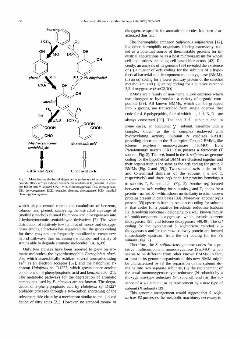
Pseudomonas sp. OX1 is an ideal model organism for studies of metabolicengineering since it can utilize benzene, toluene, and o-xylene, but not m- and p-xylene, as the sole sources of carbon and energy trough a single, well-characterizeddegradative pathway [2]. Moreover, in collaboration with the research group ofProfessor Salatino and Marzocchella (Dipartimento di Ingegneria Chimica, Universitàdi Napoli Federico II), the strain has already been used to set up a bioreactor for thetreatment of wastewaters containing aromatic hydrocarbons [18]. Two NADH-dependent monooxygenases – toluene/o-xylene-monooxygenase (ToMO) andphenol hydroxylase (PH) – act sequentially in the degradative pathway [19] toconvert aromatic hydrocarbons to the corresponding catechols. The twomonooxygenases form the so-called upper pathway. Catechols are successivelyconverted to non-cyclic molecules by C2,3O, an “extradiol ring cleavagedioxygenase” (ECD), which cleaves one of the C-C bond adjacent to the diol moietyof catechols [20, 21] (Fig. 1). ToMO and PH are able to convert o-xylene as well asm-xylene and p-xylene to 3,4-DMC, 3,5-DMC and 3,6-DMC, respectively. However,Pseudomonas sp. OX1 C2,3O can cleave effectively only 3,4-DMC [22] yielding anon-cyclic compound which can be further metabolized through the so-called lowerpathway. This is not possible in the case of 3,5-DMC and 3,6-DMC because of thevery low activity of C2,3O towards these compounds [22]. Thus, the restrictedspecificity of C2,3O is the primary metabolic determinant which limits the ability of
11
Pseudomonas sp. OX1 to efficiently grow on xylene mixtures. Moreover, the inabilityof Pseudomonas sp. OX1 to cleave 3,5- and 3,6-DMC has also an adverseconsequence on the metabolism of the microorganism since the NADH consumed bythe monooxygenase-catalyzed hydroxylations of m- and p-xylene cannot be restoredby the lower pathway reactions. This inefficiency results in a loss of metabolicreducing power when Pseudomonas sp. OX1 grows on xylene mixtures.
Fig. 1. Possible extradiolcleavage reactions for 3,6-DMC A), 3-MC B) and 3,5-DMC C). Wild type C2,3Ocatalyzes effectively only theproximal cleavage of 3-MCand at very low efficiency thecleavage of 3,6-DMC and thec l e a v a g e o f 3 , 5 - D M Cproximal to methyl 5.
Thus, understanding the molecular determinants that control substrate binding inPseudomonas sp. OX1 C2,3O could allow to develop mutated enzymes with a widersubstrate specificity and eventually engineered microorganisms with enhanced abilityto grow on substituted aromatic compounds.
The structure of Pseudomonas sp. OX1 C2,3O was modeled by homology usingthe crystal structure of P. putida MT2 C2,3O (1 mpy, [23]) as template. Successively,in order to hypothesize which residues of C2,3O are involved in substraterecognition, the structures of two ECDs, 2,3-dihydroxybiphenyl-1 ,2-dioxygenase
12
( D H B D ) f r o m P s e u d o m o n a s K K S 1 0 2 ( 1 e i m , [ 2 4 ] ) a n d t h e 3 , 4 -di hyd roxyphenylacetate 2, 3-d ioxygenase (H PCD) from Brevibacterium fuscum (1 q0c,[25]), crystallized in their active Fe(II) form with the substrate bound to the catalyticmetal, were used as templates for an homology docking of catechols into the activesite of C2,3O. These complexes where chosen because available data suggestedthat they represent the catalytically competent enzyme/substrate complex [24, 25].This strategy revealed that substrate CH groups at positions 3 and 4 point towardsmall cavities, indicated as sub-sites 1’ and 2’ in Fig. 2, which are defined byresidues Ile204, Phe302, Ile291 and Leu248. These cavities are large enough toaccommodate methyl substituents at positions 3 and 4 of a catechol molecule asverified by the docking of 3-MC, 4-MC and 3,4-DMC. Instead, the CH groups atpositions 5 and 6 of the substrate point toward the backbone of Leu-248 and theside-chain of Thr-249, respectively (Fig. 2A-B). Apparently, the close contactsbetween these two residues and the edge of the substrate ring prevent binding of3,6- and 3,5-DMC.
As CH atoms at position 6 contact the side chain of residue Thr249, wehypothesized that a reduction in the volume of this side chain might provide room forhousing a methyl substituent at this position and allow for the binding of 3,6-DMC or3,5-DMC as depicted in Figures 2D and 2F, respectively.
Fig. 2. Scheme ofpossible binding of 3-MC, 3,5-DMC and3,6-DMC to C2,3Oactive site. A) and B),binding of 3-MC and4-MC, respectively,into the active site ofwild type P. stutzeriC2,3O. C) and E)show two possibleorientations for thebinding of 3-MC tothe act ive site of(T246G)-C2,3O. D)and F), binding of 3,5-DMC and 3,6-DMC,respectively, to thea c t i v e s i t e o f(T246G)-C2,3O.
13
Based on the observations above, residue Thr-249 was substituted in silico withserine, alanine and glycine, and the molecular contacts of docked 3,6- and 3,5-DMCwere re-inspected. The progressive reduction of the side chain of residue 249created a new cavity (sub-site 3’, Fig. 2C-F) adjacent to CH atoms at position 6,resulting in the reduction of steric hindrance between a methyl group at this positionand the protein. When glycine is present at position 249 the closest contact betweenthe methyl group in the sub-site 3’ and residue 249 increased from 1.7 Å to 3.9 Å.
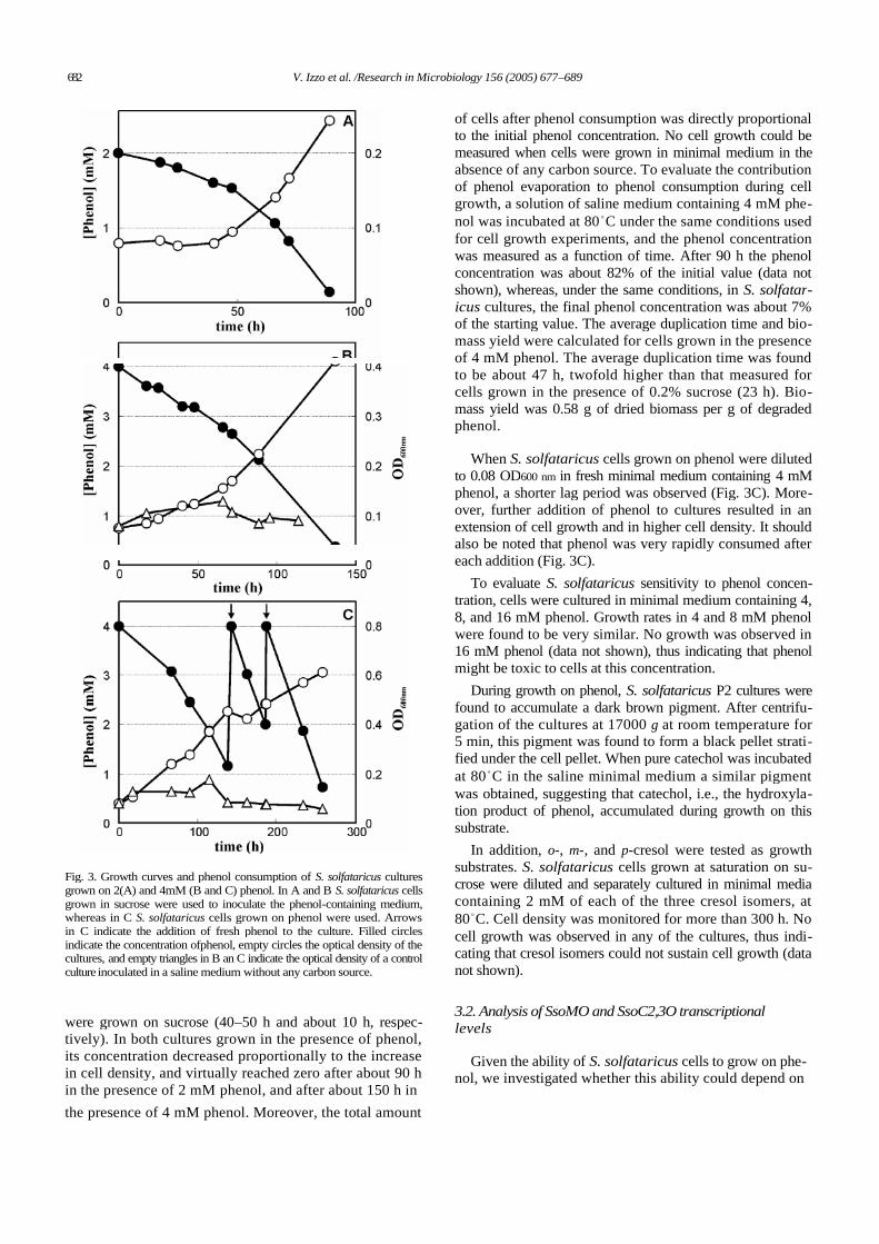
The three mutants (T249S)-, (T249A)-, and (T249G)-C2,3O were produced bysite-directed mutagenesis and assayed on catechol, 3-MC, 3,5-DMC, and 3,6-DMC.As expected the KM values showed a progressive increase as the volume of sidechain at position 249 decreased in the case of catechol, 3-MC, whereas aprogressive decrease of the KM values was observed in the case of 3,5-DMC, and3,6-DMC (Fig. 3A). These data suggested that the creation of the hypothetical 3’ sub-site improves the binding of the two larger substrates and reduces affinity for the twosmaller catechols. Surprisingly, mutations T249A and T249G, which remove thehydroxyl group from the side chain at position 249, caused also a strong decrease inthe kcat values (Fig 3B). This reduction counterbalances the improved binding of 3,5-DMC and 3,6-DMC. As a consequence mutants (T249A)-, and (T249G)-C2,3O arenot more efficient than wild type C2,3O in the cleavage of 3,5-DMC, and 3,6-DMC.
Fig. 3. Catalytic parameters ofwild-type and mutant C2,3Osmeasured at pH 7.5 are shownas function of the radius of theradius of sub-site 3’ shown inFig. 5 (radius are: 0.76, 0.98,1 .25 and 1.91 Å for wt ,(T249S)-, (T249A)-, and(T249G)-C2,3O, respectively).Filled circles, catechol; opencircles, 3-MC; filled triangles,3,6-DMC; open triangles, 3,5-DMC.For clarity in (B) thekcat/KM values on catechol and3-MC and the values on 3,5-DMC and 3 ,6 -D MC ar ereported on different scales –on the left and on the right,respectively.
A possible explanation of these findings is that mutations T249A and T249Gcause the loss of a water molecule bound to the hydroxyl group of Thr-249 visible in
14
the crystal structure of C2,3O. A water molecule in the same position has been foundalso in the crystal structures of several ECDs/substrate complexes ]. Moreover,docking experiments performed in our laboratory suggested that the water moleculecould stay in the active site during catalysis by bridging through H-bonds Thr-249side chain and substrate. These observations suggested that T249 and the boundwater could play a previously unsuspected role in catalysis [26].
In this thesis we describe the preparation of several (T249G)-C2,3O variantswith further mutations at positions 270, 267 and/or 198 designed with the aim ofrestoring the water binding site into the active site of this mutant and to obtain amutated enzyme with both a good affinity and a high catalytic efficiency on 3,5-DMC,and 3,6-DMC.
Isolation of new strains from polluted environmentsNew strains with the ability to degrade xenobiotics are usually isolated by using
enrichment techniques. The appropriate substrate is added to samples to increasethe relative abundance of the desired microorganisms, successively enrichedsamples are used to inoculate selective culture broths containing the appropriatesubstrate as the sole carbon, nitrogen, sulphur or energy source. The success of thisapproach depends on the accurate choice of the environmental sample and ofisolation conditions.
In order to isolate new strains with the ability to degrade PAH, samples of surfaceseawater were collected inside the harbour of Pozzuoli (Naples, Italy) which isheavily polluted by fuel oils due to the high number of small boats in the harbour. Likeall derivatives of petroleum, gasoline and gas-oil contain relevant amounts ofaromatic hydrocarbons. Gasoline contains about 25% of mono- and up to 10%polycyclic aromatic hydrocarbons [27]. Gas-oil, on the contrary, contains a largerfraction of PAHs – up to 15% – and only 5-6% alkylbenzenes [28]. These aromaticfractions are extremely complex including tens if not hundreds of moleculesbelonging to (poly)alkyl-benzenes, PAHs and (poly)alkyl-PAHs, naphthenes (i.e.polycyclic compounds with fused aromatic and saturated rings) and nitrogen, sulphuror oxygen-containing heterocyclic compounds.
Fuel oils degrading bacterial strains are largely diffused in the environment. Theso-called “obligate hydrocarbonoclastic bacteria” (O HCB), like Alkanivorax,Marinobacter and Oleispira [29], are among the most effective oil degraders.However, these bacteria degrade prevalently or exclusively the saturated fraction ofpetroleum and fuels. The most effective degraders of aromatic compounds belong toPseudomonadales (Gram-), Sphingomonadales (Gram-) and Mycobacteria (Gram+)[1]. Sphingomonadales are unusual alpha-proteobacteria which containglycosphingolipids instead of the more common lipopolysaccharides in the outermembrane. This peculiarity makes their cell surface more hydrophobic than those ofthe other Gram- strains and this is considered one of the reasons why severalSphingomonadales have developed the ability to degrade mono and polycyclicaromatic compounds. Likely, another reason is the fact that many Sphingomonadalesharbor several large conjugative plasmids (up to six plasmids with lengths rangingfrom less than 50 kbp to more than 500 kbp) [30, 31]. Due to these megaplasmidsseveral Sphingomonadales have “collected” genes for the degradation of xenobioticsand continuously exchange them with other strains. Some interesting examples areNovosphingobium aromaticivorans, which can use alkyl-benzenes as the sole carbonand energy sources [32], Novosphingobium pentaromativorans US6-1, whichdegrades PAHs with 3-5 rings [4], Sphingomonas paucimobilis EPA505, which
15
degrades several polycyclic compounds [33], and Sphingomonas wittichii R1, whichcan grow using dibenzofuran and dibenzo-p-dioxin and co-metabolizes mono anddichloro-derivatives of these toxic aromatic compounds [34].
In this thesis we will describe the isolation from surface seawater samples and thecharacterization of a new Sphingomonadales, Novosphingobium sp. PP1 Y. Thestrain PP1Y not only uses a surprisingly large number of mono and polycyclicaromatic compounds as the sole carbon and energy sources but shows also a veryinteresting and effective adaptation to growth on complex mixtures of aromaticcompounds dissolved in oil phases like gas-oil and gasoline.
16
MATERIALS AND METHODS
Bacterial strains
E.coli strain BL21(DE3) F-, ompT, hsdS(rB -,mB -),dcm, gal, ?. (DE3). The strain waspurchased by Novagen and used for proteins expression.E.coli strain CJ236. The strain was purchased by Bio-Rad and used for site-directmutagenesis.E.coli Top F’10 strain (F’{lacIq Tn10 (TetR)} mcrA A(mrr-hsdRMS-mcrBC) φ80AlacZAM15 AlacX74 recA1 deoR araD139 A(ara-leu)7697 galU galK rpsL (StrR) endA1nupG). The strain was purchased by Invitrogen and used for genetic manipulation.
MediumLB (Luria-Bertani) and M9 medium, solid and liquid, were prepared as described bySambroook et al.[35]KPSA medium was prepared as follows: 20 mM potassium phosphate (pH 6.9), 10mM sodium chloride and 18.7 mM ammonium chloride.
C-Goodies were added in KPSA medium at 0.5% (v/v) and were prepared asdescribed in the following table:
MgO 10.75 gCaCO3 2.0 gFeSO4 x 7H2O 7.0 gZnSO4 x 7 H2O 1.44 gMnSO4 x H2O 1.12 gCuSO4 x 5 H2O 0.25 gCoSO4 x 7 H2O 0.28 gH3BO3 0.06 gHCl 51.3 mlMgSO4 60.2 gNiCl2 x 6 H2O 4 mgNa2MoO4 x 2H2O
6 mg
H2O 1000 ml
AntibioticsAmpicillin, chloramphenicol and Kanamycin were purchased from Sigma and wereused at concentration of 100 µg/mL, 30 µg/mL and 70 µg/mL respectively.
Vectors
pET22b(-I-)AXN/C2,3O and pET22b(-I-)AXN/C2,3O(T249G), coding for C2,3Owt and(T249G)-C2,3O mutant, were available in laboratory and were used to prepare libraryof C2,3O mutants.
Substrates3,5- and 3,6-dimethylcatechol were synthesized by Doc. A. Pezzella (Dipartimento diChimica organica e Biochimica, Napoli Università Federico II) according proceduredescribed by Pezzella et al [42].
17
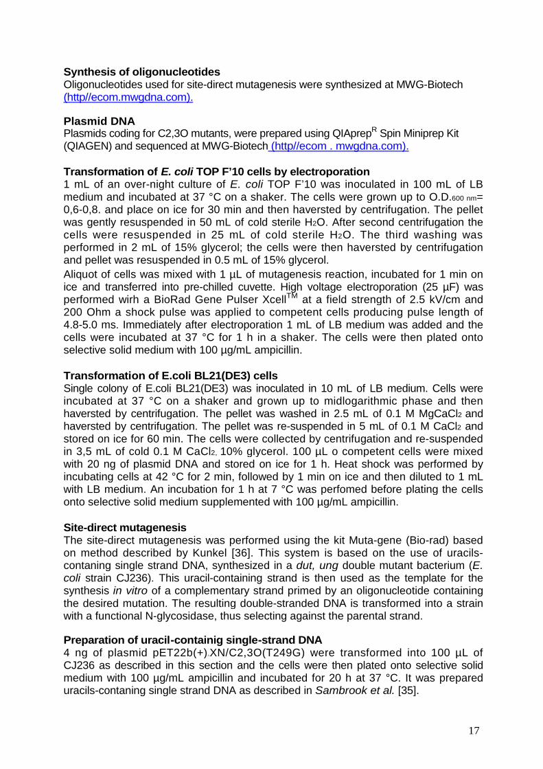
Synthesis of oligonucleotidesOligonucleotides used for site-direct mutagenesis were synthesized at MWG-Biotech(http//ecom.mwgdna.com).
Plasmid DNAPlasmids coding for C2,3O mutants, were prepared using QIAprepR Spin Miniprep Kit(QIAGEN) and sequenced at MWG-Biotech (http//ecom . mwgdna.com).
Transformation of E. coli TOP F’10 cells by electroporation1 mL of an over-night culture of E. coli TOP F’10 was inoculated in 100 mL of LBmedium and incubated at 37 °C on a shaker. The cells were grown up to O.D.600 nm=0,6-0,8. and place on ice for 30 min and then haversted by centrifugation. The pelletwas gently resuspended in 50 mL of cold sterile H2O. After second centrifugation thecells were resuspended in 25 mL of cold sterile H2O. The third washing wasperformed in 2 mL of 15% glycerol; the cells were then haversted by centrifugationand pellet was resuspended in 0.5 mL of 15% glycerol.
Aliquot of cells was mixed with 1 µL of mutagenesis reaction, incubated for 1 min onice and transferred into pre-chilled cuvette. High voltage electroporation (25 µF) wasperformed wirh a BioRad Gene Pulser XcellTM at a field strength of 2.5 kV/cm and200 Ohm a shock pulse was applied to competent cells producing pulse length of4.8-5.0 ms. Immediately after electroporation 1 mL of LB medium was added and thecells were incubated at 37 °C for 1 h in a shaker. The cells were then plated ontoselective solid medium with 100 µg/mL ampicillin.
Transformation of E.coli BL21(DE3) cellsSingle colony of E.coli BL21(DE3) was inoculated in 10 mL of LB medium. Cells wereincubated at 37 °C on a shaker and grown up to midlogarithmic phase and thenhaversted by centrifugation. The pellet was washed in 2.5 mL of 0.1 M MgCaCl2 andhaversted by centrifugation. The pellet was re-suspended in 5 mL of 0.1 M CaCl2 andstored on ice for 60 min. The cells were collected by centrifugation and re-suspendedin 3,5 mL of cold 0.1 M CaCl2, 10% glycerol. 100 µL o competent cells were mixedwith 20 ng of plasmid DNA and stored on ice for 1 h. Heat shock was performed byincubating cells at 42 °C for 2 min, followed by 1 min on ice and then diluted to 1 mLwith LB medium. An incubation for 1 h at 7 °C was perfomed before plating the cellsonto selective solid medium supplemented with 100 µg/mL ampicillin.
Site-direct mutagenesisThe site-direct mutagenesis was performed using the kit Muta-gene (Bio-rad) basedon method described by Kunkel [36]. This system is based on the use of uracils-contaning single strand DNA, synthesized in a dut, ung double mutant bacterium (E.coli strain CJ236). This uracil-containing strand is then used as the template for thesynthesis in vitro of a complementary strand primed by an oligonucleotide containingthe desired mutation. The resulting double-stranded DNA is transformed into a strainwith a functional N-glycosidase, thus selecting against the parental strand.
Preparation of uracil-containig single-strand DNA4 ng of plasmid pET22b(+)∆XN/C2,3O(T249G) were transformed into 100 µL ofCJ236 as described in this section and the cells were then plated onto selective solidmedium with 100 µg/mL ampicillin and incubated for 20 h at 37 °C. It was prepareduracils-contaning single strand DNA as described in Sambrook et al. [35].
Mutagenesis reactionMutagenesis reactions were prepared in a final volume of 10 µL containing 200 ng ofstranded uracil containing DNA, 10 pmol of phosphorilated mutagenic oligonucleotide(table 1) and 1 µL of 10x annealing buffer (200 mM Tris-HCl pH7.4, 20 mM MgCl2,500 mM NaCl). The reactions were incubated at 70 °C for 5 min and then cooledslowly, after the reactions were placed in an ice-water bath for 5 min. It was added 1µL of 10x synthesis buffer (5 mM dNTP, 10 mM ATP, 100 mM Tris/HCl pH 7,4, 50mM MgCl2, 20 mM DTT), 3 U of T4 DNA ligase and 4 U of T4 DNA polymerase. Thereaction were incubated on ice for 5 min followed by another 5 min at 25 °C, andfinally at 37 °C for 90 min. The reactions were stopped adding 90 µL of stop solution(10 mM Tris-HCl pH 7,4, 50 mM EDTA). 1 µL of reactions was used to transform TopF’ 10 and the cells were plated on LB agar with 100 µg/mL ampicillin and incubatedfor 20 h at 37 °C.The clones harbouring the desired mutations were identified by DNA sequencing.The sequences of all the clones were successively verified by sequencing.
Table 1.
Mutagenic Sequenceprimer
mutP198 5’ -GCCACGTCGTGCGGCTTGGTCGACAG-3’ P198
mutG198 5’ -GCCACGTCGTGACCCTTGGTCGACAG-3’ G198
mutH267 5’ -CTCCCCCGCAATGCACTT
Expression of C2,3Os
E. coli strain BL21 (DE3), transformecoding for the C2,3O mutants, was gr(100 µg/mL) until optical density at 60adding IPTG and Fe(NH4)2(SO4)2 arespectively. After 3 h, cells were havat 4 °C, washed with 50 mM Tris/HCpaste was stored at -80 °C until use.
PurificationThe bacterial pellet was suspended i0,08 M NaCl, 10%(v/v) glycerol, 10%
ATCCTSCCCGCAAWGCACTTCGTTGCG-3’Gln) G A (Leu)Gln) G T (His)Glu) C A (Leu)
CGTTGCG-
d with pETowth at 370 nm wast final conersted byl (pH 7.5)
n buffer A(v/v) eth
T
18
3’ H267
22b(+)∆XN containing the sequences°C in LB medium containig ampicillin0,6-0,7. Induction was performed by
centration of 0,5 mM and 100 µM,centrifugation at 4000 x g for 20 min
, and collected by centrifugation. Cell
(50 mM Tris/HCl (pH 7.5) containinganol and 2 mM dithiothreitol) in the
(His)
19
presence of 100µM Fe(NH4)2(SO4)2 and was disrupted by sonication. Cell debris wasremoved by centrifugation (12,000 × g, 1 h at 4 °C).The supernatant was loaded at 4°C onto a 15 mL Q-Sepharose column equilibrated previously in buffer A and washedwith 30 mL of equilibration buffer. Elution was carried out with 260 mL of buffer A witha NaCl linear gradient ranging from 0.08 to 0.5 M at a flow rate of 13 mL/h. Fractionscontaining dioxygenase activity were pooled and added 10% (v/v) glycerol and storedat -80 °C under nitrogen atmosphere.
Determination of the protein concentrationThe protein concentration was determined according to the Bradford’s method. Thecoomassie Brilliant (sigma) was added to the samples and the absorbance at 595 nmwas monitored. A solution of bovine serum albumin was used as a standard.
SDS-PolyAcrylamide Gel ElectrophoresisGel electrophoresis under denaturing condition was performed as described byLaemmli. The resolving gel was prepared at 18% acrylamide and the stacking gelwas prepared at 6% acrylamide.[37]
Iron determination of C2,3O mutantsThe iron content of C2,3O mutants was determined by using the iron-chelatingreagent Ferene S, which forms a complex with ferrous iron [Fe(II)].The samples were incubated at room temperature for 10 min and 25 µL of 50% (w/v)trichloracetic acid were added. Precipitated proteins were removed by centrifugation,the supernatants were transferred and 50 µL of 45% sodium acetate were added.450 µL of Ferene S reagent [0.8 mM Ferene S, 10 mM ascorbic acid and 45%sodium acetate] were added to the samples, in presence of ascorbic acid trivalentiron [Fe(III)] dissociated from proteins becomes reduced to divalent iron [Fe(II)],which forms a complex with Ferene S, then it was determined iron total content ofprotein. If Ferene S reagent was prepared without adding ascorbic acid, it wasdetermined Fe(II) concentration in proteic samples. The absorbance at 593 nm weremeasured and Fe(II) concentration was calculated using as molar extinctioncoefficient 34320 M-1cm-1.
Enzyme assayAll assay were performed at 25 °C in 50 mM Tris/HCl (pH 7.5) in a final volume of 1mL by spectrophotometric determination of the product of the reaction. Wild-type andmutant C2,3Os were used to start the reaction.The amount of the fission products was measured spectrophotometrically using theirabsorption extinction coefficient at the respective εmax values. These coefficients wereas follows: ε375 = 33,000 M-1cm-1 for 2-hydroxymuconic semialdehyde, the product ofcatechol; ε388 = 13,400 M-1cm-1 for 2-hydroxy-6-oxohepta-2,4-dienoic acid, theproduct of 3-methylcatechol; ε393 = 8,230 M-1cm-1 for 2-hydroxy-3,5-dimethyl-6-oxohexa-2,4-dienoic acid, the product of 3,5-dimetylcatechol; and ε393 = 15,200 M-
1cm-1 for 2-hydroxy-3-methyl-6-oxoepta-2,4-dienoic acid, the product of 3,6-dimetylcatechol.Kinetics parameters were determined by the program GraphPad Prism (GrapfPadSoftware; www.graphpad.com). One unit of enzyme activity was defined as theamount of enzyme required to form 1 µmol of the metacleavage product per minuteunder assay conditions.
20
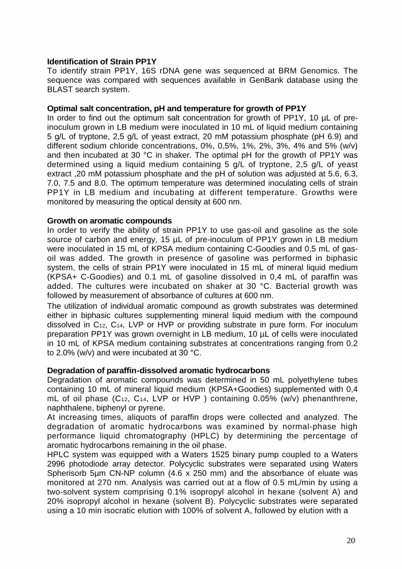
Identification of Strain PP1YTo identify strain PP1Y, 16S rDNA gene was sequenced at BRM Genomics. Thesequence was compared with sequences available in GenBank database using theBLAST search system.
Optimal salt concentration, pH and temperature for growth of PP1YIn order to find out the optimum salt concentration for growth of PP1Y, 10 µL of pre-inoculum grown in LB medium were inoculated in 10 mL of liquid medium containing5 g/L of tryptone, 2,5 g/L of yeast extract, 20 mM potassium phosphate (pH 6.9) anddifferent sodium chloride concentrations, 0%, 0,5%, 1%, 2%, 3%, 4% and 5% (w/v)and then incubated at 30 °C in shaker. The optimal pH for the growth of PP1Y wasdetermined using a liquid medium containing 5 g/L of tryptone, 2,5 g/L of yeastextract ,20 mM potassium phosphate and the pH of solution was adjusted at 5.6, 6.3,7.0, 7.5 and 8.0. The optimum temperature was determined inoculating cells of strainPP1Y in LB medium and incubating at different temperature. Growths weremonitored by measuring the optical density at 600 nm.
Growth on aromatic compoundsIn order to verify the ability of strain PP1Y to use gas-oil and gasoline as the solesource of carbon and energy, 15 µL of pre-inoculum of PP1Y grown in LB mediumwere inoculated in 15 mL of KPSA medium containing C-Goodies and 0,5 mL of gas-oil was added. The growth in presence of gasoline was performed in biphasicsystem, the cells of strain PP1Y were inoculated in 15 mL of mineral liquid medium(KPSA+ C-Goodies) and 0.1 mL of gasoline dissolved in 0,4 mL of paraffin wasadded. The cultures were incubated on shaker at 30 °C. Bacterial growth wasfollowed by measurement of absorbance of cultures at 600 nm.
The utilization of individual aromatic compound as growth substrates was determinedeither in biphasic cultures supplementing mineral liquid medium with the compounddissolved in C12, C14, LVP or HVP or providing substrate in pure form. For inoculumpreparation PP1Y was grown overnight in LB medium, 10 µL of cells were inoculatedin 10 mL of KPSA medium containing substrates at concentrations ranging from 0.2to 2.0% (w/v) and were incubated at 30 °C.
Degradation of paraffin-dissolved aromatic hydrocarbonsDegradation of aromatic compounds was determined in 50 mL polyethylene tubescontaining 10 mL of mineral liquid medium (KPSA+Goodies) supplemented with 0,4mL of oil phase (C12, C14, LVP or HVP ) containing 0.05% (w/v) phenanthrene,naphthalene, biphenyl or pyrene.At increasing times, aliquots of paraffin drops were collected and analyzed. Thedegradation of aromatic hydrocarbons was examined by normal-phase highperformance liquid chromatography (HPLC) by determining the percentage ofaromatic hydrocarbons remaining in the oil phase.HPLC system was equipped with a Waters 1525 binary pump coupled to a Waters2996 photodiode array detector. Polycyclic substrates were separated using WatersSpherisorb 5µm CN-NP column (4.6 x 250 mm) and the absorbance of eluate wasmonitored at 270 nm. Analysis was carried out at a flow of 0.5 mL/min by using atwo-solvent system comprising 0.1% isopropyl alcohol in hexane (solvent A) and20% isopropyl alcohol in hexane (solvent B). Polycyclic substrates were separatedusing a 10 min isocratic elution with 100% of solvent A, followed by elution with a
21
linear 100% to 90% solvent A gradient in 15 min and then an isocratic 90% solvent Astep. Nitrobenzene was used as internal standard.
22
RESULTS & DISCUSSION
Part 1: Protein engineering of the catechol 2,3-dioxygenasefrom Pseudomonas sp. OX1.
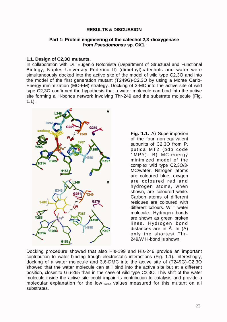
1.1. Design of C2,3O mutants.In collaboration with Dr. Eugenio Notomista (Department of Structural and FunctionalBiology, Naples University Federico II) (dimethyl)catechols and water weresimultaneously docked into the active site of the model of wild type C2,3O and intothe model of the first generation mutant (T249G)-C2,3O by using a Monte Carlo-Energy minimization (MC-EM) strategy. Docking of 3-MC into the active site of wildtype C2,3O confirmed the hypothesis that a water molecule can bind into the activesite forming a H-bonds network involving Thr-249 and the substrate molecule (Fig.1.1).
Fig. 1.1. A) Superimposionof the four non-equivalentsubunits of C2,3O from P.put ida MT2 (pdb code1MPY). B) MC-energyminimized model of thecomplex wild type C2,3O/3-MC/water. Nitrogen atomsare coloured blue, oxygena re co lou red red andhydrogen atoms, whenshown, are coloured white.Carbon atoms of differentresidues are coloured withdifferent colours. W = watermolecule. Hydrogen bondsare shown as green brokenl ines . Hydrogen bonddistances are in Å. In (A)on ly the shortest Thr-249/W H-bond is shown.
Docking procedure showed that also His-199 and His-246 provide an importantcontribution to water binding trough electrostatic interactions (Fig. 1.1). Interestingly,docking of a water molecule and 3,6-DMC into the active site of (T249G)-C2,3Oshowed that the water molecule can still bind into the active site but at a differentposition, closer to Glu-265 than in the case of wild type C2,3O. This shift of the watermolecule inside the active site could impair its contribution to catalysis and provide amolecular explanation for the low kcat values measured for this mutant on allsubstrates.
23
In order to design C2,3O mutants with improved catalytic efficiency on 3,5-DMC and3,6-DMC, (T249G)-C2,3O was chosen as starting point because, among the firstgeneration mutants, it showed the lowest KM values – and hence the highestapparent affinity – on both DMCs.
Besides residue Thr-249, four other side-chains face the hypothetical water bindingsite: Gly-250, Gly-270, Ala-198 and Phe-267 (Fig. 1.1). These four positions arepossible targets for site-directed mutagenesis. By using the Deep-View and PyMolprotein visualization and modeling softwares the four residues were mutated in silicoto find a combination of side-chains able to restore the hypothetical water moleculebinding site without hindering the sub-site for the binding of the methyl group atposition 6 of 3,6-DMC. The most promising mutants were further examined by theMC-EM strategy.Residue at position 250 was judged not suitable for mutagenesisbecause it is to close to the water binding site and even a serine residue at thisposition could partially hinder the sub-site for methyl binding (not shown).On the contrary, mutation of more distant Gly-270 residue to glutamate or glutaminecould position the hydrophilic side-chains of these residues to H-bond distance froma water molecule located at a position similar to that found in the complex betweenwild type C2,3O and 3-MC (Fig. 1.2).
Fig. 1.2. A) Comparisonbetween the model complexeswild type C2,3O/3-MC/watera n d (T2 4 9 G ) -C2 , 3 O / 3 , 6 -DMC/water. B) Comparisonbetween the model complexes(T249G, G270E)-C2,3O/3,6-D M C / wa t e r a n d ( T 2 4 9 G ,G270Q)-C2,3O/3,6-DMC/waterW = water molecule. Atoms andH-bonds are coloured as in Fig.X1 except carbon atoms whichare coloured green (wild typeC2,3O), orange [(T249G)-C2,3O], purple [(T249G,G270E)-C2,3O], and cyan[(T249G, G270Q)-C2,3O]. Allt h e a t o m s o f e a ch wa t e rmolecule are coloured like thecarbon atoms of the samecomplex.
Models of C2,3O with mutations G270E or G270Q also showed that the large side-chains of glutamate and glutamine were closely packed between the side-chains ofresidues at positions 198 and 267 (Fig. 1.2). Therefore, mutations at these twopositions could be used to control the positioning of the side-chains of Glu-270 and
24
Gln-270. For example, mutation A198G, reducing the hindrance, could allow closercontacts between the backbone of this residue and the side-chains of Glu-270/Gln-270, whereas, mutations A1 98S and A1 98P could exert the opposite effect. Similarly,a reduction of the bulky side-chain of Phe-267 could allow the shifting of the side-chains of Glu-270/Gln-270 toward the water binding site. Two types of mutations atposition 267 were considered: (i) mutations F267L and F267A reduce the volume ofthe side-chain preserving its hydrophobic nature, whereas, (ii) mutations F267H,F267N, and F267S insert hydrophilic side-chains which could form hydrogen bondswith the side-chain of Glu-270/Gln-270 and also directly with the water molecule (Fig.1.3).
Table 1.1 shows the combination of mutations which were considered particularlypromising on the basis of the modeling experiments.
1.2. Preparation of the C2,3O mutants.Due to the very high number of combinations (Table 1.1) we decided to prepare theexpression vectors for the mutants of C2,3O using the Kunkel strategy and a seriesof degenerated oligonucleotides whose combinations allow to obtain all the desiredmutations at positions 198, 267, and 270 (see Materials and Methods). For example,by using simultaneously the degenerated mutagenic oligonucleotides for positions198 and 270 we prepared a small library containing the expression vectors coding forthe six possible triple mutants at these positions (lane 8 in table 1.1).
Fig. 1.3. A) Model of thecomplex (T249G, F276H)-C2,3O/3,6-DM C/water. B)M o d e l o f t h e c o m p l e x(T249G, F276H, G270E)-C2,3O/3,6-DMC/water. W =water molecule. Atoms andH-bonds are coloured as inFig. X1 except carbon atomswhich are coloured green[(T249G, F276H)-C2,3O/3,6-DMC/water ] and purp le[(T249G, F276H, G270E)-C2,3O/3,6-DMC/water]. Allthe atoms of each watermolecule are coloured likethe carbon atoms of thesame complex.
25
Table 1.1Positions*
protein 198 249 267 270 Number ofmutants
(T249G) A G F G 1
double A G F E/Q 2mutants A G H/N G 2
triple A G H/N/S E/Q 6mutants A G A/L E/Q 4
G/S/P G F E/Q 6
quadruplemutants
S/P G A/S E/Q 8
Number of double-quadruple mutants 28* mutated residues are shown in bold.
Identification of mutations present in each clone was performed by PCR adapting aprocedure used in prenatal diagnosis which allows to discriminate two sequencesdiffering by a single base pair. The procedure is based on the use of a PCR primerwhose 3’ nucleotide binds the nucleotide which differs in the wild type and mutantsequences. As Taq polymerase lacks the 3’->5’ exonucleasic activity, if thenucleotide at the 3’ end of the oligonucleotide is not correctly paired to the templatethe oligonucleotide is not recognized as primer by the polymerase. The sequences ofall the clones were successively confirmed by sequencing.
This strategy allowed to prepare in short time thirteen double and triple mutants. Inthe following section we will describe the kinetic characterization of three doublemutants – (T249G, G270E)-, (T249G, G270Q)- and (T249G, F267H)-C2,3O – andfive triple mutants – (A198G, T249G, G270E)-, (A198S, T249G, G270E)-, (T249G,F267H, G270E)-, (T249G, F267L, G270E)-, and (T249G, F267L, G270Q)-C2,3O.
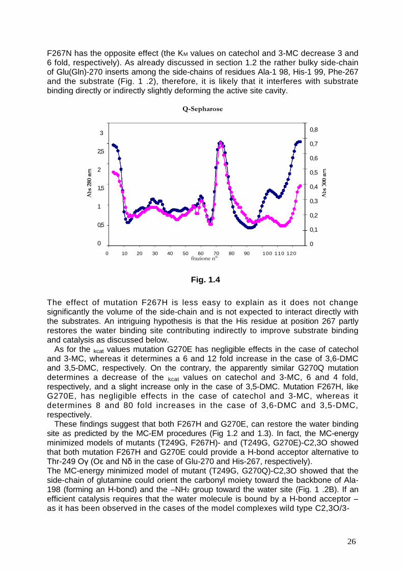
1.3. Expression, purification and characterization of C2,3O mutants.All the double and triple mutants were expressed in the E. coli strain BL21 (DE3) andpurified by ion exchange chromatography on the strong anion exchanger Q-sepharose as described in the Material and Methods section. The first generationmutant (T249G)-C2,3O was purified and used as a control. Fig. 1.4 shows a typicalchromatogram with a single major peak containing the C2,3O mutant. The fractionsof the peak were pooled and analyzed by SDS-PAGE. All mutant C2,3O showedabout 95% purity. Proteins were not purified further as every attempt to obtain purersamples lead to loss of enzyme activity (not shown).
At the end of the purification procedure all mutants were assayed to determine theiron content. All mutants showed a ratio moles of iron/moles of monomers very closeto 1 (typically 1.1 ± 0.1), as expected. Moreover, all the iron was present in thecatalytically productive Fe(II) state.
Kinetic characterization was done using catechol, 3-MC, 3,5-DMC and 3,6-DMC asdescribed in Materials and Methods.
The comparison between the KM values of the single mutant (T249G)-C2,3O andof the double mutants (T249G, G270E)-, (T249G, G270Q)- and (T249G, F267H)-C2,3O suggests that mutation of residue Gly-270 to Glu and Gln determines asignificant decrease of the apparent affinity for all the substrates, whereas mutation
26
F267N has the opposite effect (the KM values on catechol and 3-MC decrease 3 and6 fold, respectively). As already discussed in section 1.2 the rather bulky side-chainof Glu(Gln)-270 inserts among the side-chains of residues Ala-1 98, His-1 99, Phe-267and the substrate (Fig. 1 .2), therefore, it is likely that it interferes with substratebinding directly or indirectly slightly deforming the active site cavity.
The effect of mutation F267H is less easy to explain as it does not changesignificantly the volume of the side-chain and is not expected to interact directly withthe substrates. An intriguing hypothesis is that the His residue at position 267 partlyrestores the water binding site contributing indirectly to improve substrate bindingand catalysis as discussed below.
As for the kcat values mutation G270E has negligible effects in the case of catecholand 3-MC, whereas it determines a 6 and 12 fold increase in the case of 3,6-DMCand 3,5-DMC, respectively. On the contrary, the apparently similar G270Q mutationdetermines a decrease of the kcat values on catechol and 3-MC, 6 and 4 fold,respectively, and a slight increase only in the case of 3,5-DMC. Mutation F267H, likeG270E, has negligible effects in the case of catechol and 3-MC, whereas itdetermines 8 and 80 fold increases in the case of 3,6-DMC and 3,5-DMC,respectively.
These findings suggest that both F267H and G270E, can restore the water bindingsite as predicted by the MC-EM procedures (Fig 1.2 and 1.3). In fact, the MC-energyminimized models of mutants (T249G, F267H)- and (T249G, G270E)-C2,3O showedthat both mutation F267H and G270E could provide a H-bond acceptor alternative toThr-249 Oγ (Oε and Nδ in the case of Glu-270 and His-267, respectively).The MC-energy minimized model of mutant (T249G, G270Q)-C2,3O showed that theside-chain of glutamine could orient the carbonyl moiety toward the backbone of Ala-198 (forming an H-bond) and the –NH2 group toward the water site (Fig. 1 .2B). If anefficient catalysis requires that the water molecule is bound by a H-bond acceptor –as it has been observed in the cases of the model complexes wild type C2,3O/3-
3
2,5
2
1,5
1
0,5
0
0,8
0,7
0,6
0,5
0,4
0,3
0,2
0,1
0
27
MC/water, (T249G, F267H )-C2,3O/3,6-DMC/water and (T249G, G270E)-C2,3O/3,6-DMC/water (Fig. 1.1B, 1.2A, 1.3A) – the orientation of the glutamine side chain couldbe less effective than glutamate and histidine thus explaining the negligible effects oncatalysis shown by mutation G267Q.Looking at the KS values in Table 1.2 it is clear that, among the three double mutants,(T249G, F267H)-C2,3O is the most efficient catalyst for the cleavage ofdimethylcatechols.The triple mutant (T249G, F267H, G270E)-C2,3O combines the two mutations,F267H and G270E, which increase significantly the catalytic efficiency. This mutantshows a kcat value similar to that of (T249G, G270E)-C2,3O, but a KS value similar tothat of (T249G, F267H)-C2,3O, therefore the combination of the two mutations failsto provide a more effective catalyst. Again this finding could depend on the hindranceof Glu-270 side-chain, an hypothesis supported also by the features of the triplemutant (T249G, F267L, G270E)-C2,3O discussed below. An alternative explanationwas provided by the MC-EM analysis. The MC-energy minimized models showedthat, when His-267 and Glu-270 are simultaneously present, His-267 could binddirectly the carboxylate of Glu-270 so that the water molecule would interact only withGlu-270 (Figure 1 .3B).As discussed in section 1.1 the triple mutants (A198G, T249G, G270E)-, (A198S,T249G, G270E)-, (T249G, F267L, G270E)-, and (T249G, F267L, G270Q)-C2,3Owere prepared with the aim of controlling the positioning of the side-chain of residueGlu(Gln)-270. In particular, mutations A198G and F267L were chosen to try toincrease the mobility of the glutamate(glutamine) side-chain.The comparison between the catalytic constants of mutant (T249G, G270E)-C2,3Oand of triple mutant (A198G, T249G, G270E)-C2,3O shows that mutation A198Gdetermines a decrease of the apparent affinity for all the substrates – with theexception of 3-MC. The decrease is particularly high in the case of catechol and 3,5-DMC and less pronounced in the case of 3,6-DMC. Moreover, mutation A198Greduces the kcat values 3 fold in the case of 3-MC and ten fold in the case of 3,5- and3,6-DMC. As a consequence mutation A1 98G determines a marked decreases of thespecificity constant for all the substrate and is the worst catalyst for the cleavage ofdimethylcatechols. The effect of this mutation has not been investigated throughly byMC-EM, however, at the moment the most likely explanation is that the presence of aGly residue at position 198 changes the conformation of the loop Lys-197/Ala-198/His-1 99 impairing the participation to catalysis of His-199, a residue essential forthe catalytic activity of C2,3Os [21]. Interestingly, mutation A198S has oppositeeffects. In fact, it slightly decreases the KM values (2.5 fold in the case of catechol)suggesting an improved binding of the substrates. Moreover, it increases the kcat
value on catechol and 3-MC two and three fold, respectively. Unfortunately, mutationA1 98S determines a decrease of the kcat values measured on dimethylcatechols. Thecomparison between the MC-energy minimized complexes of (A198G, T249G,G270E)-C2,3O with 3-MC and 3,6-DMC indicates that the hydroxyl group of Ser-198could form a hydrogen bond with the side-chain of Glu-270 anchoring it in aconformation suitable to reconstitute the water binding site, but at the same time itwould interact closely with the second methyl group of 3,6-DMC (Fig. 1.5A). Thiscould prevent optimal positioning of dimethylcatechols for catalysis and determinethe observed selective decrease of the kcat values for these substrates.
At difference with mutation A1 98S, which selectively improves the catalytic constantsfor the physiologic substrates, mutation F267L was found to improve the catalyticconstants for dimethylcatechols, when coupled with G270E and G270Q mutations. In
28
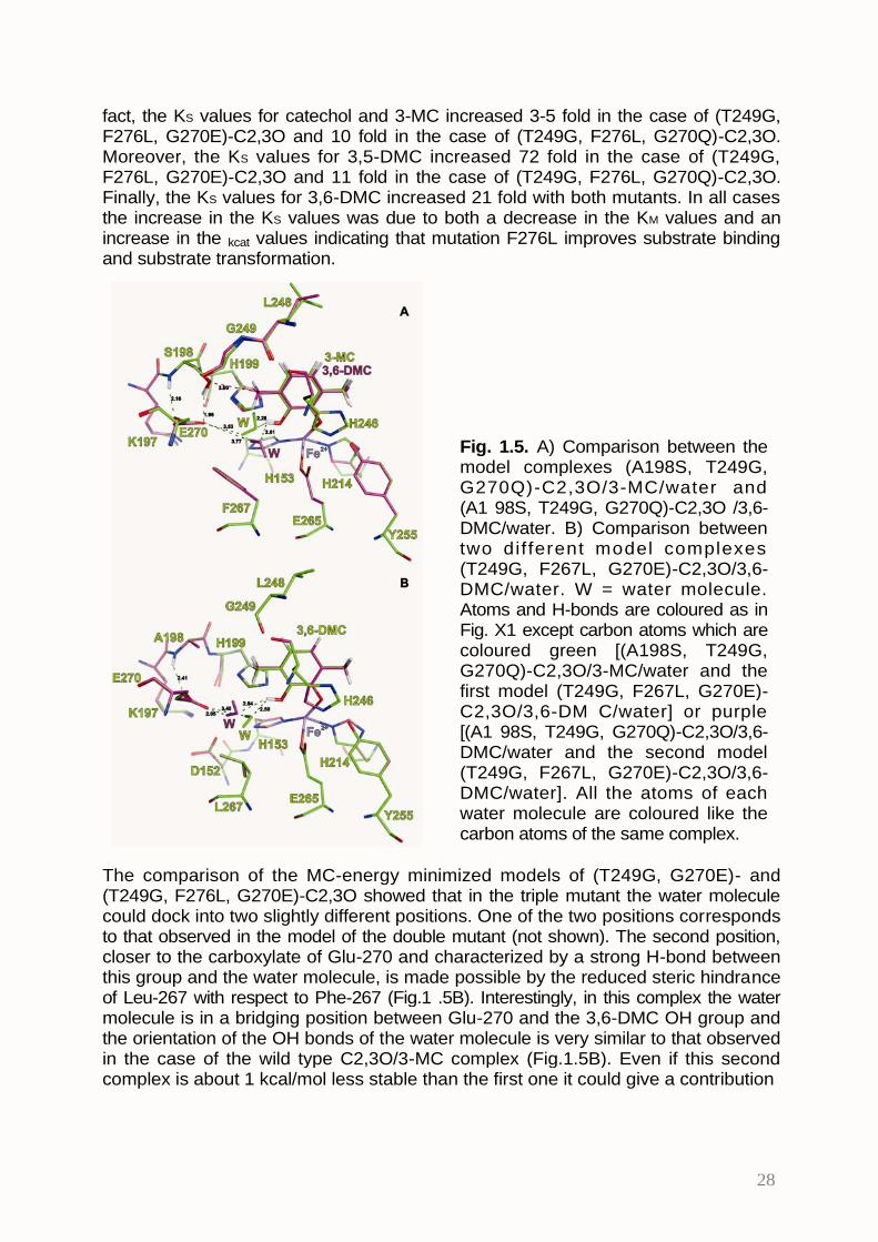
fact, the KS values for catechol and 3-MC increased 3-5 fold in the case of (T249G,F276L, G270E)-C2,3O and 10 fold in the case of (T249G, F276L, G270Q)-C2,3O.Moreover, the KS values for 3,5-DMC increased 72 fold in the case of (T249G,F276L, G270E)-C2,3O and 11 fold in the case of (T249G, F276L, G270Q)-C2,3O.Finally, the KS values for 3,6-DMC increased 21 fold with both mutants. In all casesthe increase in the KS values was due to both a decrease in the KM values and anincrease in the kcat values indicating that mutation F276L improves substrate bindingand substrate transformation.
Fig. 1.5. A) Comparison between themodel complexes (A198S, T249G,G270Q)-C2,3O/3-MC/water and(A1 98S, T249G, G270Q)-C2,3O /3,6-DMC/water. B) Comparison betweentwo dif ferent model complexes(T249G, F267L, G270E)-C2,3O/3,6-DMC/water. W = water molecule.Atoms and H-bonds are coloured as inFig. X1 except carbon atoms which arecoloured green [(A198S, T249G,G270Q)-C2,3O/3-MC/water and thefirst model (T249G, F267L, G270E)-C2,3O/3,6-DM C/water] or purple[(A1 98S, T249G, G270Q)-C2,3O/3,6-DMC/water and the second model(T249G, F267L, G270E)-C2,3O/3,6-DMC/water]. All the atoms of eachwater molecule are coloured like thecarbon atoms of the same complex.
The comparison of the MC-energy minimized models of (T249G, G270E)- and(T249G, F276L, G270E)-C2,3O showed that in the triple mutant the water moleculecould dock into two slightly different positions. One of the two positions correspondsto that observed in the model of the double mutant (not shown). The second position,closer to the carboxylate of Glu-270 and characterized by a strong H-bond betweenthis group and the water molecule, is made possible by the reduced steric hindranceof Leu-267 with respect to Phe-267 (Fig.1 .5B). Interestingly, in this complex the watermolecule is in a bridging position between Glu-270 and the 3,6-DMC OH group andthe orientation of the OH bonds of the water molecule is very similar to that observedin the case of the wild type C2,3O/3-MC complex (Fig.1.5B). Even if this secondcomplex is about 1 kcal/mol less stable than the first one it could give a contribution
29
to catalysis explaining the increased catalytic efficiency of the mutants bearing themutation Leu-267.Thus it can be concluded that mutant (T249G, F276L, G270E)-C2,3O is the bestavailable catalyst for the cleavage of 3,5-DMC and 3,6-DMC.At the moment we are preparing and characterizing further mutants with residuessmaller than leucine at position 267, as for example asparagine, alanine and serine.These mutations have been chosen to try to stabilize the second water site observedin (T249G, F276L, G270E)-C2,3O. These mutants will allow to verify if furtherreducing the hindrance at position 267 and, hence, increasing the mobility ofGlu(Gln)-270 side-chain and of the water molecule, will provide more efficientcatalysts for the cleavage of dimethylcatechols.
Table 1.2Substrate
CAT 3-MC 3,5-DMC 3,6-DMCProtein KM a kcat b KS c KM a kcat b KS c KM a kcat b KS c a
Part 2: Isolation of new strains from polluted environments
Seawater samples were collected in six different areas inside the harbor of Pozzuoli(NA) (Fig. 2.1). All samples were collected at the surface and close to oil stains. Thearea indicated by number 1 in Fig. 2.1 is a bay for the mooring of small boatsconnected to the port by a narrow canal.
Fig. 2.1. A) Satellite picture ofPozzuoli harbour showing thepoints where water sampleswe re c o l le c ted . B ) PP 1 Ycolonies surrounded by a clearh a l o o n M 9 G - A g a r p l a t econtaining phenanthrene as thesole carbon source incubated 7days at 25°C.
In order to favor the growth of microorganisms able to use aromatic hydrocarbons asenergy and carbon source, naphthalene, phenanthrene and anthracene crystals,separately or as a mixture, were added to seawater samples. After three-six weeks ofincubation at 25°C some samples showed the appearance of turbidity and/orbrown/yellow color which are indications of microorganisms growth. These samples
A
B
31
were used to inoculate saline solutions (M9G) containing naphthalene, phenanthreneand/or anthracene as the sole source of carbon and energy. After two-four weeks ofincubation turbid and/or colored cultures were used to inoculate fresh mediumcontaining PAH as the sole carbon source. After three-four rounds of enrichment,aliquots were spread on agar plates containing phenanthrene as the sole carbonsource. Several yellow colonies surrounded by clear halos were observed on platesseeded with samples collected inside area 1 (not shown). The appearance of a clearhalo in the milky layer of phenanthrene crystals is an indication that the colony is ableto degrade phenanthrene causing dissolution of crystals. Strain PP1Y (Pozzuoli;Phenanthrene; area 1; Yellow) described in this thesis was isolated starting from oneof these colonies and purified through four plating cycles on reach medium.When plated on LB-Agar strain PP1Y formed in 2-3 days bright yellow, mucoidcolonies. Small amounts of liquid cultures, deposited on M9G-Agar plates containingphenanthrene as the sole carbon source, formed light yellow colonies surrounded bya clear halo in about 5-7 days at 25°C (Fig. 2.1B).
2.1. Analysis of 16S rDNA gene and identification of strain PP1Y.A fragment of the 16S rDNA gene (Fig. 2.2), whose sequence was determined byBMR GENOMICS, was used to search the GENBANK database. 98-99% sequenceidentity was found with the rDNA sequences of several Novosphingobium strains,whereas, 90-98% sequence identity was found with the rDNA sequences of severalstrains of Sphingomonas, Sphingobium and Sphingopixis, which, together with thegenus Novosphingobium, form the Sphingomonadaceae family, the soleproteobacteria (the main group of Gram- bacteria) which do not synthesize LPS.
GCCCCTCGGTTCGGAATAACTCAGGGAAACTTGAGCTAATACCGGATAATGACTTCGGTCCAAAGATTTATTGCCGAGGGATGAGCCCGCGTAGGATTAGCTAGTTGGTGGGGTAATGGCCTACCAAGGCGACGATCCTTAGCTGGTCTGAGAGGATGATCAGCCACACTGGGACTGAGACACGGCCCAGACTCCTACGGGAGGCAGCAGTGGGGAATATTGGACAATGGGCGAAAGCCTGATCCAGCAATGCCGCGTGAGTGATGAAGGCCTTAGGGTTGTAAAGCTCTTTTACCAGGGATGATAATGACAGTACCTGGAGAATAAGCTCCGGCTAACTCCGTGCCAGCAGCCGCGGTAATACGGAGGGAGCTAGCGTTGTTCGGAATTACTGGGCGTAAAGCGCGCGTAGGCGGTTACTCAAGTCAGAGGTGAAAGCCCGGGGCTCAACCCCGGAACTGCCTTTGAAACTAGGTGACTAGAATCTTGGAGAGGTCAGTGGAATTCCGAGTGTAGAGGTGAAATTCGTAGATATTCGGAAGAACACCAGTGGCGAAGGCGACTGACTGGACAAGTATTGACGCTGAGGTGCGAAAGCGTGGGGAGCAAACAGGATTAGATACCCTGGTAGTCCACGCCGTAAACGATGATAACTAGCTGTCCGGGCACATGGTGTTTGGGTGGCGCAGCTAACGCATTAAGTTATCCGCCTGGGGAGTACGGTCGCAAGATTAAAACTCAAAGGAATTGACGGGGGCCTGCACAAGCGGTGGAGCATGTGGTTTAATTCGAAGCAACGCGCAGAACCTTACCAGCGTTTGACATCCTGATCGCGGATTAGAGAGATCTTTTCCTTCAGTTCGGCTGGATCAGTGACAGGTGCTGCATGGCTGTCGTCAGCTCGTGTCGTGAGATGTTGGGTTAAGTCCCGCAACGAGCGCAACCCTCGTCCTTAGTTGCCATCATTTGGTTGGGCACTCTAAGGAAACTGCCGGTGATAAGCCGGAGGAAGGTGGGGATGACGTCAAGTCCTCATGGCCCTTACACGCTGGGCTACACAC GT GC TACAAT GGC GGT GACAGT GGGCAGC GAGT GC GC GAGCACAAGC TAAT C T C CAAAAGCCGTCTCAGTTCGGATTGTTCTCTGCAACTCGAGAGCATGAAGGCGGAATCGCTAGTAATCGCGGATCAGCATGCCGCGGTGAATACGTTCCCAGGCCTTGTACACACCGCCCGTCACACCATGGGAGTTGGTTTCACCCGAAGGTAGTGCGCTAACCGGCAACGGAGGCAGCTAACCACGGTGGGATCAGCGACTGGGGTGAAGTC
Fig. 2.2. DNA sequence of a fragment of PP1Y 16S rRNA gene.
32
Strain PP1Y is closely related to Novosphingobium pentaromativorans US6-1T (twodifferences in a 1481 bases-long alignment) a strain isolated in Korea and able todegrade PAH with 3-5 rings, and to Novosphingobium sp. Phe-8 (five differentnucleotides) (data not shown). Therefore on the basis of the 16S rRNA sequencesstrain PP1Y could be considered a new strain of the species Novosphingobiumpentaromativorans. However, the microbiological characterization described in thefollowing sections shows that the two strains differ in several relevant physiologicalfeatures like optimal salinity, metabolic properties, membrane fatty acid composition,growth morphology and range of substrates which can be degraded. For thesereasons we suggest that strain PP1Y is a new Novosphingobium species andpropose the name Novosphingobium puteolanum PP1Y.
2.2. Phenotypic characterization.Novosphingobium puteolanum PP1Y grows effectively in LB medium and in minimalmediums containing casaminoacids or glucose as single carbon sources. Lesseffectively it can use glycerol, whereas, it does not grow using benzoate or phenol(data not shown).The strain gave positive results in the assays for the β-galactosidasic, ureasic andarginine-dihydrolase activities and for citrate assimilation. In the same tests the strainUS6-1 provides negative results.Several sphingomonadales produce carotenoids. Nostoxanthin, a yellow carotenoid,is one of the most frequently found in this group of bacteria. The UV-vis spectrum ofthe yellow pigment produced by Novosphingobium puteolanum PP1Y was found tobe identical to that of nostoxanthin reported in literature (data not shown). Theamount of the pigment depended on the growth substrate, temperature, shaking ofthe cultures and salinity. The largest amount was observed during growth on solidrich mediums (LB-Agar)Sphingomonadales are so-called because they produce an atypical outer membranecontaining glycosphingolipids (GLS) instead of lipopolysaccharides. For example S.paucimobilis outer membrane contains large quantities of GSL-1 and GSL-4 whichare formed by a sphingosine molecule bound to a 2-hydroxy fatty acid and to amono- or tetra-saccharide, respectively. The type and relative abundance of the 2-hydroxy fatty acids is typical of each strain and can be used for tassonomicpurposes. The analysis of the fatty acids (Table 2.1) showed that the lipidiccomposition of the strain PP1Y is more similar to that of the strain Novosphingobiumsubarticum JCM 10398T than to that of Novosphingobium pentaromativorans US6-1T.Moreover it showed some distinctive features such as the absence of the fatty acid17:1 and the presence of the fatty acid 18:1 methylated at position 11.Liquid cultures of strain PP1Y, both in rich and minimal mediums with several growthsubstrate, became very viscous due to the production of abundant extracellularmaterial likely containing esopolysaccharides (EPS). In fact, it is known that severalstrains of sphingomonadales produce EPS containing repeated tetrasaccharidic units(A-B-C-B)n where A is usually rhamnose or mannose, B is glucose and C isglucuronic acid. Sugars at positions A and B usually bind “side-chains” constituted byother sugars or carboxylic acids like acetate and glycerate. Several of thesepolysaccharides are patented as jellifying agents or thickeners for food industry orscientific research. Gelrite, the gel used for the preparation of plates for thermophilicbacteria, is the commercial form of “gellan”, the EPS produced by Sphingomonaselodea [38]. Preliminary analysis of the EPS produced by the strain PP1Y performedby Dr. Antonio Molinaro (Dipartimento di Chimica Organica e Biochimica, Università
33
di Napoli Federico II) showed that it contains, as expected, mainly rhamnose,glucose and glucuronic acid, but also some other less abundant sugars. At themoment we don’t know if these sugars are branches of the main polysaccharide orform one or more independent polymers.
Table 2.1. Composition of the membrane fatty acids of the strainPP1Y and of the closely related strains Novosphingobiumpentaromativorans US6-1 and Novosphingobium subarticumJCM1 0398.
2.3 Optimal salt concentration, pH and temperature for growth of PP1Y.As shown in figures 2.3A and 2.3B Novosphingobium puteolanum PP1Y was able togrow from 0% to 4% (w:v) NaCl. The highest growth rates were measured at NaClconcentrations between 0.5% and 1.5% (w:v). Growth was very slow at NaClconcentrations above 5%. These features allow the strain PP1Y to grow both inseawater and brackish water, like river estuaries and costal lagoons. It is alsointeresting to note that the behavior of strain US6-1 is very different [4]. In fact, thehighest growth rate for this strain was observed at 2,5% NaCl, moreover, it the strainis not able to grow at NaCl concentrations below 1 %.As for optimal growth temperature, in LB medium, strain PP1Y was able to grow in awide range from about 18°C to 42°C. The highest growth rates were observed atabout 32-34°C (data not shown).The effect of pH on the growth rate was studied at 22°C and 28°C (Fig. 2.4). At bothtemperatures the strain showed the ability to grow in a wide range of pH values (from5 to about 7.5) even if the optimal pH is slightly more acidic at 22°C (pH 6.0) than at28°C (pH 7.0).
Fig. 2.3. Growth curvesof Novosphingobiumputeolanum PP1Y asf u n c t i o n o f N a C lconcentration.
0% 2% 4% 6%
% NaCl
pH
pH
0% NaCl
0,5% NaCl
1% NaCl
2% NaCl
3% NaCl
4% NaCl
5% NaCl
A
1
0,5
0
0 10 20 30 40 50h
3
2,5
2
1,5
1
0,5
0
B
5
420 h
30 h3
2
1
0
5 6 7 8 9
Fig. 2.4. Growth curvesof Novosphingobiumputeolanum PP1Y asfunction of pH at 28°C A)and 22°C B).
20 h
30 h
3
2
1
0
5 6 7 8 9
5
4
35
2.4 Morphological analysis of cells and of the “biofilm” of strain PP1YCells of Novosphingobium puteolanum PP1Y, observed by phase contrastmicroscope, appeared as short rods. They could be motile (likely flagellate) or non-motile (Fig. 2.5A). Non-motile cells could form aggregates of different dimensionsincluding macroscopic “flocks” 1-10 mm long. Figure 2.5A shows the border of one ofthese flocks. The number and dimension of flocks was influenced by severalparameters including shaking, temperature and growth substrates. A completeinvestigation was not performed. However, formation of flocks was found to befavored by slow shaking, high temperature (34°-37°C) and by the presence ofcasaminoacids. Dried and stained with methylene blue, flocks showed the presenceof bunches of cells trapped inside a blue-stained matrix (Figure 2.5B).
F i g . 2 . 5 A ) P h a s econtrast microscopepicture showing thebo rde r o f f l ock (no tstained). B) Phasecontrast microscopepicture showing a flockdried and stained withmethylene blue.
A similar behavior has already been described for some other sphingomonadaleswhich show the so-called “planktonic/sessile dimorphism” [39]. These dimorphicstrains can exist in a sessile form made up of aggregated cells joined by
36
polysaccharidic extracellular material, and a planktonic form of free cells. About 5%of the free cells are motile, possessing a single polar flagellum [39]. It is interesting tonote that no planktonic/sessile dimorphism has been described for Novosphingobiumpentaromativorans US6-1T.Flocks formed by strain PP1Y showed also some peculiar features. They adsorbedand concentrated hydrophobic molecules like Red Sudan III (Fig. 2.6) and PAH (notshown), and adhered to hydrophobic surfaces as plastic polymers (for examplepolyethylene and polystyrene).
Moreover, on several types of hydrophobic surfaces N. puteolanum PP1Y formed a“biofilm” which, observed at the phase contrast microscope, appeared as astructured, two-dimensional version of the amorphous flocks. Figure 2.7A shows apolystyrene Petri plate containing 10 ml of LB medium in which N. puteolanum PP1Yformed in about 24 h a yellow biofilm layer. After about 48-72 h the plate containedan homogeneous gelatinous layer 3-4 mm thick (not shown). The biofilm, detachedfrom the plate by shaking vigorously, formed gelatinous sheets (shown in Fig 2.7B).Formation of biofilm was observed also on polyethylene surfaces and moresurprisingly at the boundary between a liquid aqueous phase and an oil phase asdescribed in the next section.
Fig. 2.7 A) Polystyrene Petri plate containing 10 ml of LB medium in whichN. puteolanum PP1Y formed in about 24 h a yellow biofilm layer. B) PP1Ybiofilm detached from the plate by shaking vigorously.
Fig. 2.6 Phase contrast microscopepicture showing the border of flockgrown in the presence of Sudan III.
A B
37
2.5 Growth on oil fuels.N. puteolanum PP1Y was found to be able to use gas-oil and gasoline as the solecarbon and energy source (Fig. 2.8).
3,5
3
2,5
2
1,5
1
0,5
0
Fig. 2.8 Growth curves ofNovosphingobiumputeolanum PP1Y insaline medium containinggas-oil (void circles) orgasoline (filled circles) assole carbon and energysources.
0 100 200 300 400
h
Gas-oil was very well tolerated – at least up to a ratio 2:1 = water:gas-oil – whereasgasoline was more toxic and the highest concentration allowing growth of the strainwas found to be 50:1 = water:gasoline. These findings are likely due to the fact thatgasoline is a light fraction of petroleum and contains low molecular weight,hydrosoluble hydrocarbons like hexane and heptane which damage cell membranesand proteins [40, 41]. In fact, we observed that gasoline diluted 3-5 fold with liquidparaffin, a mixture of medium and long chain alkanes, which can reduce partitioningof small hydrocarbon to the water phase, was less toxic than pure gasoline andallowed to obtain higher growth rates and higher cell densities (not shown).In biphasic systems water/gas-oil and water/(gasoline+paraffin) the growth of N.puteolanum PP1Y determined emulsification of the oil phase which spread in smalldrops with a diameter ≤ 1 mm. As shaking was stopped drops stratified at the surfacebut didn’t join together to form an oil phase. Analysis of the drops of gas-oil by phasecontrast microscope revealed that each drop was covered by a biofilm containingbacterial cells at a density increasing with the incubation time (Fig 2.9A and B).Irradiating the sample with UV light the drops appeared enlightened from the insideby an intense blue fluorescence typical of PAH (Fig. 2.10). This finding confirmed thatthe observed cell-covered drops contained gas-oil.Due to the strong refraction of the boundaries of oil drops it was not possible toobserve directly the existence of an extracellular matrix encapsulating cells.However, several drops showed irregularities or damages to the biofilm (Fig. 2.11A),likely due to collisions with other drops or with the tube, which suggest the existenceof a layer of extracellular material. Moreover, in aged cultures “super-aggregateswere detected” formed by two or more biofilm-covered drops clearly glued by stronglyrefractive material similar to that found in the amorphous flocks (Fig 2.11 B). All thesefinding suggest that N. puteolanum PP1Y has developed a “mechanical”
38
emulsification strategy not based on the secretion of bio-surfactants but on thephysical entrapment of small quantities of oil phase.
Fig. 2.9 Gas-oil drops covered by biofilm after 3 A) and 10 B) days of incubation.
39
Fig. 2.10 Gas-oil drop covered by biofilm observed at the phase contrast microscopeusing only visible light A) and both visible and UV light B).
40
Fig. 2.11 A) Gas-oil drop covered by damaged biofilm observed at the phasecontrast microscope. B) Super-aggregates formed by biofilm-covered drops glued byrefractive material.
2.6 Analysis of degradative potentialities of N. puteolanum PP1Y.As gasoline and gas-oil contain hundreds aromatic and saturated hydrocarbons, thedirect determination of which gas-oil/gasoline components are degraded by N.puteolanum PP1Y was too complex. Therefore, we decided to test purehydrocarbons as the sole carbon and energy sources.N. puteolanum PP1Y was not able to grow on pure linear alkanes, like hexane,decane (C10), dodecane (C12), tetradecane (C14), pentadecane (C15), on pure cyclicalkanes, like cyclohexane, and on alkane mixtures like low viscosity paraffin (LVP)and high viscosity paraffin (HVP). On the contrary, it was able to grow on asurprisingly wide range of aromatic hydrocarbons (table 2.2). Except benzene, 1,2,3-trimethylbenzene (1,2,3-TMB), and isopropilbenzene (IB), N. puteolanum PP1Y wasable to grow on all the (poly)alkylbenzenes commonly found in petroleum-derivedfuels (table 2.2). All these monocyclic hydrocarbons, except propylbenzene (PB) andbutylbenzene (BB), were good growth substrates provided either in pure form ordissolved in C12, C14, LVP, HVP or sil icon oils (polydimethylsiloxane orpolymethylphenylsiloxane) at 0.5%-2.0% (w:v) in order to obtain a low complexity
41
artificial gas-oil, allowing to obtain cultures with O.D.600nm values ≥ 1 in less than oneweek.Similar growth rates were observed using as the sole carbon sources biphenyl,naphthalene and all the available (di)methylnaphthalenes in pure or paraffin-dissolved form (table 2.2). Pure phenanthrene crystals – whose water solubility islower than 1.5 mg/l – allowed a slow growth, whereas, C12-dissolved phenanthrene(2.0% w:v) provided cultures with O.D.600nm values ≥ 1 in about 5-10 days (notshown). Paraffin-dissolved anthracene, pyrene, chrysene, benz[a]anthracene, andfluoranthene showed growth only for incubation times > 14 days. At least in part theslow growth could be attributed to the lower bio-availability of these PAH which notonly have water solubility 100-1 0000 lower than that of naphthalene (table 2.2) buthave also low solubility in liquid paraffins. For example chrysene solubility in LVP isabout 0.2% (w:v), whereas, naphthalenes and phenanthrene can be dissolved inparaffin at concentration 10-20 fold higher.Analysis of cultures at phase contrast microscope showed that no paraffin-dissolvedaromatic hydrocarbon reproduces all the features of the gas-oil grown cultures whichcontain free cells, tube-attached biofilm, amorphous flocks, biofilm-covered oil dropsand their super-aggregates. Even if all mono- and bi-cyclic aromatic hydrocarbons,provided in the paraffin-dissolved form, determined emulsification of the oil phase, oildrops were larger and covered by few cells, whereas a very large number of freecells were visible around the drops (Fig. 2.12A). The less soluble polycyclicsubstrates like anthracene, chrysene, benz[a]anthracene, and fluoranthene providedcultures with few or none free cells and small drops covered by variable amount ofencapsulated cells (not shown).Some aromatic hydrocarbons, like ethylbenzene, propylbenzene, 3-ethyltoulene,tetrahydronaphthalene, pyrene and the three heterocyclic hydrocarbonsdibenzofuran, dibenzothiophene and carbazole (dibenzopyrrole) induced theproduction of abundant mucilaginous material. Analysis of cultures at phase contrastmicroscope showed that this material is formed by a sort of refractive sheetscontaining few cells, sometimes associated in short chains (Fig. 2.12B).All the features of the gas-oil grown cultures were reproduced using as growthsubstrate LVP containing 10% (w:v) of a mixture of aromatic hydrocarbons includingone monocyclic hydrocarbon (2.0% p-X), one bicyclic hydrocarbon (4.0% 2,6-DMN),one tricyclic hydrocarbon (3.6% PHE), and one tetracyclic hydrocarbon (0.8% PYR)(Fig. 2.13A). Mixtures lacking in phenanthrene or pyrene provided oil drops withirregular shape and incomplete biofilm or irregular “hypertrophic” biofilm, respectively(Fig. 2.13B e C). These findings suggest that N. puteolanum PP1Y is specificallyadapted to grow on aromatic hydrocarbon mixtures and that some PAH, like pyrene,could by involved in the regulation of extracellular matrix/biofilm production.
42
Table 2.2.
a F = free cells (≥ 1 O.D.600nm) & amorphous flocks; f = free cells (<1 O.D.600nm) &amorphous flocks; B = emulsification & biofilm on oil drops (high cell density); b =emulsification & biofilm on oil drops (low cell density); T = biofilm on the polyethylene tube; M= emulsification & mucilage formation; - = no growth.
Hydrocarbons Abb. Sol. (mg/l) logKow Growtha
Gas-oil F,B,TGasoline F,T
Benzene BEN 1760 2.13 -Toluene TOL 515 2.69 F,bo-xylene o-X 178 2.8-3.1 F,b
Biphenyl BIP 7.5 4.09 F,bPhenanthrene PHE 1.3 4.57 F,b,TFluoranthene FAN 0.26 5.22 b
Pyrene PYR 0.13 4.88 MAnthracene ANT 0.07 4.5 b
benzo[a]anthracene BAN 0.014 5.91 bChrysene CHR 0.002 5.86 b
Dibenzofuran DBF 10 4.17 MCarbazol CAR 1.8 3.72 M
Dibenzothiophene DBT 1.47 4.38 M
43
Fig. 2.12 A) o-Xylene containing-C14 drop covered by biofilm containing cell at lowdensity. B) Phase contrast microscope picture showing the refractive sheetscontaining few cells formed by PP1Y growing on pyrene containing-C14 drops.
44
Fig. 2.13 LVP drops containing aromatic hydrocarbon mixtures covered by biofilm. A)LVP drop containing p-xylene, 2,3-DMN, phenanthrene, and pyrene. B) LVP dropcontaining p-xylene, 2,3-DMN, and phenanthrene. C) LVP drop containing p-xylene,2,3-DMN, and pyrene.
45
2.7 Degradation of paraffin-dissolved aromatic hydrocarbons.In order to verify the degradation ability of N. puteolanum PP1Y on aromatichydrocarbons, single PAH were dissolved in C12, C14, LVP or HVP and incubated at28°C. At increasing times, aliquots of paraffin drops were collected and thepercentage of aromatic hydrocarbon remaining in the oil phase was determined bynormal phase HPLC.Fig 2.14 shows the degradation of C12-dissolved phenanthrene. More than 90% ofthe initial phenanthrene was degraded in about three days. The slight decrease ofphenanthrene concentration in the control sample was likely due to transfer to waterphase, evaporation or both. Fig 2.14 shows that the presence of a monocyclicaromatic hydrocarbon (EB) didn’t prevent phenanthrene removal.
Fig. 2.14 Phenanthreneremoval from a C12 oil phasecontaining phenanthrene (0.5mg/ml) (empty squares) orphenanthrene (0.5 mg/ml) andethylbenzene (0.5 mg/ml)(empty circles). Filled symbolss h o w t h e a m o u n t o fphenanthrene in controlsamples lacking cells.
Similar results were obtained using C14, or LVP-dissolved phenanthrene, whereasphenanthrene removal was slightly slower using HVP as organic phase (not shown).It is worth noting that naphthalene removal was complete in less than 24 h also fromHVP (Fig 2.15A), whereas, only 50% biphenyl (Fig 2.15B) and 20% pyrene (Fig2.15C) were removed in 16-20 days from LVP and C12, respectively. Also in thiscase, at least in part, the observed differences could be due to the different watersolubility and LogKow values (table 2.2) and, hence, to the different bioavailability ofthe PAH.
46
Fig. 2.15 Naphthalene A), biphenyl B), and pyrene C) removal from a C12 oil phase(empty squares). Filled symbols show the amount of PAHs in control samples lackingcells. Initial concentration of PAHs was 0.5 mg/ml of C12.
47
CONCLUSIONS
Bioremediation techniques are the most promising strategies for the removal ofenvironmental pollutants. In fact, with respect to traditional approaches, they aremore effective, less expensive and more environmental-friendly. However, fullexploitation of the potential of bioremediation strategies requires not only theavailability of a large number of strains with wide degradative abilities but also anaccurate characterization of these strains both at the microbiological andbiochemical/genetic level. This knowledge is necessary to perform a rational planningof bioremediation interventions. Strains should also be selected on the basis of theirability to proliferate under the specific combination of chemical/physical parametersof the environment to be treated, or to give rise to synergic cooperation. Indeed, aconsortium of strains could be able to remove pollutants which are not degraded byany single strain.
Moreover, the knowledge of the molecular systems involved in the degradation ofxenobiotics offers the opportunity to prepare engineered microorganisms withimproved or widener degradative abilities.In the present thesis we have described two example of the two differentapproaches.Pseudomonas sp. OX1, isolated form wastewaters of an industrial area, shows avery specific adaptation being able to grow with high efficiency, among others, onthree monocyclic aromatic hydrocarbons, benzene, toluene, and o-xylene but not onsimilar hydrocarbons like ethylbenzene, m- and p-xylene. Characterization at themolecular level of the enzymes involved in the degradation of these compoundsshowed that this specificity is partly due to the first two enzymes of the pathwaywhich convert (alkyl)benzenes to (alkyl)catechols: ToMO and PH. Thesemonooxygenases posses slightly higher affinities for substrates like benzene,toluene, and o-xylene than for ethylbenzene, m- and p-xylene. However, the realbottleneck of the entire degradative pathway is the third enzyme of the pathway i.e.the C2,3O which shows a very restricted substrate specificity. Initial attempts toengineer C2,3O variants with widener substrate specificity allowed to understandthat, unfortunately, a single residue – Thr-249 – is involved both in substrateselection and in the catalytic mechanism. The results reported in the first part of theResults & Discussion section show that, using Monte Carlo modeling and sitedirected protein engineering, it was possible counterbalance the adverse effects ofmutation T249G on the catalytic efficiency of the enzyme and obtain mutants whichcombine a high affinity for dimethylcatechols and a good catalytic efficiency. The bestmutant available at the moment, (T249G, F267L, G270E)-C2,3O, shows slightlyreduced KM values and kcat values 30 and 200 fold higher than the first generationmutant (T249G)-C2,3O on 3,6-DMC and 3,5-DMC respectively. If the last group ofC2,3O mutants currently under preparation will allow a further increase of one orderof magnitude, then these improved mutants could be expressed in the OX1 strain as“isoenzymes” of wild type C2,3O to widen the degradative abilities of the strain. Thus,these improved strains could replace the wild type strain in the bioreactor systemsdeveloped in collaboration with Prof. Salatino and Marzocchella of the Dipartimentodi Ingegneria Chimica of Naples University Federico II [18].As for the strategy of searching for new strains, it should be noted thatNovosphingobium putaolanum PP1Y, the microorganism selected by enrichmentfrom seawater of the harbour of Pozzuoli, is complementary to the OX1 strain in itssubstrate specificity. In fact, it shows the ability to use a surprisingly wide panel of
48
mono and polycyclic aromatic hydrocarbons but not benzene or phenols which aregood substrates for the strain OX1. Moreover Novosphingobium putaolanum PP1 Yposses several features which make it very well suited for the use in in situbioremediations interventions. In fact, the presence of several aromatic hydrocarbonsin complex mixtures improves its growth and degradation capabilities rather impairingthem. We have observed that gas-oil and gasoline, which are among the mostcomplex known hydrocarbon mixtures, are the best growth substrates. Moreover, itsability to spontaneously form a biofilm on several surfaces could allow the adhesionto soil, sand and mud particles that reducing washing out by atmospheric agents.This feature could reduce the necessity to frequently re-inoculate the strain in thematerials under treatment.Also its ability to form emulsions reduces the need to use detergents which are oftenrequired in bioremediation treatments. Moreover, it should be noted that the ability ofPP1Y to encapsulate oil drops and to preferentially remove the aromatic fraction mayavoid the dispersion of toxic aromatic hydrocarbons in the environment. Finally, theability of the microorganism to grow in wide ranges of pH, temperature and salinityvalues allows for the use of Novosphingobium putaolanum PP1Y in a variety ofenvironments, including costal lagoons and river estuarines.In collaboration with the research group of Prof. F. Salvatore (CEINGE S.r.l.) andProf. G. Paolella (Dipartimento di Biochimica e Biologia Molecolare, Università diNapoli Federico II) we are currently sequencing and analyzing the genome ofNovosphingobium putaolanum PP1Y. The study of the complete genome will allow tobetter understand the molecular basis of the unusual features of this strain and todesign engineered strains with widened abilities.
49
BIBLIOGRAPHY
1. Samanta, S.K., O.V. Singh, and R.K. Jain, Polycyclic aromatic hydrocarbons:environmental pollution and bioremediation. Trends Biotechnol., 2002. 20(6):p. 243-8.
2. Baggi, G., et al., Isolation of a Pseudomonas stutzeri strain that degrades o-xylene. Appl. Environ. Microbiol., 1987. 53: p. 2129-2131.
3. Moody, J.D., J.P. Freeman, and C.E. Cerniglia, Degradation ofbenz[a]anthracene by Mycobacterium vanbaalenii strain PYR-1.Biodegradation., 2005. 16(6): p. 513-26.
4. Sohn, J.H., et al., Novosphingobium pentaromativorans sp. nov., a high-molecular-mass polycyclic aromatic hydrocarbon-degrading bacterium isolatedfrom estuarine sediment. Int J Syst Evol Microbiol., 2004. 54(Pt 5): p. 1483-7.
5. Zhou, H.W., et al., Different bacterial groups for biodegradation of three- andfour-ring PAHs isolated from a Hong Kong mangrove sediment. J HazardMater., 2008. 152(3): p. 1179-85. Epub 2007 Aug 8.
6. Foght, J.M. and D.W.S. Westlake, Degradation of polycyclic aromatichydrocarbons and aromatic heterocycles by a Pseudomonas species. Can. J.Microbiol., 1988. 34: p. 1135-1141.
7. Zhao, H.P., et al., Isolation and characterization of phenanthrene-degradingstrains Sphingomonas sp. ZP1 and Tistrella sp. ZP5. J Hazard Mater., 2008.152(3): p. 1293-300. Epub 2007 Aug 8.
8. Willison, J.C., Isolation and characterization of a novel sphingomonad capableof growth with chrysene as sole carbon and energy source. FEMS MicrobiolLett., 2004. 241(2): p. 143-50.
9. Rentz, J.A., P.J. Alvarez, and J.L. Schnoor, Benzo[a]pyrene degradation bySphingomonas yanoikuyae JAR02. Environ Pollut., 2008. 151(3): p. 669-77.Epub 2007 May 7.
10. Bogan, B.W., et al., Degradation of straight-chain aliphatic and high-molecular-weight polycyclic aromatic hydrocarbons by a strain ofMycobacterium austroafricanum. J Appl Microbiol., 2003. 94(2): p. 230-9.
11. Lopez, Z., et al., Simultaneous biodegradation of creosote-polycyclic aromatichydrocarbons by a pyrene-degrading Mycobacterium. Appl MicrobiolBiotechnol., 2008. 78(1): p. 165-72. Epub 2007 Dec 12.
12. Sorkhoh, N.A., et al., Crude oil and hydrocarbon-degrading strains ofRhodococcus rhodochrous isolated from soil and marine environments inKuwait. Environ Pollut., 1990. 65(1): p. 1-17.
13. Zeinali, M., et al., Hydrocarbon degradation by thermophilic Nocardiaotitidiscaviarum strain TSH1: physiological aspects. J Basic Microbiol., 2007.47(6): p. 534-9.
14. Coulter, E.D. and D.P. Ballou, Non-haem iron-containing oxygenases involvedin the microbial biodegradation of aromatic hydrocarbons. Essays Biochem.,1999. 34: p. 31-49.
15. Dagley, S., Catabolism of aromatic compounds by micro-organisms. AdvMicrob Physiol, 1971. 6: p. 1-46.
16. Mogensen, A.S., et al., Potential for anaerobic conversion of xenobiotics. AdvBiochem Eng Biotechnol., 2003. 82: p. 69-1 34.
17. Christensen, N., et al., Removal of polycyclic aromatic hydrocarbons (PAHs)from sewage sludge by anaerobic degradation. Water Sci Technol., 2004.50(9): p. 237-44.
50
18. Viggiani, A., et al., An airlift biofilm reactor for the biodegradation of phenol byPseudomonas stutzeri OX1. J Biotechnol., 2006. 123(4): p. 464-77 Epub 2006Feb 20.
19. Bertoni, G., et al., Cloning of the genes for and characterization of the earlystages of toluene catabolism in Pseudomonas stutzeri OX1. Appl EnvironMicrobiol, 1996. 62(10): p. 3704-3711.
20. Arenghi, F.L., et al., Organization and regulation of meta cleavage pathwaygenes for toluene and o-xylene derivative degradation in Pseudomonasstutzeri OX1. Appl Environ Microbiol., 2001. 67(7): p. 3304-8.
21. Viggiani, A., et al., The role of the conserved residues His-246, His-199, andTyr-255 in the catalysis of catechol 2,3-dioxygenase from Pseudomonasstutzeri OX1. J Biol Chem., 2004. 279(47): p. 48630-9 Epub 2004 Sep 4.
22. Arenghi, F.L., et al., Organization and regulation of meta cleavage pathwaygene for toluene and o-xylene derivative degradation in Pseudomonas stutzeriOX1. Appl. Environ. Microbiol., 2001. 67: p. 3304-3308.
23. Kita, A., et al., An archetypical extradiol-cleaving catecholic dioxygenase: thecrystal structure of catechol 2, 3-dioxygenase (metapyrocatechase) fromPpseudomonas putida mt-2. Structure., 1999. 7(1): p. 25-34.
24. Sato, N., et al., Crystal structures of the reaction intermediate and itshomologue of an extradiol-cleaving catecholic dioxygenase. J Mol Biol., 2002.321(4): p. 621-36.
25. Vetting, M.W., et al., Crystallographic comparison of manganese- and iron-dependent homoprotocatechuate 2,3-dioxygenases. J Bacteriol., 2004.186(7): p. 1945-58.
26. Siani, L., et al., The role of residue Thr249 in modulating the catalyticefficiency and substrate specificity of catechol-2,3-dioxygenase fromPseudomonas stutzeri OX1. Febs J., 2006. 273(13): p. 2963-76 Epub 2006May 30.
27. Sawyer, R.F., Trends in auto emissions and gasoline composition. EnvironHealth Perspect., 1993. 101 (Suppl 6): p. 5-12.
28. Liang, F., et al., The organic composition of diesel particulate matter, dieselfuel and engine oil of a non-road diesel generator. J Environ Monit., 2005.7(10): p. 983-8. Epub 2005 Aug 8.
30. Basta, T., S. Buerger, and A. Stolz, Structural and replicative diversity of largeplasmids from sphingomonads that degrade polycyclic aromatic compoundsand xenobiotics. Microbiology., 2005. 151 (Pt 6): p. 2025-37.
31. Basta, T., et al., Detection and characterization of conjugative degradativeplasmids in xenobiotic-degrading Sphingomonas strains. J Bacteriol., 2004.186(1 2): p. 3862-72.
32. Romine, M.F., et al., Complete sequence of a 184-kilobase catabolic plasmidfrom Sphingomonas aromaticivorans F199. J Bacteriol., 1999. 181(5): p.1585-602.
33. Story, S.P., et al., Degradation of aromatic hydrocarbons by Sphingomonaspaucimobilis strain EPA505. Arch Environ Contam Toxicol., 2004. 47(2): p.168-76.
51
34. Yabuuchi, E., et al., Proposal of Sphingomonas wittichii sp. nov. for strainRW1T, known as a dibenzo-p-dioxin metabolizer. Int J Syst Evol Microbiol.,2001. 51(Pt 2): p. 281-92.
35. Sambrook, J., E.F. Fritsch, and T. Maniatis, Molecular Cloning. A LaboratoryManual. 2nd ed. 1989, Cold Spring Harbor, New York: Cold Spring HarborLaboratory Press.
36. Kunkel, T.A., Rapid and efficient site-specific mutagenesis without phenotypicselection. Proc. Natl. Acad. Sci. USA, 1987. 82: p. 488-492.
37. Laemmli, U., Cleavage of structural proteins during the assembly of the headof bacteriophage T4. Nature, 1970. 227: p. 680-685.
38. Fialho, A.M., et al., Occurrence, production, and applications of gellan: currentstate and perspectives. Appl Microbiol Biotechnol., 2008. 79(6): p. 889-900Epub 2008 May 28.
39. Pollock, T.J. and R.W. Armentrout, Planktonic/sessile dimorphism ofpolysaccharide-encapsulated sphingomonads. J Ind Microbiol Biotechnol.,1999. 23(4-5): p. 436-441.
40. Goel, S.K., G.S. Rao, and K.P. Pandya, Hepatotoxic effects elicited by n-hexane or n-heptane. J Appl Toxicol., 1988. 8(2): p. 81-4.
41. Mazur, P., K.W. Cole, and A.P. Mahowald, Critical factors affecting thepermeabilization of Drosophila embryos by alkanes. Cryobiology., 1992. 29(2):p. 210-39.
42 Pezzella, A., et al.An expedient one pot entry catecholestogens and othercatechol compounds via IBX mediatec phenolic oxygenation. TetrahedronLett., 2005. 46: p. 3541-3544
52
PUBLICATION
P1) Izzo, V., Notomista, E., Picardi, A., Pennacchio, F. & Di Donato, A.The thermophilic archaeon Sulfolobus solfataricus is able to grow on phenolResearch in Microbiology (2005) 156, 677-689
COMUNICATIONS
C1) Cafaro, V, Notomista, E., Scognamiglio, R., Alfieri, F., Bozza, G., Pennacchio,F. & Di Donato, A.
Metabolic Engineering for bioremediation strategiesJapan-Italy Symposium of New Trends in Enzyme Science andTechnology Nagoya (2006).
C2) Di Donato, A., Cafaro, V., Izzo, V., Notomista, E., Scognamiglio, R., Alfieri, F.,Bozza, G., Pennacchio, F.
Metabolic Engineering: a tool for bioremediation strategies53° Congresso Nazionale SIB, Riccione (2008).
www.elsevier.com/locate/resmic
The thermophilic archaeon Sulfolobus solfataricusis able to grow on phenol
Viviana Izzo a,1, Eugenio Notomista a,1 , Alessandra Picardi a,
Francesca Pennacchio a, Alberto Di Donato a,b,∗
a Dipartimento di Biologia strutturale efunzionale, Università di Napoli Federico II, Complesso Universitario di Monte S. Angelo,
Via Cinthia, 80126 Naples, Italy
b CEINGE-BiotecnologieAvanzate S.c.ar.l., Naples, Italy
Received 11 February 2005; accepted 7 April 2005
Available online 5 May 2005
Abstract
Many eubacteria use aromatic molecules as a carbon and energy source, but only a few archaea have been reported to grow on aromatics.Degradation of aromatic hydrocarbons by aerobic bacteria is generally divided into an upper pathway, which produces dihydroxylatedaromatic intermediates by the action of monooxygenases, and a lower pathway that processes these intermediates down to molecules thatenter the citric acid cycle. Recently, analysis of the genome of the thermophilic archaeon Sulfolobus solfataricus revealed the existence oforfs coding for putative enzymes of the degradation pathway of aromatics, i.e., a cluster of orfs coding for the subunits of a hypotheticalbacterial multicomponent monooxygenase (SsoMO), an orf coding for a catechol 2,3-dioxygenase (SsoC2,3O), and an orf coding for anenzyme of the lower pathway of the catechol metabolism. In this paper we report that S. solfataricus can efficiently grow on phenol asthe sole source of carbon and energy. To our knowledge this is the first report of a thermophilic archaeon able to grow on an aromaticcompound under aerobic conditions. Moreover, the cloning and heterologous expression and characterization of the thermophilic SsoC2,3Oare reported.
The ability to use aromatic molecules as carbon and en-ergy sources is widespread in eubacteria. Strains able to de-grade benzene and phenol derivatives can be found amongboth Gram-positive and Gram-negative bacteria [10,11,39],among mesophiles and extremophiles [34,35]. Themajority of aromatic-degrading microorganisms are aerobicbut a few reports of anaerobic degradation have also beenpublished [23,30,47].
The catabolic pathways used by mesophilic bacteria arewell characterized and share common features [16,26,28].
The few catabolic pathways known for extremophiles seemto be similar to those found in mesophiles [34,35]. The aro-matic ring is first converted to dihydroxylated compounds,usually aromatic vicinal diols (Fig. 1) in the so-called up-per pathway. These dihydroxylated molecules undergo ringcleavage reactions producing non-cyclic molecules eventu-ally converted, in the lower pathway, into species that canenter the citric acid cycle (CAC). The initial hydroxylationsteps are carried out by mono- or dioxygenases (Fig. 1)[28,29,32,56]. Ring cleavage reactions are catalyzed byextradiol-cleaving dioxygenases (ECDs) and intradiol cleav-ing dioxygenases (ICDs), which incorporate both atomsof dioxygen into the aromatic substrate cleaving the aro-matic ring at positions meta and ortho to the hydroxylsubstituents, respectively (Fig. 1) [7]. For example, cate-chol 2,3-dioxygenases (C2,3O) are Fe(II)-dependent ECDs
Research in Microbiology 156 (2005) 677–689
678 V. Izzo et al. /Research in Microbiology 156 (2005) 677–689
Fig. 1. Most frequently found degradation pathways of aromatic com-pounds. Black arrows indicate benzene metabolism in B. pickettii, B. cepa-cia JS150 and P. stutzeri OX1. MO, monooxygenase; DO, dioxygenase,DH, dehydrogenase; ECD, extradiol cleaving dioxygenase; ICD, intradiolcleaving dioxygenase.
which play a central role in the catabolism of benzene,toluene, and phenol, catalyzing the extradiol cleavage of(methyl)catechols formed by mono- and dioxygenases into2-hydroxymuconic semialdehyde derivatives [7]. The widedistribution of relatively few families of mono- and dioxyge-nases among eubacteria has suggested that the genes codingfor these enzymes are frequently mobilized to create newhybrid pathways, thus increasing the number and variety ofstrains able to degrade aromatic molecules [14,16,39].
Only two archaea have been reported to grow on aro-matic molecules: the hyperthermophile Ferroglobus placi-dus, which anaerobically oxidizes several aromatics usingFe3+ as an electron acceptor [52], and the halophilic ar-chaeon Haloferax sp. D1227, which grows under aerobicconditions on 3-phenylpropionic acid and benzoic acid [21].The metabolic pathways for the degradation of aromaticcompounds used by F. placidus are not known. The degra-dation of 3-phenylpropionic acid by Haloferax sp. D1227probably proceeds through a two-carbon shortening of the
substituent side chain by a mechanism similar to the 13-oxi
dation of fatty acids [21]. However, no archaeal mono- or
dioxygenase specific for aromatic molecules has been char-acterized thus far.
The thermophilic archaeon Sulfolobus solfataricus [12],like other thermophilic organisms, is being extensively stud-ied as a potential source of thermostable proteins for in-dustrial applications or as a host microorganism for wholecell applications including cell-based bioreactors [42]. Re-cently, an analysis of its genome [39] revealed the existenceof (i) a cluster of orfs coding for the subunits of a hypo-thetical bacterial multicomponent monooxygenase (BMM),(ii) an orf coding for a lower pathway protein of the catecholmetabolism, and (iii) an orf coding for a putative catechol2,3-dioxygenase (SsoC2,3O).
BMMs are a family of non-heme, diiron enzymes whichuse dioxygen to hydroxylate a variety of organic com-pounds [39]. All known BMMs, which can be groupedinto 6 groups, are transcribed from single operons that
code for 4–6 polypeptides, four of which— ,13, N, R—are
always conserved [39]. The and 13 subunits and, in
some cases, an additional y subunit, assemble into a
complex known as the H complex endowed withhydroxylating activity. Subunit N oxidizes NADHproviding electrons to the H complex. Group 2 BMMs, liketoluene o-xylene monooxygenase (ToMO) fromPseudomonas stutzeri OX1, also possess a ferredoxin (Tsubunit, Fig. 2). The orfs found in the S. solfataricus genomecoding for the hypothetical BMM are clustered together andtheir organization is the same as the orfs coding for group 2BMMs (Fig. 2 and [39]). Two separate orfs code for N-and C-terminal domains of the subunit ( N and C,respectively) and three orfs code for proteins homologous
to subunits T, R, and 13 (Fig. 2). Another orf, located
between the orfs coding for subunits C and T, codes for aprotein—named X—which shows no similarity to other knownproteins present in data bases [39]. Moreover, another orf ispresent [39] upstream from the sequences coding for subunitN, that codes for a putative ferredoxin reductase (subunitFR, ferredoxin reductase), belonging to a well known familyof multicomponent dioxygenases which include benzenedioxygenase [51] and toluene dioxygenase [48,49]. The orfcoding for the hypothetical S. solfataricus catechol 2,3-dioxygenases and for the meta-pathway protein are locatedimmediately upstream from the orf coding for the FR
subunit (Fig. 2).Therefore, the S. solfataricus genome codes for a pu-
tative multicomponent monooxygenase (SsoMO) whichseems to be different from other known BMMs. In fact,at least in its genome organization, this new BMM mightbe characterized by (i) the separation of the subunit do-mains into two separate subunits, (ii) the replacement ofthe usual monooxygenase-type reductase (N subunit) by adioxygenase-type reductase (FR subunit), and (iii) the ab-
sence of a y2 subunit, or its replacement by a new type of
subunit (X subunit) [39].This genomic arrangement would suggest that S. solfa-
taricus P2 possesses the metabolic machinery necessary to
V. Izzo et al. /Research in Microbiology 156 (2005) 677–689 679
Fig. 2. Comparison between the operon coding for a generic group 2 BMM (top) and the region of S. solfataricus containing the homologous orfs (bottom).The names of the orfs are in italics, whereas the polypeptides are in bold. Identical filling motifs indicate homologous sequences. The schematic structure ofthe group 2 BMM and of the hypothetical SsoMO are shown. Arrows indicate electron flux.
metabolize benzene and/or benzene derivatives through apathway similar to those found in Burkholderia pickettii,Burkholderia cepacia JS 150 and P. stutzeri OX1 (Fig. 1)[1,4,29,3 1].
In this paper we report that S. solfataricus can efficientlygrow on phenol as the sole source of carbon and energy.Moreover, we show that the orfs coding for the SsoMO sub-units, including FR and X subunits, are transcribed only inthe presence of phenol. Finally, the cloning and heterologousexpression and characterization of SsoC2,3O are reported.
To our knowledge, this is the first report of a thermophilicarchaeon able to grow on an aromatic compound under aer-obic conditions, and the first characterization of an archaealring cleavage dioxygenase.
2. Materials and methods
2.1. Materials
Bacterial cultures, plasmid purifications and transfor-mations of Escherichia coli were performed according toSambrook et al. [44]. The E. coli strain BL21(DE3) waspurchased from AMS biotechnology. Vector pET22b(+)was from Novagen. E. coli strain JM101 was purchasedfrom Boehringer. The S. solfataricus P2 strain was pur-chased from DSMZ (Deutsche Sammlung von Mikroor-
ganismen und Zellkulturen GmbH). The Wizard SV Geland PCR Clean-Up System for elution of DNA fragmentsfrom agarose gel were obtained from Promega. Enzymesand other reagents for DNA manipulation were from NewEngland Biolabs. The oligonucleotides were synthesized atthe Stazione Zoologica ‘A. Dohrn’ (Naples, Italy). All otherchemicals were of the highest grade available and were fromPharmacia, Promega, New England Biolabs, Sigma, Fluka,or Applichem.
2.2. Growth curves
S. solfataricus P2 was routinely grown at 80◦C in min-imal medium prepared according to Brock et al. [6], ad-justed to pH 4.1 with H2SO4 and supplemented with 0.2%sucrose as sole source of carbon and energy. Cell growthwas followed spectrophotometrically at 600 nm. Saturatedcultures (about 1.6 OD600 nm) were used to inoculate min-imal medium supplemented with sucrose, phenol, o-, m-,or p-cresol at the appropriate concentrations as sole sourceof carbon and energy. S. solfataricus cells were consis-tently diluted to 0.08 OD600 nm and grown at 80◦C. Con-centrations of phenol and cresol isomers in the cultures
680 V. Izzo et al. /Research in Microbiology 156 (2005) 677–689
were measured spectrophotometrically. At the appropriatetimes, 1 ml aliquots of the cultures were collected, cellswere removed by centrifugation at 17 000 g at room tem-perature for 10 min, and the supernatants diluted tenfoldin 0.1% formic acid in water. The concentration of phenolin the diluted supernatants was measured using an extinc-tion coefficient ε271 nm = 1.62 mM-1 cm-1. The concen-tration of cresol isomers was measured using an extinc-tion coefficient ε271 nm = 1.47 mM-1 cm-1 for o-cresol,ε272 nm = 1.35 mM-1 cm- 1 for m-cresol, and ε277 nm =1.62 mM-1 cm-1 for p-cresol. The evaporation of phenoland cresols was evaluated by measuring their concentrationin solutions prepared as described for bacterial cultures ex-cept that cells were not added to the minimal medium con-taining the aromatic compounds.
The average duplication time (g) was calculated by plot-ting the logarithm of cells density (ln OD600 nm) as a functionof time and using the equation:
g =ln2/k
in which k is the slope of the line fitting the experimentaldata.
The yield factor is expressed as YX / S = AX/AS, whereAX is the variation of the cell mass (g of dry weight) andAS is the phenol consumed (g).
2.3. Total RNA purification and RT-PCR
S. solfataricus P2 cells grown on phenol or sucrose wereharvested at 2400 g at 4 °C for 30 min at an optical densityof about 0.37 and 0.96 OD600 nm, respectively. Cell pasteswere stored at -80 °C until use. Total RNA was extractedwith the TRI-Reagent kit (Sigma), using the manufactur-er’s protocol. Samples were then incubated with 10 Uµl-1
DNaseI (Boehringer) at 37 °C for 30 min and subjectedto an acid phenol:chloroform extraction. RNA was recov-ered from the aqueous phase by precipitation with 3 Msodium acetate pH 5.3. Total RNA was dissolved in 60 µlof diethylpyrocarbonate-treated H2O and stored at -80 °C.RNA concentration was estimated spectrophotometricallyby measuring the absorbance at 260 nm (1 OD260 nm =40 µg ml-1) [44]. The integrity of RNA was verified on adenaturant 1.5% agarose gel. An aliquot of each RNA prepa-ration was treated with 0.2 mg ml-1 RNase A at 37 °C for1h and used to run control RT-PCR reactions.
RT-PCR reactions were performed using the “AccessRT-PCR System” kit (Promega), using 48 ng of RNA astemplate for each reaction and amplification products wereexamined on 1% agarose gels. RT-PCR reactions were per-formed using the following primers: 5'-CCCCTCTAGAAG-GAGAAATGTAGATGAAATGTGAAT-3' (upstream) and5'-CCCCCCATGGATCCCACTTATATTAAAAG-3' (down-stream) for the amplification of orf SSO1225; 5'-CCCGAG-CTCAAGAAGGAGATTTACTTATGAAACCAGT-3' (up-stream) and 5'-CCCCGTCGACAAATTAAATTTTCTTTT-CCA-3' (downstream) for orf SSO1227; 5'-CCCCGTC-
GACAAGAAGGAGATACGACGATGAGTAGAG-3' (up-
stream) and 5'-CCCAAGCTTTTGTTTTACGCCTCCG-3'
(downstream) for orf SSO1228; 5'-CCGGATCCAAGA-AGGAGAAAAACAAATGAGTATAGAAG-3' (upstream)and 5'-CCCGAATTCATAATCTCACGCTCTTA-3' (down-stream) for orf SSO1229; 5'-CCCAAGCTTAAGAAGGA-GATGAGATTATGAATTACCCT-3' (upstream) and 5'-CC-CAAGCTTTCTAAAATTGTTTCAAGAGTTA-3' (down-stream) for orf SSO1230; 5'-CCCAAGCTTAAGAAGG-AGAGTGATAAATGAGTTTGGA-3' (upstream) and 5'-A-AGGAAAAAAGCGGCCGCTTTCACCTCACAAGTC-3'
(downstream) for orf SSO1231; 5'-CCCGAATTCAA-GAAGGAGAAAAGGATATGACTAGATTA-3' (upstream)and 5'-CCCGAGCTCTCTCCTTCATAGTCCTA-3' (down-stream) for orf SSO1233; 5'-AGAATTCATATGGTAAATA-AGTTACTTTCC-3' (upstream primer sscat-up) and5'-GGGGATCCAAGCTTATTAGATTTTTGCGCCTAA-3'
(downstream primer sscat-dw) for orf SSO1223.
2.4. Phylogenetic analysis
Homology searches were performed using the programsBLAST and PSI-BLAST available at the web addresshttp://www.ncbi.nlm.nih.gov/BLAST/. Default parameterswere used to search the NCBI non-redundant (nr) database.Analysis of S. solfataricus genome was performed usingtools available at the “S. solfataricus genome sequencingproject” web site (http://www-archbac.u-psud.fr/projects/sulfolobus/). Multiple alignments were prepared usingClustalW (http://www.ebi.ac.uk/clustalw/). Alignments werevisualized and examined using JalView (http://www.ebi.ac.uk/jalview/). Phylogenetic trees were prepared using Phylipand visualized using TreeView (http://taxonomy.zoology.gla.ac.uk/rod/treeview.html).
2.5. Cloning of SsoC2,3O
Genomic DNA was extracted from a 50 ml saturatedculture of S. solfataricus as described [44]. orf SSO1223coding for SsoC2,3O was amplified by PCR using the spe-cific primers sscat-up and sscat-dw. Primers sscat-up andsscat-dw were designed to add NdeI and HindIII restrictionsites upstream and downstream, respectively, of the ampli-fied orf. The amplified fragment was digested using NdeIand HindIII and ligated to the pET22b(+) commercial vec-tor previously cut with the same enzymes. Ligated vectorswere used to transform E. coli JM101. Clones harboringthe recombinant plasmid were identified by PCR using theT7 promoter and T7 terminator primers which are com-plementary to sequences located upstream and downstreamfrom the pET22b(+) multicloning site, respectively. PlasmidDNA from clones in which the expected 1000-bp fragmentwas detected were sequenced and found to contain a frag-ment whose sequence was identical to that of published orfSSO1223. The plasmid containing orf SSO1223 was namedpET22b(+)/S soC2,3O.
V. Izzo et al. /Research in Microbiology 156 (2005) 677–689 681
2.6. Expression and purification of SsoC2,3O
E. coli BL21 (DE3) cells transformed withplasmid pET22b(+)/SsoC2,3O were routinely grownin Luria–Bertani (LB) medium supplemented with50 µg ml−1 ampicillin at 37◦C for 19 h. Cellswere collected by centrifugation at 5500 g for20 min at 4◦C, suspended in 50 mM Tris–HCl (pH8.4) containing 10% ethanol, 10% glycerol, 200 µMFe(NH4)2(SO4)2, 6mM vitamin C, and 1 mgml−1
lysozyme, and lysed by sonication. Expression yieldedabout 200 mg of SsoC2,3O l−1 of culture asestimated from Coomassie blue-stained SDS–PAGE of cell lysates. Soluble and insoluble fractionsof cell lysates were separated by centrifugation at 17000 gfor 30 min at 4◦C. About 20–30% of total SsoC2,3O wasfound to be present in the soluble fraction, as detected bySDS–PAGE (data not shown). Average dioxygenase-specificactivity of the soluble fraction was found to be about 1.5–1.8Umg−1 of SsoC2,3O, one unit being the amount of proteinthat converts 1 µmol of catechol to 2-hydroxymuconicsemialdehyde under the condition described below.
Soluble SsoC2,3O in the supernatant was purified toabout 95% homogeneity by a two-step procedure includ-ing a heat precipitation step followed by ion exchangechromatography. 200 µM Fe(NH4)2(SO4)2 and 6 mM vi-tamin C were added to the soluble fraction of cell lysate.The sample was purged with nitrogen, sealed andincubated at 60◦C for 30 min, and then slowly cooled to4◦C. Precipitated proteins were removed by centrifugationat 17000 g for 30 min at 4◦C. The average dioxygenase-specific activity of the soluble fraction after heatprecipitation was found to be about 2.0–2.4 Umg−1 ofSsoC2,3O. The soluble fraction containing SsoC2,3O wasloaded onto a column (10 × 150 mm) of Q-SepharoseFast Flow (Pharmacia) equilibrated with buffer A (50 mMTris–HCl (pH 8.4), 10% ethanol, 10% glycerol). Thecolumn was washed with three column volumes ofbuffer A and eluted with a 260 ml linear gradient ofNaCl from 0 to 0.4 M in buffer A at the flow rate of 15ml h−1. Fractions containing SsoC2,3O were pooled andconcentrated by ultrafiltration in an Amicon apparatusequipped with a YM30 membrane (Millipore). Thisprocedure yielded about 10 mg of SsoC2,3O l−1 of culture,i.e., 25% yield with respect to soluble expressed protein.When the protein was assayed on catechol, it was found tobe inactive. Protein samples were stored at −80◦ C afterhaving changed by ultrafiltration the buffer to 100 mMTris–HCl (pH 7.0), containing 50% glycerol.
2.7. Reactivation of SsoC2,3O
Aliquots of the SsoC2,3O were heated as described abovefor the cell lysate. At the appropriate times the specific ac-tivity of the sample was determined as described in the fol-lowing section. 2.8. Enzyme assays
All assays were performed by measuring the amountof product of the reaction at temperatures between 25 and80◦C, in 50 mM Tris–HCl (pH 7.5) in 500 µl final vol-ume. The pH of the buffers was always measured at thetemperature of use. Freshly reactivated SsoC2,3O was usedto start the reaction. The amount of products was mea-sured spectrophotometrically by using their extinction co-efficients. These were: ε375 nm = 33000 M−1 cm−1 for2-hydroxymuconic semialdehyde, the product of catechol;ε388 nm = 13400 M−1 cm−1 for the product of 3-methyl-catechol (3-MC); ε382 nm = 28 100 M−1 cm−1 for the pro-duct of 4-methylcatechol (4-MC).
Kinetic parameters were determined by using the pro-gram GraphPad Prism (GraphPad Software, http://www.graphpad.com).
2.9. Thermostability measurements
Thermostability of SsoC2,3O was determined by incu-bating aliquots of the freshly reactivated protein at 60, 70,or 80◦C for the appropriate times. At the end of the incu-bation samples were cooled on ice and immediately assayedon catechol at 25 ◦C.
2.10. Analytic methods
SDS–PAGE was carried out according to Laemmli [33].The protein concentration was determined colorimetricallywith the Bradford reagent [5], using bovine serum albuminas a standard. Total iron and Fe2+ content was determinedcolorimetrically by complexation with Ferene S in the pres-ence and absence of vitamin C, respectively [37].
3. Results
3.1. Growth curves
In order to evaluate the ability of S. solfataricus P2to grow on aromatic compounds, the microorganism wasgrown at 80◦C in saline minimal medium containing 0.2%sucrose as the sole carbon source until saturation (about1 OD600 nm after 120 h). An inoculum of the saturated cul-ture was diluted at 0.08 OD600 nm final cell density in salineminimal medium containing phenol at a 2 mM (0.188 g l−1)or 4 mM (0.376 g l−1) concentration, as the sole source ofcarbon and energy. Control cultures were also grown by in-oculating the same amount of cells (i) into minimal mediumcontaining 0.2% sucrose and (ii) into minimal medium in theabsence of any carbon source.
As shown in Fig. 3,S. solfataricus was able to grow in thepresence of phenol at both concentrations tested. However,the lag period time was higher than that measured when cells
682 V. Izzo et al. /Research in Microbiology 156 (2005) 677–689
Fig. 3. Growth curves and phenol consumption of S. solfataricus culturesgrown on 2(A) and 4mM (B and C) phenol. In A and B S. solfataricus cellsgrown in sucrose were used to inoculate the phenol-containing medium,whereas in C S. solfataricus cells grown on phenol were used. Arrowsin C indicate the addition of fresh phenol to the culture. Filled circlesindicate the concentration ofphenol, empty circles the optical density of thecultures, and empty triangles in B an C indicate the optical density of a controlculture inoculated in a saline medium without any carbon source.
were grown on sucrose (40–50 h and about 10 h, respec-tively). In both cultures grown in the presence of phenol,its concentration decreased proportionally to the increasein cell density, and virtually reached zero after about 90 hin the presence of 2 mM phenol, and after about 150 h in
the presence of 4 mM phenol. Moreover, the total amount
of cells after phenol consumption was directly proportionalto the initial phenol concentration. No cell growth could bemeasured when cells were grown in minimal medium in theabsence of any carbon source. To evaluate the contributionof phenol evaporation to phenol consumption during cellgrowth, a solution of saline medium containing 4 mM phe-nol was incubated at 80◦C under the same conditions usedfor cell growth experiments, and the phenol concentrationwas measured as a function of time. After 90 h the phenolconcentration was about 82% of the initial value (data notshown), whereas, under the same conditions, in S. solfatar-icus cultures, the final phenol concentration was about 7%of the starting value. The average duplication time and bio-mass yield were calculated for cells grown in the presenceof 4 mM phenol. The average duplication time was foundto be about 47 h, twofold higher than that measured forcells grown in the presence of 0.2% sucrose (23 h). Bio-mass yield was 0.58 g of dried biomass per g of degradedphenol.
When S. solfataricus cells grown on phenol were dilutedto 0.08 OD600 nm in fresh minimal medium containing 4 mMphenol, a shorter lag period was observed (Fig. 3C). More-over, further addition of phenol to cultures resulted in anextension of cell growth and in higher cell density. It shouldalso be noted that phenol was very rapidly consumed aftereach addition (Fig. 3C).
To evaluate S. solfataricus sensitivity to phenol concen-tration, cells were cultured in minimal medium containing 4,8, and 16 mM phenol. Growth rates in 4 and 8 mM phenolwere found to be very similar. No growth was observed in16 mM phenol (data not shown), thus indicating that phenolmight be toxic to cells at this concentration.
During growth on phenol, S. solfataricus P2 cultures werefound to accumulate a dark brown pigment. After centrifu-gation of the cultures at 17000 g at room temperature for5 min, this pigment was found to form a black pellet strati-fied under the cell pellet. When pure catechol was incubatedat 80◦C in the saline minimal medium a similar pigmentwas obtained, suggesting that catechol, i.e., the hydroxyla-tion product of phenol, accumulated during growth on thissubstrate.
In addition, o-, m-, and p-cresol were tested as growthsubstrates. S. solfataricus cells grown at saturation on su-crose were diluted and separately cultured in minimal mediacontaining 2 mM of each of the three cresol isomers, at80◦C. Cell density was monitored for more than 300 h. Nocell growth was observed in any of the cultures, thus indi-cating that cresol isomers could not sustain cell growth (datanot shown).
3.2. Analysis of SsoMO and SsoC2,3O transcriptionallevels
Given the ability of S. solfataricus cells to grow on phe-nol, we investigated whether this ability could depend on
V. Izzo et al. /Research in Microbiology 156 (2005) 677–689 683
Fig. 4. Analysis on 1% agarose gel of the RT-PCR amplification productsobtained using primers specific for SsoC2,3O and SsoMO orfs. Lanes 1–8:amplification products obtained using as template total RNA extracted fromS. solfataricus cells grown on sucrose. Lane 9: 2 log ladder (New EnglandBiolabs). Lanes 10–17: amplification products obtained using as templatetotal RNA extracted from S. solfataricus cells grown on phenol.
the expression of putative genes for the catabolism of aro-matic compounds present in the genome of the microorgan-ism [39].
S. solfataricus cells grown on phenol or sucrose werelysed and total RNA was purified and subjected to RT-PCRas described in Section 2. Specific couples of primers weredesigned for the amplification of the orfs coding for theSsoMO subunits αN, αC, β, R, T, FR, X, and for SsoC2,3O.For each couple, the upstream and the downstream primerwere always complementary to the 5' and to the 3' end ofan orf. The primer couples are expected to yield the ampli-fication of fragments of 839 bp (αN), 967 bp (αC), 1,166 bp(β), 386 bp (R), 446 bp (T), 1,247 bp (FR), 470 bp (X), and1032 bp (SsoC2,3O). As shown in Fig. 4, RT-PCR experi-ments carried out using primers specific for the orfs codingfor the SsoMO subunits yielded fragments of the expectedlength only when the RNA extracted from cells grown onphenol was used. Little or no amplification was detected inthe case of cells grown on sucrose. Moreover, when the cou-ple of primers designed for the orf coding for SsoC2,3Owas used, the amplification of fragments of the expectedsize was observed in the case of cells grown both on phe-nol and on sucrose. No amplification was observed usingDNaseI and RNase A-treated RNA samples nor when per-forming a PCR reaction on DNaseI-treated RNA samples,hence excluding DNA contamination of the RNA samples.These findings suggest that SsoC2,3O is constitutively tran-scribed in S. solfataricus, whereas the orfs coding for all theputative SsoMO subunits, including FR and X subunits, aretranscribed only in cells grown on phenol. 3.3. Sequenceanalysis of SsoC2,3O
BLAST search with several ECD sequences in the ge-nome of S. solfataricus revealed that it contained a secondorf (SSO2054) homologous to ECDs. It should be under-lined that this orf was adjacent to an orf coding for a hy-pothetical 4-hydroxyphenylacetate 3-monooxygenase (orfSSO2053). The protein sequence coded by orf SSO2054,named SsoECD-2, was aligned to the sequence of SsoC2,3Oand the two proteins were found to possess 19% identityand 40% similarity. Therefore, it can be concluded thatS. solfataricus genomes coded for two different but homol-ogous ECDs. These two dioxygenase sequences were usedto search homologous sequences by PSI-Blast. A set of 122sequences conserving the three iron ligands, H153, H214,and E265, and the three catalytic residues, H199, H246, andY255 [55] (P. putida MT2 C2,3O numbering), was obtained.Only four sequences out of the 122 in the set did not belongto eubacteria: the two S. solfataricus dioxygenases, a dioxy-genase from Sulfolobus tokodaii (a thermophilic archaeonrelated to S. solfataricus), and a single eukaryotic sequencefrom Neurospora crassa. The sequences were aligned byClustalW and multiple alignment was used to obtain themaximum parsimony (Fig. 5) and neighbor-joining (notshown) phylogenetic trees. Both trees showed that the major-ity of the sequences clustered into 6 groups sharing similarsubstrate specificity. Group 1 contained C2,3Os, includingPseudomonas putida MT2 and P. stutzeri OX1 C2,3Os [3,55]. Group 2 contained dioxygenases acting on catecholswith large substituents at positions 3 and/or 4. Group 3 in-cluded 3,4-dihydroxyphenylacetate-2,3-dioxygenases (DH-PAD). The second S. solfataricus dioxygenases SsoECD-2,and the dioxygenase from S. tokodaii were very closelyrelated—77% identity and 88% similarity—and formed themost divergent branch of this group. As bootstrap confi-dence was very high both in the maximum parsimony andthe neighbor-joining trees, these two proteins were tenta-tively classified as DHPADs. Four out of seven membersof Group 4 were from thermophilic organisms: SsoC2,3Ofrom S. solfataricus, DHBD from Bacillus sp. JF8 (BP-BaciJF8), C2,3O from Bacillus stearothermophilus FDTP(C-BsteFDTP) and the hypothetical dioxygenase from Chlo-roflexus aurantiacus (D-Caur). Thus, it may be hypothesizedthat thermophilicity rather than substrate specificity is thedistinctive feature of the members of this group. Group 5 in-cluded the 2,3-dihydroxy-p-cumate 3,4-dioxygenases [19,38]. Group 6 included two dioxygenases acting on 2,4,5-trihydroxytoluene, an intermediate of 2,4-dinitrotoluenedegradation [46,5 0], and the only known eukaryotic dioxy-genase (D-Ncra in Fig. 5).
3.4. Cloning, expression and purification of SsoC2,3O
Preparative expression of SsoC2,3O, carried out as de-scribed in Section 2, enabled obtaining about 40–60 mg ofSsoC2,3O l−1 in the soluble fraction of cell lysates. This
684 V. Izzo et al. /Research in Microbiology 156 (2005) 677–689
Fig. 5. Maximum parsimony tree of ECDs. Number in italics at each node is the percentage ofbootstrap replicates that possess that node. # indicates a sequence
from a thermophile; § indicates a sequence from an archaea; ˆ indicates a sequence from a eukarya. For clarity the names of the proteins were abbreviated asfollows: D, non-characterized dioxygenase; C, C2,3O; 3mC, 3-methyl-C2,3O; IpC, 3-isopropyl-C2,3O; BP, DHBD; ND, dihydroxynaphthalene dioxygenase;PA, DHPAD; THBD, trihydroxybiphenyl dioxygenase; THTD, trihydroxytoluene dioxygenase; pCD, dihydroxy-p-cumate dioxygenase. Abbreviations of thenames of the microorganisms were obtained from the first letter of the genus and the first three letters of the species; the strain is indicated only in case ofambiguity. When more sequences were identified in a single organism they were numbered progressively.
fraction showed catechol dioxygenase activity which in-creased about 20–30% after heat precipitation at 60◦C for15–30 min under an N2 atmosphere and in the presenceof Fe2+ salts and DTT or vitamin C. Iron and reducing
agents were added to avoid oxidation and/or loss of the cat-
alytic metal. A slight decrease in activity was observed whenheat precipitation was carried out at 70◦C (data not shown).
When growth of E. coli/pET22b(+)/SsoC2,3O cells and heat
V. Izzo et al. /Research in Microbiology 156 (2005) 677–689 685
Fig. 6. (A) Reactivation of SsoC2,3O by Fe(II) and vitamin C at 60◦C. (B) inactivation caused by exposure to air at 4◦C. In A specific activity isreported as percentage of the maximum value. In B specific activity is reported as percentage of the initial value; empty and filled circles indicate specificactivity of samples of reactivated SsoC2,3O after exposure to air and storage in an N2 atmosphere, respectively.
precipitation of cell lysates was carried out in thepresence of Mn2+ no activity was detected.
Recombinant SsoC2,3O was purified to about 95% ho-mogeneity by heat precipitation followed by ion-exchangechromatography. This procedure led to obtaining about10 mg of SsoC2,3O l−1 of culture, i.e., 25% yield with re-spect to soluble SsoC2,3O in the cell lysate, but with nodioxygenase activity.
3.5. Reactivation of SsoC2,3O and stability in storage
Based on the finding (data not shown) that during purifi-cation, SsoC2,3O was found to be active after heat precip-itation, it was concluded that activity had been lost duringthe chromatographic step. Addition of 200 µM Fe(NH4)2
(SO4)2 or 6 mM vitamin C or both to the elution buffer ofthe Q-Sepharose column did not lead to obtaining any activeprotein. However, determination of the iron content of thepurified SsoC2,3O showed that only 0.25 moles of iron werepresent per mol of protein, fourfold less than expected [7].Moreover, about 50% of iron was found to be in the Fe3+
form, thus indicating that the absence of activity was likelydue either to the loss or to the oxidation of the catalytic iron.It has already been reported that other ECDs are inactive af-ter purification because they lose the catalytic metal, andthat they can be reactivated by addition of the appropriatemetal ions [22,43]. We thus incubated purified SsoC2,3O forincreasing times, at 60◦C under a nitrogen atmosphere, inthe presence of variable concentrations of Fe(NH4)2(SO4)2(100–200 µM), vitamin C (0.5–8.0 mM), and glycerol (10–5 0%), and measured the activity of the protein after incuba-tion. Iron salts and vitamin C proved to be essential for re-activation; however, different concentrations did not signifi-cantly change activity recovery. Specific activity of the pro-tein increased with glycerol concentration. Moreover, higherspecific activities were found when performing reactivationat pH 7.0 than at pH 8.0 (data not shown). Also, the durationof the incubation at 60◦C proved to be crucial. As shown in
Fig. 6A, specific activity rapidly increased, reached a max-imum after 30 min of incubation and then decreased veryslowly. On the basis of these findings SsoC2,3O reactivationwas routinely carried out by incubating the purifiedprotein at 60◦C under a nitrogen atmosphere for 30 min inTris–HCl (pH 7.0) containing 50% glycerol, 200 µMFe(NH4)2(SO4)2, and 6mM vitamin C. Specific activity at25◦C of SsoC2,3O samples reactivated under theseconditions always ranged between 1.8–2.2 U mg−1.Reactivated SsoC2,3O was found to contain about 2 molesof iron per mole of protein. Iron was present as Fe2+.Reactivated protein retained complete activity for more than350 min when kept under an N2 atmosphere at 4◦C,whereas it lost about 80% of its activity when stored forthe same time at 4◦C in the absence of nitrogen (Fig. 6B).
Finally, the stability in storage of the inactive form ofSsoC2,3O was investigated. The protein purified with theprocedure described above was immediately stored at differ-ent temperatures and glycerol concentrations. At differenttimes the protein was reactivated as described above andassayed for enzymatic activity. We found that both low tem-peratures and high glycerol concentrations improved its sta-bility in storage. Storage at −80◦C in the presence of 50%glycerol was found to be the optimal condition.
3.6. Biochemical characterization ofSsoC2,3O
Thermostability of SsoC2,3O was studied by measuringresidual activity on catechol of freshly reactivated proteinafter incubation at various temperatures. The half-life ofSsoC2,3O was found to be about 230–240 min at 60◦C.This value decreased to 100–110 min at 70◦C, and toabout 20 min at 80◦C (Fig. 7A). As all incubations wereperformed under a nitrogen atmosphere and in the presenceof vitamin C, inactivation cannot be attributed to oxidationof
686 V. Izzo et al. /Research in Microbiology 156 (2005) 677–689
Fig. 7. Thermostability (A) and thermophilicity (B) of SsoC2,3O. In A specific activity is reported as percentage of the initial value. Thermostability was
measured at 60 (filled circles), 70 (empty circles), and 80◦C (empty squares). In B specific activity is reported as percentage of the maximum value.
Table 1
Kinetic parameters measured at 25 and 60◦C for catechol, 3-MC, and 4-MC
Protein Substrate
Catechol 3-MC 4-MC
a Values from Ref. [55].b Ratios between the kinetic parameters measured at 60 and 25◦C.
the catalytic iron, but is probably the consequence of heatdenaturation of the protein.
The optimal temperature of SsoC2,3O was determinedby assaying the activity of the freshly reactivated proteinon catechol, at different temperatures. The specific activityincreased linearly with temperature, reached a maximum at70◦C, and remained constant up to 80◦C (Fig. 7B).
Kinetic parameters for catechol, 3-MC and 4-MC, weredetermined at 25 and 60◦C (Table 1). At 25 ◦C the highestspecificity constant was measured when catechol was usedas substrate, whereas the KS values for 3-MC and 4-MCwere very similar and about threefold lower than the KS
value for catechol. The KM values for the three substrateswere very similar to those measured for the mesophilicC2,3O from P. stutzeri OX1 at the same temperature (Ta-ble 1). On the contrary, the kcat values were about 40–60-foldlower than those measured for the P. stutzeri C2,3O depend-ing on the substrate. At 60◦C all the kcat values increased6–9-fold with respect to those measured at 25 ◦C, whereasthe KM values increased 2–4-fold. Moreover, at 60◦C the
KS values measured for catechol and 3-MC were identicalwithin the experimental error.
4. Discussion
4.1. Phenol metabolism in S. solfataricus
S. solfataricus is a thermoacidophilic archaea thatgrows at temperatures between 80 and 90◦C and at pHvalues of around 4 [12]. Moreover, it is metabolicallyversatile, being able to oxidize, under aerobic conditions,several organic molecules and reduced sulfur compounds[6,15]. The occurrence in its genome of DNA sequencescoding for putative proteins of the catabolism of aromaticcompounds has led to the hypothesis that S. solfataricusmight be able to grow on these molecules, an ability that hasnever been reported previously. Indeed, our data show thatS. solfataricus is able to use phenol as the sole source ofcarbon and energy, with an average duplication timewhich is only twofold higher
P.stutzeriOX1 kcat(s−1)
C2,3O KM (µM)(25 ◦C)a kcat/KM (µM−1 s−1)
SsoC2,3O kcat (s−1)
(25◦C) KM (µM)kcat/KM (µM−1s−1)
SsoC2,3O kcat (s−1)
(60◦C) KM (µM)
kcat/KM (µM−1s−1)
SsoC2,3O kcatb
(60/25◦C) KMb
kcat/KMb
52±5 29±3 50±51.5± 0.3 3.8± 0.6
35 7.6
1.3± 0.1 0.58± 0.06 0.76± 0.07
1.6±0.2 1.7±0.3 3.0±0.20.81 0.34 0.25
7.8 ± 0.6 5.2 ± 0.2 4.7 ± 0.2
5.9 ± 0.5 3.6 ± 0.4 11.7 ± 1.7
1.3 1.4 0.4
6 9 6.2
3.7 2.1 3.9
1.6 4.1 1.6
V. Izzo et al. /Research in Microbiology 156 (2005) 677–689 687
than that observed on sucrose. Moreover, the biomass yieldfor growth on phenol—0.58 g per g of phenol—is compa-rable to that reported for other phenol-consuming organisms(0.4–0.52 g per g of phenol) [20], thus confirming that thiscompound is a good growth substrate for S. solfataricus.
During growth on phenol, we observed the accumulationof a dark polymer similar to that obtained by the spontaneousoxidation of catechol in the S. solfataricus saline medium.This latter finding supports the hypothesis that phenol ismetabolized via oxidation to catechol. Indeed, the accumu-lation of dark pigments in cultures of organisms produc-ing catechols has already been reported [17] and catecholsare known to undergo oxidative polymerization in the pres-ence of molecular oxygen as oxidant and transition metalswhich act as catalysts [13]. It is worth noting that S. sol-fataricus saline medium contains several transition metalsalts [6].
The finding that the orfs coding for SsoMO are specif-ically transcribed only in cells grown on phenol but notin cells grown on sucrose strongly suggests that this en-zyme is the catalyst involved in the hydroxylation of thesubstrate, the first reaction of most of the known pathwaysfor the degradation of phenol [1,17,18,25,45]. Moreover, thefinding that the dioxygenase gene coding for SsoC2,3O isalso transcribed in S. solfataricus reinforces the hypothe-sis that the microorganism makes use of a catabolic path-way for the degradation of aromatic molecules similar tothose found in other microorganisms (Fig. 1) [1,17,18,25,45]. However, constitutive transcription of the orf coding forSsoC2,3O is unexpected and cannot be easily explained. Ithas been reported [2] that in P. stutzeri OX1 the enzymesresponsible for its ability to grow on toluene are inducedby the cresols formed by the low constitutive expression oftoluene-o-xylene monooxygenase. Thus, the hypothesis canbe advanced that a similar role might be played in S. sol-fataricus by SsoC2,3O, which could be involved in reg-ulation of the metabolic process. It should also be addedthat, even though the selective transcription of the SsoMOorfs strongly suggests the involvement of the monooxyge-nase in phenol metabolism in S. solfataricus, it does notrule out that other monooxygenases might be involved inthe process. We have searched the S. solfataricus genomefor sequences coding for proteins homologous to other oxy-genases of aromatic compounds. Only two new orfs werefound, orf SSO2053 coding for a 4-hydroxyphenylacetate3-monooxygenase, and the adjacent orf SSO2054 codingfor a second ECD (SsoECD-2). Phylogenetic analysis of alarge set of ECDs showed that SsoC2,3O belongs to a smallheterogeneous subfamily including both a C2,3O and a di-hydroxybiphenyl dioxygenase, whereas SsoECD-2 and thevery similar dioxygenase from S. tokodaii form the most di-vergent branch of the subfamily of DHPADs. Thus, it maybe concluded that both orf SSO2053 and orf SSO2054 codefor enzymes involved in phenylacetate metabolism, whereasSsoMO and SsoC2,3O would appear to be the oxygenasesinvolved in phenol metabolism.
It is worth noting that the genome of S. tokodaii
contains a cluster of orfs similar to the S. solfataricus orfscoding for the oxygenases that we have hypothesized to beinvolved in phenylacetate metabolism, but it does notcontain orfs coding for proteins homologous to SsoMO orSsoC2,3O. Moreover, no other known archaeal genomecontains orfs homologous to that coding for SsoMO or forSsoC2,3O [39]. Thus, it would appear that these two genesare a specific acquisition of the S. solfataricus P2 genome,perhaps obtained through a horizontal gene transfer eventsimilar to those hypothesized for the genes of the metabolicpathways of aromatics of several bacteria [14,16,39]. Thishypothesis is reinforced by the finding that the S.solfataricus genome is very rich in mobile elements (IS,insertion sequence elements) which represent more than10% of the genome, making it extremely variable [8,9].Nine orfs homologous to orfs of known transposons(including two orfs coding for transposases) have been founddownstream of the last SsoMO orf (on the right of SsoMO βin Fig. 2) and two other transposon orfs are located upstreamof the SsoC2,3O orf. These findings support the hypothesisthat the entire region is the result of a genomicrearrangement.
Finally, we found that the three cresol isomers do not sup-port S. solfataricus growth. This finding is surprising giventhe ability of SsoC2,3O to use methylcatechols, i.e., the hy-droxylation products of cresols, as substrates. The inabilityof S. solfataricus to use cresols as growth substrates maybe subject to different explanations, including a restrictedsubstrate specificity of SsoMO, which acts upstream fromthe C2,3O dioxygenase or of the enzymes which act down-stream from SsoC2,3O.
4.2. Characterization of SsoC2,3O
We have found that orf SSO1223, when expressed inE. coli, yields a protein, SsoC2,3O, active toward catechol,3-MC and 4-MC. In the SsoC2,3O sequence all the residuesthat are known to play a role in dioxygenases catalysis, i.e.,the three iron ligands, the two active site histidine residuesand the tyrosine residue [55], are present. SsoC2,3O has avery low enzymatic activity at 25 ◦C, as observed for sev-eral other thermophilic proteins [54] (Table 1), with a kcat
value 40-fold lower than that of the mesophilic C2,3O fromP. stutzeri OX1. However, its kcat value increases with tem-perature, reaching a maximum at a temperature near theoptimal growth temperature. At 80◦C SsoC2,3O is only4– 5-fold less active than the P. stutzeri C2,3O at 25 ◦C.
SsoC2,3O activity, like that of many other ECDs, is de-pendent on the presence of Fe(II). In the recombinant proteinthe catalytic metal seems to be very easily oxidized andlabile. The purified protein contains only 25% of the ex-pected iron and half of the metal ions are in the Fe(III),non-catalytic form. A similar feature has been observed forother ECDs [22,43]. Sensitivity to oxidation of the recombi-nant protein is further confirmed by the fact that recombinantSsoC2,3O can be reactivated by providing Fe(II) in a reduc-
688 V. Izzo et al. /Research in Microbiology 156 (2005) 677–689
ing environment but the reactivated protein rapidly loses itsactivity when exposed to air and not when it is stored undera nitrogen atmosphere. It should be noted, however, that cy-tosol is a reducing environment and hence SsoC2,3O couldbe at least partially protected from oxidation in vivo. More-over, inside the cell, protective molecules could be present,like the small ferredoxines which have been found to reacti-vate several ECDs from mesophiles [27,40,41,53].
SsoC2,3O is a thermostable enzyme with a half-lifelonger than 200 min at 60◦C and about 110 min at 70◦C.These values are comparable to those reported for the mostthermostable known ECD, the DHBD from Bacillus sp. JF8[24]. Interestingly, at 80◦C the half-life of SsoC2,3O isonly 20 min. Above this temperature, the dioxygenaseundergoes rapid inactivation in spite of the fact that the optimalgrowth temperature of S. solfataricus P2 is around 87◦C[6,15]. However, the half-life at this temperature could belonger inside the cell due to the presence of stabilizingmolecules. It has been demonstrated that B.stearothermophilus A2, which degrades phenol at 60◦C,produces a thermolabile C2,3O and compensates for theinactivation of the enzyme by increased protein production[36]. A similar mechanism might be present in S.solfataricus.
Acknowledgements
This work was supported by grants from the Ministry ofUniversity and Research (PRIN/2002 and PRIN/2004).
References
[1] F.L. Arenghi, D. Berlanda, E. Galli, G. Sello, P. Barbieri, Organiza-tion and regulation of meta-cleavage pathway gene for toluene ando-xylene derivative degradation in Pseudomonas stutzeri OX1, Appl.Environ. Microbiol. 67 (2001) 3304–3308.
[2] F.L. Arenghi, M. Pinti, E. Galli, P. Barbieri, Identification of thePseudomonas stutzeri OX1 toluene-o-xylene monooxygenase regula-tory gene (touR) and of its cognate promoter, Appl. Environ. Micro-biol. 65 (1999) 4057–4063.
[3] I. Bertini, F. Briganti, S. Mangani, H.F. Nolting, A. Scozzafava, X-rayabsorption studies on catechol 2,3-dioxygenase from Pseudomonasputida mt2, Biochemistry 33 (1994) 10777–10784.
[4] G. Bertoni, F. Bolognesi, E. Galli, P. Barbieri, Cloning of thegenes for and characterization of the early stages of toluenecatabolism in Pseudomonas stutzeri OX1, Appl. Environ. Microbiol.62 (1996) 3704–3711.
[5] M.M. Bradford, A rapid and sensitive method for the quantitation ofmicrogram quantities of protein utilizing the principle of protein–dyebinding, Anal. Biochem. 72 (1976) 248.
[6] T.D. Brock, K.M. Brock, R.T. Belly, R.L. Weiss, Sulfolobus: A newgenus of sulfur-oxidizing bacteria living at low pH and high tempera-ture, Arch. Mikrobiol. 84 (1972) 54–68.
[8] K. Brugger, P. Redder, Q. She, F. Confalonieri, Y. Zivanovic, R.A. Gar-rett, Mobile elements in archaeal genomes, FEMS Microbiol. Lett. 206(2002) 13 1–141.
[9] K. Brugger, E. Torarinsson, P. Redder, L. Chen, R.A. Garrett, Shufflingof Sulfolobus genomes by autonomous and non-autonomous mobileelements, Biochem. Soc. Trans. 32 (2004) 179–183.
[10] L. Cavalca, E. Dell’Amico, V. Andreoni, Intrinsic bioremediabilityof an aromatic hydrocarbon-polluted groundwater: Diversity of bac-terial population and toluene monoxygenase genes, Appl. Microbiol.Biotechnol. 64 (2004) 576–587.
[11] L. Cavalca, P. Di Gennaro, M. Colombo, V. Andreoni, S. Bernasconi,
I. Ronco, G. Bestetti, Distribution of catabolic pathways in somehydrocarbon-degrading bacteria from a subsurface polluted soil, Res.Microbiol. 151 (2000) 877–887.
[12] M. Ciaramella, F.M. Pisani, M. Rossi, Molecular biology of ex-tremophiles: Recent progress on the hyperthermophilic archaeon Sul-folobus, Antonie Van Leeuwenhoek 81(2002) 85–97.
[13] M.L. Colarieti, G. Toscano, G. Greco Jr., Soil-catalyzed polymeriza-
tion ofphenolics in polluted waters, Water Res. 36(2002) 3015–3022.
[14] S.D. Copley, Evolution of a metabolic pathway for degradation of atoxic xenobiotic: The patchwork approach, Trends Biochem. Sci. 25(2000) 261–265.
[15] M. De Rosa, A. Gambacorta, J.D. Bu’lock, Extremely thermophilicacidophilic bacteria convergent with Sulfolobus acidocaldarius,J. Gen. Microbiol. 86 (1975) 156–164.
[16] E. Diaz, Bacterial degradation of aromatic pollutants: A paradigm of
[17] F.M. Dong, L.L. Wang, C.M. Wang, J.P. Cheng, Z.Q. He, Z.J. Sheng,R.Q. Shen, Molecular cloning and mapping of phenol degradationgenes from Bacillus stearothermophilus FDTP-3 and their expressioninEscherichia coli, Appl. Environ. Microbiol. 58(1992)2531–2535.
[18] F.M. Duffner, U. Kirchner, M.P. Bauer, R. Muller, Phenol/cresoldegradation by the thermophilic Bacillus thermoglucosidasius A7:Cloning and sequence analysis of five genes involved in the pathway,Gene 256 (2000) 215–221.
[19] R.W. Eaton, p*-Cumate catabolic pathway in Pseudomonasputida Fl:Cloning and characterization of DNA carrying the cmt operon, J. Bac-teriol. 178 (1996) 1351–1362.
[20] H. Feitkenhauer, S. Schnicke, R. Muller, H. Markl, Determination ofthe kinetic parameters of the phenol-degrading thermophile Bacillusthermoleovorans sp. A2, Appl. Microbiol. Biotechnol. 57(2001) 744–750.
[21] W. Fu, P. Oriel, Degradation of 3-phenylpropionic acid by Haloferaxsp. D1227, Extremophiles 3 (1999) 45–53.
[22] K. Furukawa, N. Arimura, Purification and properties of 2,3-dihydroxybiphenyl dioxygenase from polychlorinated biphenyl-degrading Pseudomonaspseudoalcaligenes and Pseudomonas aerugi-nosa carrying the cloned bphC gene, J. Bacteriol. 169(1987)924–927.
[23] J. Gibson, C.S. Harwood, Metabolic diversity in aromatic compoundutilization by anaerobic microbes, Annu. Rev. Microbiol. 56 (2002)345–369.
[24] T. Hatta, G. Mukerjee-Dhar, J. Damborsky, H. Kiyohara, K. Kim-bara, Characterization of a novel thermostable Mn(II)-dependent 2,3-dihydroxybiphenyl 1,2-dioxygenase from a polychlorinated biphenyl-and naphthalene-degrading Bacillus sp. JF8, J. Biol. Chem. 278 (2003)21483–2 1492.
[25] K. Heesche-Wagner, T. Schwarz, M. Kaufmann, Phenol degradationby an enterobacterium: A Klebsiella strain carries a TOL-like plasmidand a gene encoding a novel phenol hydroxylase, Can. J. Microbiol. 45(1999) 162–171.
[26] E. Heinaru, J. Truu, U. Stottmeister, A. Heinaru, Three types of phenoland p-cresol catabolism in phenol- and p-cresol-degrading bacteriaisolated from river water continuously polluted with phenolic com-pounds, FEMS Microbiol. Ecol. 31(2000)195–205.
[27] N. Hugo, J. Armengaud, J. Gaillard, K.N. Timmis, Y. Jouanneau,A novel –2Fe–2S– ferredoxin from Pseudomonas putida mt2 pro-motes the reductive reactivation of catechol 2,3-dioxygenase, J. Biol.Chem. 273 (1998) 9622–9629.
V. Izzo et al. /Research in Microbiology 156 (2005) 677–689 689
[28]E. Jindrova, M. Chocova, K. Demnerova, V. Brenner, Bacterial aero-bic degradation of benzene, toluene, ethylbenzene, and xylene, FoliaMicrobiol. (Praha) 47 (2002) 83–93.
[30]P. Jothimani, G. Kalaichelvan, A. Bhaskaran, D.A. Selvaseelan, K. Ra-masamy, Anaerobic biodegradation of aromatic compounds, Indian J.Exp. Biol. 41(2003) 1046–1067.
[31]J.J. Kukor, R.H. Olsen, Genetic organization and regulation of a metacleavage pathway for catechols produced from catabolism of toluene,benzene, phenol, and cresols byPseudomonaspickettii PKO1, J. Bac-teriol. 173 (1991) 4587–4594.
[32]J.J. Kukor, R.H. Olsen, Molecular cloning, characterization, and regu-lation ofaPseudomonaspickettii PKO1 gene encoding phenol hydrox-ylase and expression of the gene in Pseudomonas aeruginosa PAO1c,J. Bacteriol. 172 (1990) 4624–4630.
[33]U. Laemmli, Cleavage of structural proteins during the assembly of
the head of bacteriophage T4, Nature 227 (1970) 680–685.
[34]R. Margesin, S. Gander, G. Zacke, A.M. Gounot, F. Schinner, Hydro-carbon degradation and enzyme activities of cold-adapted bacteria andyeasts, Extremophiles 7 (2003) 451–458. Epub 2003 Aug 2026.
[35]R. Margesin, F. Schinner, Biodegradation and bioremediation of hy-drocarbons in extreme environments, Appl. Microbiol. Biotechnol. 56(2001) 650–663.
[37]L.M. Newman, L.P. Wackett, Purification and characterization oftoluene 2-monooxygenase from Burkholderia cepacia G4, Biochem-istry 34 (1995) 14066–14076.
[38]H.Z. Ninnekar, Purification and properties of 2,3-dihydroxy-p-cumate-3,4-dioxygenase from Bacillus species, Biochem. Int. 28(1992) 97–103.
[39]E. Notomista, A. Lahm, A. Di Donato, A. Tramontano, Evolutionof bacterial and archaeal multicomponent monooxygenases, J. Mol.Evol. 56 (2003) 435–445.
[40]D.W. Park, J.C. Chae, Y. Kim, T. Iida, T. Kudo, C.K. Kim, Chloroplast-type ferredoxin involved in reactivation of catechol 2,3-dioxygenasefrom Pseudomonas sp. S47, J. Biochem. Mol. Biol. 35 (2002) 432–436.
[41]A. Polissi, S. Harayama, In vivo reactivation of catechol 2,3-dioxy-genase mediated by a chloroplast-type ferredoxin: A bacterial strategyto expand the substrate specificity of aromatic degradative pathways,EMBO J. 12 (1993) 3339–3347.
[42]H. Radianingtyas, P.C. Wright, 2-Propanol degradation by Sulfolobussolfataricus, Biotechnol. Lett. 25 (2003) 579–583.
[43]U. Riegert, G. Heiss, A.E. Kuhm, C. Muller, M. Contzen, H.J. Knack-muss, A. Stolz, Catalytic properties of the 3-chlorocatechol-oxidizing
2,3-dihydroxybiphenyl 1,2-dioxygenase from Sphingomonas sp. strainBN6, J. Bacteriol. 181 (1999) 4812–4817.
[44] J. Sambrook, E.F. Fritsch, T. Maniatis, Molecular Cloning. A Labora-tory Manual, second ed., Cold Spring Harbor Laboratory Press, ColdSpring Harbor, NY, 1989.
[45] V. Shingler, J. Powlowski, U. Marklund, Nucleotide sequenceand functional analysis of the complete phenol/3,4-dimethylphenolcatabolic pathway of Pseudomonas sp. strain CF600, J. Bacteriol. 174(1992) 71 1–724.
[46] R.J. Spanggord, J.C. Spain, S.F. Nishino, K.E. Mortelmans, Biodegra-dation of 2,4-dinitrotoluene by a Pseudomonas sp., Appl. Environ.Microbiol. 57 (1991) 3200–3205.
[47] A.M. Spormann, F. Widdel, Metabolism of alkylbenzenes, alkanes,and other hydrocarbons in anaerobic bacteria, Biodegradation 11(2000) 85–105.
[48] V. Subramanian, T.N. Liu, W.K. Yeh, M. Narro, D.T. Gibson, Purifica-tion and properties of NADH-ferredoxin TOL reductase: Acomponent of toluene dioxygenase from Pseudomonasputida, J. Biol.Chem. 256 (1981) 2723–2730.
[49] V. Subramanian, T.N. Liu, W.K. Yeh, C.M. Serdar, L.P. Wackett, D.T.Gibson, Purification and properties of ferredoxin TOL: A compo-nent of toluene dioxygenase from Pseudomonas putida F1, J. Biol.Chem. 260 (1985) 2355–2363.
[50] W.C. Suen, J.C. Spain, Cloning and characterization of Pseudomonassp. strain DNT genes for 2,4-dinitrotoluene degradation, J. Bacte-riol. 175 (1993) 1831–1 837.
dioxygenase are unusual in codon usage and low in G + C content,Gene 130 (1993) 3 3–39.
[52] J.M. Tor, D.R. Lovley, Anaerobic degradation of aromatic compoundscoupled to Fe(III) reduction by Ferroglobus placidus, Environ. Micro-biol. 3 (2001) 28 1–287.
[53] D. Tropel, C. Meyer, J. Armengaud, Y. Jouanneau, Ferredoxin-mediated reactivation of the chlorocatechol 2,3-dioxygenase fromPseudomonas putida GJ31, Arch. Microbiol. 177 (2002) 345–351.Epub 2002 Feb 2002.
[54] C. Vieille, G.J. Zeikus, Hyperthermophilic enzymes: Sources, uses,and molecular mechanisms for thermostability, Microbiol. Mol. Biol.Rev. 65 (2001) 1–43.
[55] A. Viggiani, L. Siani, E. Notomista, L. Birolo, P. Pucci, A. Di Donato,The role of conserved residues H246, H199 and Y255 in the catalysisof catechol 2,3-dioxygenase from Pseudomonas stutzeri OX1, J. Biol.Chem. 279 (2004) 48630–48639.
[56] G.M. Whited, D.T. Gibson, Separation and partial characterization ofthe enzymes of the toluene-4-monooxygenase catabolic pathway inPseudomonas mendocina KR1, J. Bacteriol. 173 (1991) 3017–3020.