28

Created by Students
AbdulAziz AlMahmoud
AbdulRahman AlZaydi
Abdullah AlWeleyi

Contents:
First Speaker (AbdulAziz AlMahmoud)
A. Introduction
B. Radiopharmaceuticals
Second Speaker (AbdulRahman AlZaydi)
C. Gamma Camera
Third Speaker (Abdullah AlWeleyi)
D. Single Photon Emission Computed
Tomography (SPECT)
E. Conclusion

What is radiation?
Radiation is a type of energy,
which exists in our environment in
many forms and comes from both
natural and man-made sources.
Light that allows us to see and the
warmth we get from the sun or
from nature are natural forms of
radiation.
e.g. of man-made radiation
include the microwave radiation
that is used for cooking and radio
waves for communication over
long distances ionizing radiation
comes from both natural and
man-made sources.

What is nuclear medicine ?
This is a branch of medicine that
uses radiation from radioactive
tracers to provide information
about the function of specific
organs.
In some cases, radioactivity can
be used to treat certain conditions
such as an overactive thyroid.

Follow.. Nuclear medicine studies use ionizing radiation,
as do x-ray studies.
radioactive tracers or radiopharmaceuticals commonly used are quickly eliminated from the
body through its natural functions.
In addition, the tracers used rapidly lose their
radioactivity.
In most cases, the dose of radiation necessary for
a scan is very small.
For example, a patient having a lung scan is
exposed to the same dose of radiation they
would receive from eight return air flights
between Sydney and London.

What is pharmaceutical?
The radioactive materials administered to
patients are known as radiopharmaceuticals.
These consist of :
Chemical molecule which determines the
behavior of the radiopharmaceutical in the
body a radionuclide.
The radiation emitted by the radionuclide may
be detected from outside the body by a
radionuclide imaging device (a gamma
camera) or may be detected
In a sample of a body fluid (e.g. plasma or urine)

Radiopharmaceuticals
Diagnostic radiopharmaceuticals must
deliver the minimum possible radiation
dose to the patient while still obtaining
the required diagnostic information.
Therapy radiopharmaceuticals must
deliver the maximum radiation dose to
the diseased organ or tumor, while
minimizing the radiation dose to non
target tissues such as the bone marrow.
Ensure minimal irradiation of other parts.
What is a half-life?
Nuclear medicines used for diagnosis
or treatment generally have short half-
lives.
A half-life is the time it takes for the
level of radioactivity to drop to half the
starting level.
Nuclear medicines typically have a
half-life of several hours or days.
This means they rapidly lose their
radioactivity level within the
predetermined half-life.

GAMMA CAMERA
1-Principle:
The Gamma or Scintillation Camera is an imaging device
that is most commonly used in nuclear medicine. It is also
called the Anger Camera.
Gamma Cameras detect radiation from the entire field of view simultaneously and therefore are capable of
recording dynamic as well as static images of the area
of interest in the patient.
The gamma cameras usually consists of several
components: a detector, a collimator, PM tube, a
preamplifier, an amplifier, a pulsed-height analyzer
(PHA), an X-, Y-positioning circuit, and display or recording device.


GAMMA CAMERA
2-Operation:The gamma rays emitted by the
radiopharmaceutical (in the patient)
are first collimated (by a specific
collimator) and then detected by a
detector (usually scintillator)


Single Photon Emission
Computed Tomography
(SPECT)
Tomograms: a series of views
(profiles) are acquired at different
angles around
Filters: Fourier transform "Sampling
and correction

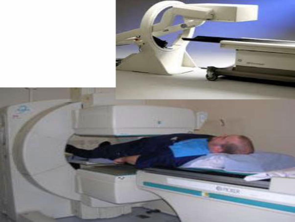
(SPECT)
SPECT is also widely used and the
process of injecting a radioactive tracer
is the same as the PLANAR technique.
Instead of being stationary, the gamma
camera moves around the body
providing a series of images. This takes
about 20-30 minutes.
SPECT and PLANAR imaging are highly
convenient technologies as they use
radiopharmaceuticals, which can be
easily distributed, stored and mixed
ready for use at nuclear medicine clinics
and hospitals.

Collimator
A collimator is a device that narrows a beam of
particles or waves. To "narrow" can mean either
to cause the directions of motion to become
more aligned in a specific direction (i.s.,
collimated or parallel) or to cause the spatial
cross section of the beam to become smaller.
Gamma rays are emitted isotropically (in all
directions)
Using only a detector would not result in an
image because there will be no relationship
between the position at which the gamma rays
hit the detector and the origin of the gamma
rays (in the patient)


Detector
Detector, by which increase the
thickness of a detector increase
probability of Complete
absorption of gamma ray.
Detector are used in gamma
camera, but this decrease the
sensitivity Of the camera, because
many gamma rays () may escape
from the detector without
interaction.
Detector ٠Spatial resolution: the smallest separation
required between two small objects to be detected and distinguished as two separated objects
٠Energy resolution: bill width half maximum(FWHM)
٠Non-Uniformity: the slightly different response of different areas of the detector of the camera to a uniform radioactive source
٠Spatial distortion: random or systematic error of determining event location
٠Counting-rate: counts / unit of time. It is assessed as the Observed count-rate (gamma camera) vs. True count-rate (source activity)
٠Sensitivity: count-rate / unit of radioactivity
References:
Books:
1_Nuclrear Medicine and PET/CT, sixth edition.
2_The Essential Physics of Medical Imaging, third edition.
3-Physics Radiobiology of Nuclear Medicine, third edition.
Others:
1_Nuclear Medicine.ppt
2_Introduction to Nuclear Medicine.ppt
3_Imaging with Radionuclides.ppt

PLEASE
Give us your POSITIVE
feedback!

Thank You
All The Best


