38
NUMERICAL ABNORMALITIES IN CHROMOSOMES DR BETTY JOHN FRCOG, MRCOG, MD, DGO CONSULTANT GYNAECOLOGIST ZULEKHA HOSPITAL SHARJAH

NUMERICAL ABNORMALITIES
IN
CHROMOSOMES
DR BETTY JOHN
FRCOG, MRCOG, MD, DGO
CONSULTANT GYNAECOLOGIST
ZULEKHA HOSPITAL SHARJAH
Numerical and Structural Abnormalities Of Chromosomes
�Chromosomal abnormalities or aberration is a missing ,
extra or irregular portion of chromosomal DNA .
�They usually occur as a result of errors in meiotic / mitotic
cell division.
�They can be inherited from a parent or be “ de novo “

TYPES OF CHROMOMOSOMAL
ABNORMALITIES
There are 2 main types of chromosomal abnormalities :
� NUMERICAL
� STRUCTURAL
NUMERICAL ABNORMALITIES
�Known as aneuploidy (abnormal number chromosomes ).
�Usually caused by failure of chromosome division (NON -
DISJUNCTION) which results in cells with an extra
chromosome or deficient chromosome.
�The causes of non – disjunction are :-
�Aging effect
� Radiation
� Delayed fertilization after ovulation.
Common Numerical Abnormalities
�Triploidy , Trisomy } Autosomal Chromosomes
�Monosomy } Sex chromosomes
�Mosaicism
Most aneuplodies are incompatible with life
resulting in spontaneous abortions except for
trisomy 21 , 13 and 18 and monosomy X which can
result in viable pregnancies.

Most frequent numerical anomalies in liveborn
Autosomes
Down syndrome (trisomy 21: 47,XX,+21)
Edwards syndrome (trisomy 18: 47,XX,+18)
Patau syndrome (trisomy 13: 47,XX+13)
Sex chromosomes
Turner syndrome 45,X
Klinefelter syndrome 47,XXY
All chromosomes
Triploidy (69 chromosomes)
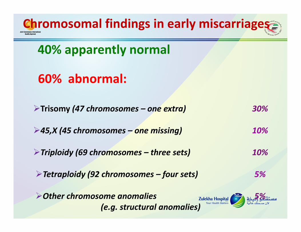
Chromosomal findings in early miscarriages
40% apparently normal
60% abnormal:
�Trisomy (47 chromosomes – one extra) 30%
�45,X (45 chromosomes – one missing) 10%
�Triploidy (69 chromosomes – three sets) 10%
�Tetraploidy (92 chromosomes – four sets) 5%
�Other chromosome anomalies 5%
(e.g. structural anomalies)
TRIPLOIDY� Three copies of each chromosome making a total of 69
chromosomes.
� It occurs in 1 to 2 % of all pregnancies.
� Most Triploid die early in preg - spontaneous
miscarriages(~10%).Almost all other babies die later or are
stillborn. Live born very rare.
� It is not hereditary.
� There are no specific risk factors.
� Not more common in older mothers.
� No increased risk in future pregnancies

TRIPLOIDY
Failure of meiotic division -2 N gamete + haploid gamete of
other parent= Triploid Zygote (69 XXX ,69 XXY ,69 XYY)

TRISOMY
All trisomies ( trisomy 21 , 13 and 18 ) could be due
to the following three causes :-
�NONDISJUNCTION
�TRANSLOCATION
�MOSAICISM
NON- DISJUNCTION ERROR
� In either mitosis or meiosis .
� If a meiotic error produces a gamete with 2
copies of an A chromosome , fertilization with a
normal gamete will result in trisomy for the A
chromosome.
�Non -disjunction often occurs in the maternal
oocyte , the incidence of which increases with
maternal age.

In meiosis 1 , a
pair of
homologous
chromosomes
fail to
separate 46 /2= 23 ideally but non-
disjunction causes
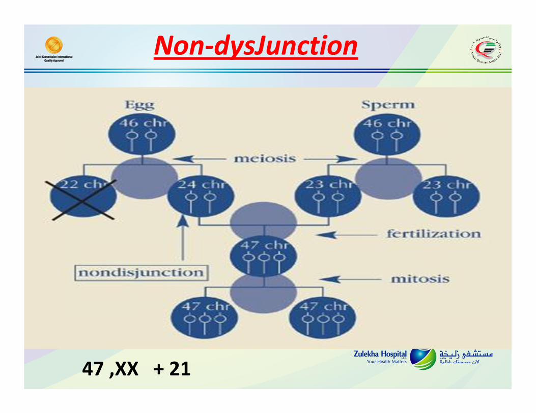
Non-dysJunction
47 ,XX + 21
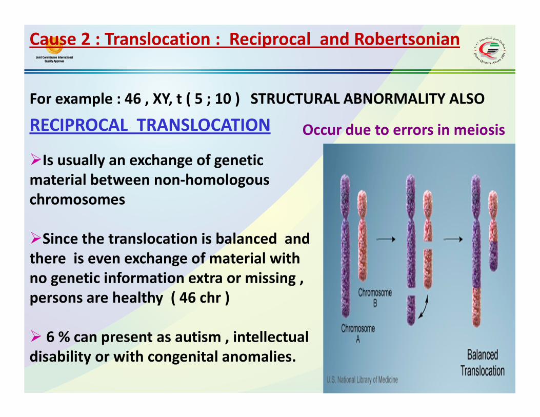
Cause 2 : Translocation : Reciprocal and Robertsonian
�Is usually an exchange of genetic
material between non-homologous
chromosomes
�Since the translocation is balanced and
there is even exchange of material with
no genetic information extra or missing ,
persons are healthy ( 46 chr )
� 6 % can present as autism , intellectual
disability or with congenital anomalies.
RECIPROCAL TRANSLOCATION Occur due to errors in meiosis
For example : 46 , XY, t ( 5 ; 10 ) STRUCTURAL ABNORMALITY ALSO

Unbalanced Translocation
A person with a unbalanced
translocation has an
increased risk of creating
gametes with unbalanced
chromosomes
Unequal exchange = extra or
missing chromosomes : 1 to 2 %
chance of trisomic fetus
Others will be carrier and normal
offspring

Robertsonian translocation
Two long arms of acrocentric
chromosomes ( 14 and 21 ) join at the
centromere with loss of short arms,
producing balanced two copies of all major
chromosomal arms and essential genes.
The acrocentric chromosomes that are
lost ( therefore 45 chr ) do not have much
important genetic material .
Carriers of this translocation have a
5 % of having a child with trisomy 21
due to inheritance of a long arm of
chromosome 21.
45 , XX , t ( 14 ;21 ) balanced carrier
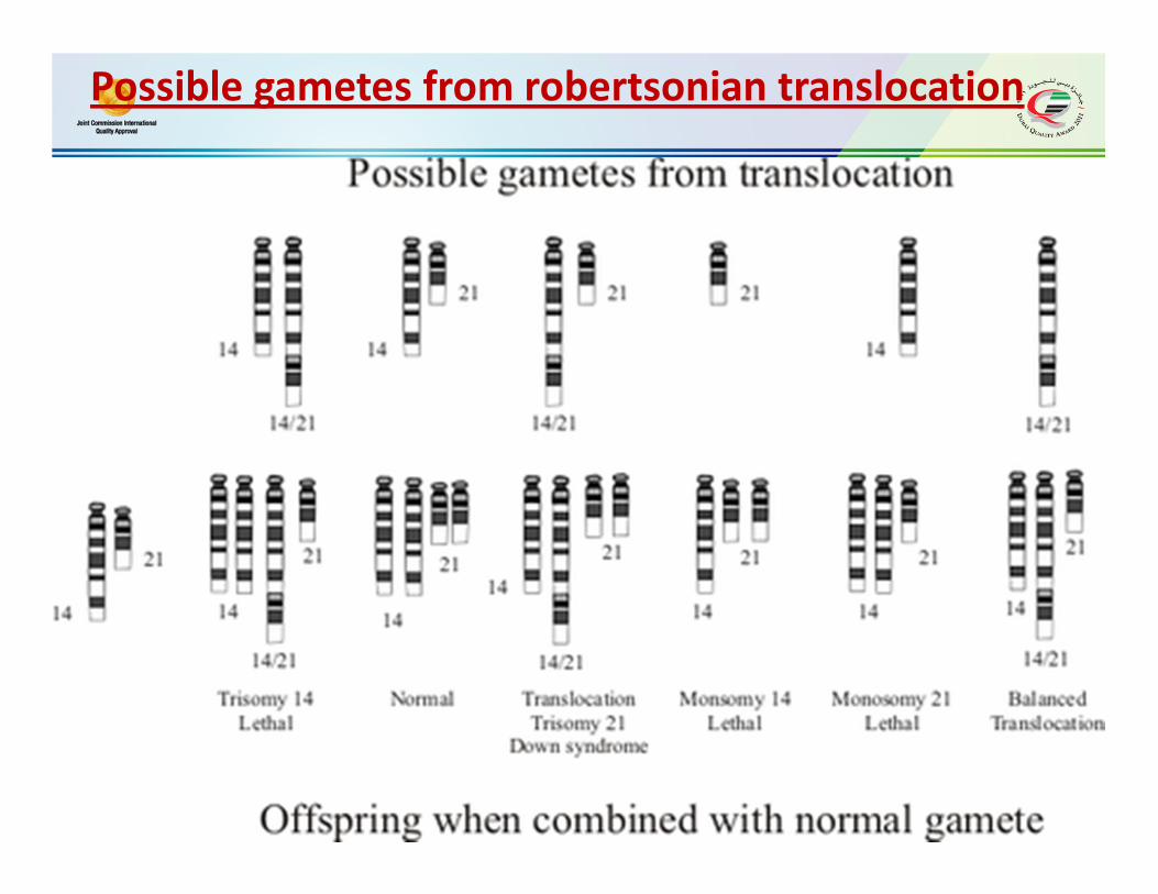
Possible gametes from robertsonian translocation
Cause 3 : MOSAICISM
• Mosaic : When an individual has two or more cell populations
with a different chromosomal make up .
• Results when some of the cells in the body are normal and other
cells have a trisomic or monosomic complement eg : trisomy 21
( 46 XX / 47 XX + 21 ) ( 45% /55%)
Some chromosomal anomalies can happen after conception like
mosaicism. This can result from
1. non –disjunction event during an early mitotic cell division in a
normal embryo or
2 . A trisomic embryo undergoes non –disjunction and some of the
cells revert to a normal chromosomal rearrangement.
Because normal cells are also present in an mosiac ,the
clinical effect may be less severe.
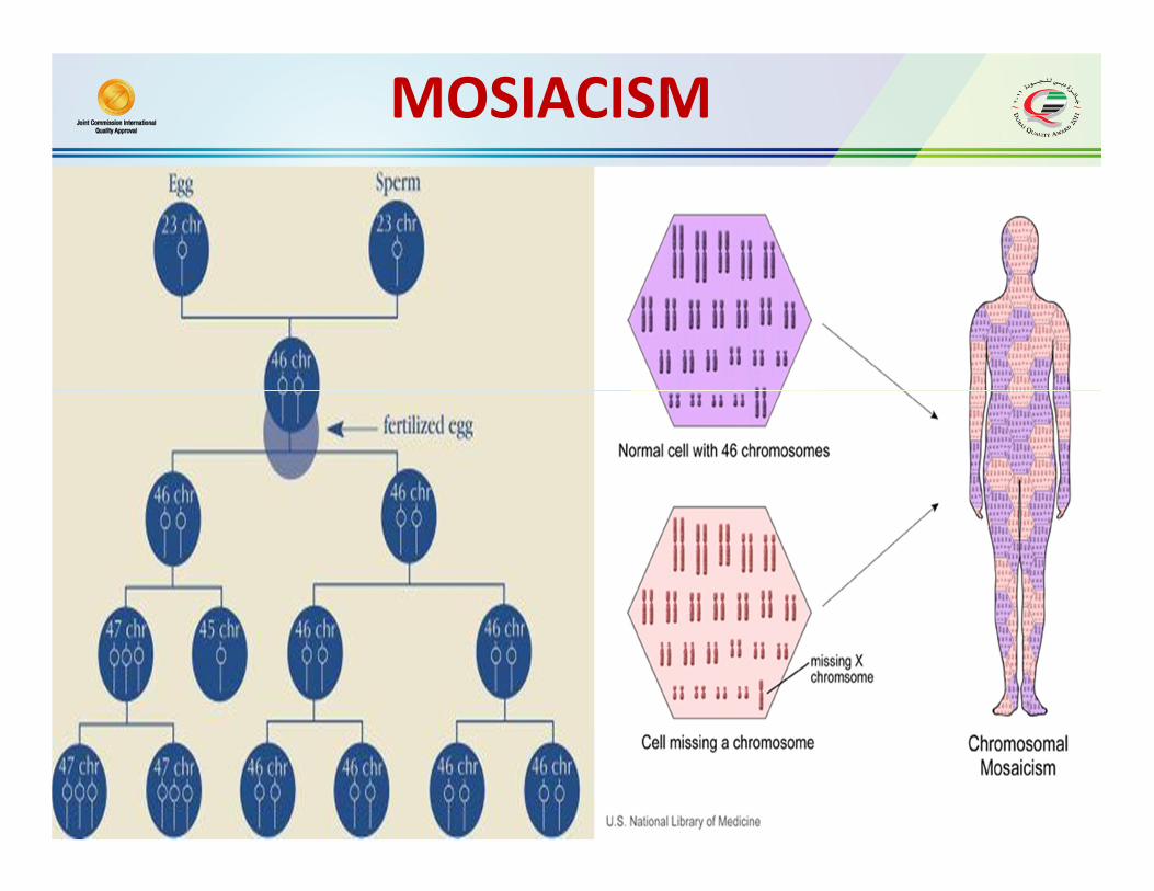
MOSIACISM

TRISOMY 21- DOWNS
• The incidence is 1 in 700 live births.
• Three causes of trisomy 21 are :
� Non –disjunction : 47 , XY + 21 ( 92 to 95 % of cases )
� Translocation : 4.8 % of cases ( most of the cases are
sporadic ( de novo ) , 1/3 rd of the cases the parents are
carriers )
�Mosaics : 46 XX / 47 XX + 21 ( 2.7 % of the cases)

Trisomy 21 due to non-disjunction Karyotype due to Robertsonian
translocation for trisomy 21
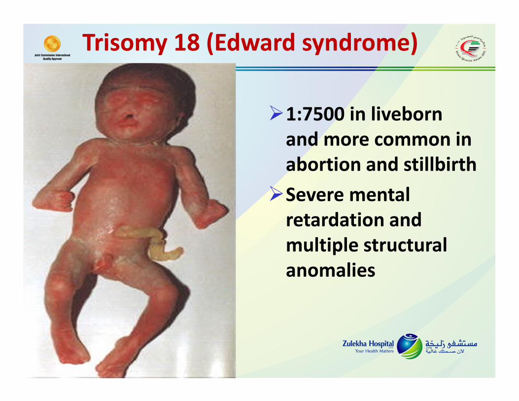
Trisomy 18 (Edward syndrome)
�1:7500 in liveborn
and more common in
abortion and stillbirth
�Severe mental
retardation and
multiple structural
anomalies

Trisomy 13((((Patau syndrome))))• 1:20,000 in liveborn
and more common in abortion and stillbirth
• Severe structural anomalies lead to death in one month
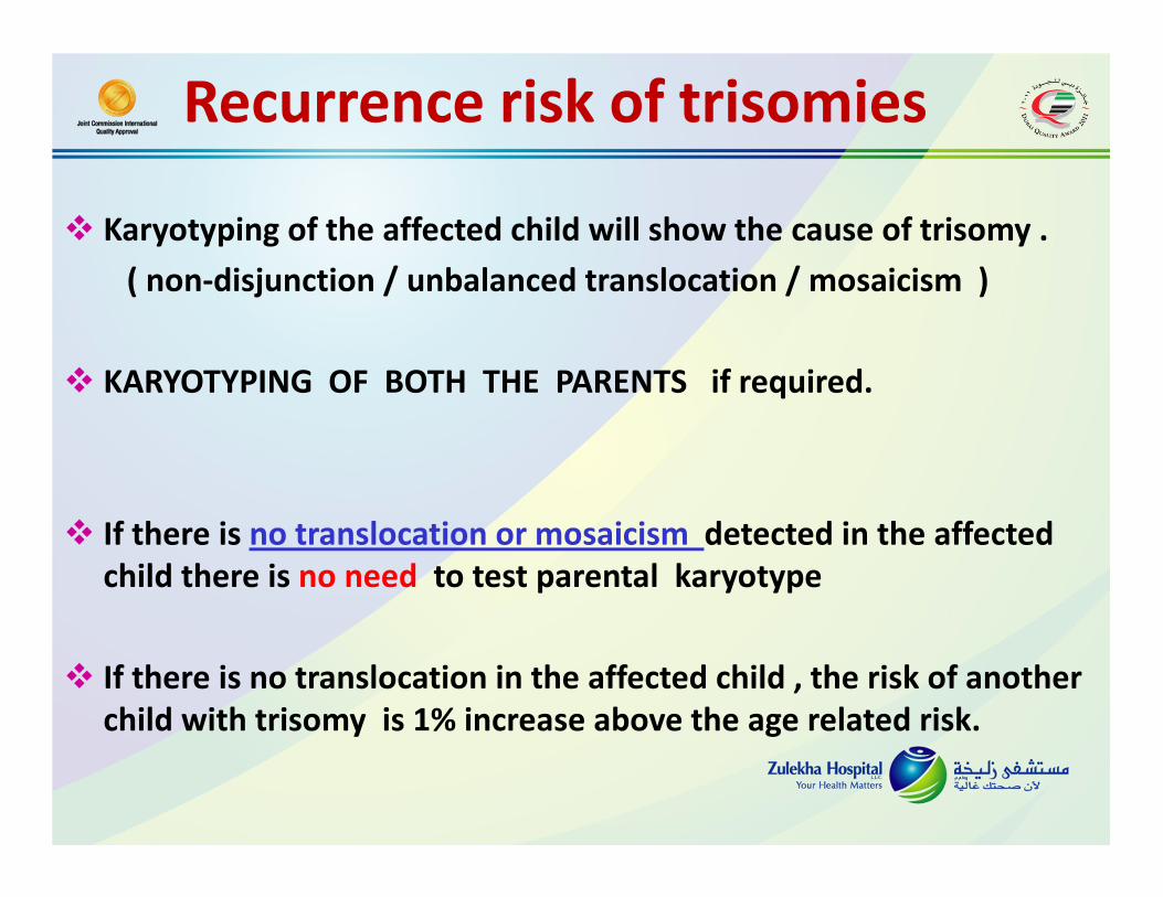
Recurrence risk of trisomies
� Karyotyping of the affected child will show the cause of trisomy .
( non-disjunction / unbalanced translocation / mosaicism )
� KARYOTYPING OF BOTH THE PARENTS if required.
� If there is no translocation or mosaicism detected in the affected
child there is no need to test parental karyotype
� If there is no translocation in the affected child , the risk of another
child with trisomy is 1% increase above the age related risk.

Recurrence risk of trisomies
� If balanced translocation is detected in the parents , then the
recurrence risk is 1 % in male carriers and 12 % in female carriers.
� In a parent having a balanced translocation between
chromosomes 21 : 21 , the recurrence risk is 100 % .
� If the translocation in the affected child is not inherited ( de novo ) ,
then the parents have a less than 1 % risk of having another
affected child with Downs syndrome.

Recurrence risk of trisomies
� The risk of recurrence for a mosiac pattern is also 1 % above the
age related risk.
� In cases where translocation or mosaic pattern is detected in
parents prenatal invasive testing is MANDATORY .
� There is no increased risk in second degree relatives , unless caused
by unbalanced translocation in the index case.
But routine screening as done for all pregnancies should be done
(Nuchal translucency and 11 to 14 week scan , blood screening and
detailed anomaly scan )

RECURRENCE RISK OF DOWNS SYNDROME
CHROMOSOMAL CONSTITUTION RISK TO OFFSPRING
AFFECTED CHILD FATHER MOTHER
TRISOMY 21 N N MOTHER < 30 YRS 2-3%
IN PRESENT PREG
MOTHER > 30 YRS, MOTHER`S AGE +1%
HAD BABY WITH DOWNS
BEFORE AGE 30
MOTHER >30 YRS, MOTHER`S AGE
HAD BABY WITH DOWNS
AFTER 30 YRS

RECURRENCE RISK OF DOWNS SYNDROME
CHROMOSOMAL CONSTITUTION RISK TO OFFSPRING
AFFECTED CHILD FATHER MOTHER
TRANSLOCATION
14/21, 15/21,13/21,21/22 N N <1%
N C 12%
C N 2- 3%
TRANSLOCATION 21 / 21 100%
MOSAIC N N 2-3%

Sex chromosome abnormalities ( aneuplodies )
� Sex chromosome abnormalities are less severe in their effects
because all but one of the X chromosome gets inactivated because
of the Lyon hypothesis ( Barr body ) and the number of genes on the
Y chromosome are limited.
� Cause of sex chromosome aneuplodies are non-disjunction errors
during meiosis .
However pre-nantal invasive testing should be done.
� Recurrence risk in sex chromosome aneuploidies is very low.
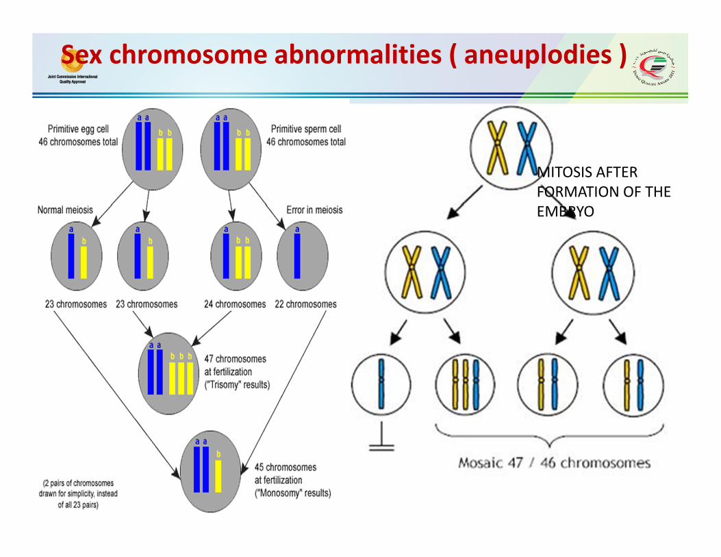
Sex chromosome abnormalities ( aneuplodies )
MITOSIS AFTER
FORMATION OF THE
EMBRYO

TRIPLE XXX Syndrome
�Often goes undetected throughout life .
�They are often taller than normal and may have learning difficulties
�Fully fertile and generally have chromosomally normal children
47, XXX
TURNER SYNDROME ( MONOSOMY X ,45 XO)
� 45 X karyotype
� Only monosomy compatible with life
� Live born females are usually mosaics ( 45 ,X / 46 XX 45 , X / 46, XY)
Pure 45 X is often lethal.
� Cause:Occurs due to loss of the paternal X chromosome
Nondisjunction in male gamete
Structural abnormalities of X chromosome
One X chromosome is missing
Mitotic nondisjunction
� Phenotype is highly variable in mosaics .
� This abnormality is unrelated to maternal age.
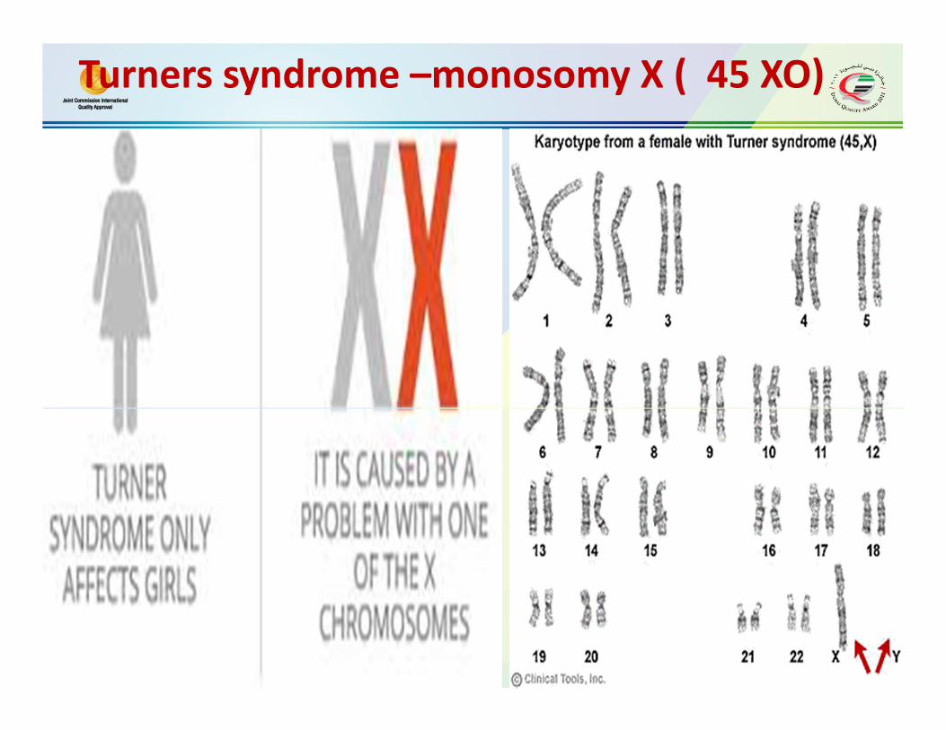
Turners syndrome –monosomy X ( 45 XO)

Turners syndrome

47 , XYY
�Patients are clinically
indistinguishable from 46 XY.
�XYY often goes undetected
throughout life.
�XYY affects 1 in 1000 live
births and is the failure of
paternal meiosis.
�Characteristics include tall
structure , normal intelligence
and normal fertility
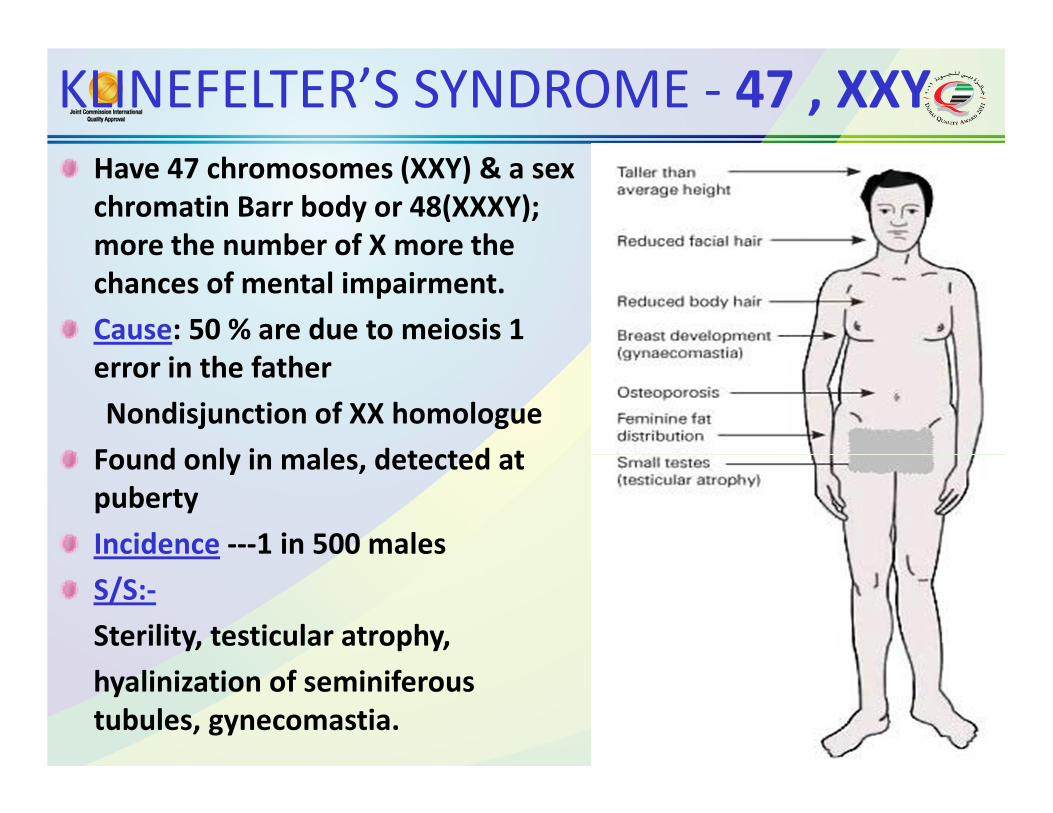
KLINEFELTER’S SYNDROME - 47 , XXY
Have 47 chromosomes (XXY) & a sex
chromatin Barr body or 48(XXXY);
more the number of X more the
chances of mental impairment.
Cause: 50 % are due to meiosis 1
error in the father
Nondisjunction of XX homologue
Found only in males, detected at
puberty
Incidence ---1 in 500 males
S/S:-
Sterility, testicular atrophy,
hyalinization of seminiferous
tubules, gynecomastia.

KLINEFELTER’S SYNDROME - 47 , XXY


