WILLIAM E. YARGER, DOUGLASD. SCHOCKEN,and ROBERTH. HARRIS, with thetechnical assistance of ROBERTMcRAE, MICHAELA. BOYD, CHRISTOPHERH. BEST,BARBARAKENNEDY,JAMES GILL, and NELL W. SCHRADER,Departments ofAledicitne and Physiology, Dluke University Medical Center, atndThe Durhaml Veteratns Adin inistrationt Hospital, Durha in, Northt Carolinia 27705
A B S T R A C T Relief of unilateral ureteral obstruiction(UUO) of 24 h duration in rats is followed by severerenal vasoconstriction in the postobstructive kidney(POK). The present study examined possible roles ofrenal prostaglandins (PG) and thromboxanes (TX), as wellas the renin-angiotensin system, in this vasoconstriction.
Administration of the cyclooxygenase inhibitor in-domethaciin, which blocks both PGand TX production,failed to improve POK hemodynamics in UUOrats.To explore the possible role of the TX compounds,which include the potent vasoconstrictor thromboxaneA2 (TXA2), UUOrats were infused with imidazole, anagent that blocks synthesis of TX, but not of PG. Imid-azole led to two- to threefold increases in the clearanceof both inulin and p-aminohippuric acid by the POK.This effect of imidazole was abolished by indomethacin,suggesting that the amelioration of POKvasoconstric-tion by imidazole was a result of inhibition of vaso-constrictor TX synthesis (e.g. TXA2), with PGvasodila-tors (e.g. PGE2or PGI2) still active. Urea, infused in a
solution whose osmolality and voluime were identicalto the imidazole infusion, failed to improve hemo-dynamics in the POK, making it unlikely that non-specific effects of volume expansion or osmotic diuresismediated the beneficial effect of imidazole.
Further studies examined the possible role of therenin-angiotensin systems in the vasocoinstriction ofthe POK. UUOrats infused with the angiotensii II
Ai al)stract of this work was published in 1978. Clin. Res.26: 564A. (Abstr.)
Dr. Harris is an Establislhed Investigator of the AmericanHeart Association. He was a Research Associate of the VeteransAdministration during the performance of part of these studies.
Received for publication 25 Jiuly 1978 antd in revised form25 September 1979.
400
antagonist, Saralasin, exhibited no significanit imiiprove-merit in POKfuncetion, a finding that might be at leastpartly attributable to agoIi st/vasoconstrictor propertiesof Saralasin. In other experiments, treatment of UUOrats with the anigiotensin-converting enzyme blockerSQ 14225 (Captopril), in order to inhibit angiotensiinII formation, led to at least twofold increases in the clear-ance of both inulin and p-aminohippuric acid in thePOK. It is unlikely that Captopril exerted this beneficialeffect by potentiating the vasodilator kinins, becausethe effect was not diminished by administration ofeither carboxypeptidase B (which destroys the kinins)or Trasylol (which blocks kinin synthesis).
Thus, these resuilts suggest that both angiotensin II,as well as metalbolites of the PG-TX system, may beimportant determiinants of postobstructive renal hemo-dynamics in the rat.
INTRODUCTION
Chronic obstruction of the urinary tract is associatedwith a severe reduction in renal blood flow aind glomer-ular filtration rate, eveni after the obstruction is relieved(1, 2). Although structural alterations are undoubtedlyimportant in increasing renal vascular resistance whenhydronephrotic destructioni of the kidney parenchymahas occurred, both blood flow and glomerular filtrationhave been shown to be severely decreased after therelease of short periods of ureteral ligation, at a timewhen morphological changes are minimal or absent.This inerease in renal vascular resistance can be some-what ameliorated by extracellular volume expansion(3) or the infuision of renal vasodilators (4), suggestingthat functional changes are at least partially responsiblefor the observed decrease in renal blood flow aindglomerular filtration rate.
In this study, we explored the potential contributionof renin and angiotensin as well as renal prostaglandinsand thromboxanes to postobstructive renal function bytreating rats with pharmacological agents which arecapable of modifying these two hormonal systems. Therenin-angiotensin system was investigated becauserenal renin production is known to be increased afteracute ureteral obstruction (5) and because vasoconstric-tion in the postobstructive kidney (POK)' is most severein the outer cortex (1, 2)-an area rich in renin-con-taining glomeruli (6). The potential role of renalprostaglandins and thromboxanes was explored becausethe production of prostaglandin E2 (PGE2) and throm-boxane A2 (TXA2) by the in vitro perfused hydro-nephrotic rabbit kidney have been shown to be in-creased (7-9). The rat was chosen for this study, how-ever, because this experimental animal demonstratesparticularly severe vasoconstriction after the release ofonly 24 h of unilateral ureteral obstruction (UUO) (2).
Knowledge of prostaglandin biochemistry is expand-ing rapidly, and it is becoming clear that importantdifferences exist in the effects that various prostaglan-dins produce in different organs and species. There-fore, some of the features of prostaglandin metabolismthat are of importance to the present study are shownin Fig. 1. The rate-limiting step in prostaglandin bio-synthesis appears to be the release of arachidonic acid
' Abbreviations used in this paper: Al, AII, angiotensins Iand II; Cln, CPAH, COsm, CNa, clearance rates of inulin,p-aminohippuric acid, osmoles, and sodium, respectively; CK,contralateral kidney; PG-, prostaglandins such as: PGG2,PGH2, PGE2, PGI2; POK, previously obstructed kidney; TX,thromboxanes such as TXA2, TXB2; UUO, unilateral ureteralobstruction.
from membrane-bound phospholipids. The phospho-lipase responsible for this release of arachidonic acidis stimulated by a variety of conditions including: in-jury, anoxia, and the action of several peptide hormonessuch as angiotensin II (All) and bradykinin. Arachidonicacid is converted by the enzyme cyclooxygenase tothe cyclic endoperoxides PGG2 and PGH2. Cyclo-oxygenase can be inhibited by a variety of nonsteroidalanti-inflammatorv agents such as aspirin, indomethacin,and meclofenamate, thereby blocking further prosta-glandin and thromboxane production. The cyclic endo-peroxides can be converted into a variety of prostaglan-dins (PGE2, PGI2 or prostacycline, PGF2a, PGD2,etc.) or into the potent vasoconstrictor TXA2. TXA2,which has a very short half-life, decomposes spon-taneously into TXB2, which is stable but biologicallyinactive. The enzyme which converts the cyclic endo-peroxides into TXA2, thromboxane synthetase, hasbeen detected in the glomeruli of normal rats (10). Thisenzyme has been shown to be inhibited by the pharma-cological agent imidazole in microsomes isolated fromplatelets (11) and hydronephrotic rabbit kidneys (8, 9).
METHODS
133 male Sprague-Dawley rats weighing 250-350 g were stud-ied. The animals were divided into 12 groups as shown inTable I. All animals, except the 11 rats in group VI, weresubjected to ureteral obstruction 25 h before beginning clear-ance studies, by anesthetizing them with ether, making a smallsuprapubic incision, and ligating the left ureter between thebladder and the left spermatic cord. The incision was suturedand the animals were allowed to recover in separate cages.Solid food was withheld, but the rats received 50 ml of a 5%sucrose solution containing 3 meq each of NaCl and KC1.They also received tap water ad lib.
FIGURE 1 Metabolic pathways of prostaglandin and thromboxane synthesis in the kidney. Thecyclooxygenase blockers, indomethacin and meelofenamate, inhibit the conversion of arachidonicacid to PGG2, thus inhibiting subsequent synthesis of both prostaglandins and thromboxanes.Imidazole, by selectively blocking thromboxane synthesis, inhibits synthesis of the vasocon-strictor thromboxane A2 (TXA2) without inhibiting synthesis of the vasodilators PGE2and PGI2.
Vasoactive Factors in the Postobstructive Kidney 401
TABLE IExperimental Groups, Number of Rats (n), Experimental
Models, and Drug Treatments duringEach Clearance Period
Treatment
Group n Model Period 1 Period 2 Period 3
I 10 UUO* None Indo xtII 22 UUO None Imid xIII 10 UUO None Urea xIV 11 UUO Imid Imid-Indo xV 11 UUO Imid Imid-Buff xVI 11 Normal Imid x xVII 7 UUO Sarl x xVIII 6 UUO Cap-Pre x xIX 6 UUO Veh-Pre x xX 20 UUO None Cap xXI 8 UUO None Cap Cap-Cp BXII 11 UUO None Cap Cap-Tras
* Abbreviations used in this table: Normal, otherwise healthy,unoperated rats; None, no drugs were infused during theseperiods; Indo, indomethacin was given intravenously 30min before this period; Imid, imidazole was infused intra-arterially during these periods; Urea, was infused intra-arterially during this period; Imid-Indo, imidazole infusionwas resumed after indomethacin treatment; Imid-Buff,imidazole infusion was resumed after these rats had beeninfused with the buffer used to dissolve the indomethacin;Sarl, Saralasin; Cap-Pre, these rats were treated withCaptopril before ureteral obstruction was produced; Veh-Pre,these rats were treated with the vehicle used to dissolveCaptopril before ureteral obstruction was produced; Cap,these animals received Captopril just before these periods;Cap-Cp B, the rats received carboxypeptidase B 1 h afterCap; Cap-Tras, these rats received Trasylol beginning 1 hafter Cap.t There were no studies performed during these times.
On the day of study, all animals were anesthetized with100 mg/kg Inactin i.p., placed on a thermoregulated table,and a short segment of polyethylene (PE)-240 catheter wasinserted into the trachea to assist in spontaneous respiration.A PE-50 catheter was placed into the left femoral artery topermit periodic sampling of arterial blood and to measurearterial blood pressure. Blood pressure was monitored with anelectronic pressure transducer (model P23Db, Gould Inc.,Measurement Systems Div., Oxnard, Calif.) connected to adirect writing recorder (model 7754 B, Hewlett Packard Co.,Palo Alto, Calif.). A PE-50 catheter was also inserted into theleft femoral vein to infuse a priming dose of [carboxyl-14C]-inulin and [glycyl-3H]p-aminohippuric acid (New EnglandNuclear, Boston, Mass.) followed by a continuous infusionof these isotopes in a solution of Ringer's lactate at a rate of0.02 ml/min. In 65 rats a PE-10 catheter was also insertedthrough the left carotid or right femoral artery and was posi-tioned in the aorta just above the renal arteries to infuse asolution of imidazole or Ringer's lactate. PE-50 catheters werealso inserted into the left ureter above the ligature and intothe bladder to collect urine from both the POKand the con-tralateral kidney (CK) for calculation of urine flow rate andthe determination of inulin (CIn) and p-aminohippuric acid
(CPAH) clearance. During this period of surgical preparation,the rats in groups I-VI received 1% of their body weight ofa solution of Ringer's lactate. Because the type of surgicalpreparation used in these studies may lead to loss of plasmaprotein and a contraction of the plasma volume (12), to studythe role of the renin-angiotensin system in the pathogenesis ofureteral obstruction, the rats in groups VII-XII received 1%of their body weight of a solution containing bovine serumalbumin (7 g/dl) in Krebs-Henseleit buffer over a 1-h period toinsure euvolemia during the subsequent clearance studies (13).
Urine collections were begun -60 min after inserting theleft ureteral catheter and continued for 2-3 h thereafter. Urinewas collected in weighed containers for clearance periodslasting 20-30 min. Arterial blood was sampled at the midpointof each urine collection and arterial blood pressure was meas-ured throughout the experiment.
Prostaglandin and thromboxane studies. 75 rats in six ex-perimental groups (groups I-VI, Table I) were used to studythe effects of altering prostaglandin and thromboxane me-tabolism on postobstructive renal function. These rats weretreated with several pharmacologic agents. 21 rats (groupsI and IV) were treated with indomethacin to block both prosta-glandin and thromboxane synthesis. The indomethacin wasprepared by dissolving 0.179 g of indomethacin (SigmaChemical Co., St. Louis, Mo.) and 0.106 g Na2CO3 in 10 mlof distilled water. This solution was diluted with pH 7.4 0.3 Mphosphate buffer to a final indomethacin concentration of 3mg/ml. The rats were infused intravenously with the solutionover a 30-min period to administer a dosage of 10 mg/kg.
55 rats (groups II, IV-VI) were infused with imidazole toblock the production of TXA2 (11). The imidazole (0.6 M)was prepared fresh daily by dissolving 0.408 g of imidazole(Sigma Chemical Co.) in 10 ml of a 0.9% solution of NaCland adjusting the initially alkaline pH to a final value of 7.5with HCI. This solution was infused intraarterially through acatheter positioned in the aorta above the renal arteries, ata rate of 0.075 ml/kg per min.
To assess the effects of the osmotic and volume load im-posed by the imidazole infusion, 10 animals (group III) re-ceived an intraarterial infusion of 0.6 M urea solution. Thisurea solution was prepared by dissolving 0.360 g of urea(Sigma Chemical Co.) in 10 ml of a 0.9% solution of NaCland then adjusting the pH to 7.5 with NaOH. The position ofthe catheter and the rate of administration were identical tothat described for imidazole above.
11 rats (group V) were infused with the same buffer solutionwhich was used to administer the indomethacin under identi-cal conditions. The imidazole infusion in groups IV and Vwas temporarily discontinued and was replaced by an infusionof Ringer's lactate solution (0.075 ml/kg per min) during theperiod (2-2½2 h after release) in which either indomethacin(group IV) or its buffer solution (group V) was infused; theimidazole infusion was then resumed.
Finally, group VI of normal healthy rats, not previouslysubjected to ureteral obstruction, was infused with imidazoleto determine if this drug is a vasodilator in normal kidneysas well as in the kidneys of rats after the release of ureteralobstruction.
All studies. 58 rats in six experimental groups (groupsVII-XII, Table I) were used to determine whether blockadeof AII activity (group VII) or its production (groups VIII-XII)can improve renal function after the release of 24 h of UUO.
Seven UUOrats (group VII) were given an intravenous in-fusion of the AII antagonist, [1-sarcosine, 8-alanine] AII(Saralasin) at rates of 2, 5, or 10 Ag/kg per min after release ofUUO. In two of these seven rats, the antagonist was infusedboth before and after release of UUO.
45 rats (groups VIII, X-XII) were subjected to blockade of
402 Yarger, Schocken, and Harris
angiotensin-converting enzyme with Captopril (SQ 14225,kindly supplied by Dr. Z. P. Horovitz, The Squibb Institutefor Medical Research, Princeton, N. J.). 50 mg/ml Captoprilin 0.9% NaCl, was injected intravenously over a 5-min periodto administer a 10-mg/kg dose. This dosage is sufficient toblock at least 50% of converting enzyme activity in the ratin vivo for 24 h.2 This dose of Captopril was given to thesix rats in group VIII under ether anesthesia 30 min beforethe ureter was ligated. As a control group for the rats in groupVIII, six rats (group IX), matched for weight with group VIIIrats, received a similar volume of 0.9% saline solution underidentical conditions. 39 rats (groups X-XII) received Capto-pril 2 h after the ureter was released. Clearance studies inthese rats were initiated before Captopril treatment-hours1-2 after releasing the ureter-so that the values from thisinitial period could serve as an untreated control with whichvalues from subsequent treatment periods could be compared.
The angiotensin-converting enzyme not only converts Alto AII, but also inactivates vasodilator kinins (bradykinin, kal-lidin, and met-lys-bradykinin) (14). Therefore, to determineif the beneficial effects of Captopril treatment observed ingroup X were the result of decreased All production or, con-versely, to decreased kinin destruction, two additional groupsof rats (groups XI and XII) were studied. In group XI, thedestruction of bradykinin and other kinins was increased byinfusing carboxypeptidase B (Worthington Biochemical Corp.,Freehold, N. J.), prepared by dissolving 10 mg in 1.0 ml of a0.9% saline solution. Because this enzyme is rapidly clearedby the kidney (15), a 3-mg/kg dose was injected at 3 and 31/2 hafter the release of the ureter while clearance studies wereperformed as shown in Table I.
In 11 rats (group XII) the production of bradykinin andother kinins was suppressed by infusing the kallikrein in-hibitor (16, 17), Trasylol (registered trademark Bayer A. G.,kindly supplied by Professor Dr. Gurt L. Haberland, Farben-fabriken, Bayer A. G., Wuppertal-Elberfeld, Germany). TheTrasylol was prepared by dissolving 10.4 mg in 1.0 ml of a0.9% solution of NaCl (69,000 KIU/ml). This solution wasinfused for 1 h at a rate of 0.033 ml/min beginning 3 h afterthe ureter was released as shown in Table I.
Osmolality and the concentration of sodium of the urineand plasma were measured in 10 of the 22 rats in group IIand the 10 rats in group III. Osmolality was measured by avapor pressure osmometer (model 5100, Wescor Inc., Logan,Utah). Sodium was measured by a lithium internal standardflame photometer (model 443, Instrumentation Laboratory,Inc., Lexington, Mass.). Plasma calcium was measured inseven rats in group II and seven rats in group IV by fluorometrictitration with a Calcett Automatic Calcium Titrator (PrecisionSystems, Inc., Sudbury, Mass.). Imidazole concentrationswere measured in deproteinized plasma from 11 rats in groupII and six rats from group IV by modifying the procedure ofKoessler and Hanke (18). The color developed by the reactionbetween imidazole and p-phenyldiazonium sulfonate wasread at 520 nm at 15, 20, and 25 min after combining thereagents.
Calculations. Statistics were calculated according tostandard methods. To obviate the effect that seasonal differ-ences appear to exert on basal postrelease renal function (19),the animals were used as their own controls wherever possible,and the data were analyzed by the paired Student's t test.Where data from different groups are compared, the animalswere matched by weight and an unpaired t test was utilized.A probability value <0.05 was regarded as significant. Valuesare presented as means ±standard error of the mean.
2 Dr. Z. P. Horovitz. Personal communication.
RESULTS
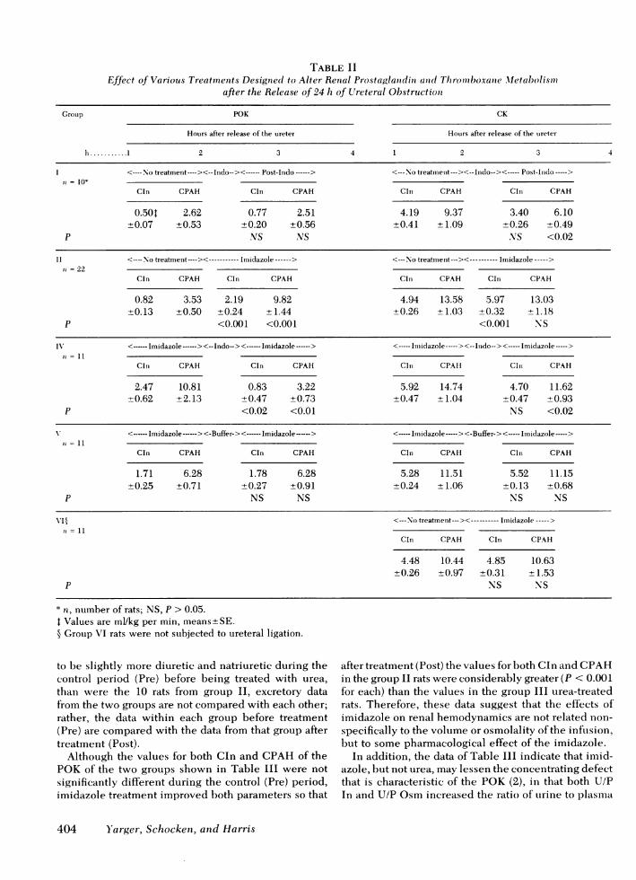
Previous studies in our laboratory have demonstratedthat, after relief of 24 h of UUOin rats, the POK isintensely vasoconstricted (2). The present experimentswere designed to examine the mechanisms of this vaso-constriction. It has been shown that the in vitro per-fused hydronephrotic rabbit kidney exhibits increasedproduction of both the vasoconstrictor TXA2 (8, 9) andPGE2 (7), which is a vasodilator in the rabbit kidney(20). Accordingly, we studied the role of prostaglandinand thromboxane metabolism in vivo in rats previouslysubjected to 24 h of UUO. The results of these studiesare presented in Tables II and III and in Figs. 2 and 3.
The initial step in clarifying the roles of prostaglan-dins and thromboxanes after UUOwas to suppress theproduction of both by blocking the enzyme cyclooxy-genase (Fig. 1) with indomethacin. Because this en-zyme converts arachadonic acid to the cyclic endo-peroxides (PGG2 and PGH2), which are the precursorsof both prostaglandins and thromboxanes, indomethacintreatment should allow examination of postobstructiverenal function under circumstances in which the con-tribution of all of the metabolites of arachidonic acidhave been markedly reduced or abolished. As shownin Table II, the group I data demonstrate that indo-methacin treatment improves neither CIn nor CPAHby the POK. CPAHby the CKwas slightly decreased.
These data are subject to several possible interpre-tations, one being that reduced production of both vaso-dilators (e.g. PGE2or PGI2) as well as vasoconstrictors(e.g. TXA2) might have no net beneficial effect on thekidney. To test this hypothesis, group II rats weretreated with imidazole, an agent which should decreasethe production ofthromboxanes, but not the productionof prostaglandins (Fig. 1). As shown by the data inTable II and Fig. 2, imidazole treatment results in asignificant increase in the values for both Cln and CPAHin the POK, suggesting that specific blockade of throm-boxane production is quite beneficial to the POK.
However, as CIn (but not CPAH) also increased inthe CK, and as function in the POK is known to beincreased by volume expansion (3), it is possible thatsome nonspecific effect of an intraarterial infusion of asolution of high osmolality (850-900 mosmol/kg H20),rather than a metabolic effect of imidazole, might havebeen responsible for this improvement in renal func-tion. To evaluate this possibility, a solution of urea insaline that was of similar volume, osmolality, and pHwas infused in the rats in group III. The data fromthese rats were compared with those of 10 of the groupII rats, as shown in Table III.
The data in Table III demonstrate the contrast be-tween imidazole and urea with regard to their effectson renal hemodynamics and on the excretion of waterand electrolytes. Because the rats in group III appeared
Vasoactive Factors in the Postobstructive Kidney 403
TABLE IIEffect of Various Treatments Designedl to Alter Rental Prostaglanidini and(1 Thlrotinboxanie Metabolismll
after the Release of 24 h of Ureteral Obstructioni
Group POK CK
Hours after release of the ureter Hoturs after release of the uireter
* t, number of rats; NS, P > 0.05.Values are ml/kg per min, means+SE.
§ Group VI rats were not subjected to ureteral ligation.
to be slightly more diuretic and natriuretic during thecontrol period (Pre) before being treated with urea,than were the 10 rats from group II, excretory datafrom the two groups are not compared with each other;rather, the data within each group before treatment(Pre) are compared with the data from that group aftertreatment (Post).
Although the values for both Cln and CPAHof thePOKof the two groups shown in Table III were notsignificantly different during the control (Pre) period,imidazole treatment improved both parameters so that
after treatment (Post) the values for both Cln and CPAHin the group II rats were considerably greater (P < 0.001for each) than the values in the group III urea-treatedrats. Therefore, these data suggest that the effects ofimidazole on renal hemodynamics are not related non-specifically to the volume or osmolality of the infusion,but to some pharmacological effect of the imidazole.
In addition, the data of Table III indicate that imid-azole, but not urea, may lessen the concentrating defectthat is characteristic of the POK (2), in that both U/PIn and U/P Osm increased the ratio of urine to plasma
404 Yarger, Schocken, and Harris
28
24
20cx-
c
E
IE14I-
16
12
8
4
Before During
FIGURE 2 Cln and CPAHin the POhibition of thromboxane synthetase M
osmolality and inulin concentWhether this improvement resulleffect of imidazole on the conceper se, or derived secondarily fr4in renal hemodynamics, cannotthese data, however.
Although imidazole increased t]dium excretion, this appeared tothe increase in filtered load, sfiltered sodium excreted in the uiThe value for fractional osmolar cdecreased slightly, but not signifi(of rats. This suggests that any ecannot be explained by an osmotsion further suipported by the obsagent produced a significant increaity. Finally, imidazole produced,in blood pressure whereas urea din blood pressure after imidazolcurred in all other rats in group I
Comparison of the data in grotthat inhibition of thromboxane prois not beneficial unless prostaglaitinues unimpeded (imidazole alkpothesis further, the rats in groupimidazole before and after beinlmethacin. The results are shown3. Inspection of Table II revealfindings. First, as would be expein group II, it is clear that theduring the first imidazole period,indomethacin, is considerably belobserved in rats in groups I-II]treatment was initiated. Second, itby the POK during the secondperiod, which followed the infus
deteriorated substantially and significantly as comparedto values observed before indomethacin was given.
Because imidazole is an agent with many pharma-cological effects, and because we found in preliminarystudies3 that prolongation of the imidazole infusion for4 h or more resulted in depressed renal function, hypo-tension, hypocalcemia, and death, it is possible thatthe deterioration of renal function in the POKof therats in group IV during the second imidazole periodmight have been a result of the increased amount ofimidazole that those rats received, rather than to aneffect of indomethacin. Therefore, the rats in group Vwere subjected to a similar protocol to that used ingroup IV, except that, insted of indomethacin, theywere infused with an equivalent volume of only thebuffer that was used as the indomethacin vehicle. As
Before During shown in Table II, elimination of the indomethacinK before and during in- also eliminated the deterioration in renal function dur-vith imidazole. ing the second imidazole infusion period.
Finally, inasmuch as imidazole treatment increasedtration significantly. CIn (but not CPAH) in the CK as well as the POKinted from some direct group II rats, it is possible that imidazole is a non-entrating mechanism specific vasodilator, and that its beneficial effect in theom the improvement POKwas not related to any consequence of ureteralbe determined from obstruction. To test this possibility, the normal healthy
rats of group VI (not subjected to previous obstructionhe absolute rate of so- of the ureter) were studied before and during imidazoleresult primarily from infusion. As shown on the right side of Table II, neither;ince the fraction of Cln nor CPAHwere significantly increased by suchrine did not increase. treatment.learance by the POK Because of the known effect of imidazole, in massive
cantly, in both groups doses, to produce marked hypocalcemia (21), plasmaffect of either agent calcium was measured in seven rats each in groups II
tic diuresis, a conclu- and IV. Plasma calcium in group II rats before imidazoleservation that neither treatment, 10.15+0.14 mg/dl, decreased (P < 0.05) to aise in plasma osmolal- value of 9.52±0.23 after completing the imidazole in-a significant increase fusion. Plasma calcium after the first imidazole infusionlid not. This increase in group IV, 9.50±0.28, also decreased (P < 0.05) whenle treatment also oc- measured at the end of the second imidazole period1I. (8.56±0.30). Although statistically significant, theseaps I and II suggests values are considerably greater than the values forduction (both groups) plasma calcium observed after 4 h of imidazole infusionndin production con- (5.41+0.43 mg/dl) at which time blood pressure)ne). To test this hy- began to decrease and renal and cardiovascular functionIV were treated with deteriorated.
g infused with indo- The data in Table II suggest that, although selectivein Table II and Fig. inhibition of the vasoconstrictor TXA2 improves renal
Is several interesting function in the POK, this improvement depends, incted from the results some part, on the continued production of some prosta-function of the POK glandin, presumably a vasodilator. These data do not,before the infusion of however, identify the source of the residual vasocon-tter than the function striction observed in the POKafter the production ofI before any type of both types of arachidonic acid metabolites have beent is clear that function blocked by indomethacin treatment. In an attempt toimidazole treatmention of indomethacin, 3 Unpublished observations.
Vasoactive Factors in the Postobstructive Kidney 405
TABLE IIIEffect of ant Initraarterial Inifusioni of Either Imiidazole (Imiid; 10 Rats) or Urea (10 Rats) oni Renial Hernodnaicsanid the
Excretion of Salt and Water after the Release of Ureteral Obstruction*
*Values are means±SE.tAbbreviationis used in this table: U/P In, ratio of urine to plasmia iniulini concentration; V, urine flow rate; COsm, clearance
rate of osmoles; CNa/Cln, fractioni of filtered sodiumi excreted; BP, blood pressure; UNaV, absolute excretion rate for sodiumi;U/P Osm, ratio of urine to plasmia osmnolalitv; P Osmi, plasma osmolality; Pre, before treatment; Post, after treatmnent; NS,P > 0.05.
characterize other vasoconstrictors that might act on theP0K, we investigated the possil)le role of the renin-angiotensin system. These data are shown in TableIV and Fig. 4.
Fig. 4 demonstrates the effect of infuising the Allantagonist 1-sarcosine, 8-alanine, All (Saralasin) intorats either before (rats E and F) or after (rats A, B,C, D, and G) releasing UUO. When infused after therelease of UUO, renal function was improved in theP0K in only one rat (rat C) by doses of 2 and 5 Agikgper min In all other animals, both Cln and CPAHdeteriorated when the drug was infused. These changesin function of the P0K were not associated with sig-nificant changes in either function of the CKor in bloodpressure. When Saralasin was infused before the re-lease of UUOand continued during the houir immii-edi-
ately after release, as well as during the first clearanceperiod,) neither a 5-.tigikg per min dose (rat E) nior a
2-.tig/kg per min dose (rat F) appeared to be benie-ficial as compared with the values obtained after theSaralasin infusion was stopped.
These data do not establish a clear role for All asa vasoconstrictor in the P0K. However, because ofemnerging evidence suiggesting that Saralasin may haveagonistic properties (22), fu-rther studies to characterizethe role of the renin-an-giotensin systemi- in the P0Kwvere conduicted, using the Al-converting enzyme in-hibitor, SQ14225 (Captopril). These data are presentedin Table IV.
In group VIII rats, Captopril was infused shortly be-fore UUOwas produced. Data from these animals are
comipared with valutes observed in rats, matched forr
406 Y'arger, Schockeni, antd Harris
0
EI-,
0b
C%
I~
U
PRE POST
FIGURE 3 CIn and CPAH in the POKthromboxane synthetase with imidazolquently after (Post) inhibition of bothindomethacin and thromboxane synthets
weight with group VIII rats, that weequal volume of the vehicle useCaptopril (group IX). Both CIn ancnificantly better in the rats treated M%in those rats treated with the vehiment with Captopril did not affectthe CK.
To use animals as their own contthe effects of Captopril, an additi((group X) was studied in the absenc(ment, and again after Captopril treresults in groups VIII and IX, Captoaccompanied by significant improv(and CPAH in the POK, with valueunchanged.
These data suggest that All migimportant vasoconstrictor in the P(Al-converting enzyme (kininase II)specific with many actions, includirof bradykinin and other vasodilatorfore, the improvement in renal functiCaptopril treatment might have resuldegradation of kinins rather than de(of All. To clarify this possibility, tof animals were studied.
Group XI experiments resembledin that renal function was studied aftin the absence of drug treatment (again after Captopril administratioiObservations were continued duriniwhich the rats were additionally tre;peptidase B, an enzyme that destrckinins (15), but does not affect Al o
As shown in Table IV, the valuesCPAHwere significantly increased
ment in the rats in group XI as compared with thevalues observed in these animals during the prior pe-riod with no treatment. Table IV further shows thatthe addition of carboxypeptidase B treatment did notdiminish these beneficial effects of Captopril in thePOK, but rather, appeared to augment them. Cln andCPAHin the POKwere both slightly, though not sig-nificantly, greater during combined treatment withboth agents, than during Captopril alone. In the CK,Captopril treatment increased Cln (P < 0.02), and thisincrease was further augmented (P < 0.02) by the addi-tion of carboxypeptidase B treatment. In both the POKand CK, the values for both CIn and CPAHduringthe final period (carboxypeptidase B) were significantly
PRE POSTgreater than the values observed during the initial pe-riod in which no drugs were administered.
during inhibition of Because the administration of carboxypeptidase Ble (Pre), and subse- may have other effects in addition to the inactivationcyclooxygenase with of kinins, an alternate method of reducing bradykininase with imidazole.
levels was performed in the rats in group XII. In theseanimals, again after an initial period without pharma-
re infused with an cological manipulation, and a second period of Capto-d to dissolve the pril treatment, the rats were treated during a third pe-I CPAHwere sig- riod with the kallikrein inhibitor, Trasylol, to blockvith Captopril than the production of the kinins (16, 17).cle only. Pretreat- Similar to the results in groups X and XI, Captopril
renal function in treatment again significantly increased both CIn andCPAHin the POKin group XII. After the addition of
trol when studying Trasylol, CIn improved even further (P < 0.02) in theonal group of rats POK. No values for CPAHare presented during thise of any drug treat- third period because the values during the first 30 min~atment. As in the increased sharply and then fell dramatically during the)pril treatment was second 30 min to levels which were similar to Cln inement in both Cln both kidneys. Whether this fall in CPAHrepresents as in the CK being true decrease in renal blood flow, an effect on PAH
secretion, or an artifact resulting from the effect of1ht, indeed, be an rapidly changing blood concentrations is not clear.OK. However, the Therefore, the effect on renal blood flow of Captopril
is relatively non- and Trasylol in combination remains problematic.ng the degradationkinins (14). There-ion which followedted from decreased:reased production:wo further groups
I those of group Xter release of UUO'first period), thenn (second period).g a third period, inated with carboxy-)ys the vasodilator,r All.s for both CIn andby Captopril treat-
DISCUSSION
After the release of as little as 24 h of UUO, there isintense renal vasoconstriction in the POK, accompaniedby a decrease in renal blood flow and glomerularfiltration rate. In a previous study using a vascular in-jection technique, we demonstrated (2) that this post-obstructive vasoconstriction tended to spare the medullaand to involve the cortex heterogeneously, in that therewere relatively large areas of the cortex in which per-fusion beyond interlobular arteries could not be dem-onstrated. Because anatomic hydronephrosis is rela-tively slight at this time, and because both acute ex-tracellular volume expansion (3) and infusion ofvasodilators (4) have been shown to improve renalblood flow in the POK, these findings suggest that the
Vasoactive Factors in the Postobstructive Kidney 407
TABLE IVEffect of Blocking the Production of A II with the Converting Enzyme Inhibitor, Captopril, on Renal Function after the
Release of 24 h of UUO, and the Inability of a Bradykinin-Inactivating Agent (Carboxypeptidase B)or a Kallikrein-Inhibiting Agent (Trasylol) to Block the Effect
Group POK CK
Hours after release of the ureter Hours after release of the ureter
* P, probability of significant difference compared with previous treatment period; P', difference of group IX compared withgroup VIII; P", difference of values in hours 3-4 compared with values in hours 1-2; NS, P > 0.05.t Values are ml/kg per min, means±SE.§ No P-aminohippuric acid data are reported because of potential methodological errors, see text.
vasoconstriction observed after the release of 24 h of and the renin-angiotensin system in these alterationsUUOis predominantly functional rather than structural. of renal hemodynamics.The present studies were undertaken in an attempt to Other investigators (7-9) have provided evidenceidentify possible roles for prostaglandins, thromboxanes, that prostaglandins and thromboxanes might be impor-
408 Y'arger, Schocken, and Harris
4 -- Cl1n- CPAH
A
4 12
2 6
[B
0
Ec1E
12
6
1.-.1C---I
2
D
E2 6
F ~2 6
G I-- -.
2 0
-t
E)E
I
0 2 5 10Sorolosin Dose, Mg/kg per min
FIGURE 4 The effects of infusing various doses of the Allantagonist Saralasin on the CIn and CPAHby the POK inseven rats. In rats E and F (*), Saralasin was infused bothbefore and after release of obstruction; in the other five rats,Saralasin was infused only after release.
tant mediators of postobstructive renal function. Nishi-kawa et al. (7) reported that, after 72 h of UUO, thein vitro perfused hydronephrotic rabbit kidney dem-onstrates increased production of PGE2 when stimu-lated with either bradykinin or All, as compared withthe intact CK. Subsequently, this same group demon-strated that the in vitro perfused hydronephrotic rabbitkidney also exhibits increased production of TXA2, inresponse to infusion of bradykinin (8, 9).
In previous attempts to define a role for prostaglan-dins after UUOin rats, both Edwards et al. (23) and we(24) treated animals with indomethacin at the timeUUOwas produced and, again, just before it was re-leased. Such treatment has been shown to significantlydecrease rat renal prostaglandin synthesis (23); how-ever, in both studies, it failed to improve function inthe POK. One explanation for this lack of improvementis that indomethacin, in the dosages used, may haveimpaired cardiac function during the 24 h of UUO.However, administration of indomethacin only as anacute injection of the drug after the release of UUOin the present studies (group I) also failed to improverenal function.
Although these results can be interpreted in severalways, one interpretation is that indomethacin inhibited
the synthesis of both a vasodilator and a vasocon-strictor, and thus, had no net beneficial effect on thePOK. To examine this possibility, we infusedimidazole, an inhibitor of thromboxane synthetase(8, 9), into rats after the release of UUO(group II).Our finding that imidazole infusion resulted in asignificant improvement in renal function in the POKis compatible with a role for TXA2 as a renal vaso-constrictor in the POK.
Although the reversal of vasoconstriction in the POKby imidazole may have resulted from blockade ofthromboxane synthesis, other explanations are possible.First, imidazole infusion was accompanied by an in-crease in Cln in the intact CK, raising the possibilitythat imidazole might be a nonspecific renal vasodilator.This appears unlikely, however, because imidazole hadno measurable effect on renal hemodynamics in normalrats (group VI).
Second, because acute saline expansion has beenshown to decrease vasoconstriction in the POK(3), itis possible that part of the beneficial effect of imid-azole treatment was a nonspecific consequence of theosmotic or volume load of the infusion. Weexaminedthis possibility by infusing urea into rats (group III)in a solution of the same volume, osmolality, and pHas that used for imidazole. Urea was chosen becauseit is of similar molecular weight and, like imidazole,is not confined to the extracellular space. Our findingthat urea infusion did not improve function of the POK(Table III) is evidence against an important nonspecificosmotic or volume effect of the imidazole infusion. Al-though we previously reported that urea loading im-proved POKfunction in rats, the concentration of ureain the infusion solution in those experiments was 10times greater than in the present studies (25). Theseresults, then, support the concept that imidazole ex-erted its effect of reversing vasoconstriction in the POKprimarily by inhibiting the renal synthesis of vaso-constrictor TXA2.
This beneficial effect of imidazole in the POKwasabolished by the addition of indomethacin treatment(group IV) while the imidazole infusion was extended.This finding suggests that the ability of imidazole toimprove POKfunction depends on prostaglandin me-tabolism being intact except for the blockade of throm-boxane synthesis. However, there are alternative inter-pretations of these results. First, imidazole is a stimu-lator of phosphodiesterase (26), whereas indomethacinmay be a phosphodiesterase inhibitor (27). Thus, it ispossible that the beneficial effects of imidazole, andtheir reversal by indomethacin, might be mediated byalterations in cyclic nucleotide metabolism. Plasmaconcentrations of imidazole, measured in the presentstudy, did not exceed 2.5 mM. By comparison, imid-azole levels as high as 6 mMhave been shown to inhibit
Vasoactive Factors in the Postobstructive Kidney
-~~~~~~~~
2
409
platelet thromboxane synthesis without altering eitherbasal or stimulated cyclic AMPlevels (11). Althoughthese data do not exclude the possibility that alteredcyclic nucleotide metabolism might contribute to theimprovement in POKfunction induced by imidazole,they suggest that other mechanisms are more important.
Second, imidazole in large doses may lead to hypo-calcemia (21), an effect possibly related to stimulationof phosphodiesterase. Indeed, we have observed3 thatimidazole, when infused into normal rats continuouslyfor 4 h or more, often leads to marked hypocalcemiaand tetany. Plasma imidazole concentrations in theserats generally exceeded 8 mM, compared with levelsof <2.5 mMin group II and group IV rats in the presentstudy. This finding, in itself, does not remove the pos-sibility that the modest decrease in plasma calciumnoted in group IV rats could have contributed to thedepression of POK function after indomethacin ad-ministration. Such a possibility seems unlikely, how-ever, because renal function did not decrease in eitherthe POKor CKof group V rats given the same total doseof imidazole, but without indomethacin. AlthoughCPAHdid decrease in the CK of group IV rats duringthe second imidazole period, CPAH also decreasedin group I rats after indomethacin treatment alone.Both of these decreases seem likely to be an effect ofindomethacin per se, not of hypocalcemia. This inter-pretation is supported by the observations of Terragnoet al. (28) that indomethacin treatment decreases renalblood flow in anesthetized, surgically stressed animals.
Considered together, the data from groups I to VIsuggest that both vasodilator and vasoconstrictor me-tabolites of arachidonic acid are important in postob-structive renal hemodynamics in the rat; they do not,of course, identify the compounds responsible. Althoughthe beneficial response to imidazole suggests that TXA2is an important determinant of function in the POK,the nature of the prostaglandin vasodilator is less clear.The hydronephrotic rabbit kidney produces increasedamounts of PGE2-a compound which is a vasodilatorin that species. However, extrapolation of these datato infer that PGE2 is a vasodilator in the POKof therat, is probably not warranted at the present time forat least two reasons. First, increased PGE2productionby the hydronephrotic rat kidney has not yet been dem-onstrated. Second, the preponderance of data at thepresent time suggests that PGE2 is actually a vaso-constrictor in the rat (20, 29, 30). Thus, the presentdata suggest that, although its identity is not yet es-tablished, the interplay between some prostaglandinvasodilator and TXA2 is important in determining func-tion after release of 24 h of UUOin the rat.
The observation that renal function deterioratedwhen imidazole infusion was resumed after indometh-acin treatment also implies that there is some non-prostaglandin vasoconstrictor acting on the POK. Be-
cause it is known that acute ureteral obstruction is ac-companied by an increase in renin production (5), weinvestigated the possibility that AII was a vasocon-strictor in the POK.
We first infused the All antagonist, Saralasin. Asshown in Fig. 2, except for one rat out of seven, therewas no evidence that Saralasin improved renal hemo-dynamics in the POK. However, because Saralasin mayitself act as a renal vasoconstrictor (22) as a result ofits angiotensin agonistic properties, we further evalu-ated the role of AII by infusing the converting enzymeinhibitor, Captopril.
Whenpostobstructive renal function in animals pre-treated with Captopril before the production of UUO(group VIII) was compared to function of UUOratssimilarly pretreated with only the Captopril vehicle(group IX), renal function was significantly improvedin the former group. In addition to the group VIII andgroup IX studies, in which AII production was inhib-ited by Captopril during the 24 h of UUO, we alsoexamined the effects of Captopril given after UUOwasreleased (group X rats), and again observed that Capto-pril substantially improved renal function in the POK.Thus, the data from groups VIII-X indicate that Capto-pril improves renal function in the POK(but not CK)if given either before UUOis produced, or after it isreleased.
Converting enzyme, which is blocked by Captopril,is a relatively nonspecific dipeptidylpeptidase. In ad-dition to converting Al to AII, it also degrades brady-kinin and other vasoactive kinins, such as kallidin (14).Thus, the beneficial effect of Captopril treatment mighthave resulted from an increase in the level of the vaso-dilator kinins, rather than a decreased production ofthe vasoconstrictor AII.
To examine the possible role of increased kinins inthe improvement in function of the POK, two differentprocedures were used to decrease kinin levels. First,the enzyme carboxypeptidase B, which cleaves a basiccarboxyl terminal amino acid, was infused intrave-nously. All the vasoactive kinins have a carboxyl termi-nal arginine, cleavage of which will inactivate thepeptide (15). Thus, this enzyme should lower kininlevels, even if they had been previously increased bythe inhibition of kininase II (converting enzyme) withCaptopril. Indeed, Erdos and colleagues have shownthat the infusion of carboxypeptidase B blocks the ef-fect of intravenously infused bradykinin when testedin the rat, cat (31), or guinea pig (15).
A second method used to examine the possible con-tribution of an increase in kinin levels to the effectof Captopril on the POKwas to infuse Trasylol. Thispeptide, isolated from bovine lung, has been shownto block the effect of both trypsin and kallikrein (17),enzymes that are both capable of generating kinins.
The observation that renal function does not deterio-
410 Y'arger, Schocken, and Harris
rate with either carboxypeptidase B or Trasylol, butactually improves, suggests strongly that increased ki-nin activity was not the explanation for the observedincrease in renal function that follows Captopril treat-ment. These data do not, of course, explain why renalfunction should improve further when agents such ascarboxypeptidase B or Trasylol, which are capable ofdecreasing kinin levels are infused. Irrespective of theunanticipated beneficial effect of these two agents, thedata do demonstrate that the reversal of vasoconstric-tion in the POK after Captopril administration doesnot depend on an increase in the activity of vasodilatorkinins. It would appear, rather, that the major effectof Captopril was to inhibit All production. Thus, itseems reasonable to conclude that an increased levelof intrarenal All activity explains the nonprostaglandincomponent of the vasoconstriction observed in the POK.
Taken together, the data presented herein suggestthat both metabolites of arachidonic acid and the renin-angiotensin system make important contributions tothe determination of postobstructive renal functionafter the release of 24 h of UUOin the rat. That thesetwo intrarenal hormones should interact in determiningrenal function is not surprising in that some prostaglan-dins have been shown to stimulate renin production(32), whereas AII is a known activtor of the phospho-lipase, which, by releasing arachidonic acid from mem-brane bound phospholipids, controls the rate-limitingstep in prostaglandin and thromboxane biosynthesis.The data also suggest that both vasoconstrictor (TXA2)and vasodilator metabolites of arachidonic acid are ofimportance, although the identity of the latter has notbeen established for the rat. Finally, given that phar-macological agents are capable of modulating the effectof these hormones, the present findings suggest areasof exploration for therapeutic manipulation of post-obstructive renal function in man.
ACKNOWLEDGMENTS
The authors wish to thank Ms. Carol Bohannon and Ms. JanetShipley for secretarial assistance, and Mr. Don Powell forassistance in preparation of the illustrations.
This work was supported, in part, by a research grant fromthe Veterans Administration 9750-01, National Institutes ofHealth grant AM17195, and a Grant-in-Aid from the AmericanHeart Association.
REFERENCES
1. Yarger, W. E., and L. D. Griffith. 1974. Intrarenal hemo-dynamics following chronic unilateral ureteral obstruc-tion in the dog. Am. J. Physiol. 227: 816-826.
2. Harris, R. H., and W. E. Yarger. 1974. Renal functionafter release of unilateral ureteral obstruction in rats. Am.
J. Physiol. 227: 806-815.3. Wilson, D. R. 1974. The influence of volume expansion
on renal function after relief of chronic unilateral ureteralobstruction. Kidney Int. 5: 402-410.
4. Shenasky, J. H., J. Y. Gillenwater, S. D. Graham, and L. D.Wooster. 1972. Effects of vasoactive drugs on renal vascu-lar resistance in obstructive disease.J. Urol. 106: 355-359.
5. Vaughan, E. D., R. C. Sweet, and J. Y. Gillenwater. 1970.Peripheral renin and blood pressure changes followingcomplete unilateral ureteral occlusion.J. Urol. 104: 89-92.
6. Brown, J. J., D. L. Davies, A. F. Lever, R. A. Parker, andJ. I. S. Robertson. 1963. Assay of renin in single glomeruli.Lancet. II: 668. (Abstr.)
7. Nishikawa, K., A. Morrison, and P. Needleman. 1977.Exaggerated prostaglandin biosynthesis and its influenceon renal resistance in the isolated hydronephrotic rabbitkidney.J. Clin. Invest. 59: 1143-1150.
8. Morrison, A. R., K. Nishikawa, and P. Needleman. 1978.Thromboxane A2 biosynthesis in the ureter obstructedisolated perfused kidney of the rabbit.J. Pharmacol. Exp.Ther. 205: 1-8.
9. Morrison, A. R., K. Nishikawa, and P. Needleman. 1977.Unmasking of thromboxane A2 synthesis by ureteral ob-struction in the rabbit kidney. Nature (Lond.). 267:259-260.
10. Hassid, A., and M. J. Dunn. 1978. Prostaglandin synthesisin isolated rat kidney glomeruli. Kidney Int. 14: 740.(Abstr.)
11. Needleman, P., A. Raz, J. A. Ferrendelli, and M. Minkes.1977. Application of imidazole as a selective inhibitor ofthromboxane synthetase in human platelets. Proc. Natl.Acad. Sci. U. S. A. 74: 1716-1720.
12. Maddox, D., D. C. Price, and F. C. Rector. 1977. Effectsof surgery on plasma volume and salt and water excretionin rats. Am. J. Physiol. 233: F600-F606.
13. Ichikawa, I., and B. M. Brenner. 1979. Local intrarenalvasoconstrictor-vasodilator interactions in mild partialureteral obstruction. Am. J. Physiol. 236: F131-F140.
14. Yang, H. Y. T., E. G. Erdos, and Y. Levin. 1970. A di-peptidyl carboxypeptidase that converts angiotensin I andinactivates bradykinin. Biochim. Biophys. Acta. 214: 374-376.
15. Erdos, E. G., R. Wohler, and M. I. Levine. 1963. Blockingof the in vivo effects of bradykinin and kallidin with car-boxypeptidase BI, 2.J. Pharmacol. Exp. Ther. 142: 327-334.
16. Werle, E., I. Trautschold, H. Kaendle, and H. Fritz. 1968.Physiologic, pharmacologic, and clinical aspects of pro-teinase inhibitors. Ann. N. Y. Acad. Sci. 146: 464-478.
17. Trautschold, I., E. Werle, and G. Zickgraf-rudel. 1967.Trasylol. Biochem. Pharmacol. 16: 59-72.
18. Koessler, K. K., and M. T. Hanke. 1919. Studies on pro-teinogenous amines.J. Biol. Chem. 39: 497-519.
19. Boucher, R., J. Menard, and J. Genest. 1967. A micro-method for measurement of renin in the plasma and kidneyof rats. Can. J. Physiol. Pharmacol. 45: 881-890.
20. Malik, K. U., and J. C. McGiff. 1975. Modulation by pros-taglandins of adrenergic transmission in the isolated per-fused rabbit and rat kidney. Circ. Res. 36: 599-609.
21. Wells, H., and W. Lloyd. 1963. Hypocalcemic effect ofimidazole in rats. Endocrinology. 83: 521-529.
22. Mimran, H., K. J. Hinrichs, and N. K. Hollenberg. 1974.Characterization of smooth muscle receptors for angio-tensin. Am. J. Physiol. 226: 185-190.
23. Edwards, B. S., G. R. Marchand, W. S. Spielman, H. Os-swald, J. C. Romero, and F. G. Knox. 1978. Lack of effectof indomethacin on glomerular filtration rate after releaseof 24-hour ureteral obstruction in the rat. Mineral Elec-trolyte Metab. 1: 170-178.
24. Yarger, W. E., and R. H. Harris. 1978. Thromboxane A2and prostaglandin E2: vasoactive agents in hydronephrotickidneys in vivo? Clin. Res. 26: 546A. (Abstr.)
Vasoactive Factors in the Postobstructive Kidney 411
25. Harris, R. H., and W. E. Yarger. 1975. The pathogenesisof postobstructive diuresis. The role of circulating natri-uretic and diuretic factors, including urea.J. Clin. Invest.56: 880-887.
26. Schoolwerth, A. C., A. R. Morrison, J. Yates, and S. Klahr.1976. Effect of imidazole on renal gluconeogenesis. Bio-chim. Biophys. Acta. 444: 674-684.
27. Flores, A. G. A., and G. W. G. Sharp. 1972. Endogenousprostaglandins and osmotic water flow in the toad bladder.Am.J. Physiol. 233: 1392-1397.
28. Terragno, N. A., D. A. Terragno, and J. C. McGiff. 1974.Contribution of prostaglandins to the renal circulationin conscious, anesthetized and laparotomized dogs. Circ.Res. 40: 590-595.
29. Armstrong, J. M., G. J. Blackwell, R. J. Flower, J. C. Mc-Giff, K. M. Mullane, and J. R. Vane. 1976. Genetic hyper-tension in rats is accompanied by a defect in renal pros-taglandin catabolism. Nature (Lond.). 260: 582-586.
30. Gerber, J. G., and A. S. Nies. 1979. The hemodynamiceffects ofprostaglandins in the rat. Circ. Res. 44:406-410.
31. Erdos, E. G., and H. Y. T. Yang. 1966. Inactivation andpotentiation of the effects of bradykinin. In HypotensivePeptides. E. G. Erdos et al., editors. Springer-Verlag NewYork, Inc., New York. 235-250.
32. Weber, P. C., C. Larsson, and B. Scherer. 1977. Prostaglan-din E2-9-ketoreductase as a mediator of salt intake-relatedprostaglandin-renin interaction. Nature (Lond.). 266:65-66.


![Chronic Obstructive Pulmonary Diseaseopenaccessebooks.com/chronic-obstructive-pulmonary...Chronic Obstructive Pulmonary Disease 5 a-MCI is made [32]. COPD patients without significant](https://static.documents.pub/doc/80x56/5f853ccf82a2412fd65b9e28/chronic-obstructive-pulmonary-dis-chronic-obstructive-pulmonary-disease-5-a-mci.jpg)


