ORIGINAL PAPER Description, molecular characterization, and patterns of distribution of a widespread New World avian malaria parasite (Haemosporida: Plasmodiidae), Plasmodium (Novyella) homopolare sp. nov. Erika L. Walther & Gediminas Valkiūnas & Angie D. González & Nubia E. Matta & Robert E. Ricklefs & Anthony Cornel & Ravinder N. M. Sehgal Received: 27 February 2014 /Accepted: 6 June 2014 # Springer-Verlag Berlin Heidelberg 2014 Abstract Plasmodium (Novyella) homopolare, a newly de- scribed Plasmodium species, was found in a wide range of Passeriformes species in California, USA, and Colombia. This parasite infected more than 20 % of the sampled bird commu- nity (N = 399) in California and was found in 3.6 % of birds sampled (N = 493) in Colombia. Thus far, it has been con- firmed in North and South America where it is present in numerous species of migratory and resident birds from six families. Based on 100 % matches, or near-100 % matches (i.e., ≤2-nucleotide difference), to DNA sequences previously deposited in GenBank, this parasite is likely also distributed in the Eastern USA, Central America, and the Caribbean. Here, we describe the blood stages of P. homopolare and its mtDNA cytochrome b sequence. P. homopolare belongs to the subge- nus Novyella and can be readily distinguished from the ma- jority of other Novyella species, primarily, by the strictly polar or subpolar position of meronts and advanced trophozoites in infected erythrocytes. We explore possible reasons why this widespread parasite has not been described in earlier studies. Natural malarial parasitemias are usually light and co- infections predominate, making the parasites difficult to detect and identify to species when relying exclusively on micro- scopic examination of blood films. The combined application of sequence data and digital microscopy techniques, such as those used in this study, provides identifying markers that will facilitate the diagnosis of this parasite in natural avian popu- lations. We also address the evolutionary relationship of this parasite to other species of Plasmodium using phylogenetic reconstruction. Keywords Plasmodium . Avian malaria . Hematozoa . Morphospecies . California . Colombia Introduction Plasmodium is a genus of apicomplexan (Apicomplexa, syn. Sporozoa) parasites within the order Haemosporida. Plasmodium species cause avian malaria (as well as mammali- an and reptilian malarias) and affect a wide range of wild birds in most parts of the world (Valkiūnas 2005). Approximately 50 species of avian Plasmodium have been described to date, which vary widely in geographic distribution, pathogenicity, and host specificity (Atkinson 2008). Plasmodium parasites are closely related to haemosporidian parasites of the genera Haemoproteus and Leucocytozoon. While species of the three genera differ in morphology and certain developmental charac- teristics, they share a complex life cycle requiring a susceptible vertebrate host for asexual reproduction and a competent E. L. Walther : R. N. M. Sehgal (*) Department of Biology, San Francisco State University, 1600 Holloway Avenue, San Francisco, CA 94132, USA e-mail: [email protected]G. Valkiūnas Nature Research Centre, Akademijos 2, 08412 Vilnius, Lithuania A. D. González : N. E. Matta Departamento de Biología, Facultad de Ciencias, Universidad Nacional de Colombia, Sede Bogotá, Carrera 30 No 45–03, Bogotá, Colombia R. E. Ricklefs Department of Biology, University of Missouri-St. Louis, One University Boulevard, St. Louis, MO 63121, USA A. Cornel Mosquito Research Control Laboratory, Department of Entomology, University of California, Davis, 9240 S. Riverbend Avenue, Parlier, CA 93648, USA Parasitol Res DOI 10.1007/s00436-014-3995-5
Transcript
ORIGINAL PAPER
Description, molecular characterization, and patternsof distribution of a widespread New World avian malariaparasite (Haemosporida: Plasmodiidae), Plasmodium (Novyella)homopolare sp. nov.
Erika L. Walther & Gediminas Valkiūnas & Angie D. González &
Nubia E. Matta & Robert E. Ricklefs & Anthony Cornel &Ravinder N. M. Sehgal
Received: 27 February 2014 /Accepted: 6 June 2014# Springer-Verlag Berlin Heidelberg 2014
Abstract Plasmodium (Novyella) homopolare, a newly de-scribed Plasmodium species, was found in a wide range ofPasseriformes species in California, USA, and Colombia. Thisparasite infected more than 20 % of the sampled bird commu-nity (N = 399) in California and was found in 3.6 % of birdssampled (N = 493) in Colombia. Thus far, it has been con-firmed in North and South America where it is present innumerous species of migratory and resident birds from sixfamilies. Based on 100 % matches, or near-100 % matches(i.e., ≤2-nucleotide difference), to DNA sequences previouslydeposited in GenBank, this parasite is likely also distributed inthe Eastern USA, Central America, and the Caribbean. Here,we describe the blood stages of P. homopolare and its mtDNAcytochrome b sequence. P. homopolare belongs to the subge-nus Novyella and can be readily distinguished from the ma-jority of other Novyella species, primarily, by the strictly polar
or subpolar position of meronts and advanced trophozoites ininfected erythrocytes. We explore possible reasons why thiswidespread parasite has not been described in earlier studies.Natural malarial parasitemias are usually light and co-infections predominate, making the parasites difficult to detectand identify to species when relying exclusively on micro-scopic examination of blood films. The combined applicationof sequence data and digital microscopy techniques, such asthose used in this study, provides identifying markers that willfacilitate the diagnosis of this parasite in natural avian popu-lations. We also address the evolutionary relationship of thisparasite to other species of Plasmodium using phylogeneticreconstruction.
Keywords Plasmodium . Avianmalaria . Hematozoa .
Morphospecies . California . Colombia
Introduction
Plasmodium is a genus of apicomplexan (Apicomplexa, syn.Sporozoa) parasites within the order Haemosporida.Plasmodium species cause avian malaria (as well as mammali-an and reptilian malarias) and affect a wide range of wild birdsin most parts of the world (Valkiūnas 2005). Approximately 50species of avian Plasmodium have been described to date,which vary widely in geographic distribution, pathogenicity,and host specificity (Atkinson 2008). Plasmodium parasites areclosely related to haemosporidian parasites of the generaHaemoproteus and Leucocytozoon. While species of the threegenera differ in morphology and certain developmental charac-teristics, they share a complex life cycle requiring a susceptiblevertebrate host for asexual reproduction and a competent
E. L. Walther : R. N. M. Sehgal (*)Department of Biology, San Francisco State University, 1600Holloway Avenue, San Francisco, CA 94132, USAe-mail: [email protected]
G. ValkiūnasNature Research Centre, Akademijos 2, 08412 Vilnius, Lithuania
A. D. González :N. E. MattaDepartamento de Biología, Facultad de Ciencias, UniversidadNacional de Colombia, Sede Bogotá, Carrera 30 No 45–03, Bogotá,Colombia
R. E. RicklefsDepartment of Biology, University of Missouri-St. Louis, OneUniversity Boulevard, St. Louis, MO 63121, USA
A. CornelMosquito Research Control Laboratory, Department of Entomology,University of California, Davis, 9240 S. Riverbend Avenue, Parlier,CA 93648, USA
Parasitol ResDOI 10.1007/s00436-014-3995-5
dipteran vector for sexual reproduction and sporogony(Garnham 1966; Valkiūnas 2005; Atkinson 2008).
The study and analysis of Plasmodium sp.-vector-hostsystems require proper species identification and a resolvedtaxonomy of Plasmodium species (Valkiūnas et al. 2008a;Ilgūnas et al. 2013; Mantilla et al. 2013a). While polymerasechain reaction (PCR) has greatly increased our understandingof the range and genetic diversity of these parasites, fewmorphospecies have been formally described, even amongwell-studied avian hosts, despite the increasing number ofnucleotide sequences deposited in GenBank (Valkiūnas et al.2008a). Identification of Plasmodiummorphospecies requiresdetailed microscopy and a high degree of competence todistinguish these parasite species from one another in variouslife stages, which is particularly difficult in cases of lightparasitemias, which predominate in wildlife. Consequently,comprehensive taxonomic identifications are seldom done,misidentifications are common, and much work remains todetermine the geographic ranges, phylogenetic relationships,and host and vector distributions of avian Plasmodium species(Valkiūnas et al. 2008b). The combined application of PCRand microscopy has made some headway in facilitating therecognition of new haemosporidian parasite lineages, espe-cially those that appear to be cryptic (Sehgal et al. 2006;Valkiūnas et al. 2010; Levin et al. 2012; Mantilla et al.2013b). The development of the MalAvi database also aimedto help address this knowledge gap by providing a singledatabase for avian blood parasites of the Plasmodium,Haemoproteus, and Leucocytozoon genera that have beenamplified using a specified portion of the cytochrome b (cytb)sequence (Bensch et al. 2009). However, biological questionssuch as host and vector specificity and competency must beanswered to provide a more complete picture of the diversityof parasite lineages and relationships.
Here, we utilize microscopy and genetic sequencing todescribe and name a New World species of Plasmodium,Plasmodium homopolare sp. nov., which has been identifiedfrom hosts in Colombia and California, USA, and, based onDNA sequences, has likely distributions in the Eastern USA,Central America, and the Caribbean. We also address theevolutionary relationship of this parasite to other species ofPlasmodium using phylogenetic reconstruction. Finally, weexplore some possible reasons why this apparently wide-spread parasite has not been described in earlier studies.
Materials and methods
Ethical statement
The bird sampling methods in California were approved by theInstitutional Animal Care andUse Committee (IACUC). Samplecollection was performed under permits supplied by the US
Geological Survey Bird Banding Laboratory and a ScientificCollecting Permit issued by the California Natural ResourcesAgency, Department of Fish and Game. The bird samplingmethods in Colombia were approved by the Comité deBioética of Departamento de Ciencias para la Salud Animal ofFacultad de Medicina Veterinaria y de Zootecnia (act 005 of2010). Sample collection was performed under permits suppliedby the Unidad Administrativa Especial del Sistema de ParquesNacionales Naturales de Colombia (UAESPNN)Subdireccióntécnica and Autoridad Nacional de LicenciasAmbientales (ANLA).
Study sites
Sampling of the California birds took place over 38 trappingdays between April 2011 and January 2013, during themonths of April, May, June, October, and January. Trappingtook place at China Creek County Park, a 120-ac riparianhabitat in the southern Central Valley of California, approxi-mately 20 mi east of the city of Fresno (36° 44′N, 119° 29′W,120 m above sea level). The site is located in the King’s Riverwatershed of the Sierra Nevadamountain range and includes asmall stream and two ponds, as well as areas of oak grassland.
In Colombia, birds were sampled over 58 trapping days infour periods: April 2011, between June and July 2011, be-tween December 2011 and January 2012, and December2012. Sampling took place at Los Nevados National NaturalPark (04° 43′ N, 75° 27′W, 3,300 m above sea level) and twolocalities inside Ucumari Natural Regional Park (04° 42′ N,75° 32′ W; 04° 42′ N, 75° 29′ W, 2,400 m above sea level).These localities included Andean and high Andean forests aswell as small areas of grasslands used for cattle raising, severalsmall streams, and the Otun River crossing the study sites.
Samples and blood film examination
We obtained 25–50 μl of blood from each bird via brachialvenipuncture and prepared two blood smears using a drop ofblood on each slide. Slides were air-dried and fixed in absolutemethanol for 1–5 min and stained with Giemsa as describedby Valkiūnas (2005). Remaining blood from California sam-ples was placed in lysis buffer (10 mM Tris-HCl, pH 8.0,100 mM EDTA, 2 % SDS) and stored at ambient temperaturewhile in the field and then preserved at −80 °C upon returningto the laboratory. For the Colombian birds, whole blood wasstored in SET buffer (0.05 M Tris, 0.15 M NaCl, 0.5 MEDTA, pH 8.0) and stored at ambient temperature while inthe field and then preserved at −20 °C in the laboratory.
Morphometrics and the species description were based onblood smears and performed using an Olympus BX51 lightmicroscope equipped with Olympus DP12 digital camera andimaging software. Olympus DP-SOFT was used to examineslides, to prepare illustrations, and to take measurements from
Parasitol Res
Californian samples. Blood smears from Colombian birds wereexamined using a Leica DM750 microscope (LeicaMicrosystems, Heerbrugg, Switzerland), and digital images wereprepared using the Leica EC3 digital camera and processed withLAS EZ software (Leica Microsystems Switzerland Limited2012). Measurements were taken from digital images usingImageJ (Schneider et al. 2012). For both Californian andColumbian smears, approximately 100–150 fields were exam-ined at low magnification (×400), and then, at least 100 fieldswere studied at high magnification (×1,000). Parasitemia wasestimated based on the number of infected erythrocytes per10,000 cells examined, by counting 100 fields at a magnificationof ×1,000 in areas of the slide where the blood cells formed amonolayer. A single monolayer field contained approximately100 cells under ×1,000 magnification (Muñoz et al. 1999). Todetermine the possible presence of co-infections with other hae-mosporidian parasites in the type material of new species, theentire blood films from hapantotype and parahapantotype serieswere examined microscopically at low magnification. The mor-phometric features studied were those defined by Valkiūnas(2005). Student’s t test for independent samples was used todetermine statistical significance between mean linear parame-ters. A P value of 0.05 or less was considered significant.
DNA extraction, PCR amplification, and sequencing
California samples
Parasite DNAwas extracted from whole blood stored in lysisbuffer following animal tissue protocols recommended for theWizard SV Genomic DNA Purification kits (PromegaCorporation, Madison, WI). Success of each DNA extractionwas verified with primers that amplify the brain-derived neu-rotrophic factor (BDNF) (Sehgal and Lovette 2003).Plasmodium spp. were detected by nested PCRs that amplifysections of the mitochondrial cytb gene. The primers used forthe first and second reactions were HaemNF/HaemNR2 andHaemF/HaemR2, respectively and have been described pre-viously (Waldenström et al. 2004).
All PCR reactions were carried out in 25-μl reactions andwere accompanied by negative controls (ddH2O) and positivecontrols (samples from infected birds previously confirmed bysequencing and microscopy) to control for any contaminationand to confirm success of the PCR. The resulting PCR prod-ucts were run out on a 1.8 % agarose gel using 1× TBE andvisualized by ethidium bromide staining under ultravioletlight to check for positive infections.
PCR products from positive infections were purified usingExoSap following the manufacturer’s instructions (UnitedStates Biochemical Corporation, Cleveland, OH).Bidirectional sequencing of the PCR fragments was per-formed using the BigDye® version 1.1 sequencing kit(Applied Biosystems, Inc., Foster City, California) and an
ABI Prism 3100 automated sequencer (Applied Biosystems,Inc., Foster City, CA).
Colombian samples
DNA extraction was made only from infected blood samplesdiagnosed by microscopy. DNA was extracted by the phenol-chloroform method (Sambrook et al. 1989). A fragment of thecytb gene was amplified using the nested PCR protocol de-scribed by Hellgren et al. (2004), modified as follows: firstreaction with total volume of 12.5 μl included 50-ng DNAtemplate, 0.6 mM of each primer (HaemNFI/HaemNR3), and5.624 μl of GoTaq® Green Master Mix (Promega, USA). Thethermal profile of the first reaction consisted of an initial step of94 °C for 3 min; followed by 5 cycles with denaturation at 94 °Cfor 1 min, annealing at 45 °C for 1.5 min, and extension at 72 °Cfor 1.5 min; followed by 25 cycles with denaturation at 94 °C for30 s, annealing at 50 °C for 30 s, and extension at 72 °C for 45 s;and followed by a final extension at 72 °C for 10 min. Thesecond PCR reaction in 25-μl volumes included 2 μl of DNAtemplate from the previous PCR, 0.6 mM of each primer(HAEMF/HAEMR2), and 11.249 μl of GoTaq® Green MasterMix (Promega,USA). The second thermal profilewas conductedas described by Hellgren et al. (2004) except that an annealingtemperature of 52 °C was used instead of 50 °C.
The amplification product was evaluated by running 2 μl ofthe final PCR product on a 2 % agarose gel. Every reaction wasaccompanied by negative controls (ddH2O) and positive controls(samples from infected birds as confirmed by sequencing andmicroscopy); no false positives or negatives were recorded.Amplified products were precipitated with ammonium acetateand 95 % ethanol (Bensch et al. 2000) and sequenced in bothdirections in a 3730xl DNA analyzer (Applied Biosystems, Inc.,Foster City, California) by Macrogen, Inc.
Phylogenetic analysis
Sequences were edited using Sequencher 4.8 (GeneCodes,Ann Arbor, MI) and aligned using SEAVIEW software(Galtier et al. 1996). In addition toP. homopolare, our analysisincluded reference sequences from GenBank that have well-established positive morphological identifications (Valkiūnas2005; Bensch et al. 2009). Leucocytozoon sp. LSISKIN2(accession no. AY393796) served as the out-group.
The appropriate model of sequence evolution was deter-mined by the software MrModeltest (Nylander et al. 2004) tobe GTR+Γ. Phylogenetic reconstruction was done usingBayesian Inference in MrBayes version 3.1.2 (Ronquist andHuelsenbeck 2003) using the appropriate models of sequenceevolution described above. Two Markov Chain Monte Carlo(MCMC) simulations were run simultaneously for 10 milliongenerations with sampling every 200 generations, generating50,000 trees. The first 12,500 trees were discarded from the
Parasitol Res
sample as the “burn-in” period, accounting for 25 % of thetrees. The remaining 37,500 trees were used to construct amajority rule consensus tree and to calculate the posteriorprobabilities of the individual clades.
The sequence divergence between the different lineageswas calculated using the uncorrected p distance, implementedin the program PAUP (Swofford 2001).
Results
Of 399 birds sampled in our California study, 31% harbored aPlasmodium infection. The majority, 68 %, of those infectedwith Plasmodium were infected with P. homopolare. Thissuggests that more than one in five birds in our study areawere infected with this species. We found that P. homopolarewas a host generalist, infecting nine different passerine speciesacross five families at the California field site, but 80 % ofthese infected birds were from the Emberizidae family. TheSpotted towhee (Pipilo maculatus) and Song sparrow(Melospiza melodia), both resident emberizids, had infectionprevalences approaching 70 %.
P. homopolare primarily infected resident birds at theCalifornia study site. Four individuals aged as hatch year(HY) birds were infected, which indicates that the parasite istransmitted locally. In addition, six individuals representingthree migratory species—Lincoln’s sparrow (Melospizalincolnii), Fox sparrow (Passerella iliaca), and Wilson’s war-bler (Cardellina pusilla)—were also infected (Table 1). All ofthese migrants were after-hatch year (AHY) birds.
Of 493 birds sampled in Colombia, 3.6 % were infectedwith P. homopolare. This parasite was found infecting birds ofthree families: 67 % of birds infected were from theEmberizidae, 22 % from the Parulidae, and 11 % from theThraupidae (Table 1). P. homopolare was found in six passer-ine species, with prevalences between 3.8 and 25 %: White-naped brush finch (Atlapetes albinucha), Rufous-collaredsparrow (Zonotrichia capensis), Russet-crowned warbler(Myiothlypis coronatus), Golden-fronted whitestart(Myioborus ornatus), Blue-winged mountain tanager(Anisognathus somptuosus), and Yellow-throated bush tana-ger (Chlorospingus flavigularis). All infected hosts were res-ident birds of Colombia. Although the boreal migrant speciesBlackburnian warbler (Setophaga fusca) was captured (dataare not shown), none was infected with P. homopolare.
GenBank queries revealed that the California andColombia sequences matched 100 % with two previouslydeposited sequences in GenBank: AF465555 and JN819334.These sequences were isolated from the Silver-throated tana-ger (Tangara icterocephala), a resident bird of Costa Rica, anda range of neo-tropical migrants and resident birds in theEastern USA and the Caribbean, respectively (see full list ofhosts in Table 2). However, blood smears either were
unavailable or were not examined, and therefore, the se-quences deposited under accession numbers AF465555 andJN819334 are likelyP. homopolare but this was not confirmedmicroscopically.
Phylogenetic analysis
The final alignment included 20 taxa, eachwith sequences of 468nucleotides of the cytb gene. The phylogenetic reconstruction(Fig. 1), as well as morphological evidence (see “Parasite de-scription” section below, and Fig. 2), confirms thatP. homopolare is in the Plasmodium subgenus Novyella. TheBayesian phylogenetic tree shown in Fig. 1 includes sevenadditional species within the subgenus Novyella: Plasmodiumhomonucleophilum, Plasmodium vaughani, Plasmodiumparahexamerium, Plasmodium globularis, Plasmodium rouxi,Plasmodium multivacuolaris, and Plasmodium ashfordi, as wellas one species from the subgenus Bennettinia, Plasmodiumjuxtanucleare, which produces small Novyella-like erythrocyticmeronts. Genetic distance analysis indicates that P. homopolareis most closely related to P. parahexamerium, with a geneticdivergence of 3.6 % (Table 3).
Parasite description
P. (Novyella) homopolare sp. nov.
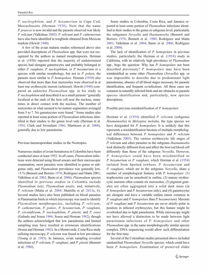
Trophozoites Trophozoites (Fig. 2a–d) are seen in matureerythrocytes. The earliest forms can be found anywhere in thehost cells but are more often seen in a position subpolar or polarto the nuclei of erythrocytes. Advanced trophozoites are seenonly in positions subpolar or polar to the nuclei of erythrocytes,are variable in shape, usually markedly amoeboid in outline,and often possess one or several long outgrowths (Fig. 2c, d).The nuclei and cytoplasm are prominent in advanced tropho-zoites (Fig. 2c). Each fully grown trophozoite usually possessesa prominent diffuse nucleus, readily visible cytoplasm, oneglobule (which may be invisible in some parasites), and twoor three small, roundish brown pigment granules (Fig. 2b),which are usually located close to each other near the edge ofthe parasite and may be clumped together (Fig. 2d). The cyto-plasm contains one or several small vacuoles. Trophozoites donot adhere to the nuclei of erythrocytes. The “ring” stage is notseen. One tiny bluish refractive globule appears in advancedtrophozoites (Fig. 2c, d) but is not visible in all parasites(Fig. 2b). The influence of trophozoites on the morphology ofinfected erythrocytes is not pronounced.
Erythrocytic meronts Erythrocytic meronts (Fig. 2e–l) areseen mainly in mature erythrocytes, but also occasionally innearly mature erythrocytes. The cytoplasm is prominent and
Parasitol Res
stains pale blue in early meronts (Fig. 2e–g). It graduallybecomes poorly visible or even invisible in fully grownmeronts (Fig. 2h–k). Nuclei are markedly variable in sizeand form in growing meronts but generally decrease in sizeas the parasite matures (compare Fig. 2e, f with Fig. 2i, j).Vacuoles are visible in some growing meronts (Fig. 2h). Onesmall bluish refractive globule is often seen in growingmeronts (Fig. 2h) and is more often seen in binuclear parasites.However, the globule is not visible in all meronts and gradu-ally degrades in mature meronts to a point where it is oftenonly seen as a bluish rim close to pigment granules (Fig. 2i).The globule is invisible in the majority of mature meronts(Fig. 2, j–l). Occasionally, two small refractive globules areseen in young meronts (Fig. 2h). Growing meronts are mark-edly variable in shape (elongate, oval, and roundish formspresent) and usually possess one or several long amoeboidoutgrowths (Fig. 2f, g). Fully grown meronts are usuallyroundish or slightly oval (Fig. 2j, k) and sometimes fan-likein shape (Fig. 2i). Nuclei are usually arranged irregularly inmature meronts, which produce between four and eight mer-ozoites. The majority of mature meronts (>80 % in all typepreparations) possess six and eight merozoites, but merontswith six merozoites predominate. Meronts contain two to fourminute brown pigment granules, which, in mature meronts,are usually clumped together and look like a solid mass
(Fig. 2j–l). Both growing and mature meronts are strictly ofpolar or subpolar position to the nuclei of erythrocytes(Fig. 2e–k) and do not adhere to the nuclei; these characteris-tics appear to be the most distinctive features of this parasitespecies’ development. Mature merozoites do not exceed 1 μmin diameter; their cytoplasm usually is poorly visible or invis-ible (Fig. 2l). Merozoites persist in infected erythrocytes forsome time after the rupture of meronts (Fig. 2l). The influenceofmeronts on themorphology of infected erythrocytes usuallyis not pronounced, but nuclei of some infected erythrocytesmay be slightly displaced laterally (Fig. 2j, l; Table 4).
Macrogametocytes Macrogametocytes (Fig. 2m–r) developin mature erythrocytes; the cytoplasm is heterogeneous inappearance, often containing oval bluish globular-like inclu-sions (Fig. 2q). Gametocytes are elongated in form and slight-ly irregular in outline from the earliest stages of their devel-opment. Growing gametocytes occur lateral to, but nevertouch, the nuclei of infected erythrocytes and usually adhereto the erythrocytic envelope and extend along the nuclei(Fig. 2m), yet another characteristic feature that is observedin the parasite’s development. As the parasite matures, thecentral part of the gametocyte adheres to the nucleus, leavingits ends unattached (Fig. 2n–q). Mature gametocytes usuallyadhere to both the nuclei and erythrocytic envelope of
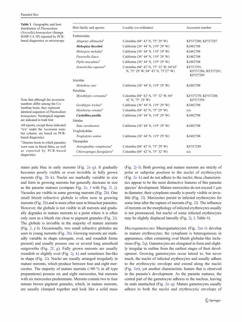
Table 1 Geographic and hostdistribution of Plasmodium(Novyella) homopolare (lineageSOSP CA 3P) reported by PCR-based diagnostics or microscopy
Note that although the accessionnumbers differ among the Co-lombian hosts, they representidentical sequences of Plasmodiumhomopolare. Neotropical migrantsare indicated in bold font
All reports, except those indicated“n/a” under the Accession num-ber column, are based on PCR-based diagnosticsa Denotes hosts in which parasiteswere seen in blood films, as wellas reported by PCR-baseddiagnostics
Bird family and species Locality (co-ordinates) Accession number
Melospiza lincolnii California (36° 44′ N, 119° 29′ W) KJ482708
Melospiza melodiaa California (36° 44′ N, 119° 29′ W) KJ482708
Passerella iliaca California (36° 44′ N, 119° 29′ W) KJ482708
Pipilo maculatusa California (36° 44′ N, 119° 29′ W) KJ482708
Zonotrichia capensisa Colombia (04° 42′ N, 75° 32′ W; 04°42′N, 75° 29′ W; 04° 43′ N, 75°27′ W)
KF537291;KF537286; KF537281;KF537289
Icteridae
Molothrus ater California (36° 44′ N, 119° 29′ W) KJ482708
Parulidae
Myiothlypis coronatusa Colombia (04° 42′ N, 75° 32′ W; 04°42′ N, 75° 29′ W)
KF537278; KF537288;KF537294
Geothlypis trichasa California (36° 44′ N, 119° 29′ W) KJ482708
Myioborus ornatusa Colombia (04° 42′ N, 75° 29′ W) n/a
Cardellina pusilla California (36° 44′ N, 119° 29′ W) KJ482708
Sittidae
Sitta carolinensis California (36° 44′ N, 119° 29′ W) KJ482708
Troglodydidae
Troglodytes aedon California (36° 44′ N, 119° 29′ W) KJ482708
Thraupidae
Anisognathus somptuosusa Colombia (04° 42′ N, 75° 29′ W) KF537289
Chlorospingus flavigularisa Colombia (04° 42′ N, 75° 32′ W) n/a
Parasitol Res
erythrocytes, filling-up the poles of erythrocytes and slightlyenclosing the erythrocyte nuclei with their ends, but notencircling them completely (Fig. 2r). The macrogametocytenucleus appears relatively diffuse and irregular in shape andhas faint boundaries and a central or subcentral position.Clumps of chromatin are very visible within the parasitenucleus (Fig. 2o). Small- (<0.5 μm) and medium- (0.5–1.0 μm) sized roundish and oval pigment granules can be seenanywhere within the gametocyte (Fig. 2p, q). The influence ofgametocytes on the morphology of infected erythrocytes isusually not pronounced, but nuclei of some infected erythro-cytes are slightly displaced laterally (Table 4).
Microgametocytes The general configuration and other fea-tures are similar to macrogametocytes (Fig. 2s, t), with theusual haemosporidian sexual dimorphic characters; however,the granular appearance of the cytoplasm is less evident thanthat in macrogametocytes. The nucleus is markedly diffuse tothe point where its size cannot be easily measured.
Remarks
Because of its small erythrocytic meronts and elongatedmicrogametocytes, P. homopolare belongs to the subgenusNovyella (Corradetti et al. 1963, Garnham 1966). This malariaparasite can be readily distinguished from other species ofNovyella primarily due to its strictly polar or subpolar positionof meronts and advanced trophozoites in infected erythrocytes(Fig. 2a–k). The positioning of the meronts and advancedtrophozoites is shared with the relatively rare avian parasitePlasmodium polare (Manwell 1935; Bishop and Bennett1992), which is reflected in the name of this new species:P. homopolare. P. polare produces meronts of greater size thanP. homopolare, and, in this regard, is closer to species of thesubgenus Giovannolaia (Valkiūnas 2005). The maximumnumber of merozoites in mature meronts of P. homopolare is8, as opposed to 16 in those of P. polare, allowing for differ-entiation of these two species.
The morphology of blood stages of P. homopolare wasstrictly consistent across the eight California and Colombiabird species represented in the Emberizidae, Parulidae, andThraupidae families (Fig. 2), indicating that the parasite didnot change main morphological characters in different avianhosts belonging to Passeriformes. Morphometric comparisonsof mature meronts, macrogametocytes, and microgametocytesof P. homopolare in the Song sparrow (Melospiza melodia)and the Rufous-collared sparrow (Z. capensis) sampled inCalifornia and Colombia, respectively, did not differ signifi-cantly (P ≥ 0.4, for all features, except the gametocyte area, forwhich P = 0.09, data not shown). Due to insufficient intensityof parasitemia with some blood stages (usually mature game-tocytes and mature meronts), we were unable to complete asimilar morphometric analysis for the other bird species in ourstudy. Morphometric characters of parasites in the type verte-brate host (Melospiza melodia) are given in Table 4.
Taxonomic summary
Type host: Song sparrow Melospiza melodia (Passeriformes,Emberizidae).
DNA sequences: Mitochondrial cytb lineage SOSP CA 3P(478 bp, GenBank accession no. KJ482708).
Additional hosts: Numerous species of passeriform birds,belonging mainly to Emberizidae (Table 1).
Table 2 Geographic and host distribution of Plasmodium spp. with100 % or nearly 100 % (≤2-nucleotide difference) sequence matches toPlasmodium homopolare (lineage SOSP CA 3P), but lacking examina-tion of blood smears
Type locality: China Creek County Park, Fresno County,California, USA (36° 44′N, 119° 29′W, 120 m above sea level).
Prevalence: The prevalence determined using combinedPCR-based detection and microscopy methods was 28 of 51(54.9 %) in Melospiza melodia at the type locality. OverallPCR-based prevalence in all investigated birds was 21 and3.6 % in California and Colombia, respectively. However, inCalifornia, all bird blood samples were analyzed using PCRwhereas, in Colombia, only those birds diagnosed by micros-copy were analyzed using PCR.
Geographical distribution: P. homopolare and its lineageshave been recorded only in North and South America wherethe parasite is present in numerous species of migratingand resident birds. P. homopolare is a host generalist(Table 1), but its transmission has not been reportedoutside of the American continents.
Site of infection: Mature or nearly mature erythro-cytes; no other data.
Type specimens: Hapantotype (accession no. 48740 NS,lineage SOSP CA 3P, intensity of parasitemia is approximately
8 %, Melospiza melodia, China Creek County Park, FresnoCounty, California, 36° 44′N, 119° 29′W, 21 January 2013, E.Walther) is deposited in the Nature Research Centre, Vilnius,Lithuania. Parahapantotypes are deposited in the QueenslandMuseum, Queensland, Australia (accession no. G465687, otherdata as for the hapantotype) and in the Nature Research Centre(accession nos. 48741, 48742 NS, lineage SOSP CA 3P, host isPipilo maculatus, China Creek County Park, Fresno County,CA, 24 June 2011, collected by E. Walther).
Additional material: Blood films from Pipilo maculatus(accession nos. 48744–48747 NS, 48751–48754 NS),Melospiza melodia (48748–48750 NS), and Geothlypistrichas (48755 NS) were deposited in the Nature ResearchCentre, Vilnius, Lithuania. Blood films from Myiothlypiscoronatus (accession nos. GERPH-07316, GERPH-07317,and GERPH-07318) and Z. capensis (accession nos.GERPH-04822, GERPH-04823, and GERPH-04824) weredeposited in the collection Grupo de Estudio RelaciónParásito Hospedero (GERPH), Department of Biology,Universidad Nacional de Colombia, Bogotá, Colombia.
Fig. 1 Bayesian inferencephylogenetic reconstructionbased on 468 nucleotides of thecytochrome b gene and includingPlasmodium (Novyella)homopolare, as well as 19reference sequences of knownmorphospecies from GenBank.Leucocytozoon sp. SISKIN2(accession no. AY393796) servesas the out-group. Note thatP. homopolare groups withPlasmodium species that areprimarily in the subgenusNovyella
Parasitol Res
Parasitol Res
Digital images of blood stages of the new parasite are avail-able on request from GERPH.
Etymology: The species name reflects the similarity oferythrocytic meronts and advanced trophozoites of this para-site to P. polare. In both parasites, these blood stages assume astrictly polar or subpolar position in infected erythrocytes, acharacteristic feature of their development.
Discussion
P. homopolare was identified to morphospecies based onblood samples from neo-tropical migrants and resident birdsin California, USA, and Colombia. We believe P. homopolareis present in birds throughout the New World because theCalifornia and Colombia cytb sequences matched 100 % ornearly 100% (i.e., ≤2-nucleotide difference) to two previouslydeposited parasite sequences in GenBank: accession nos.AF465555 and JN819334. These sequences were isolatedfrom the Silver-throated tanager (T. icterocephala), a residentbird of Costa Rica, and a variety of Neotropical migrants andresident birds in the Eastern USA and the Caribbean, respec-tively (see full list of hosts in Table 2). Because blood smearseither were unavailable or were not examined for these hosts,we cannot unequivocally confirm AF465555 and JN819334to be P. homopolare; it is likely based on the available data(Table 1) but would require additional testing because geneticdifferences among cytb gene partial sequences in some readilydistinguishable haemosporidian species is small (Hellgrenet al. 2007). Additionally, it should be noted that the
development of Plasmodium spp. and related haemosporidi-ans can be abortive in resistant or partly resistant hosts,resulting in no development of gametocytes (Valkiūnas et al.2011; Cannell et al. 2013). This can happen when (1) sporo-zoites are inoculated by a vector into the blood but are inca-pable of intracellular invasion, so remain in the circulation forsome time, or (2) remnants of tissue meronts, the developmentof which was aborted before the gametocyte stage, are pro-duced and remain in hosts, serving as a PCR template. In bothcases, the bird would be scored as infected by PCR when, infact, it might not be a competent host of the parasite.Therefore, conclusions regarding the full range of avian hostswould require microscopic examination of blood films inaddition to comparison of molecular results.
Assuming that P. homopolare is broadly distributedthroughout the New World and infects a wide range of hostsin geographic areas where haemosporidian studies have oc-curred, it is notable that this parasite has not previously beenidentified in prior studies from North America and theNeotropics, particularly those conducted in California andColombia.
Previous haemosporidian studies in the Nearctic
Numerous studies examining the prevalence and diversity ofPlasmodium spp. have been conducted in the Nearctic sincethe 1930s (Herman 1938; Hewitt 1940; Herman et al. 1954;Clark and Swinehart 1966; Greiner et al. 1975; Super and vanRiper 1995; Ricklefs et al. 2005; Martinsen et al. 2008). Inparticular, a 6-year study by Herman et al. (1954) was under-taken in Kern County, California, approximately 160 km fromthe California study site at China Creek County Park. In KernCounty, numerous species of Plasmodium were identifiedmorphologically, including Plasmodium elongatum,Plasmodium hexamerium, Plasmodium nucleophilum,P. polare, Plasmodium relictum, P. rouxi, and P. vaughani,as well as several unknown species from 7,440 passerine and
�Fig. 2 Plasmodium (Novyella) homopolare sp. nov. (lineage SOSP CA3P) from the blood of Song sparrow Melospiza melodia. a–dTrophozoites. e–l Erythrocytic meronts. m–r Macrogametocytes. s, tMicrogametocytes. Short arrows nuclei of parasites, long arrows bluishrefractive globule, triangle arrowhead pigment granules, simplearrowhead globular-like inclusions in the cytoplasm. Giemsa-stainedthin blood films. Scale bar = 10 μm
Table 3 Genetic distances between Plasmodium homopolare and seven other Plasmodium spp. of the subgenus Novyella, as well as an out-group(P. elongatum)
Genetic divergence is given as a percentage and was calculated using uncorrected p distance analysis, based on 468 nucleotides of the cytb gene
Parasitol Res
near-passerine blood smears. P. relictum was the most com-mon parasite, infecting 792 (10.7 %) birds. In Kern County,the vast majority of birds sampled were House sparrows(Passer domesticus) (2,880/7,440) and House finches(Carpodacus mexicanus) (1,741/7,440). Two hundred forty-three (8.1%) House sparrows and 424 (24.4 %) House fincheswere infected with P. relictum. At China Creek, we collectedsamples from six House finches, none of which was infectedwith P. relictum. We sampled no House sparrows. Only oneisolate ofP. relictumwas obtained at China Creek and that wasfrom a Cliff swallow (Petrochelidon pyrrhonota) (data notshown). In the Kern County study, blood smears were alsoobtained from the two host species at China Creek found tohave high rates of infection with P. homopolare: Song spar-rows (Melospiza melodia) and Spotted towhees (Pipilomaculatus). Herman et al. (1954) trapped 14 Song sparrows,of which 2 (14.3 %) were infected with P. relictum, and 42Spotted towhees, of which 4 (9.5 %) were infected withP. relictum, 1 (2.4 %) with P. hexamerium, and 1 (2.4 %) withP. vaughani. None of the parasites in these infected birds wascategorized as unknown Plasmodium species, as would beexpected if the authors had found the then undescribedP. homopolare in the hosts. Conversely, none of the six hostsin which the authors identified an unknown Plasmodiumspecies was host that harbored P. homopolare in the ChinaCreek study.
Additional studies in the Nearctic utilizing microscopyidentified several Plasmodium spp., including Plasmodiumpraecox, Plasmodium circumflexum, and P. relictum inCalifornia (Herms et al. 1939; Super and van Riper 1995;Martinsen et al. 2008) and Plasmodium cathemerium,P. praecox, P. circumflexum, P. vaughani, P. elongatum,
Table 4 Morphometry of mature erythrocytic meronts, gametocytes, andhost-cells of Plasmodium homopolare (lineage SOSP CA 3P) from theblood of the Song sparrow Melospiza melodia
Feature Measurementsa
Uninfected erythrocyte
Length 10.3–12.8 (11.4 ± 0.5)
Width 5.2–6.8 (6.0 ± 0.3)
Area 49.7–64.8 (55.2 ± 4.2)
Uninfected erythrocyte nucleus
Length 4.8–6.3 (5.3 ± 0.3)
Width 2.4–3.1 (2.5 ± 0.2)
Area 9.5–19.4 (12.2 ± 1.3)
Meront
Length 3.2–6.0 (4.3 ± 0.3)
Width 1.9–3.6 (2.5 ± 0.5)
Area 6.1–15.2 (8.4 ± 1.4)
No. of merozoites 4–8 (6.9 ± 0.7)
Infected erythrocyte
Length 10.1–13.8 (11.8 ± 0.7)
Width 5.0–6.4 (5.6 ± 0.4)
Area 47.4–62.8 (52.7 ± 4.6)
Infected erythrocyte nucleus
Length 4.4–6.6 (5.4 ± 0.4)
Width 2.1–3.2 (2.4 ± 0.3)
Area 9.3–18.8 (12.7 ± 1.6)
Macrogametocyte
Length 10.7–16.7 (13.8 ± 1.5)
Width 1.3–3.0 (2.1 ± 0.4)
Area 18.9–34.0 (27.0 ± 3.7)
Gametocyte nucleus
Length 1.6–5.5 (2.4 ± 0.8)
Width 0.9–2.3 (1.5 ± 0.3)
Area 1.3–5.4 (3.2 ± 2.0)
Pigment granules 5–15 (9.5 ± 1.8)
NDR 0.6–1.1 (0.9 ± 0.1)
Infected erythrocyte
Length 10.5–13.8 (11.6 ± 0.9)
Width 5.0–7.2 (6.2 ± 0.5)
Area 49.0–68.9 (58.2 ± 4.9)
Infected erythrocyte nucleus
Length 4.5–6.0 (5.0 ± 0.3)
Width 2.2–3.0 (2.4 ± 0.2)
Area 9.3–17.4 (11.2 ± 1.0)
Microgametocyte
Length 10.2–18.9 (14.2 ± 1.5)
Width 1.4–3.0 (2.4 ± 0.5)
Area 22.2–36.7 (28.8 ± 3.8)
Gametocyte nucleus
Length –b
Width –b
Area –b
Table 4 (continued)
Feature Measurementsa
Pigment granules 7–17 (10.3 ± 2.6)
NDR 0.5–1.1 (0.8 ± 0.2)
Infected erythrocyte
Length 10.3–13.8 (11.7 ± 0.9)
Width 5.2–7.0 (6.2 ± 0.4)
Area 47.9–69.8 (58.2 ± 5.0)
Infected erythrocyte nucleus
Length 4.4–6.3 (5.3 ± 0.4)
Width 2.0–3.1 (2.5 ± 0.3)
Area 9.2–18.4 (12.0 ± 1.5)
NDR nucleus displacement ratio according to Bennett and Campbell(1972)a All measurements (n = 21) are given in micrometers. Minimum andmaximum values are provided, followed in parentheses by the arithmeticmean and standard deviationbDifficult to measure feature; see the text for explanation
Parasitol Res
P. nucleophilum, and P. hexamerium in Cape Cod,Massachusetts (Herman 1938). Note that the nameP. praecox is now invalid and the parasite observed was likelyP. relictum (Valkiūnas 2005). P. relictum and P. cathemeriumhave also been identified in songbirds obtained fromMexicanmarkets (Hewitt 1940).
A few of the avian malaria studies referenced above alsoprovided descriptions of Plasmodium spp. that were not rec-ognized by the authors as named morphospecies. Hermanet al. (1938) reported that the majority of undeterminedspecies had elongate gametocytes and probably belonged toeither P. vaughani, P. nucleophilum, or P. hexamerium or aspecies with similar morphology, but not to P. polare, theparasite most similar to P. homopolare. Herman (1938) alsoobserved that more than four merozoites were observed in atleast one erythrocytic meront (schizont). Hewitt (1940) com-pared an unknown Plasmodium spp. in his study toP. nucleophilum and described it as a small parasite “generallylocalized at the ends of the host cell near the nucleus, manytimes in direct contact with the nucleus. The number ofmerozoites in what seemed to be mature segmenters averagedfrom 5 to 7. No gametocytes were found.” Some studies alsoreported at least some portion of Plasmodium infections iden-tified in their studies to the genus level only (Herman et al.1954; Clark and Swinehart 1966; Martinsen et al. 2008),generally due to low parasitemia.
Previous haemosporidian studies in the Neotropics
Numerous studies of avian hematozoa in Colombia have beenconducted since at least 1952. In all cases, Plasmodium infec-tions were detected using blood smears and their microscopicexamination, most parasites were identified to genus or sub-genus only, and Plasmodium prevalence was generally low,<5 % (Bennett and Borrero 1976; Rodriguez and Matta 2001;Valkiūnas et al. 2003; Basto et al. 2006). Plasmodium speciesidentified in previous studies in Colombia includePlasmodium lutzi, Plasmodium unalis, and, tentatively,P. relictum (Matta et al. 2004; Mantilla et al. 2013a, b).Several studies have also been published on blood parasitesin Panamanian birds in whichmicroscopywas used to identifyPlasmodium morphospecies, including P. relictum,P. cathemerium, P. polare, P. hexamerium, P. vaughani,P. circumflexum, P. nucleophilum, P. pinotii, and P. rouxi(Galindo and Sousa 1966; Sousa and Herman 1982), thoughthe authors acknowledged that low parasitemia and limitedsampling may have resulted in erroneous identifications(Sousa and Herman 1982). In a Monteverde, Costa Rica studyutilizing microscopy, P. relictum was found in low prevalence(Young et al. 1993). In Jamaica, avian sampling revealedinfections of P. relictum, P. vaughani, and P. pinotii (Bennettet al. 1980).
Some studies in Colombia, Costa Rica, and Jamaica re-ported at least some portion of Plasmodium infections identi-fied in their studies to the genus or subgenus level, particularlythe subgenera Novyella and Haemamoeba (Bennett andBorrero 1976; Bennett et al. 1980; Rodríguez and Matta2001; Valkiūnas et al. 2004; Basto et al. 2006; Rodríguezet al. 2009).
The lack of identification of P. homopolare in previousstudies, particularly the Herman et al. (1954) study inCalifornia, with its relatively high prevalence of Plasmodiumspp., begs the question: Why has P. homopolare not beendescribed previously? P. homopolare may have beenmisidentified as some other Plasmodium (Novyella) spp. orwas impossible to describe due to predominant lightparasitemias, absence of all blood stages necessary for parasiteidentification, and frequent co-infections. All these cases arecommon in naturally infected birds and are obstacles in parasitespecies identification and, particularly, new speciesdescriptions.
Possible previous misidentification of P. homopolare
Herman et al. (1954) identified P. relictum (subgenusHaemamoeba) in Melospiza melodia, the type species wehave designated for P. homopolare. It is unlikely that thisrepresents a misidentification because of multiple morpholog-ical differences between P. homopolare and P. relictum(Valkiūnas 2005). The various erythrocytic life stages ofP. relictum and other parasites in the subgenus Haemamoebalook distinctly different from and affect the host red blood celldifferently than those of the subgenus Novyella. However,P. homopolare could have been misidentified asP. hexamerium or P. vaughani, which Herman et al. (1954)isolated from Spotted towhees. P. hexamerium andP. vaughani, which are in the subgenus Novyella, share anumber of morphological features with P. homopolare: (1)trophozoites can be amoeboid in outline, (2) mature erythro-cytic meronts often contain six merozoites, (3) pigment gran-ules are often aggregated into a solid dark mass (inP. homopolare and P. hexamerium only), and (4) gametocytesare elongate and have a variable outline (more common inP. vaughani and P. homopolare than P. hexamerium). Merontsof P. vaughani and P. hexamerium are never strictly polar inposition in infected erythrocytes, but this feature might beoverlooked due to light parasitemia. While microscopy mightnot have allowed a distinction to be made between lightparasitemia infections of P. homopolare and otherPlasmodium spp. in the same morphologically similar speciescomplex, DNA sequencing would allow such differentiationfor the first time.
Several of the Colombian studies presented above includedunidentified Plasmodium Novyella species, which could havebeen P. homopolare. Examination of preserved slides
Parasitol Res
deposited in the GERPH collection from some of these studies(Rodríguez and Matta 2001; Matta et al. 2004; Basto et al.2006; Rodríguez et al. 2009) showed that Plasmodium(Novyella) species found in Emberizidae, Icteridae, andThraup idae hos t s cor respond to P. una l i s andP. nucleophilum (data not shown). Unfortunately, theidentity of unknown Plasmodium species in the Novyellasubgenus from Bennett and Borrero (1976) and Valkiūnaset al. (2003) cannot be confirmed since morphological de-scriptions or slides are not available. Future studies shouldinclude an examination of preserved slides from some of theseolder studies, to determine the possible presence ofP. homopolare.
Light parasitemias and co-infections in earlier studiesprevented description of P. homopolare
The disease dynamics of avian haemosporidians include abrief pre-patent period in which the parasites are found onlyin the host tissues, followed by a patent stage that begins withshort acute infection lasting on the order of weeks and char-acterized by high parasitemia (Valkiūnas 2005). Assumingthat the host survives, the acute infection is followed by anindefinite period of chronic infection, which is characterizedby low parasitemia (Garnham 1966; Valkiūnas 2005;Atkinson 2008). It is during this chronic infection that re-searchers are most likely to trap a bird because this period isrelatively long and the bird is more likely to be active duringthe chronic infection than during the acute infection. In fact,the acute stage of Plasmodium is relatively short, and theparasitemia level during chronic infections is relatively low,compared to other haemosporidians (Greiner et al. 1975;Valkiūnas 2005). In addition, the patent infection is affectedby season; for example, Plasmodium spp. are unlikely to befound in hosts in early spring and late fall in the Holarcticregion using microscopic examination of blood films(Valkiūnas 2005). Moreover, co-infections of haemosporidianparasites are common in many bird species and confound themorphological identification of these parasites (Valkiūnaset al. 2008a). All of these factors present a challenge to themalaria researcher relying solely on microscopy to quantifyprevalence and parasite identity in a population and wouldnecessitate frequent sampling and examination of multipleblood smears to find and identify morphospecies ofPlasmodium parasites. All of the previous haemosporidianstudies described above relied solely on microscopy andsometimes on a single blood smear for each host examined.It is not always clear in which season(s) sampling took place,but it is possible that some early researchers in the Holarcticwere unaware of the seasonal dynamics of Plasmodium infec-tions and may have collected samples during bird migration inearly spring and late fall when parasitemias are light or absentfor certain Plasmodium species. Many studies were able to
identify Plasmodium parasites to morphospecies, but mostincluded samples for which there was not enough morpholog-ical data to identify the parasites to species.
Phylogenetic relationship of P. homopolareto other morphospecies
Phylogenetic analysis placed P. homopolare in a clade withseven other species within the subgenusNovyella. Parasites ofthe Novyella subgenus are distinguished from other subgeneraby the following morphological characteristics: (1) scantycytoplasm in erythrocytic meronts, (2) size of fully grownerythrocytic meronts does not exceed or only slightly exceedsthat of the nuclei of infected erythrocytes, (3) fully growngametocytes are elongated, and (4) exoerythrocytic merogonytakes place in cells of the reticuloendothelial system(Garnham 1966; Valkiūnas 2005). P. vaughani is the typespecies of the Novyella subgenus and has a worldwide geo-graphic distribution with the exception of Antarctica.Novyellaspecies are known to infect over 240 species of Passeriformes(Valkiūnas 2005; Bensch et al. 2009) including species fromthe Sylviidae, Turdidae, Fringillidae, Petroicidae,Muscicapidae, Meliphagidae, and Alaudidae families.Genetic distance analysis indicates that P. homopolare hasthe l owes t l eve l s o f gene t i c d ive rgence f romP. parahexamerium. The latter parasite was first identified in2009 in two species from the Turdidae family, as well as anOld world warbler, Hylia prasina (Green hylia), in Africa(Valkiūnas et al. 2009; Bensch et al. 2009). Interestingly,P. homopolare, thus far, has only been confirmed to infecthosts in the New World. However, like P. homopolare,P. parahexamerium was identified relatively recently, andfurther study may reveal that one or both of these speciesinfect a wider range of hosts on a broader geographical scalethan is currently understood.
The application of molecular markers opens new opportu-nities for Plasmodium spp. taxonomic studies due to thepossibility of using sequence information to strengthen studiesof wild hosts with light parasitemias. Many species ofPlasmodium occur in birds belonging to different species,genera, families, and even orders. Within this host range, thesame morphospecies may exhibit slightly different morpho-logical forms, resulting in strains, varieties, and transmissionby different vectors throughout their distributional range. Dueto these morphological variants, it has been the convention inearly avian malaria studies (Garnham 1966; Atkinson 2008)that any new species should only be established if supportedby a “package” of taxonomic data, including the full range ofblood forms, and also data on tissue merogony, periodicity,vectors, and other features. It is difficult and time-consumingto collect such information, which resulted in few descriptionsof new Plasmodium species before DNA sequencing becameavailable (Garnham 1966; Valkiūnas 2005). The combined
Parasitol Res
application of sequence data and digital microscopy tech-niques provides new opportunities to repeat studies and uselight microscopy for parasite species identification; this strat-egy has benefitted taxonomic research and has resulted in thecurrent renaissance in haemosporidian parasite taxonomy anddescriptions of numerous new haemosporidians parasitizingbirds (Valkiūnas et al. 2007; Valkiūnas et al. 2008c; Valkiūnaset al. 2009; Levin et al. 2012; Ilgūnas et al. 2013; Mantillaet al. 2013a; Valkiūnas et al. 2013) and reptiles (Perkins andAustin 2009). Importantly, the descriptions of new morpho-species are in accord with recent PCR-based studies indicatingremarkable genetic diversity of haemosporidian parasites(Bensch et al. 2000; Ricklefs and Fallon 2002; Beadell et al.2004; Ricklefs et al. 2005; Valkiūnas 2005; Loiseau et al.2010). This study thus reports the identifying molecular andmorphological characters for the detection of P. homopolare, awidespread malaria parasite of American birds.
Acknowledgments Tatjana Iezhova is acknowledged for technical as-sistance during preparation of the plate of the illustrations. Work inCalifornia was partially supported by a grant from the San FranciscoState University Arthur Nelson Scholarship and the San Francisco StateUniversity Instructionally Related Activities Grant. Work in Colombiawas supported by the Departamento Administrativo de Ciencias,Tecnología e Innovación COLCIENCIAS contract no. 359 andECOPETROL SA. The authors wish to thank the students belonging tothe Host-Parasite Relationship Research Group: avian haemoparasitemodel of the Universidad Nacional de Colombia and the staff of theUnidad Administrativa Especial del Sistema de Parques NacionalesNaturales for support with field logistics and work, as well as the manyfield assistants who aided in sampling California birds. R. E. Ricklefs’sstudies on avian haemosporidians have been supported by the NationalGeographic Society, the US National Science Foundation, and the Cura-tors of the University of Missouri, with field and laboratory assistancefrommany individuals.We thankHolly Archer for contributing data fromher research on avian malaria in Costa Rican birds.
Basto N, Rodríguez OA, Marinkelle CJ, Gutierrez R, Matta N (2006)Haemoatozoa in birds from La Macarena National Natural Park(Colombia). Caldasia 28(2):371–377
Beadell JS, Gering E, Austin J, Dumbacher JP, Peirce MA, Pratt TK,Atkinson CT, Fleischer RC (2004) Prevalence and differential host-specificity of two avian blood parasite genera in the Australo-Papuan region. Mol Ecol 3:3829–3844
Bennett GF, Campbell AG (1972) Avian Haemoproteidae. I. Descriptionof Haemoproteus fallisi n. sp. and a review of the haemoproteids ofthe family Turdidae. Can J Zool 50:1269–1275
Bennett GF, Borrero JI (1976) Blood parasites of some birds fromColombia. J Wildl Dis 12:454–458
Bennett GF, Whitt H, White H (1980) Blood parasites of some Jamaicanbirds. J Wildl Dis 16:29–38
Bensch S, Sternman M, Hasselquist D, Ostman O, Hansson B,Westerdahl H, Pinheiro RT (2000) Host specificity in avian blood
parasites: a study of Plasmodium and Haemoproteus mito-chondrial DNA amplified from birds. Proc R Soc Lond BBiol Sci 267:1583–1589
Bensch S, Hellgren O, Pérez-Tris J (2009) MalAvi: a public database ofmalaria parasites and related haemosporidians in avian hosts basedon mitochondrial cytochrome b lineages. Mol Ecol 9:1353–1358.doi:10.1111/j.1755-0998.2009.02692.x
Bishop MA, Bennett GF (1992) Host-parasite catalogue of the avianhaematozoa: supplement 1 and bibliography of the avian blood-inhabiting haematozoa: supplement 2. Mem Univ NewfoundlOccas Pap Biol 15:1–244
Cannell BL, Krasnec KV, Campbell K, Jones HI, Miller RD, Stephens N(2013) The pathology and pathogenicity of a novel Haemoproteusspp. infection in wild Little Penguins (Eudyptula minor). VetParasitol 197(1-2):74–84. doi:10.1016/j.vetpar.2013.04.025
Clark GW, Swinehart B (1966) Blood protozoa of passerine birds of theSacramento (Calif.) region. Bull Wildl Dis Assoc 2:53–54
Corradetti A, Garnham PCC, Laird M (1963) New classification of theavian malaria parasites. Parassitologia 5:1–4
Galindo P, Sousa O (1966) Blood parasites of birds from Almirante,Panama, with ecological notes on the hosts. Rev Biol 14(1):27–46
Galtier N, GouyM, Gautier C (1996) SEAVIEWand PHYLO_WIN: twographic tools for sequence alignment and molecular phylogeny.Comput Appl Biosci: CABIOS 12(6):543–548
Garnham PCC (1966) Malaria parasites and other Haemosporidia.Blackwell Scientific Publications, Oxford
Greiner EC, Bennett GF, White EM, Coombs RF (1975) Distribution ofthe avian hematozoa of North America. Can J Zool 53:1762–1787
Hellgren O, Waldenström J, Bensch S (2004) A new PCR assay forsimultaneous studies of Leucocytozoon, Plasmodium, andHaemoproteus from avian blood. J Parasitol 90:797–802
Hellgren O, Križanauskienė A, Valkiūnas G, Bensch S (2007) Diversityand phylogeny of mitochondrial cytochrome b lineages from sixmorphospecies of avian Haemoproteus (Haemosporida:Haemoproteidae). J Parasitol 93:889–896
Herman CM (1938) The relative incidence of blood protozoa in somebirds from Cape Cod. Tr Am Micr Soc 47:132–141
Herman CM, Reeves WC, McClure HE, French EM, Hammon W (1954)Studies on avian malaria in vectors and hosts of encephalitis in KernCounty, CA: infections in avian hosts. Am J TropMed Hyg 3:676–695
Herms WB, Kadner CG, Galindo P, Armstrong DF (1939) Blood para-sites of California birds. J Parasitol 25(6):511–512
Hewitt R (1940) Studies on blood protozoa obtained from Mexican wildbirds. J Parasitol 26(4):287–295
Ilgūnas M, Palinauskas V, Iezhova TA, Valkiūnas G (2013) Molecularand morphological characterization of two avian malaria parasites(Haemosporida: Plasmodiidae), with description of Plasmodiumhomonucleophilum n. sp. Zootaxa 3666(1):49–61
Levin II, Valkiūnas G, Iezhova TA, O’Brien SL, Parker PG (2012) NovelHaemoproteus species (Haemosporida: Haemoproteidae) from theSwallow-tailed gull (Lariidae), with remarks on the host range ofHippoboscid-transmitted avian hemoproteids. J Parasitol 98(4):847–854
Loiseau C, Iezhova T, Valkiūnas G, Chaser A, Hutchinson W, BuermannT, Smith TB, Sehgal RNM (2010) Spatial variation ofhemosporidian parasite infection in African rainforest bird species.J Parasitol 96:21–29
Mantilla JS, González AD, Valkiūnas G, Moncada LI, Matta NE (2013a)Description and molecular characterization of Plasmodium(Novyella) unalis sp. nov. from the Great Thrush (Turdus fuscater)in highland Colombia. Parasitol Res 112:4193–4204
Mantilla JS, Matta NE, Pacheco MA, Escalante AA, González AD,Moncada LI (2013b) Identification of Plasmodium (Haemamoeba)lutzi (Lucena, 1939) from Turdus fuscater (Great Thrush) inColombia. J Parasitol 99(4):662–668
Manwell RD (1935) How many species of avian malaria parasites arethere? Am J Trop Med 15(3):265–283
Martinsen ES, Blumberg BJ, Eisen RJ, Schall JJ (2008) Avianhemosporidian parasites from northern California oak woodlandand chaparral habitats. J Wildl Dis 44(2):260–268
Matta NE, Basto N, Gutierrez R, Rodríguez OA, Greiner EC (2004)Prevalence of blood parasites in Tyrannidae (flycatchers) in theeastern plains of Colombia. Mem Inst Oswaldo 99(3):271–274
Muñoz E, Ferrer D, Molina RRA (1999) Prevalence of haematozoa inbirds of prey in Catalonia, northeast Spain. Vet Rec 144:623–636
Perkins SL, Austin CC (2009) Four new species of Plasmodium fromNew Guinea lizards: integrating morphology and molecules. JParasitol 95:424–433
Ricklefs RE, Fallon SM (2002) Diversification and host switching inavian malaria parasites. Proc R Soc London 269:885–892
Ricklefs RE, Swanson BL, Fallon SM, Martinez-Abrain A, Scheuerlein A,Gray J, Latta SC (2005) Community relationships of avian malariaparasites in Southern Missouri. Ecol Monogr 75(4):543–559
Rodríguez OA, Matta NE (2001) Blood parasites in some birds fromeastern plains of Colombia. Mem Inst Oswaldo Cruz 96:000–000
Rodríguez OA, Moya H, Matta NE (2009) Avian blood parasites in theNational Natural Park Chingaza: high Andes of Colombia. Hornero24(1):1–6
Sambrook J, Fritsch EF, Maniatis T (1989) Molecular cloning: a laboratorymanual, 2nd edn. Cold Harbour Laboratory Press, New York, 2222
Schneider CA, Rasband WS, Eliceiri KW (2012) NIH Image to ImageJ:25 years of image analysis. Nat Methods 9:671–675. doi:10.1038/nmeth.2089
Sehgal RNM, Lovette IJ (2003) Molecular evolution of three avianneurotrophin genes: implications for proregion functional con-straints. J Mol Evol 57:335–342
Sehgal RNM, Hull AC, Anderson NL, Valkiūnas G, Markovets MJ,Kawamura S, Tell LA (2006) Evidence for cryptic speciation ofLeucocytozoon spp. (Haemosporida, Leucocytozoidae) in diurnalraptors. J Parasitol 92(2):375–379
Sousa OE,HermanCM (1982) Blood parasites of birds fromChiriqui andPanama provinces in the Republic of Panama. J Wildl Dis 16(2):205–221
Super PE, van Riper C (1995) A comparison of avian hematozoanepizootiology in two California coastal scrub communities. J WildlDis 31(4):447–461
Swofford D (2001) PAUP* 4.0. Sinauer AssociatesValkiūnas G, Salaman P, Iezhova TA (2003) Paucity of Hematozoa in
CauseyD (2004)Additional observations on blood parasites of birdsin Costa Rica. J Wildl Dis 40(3):555–561
Valkiūnas G (2005) Avian malaria parasites and other haemosporidia.CRC Press, Boca Raton
Valkiūnas G, Zehtindjiev P, Hellgren O, Ilieva M, Iezhova TA, Bensch S(2007) Linkage between mitochondrial cytochrome b lineages andmorphospecies of two avian malaria parasites, with a description ofPlasmodium (Novyella) ashfordi sp. nov. Parasitol Res 100:1311–1322
Valkiūnas G, Iezhova TA, Krizankauskiene A, Palinauskas V, Sehgal RN,Bensch S (2008a) A comparative analysis of microscopy and PCR-based detection methods for blood parasites. J Parasitol 94(6):1395–1401. doi:10.1645/GE-1570.1
Valkiūnas G, Atkinson CT, Bensch S, Sehgal RNM, Ricklefs R (2008b)Parasite identifications in GenBank: how to minimize their number?Trends Parasitol 24(6):247–248
Valkiūnas G, Iezhova TA, Loiseau C, Chasar A, Smith TB, Sehgal RNM(2008c) New species of haemosporidian parasites (Haemosporida)from African rainforest birds, with remarks on their classification.Parasitol Res 103:1213–1228
Valkiūnas G, Iezhova TA, Loiseau C, Smith TB, Sehgal RNM (2009)Newmalaria parasites of the subgenusNovyella in African rainforestbirds, with remarks on their high prevalence, classification anddiagnostics. Parasitol Res 104:1061–1077
Valkiūnas G, Sehgal RNM, Iezhova TA, Hull AC (2010) Identification ofLeucocytozoon toddi group (Haemosporida: Leucocytozoidae), withremarks on the species taxonomy of Leucocytozoids. J Parasitol96(1):170–177
Valkiūnas G, Ashford RW, Bensch S, Killick-Kendrick R, Perkins S(2011) A cautionary note concerning Plasmodium in apes. TrendsParasitol 27(6):231–232
Valkiūnas G, Iezhova TA, Evans E, Carlson JS, Martinez-Gomez JE,Sehgal R (2013) Two new Haemoproteus (Haemosporida:Haemoproteidae) species from columbiform birds. J Parasitol99(3):513–521
Waldenström J, Bensch S, Hasselquist D, Östman Ö (2004) A new nestedpolymerase chain reaction method very efficient in detectingPlasmodium and Haemoproteus infections from avian blood. JParasitol 90(1):191–194. doi:10.1645/GE-3221RN
Young BE, Garvin MC, McDonald DB (1993) Blood parasites in birdsfrom Monteverde, Costa Rica. J Wildl Dis 29(4):555–560