45
Operative vaginal delivery By: Dr. Mojgan Rajati Assistant Professor of Ob. & Gvn, Kermanshah University of Medical Sciences

Operative vaginal
delivery By:
Dr. Mojgan Rajati
Assistant Professor of Ob. & Gvn, Kermanshah University of Medical Sciences
Operative vaginal delivery
OVD refers to delivery in which the operator uses forceps, a vacuum, or other devices to extract the fetus from the vagina, with or without the assistance of maternal pushing.
The decision to use an instrument to deliver the fetus balances the maternal, fetal, and neonatal impact of the procedure against the alternative options of cesarean birth or expectant management.
2 Dr. Mojgan Rajati

Frequency of OVD
In the United States, 3.1 percent of all deliveries in 2017
were accomplished via an operative vaginal approach .
Forceps deliveries accounted for 0.5 percent of vaginal
births, and vacuum deliveries accounted for 2.6 percent of
vaginal births.
3 Dr. Mojgan Rajati

Operative vaginal delivery
In low- and middle-income countries
the operative delivery rate declined from 1.6 to
0.3 percent
while the cesarean rate more than doubled to
reach 14.4 percent.
4 Dr. Mojgan Rajati

INDICATIONS
Maternal exhaustion and an inability to push
effectively.
Maternal medical indications, such as maternal
cardiac disease and a need to avoid pushing in the
second stage of labor.
Prolonged second stage of labor.
Suspicion of immediate or potential fetal
compromise.
5 Dr. Mojgan Rajati

However, no indication is absolute, and cesarean
delivery is also an option in these clinical
settings.
The decision to proceed with operative vaginal
delivery is an ongoing process with constant
reconsideration based on assessment of the
success of sequential steps in the procedure.
Pre-procedure risk factors do not accurately
predict whether an operative vaginal delivery
attempt will succeed or fail.
6 Dr. Mojgan Rajati
Prolonged second stage of labor
ACOG and the Society for Maternal-Fetal Medicine, in
order to reduce the rate of cesarean delivery due to
failure to progress in the second stage recommended
when maternal and fetal conditions permit :
2 hrs in multiparous women
3 hrs in nuliparous women
They also opined that longer durations may be appropriate
on an individual basis (eg, epidural anesthesia, fetal
malposition) as long as progress is being documented but
did not provide specific criteria for the upper limit of the
second stage. 7 Dr. Mojgan Rajati

For patients whose second stage is prolonged by these
criteria and who have a normal fetal heart tracing and no
other indication for expediting delivery, we evaluate the
relative value of an operative delivery versus expectant
management.
We favor expectant management when we believe a
spontaneous delivery is likely because fetal descent is
progressing, albeit slowly, or because there has been a
recent favorable change in the clinical situation, such as
rotation from occiput posterior to occiput anterior or more
effective pushing.
8 Dr. Mojgan Rajati
We favor operative vaginal delivery when further
progress seems unlikely, and we believe operative
vaginal delivery is the least morbid operative
strategy, given the fetal station, position, and
estimated size.
Many of these cases appear to be related to
ineffective pushing due to maternal exhaustion or,
less commonly, to a maternal neurologic or
muscular disease. Women with a prolonged
second stage who are not good candidates for
operative vaginal delivery are delivered by
cesarean.
9 Dr. Mojgan Rajati

Fetal compromise
Forceps or vacuum expeditious delivery
when fetal compromise or probably imminent
fetal compromise (eg, acute abruption)
Operative vaginal delivery can be safely and
readily accomplished; otherwise, cesarean
delivery is the better option.
10 Dr. Mojgan Rajati

Maternal medical disorder
Forceps or vacuum shortening second stage
the Valsalva maneuver contraindicated
because of maternal medical disorders (cardiac or
neurologic disease, cystic lung disease).
maternal neurologic or muscular disease
disability of pushing
11 Dr. Mojgan Rajati

CONTRAINDICATIONS
Fetal demineralizing disease (eg, osteogenesis
imperfecta).
Fetal bleeding diathesis (eg, fetal hemophilia,
neonatal alloimmune thrombocytopenia ).
Unengaged head. (The head is engaged when the
widest diameter has reached or passed through
the pelvic inlet. When the leading bony part has
reached through the ischial spines).
Unknown fetal position.
Brow or face presentation.
12 Dr. Mojgan Rajati

Relative contraindications to vacuum
extraction
Gestational age <34 weeks or
prior scalp sampling
13 Dr. Mojgan Rajati

CLASSIFICATION
Forceps deliveries is based on :
Station: fetal station is measured using the -5 to +5
centimeter classification system.
Extent of rotation (eg, lower fetal station and smaller
degree of head rotation are associated with less risk of
maternal and fetal injury).
14 Dr. Mojgan Rajati
Outlet forceps
The leading point of the fetal skull has reached
the pelvic floor, and at or on the perineum, the
scalp is visible at the introitus without separating
the labia.
The sagittal suture is in anteroposterior diameter
or a right or left occiput anterior or posterior
position.
Rotation does not exceed 45 degrees.
15 Dr. Mojgan Rajati
Low forceps
The leading point of the fetal skull is ≥2 cm beyond the
ischial spines but not on the pelvic floor (ie, station is at
least +2/5 cm).
Low forceps have two categories that are based on
whether rotation of the head is more or less than 45
degrees from the median sagittal plane:
-Without rotation: Rotation ≤45 degrees (right or left
occiput anterior to occiput anterior, or right or left
occiput posterior to occiput posterior).
With rotation: Rotation ≥ 45 degrees.
16 Dr. Mojgan Rajati

Midforceps
The head is engaged (ie, at least 0 station)
but the leading point of the skull is not ≥2 cm beyond the ischial
spines (ie, station is 0 to +1/5 cm).
Midforceps is not routinely used
Vacuum deliveries do not have a separate classification
system. The clinician should document the station at
which the vacuum was applied.
Rotational maneuvers because of the risk of severe scalp
lacerations and concern for causing subgaleal hemorrhage
should not be performed with vacuum.
17 Dr. Mojgan Rajati

18
The correct application of vacuum
Dr. Mojgan Rajati

PREREQUISITES
Filling informed the consent
Cervix is fully dilated.
Membranes are ruptured.
Head is engaged (at least 0/5 cm station). Forceps
should never be used when the head is not engaged.
Cephalic presentation(unless the purpose is to use Piper
forceps), and any asynclitism are known.
Expert operator
No coagulopathy in fetus
No demineralisation disorder in fetus
o Large infants, extreme molding, extension of the fetal
head, pelvic deformities, and asynclitism may falsely
suggest engagement.
19 Dr. Mojgan Rajati

No CPD
Clinical pelvimetry which suggests an inadequate pelvis relative to
estimated fetal size includs:
o A flat sacrum,
o a narrow subpubic angle, or
o a narrow distance between the ischial spines
The patient has adequate anesthesia for the planned procedure.
The maternal bladder is empty more room for fetal descent and
possibly reduce injury to the bladder.
20
PREREQUISITES
Dr. Mojgan Rajati
Upper threshold
Multiple maternal factors (eg, diabetes, body mass index
[BMI], prior infant size in successful vaginal deliveries,
clinical pelvimetry, progress in the second stage)
Fetal factors (eg, head position and station, caput and
molding, estimated abdominal circumference compared
with head circumference).
a BMI >40 kg/m2, diabetes, slow progress in the second
stage of labor with significant caput/molding, or an infant
estimated to be over 4000 grams.
21
decision making to
operative delivery
No ideal candidate
for operative
vaginal delivery
Dr. Mojgan Rajati

Upper threshold
If maternal pelvis and progress of labor are adequate, in
macrosomic fetuses there is no contraindicate for OVD.
However, the obstetrician should be aware of the risk of
shoulder dystocia, especially when the second stage of labor
is prolonged.
22 Dr. Mojgan Rajati

Lower threshold
Use of vacuum devices is limited to deliveries ≥34 weeks of G.A the risk
of intraventricular hemorrhage appears to be increased above baseline
when these devices are employed at earlier gestational ages.
"Baby" Elliot and "baby" Simpson forceps have smaller dimensions
deliver fetuses as small as 1000 grams.
Using standard forceps: when clinically indicated, for at least EFW ≥2000
g.
23 Dr. Mojgan Rajati

PATIENT PREPARATION
Anesthesia: Before beginning an OVD, maternal
anesthesia should be satisfactory.
Neuraxial anesthesia provides more effective analgesia
than pudendal block.
Pudendal block may be adequate for vacuum extraction.
24 Dr. Mojgan Rajati

Ancillary procedures
Ultrasound:
Always is performed an ultrasound examination when uncertain of
the head position suspected.
Multiple studies comparing ultrasound with digital vaginal
examination of head position have shown digital examination is
incorrect in approximately 20 to 40 percent of cases, regardless of
the experience of the person performing the examination, whereas
ultrasound is incorrect in only 1 to 2 percent of cases.
Use of ultrasound to determine fetal position during late labor
when cervical dilation is ≥8 cm may have unanticipated
consequences. In a randomized trial comparing digital examination
versus both ultrasound and digital examination at ≥8 cm dilation,
occiput posterior and occiput transverse positions were under-
detected on digital examination alone and knowing correct fetal
position actually increased the likelihood of cesarean delivery.
25 Dr. Mojgan Rajati

Antibiotics
Prophylactic antibiotics are not routinely administered before operative
vaginal delivery.
But to a single dose of intravenous co-amoxiclav versus placebo after OVD
a 42 percent reduction in infection.
The ACOG recommends a single dose of prophylactic antibiotics at the time of
repair of obstetric anal sphincter injuries, regardless of mode of delivery.
26 Dr. Mojgan Rajati

Choice of vacuum cup
All vacuum extraction devices soft or rigid plastic cup
A soft vacuum cup is appropriate for most deliveries.
Rigid cups may be prefered for occiput posterior, occiput transverse,
and difficult occiput anterior deliveries.
27 Dr. Mojgan Rajati

Choice of forceps
The type of forceps selected for a particular procedure depends on:
The size and shape of the fetal head
maternal pelvis
Simpson type forceps for a molded head
28
match the size, cephalic curve,
and pelvic curve of the forceps
Dr. Mojgan Rajati

Elliott type forceps (picture 1) or Tucker-McLane type forceps (picture 2) are better suited
to a round, unmolded head as the cephalic curve of the forceps is more concave.
29
picture 2 picture 1
Dr. Mojgan Rajati

Fenestrated blades (picture 4) allow for a better grip and therefore are less likely to slip,
but the fenestrations increase the risk for tissue laceration when greater forces are applied.
Solid blades (picture 3) are less likely to lacerate the fetal head but may be more likely to
slip with increased traction. Pseudo fenestrated blades have a shallow indentation rather
than a true fenestrated, which may reduce slippage while also reducing risks of laceration.
Picture 3
30
picture 4
Dr. Mojgan Rajati
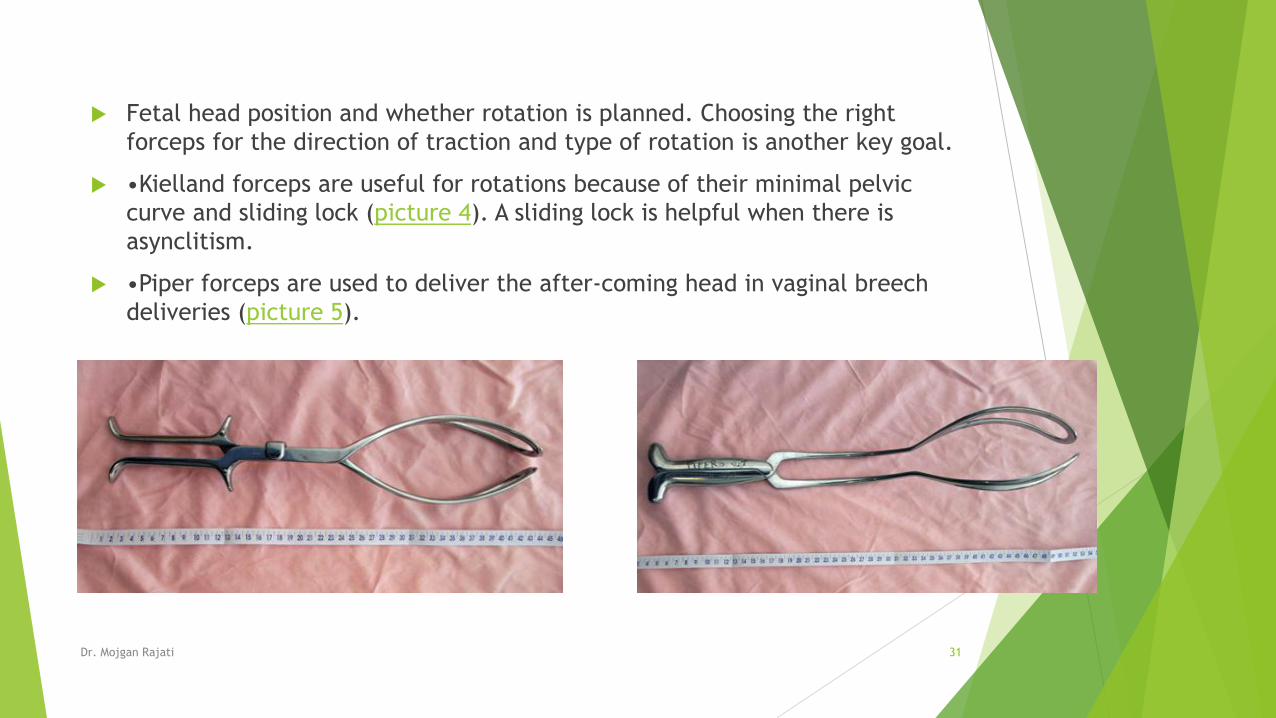
Fetal head position and whether rotation is planned. Choosing the right
forceps for the direction of traction and type of rotation is another key goal.
•Kielland forceps are useful for rotations because of their minimal pelvic
curve and sliding lock (picture 4). A sliding lock is helpful when there is
asynclitism.
•Piper forceps are used to deliver the after-coming head in vaginal breech
deliveries (picture 5).
31 Dr. Mojgan Rajati

Bill's axis traction handle
Irving forceps.
Traction is applied in the axis of the pelvis, which is curved in most women.
Operator experience and preference.
Application of forceps is more difficult, requires more manipulation for a good
application, and is more likely to result in maternal or fetal trauma with higher stations.
32 Dr. Mojgan Rajati
Midpelvic deliveries:

33 Dr. Mojgan Rajati
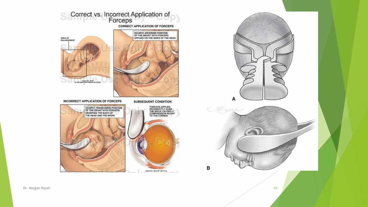
PROCEDURE
Application: Appropriately applied forceps grasp the occiput anterior
(OA) fetal head such that:
The long axis of the blades corresponds to the occipitomental
diameter (figure 2).
The tips of the blades lie over the cheeks (figure 3).
The blades are equidistant from the sagittal suture, which should
bisect a horizontal plane through the shanks.
The posterior fontanelle should be one finger breadth anterior to this
plane.
Fenestrated blades should admit no more than one finger breadth
between the heel of the fenestration and the fetal head.
No maternal tissue has been grasped.
34 Dr. Mojgan Rajati

PROCEDURE
Midforceps
Having higher rate of severe perinatal morbidity/mortality compared with
cesarean deliveries performed in the second stage.
They are also associated with higher rates maternal trauma.
Therefore Midforceps generally avoided.
35 Dr. Mojgan Rajati

PROCEDURE
Rotation – A rotational delivery is an appropriate option in select
clinical circumstances.
Rotation, when needed, is performed between contractions.
Rotation followed by extraction is more difficult and associated
with a higher risk of maternal and fetal complications than simple
traction applied to the non- or minimally-rotated head.
Traction – Traction should be steady (not rocking) and in the line
of the birth canal, rotating under the symphysis pubis, along the
curve of Carus (ie, pelvic axis).
The forceps pressure on the fetal head can be relaxed between
contractions to reduce fetal cranial compression.
Removal – To reduce the risk of laceration, forceps are
disarticulated and removed when expulsion is certain.
The head can then be delivered with no or minimal maternal
assistance.
36 Dr. Mojgan Rajati

When to abandon the procedure
Operative vaginal delivery should be abandoned:
if it is difficult to apply the instrument
Descent does not easily proceed with traction
The fetus has not been delivered within a reasonable time (within 15 to 20 min or
after three pulls)
The most common and highest risk clinical factors associated with failed
operative vaginal delivery are occiput posterior position, macrosomia, prolonged
second stage, primiparity, and maternal obesity.
Higher rates of neonatal morbidity have been observed when cesarean delivery
was performed after a failed operative vaginal delivery than when performed
during labor without such attempts.
37 Dr. Mojgan Rajati
SUCCESS RATE
failed delivery occurred in :
9 percent of forceps deliveries
14 percent of vacuum deliveries
Midforceps delivery is more likely to fail than low forceps delivery;
failure rates were 8.9 and 0.3 percent, respectively, in one large
prospective study.
Historically, failed forceps was more likely to lead to cesarean
delivery than failed vacuum since failed vacuum extraction was
sometimes followed by a successful trial of forceps, but the converse
rarely occurs. As sequential use of instruments carries much higher
morbidities, it is no longer considered acceptable to perform
sequential instrument use. 38 Dr. Mojgan Rajati

Morbidity
Maternal and fetal/neonatal complication rates vary
widely .
These factors include type of instrument, head position at
application, station, indication for intervention, and
operator experience. Rotation, higher station, longer
active second stage of labor, and operator inexperience
variably increase the risk of complications.
Infection
Laceration (3rd and 4th degree)
Episiotomy is done when it is indicated.
Laceration has higher rate OP versus OA
39 Dr. Mojgan Rajati

Pelvic floor disorders
Including: Urine incontinence, anal incontinence, pelvic organs prolapse
Mechanisms: Pelvic floor denervation, anatomic disorder
Risk factors: Parity special vaginal delivery
But there is not difference between rate of incontinency in spontaneous NVD
versus OVD.
40 Dr. Mojgan Rajati

Perinatal morbidity
Acute perinatal injuries are seen in OVD (both forceps and vacuum).
Facial palsies, other facial injuries, and depressed skull fractures are more
common with use of forceps than vacuum devices.
Cephalohemathoma, subgaleal hemorrhage, retinal hemorrhage, icter
shoulder dystocia, clavicular FX, scalp laceration in vacuum delivery are
more common.
In OVD the rate of fetal academia is not increased in compare with c/s.
The rate of intracranial hemorrhage is same in OVD and c/s.
41 Dr. Mojgan Rajati

Delivery with forceps
42 Dr. Mojgan Rajati
43 Dr. Mojgan Rajati
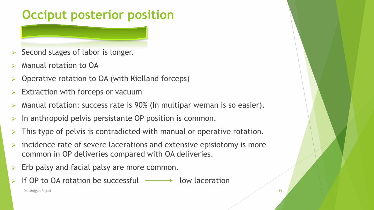
Occiput posterior position
Second stages of labor is longer.
Manual rotation to OA
Operative rotation to OA (with Kielland forceps)
Extraction with forceps or vacuum
Manual rotation: success rate is 90% (In multipar weman is so easier).
In anthropoid pelvis persistante OP position is common.
This type of pelvis is contradicted with manual or operative rotation.
incidence rate of severe lacerations and extensive episiotomy is more
common in OP deliveries compared with OA deliveries.
Erb palsy and facial palsy are more common.
If OP to OA rotation be successful low laceration
44 Dr. Mojgan Rajati

Thank you for Your attention
If you have any question, please send an E-mail to:
Dr. Mojgan Rajati 45


