37
11/16/2012 1 Chonticha Srivanitchapoom, M.D. Otolaryngology Department, Faculty of Medicine Nov 14 th ,2012

11/16/2012
1
Chonticha Srivanitchapoom, M.D.
Otolaryngology Department, Faculty of Medicine
Nov 14th ,2012
11/16/2012
2
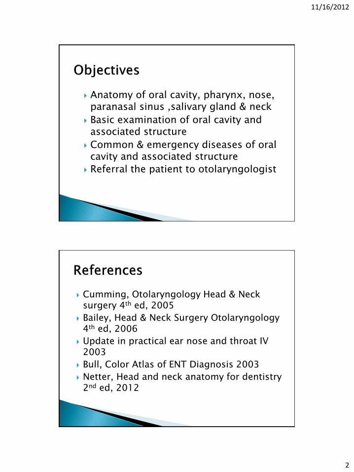
Anatomy of oral cavity, pharynx, nose, paranasal sinus ,salivary gland & neck
Basic examination of oral cavity and associated structure
Common & emergency diseases of oral cavity and associated structure
Referral the patient to otolaryngologist
Cumming, Otolaryngology Head & Neck surgery 4th ed, 2005
Bailey, Head & Neck Surgery Otolaryngology 4th ed, 2006
Update in practical ear nose and throat IV 2003
Bull, Color Atlas of ENT Diagnosis 2003
Netter, Head and neck anatomy for dentistry 2nd ed, 2012

11/16/2012
3
Examination of ENT tonsil & air sinus
◦ Antomy
◦ Basic examination & investigation
ENT tonsil & air sinus diseases related to dental
◦ Sinonasal diseases
◦ Tonsillar diseases
◦ Deep neck infection
◦ Salivary gland diseases
◦ Oral cavity carcinoma

11/16/2012
4

11/16/2012
5
nasopharynx
oropharynx
hypopharynx
epiglottis
False vocal cord
pyriform Arytenoid, corniculate, cuneiform cartilage
True vocal cord

11/16/2012
6
Nose Maxillary sinus

11/16/2012
7
Oral cavity Neck

11/16/2012
8
Anatomy : nose
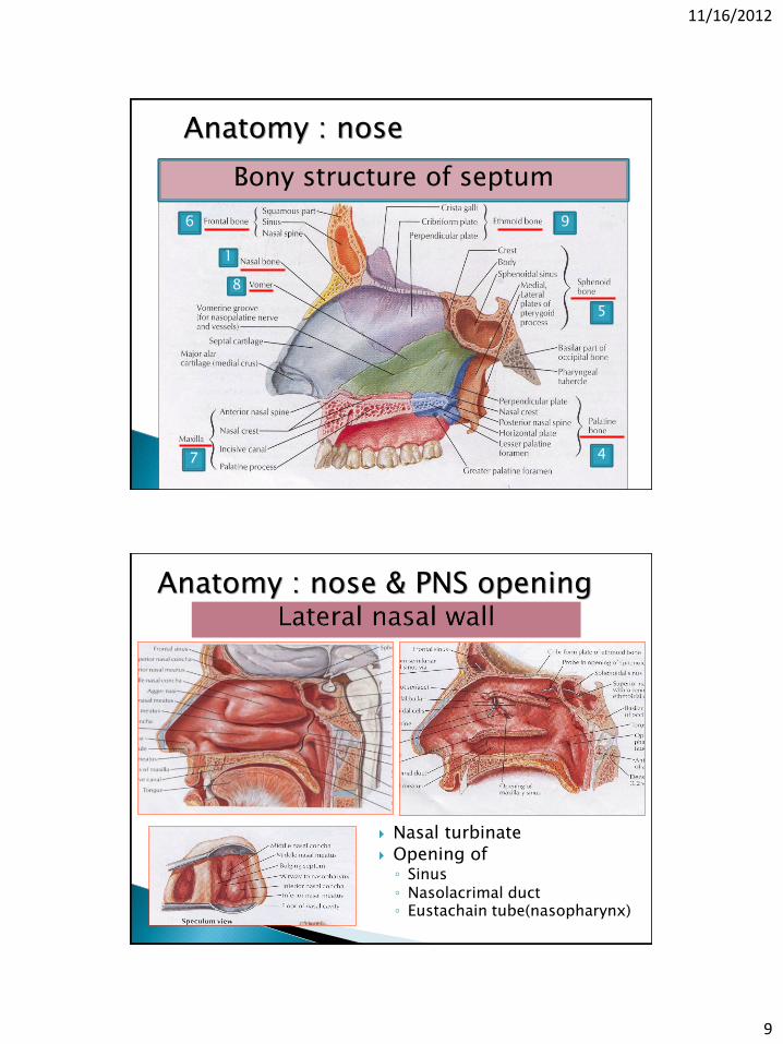
11/16/2012
9
Bony structure of septum
1
9
5
4 7
6
8
Anatomy : nose
Nasal turbinate
Opening of ◦ Sinus ◦ Nasolacrimal duct ◦ Eustachain tube(nasopharynx)
Anatomy : nose & PNS opening

11/16/2012
10
Bony structure of lateral wall
1. Nasal bone
2. Lacrimal bone
3. Inferior concha
4. Palatine bone
5. Sphenoid bone
6. Frontal bone
7. Maxillary bone
8. Vomer
9. Perpendicular plate of ethmoid
3
9
2
1
6
5
4 7
Anatomy : nose
Anatomy : nose

11/16/2012
11
1.External carotid a.system 1.1 maxillary a.
-sphenopalatine a. -descending palatine a. -infraorbital a.
1.2 facial a. -sup. labial a. 2. Internal carotid a.system 2.1 opthalmic a.
-post ethmoidal artery -ant ethmoidal artery -dorsal nasal artery
Anatomy : nose
Nerve supply Anatomy : nose
Sensory innervation => Olfactory nerve (olfaction), Ophthalmic & maxillary division of CN V (general
sensation) Autonomic innervation=> through the sensory br of
maxillary division of CN V

11/16/2012
12
Anatomy : paranasal sinus
Anatomy : PNS

11/16/2012
13
Anatomy : PNS (lateral view)
Anatomy : PNS

11/16/2012
14
Anatomy : PNS (axial view)
Anatomy : PNS

11/16/2012
15
Anatomy : PNS & lateral wall of nose
Maxillary sinus opening

11/16/2012
16

11/16/2012
17
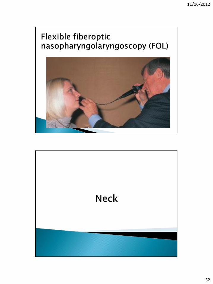
Rigid endoscopy
Imaging
◦ Plain film (Caldwell’s view, Waters’ view, submentovertex view & lateral view)
◦ CT scan
◦ MRI

11/16/2012
18

11/16/2012
19

11/16/2012
20
1. Lip 2. Alveolar ridge 3. Hard palate 4. Retromolar
trigone 5. Buccal mucosa 6. Oral tongue 7. Floor of mouth
Branches of external carotid arteries
Vascular supply

11/16/2012
21
Branch of
• Glossopharyngeal nerve ( CN IX)
• Trigeminal nerve (CN V)
• Facial nerve (CN VII)
Nerve supply
Muscle of mastication
Muscle of tongue : extrinsic & intrinsic muscle of tongue
Muscle of soft palate group
Muscular part

11/16/2012
22
Taste zone and taste bud
Parotid gland Stensen’s duct
opening at upper 2nd molar
Main of serous secretion

11/16/2012
23
Submandibular gland
Wharton’s duct opening
Mixed of serous & mucous secretion
Largest duct (duct of
Batholin) & small duct (duct
of Rivinus) ,some drain into submandibular gland, some opening in elevated crest of mucous membrane (plica sublingualis)
Main of mucous secretion
Sublingual gland

11/16/2012
24
• Salivary gland and opening
Submucosal of upper aerodogestive tract
Oral cavity : hard palate, buccal, lip, pharyngeal wall

11/16/2012
25

11/16/2012
26
Plain film
◦ Bony lesion
◦ Calcification : sialolithiasis
Sialography

11/16/2012
27

11/16/2012
28
Tonsil / Base of tongue / Soft palate/ Oropharyngeal wall

11/16/2012
29
◦ A pair of tubal tonsils
◦ A pair of palatine tonsils
◦ A single lingual tonsil
◦ A single pharyngeal tonsil
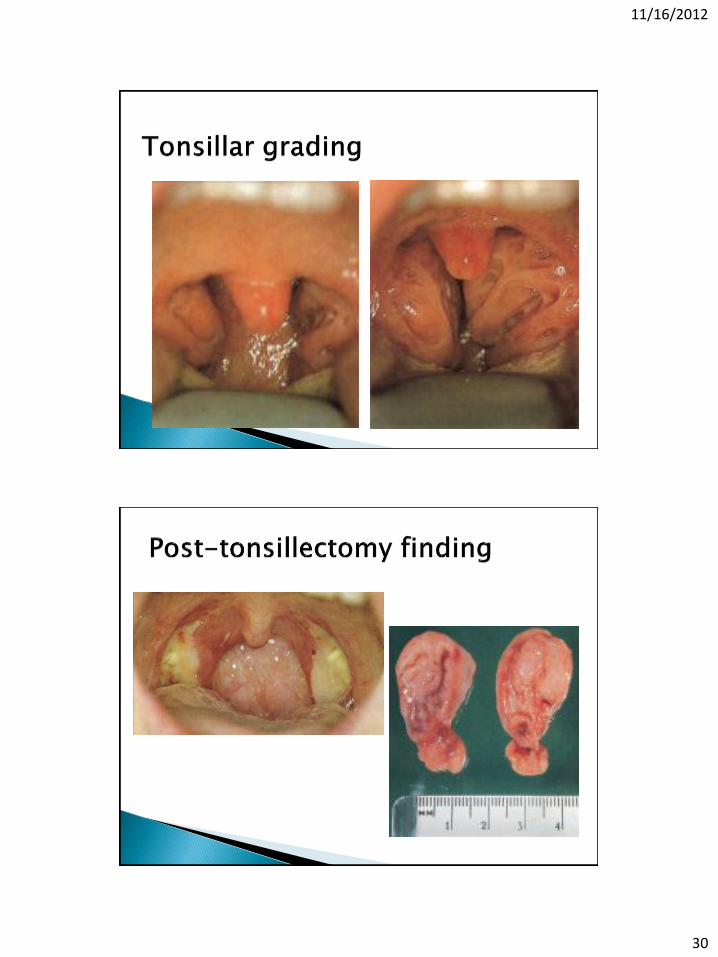
11/16/2012
30

11/16/2012
31
11/16/2012
32

11/16/2012
33
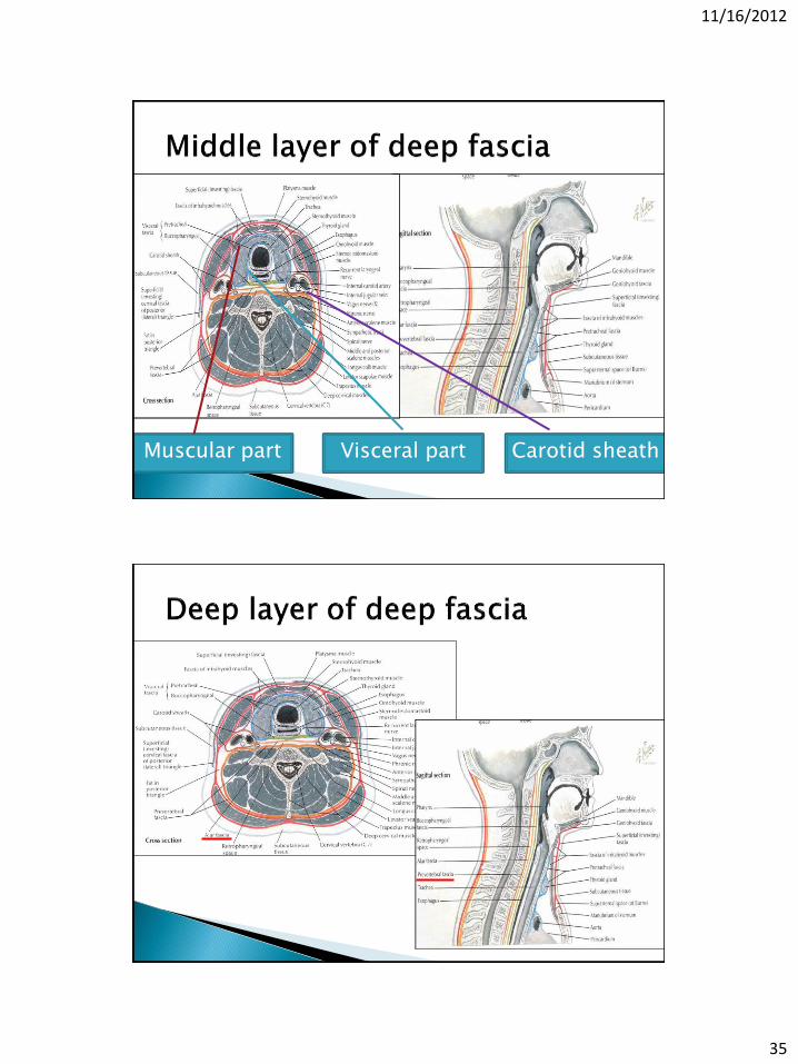
Superficial cervical fascia Deep cervical fascia ◦ Superficial Lower : sternum, clavicle, scapular, cervical
spine Upper : lower border of mandible, zygomatic
arch, mastoid, superior nuchal line ◦ Middle Middle cervical fascia “ muscular part” Cervical visceral fascia “visceral part” Carotid sheath “3 layers of deep cervical fascia”
◦ Deep Prevertebral fascial Alar fascia
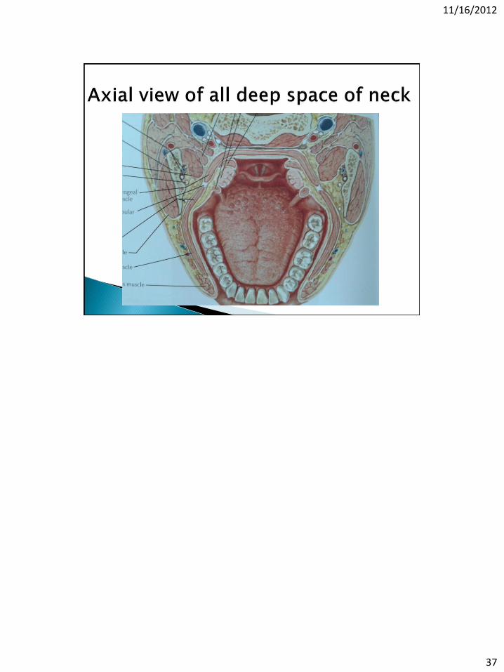
Deep space of neck
(potential space)

11/16/2012
34
Fibrous connective tissue connecting from ◦ Lower : shoulder,
chest wall, axillar ◦ Upper : lower border
of mandible Content in this layer :
platysma muscle, subcutaneous fat, external jugular vein, superficial cervical lymph node
Lower : sternum, clavicle, scapular, cervical spine
Upper : lower border of mandible, zygomatic arch, mastoid, superior nuchal line
11/16/2012
35
Muscular part Visceral part Carotid sheath

11/16/2012
36
11/16/2012
37


