74
Lecture Presentation by Steven Bassett Southeast Community College Chapter 5 The Skeletal System Osseous Tissue and Skeletal Structure © 2015 Pearson Education, Inc.

Lecture Presentation by
Steven Bassett
Southeast Community College
Chapter 5
The Skeletal
System
Osseous Tissue and
Skeletal Structure
© 2015 Pearson Education, Inc.

Introduction
• The skeletal system is made of:
• Skeletal bones
• Cartilage
• Ligaments
• Connective tissue to stabilize the skeleton
• Bones are dynamic organs, which consist of
several tissue types
© 2015 Pearson Education, Inc.

Introduction
• Functions of the Skeletal System
• Support
• Provides the framework for the attachment of other
organs
• Storage of minerals
• Calcium ions: 98 percent of the body’s calcium ions
are in the bones
• Phosphate ions
• Blood cell production
• Bone marrow produces erythrocytes, leukocytes,
and platelets
© 2015 Pearson Education, Inc.
Introduction
• Functions of the Skeletal System (continued)
• Leverage
• Muscles pull on the bones to produce movement
• Protection
• Ribs protect heart and lungs
• Skull protects the brain
• Vertebrae protect the spinal cord
• Pelvic bones protect the reproductive organs
© 2015 Pearson Education, Inc.

Structure and Function of Bone
• Bones (Osseous Tissue)
• Supporting connective tissue
• Specialized cells
• Solid matrix
• Outer lining
• Called the periosteum
• Inner lining
• Called the endosteum
© 2015 Pearson Education, Inc.

Structure and Function of Bone
• The Histological Organization of Mature Bone
• The matrix of bone
• Calcium phosphate eventually converts to
hydroxyapatite crystals
• Calcium phosphate makes up 2/3 of the bone mass
• Hydroxyapatite crystals resist compression
© 2015 Pearson Education, Inc.

Structure and Function of Bone
• The Histological Organization of Mature Bone
• Collagen fibers
• Make up 1/3 of the bone matrix
• Contribute to the tensile strength of bones
• Collagen and hydroxyapatite make bone tissue
extremely strong
• Bone cells
• Contribute only 2 percent of the bone mass
© 2015 Pearson Education, Inc.

Structure and Function of Bone
• The Cells of Mature Bone
• Osteocytes
• Mature bone cells
• Maintain the protein and mineral content of the
matrix
• Cause the release of calcium ions from the bone to
the blood
• Sit in depressions called lacunae
• Matrix layer associated with osteocytes is lamellae
• Small channels extending from the osteocytes to
the bone capillaries are called canaliculi
© 2015 Pearson Education, Inc.

Structure and Function of Bone
• The Cells of Mature Bone
• Osteoblasts
• Immature bone cells
• Found on the inner and outer surfaces of bones
• Produce osteoid, which is involved in making the
matrix
• Osteoblasts are involved in making new bone. This
is a process called osteogenesis
© 2015 Pearson Education, Inc.

Structure and Function of Bone
• The Cells of Mature Bone (continued)
• Osteoprogenitor cells
• These are bone stem cells
• Found on the innermost layer of the periosteum
and the inner lining of the endosteum
• Differentiate to form new osteoblasts
• Heavily involved in the repair of bones after a break
© 2015 Pearson Education, Inc.

Structure and Function of Bone
• The Cells of Mature Bone (continued)
• Osteoclasts
• Multinucleated cells
• Secrete acids, which dissolve the bones thereby
causing the release of stored calcium ions and
phosphate ions into the blood
• This process is called osteolysis
© 2015 Pearson Education, Inc.

Figure 5.1 Histological Structure of a Typical Bone
© 2015 Pearson Education, Inc.
Canaliculi
a
c d b
Osteocyte Matrix
Osteoblast Osteoid Matrix
Osteon
Osteocyte: Mature bone cell
that maintains the bone matrix
Osteoblast: Immature bone
cell that secretes organic
components of matrix
Lacunae
Central
canals
Lamellae
Osteons SEM × 182 Osteons
The cells of bone.
A scanning electron micrograph of
several osteons in compact bone. A thin section through compact
bone; in this procedure the intact
matrix and central canals appear
white, and the lacunae and canaliculi
are shown in black.
LM × 220 LM × 343 Osteon
A single osteon at higher
magnification.
Osteoclast: Multinucleate cell
that secretes acids and enzymes
to dissolve bone matrix.
Osteoprogenitor cell: Stem cell
whose divisions produce osteoblasts
Matrix
Endosteum Osteoprogenitor cell
Osteoclast
Medullary
cavity
Medullary
cavity
Canaliculi
Concentric
lamellae
Central canals
Osteon
Lacunae
Structure and Function of Bone
• Two Types of Osseous Tissue
• Compact bone (dense bone)
• Compact bones are dense and solid
• Forms the walls of bone
• Spongy bone (trabecular, or cancellous, bone)
• Open network of plates
• Surrounds the medullary cavity
• Medullary cavity consists of bone marrow
© 2015 Pearson Education, Inc.

Structure and Function of Bone
• Compact Bone (details)
• Consists of osteons
• Basic functional unit of bone
• Consists of:
• Central canal
• Canaliculi
• Osteocytes
• Lacunae
• Lamellae
© 2015 Pearson Education, Inc.

Figure 5.2a-c The Internal Organization in Representative Bones
© 2015 Pearson Education, Inc.
Spongy bone
a Concentric
lamellae
b
c
Gross anatomy of
the humerus.
The organization of collagen fibers
within concentric lamellae.
Diagrammatic view of the
histological organization of
compact and spongy bone.
Blood vessels
Compact bone
Medullary cavity
Endosteum
Periosteum
Compact
bone
Spongy
bone Medullary
cavity
Collagen fiber
orientation
Concentric
lamellae
Central
canal
Endosteum
Interstitial
lamellae
Capillary Small vein Circumferential
lamellae
Osteons
Periosteum
Artery Vein
Central
canal Perforating
canal
Trabeculae of
spongy bone

Figure 5.2a The Internal Organization in Representative Bones
© 2015 Pearson Education, Inc.
Spongy bone
a
Blood vessels
Compact bone
Medullary cavity
Endosteum
Periosteum
Compact
bone
Spongy
bone Medullary
cavity
Gross anatomy of
the humerus.

Figure 5.2c The Internal Organization in Representative Bones
© 2015 Pearson Education, Inc.
The organization of collagen fibers
within concentric lamellae.
Collagen fiber orientation
Concentric lamellae
Central canal
Endosteum
c

Figure 5.2b The Internal Organization in Representative Bones
© 2015 Pearson Education, Inc.
Concentric
lamellae
b Diagrammatic view of the
histological organization of
compact and spongy bone.
Interstitial
lamellae
Capillary Small vein Circumferential
lamellae
Osteons
Periosteum
Artery Vein
Central
canal
Perforating
canal
Trabeculae of
spongy bone
Structure and Function of Bone
• Spongy Bone (details)
• Spongy bone
• Arranged in parallel struts
• Forms branching plates called trabeculae
• Trabeculae form an open network
• Creates the lightweight nature of bones
© 2015 Pearson Education, Inc.

Figure 5.2d The Internal Organization in Representative Bones
© 2015 Pearson Education, Inc.
Location and structure of spongy bone. The
photo shows a sectional view of the proximal
end of the femur.
Trabeculae of
spongy bone
Endosteum
Lamellae
Canaliculi opening
on surface
d

Structure and Function of Bone
• Functional Differences between Compact and
Spongy Bone
• Compact bone
• Conducts stress from one end of the long bone to
the other end of the long bone
• Generates tremendous strength from end to end
• Weak strength when stress is applied to the side
• Osteon arrangement is parallel to the bone axis
• Generates tremendous strength from end to end
© 2015 Pearson Education, Inc.

Structure and Function of Bone
• Functional Differences between Compact and
Spongy Bone
• Spongy bone
• Trabeculae create strength to deal with stress from
the side
• Trabeculae are oriented along the stress lines
• Has extensive cross-bracing
• Also supports yellow marrow in the shaft of the
bone
• Also supports red marrow in the epiphysis of the
bone
© 2015 Pearson Education, Inc.

Figure 5.3a Anatomy of a Representative Bone
© 2015 Pearson Education, Inc.
Articular
surface of
head of femur
a
Epiphysis
Metaphysis
Diaphysis
(shaft)
Metaphysis
Epiphysis
Spongy bone
Compact bone
Medullary
cavity
Posterior view Sectional view
The femur, or thigh bone, in posterior and sectional views. The femur
has a diaphysis (shaft) with walls of compact bone and epiphyses
(ends) filled with spongy bone. A metaphysis separates the diaphysis
and epiphysis at each end of the shaft. The body weight is transferred
to the femur at the hip joint. Because the hip joint is off center relative
to the axis of the shaft, the body weight is distributed along the bone
so that the medial portion of the shaft is compressed and the lateral
portion is stretched.

Figure 5.3b Anatomy of a Representative Bone
© 2015 Pearson Education, Inc.
b An intact femur chemically
cleared to show the orientation of
the trabeculae in the epiphysis.

Figure 5.3c Anatomy of a Representative Bone
© 2015 Pearson Education, Inc.
c
Articular
surface of
head of
femur
Trabeculae of
spongy bone
Cortex
Medullary cavity
Compact bone
A photograph showing the
epiphysis after sectioning.

Structure and Function of Bone
• Functional Differences between Compact and
Spongy Bone
• Epiphysis (ends of the long bones)
• Consists of red marrow
• Diaphysis (shaft of the long bones)
• The medullary cavity of the diaphysis consists of
yellow marrow
• Metaphysis
• Narrow growth zone between the epiphysis and the
diaphysis
© 2015 Pearson Education, Inc.

Structure and Function of Bone
• The Periosteum and Endosteum
• Periosteum
• Outer surface of the bone
• Isolates and protects the bone from surrounding
tissue
• Provides a route and a place for attachment for
circulatory and nervous supply
• Actively participates in bone growth and repair
• Attaches the bone to the connective tissue network
of the deep fascia
© 2015 Pearson Education, Inc.
Structure and Function of Bone
• The Periosteum and Endosteum
• Endosteum
• Inner surface of bone
• Lines the medullary cavity
• Consists of osteoprogenitor cells
• Actively involved in repair and growth
© 2015 Pearson Education, Inc.

Figure 5.4a Anatomy and Histology of the Periosteum and Endosteum
© 2015 Pearson Education, Inc.
a The periosteum contains outer (fibrous) and
inner (osteogenic) layers. Collagen fibers of the
periosteum are continuous with those of the
bone, adjacent joint capsules, and attached
tendons and ligaments.
Joint capsule
Osteogenic layer
of periosteum
Fibrous layer
of periosteum
Endosteum Compact bone
Circumferential
lamellae
Periosteum
Fibrous layer
Osteogenic layer
Canaliculi
Lacuna
Osteocyte
Perforating
fibers

Bone Development and Growth
• Before six weeks of development, the skeleton is
hyaline cartilage
• Cartilage cells will be replaced by bone cells
• This is called ossification
• Osteogenesis
• Bone formation
• Calcification
• The deposition of calcium ions into the bone tissue
© 2015 Pearson Education, Inc.

Bone Development and Growth
• There are two types of ossification
• Intramembranous ossification
• Involved in the development of clavicle, mandible,
skull, and face
• Endochondral ossification
• Involved in the development of limbs, vertebrae,
and hips
© 2015 Pearson Education, Inc.

Bone Development and Growth
• Intramembranous Ossification
• Mesenchymal cells differentiate to form
osteoblasts
• Osteoblasts begin secreting a matrix
• Osteoblasts become trapped in the matrix
• Osteoblasts differentiate and form osteocytes
• More osteoblasts are produced, thus move outward
• Blood vessels are trapped by the formation of
spicules
© 2015 Pearson Education, Inc.

Figure 5.6.1 Intramembranous Ossification
© 2015 Pearson Education, Inc.
Bone matrix
Mesenchymal tissue becomes
highly vascularized, and the mesen-
chymal cells aggregate, enlarge, and
then differentiate into osteoblasts.
The osteoblasts then cluster
together and start to secrete the
organic components of the matrix.
The resulting osteoid then
becomes mineralized through the
crystallization of calcium salts. The
location where ossification begins is
called an ossification center.
Differentiation of Osteoblasts within Mesenchyme 1
Osteoid
Mesenchymal cell
Ossification center
Blood vessel
Osteoblast
Mandible
Occipital bone
Frontal bone
Parietal bone

Figure 5.6.2 Intramembranous Ossification
© 2015 Pearson Education, Inc.
Spicules
Lacuna
Osteocyte
2
As ossification proceeds, osteoblasts
that become surrounded by osteoid
differentiate into osteocytes. These
cells will remain trapped within tiny
spaces known as lacunae (singular:
lacuna). The developing bone grows
outward from the ossification center into
small struts called spicules. Although
osteoblasts are still being trapped in
the expanding bone, mesenchymal
cell divisions continue to produce
additional osteoblasts.
Formation of Bony Spicules
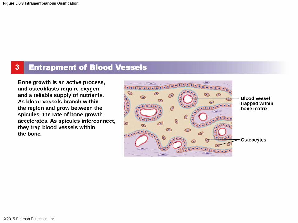
Figure 5.6.3 Intramembranous Ossification
© 2015 Pearson Education, Inc.
3
Bone growth is an active process,
and osteoblasts require oxygen
and a reliable supply of nutrients.
As blood vessels branch within
the region and grow between the
spicules, the rate of bone growth
accelerates. As spicules interconnect,
they trap blood vessels within
the bone.
Entrapment of Blood Vessels
Osteocytes
Blood vessel trapped within bone matrix
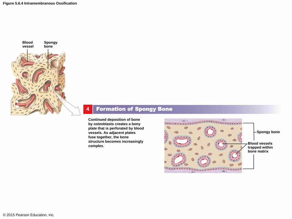
Figure 5.6.4 Intramembranous Ossification
© 2015 Pearson Education, Inc.
4
Continued deposition of bone
by osteoblasts creates a bony
plate that is perforated by blood
vessels. As adjacent plates
fuse together, the bone
structure becomes increasingly
complex.
Formation of Spongy Bone
Blood vessels trapped within bone matrix
Spongy bone
Spongy bone
Blood vessel
Bone Development and Growth
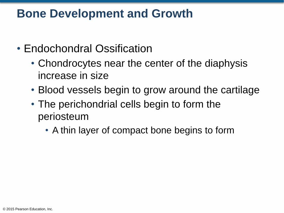
• Endochondral Ossification
• Chondrocytes near the center of the diaphysis
increase in size
• Blood vessels begin to grow around the cartilage
• The perichondrial cells begin to form the
periosteum
• A thin layer of compact bone begins to form
© 2015 Pearson Education, Inc.

Bone Development and Growth
• Endochondral Ossification
• Cartilage cells die and are replaced by osteoblasts
• Osteoblasts begin to form spongy bone
• This is the primary ossification center
• The cartilage in the metaphysis region is invaded
by osteoblasts
• An increase in bone length and diameter begins
© 2015 Pearson Education, Inc.

Figure 5.7.1 Endochondral Ossification (2 of 3)
© 2015 Pearson Education, Inc.
Spongy bone
4 3 2 1
Superficial bone
Primary
ossification center
Medullary cavity
Perichondrium
Bone collar
Blood vessel
Periosteum formed from
perichondrium
Enlarging chondrocytes within calcifying matrix
Hyaline cartilage
Disintegrating chondrocytes
Medullary cavity
See Figure 5.9
Metaphysis
As the cartilage enlarges,
chondrocytes near the
center of the shaft
increase greatly in size,
and the surrounding
matrix begins to calcify.
Deprived of nutrients,
these chondrocytes die
and disintegrate, leaving
cavities within the
cartilage.
Blood vessels grow around the
edges of the cartilage, and the
cells of the perichondrium
begin differentiating
into osteoblasts. The
perichondrium has now been
converted into a periosteum,
and the inner osteogenic
(os-te-o-JEN-ik) layer soon
produces a bone collar, a thin
layer of compact bone around
the shaft of the cartilage.
¯ ¯
While these changes are under
way, the blood supply to the
periosteum increases, and
capillaries and osteoblasts
migrate into the heart of the
cartilage, invading the spaces
left by the disintegrating
chondrocytes. The calcified
cartilaginous matrix then breaks
down, and osteoblasts replace
it with spongy bone. Bone
development proceeds from this primary ossification center
in the shaft, toward both ends of
the cartilaginous model.
While the diameter is small, the entire
shaft is filled with spongy bone, but as it
enlarges, osteoclasts erode the central
portion and create a medullary cavity.
The bone of the shaft becomes thicker,
and the cartilage of the metaphysis is
invaded by osteoblasts that produce
columns of bone. Further growth
involves two distinct processes: an
increase in length and an enlargement in diameter (Figure 5.9).

Bone Development and Growth
• Endochondral Ossification
• Osteoblasts begin to migrate into the epiphysis
region
• This is the secondary ossification center
• Osteoblasts begin to replace cartilage with bone
• This results in pushing the epiphysis away from
the diaphysis thus resulting in longer bones
© 2015 Pearson Education, Inc.

Figure 5.7.1 Endochondral Ossification (3 of 3)
© 2015 Pearson Education, Inc.
5
Diaphysis
Capillaries and osteoblasts then migrate
into the centers of the epiphyses, creating
secondary ossification centers.
The time of appearance of secondary
ossification centers varies from one bone
to another and from individual to individual.
Secondary ossification centers may be
present at birth in both ends of the humerus
(arm), femur (thigh), and tibia (leg), but the
epiphyses of some other bones remain
cartilaginous through childhood.
6 7
Hyaline cartilage Secondary ossification center
Epiphysis
Metaphysis
Periosteum
Compact bone
Spongy bone
Epiphyseal cartilage
Within the epiphyseal
cartilage, the chondrocytes
are organized into zones.
Chondrocytes at the
epiphyseal side of the
cartilage continue to divide
and enlarge.
Chondrocytes degenerate
at the diaphyseal side.
Osteoblasts migrate
upward from the diaphysis,
and the degenerating
cartilage is gradually
replaced by bone.
Secondary ossification
center
Spongy bone
Epiphyseal line Articular cartilage
Medullary cavity
The epiphyses eventually become filled with spongy
bone. The epiphysis and diaphysis are now separated
by a narrow epiphyseal cartilage, or epiphyseal
plate, within the metaphysis. Osteoblasts invade the
shaft side of the epiphyseal cartilage, replacing the
cartilage with bone, at the same rate that the
epiphyseal cartilage enlarges through the interstitial
growth. This enlargement pushes the epiphysis away
from the diaphysis, and the length of the bone
Increases.
At maturity, the rate of epiphyseal cartilage
enlargement slows and the rate of
osteoblast activity accelerates. As a result,
the epiphyseal cartilage gets narrower and
narrower, until it ultimately disappears. This
event is called epiphyseal closure. The
former location of the epiphyseal cartilage
becomes a distinct epiphyseal line that
remains after epiphyseal growth has ended.
A thin cap of the original cartilage model
remains exposed to the joint cavity as
the articular cartilage. This cartilage
prevents damaging bone-to-bone
contact within the joint.

Bone Development and Growth
• Increasing the Diameter of a Developing Bone
• Appositional growth
• Inner layer of the periosteum differentiates to form
osteoblasts and adds bone matrix to the surface
• This forms circumferential lamellae to the outer
surface
• Osteons form
• Bone continues to enlarge in diameter
© 2015 Pearson Education, Inc.

Figure 5.9 Appositional Bone Growth
© 2015 Pearson Education, Inc.
Ridge
1 2 3
4 5 6
Artery
Periosteum Perforating canal
Circumferential lamellae
Periosteum Central canal of new osteon
Infant Child
Bone resorbed
by osteoclasts
Bone
deposited by
osteoblasts Young adult Adult
Bone formation at the surface
of the bone produces ridges
that parallel a blood vessel.
The ridges enlarge and create
a deep pocket.
The ridges meet and fuse, trapping
the vessel inside the bone.
Bone deposition proceeds inward
toward the vessel, beginning the
creation of a typical osteon.
Additional circumferential lamellae
are deposited and the bone
continues to increase in diameter.
Osteon is complete with new central
canal around the blood vessel. Second
blood vessel becomes enclosed.
Three-dimensional diagrams illustrate the mechanism
responsible for increasing the diameter of a growing bone.
A bone grows in diameter as new bone is added to the outer surface. At the same
time, osteoclasts resorb bone on the inside, enlarging the medullary cavity.
a
b

Bone Development and Growth
• Epiphyseal Plate
• Area of cartilage in the metaphysis
• Cartilage near the diaphysis is converted to bone
• The width of this zone gets narrower as we age
• Marks the former location of the epiphyseal
cartilage
© 2015 Pearson Education, Inc.

Figure 5.8 Epiphyseal Cartilages and Lines
© 2015 Pearson Education, Inc.
X-ray of the hand of a young
child. The arrows indicate
the locations of the
epiphyseal cartilages.
a b X-ray of the hand of an
adult. The arrows indicate
the locations of epiphyseal
lines.
Bone Development and Growth
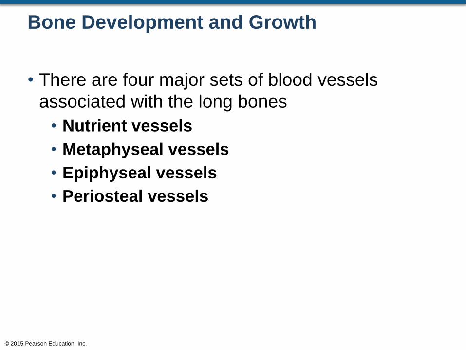
• There are four major sets of blood vessels
associated with the long bones
• Nutrient vessels
• Metaphyseal vessels
• Epiphyseal vessels
• Periosteal vessels
© 2015 Pearson Education, Inc.

Bone Development and Growth
• Nutrient Vessels
• Enter the diaphysis and branch toward the
epiphysis
• Enter through the nutrient foramen of the bone
• Penetrates the shaft and enters the medullary
cavity
• Divides into ascending and descending branches
to go toward the epiphysis regions
• Vessels branch to form perforating vessels
© 2015 Pearson Education, Inc.
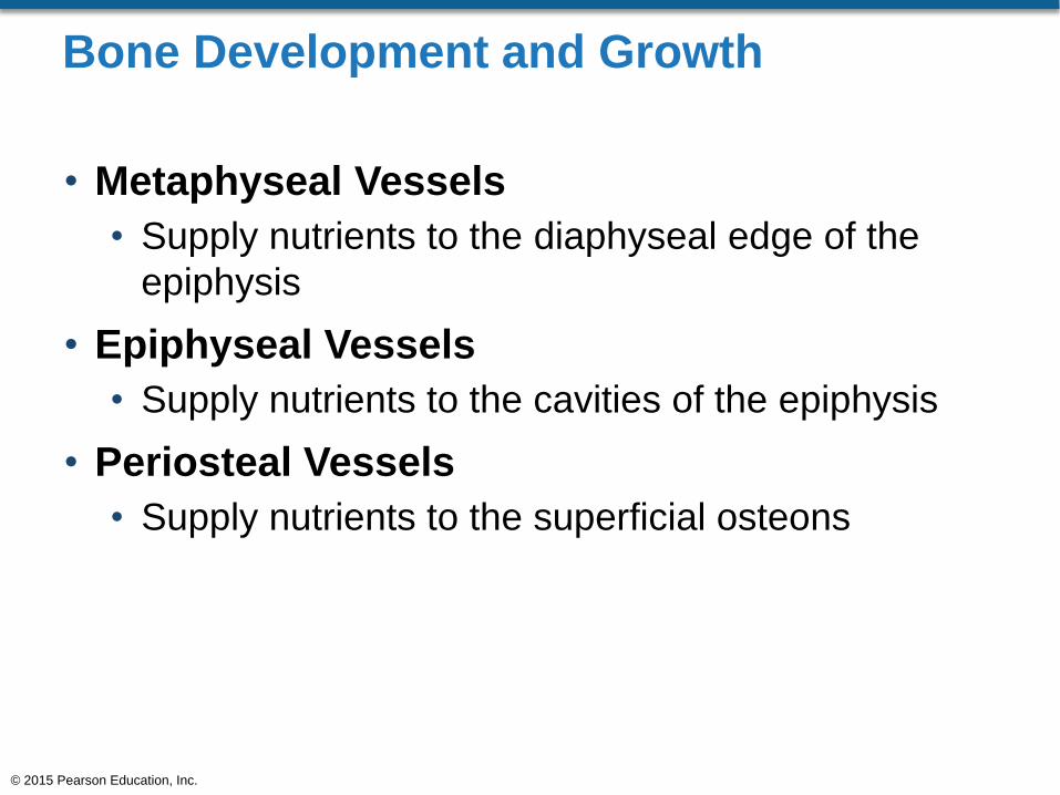
Bone Development and Growth
• Metaphyseal Vessels
• Supply nutrients to the diaphyseal edge of the
epiphysis
• Epiphyseal Vessels
• Supply nutrients to the cavities of the epiphysis
• Periosteal Vessels
• Supply nutrients to the superficial osteons
© 2015 Pearson Education, Inc.

Figure 5.10 Circulatory Supply to a Mature Bone
© 2015 Pearson Education, Inc.
Metaphysis
Epiphyseal
artery and vein
Metaphyseal
artery and
vein
Nutrient artery
and vein
Periosteal
arteries and veins
Periosteum
Connections to
superficial osteons
Metaphyseal
artery and vein
Articular
cartilage
Periosteum
Compact
bone
Medullary
cavity
Nutrient
foramen
Branches of
nutrient artery
and vein
Epiphyseal
line
Vessels in Bone

Bone Development and Growth
• Bone Innervation
• Nerves penetrate the bone with the nutrient artery
• Innervates throughout the periosteum
• Innervates the endosteum
• Innervates the medullary cavity
• Innervates the epiphysis
© 2015 Pearson Education, Inc.

Bone Development and Growth
• Factors Regulating Bone Growth
• Nutrition
• Calcium ions
• Phosphate ions
• Magnesium ions
• Citrate
• Carbonate ions
• Sodium ions
• Vitamins A, C, D (calcitriol)
© 2015 Pearson Education, Inc.

Bone Development and Growth
• Factors Regulating Bone Growth (continued)
• Hormones
• Parathyroid gland releases parathyroid hormone
• Stimulates osteoclasts
• Stimulates osteoblasts
• Increases calcium ion absorption from the small
intestine to the blood
• Reduces the rate of calcium ion loss from the
kidneys
© 2015 Pearson Education, Inc.

Bone Development and Growth
• Factors Regulating Bone Growth (continued)
• Hormones
• Thyroid gland releases calcitonin
• Inhibits osteoclasts
• Increases rate of calcium ion loss in the urine
• Removes calcium ions from blood and adds it to
bone
• Thyroid gland releases thyroxine
• Maintains normal activity in the epiphyseal region
© 2015 Pearson Education, Inc.

Bone Development and Growth
• Factors Regulating Bone Growth (continued)
• Hormones
• Pituitary gland releases growth hormone
(somatotropin)
• Stimulates bone growth
• Maintains normal activity of the epiphyseal
cartilage
© 2015 Pearson Education, Inc.

Bone Development and Growth
• Factors Regulating Bone Growth (continued)
• Hormones
• Estrogen and testosterone stimulate osteoblast
activity
• Osteoblast activity produces bone faster than
epiphyseal cartilage expansion
• Ultimately the epiphyseal cartilage narrows and
bone growth ceases (about age 25)
© 2015 Pearson Education, Inc.

Bone Maintenance, Remodeling, and Repair
• Remodeling of Bone
• Realignment of teeth can change the shape of
tooth sockets
• Increased muscular development
• Bone changes occur due to stress
• Different features develop on the bone
• Attachment of ligaments
• Attachment of tendons
• Stressed bones become thicker and stronger
© 2015 Pearson Education, Inc.

Bone Maintenance, Remodeling, and Repair
• Remodeling of Bone
• Inactivity of bones can cause degeneration
• After a few weeks, unstressed bones can lose
about a third of their mass
© 2015 Pearson Education, Inc.

Bone Maintenance, Remodeling, and Repair
• Injury and Repair
• Fractures
• Transverse fractures
• Break transverse to the long axis
• Displaced fractures
• Produces new and abnormal bone arrangements
• Compression fractures
• Bones “jam” together
• Spiral fractures
• Bones twist along the length of the bone
© 2015 Pearson Education, Inc.

Clinical Note 5.2 Fractures and Their Repair (1 of 4)
© 2015 Pearson Education, Inc.
Tra
nsvers
e f
ractu
re
Compression fracture

Bone Maintenance, Remodeling, and Repair
• Injury and Repair
• Fractures
• Epiphyseal fractures
• Fractures within the epiphyseal region
• Comminuted fractures
• The fractured area shatters into many bony
fragments
• Greenstick fractures
• Only one edge of the bone breaks while the other
edge bends
© 2015 Pearson Education, Inc.

Clinical Note 5.2 Fractures and Their Repair (2 of 4)
© 2015 Pearson Education, Inc.
Epiphyseal fracture
Colles fracture

Bone Maintenance, Remodeling, and Repair
• Injury and Repair
• Fractures
• Colles fracture
• A break at the distal portion of the radius
• Pott fracture
• Occurs at the ankles and affects both the tibia and
the fibula
© 2015 Pearson Education, Inc.

Bone Maintenance, Remodeling, and Repair
• Injury and Repair
• When a bone is broken, bleeding occurs
• A network of spongy bone forms
• Osteoblasts are overly activated, thus resulting in
enlarged callused area
• This area is now stronger and thicker than normal
bone
© 2015 Pearson Education, Inc.

Clinical Note 5.2 Fractures and Their Repair (3 of 4)
© 2015 Pearson Education, Inc.
Fracture
hematoma
Dead
bone
Bone
fragments
Spongy bone of
external callus
Periosteum
Repair
of a
fracture
Immediately after the fracture,
extensive bleeding occurs.
Over a period of several hours, a
large blood clot, or fracture hema-
toma, develops.
An internal callus forms as a
network of spongy bone unites
the inner edges, and an external
callus of cartilage and bone stabilizes
the outer edges.
1 2

Clinical Note 5.2 Fractures and Their Repair (4 of 4)
© 2015 Pearson Education, Inc.
Internal
callus
The cartilage of the external
callus has been replaced by bone,
and struts of spongy bone now unite
the broken ends. Fragments of dead
bone and the areas of bone closest to
the break have been removed and
replaced.
3
External
callus
External
callus
4 A swelling initially marks
the location of the fracture.
Over time, this region will be
remodeled, and little evidence of
the fracture will remain.

Bone Maintenance, Remodeling, and Repair
• Aging and the Skeletal System
• When we’re young, osteoblast activity balances
with osteoclast activity
• When we get older, osteoblast activity declines
• Osteoclast activity maintains its previous level of
activity
• When osteoclast activity is faster than osteoblast
activity, bones become porous
© 2015 Pearson Education, Inc.

Bone Maintenance, Remodeling, and Repair
• Aging and the Skeletal System
• As women age, estrogen levels drop
• Osteoclast control is lost
• Osteoclasts are overactive
• Bones become porous
• This is osteoporosis
© 2015 Pearson Education, Inc.

Clinical Note 5.3 Osteoporosis and Age-Related Skeletal Abnormalities
© 2015 Pearson Education, Inc.
Normal spongy bone SEM × 25
Osteomyelitis
of great toe
Spongy bone
in osteoporosis
SEM × 21
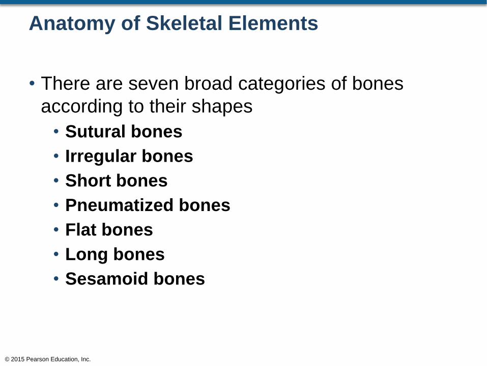
Anatomy of Skeletal Elements
• There are seven broad categories of bones
according to their shapes
• Sutural bones
• Irregular bones
• Short bones
• Pneumatized bones
• Flat bones
• Long bones
• Sesamoid bones
© 2015 Pearson Education, Inc.

Figure 5.11 Shapes of Bones
© 2015 Pearson Education, Inc.
Sutures Sutural
bone
Sutural Bones
Ethmoid
Air cells
Sutural (Wormian)
bones are small, flat,
oddly shaped bones
found between the flat
bones of the skull in
the suture line. They
develop from separate
centers of ossification,
regarded as a type of
flat bone.
Pneumatized Bones
Pneumatized bones are bones
that are hollow or contain
numerous air pockets,
such as the ethmoid.
Flat Bones
Flat bones have thin, roughly parallel surfaces of
compact bone. In structure a flat bone resembles
a spongy bone sandwich; such bones are strong
but relatively light. Flat bones form the roof of the
skull, the sternum, the ribs, and the scapulae. They
provide protection for underlying soft tissues and
offer an extensive surface area for the attachment
of skeletal muscles. Special terms are used when
describing the flat bones of the skull such as the
parietal bones. Their relatively thick layers of
compact bone are called the internal and external
tables, and the layer of spongy bone between the
tables is called the diploë.
Short Bones
Short bones are boxlike in
appearance. Their external
surfaces are covered by
compact bone, but the
interior contains spongy
bone. Examples of
short bones include
the carpal bones
(wrists) and tarsal
bones (ankles).
Carpal
bones
Vertebra
Irregular Bones
Irregular bones have
complex shapes with short,
flat, notched, or ridged
surfaces. Their internal
structure is equally varied.
The vertebrae that form the
spinal column and several
bones in the skull are
examples of irregular bones.
Patella
Humerus
Parietal bone
Internal
table
Diploë
(spongy bone)
External table
Long Bones
Sesamoid Bones
Long bones are relatively long
and slender. They have a
diaphysis, two metaphyses, two
epiphyses, and a medullary
(marrow) cavity, as detailed in
Figure 5.3. Long bones are
found in the upper and lower
limbs. Examples include the
humerus, radius, ulna, femur,
tibia, and fibula.
Sesamoid bones are usually
small, round, and flat. They
develop inside tendons and
are most often encountered
near joints at the knee, the
hands, and the feet. Few
individuals have sesamoid
bones at every possible
location, but everyone has
sesamoid patellae, or
kneecaps.

Anatomy of Skeletal Elements
• Bone markings include:
• Projections
• Depressions
• Fossa
• Openings
• Sinuses/canals/fissures/foramen
• Processes
• Trochanter/crest/spine/line/tubercle/tuberosity/
head/neck/facet/condyle/trochlea
© 2015 Pearson Education, Inc.

Figure 5.12 Examples of Bone Markings (Surface Features) (1 of 2)
© 2015 Pearson Education, Inc.
Skull, sagittal section
Skull, anterior view
Openings
Sinus or
antrum:
A chamber within
a bone, normally
filled with air
Meatus
or canal:
A passageway
through the
substance of a bone
Fissure: An elongated cleft
Foramen: A rounded
passageway for blood
vessels and/or nerves
Process: Any projection
or bump
Elevations and Projections
Ramus: An extension of a
bone making an
angle to the rest
of the structure

Figure 5.12 Examples of Bone Markings (Surface Features) (2 of 2)
© 2015 Pearson Education, Inc.
Ramus
Head: The expanded articular end of an epiphysis, often separated from the shaft by a narrower neck
Processes formed where
tendons or ligaments
attach
Processes formed for
articulations with
adjacent bones
Fossa
Foramen
Pelvis
Head
Neck
Humerus
Condyle
Femur
Depressions
Neck: A narrower connection between the epiphysis and diaphysis
Facet: A small, flat articular surface
Condyle: A smooth, rounded articular process
Trochlea: A smooth, grooved articular process shaped like a pulley
Trochanter: A large, rough projection
Crest: A prominent ridge
Spine: A pointed process
Line: A low ridge
Tubercle: A small, rounded projection
Tuberosity: A rough projection
Sulcus: A narrow groove
Fossa: A shallow depression

Integration with Other Systems
• Bones are not Inert
• Bones are Dynamic Structures
• Attached to the muscles
• Under physiological control via the endocrine
system
• Digestive and excretory system
• Provides calcium and phosphate needed for growth
• Serves as a store of calcium, phosphate, and
other minerals
© 2015 Pearson Education, Inc.


