24
Overview of the Toxicity Issues with Nanoparticles
| Date post: | 29-Dec-2015 |
| Category: |
Documents |
| Upload: | luke-owens |
| View: | 223 times |
| Download: | 3 times |

Overview of the Toxicity Issues with Nanoparticles

Designing a realistic test
Nature nanotechnology (2009) , Vol. 4, pp 395
Meaningful results on the toxicity of nanomaterials are achieved when the conditions of possible exposure are reproduced accurately
Different methods of exposure of the nanoparticle might produce different results

Influence of Properties on Lung Deposition as well as Toxicity
• Ultra-fine or nanoparticles may deposit as aggregates due to high Van Der Waals forces, rather than discrete particles.
• If an inhaled particle with a diameter of 50-100 nm forms an aggregate of 5-10 particle types, in terms of deposition it may have the properties of a 200-500 nm particle.
• Inhaled agglomerates may dissociate when in contact with lung surfactants.

Translocation of Probes in the Blood Circulation to Bone Marrow in Rodent Models
10 nm 100 nm <200 nm >200 nm
PEG-QDots Metallo-C60 HAS-coated PLA NP PS beads
Fast appearance in liver, spleen, lymph nodes and bone marrow (mouse)
Highest accumulation bone marrow after liver, continued increase in bone marrow and decrease in liver,
Significant accumulation in bone marrow after liver
Rapid passage through endothelium in bone marrow, uptake by phagocytizing cells in tissue (mouse)
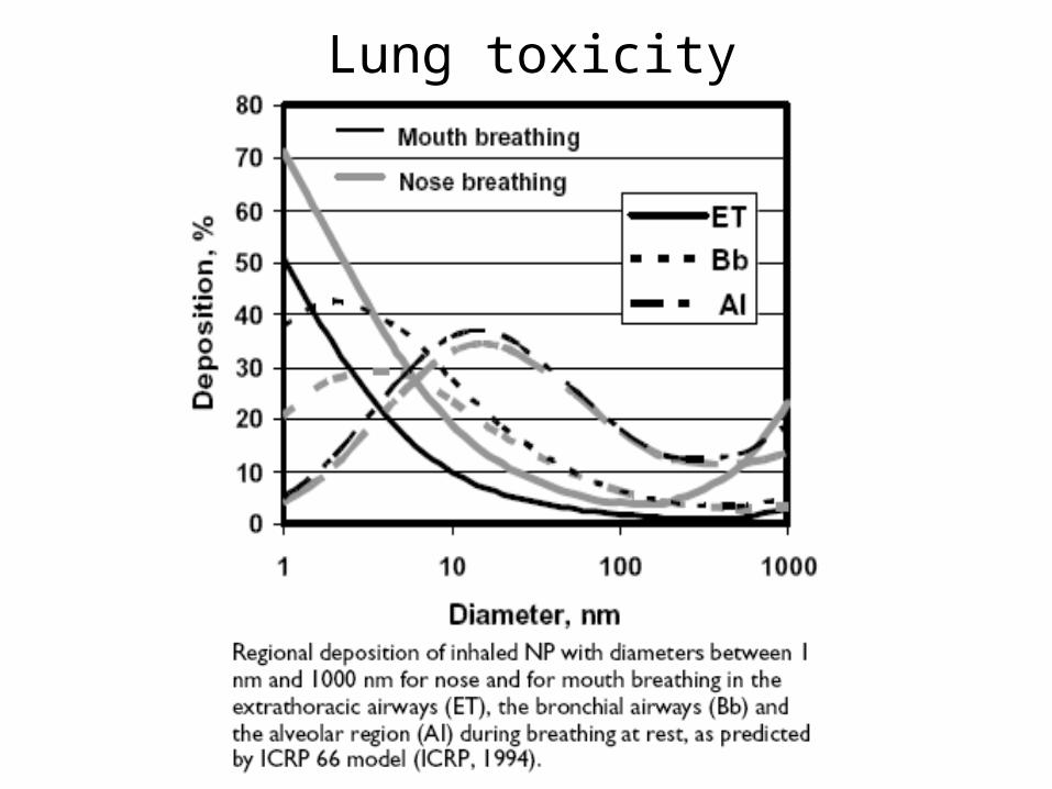
Lung toxicity

Cardiotoxicity
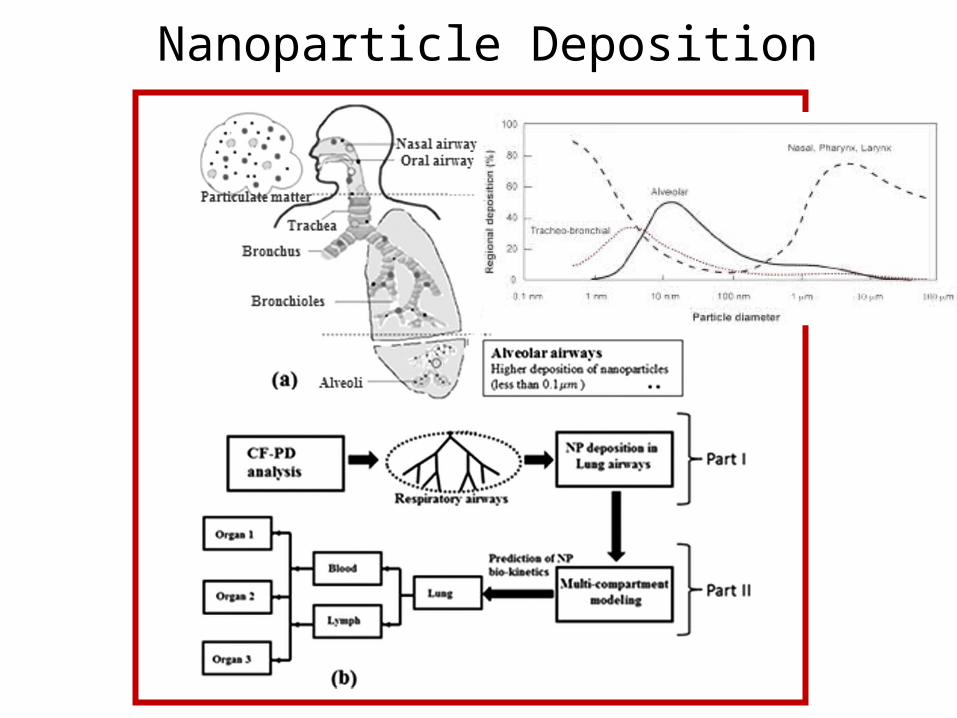
Nanoparticle Deposition

(a) SEM image of lung trachea epithelium, showing cilia (mucociliary escalator), (b) Human alveolar macrophage (center, yellow) phagocytosis of Escherichia coli(multiple ovoids, green), together with a red blood cell (red). (c), (d) Alveoli in the lung. (e) Deposition of inhaled particles in the human respiratory tract versus the particle diameter, after.

Rats that were instilled with high doses of SWCNT’s died of respiratory blockage rather than pulmonary intoxication
Instillation of CNT’s in Rats
Micrograph of Lung Tissue in Rats
Toxicological Sciences (2004) , Vol. 77, pp 117
The picture shows that the respiratory airways are mechanically blocked by carbon nanotubes. This led to the asphyxiation of 15% of the test population
Methods
• Four kind of particles including SWCNT’s
• Pressurized Intratraqueal instillation
• Tracking of alveolar response
• Observation periods at 24h, 1 week, 1 month and 3 months
Results Inflammation, no cytotoxicity

Inhalation of CNT’s in Rats
Nature nanotechnology (2009) , Vol. 4, pp 451
Exposing rats to air contaminated with CNT’s led to immune-suppression
Mechanism for Immune-
suppression by CNT’s
A signal, likely TGFβ, is released when the carbon nanotube is inhaled. This was tested by isolating the BALF protein from both exposed and control rats. It was shown that the protein from exposed mice cause immune-suppression
Methods• Air contaminated with low
concentration CNT’s• Exposure 6h per day during 14 days
• Tracking of proteins and immune response
ResultsImmune-suppression

Fullerene Disruption of Cell Membranes
Migration of a Single Fullerene
Migration of a Fullerene Aggregate
Unbiased MD simulations show that the fullerenes easily pass through the lipid head group, to further diffuse slowly within the bi-layer region
Nature nanotechnology (2008) , Vol. 3, pp 363
Average penetration time is 500 ps
Average penetration time is 1 µs
Pore formation appears not to be induced by the
presence of fullerene
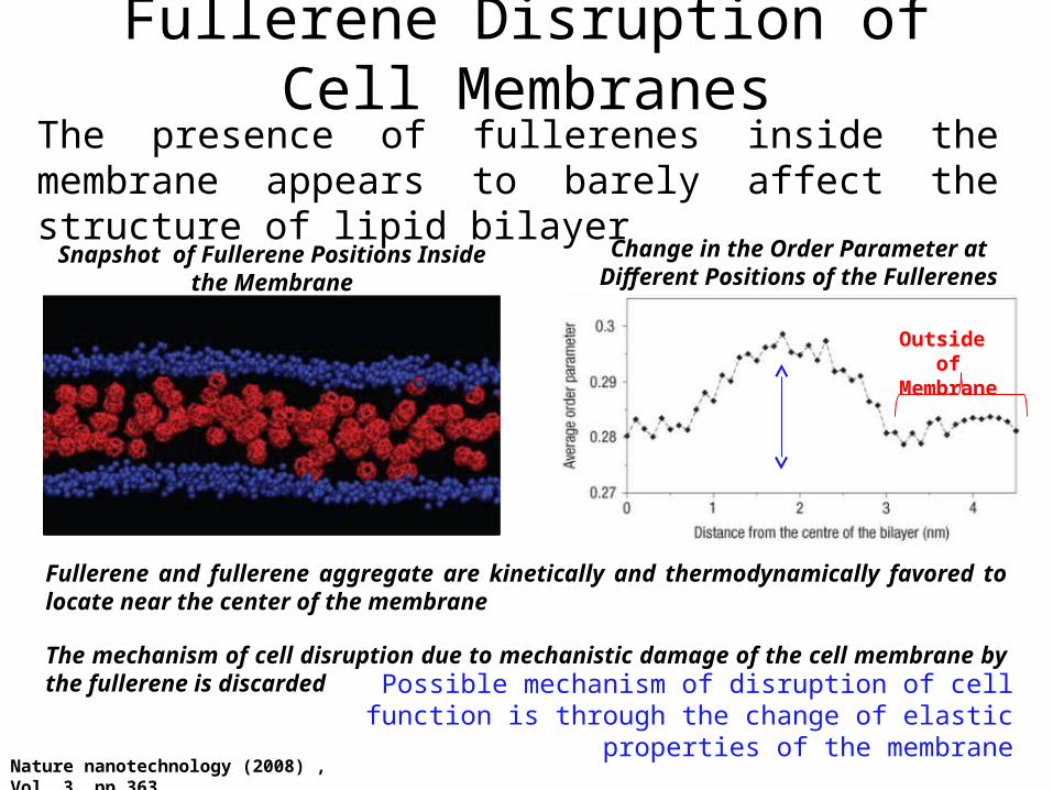
Fullerene Disruption of Cell MembranesThe presence of fullerenes inside the membrane appears to barely affect the structure of lipid bilayer
Snapshot of Fullerene Positions Inside the Membrane
Change in the Order Parameter at Different Positions of the Fullerenes
Nature nanotechnology (2008) , Vol. 3, pp 363
Outside of Membrane
Fullerene and fullerene aggregate are kinetically and thermodynamically favored to locate near the center of the membrane
The mechanism of cell disruption due to mechanistic damage of the cell membrane by the fullerene is discarded
Possible mechanism of disruption of cell function is through the change of elastic properties of the membrane

Silver Nanoparticles ToxicityThe toxicity of silver nanoparticles was tested using embryos of zebra fish
Nanotechnology (2008) 4, 873
Two kind of nanoparticles were used. One capped with BSA and
the other one with starch
TEM images of Ag Nanoparticles
starch BSA
The coating of the nanoparticle confer them the desired solubility and stability properties in water
Extent of toxicity is to be measure in term of mortality rate, hatching,
heart rate and abnormal phenotypes
Optical characterization

Silver Nanoparticles ToxicityThe toxicity of silver nanoparticles was tested using embryos of zebra fish
Nanotechnology (2008) 4, 873
The zebra fish eggs were taken to a 96-well plate, and a solution of silver nanoparticles at different concentrations was added to each well.
Normal Embryo
The images show the appearance of normal, malformed and dead embryos. Visual counting was made
Dead EmbryoMalformed Embryo
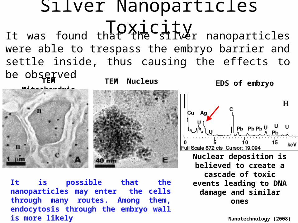
Silver Nanoparticles ToxicityIt was found that the silver nanoparticles were able to trespass the embryo barrier and settle inside, thus causing the effects to be observed
Nanotechnology (2008) 4, 873
It is possible that the nanoparticles may enter the cells through many routes. Among them, endocytosis through the embryo wall is more likely
TEM Mitochondria TEM Nucleus EDS of embryo
Nuclear deposition is believed to create a cascade of toxic
events leading to DNA damage and similar ones

DNA Damage of Cobalt-Chromium NPThe increasing use of nanoparticles (NP) in medicine has raised concerns over their ability to reach privileged sites in the body.
CoCr NP can be created by wear of orthopedic joint replacements
Schematic of exposure setup
The indirect effect of CoCr nanoparticles on human fibroblasts cells was evaluated. The fibroblast cells were protected by a cell barrier made out of BeWo (a human choriocarcinoma). The set up models the protein transport through placenta and similar barriers
Nature nanotechnology (2009) 4, 873
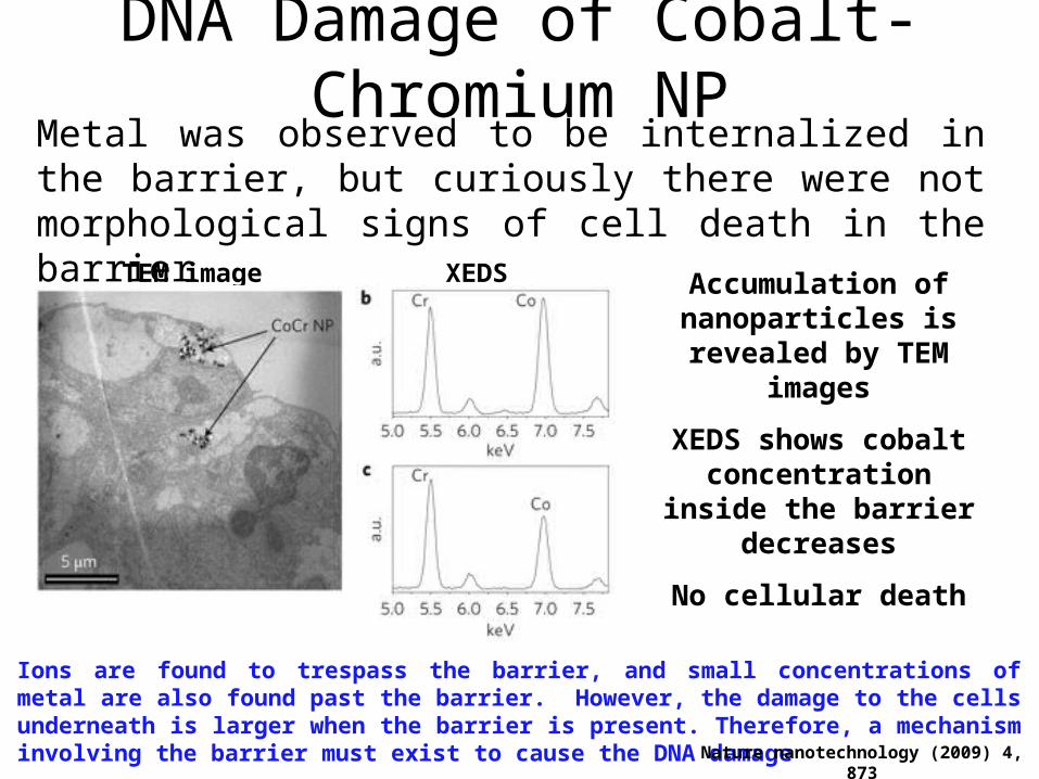
DNA Damage of Cobalt-Chromium NPMetal was observed to be internalized in the barrier, but curiously there were not morphological signs of cell death in the barrier
TEM image Accumulation of nanoparticles is revealed
by TEM images
XEDS
XEDS shows cobalt concentration inside the
barrier decreases
No cellular death
Ions are found to trespass the barrier, and small concentrations of metal are also found past the barrier. However, the damage to the cells underneath is larger when the barrier is present. Therefore, a mechanism involving the barrier must exist to cause the DNA damage
Nature nanotechnology (2009) 4, 873

DNA Damage of Cobalt-Chromium NPThe DNA damage of the cells below the barrier occurs through a chain of events starting with the damage of the mitochondria in the top layer of the cell barrier which end up in secretion of ATP from the bottom layer to the fibroblasts
DNA Damage Mechanism Schematics
Nature nanotechnology (2009) 4, 873
NSF and Why Gd Should be Avoided
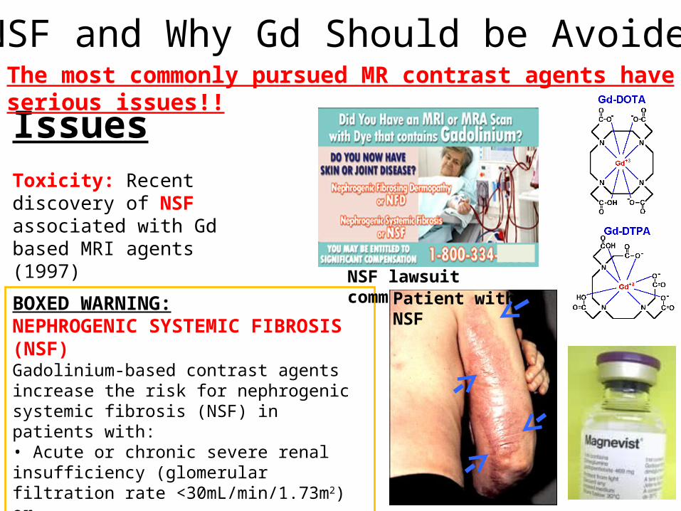
IssuesToxicity: Recent discovery of NSF associated with Gd based MRI agents (1997)
BOXED WARNING:NEPHROGENIC SYSTEMIC FIBROSIS (NSF)Gadolinium-based contrast agents increase the risk for nephrogenic systemic fibrosis (NSF) in patients with:• Acute or chronic severe renal insufficiency (glomerular filtration rate <30mL/min/1.73m2) or• Acute renal insufficiency of any severity due to the hepato-renal syndrome or in the perioperative liver transplantation period.
NSF lawsuit commercial
The most commonly pursued MR contrast agents have serious issues!!
Patient with NSF
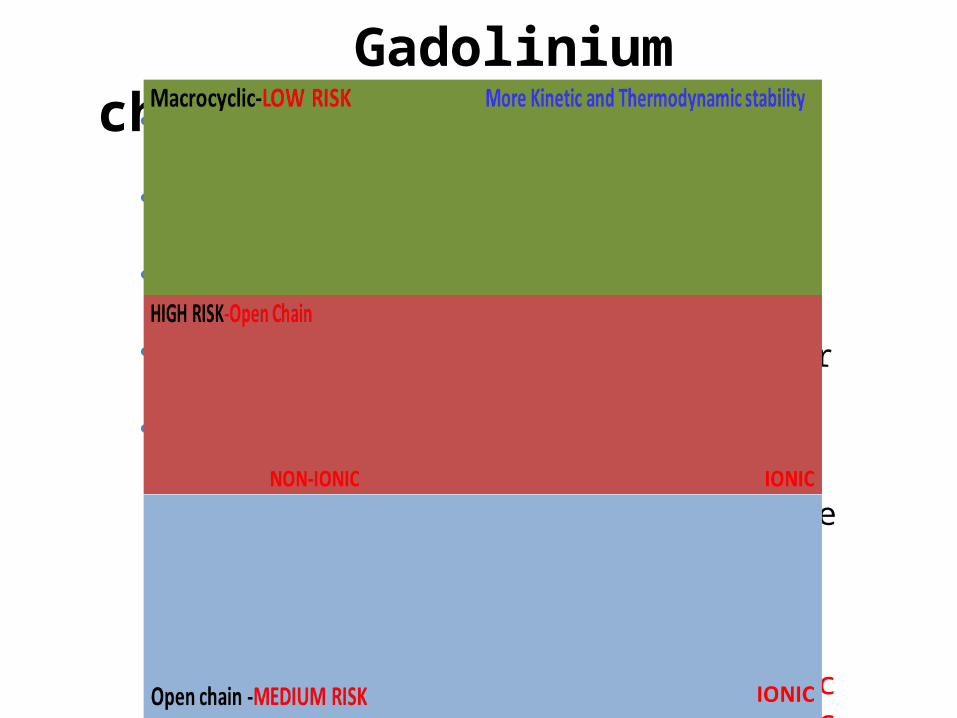
Gadolinium characteristics Rare-earth lanthanide metal (atomic #64) Gd3+ highly toxic, so bound to chelate Chelate prevents dissociation in vivo Eliminated unchanged in glomerular filterate Patients with impaired kidney or renal function is
affected• *Most reported cases of NSF are with a nonionic,
linear chelate• http://cds.ismrm.org/protected/NSF/• Stability: Linear nonionic complexes <linear ionic
<cyclic ones.

Understanding the Properties Before Using In Vivo
NCL assay cascade
In Vivo:– Absorption– Pharmacokinetics– Serum half-life– Tissue distribution– Excretion– SafetyPlasma PK profile/Tissue distribution(Liver, lungs, kidney, heart, spleen)
PhysicalCharacterization:– Size– Size distribution– Molecular weight– Morphology– Surface area– Porosity– Solubility– Surface charge density– Purity– Sterility– Surface chemistry– Stability– No of ligands, CAs, drugs
In Vitro:– Binding– Pharmacology– Blood contact properties– Cellular uptake– Cytotoxicity

Failed Phase I Clinical Trial (Kereos, Inc.)
X
22
Another Aspect to Worry
Cell Lysis
• >100K Gd on the surface
• Needed for detecting angiogenesis
Solution: Increase safety by replacing or reducing GdSource: wiki
Complement Activation
100,000Gd
NP decorated with Gd generates complement activation by eliciting immune response
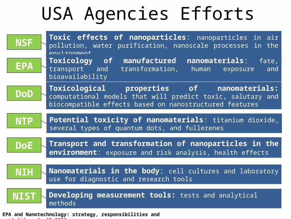
USA Agencies EffortsNSF Toxic effects of nanoparticles: nanoparticles in air pollution, water purification,
nanoscale processes in the environment
DoD Toxicological properties of nanomaterials: computational models that will predict toxic, salutary and biocompatible effects based on nanostructured features
Toxicology of manufactured nanomaterials: fate, transport and transformation, human exposure and bioavailability
Transport and transformation of nanoparticles in the environment: exposure and risk analysis, health effects
DoE
NIH Nanomaterials in the body: cell cultures and laboratory use for diagnostic and research tools
NIST Developing measurement tools: tests and analytical methods
EPA
NTP Potential toxicity of nanomaterials: titanium dioxide, several types of quantum dots, and fullerenes
EPA and Nanotechnology: strategy, responsibilities and activities, April 2006