p38 MAP kinase inhibition enablesproliferation of adult mammaliancardiomyocytesFelix B. Engel,1 Michael Schebesta,1 Mychelle T. Duong,1 Gang Lu,2 Shuxun Ren,2
Jeffery B. Madwed,3 Huiping Jiang,3 Yibin Wang,2 and Mark T. Keating1,4
1Howard Hughes Medical Institute, Department of Cell Biology, Harvard Medical School, Department of Cardiology,Children’s Hospital, Boston, Massachusetts 02115, USA; 2Molecular Biology Institute, Department of Anesthesiologyand Medicine, David Geffen School of Medicine, University of California, Los Angeles, California 90095, USA;3Department of Pharmacology, Boehringer Ingelheim Pharmaceuticals, Inc., Ridgefield, Connecticut 06410, USA
Adult mammalian cardiomyocytes are considered terminally differentiated and incapable of proliferation.Consequently, acutely injured mammalian hearts do not regenerate, they scar. Here, we show that adultmammalian cardiomyocytes can divide. One important mechanism used by mammalian cardiomyocytes tocontrol cell cycle is p38 MAP kinase activity. p38 regulates expression of genes required for mitosis incardiomyocytes, including cyclin A and cyclin B. p38 activity is inversely correlated with cardiac growthduring development, and its overexpression blocks fetal cardiomyocyte proliferation. Activation of p38 in vivoby MKK3bE reduces BrdU incorporation in fetal cardiomyocytes by 17.6%. In contrast, cardiac-specific p38�knockout mice show a 92.3% increase in neonatal cardiomyocyte mitoses. Furthermore, inhibition of p38 inadult cardiomyocytes promotes cytokinesis. Finally, mitosis in adult cardiomyocytes is associated withtransient dedifferentiation of the contractile apparatus. Our findings establish p38 as a key negative regulatorof cardiomyocyte proliferation and indicate that adult cardiomyocytes can divide.
Supplemental material is available at http://www.genesdev.org.
Received February 15, 2005; revised version accepted April 4, 2005.
Highly differentiated mammalian cells are thought to beincapable of proliferation. These cells have exited thecell cycle. Proteins critical for cellular specializationhave accumulated and driven these cells to their finalform and function (Studzinski and Harrison 1999). Incontrast with mammals, differentiated cells in teleostfish (Poss et al. 2002, 2003) and urodele amphibians(Brockes and Kumar 2002) can dedifferentiate and/or pro-liferate, enabling regeneration. For example, zebrafishhearts regenerate through cardiomyocyte proliferation(Poss et al. 2002). Thus, a thorough understanding ofmechanisms regulating cell cycle exit, and the develop-ment of approaches to reactivate proliferation of mam-malian cells, will be of great therapeutic value.
Mammalian cardiac regeneration has been studiedsince the mid-nineteenth century. The consistent con-clusion of these studies has been that the heart has littleor no regenerative capacity (Rumyantsev 1977; Mum-mery 2005). This is a major medical problem, as is-
chaemic heart disease, resulting in cardiac muscle loss,is the leading cause of morbidity and mortality in theindustrialized world. Here, we have studied the prolif-eration potential of adult mammalian cardiomyocytes.
In contrast to adult cardiomyocytes, mammalian car-diomyocytes do proliferate during fetal development.Shortly after birth, these cardiomyocytes down-regulatecell-cycle-perpetuating factors like cyclin A and cdk2.The loss of proliferation capacity coincides with in-creased levels of the cell cycle inhibitors p21 and p27(Pasumarthi and Field 2002). At this point of develop-ment, post-natal cardiac growth is mediated by cardio-myocyte hypertrophy. This transition from hyperplasticto hypertrophic growth is characterized by maturation ofthe contractile apparatus, a cytoplasmic structure that isthought to preclude cytokinesis (Rumyantsev 1977).Thus, primary adult mammalian cardiomyocytes arethought to be incapable of cytokinesis.
In general, there is an inverse relationship betweenproliferation and differentiation (Studzinski and Harri-son 1999), and molecules that promote differentiationmay also repress cell cycle re-entry. It has been shownthat the signaling molecule p38 mitogen-activated pro-tein (MAP) kinase (p38) induces cell cycle exit and dif-
4Corresponding author.E-MAIL [email protected]; FAX (617) 730-8317.Article published online ahead of print. Article and publication date areat http://www.genesdev.org/cgi/doi/10.1101/gad.1306705.
ferentiation of many cell types (Nebreda and Porras2000; Wu et al. 2000; Ambrosino and Nebreda 2001; Bu-lavin et al. 2002; Shi and Gaestel 2002), including differ-entiation of P19 cells to cardiomyocytes (Eriksson andLeppa 2002). Activated p38 phosphorylates downstreamsignaling molecules important for cardiomyocyte differ-entiation and hypertrophy (Liang and Molkentin 2003).Four different p38 isoforms have been identified. Themain isoform expressed in the heart is p38�. p38� andp38� are expressed at low levels, and p38� is not ex-pressed in heart (Wang et al. 1997; Liao et al. 2001; Liangand Molkentin 2003). Previously, it was not known if theeffects of p38 on differentiation and proliferation are re-versible.
Here, we show that post-mitotic mammalian cardio-myocytes can proliferate. One mechanism of cell cycleregulation for mammalian cardiomyocytes is p38 activ-ity; that is, p38 is a key negative regulator of mammaliancardiomyocyte division. p38 activity is inversely corre-lated with cardiac growth during development, and itsoverexpression blocks proliferation of fetal cardiomyo-
cytes in vitro. Genetic activation of p38 in vivo reducesfetal cardiomyocyte proliferation, whereas targeted dis-ruption of p38� increases neonatal cardiomyocyte mito-ses. Furthermore, we demonstrate that growth factorstimulation and p38 inhibition induce cytokinesis inadult cardiomyocytes. These results indicate that the in-hibitory effects of p38 on cardiomyocyte proliferation arereversible and that post-mitotic, differentiated cells arecapable of proliferation.
Results
p38 inhibition regulates genes critical for mitosisin cardiomyocytes
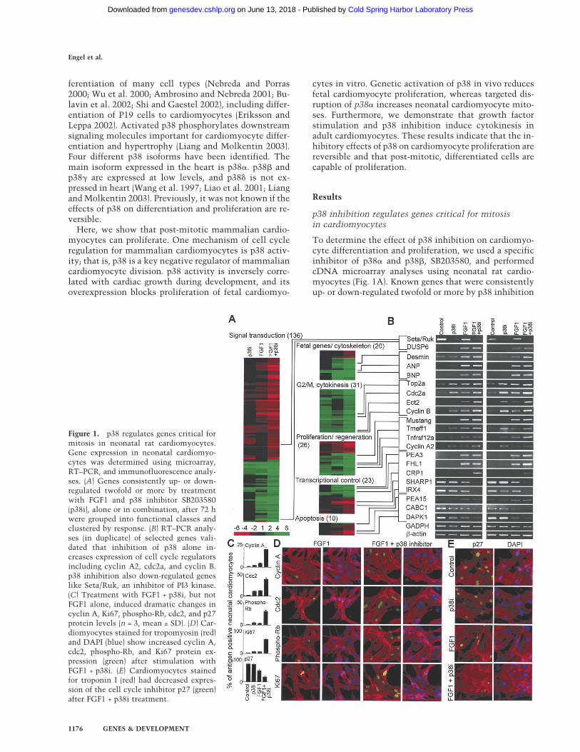
To determine the effect of p38 inhibition on cardiomyo-cyte differentiation and proliferation, we used a specificinhibitor of p38� and p38�, SB203580, and performedcDNA microarray analyses using neonatal rat cardio-myocytes (Fig. 1A). Known genes that were consistentlyup- or down-regulated twofold or more by p38 inhibition
Figure 1. p38 regulates genes critical formitosis in neonatal rat cardiomyocytes.Gene expression in neonatal cardiomyo-cytes was determined using microarray,RT–PCR, and immunofluorescence analy-ses. (A) Genes consistently up- or down-regulated twofold or more by treatmentwith FGF1 and p38 inhibitor SB203580(p38i), alone or in combination, after 72 hwere grouped into functional classes andclustered by response. (B) RT–PCR analy-ses (in duplicate) of selected genes vali-dated that inhibition of p38 alone in-creases expression of cell cycle regulatorsincluding cyclin A2, cdc2a, and cyclin B.p38 inhibition also down-regulated geneslike Seta/Ruk, an inhibitor of PI3 kinase.(C) Treatment with FGF1 + p38i, but notFGF1 alone, induced dramatic changes incyclin A, Ki67, phospho-Rb, cdc2, and p27protein levels (n = 3, mean ± SD). (D) Car-diomyocytes stained for tropomyosin (red)and DAPI (blue) show increased cyclin A,cdc2, phospho-Rb, and Ki67 protein ex-pression (green) after stimulation withFGF1 + p38i. (E) Cardiomyocytes stainedfor troponin I (red) had decreased expres-sion of the cell cycle inhibitor p27 (green)after FGF1 + p38i treatment.
Engel et al.
1176 GENES & DEVELOPMENT
Cold Spring Harbor Laboratory Press on June 13, 2018 - Published by genesdev.cshlp.orgDownloaded from
after 72 h were grouped into functional classes and clus-tered by response (Fig. 1A; Supplementary Table S1). Wevalidated expression changes of a subset of genes by RT–PCR (Fig. 1B).
Down-regulation of cyclin A is an early sign of cellcycle exit in mammalian cardiomyocytes (Yoshizumiet al. 1995). In addition, it has been shown that cardiac-specific overexpression of cyclin A2 from embryonicday 8 into adulthood increases cardiomyocyte mitosisduring post-natal development (Chaudhry et al. 2004).We discovered that p38 inhibition up-regulated cyclinA2 (Fig. 1B). p38 inhibition also regulated othergenes involved in mitosis and cytokinesis, includingcyclin B, cdc2, and aurora B (Wheatley et al. 2001;Murray 2004). We expected that these changes mightalso be associated with evidence of dedifferentia-tion, such as induction of fetal genes. However, weobserved only a slight induction of ANP. Thus, p38 ac-tivity regulates genes important for mitosis in cardio-myocytes.
Stimulation of neonatal cardiomyocytes with FGF1 in-duces fetal gene expression (Parker et al. 1990). To de-termine if FGF1, in combination with p38 inhibition,can reverse differentiation and induce cell cycle re-entry,we repeated cDNA microarray analyses (Fig. 1A,B;Supplementary Table S1). FGF1 up-regulated genesthat are associated with fetal cardiac development, in-cluding ANP and BNP (Cameron and Ellmers 2003), andthe Ets-related transcription factor PEA3 (Chotteau-Lelievre et al. 1997). In addition, FGF1 up-regulatedgenes previously implicated in regeneration and cellcycle control, including Mustang (Lombardo et al. 2004).Finally, FGF1 down-regulated pro-apoptotic genes, likeCABC1 (Iiizumi et al. 2002), and up-regulated anti-apo-ptotic genes, like PEA15 (Kitsberg et al. 1999). Takentogether, these data suggest that FGF1 induces partialdedifferentiation and possibly protects cardiomyocytesfrom apoptosis.
Expression analysis revealed that p38 inhibition andFGF1 together modulate expression of specific genes,whereas p38 inhibition or FGF1 stimulation alone hadlittle or no effect. For example, p38 inhibition and FGF1dramatically modulated expression of the cytokinesisregulator Ect2 (Saito et al. 2004), the bHLH factorSHARP1 (Azmi et al. 2003), the cell-cycle-regulatedprotein CRP1 (Weiskirchen et al. 1995), and the media-tor of ventricular cardiomyocyte differentiation, IRX4(Fig. 1B; Bao et al. 1999). For a subset of cell-cycle-per-petuating factors, including Ki67, cdc2, and cyclin A, andthe cell cycle inhibitor p27, the combined effect of p38inhibition and FGF1 stimulation was even greater at theprotein level (Fig. 1C–E). The proliferation marker Ki67(Brown and Gatter 2002), for example, was increasedsevenfold. Finally, p38 inhibitor and FGF1, but neitherfactor alone, led to phosphorylation of Rb, a key cellcycle regulator (Fig. 1C,D; Classon and Harlow 2002).Taken together, our data indicate that p38 inhibitionand FGF1 stimulation act synergistically to induce ex-pression of genes involved in proliferation and regenera-tion.
Fetal cardiomyocytes proliferate during development butlose this capacity shortly after birth. The switch fromproliferative to hypertrophic growth has been associatedwith up- and down-regulation of many factors. However,its mechanism is not understood. To determine if p38regulates fetal cardiomyocyte proliferation, we exam-ined prenatal cardiac growth. We collected rat hearts atsequential developmental stages (embryonic days 12–21[E12–E21], post-natal day 2 [P2], and adult), and assessedthe cardiac growth rate (n = 18–40 per time point) andp38 activity (n = 5 litters) (Fig. 2A–C). Cardiac growthrate mediated predominantly by fetal cardiomyocyteproliferation (Rumyantsev 1977; Pasumarthi and Field2002) was defined as the percentage increase of maximalventricular area (Fig. 2A). The rate of cardiac growth de-creased sharply from E13 to E15 (p < 0.01), acceleratedfrom E17 to E19 (p < 0.01), and decreased again (Fig. 2B).p38 activity, in contrast, was inversely correlated withcardiac growth (Fig. 2B,C). p38 activity was low at E12,peaked at E15, declined to a second low at E19, roseagain, and stayed high in adults (p < 0.01). At E13, forexample, cardiac area doubled and p38 activity was low(4.51). In contrast, at E15, cardiac area increased only35% and p38 activity was high (11.89). These data indi-cate an association between p38 activity and fetal car-diomyocyte proliferation.
To directly assess the role of p38 in regulating fetalcardiomyocyte proliferation, we overexpressed GFP,p38�, and a dominant-negative form of p38� (p38�DN)in fetal (E19) cardiomyocytes. p38�DN is mutated in itsdual phosphorylation site, causing lack of kinase activity(Raingeaud et al. 1995). Cells were electroporated, cul-tured for 36 h, and stimulated for 24 h with FGF1 in thepresence of BrdU (5-bromo-2�-deoxyuridine), a marker ofDNA synthesis. The rate of BrdU incorporation in mock-transfected cells (GFP) was 23% ± 5.2%. Overexpressionof p38� (3.4% ± 1.9%), but not p38�DN (19.2% ± 4.8%),decreased FGF1-induced BrdU incorporation signifi-cantly (Fig. 2D,E). As shown in Figure 2C, p38 activity isvery low in the fetal heart at this stage of development,thus overexpression of p38�DN was not expected tohave a significant effect. These results indicate that p38�is a potent regulator of fetal cardiomyocyte proliferationin vitro.
To determine the role of p38 activation in vivo, weexamined transgenic animals with cardiomyocyte-spe-cific expression of a constitutively active upstream ki-nase for p38, MKK3bE. Targeted activation of p38 in ven-tricular myocytes was achieved in vivo by using a gene-switch transgenic strategy resulting in the expression ofMKK3bE mutant protein under the control of the �MHC promoter (Liao et al. 2001). Previously, it has beendemonstrated that activation of p38 kinase activitycauses a thin ventricular wall (Liao et al. 2001). The un-derlying mechanism of this phenotype is unclear, butinduction of apoptosis was excluded. As shown in Fig-ure 2F, BrdU incorporation in fetal cardiomyocytes (E21)was reduced from 18.2% ± 3.4% to 15.0% ± 2.9% in
p38 controls cardiomyocyte proliferation
GENES & DEVELOPMENT 1177
Cold Spring Harbor Laboratory Press on June 13, 2018 - Published by genesdev.cshlp.orgDownloaded from
MKK3bE transgenic hearts. This is a reduction of 17.6%(p < 0.05) in cardiomyocyte proliferation. Taken to-gether, our results indicate that p38 activity is a potentnegative regulator of fetal cardiomyocyte proliferation invitro and in vivo.
p38� inhibition promotes neonatal cardiomyocyteproliferation in vitro
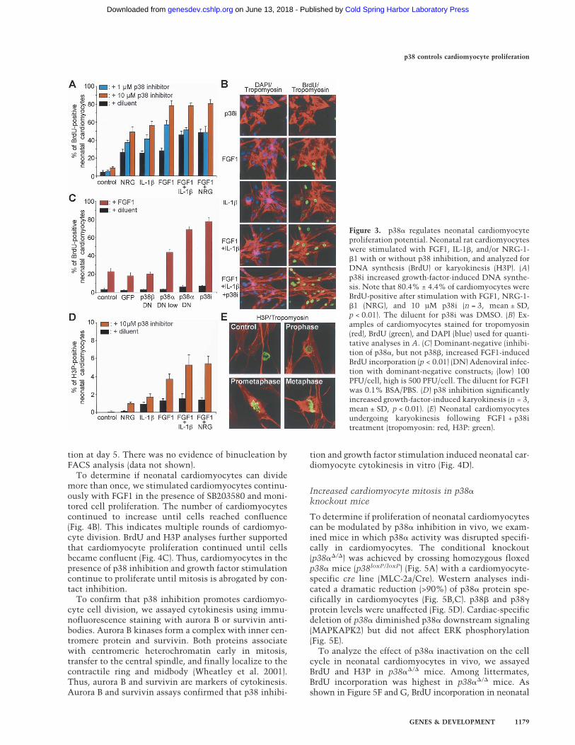
Several growth factors have a limited capacity to induceDNA synthesis in neonatal cardiomyocytes, includingFGF1 (Pasumarthi and Field 2002). We screened 45 ex-tracellular factors at two different concentrations fortheir ability to induce BrdU incorporation in neonatal(P2) cardiomyocytes. Cells were stimulated every 24 hfor 3 d and pulse-labeled with BrdU for the final 24 h. Weconfirmed previous studies showing that FGF1, IL-1�,and NRG-1-�1 are potent growth factors for neonatalcardiomyocytes (Fig. 3A,B; Supplementary Table S2; Pa-sumarthi and Field 2002).
Inhibition of p38 activity by SB203580 increased BrdUincorporation 2.8-fold in neonatal cardiomyocytesstimulated with FGF1 (p < 0.01) (Fig. 3A,B). Similar re-sults were obtained after stimulation with IL-1� andNRG-1-�1. Thus, inhibition of p38 activity augmentsgrowth factor-mediated DNA synthesis in neonatal car-diomyocytes.
To support the specificity of SB203580, we repeatedthese experiments with dominant-negative forms ofp38� (p38�DN) and p38� (p38�DN). Adenovirus-medi-ated expression of p38�DN was as effective as SB203580in increasing growth-factor-mediated BrdU incorpora-
tion (Fig. 3C). In contrast, expression of p38�DN had noeffect on DNA synthesis. These results are consistentwith previous findings showing that p38� and p38� havedistinct downstream targets (Enslen et al. 1998; Wanget al. 1998). Taken together, our data indicate that theeffect of p38 on DNA synthesis in neonatal cardiomyo-cytes is mediated by p38�.
To determine if p38 also regulates karyokinesis in neo-natal cardiomyocytes, we assayed mitosis by immuno-fluorescence staining of phosphorylated histone-3 (H3P).Inhibition of p38 activity using SB203580 increased thenumber of H3P-positive cells 3.9-fold in the presence ofFGF1 + NRG-1-�1, resulting in 5.4% ± 0.8% H3P-posi-tive cardiomyocytes (p < 0.01) (Fig. 3D,E). This value iscomparable to that of proliferating cell lines and the mi-totic index of fetal cardiomyocytes during embryonic de-velopment (E12, 3.7% ± 0.6%) (data not shown). Thus,p38 activity regulates neonatal cardiomyocyte karyo-kinesis.
During post-natal development, mammalian cardio-myocytes frequently undergo karyokinesis without cy-tokinesis, and ∼60% of human, and 85% of rat, adultcardiomyocytes are binucleated (Brodsky 1991). To testif p38 regulates cell division in neonatal cardiomyocytes,we performed cell count experiments. The percentage ofcardiomyocytes was determined by tropomyosin stain-ing and FACS analyses. Cells were incubated withSB203580 and stimulated once with growth factors onday 0. As shown in Figure 4A, this resulted in signifi-cantly increased cell numbers (day 3: p < 0.05; days 4and 5: p < 0.01). The maximal increase in cardiomyocytenumber of 2.6-fold was seen with FGF1 + IL-1� stimula-
Figure 2. p38 controls fetal cardiomyocyte prolif-eration in vitro and in vivo. (A) Rat hearts grew rap-idly during embryonic development. (B) The rate ofcardiac growth (black line) was inversely correlatedwith p38 activity (orange bars, n = 5, mean ± SD).p38 activity was measured by its ability to phos-phorylate ATF-2. p38 activity was biphasic duringdevelopment, low at E12 and E19, and high at E15and E21–adult. (C) Western analysis showed bipha-sic p38 activity, while p38 protein levels were con-stant. (D,E) Overexpression of wild-type p38� (D),but not dominant-negative p38� (p38�DN) (E),blocked BrdU incorporation in fetal cardiomyocytes(n = 3, mean ± SD, p < 0.05). Fetal cardiomyocytesstained for Nkx2.5 (red), BrdU (green), and Flag-tagged p38� or p38�DN (blue). Arrowheads indicateBrdU-positive cardiomyocytes expressing p38�DN.(F) Quantitative analysis of BrdU incorporation infetal cardiomyocytes (MEF2-positive, E21) of MKK3bEtransgenic (MKK3bE+) and nontransgenic (MKK3bE−)littermates in vivo. Note that BrdU incorporationin cardiomyocytes of MKK3bE transgenic micedecreased from 18.2% ± 3.4% to 15.0% ± 2.9%(mean ± SD, 17.6% reduction, p < 0.05). Mean is indi-cated by a red line.
Engel et al.
1178 GENES & DEVELOPMENT
Cold Spring Harbor Laboratory Press on June 13, 2018 - Published by genesdev.cshlp.orgDownloaded from
tion at day 5. There was no evidence of binucleation byFACS analysis (data not shown).
To determine if neonatal cardiomyocytes can dividemore than once, we stimulated cardiomyocytes continu-ously with FGF1 in the presence of SB203580 and moni-tored cell proliferation. The number of cardiomyocytescontinued to increase until cells reached confluence(Fig. 4B). This indicates multiple rounds of cardiomyo-cyte division. BrdU and H3P analyses further supportedthat cardiomyocyte proliferation continued until cellsbecame confluent (Fig. 4C). Thus, cardiomyocytes in thepresence of p38 inhibition and growth factor stimulationcontinue to proliferate until mitosis is abrogated by con-tact inhibition.
To confirm that p38 inhibition promotes cardiomyo-cyte cell division, we assayed cytokinesis using immu-nofluorescence staining with aurora B or survivin anti-bodies. Aurora B kinases form a complex with inner cen-tromere protein and survivin. Both proteins associatewith centromeric heterochromatin early in mitosis,transfer to the central spindle, and finally localize to thecontractile ring and midbody (Wheatley et al. 2001).Thus, aurora B and survivin are markers of cytokinesis.Aurora B and survivin assays confirmed that p38 inhibi-
tion and growth factor stimulation induced neonatal car-diomyocyte cytokinesis in vitro (Fig. 4D).
Increased cardiomyocyte mitosis in p38�knockout mice
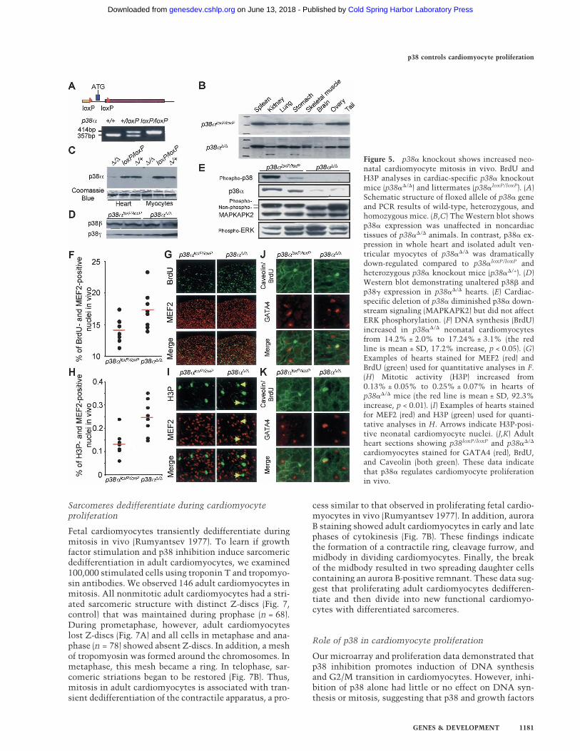
To determine if proliferation of neonatal cardiomyocytescan be modulated by p38� inhibition in vivo, we exam-ined mice in which p38� activity was disrupted specifi-cally in cardiomyocytes. The conditional knockout(p38��/�) was achieved by crossing homozygous floxedp38� mice (p38loxP/loxP) (Fig. 5A) with a cardiomyocyte-specific cre line (MLC-2a/Cre). Western analyses indi-cated a dramatic reduction (>90%) of p38� protein spe-cifically in cardiomyocytes (Fig. 5B,C). p38� and p38�protein levels were unaffected (Fig. 5D). Cardiac-specificdeletion of p38� diminished p38� downstream signaling(MAPKAPK2) but did not affect ERK phosphorylation(Fig. 5E).
To analyze the effect of p38� inactivation on the cellcycle in neonatal cardiomyocytes in vivo, we assayedBrdU and H3P in p38��/� mice. Among littermates,BrdU incorporation was highest in p38��/� mice. Asshown in Figure 5F and G, BrdU incorporation in neonatal
Figure 3. p38� regulates neonatal cardiomyocyteproliferation potential. Neonatal rat cardiomyocyteswere stimulated with FGF1, IL-1�, and/or NRG-1-�1 with or without p38 inhibition, and analyzed forDNA synthesis (BrdU) or karyokinesis (H3P). (A)p38i increased growth-factor-induced DNA synthe-sis. Note that 80.4% ± 4.4% of cardiomyocytes wereBrdU-positive after stimulation with FGF1, NRG-1-�1 (NRG), and 10 µM p38i (n = 3, mean ± SD,p < 0.01). The diluent for p38i was DMSO. (B) Ex-amples of cardiomyocytes stained for tropomyosin(red), BrdU (green), and DAPI (blue) used for quanti-tative analyses in A. (C) Dominant-negative (inhibi-tion of p38�, but not p38�, increased FGF1-inducedBrdU incorporation (p < 0.01) (DN) Adenoviral infec-tion with dominant-negative constructs; (low) 100PFU/cell, high is 500 PFU/cell. The diluent for FGF1was 0.1% BSA/PBS. (D) p38 inhibition significantlyincreased growth-factor-induced karyokinesis (n = 3,mean ± SD, p < 0.01). (E) Neonatal cardiomyocytesundergoing karyokinesis following FGF1 + p38itreatment (tropomyosin: red, H3P: green).
p38 controls cardiomyocyte proliferation
GENES & DEVELOPMENT 1179
Cold Spring Harbor Laboratory Press on June 13, 2018 - Published by genesdev.cshlp.orgDownloaded from
cardiomyocytes (P4) was increased from 14.2% ± 2.0%to 17.2% ± 3.1% (17.2% increase, p < 0.05). These dataindicate that reduced p38� protein causes increased car-diomyocyte DNA synthesis in vivo. H3 phosphorylationwas increased from 0.13% ± 0.05% to 0.25% ± 0.07%(92.3% increase, p < 0.01), indicating that reduced p38�protein resulted in increased mitosis in cardiomyocytesin vivo (Fig. 5H,I).
Furthermore, we examined the effects of p38� proteinreduction on BrdU incorporation in adult cardiomyo-cytes. To distinguish between adult cardiomyocytes andinterstitial cells, hearts were sectioned and stained forthe cardiac transcription factor GATA4 and a marker forcell membranes, Caveolin (Fig. 5J,K). We detected BrdU-positive adult cardiomyocytes in vivo. The number ofBrdU-positive cardiomyocytes per longitudinal sectionin p38��/� mice (1.7 ± 0.4) was 20-fold greater than ob-served in p38loxP/loxP mice (0.08 ± 0). Taken together, ourdata indicate that p38� is a negative regulator of cardio-myocyte proliferation in vivo.
Adult cardiomyocytes divide
In contrast to neonatal cardiomyocytes, previous studiesindicate that no DNA synthesis, karyokinesis, or cyto-kinesis occurs in rat cardiomyocytes 3 wk after birth(Rumyantsev 1977; Pasumarthi and Field 2002). To de-termine if p38 inhibition promotes growth-factor-mediated DNA synthesis in adult cardiomyocytes,we repeated cell proliferation assays using ventricularcardiomyocytes from 12-wk-old rats. As an additionalcardiomyocyte-specific marker, we used the transcrip-tion factor Nkx2.5. Cardiomyocytes were isolated at day0, and allowed to recover for 24 h. Cells were then stimu-lated every 3 d with growth factors in the presence orabsence of SB203580 for 12 d and assayed for BrdU. FGF1
alone and FGF1 + IL-1� induced BrdU incorporation in>2% of adult cardiomyocytes. Inhibition of p38 doubledthe effect of growth factors (p < 0.01) (Fig. 6A). Thesedata demonstrate that p38 inhibition promotes growth-factor-induced DNA synthesis in adult cardiomyocytes.
To determine if adult cardiomyocytes can undergokaryokinesis, we performed H3P analyses. Inhibition ofp38 activity increased the number of H3P-positive car-diomyocytes 3.7-fold in the presence of FGF1 (p < 0.01)(Fig. 6B,D). These findings indicate that p38 regulateskaryokinesis of adult cardiomyocytes.
To learn if adult mammalian cardiomyocytes can un-dergo cytokinesis, we assayed aurora B. Inhibition of p38increased cytokinesis 3.8-fold (p < 0.01) (Fig. 6C). Themaximum effect was observed with p38 inhibition andFGF1. Although most proliferating adult cardiomyocyteswere mononucleated (Fig. 6E,D), we also observed bi-nucleated cells undergoing cytokinesis (Fig. 6G,H).These data indicate that adult ventricular cardiomyo-cytes can divide.
To estimate how many cardiomyocytes proliferate af-ter 12 d of stimulation, we repeated these experimentsusing Ki67 (Brown and Gatter 2002). In neonatal cardio-myocytes, FGF1 induced DNA synthesis, but failed toinduce proliferation and Ki67 expression (Fig. 1C,D). Incontrast, FGF1 stimulation in the presence of SB203580resulted in both cardiomyocyte proliferation and Ki67expression. Thus, Ki67 is an excellent marker for cardio-myocyte proliferation. In adult cardiomyocytes, stimu-lation with FGF1 alone resulted in 1.7% ± 0.5% Ki67-positive cells (data not shown). However, stimulationwith FGF1 and p38 inhibitor resulted in 7.2% ± 1.2%Ki67-positive adult cardiomyocytes (p < 0.01). Taken to-gether, these data indicate that adult cardiomyocytes canproliferate in vitro, and that p38 potently controls thisprocess.
Figure 4. p38 controls neonatal cardiomyocyteproliferation. Neonatal cardiomyocyte prolifera-tion was analyzed by cell count, FACS, BrdU,H3P, survivin, and aurora B staining. (A) p38 in-hibition augmented growth-factor-induced car-diomyocyte proliferation as measured by cellcount (n = 2 or 3 for each time point, mean ± SD;day 3: p < 0.05, days 4 and 5: p < 0.01). Note thata single stimulation with FGF1 and IL-1� in thepresence of p38i increased cardiomyocyte num-bers by 2.6-fold after 5 d of stimulation. (B) Car-diomyocytes were seeded in a labeled microtiterwell, continuously stimulated with FGF1 (day 0,day 3, day 6) in the presence of p38i, and analyzedover time. (C) Continuous stimulation withFGF1 in the presence of p38i resulted in BrdUincorporation and H3 phosphorylation until cellsreached confluence at day 8, suggesting prolifera-tion. (D) Neonatal cardiomyocytes undergoingcytokinesis in response to FGF1 + p38i. The con-tractile ring and midbody of dividing cells werestained with survivin or aurora B (green). Cardio-myocytes were identified by tropomyosin or tropo-nin T (red). Controls show dividing nonmyocytes.
Engel et al.
1180 GENES & DEVELOPMENT
Cold Spring Harbor Laboratory Press on June 13, 2018 - Published by genesdev.cshlp.orgDownloaded from
Sarcomeres dedifferentiate during cardiomyocyteproliferation
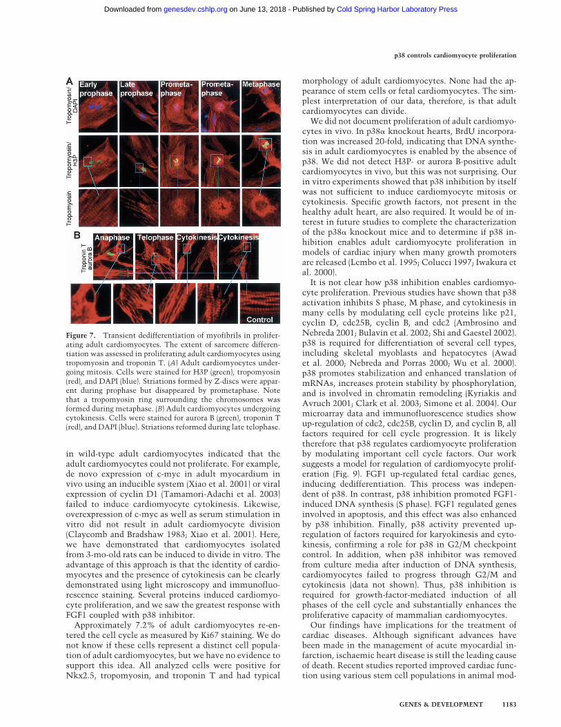
Fetal cardiomyocytes transiently dedifferentiate duringmitosis in vivo (Rumyantsev 1977). To learn if growthfactor stimulation and p38 inhibition induce sarcomericdedifferentiation in adult cardiomyocytes, we examined100,000 stimulated cells using troponin T and tropomyo-sin antibodies. We observed 146 adult cardiomyocytes inmitosis. All nonmitotic adult cardiomyocytes had a stri-ated sarcomeric structure with distinct Z-discs (Fig. 7,control) that was maintained during prophase (n = 68).During prometaphase, however, adult cardiomyocyteslost Z-discs (Fig. 7A) and all cells in metaphase and ana-phase (n = 78) showed absent Z-discs. In addition, a meshof tropomyosin was formed around the chromosomes. Inmetaphase, this mesh became a ring. In telophase, sar-comeric striations began to be restored (Fig. 7B). Thus,mitosis in adult cardiomyocytes is associated with tran-sient dedifferentiation of the contractile apparatus, a pro-
cess similar to that observed in proliferating fetal cardio-myocytes in vivo (Rumyantsev 1977). In addition, auroraB staining showed adult cardiomyocytes in early and latephases of cytokinesis (Fig. 7B). These findings indicatethe formation of a contractile ring, cleavage furrow, andmidbody in dividing cardiomyocytes. Finally, the breakof the midbody resulted in two spreading daughter cellscontaining an aurora B-positive remnant. These data sug-gest that proliferating adult cardiomyocytes dedifferen-tiate and then divide into new functional cardiomyo-cytes with differentiated sarcomeres.
Role of p38 in cardiomyocyte proliferation
Our microarray and proliferation data demonstrated thatp38 inhibition promotes induction of DNA synthesisand G2/M transition in cardiomyocytes. However, inhi-bition of p38 alone had little or no effect on DNA syn-thesis or mitosis, suggesting that p38 and growth factors
Figure 5. p38� knockout shows increased neo-natal cardiomyocyte mitosis in vivo. BrdU andH3P analyses in cardiac-specific p38� knockoutmice (p38��/�) and littermates (p38�loxP/loxP). (A)Schematic structure of floxed allele of p38� geneand PCR results of wild-type, heterozygous, andhomozygous mice. (B,C) The Western blot showsp38� expression was unaffected in noncardiactissues of p38��/� animals. In contrast, p38� ex-pression in whole heart and isolated adult ven-tricular myocytes of p38��/� was dramaticallydown-regulated compared to p38�loxP/loxP andheterozygous p38� knockout mice (p38��/+). (D)Western blot demonstrating unaltered p38� andp38� expression in p38��/� hearts. (E) Cardiac-specific deletion of p38� diminished p38� down-stream signaling (MAPKAPK2) but did not affectERK phosphorylation. (F) DNA synthesis (BrdU)increased in p38��/� neonatal cardiomyocytesfrom 14.2% ± 2.0% to 17.24% ± 3.1% (the redline is mean ± SD, 17.2% increase, p < 0.05). (G)Examples of hearts stained for MEF2 (red) andBrdU (green) used for quantitative analyses in F.(H) Mitotic activity (H3P) increased from0.13% ± 0.05% to 0.25% ± 0.07% in hearts ofp38��/� mice (the red line is mean ± SD, 92.3%increase, p < 0.01). (I) Examples of hearts stainedfor MEF2 (red) and H3P (green) used for quanti-tative analyses in H. Arrows indicate H3P-posi-tive neonatal cardiomyocyte nuclei. (J,K) Adultheart sections showing p38loxP/loxP and p38��/�
cardiomyocytes stained for GATA4 (red), BrdU,and Caveolin (both green). These data indicatethat p38� regulates cardiomyocyte proliferationin vivo.
p38 controls cardiomyocyte proliferation
GENES & DEVELOPMENT 1181
Cold Spring Harbor Laboratory Press on June 13, 2018 - Published by genesdev.cshlp.orgDownloaded from
act sequentially to control progression through the dif-ferent cell cycle phases. The fact that p38 inhibition canpromote induction of DNA synthesis suggested that p38and growth factors also act synergistically to control car-diomyocyte proliferation. To find a molecular explana-tion for this synergy, we re-examined our cDNA micro-array data. We discovered that p38 inhibition down-regulated Seta/Ruk (Fig. 1B), an adaptor protein thatbinds and inhibits PI3 kinase (Gout et al. 2000). More-over, we found that Akt, a downstream target of PI3 ki-nase, is significantly phosphorylated in p38� knockoutmice (Fig. 8A). To determine if PI3 kinase is required forFGF1 signaling in cardiomyocytes, we used the specificPI3 kinase inhibitor LY294002 (10 µM) (Vlahos et al.1994). LY294002 abolished FGF1-induced DNA synthe-sis (Fig. 8B,C), suggesting that this process may requirePI3 kinase activity. Thus, p38 inhibition may act syner-gistically with growth factors by down-regulating an-tagonists of PI3 kinase.
Discussion
We conclude that adult mammalian ventricular cardio-myocytes can divide. One important mechanism used bymammalian cardiomyocytes to control proliferation isp38 MAP kinase activity. Several lines of evidence sup-port these conclusions. First, p38 regulates expression ofgenes required for mitosis in cardiomyocytes. Second,p38 activity is inversely correlated with cardiac growthduring development, and its overexpression blocks pro-liferation of fetal cardiomyocytes. Third, activation of
p38 in vivo by MKK3bE reduces BrdU incorporation infetal cardiomyocytes. Fourth, p38� knockout increasedcardiomyocyte mitoses in neonatal mice. Furthermore,inhibition of p38 in cultures of adult cardiomyocytespromotes cytokinesis. Finally, mitosis is associated withtransient dedifferentiation of the contractile apparatus.Thus, our data indicate that p38 is a key negative regu-lator of cardiomyocyte proliferation and that post-mitotic cells can divide.
Our work indicates that adult mammalian cardiomyo-cytes can be induced to divide. Many studies have sug-gested that adult mammalian cardiomyocytes may pos-sess the ability to proliferate, but these suggestions arecontroversial. Mitotic figures inside muscle fibers bor-dering necrotic myocardium were described as early as1888 (Rumyantsev 1977). These findings have been con-firmed by other researchers (Rumyantsev 1977; Anversaet al. 1991; Anversa and Kajstura 1998; Beltrami et al.2001). However, it is not clear that these mitotic figuresare in cardiomyocytes and the conclusions have beenquestioned (Soonpaa and Field 1998; Mummery 2005;Pasumarthi et al. 2005). Transgenic overexpression ofoncogenes or cell cycle promoters has led to cardiomyo-cyte proliferation in adult animals (Sen et al. 1988; Jack-son et al. 1990; Katz et al. 1992; Soonpaa et al. 1997;Chaudhry et al. 2004; Pasumarthi et al. 2005). In allcases, however, transgene expression began in fetal de-velopment when cardiomyocytes normally proliferate.In these studies it is possible that cardiomyocyte differ-entiation was altered by the transgene. Experiments try-ing to confirm the effect of these genes on proliferation
Figure 6. Adult cardiomyocyte prolifera-tion is controlled by p38. Adult rat cardio-myocytes were analyzed using BrdU, H3P,and aurora B. (A) p38 inhibition increasedgrowth-factor-induced DNA synthesis (BrdU)in adult cardiomyocytes (n = 3, mean ± SD,p < 0.01). (B) Mitotic activity (H3P) inadult cardiomyocytes was increased byp38 inhibition (n = 4, mean ± SD,p < 0.01). (C) Adult cardiomyocytes un-dergo cytokinesis (aurora B) when incu-bated with growth factors and p38i (n = 4,mean ± SD, p < 0.01). (D) Adult cardio-myocytes undergoing mitosis (tropomyo-sin, red; H3P, green; DAPI, blue). (E,F) Di-viding adult cardiomyocytes stained foraurora B (green) and Nkx2.5 or troponin T(red). Arrows point to daughter nuclei.(G,H) Binucleated adult cardiomyocytescan undergo cytokinesis. Note that the bi-nucleated cardiomyocyte in G is dividinginto two mononucleated and one binucle-ated cell (arrows point at cleavage fur-rows), whereas the cardiomyocyte in H isdividing into two binucleated cells.
Engel et al.
1182 GENES & DEVELOPMENT
Cold Spring Harbor Laboratory Press on June 13, 2018 - Published by genesdev.cshlp.orgDownloaded from
in wild-type adult cardiomyocytes indicated that theadult cardiomyocytes could not proliferate. For example,de novo expression of c-myc in adult myocardium invivo using an inducible system (Xiao et al. 2001) or viralexpression of cyclin D1 (Tamamori-Adachi et al. 2003)failed to induce cardiomyocyte cytokinesis. Likewise,overexpression of c-myc as well as serum stimulation invitro did not result in adult cardiomyocyte division(Claycomb and Bradshaw 1983; Xiao et al. 2001). Here,we have demonstrated that cardiomyocytes isolatedfrom 3-mo-old rats can be induced to divide in vitro. Theadvantage of this approach is that the identity of cardio-myocytes and the presence of cytokinesis can be clearlydemonstrated using light microscopy and immunofluo-rescence staining. Several proteins induced cardiomyo-cyte proliferation, and we saw the greatest response withFGF1 coupled with p38 inhibitor.
Approximately 7.2% of adult cardiomyocytes re-en-tered the cell cycle as measured by Ki67 staining. We donot know if these cells represent a distinct cell popula-tion of adult cardiomyocytes, but we have no evidence tosupport this idea. All analyzed cells were positive forNkx2.5, tropomyosin, and troponin T and had typical
morphology of adult cardiomyocytes. None had the ap-pearance of stem cells or fetal cardiomyocytes. The sim-plest interpretation of our data, therefore, is that adultcardiomyocytes can divide.
We did not document proliferation of adult cardiomyo-cytes in vivo. In p38� knockout hearts, BrdU incorpora-tion was increased 20-fold, indicating that DNA synthe-sis in adult cardiomyocytes is enabled by the absence ofp38. We did not detect H3P- or aurora B-positive adultcardiomyocytes in vivo, but this was not surprising. Ourin vitro experiments showed that p38 inhibition by itselfwas not sufficient to induce cardiomyocyte mitosis orcytokinesis. Specific growth factors, not present in thehealthy adult heart, are also required. It would be of in-terest in future studies to complete the characterizationof the p38� knockout mice and to determine if p38 in-hibition enables adult cardiomyocyte proliferation inmodels of cardiac injury when many growth promotersare released (Lembo et al. 1995; Colucci 1997; Iwakura etal. 2000).
It is not clear how p38 inhibition enables cardiomyo-cyte proliferation. Previous studies have shown that p38activation inhibits S phase, M phase, and cytokinesis inmany cells by modulating cell cycle proteins like p21,cyclin D, cdc25B, cyclin B, and cdc2 (Ambrosino andNebreda 2001; Bulavin et al. 2002; Shi and Gaestel 2002).p38 is required for differentiation of several cell types,including skeletal myoblasts and hepatocytes (Awadet al. 2000; Nebreda and Porras 2000; Wu et al. 2000).p38 promotes stabilization and enhanced translation ofmRNAs, increases protein stability by phosphorylation,and is involved in chromatin remodeling (Kyriakis andAvruch 2001; Clark et al. 2003; Simone et al. 2004). Ourmicroarray data and immunofluorescence studies showup-regulation of cdc2, cdc25B, cyclin D, and cyclin B, allfactors required for cell cycle progression. It is likelytherefore that p38 regulates cardiomyocyte proliferationby modulating important cell cycle factors. Our worksuggests a model for regulation of cardiomyocyte prolif-eration (Fig. 9). FGF1 up-regulated fetal cardiac genes,inducing dedifferentiation. This process was indepen-dent of p38. In contrast, p38 inhibition promoted FGF1-induced DNA synthesis (S phase). FGF1 regulated genesinvolved in apoptosis, and this effect was also enhancedby p38 inhibition. Finally, p38 activity prevented up-regulation of factors required for karyokinesis and cyto-kinesis, confirming a role for p38 in G2/M checkpointcontrol. In addition, when p38 inhibitor was removedfrom culture media after induction of DNA synthesis,cardiomyocytes failed to progress through G2/M andcytokinesis (data not shown). Thus, p38 inhibition isrequired for growth-factor-mediated induction of allphases of the cell cycle and substantially enhances theproliferative capacity of mammalian cardiomyocytes.
Our findings have implications for the treatment ofcardiac diseases. Although significant advances havebeen made in the management of acute myocardial in-farction, ischaemic heart disease is still the leading causeof death. Recent studies reported improved cardiac func-tion using various stem cell populations in animal mod-
Figure 7. Transient dedifferentiation of myofibrils in prolifer-ating adult cardiomyocytes. The extent of sarcomere differen-tiation was assessed in proliferating adult cardiomyocytes usingtropomyosin and troponin T. (A) Adult cardiomyocytes under-going mitosis. Cells were stained for H3P (green), tropomyosin(red), and DAPI (blue). Striations formed by Z-discs were appar-ent during prophase but disappeared by prometaphase. Notethat a tropomyosin ring surrounding the chromosomes wasformed during metaphase. (B) Adult cardiomyocytes undergoingcytokinesis. Cells were stained for aurora B (green), troponin T(red), and DAPI (blue). Striations reformed during late telophase.
p38 controls cardiomyocyte proliferation
GENES & DEVELOPMENT 1183
Cold Spring Harbor Laboratory Press on June 13, 2018 - Published by genesdev.cshlp.orgDownloaded from
els of myocardial injury, but this work is controversial(Pearson 2004). Our studies suggest an alternative ap-proach—cardiac regeneration through cardiomyocyteproliferation. This approach is appealing because mam-malian heart growth during fetal development is medi-ated by cardiomyocyte proliferation and not throughstem cells. This concept resembles liver regenerationthat is based on the proliferation of differentiated hepa-tocytes. Similar to the heart, the majority of hepatocytesare tetraploid and previous studies have shown that dip-loid, tetraploid, and octoploid hepatocytes have similarcapacities to proliferate (Weglarz et al. 2000). Interest-ingly, liver regeneration is inversely correlated with p38activity (Awad et al. 2000). In addition, EGR-1-deficientmice exhibiting impaired liver regeneration are charac-
terized by increased p38 activity and inhibition of mi-totic progression (Liao et al. 2004). Furthermore, werecently demonstrated that cardiac regeneration inzebrafish is achieved through cardiomyocyte prolifera-tion. The mitotic index in this study was <0.5% in thewound area (Poss et al. 2002). Our results show a similarmitotic index (0.14%) for adult mammalian cardiomyo-cytes. Thus, this study suggests that mammalian cardiacregeneration might be possible. Due to the extraordinaryclinical importance of heart disease, it will be critical tolearn in future studies if transgenic or pharmacologic p38inhibition can be used to induce growth-factor-mediatedmammalian cardiac regeneration.
Materials and methods
Animals, cells, and stimulation
Animal experiments were performed in accordance with guide-lines of Children’s Hospital in Boston, and Univerity of Califor-nia, Los Angeles. Ventricular cardiomyocytes from fetal (E19),2-d-old (P2), and adult (250–350 g) Wistar rats (Charles River)were isolated as described with minor modifications (Engel etal. 1999, 2003). After digestion of fetal or neonatal hearts (0.14mg/mL collagenase II [Invitrogen], 0.55 mg/mL pancreatin[Sigma]), cells were cultured in DMEM/F12 (GIBCO) containing3 mM Na-pyruvate, 0.2% BSA, 0.1 mM ascorbic acid (Sigma),0.5% Insulin-Transferrin-Selenium (100×), penicillin (100 U/mL),streptomycin (100 µg/mL), and 2 mM L-glutamine (GIBCO).Adult cardiomyocytes were cultured for 1 d in standard medium(DMEM, 25 mM HEPES, 5 mM taurine, 5 mM creatine, 2 mML-carnitine [Sigma], 20 U/mL insulin [GIBCO], 0.2% BSA, peni-cillin [100 U/mL], and streptomycin [100 µg/mL]). Cells werestimulated in culture medium without BSA containing 2 mML-glutamine. Neonatal and adult cardiomyocytes were initiallycultured for 48 h in the presence of 20 µM cytosine �-D-arabi-nofuranoside (araC; Sigma) and 5% horse serum before stimu-lation to prevent proliferation of nonmyocytes. Adult cardio-myocytes were incubated another 3 d with araC during stimu-lation. Neonatal cardiomyocytes were stimulated every daywith growth factors for BrdU and H3P analyses (FGF1 andNRG-1-1� at 50 ng/mL, IL-1� at 100 ng/mL; R&D Systems; alldiluted in 0.1% BSA/PBS). SB203580 and LY294002 (Calbio-chem) were added every day. Adult cardiomyocytes were stimu-lated with fresh medium and SB203580 every 3 d.
Transgenic animals
The MKK3bE transgenic animals were reported previously (Liaoet al. 2001).
Figure 8. Potential role of PI3 kinase–Akt pathway in cardio-myocyte proliferation. PI3K activation in p38� knockout heartsat basal condition. (A) PI3 kinase activity was determined fromthe phosphorylation status of downstream substrate Akt usingboth anti-phospho-Ser473-Akt and anti-phospho-Thr308-Aktantibodies from wild-type (p38�+/+) and conditional knockout(p38��/�) hearts. The status of p38� inactivation was confirmedby immunoblot for total p38�. Equal loading of the samples wasdemonstrated by Coomassie staining pattern of the gel as indi-cated. (B) Inhibition of PI3 kinase blocks neonatal cardiomyo-cyte proliferation potential. Neonatal rat cardiomyocytes werestimulated with FGF1, IL-1�, and/or NRG-1-� in the absence orpresence of LY294002, and analyzed for DNA synthesis (BrdU).Inhibition of PI3 kinase by LY294002 (10 µM) abolished growth-factor-induced DNA synthesis. The diluent for LY294002 wasDMSO. (C) Examples of cardiomyocytes stained for tropomyo-sin (red), BrdU (green), and DAPI (blue) used for quantitativeanalyses in B.
Figure 9. Model for cardiomyocyte proliferation. p38 inhibitsthe transition from S phase to mitosis by down-regulating mi-totic genes. p38 inhibition acts synergistically with FGF1 topromote cell cycle progression, possibly through molecules likePI3 kinase.
Engel et al.
1184 GENES & DEVELOPMENT
Cold Spring Harbor Laboratory Press on June 13, 2018 - Published by genesdev.cshlp.orgDownloaded from
The p38� floxed allele was generated by homologous recom-bination in embryonic stem cells (Lexicon) in which the firstexon (containing ATG) was flanked by two loxP sites. SeeSupplemental Material for details. The floxed allele was bredinto homozygosity and genotyped using Southern blot and PCRanalysis. The conditional knockout was generated by crossingMLC-2a/Cre with homozygous floxed p38� mice. The MLC-2a/Cre mice contain CRE coding sequence knocked into the MLC-2a allele. All transgenic animals were maintained in a C57Blackbackground. Only male animals were used for adult studies.
In vivo BrdU labeling
Pregnant MKK3bE (E21) and newborn p38� knockout mice (P3)were injected i.p. with 10 mL/kg body weight of BrdU (10 mMin saline) and sacrificed 18 h later. Adult mice (10 wk) wereinjected with BrdU solution 96 h and 48 h before tissue collec-tion. Neonatal hearts were fixed in ice-cold 10% buffered for-malin, incubated in 30% sucrose (both overnight at 4°C), em-bedded in tissue-freezing medium (Fisher), stored for 24 h at−20°C, and sectioned (10 µm; Leica 3050S). Adult hearts wereembedded in tissue-freezing medium (Fisher) without fixation.
Heart growth
Images of hearts were analyzed with NIH Image 1.62 software todetermine the maximal area (ma). Heart growth was calculatedas (maEx/maEx − 1) × 100 − 100, where Ex is the specific embry-onic day.
Immunofluorescence staining
Staining was performed as described (Supplementary Table S3;Engel et al. 1999, 2003). Immune complexes were detected withALEXA 350-, ALEXA 488-, or ALEXA 594-conjugated secondaryantibodies (1:200; Molecular Probes). DNA was visualized withDAPI (4�,6�-diamidino-2-phenylindole, 0.5 µg/mL; Sigma). ForBrdU, cells were cultured in 30 µM BrdU, incubated after per-meabilization for 90 min in 2 N HCl/1% Triton X-100, andwashed three times in PBS.
p38 kinase assay and Western blotting
p38 kinase activity was determined with the p38 MAP KinaseAssay kit (Cell Signaling). Hearts were homogenized in lysisbuffer (10× tissue volume) containing 1 mM Pefabloc SC(Roche), sonicated, and centrifuged. Anti-phospho-p38 immu-noprecipitates for kinase reactions were derived from 200 µg ofprotein. Extracts containing 20 µg of protein or 20 µL of kinasereaction were resolved by NuPAGE Novex Bis-Tris Gels (Invit-rogen) and detected as described (Supplementary Table S3). Sig-nals were quantified by NIH Image 1.62 software.
Electroporation and adenoviral infection
Plasmids to overexpress p38� and p38�DN (Raingeaud et al.1995) were electroporated into fetal cardiomyocytes accordingto the manufacturer’s instructions (Amaxa). The transfectionefficiency of cardiomyocyte cultures was >30% (Gresch et al.2004). Neonatal cardiomyocyte cultures were infected with ad-enoviral constructs Ad-p38�DN, Ad-p38�DN (Wang et al.1998), and Ad-GFP (Clontech) after preplating. Infection effi-ciency of cardiomyocyte cultures was >90% as determined byindirect immunofluorescence.
Proliferation assay
Cells were trypsinized and washed in ice-cold PBS, and cellnumber was determined with a hemocytometer. The percent-age of cardiomyocytes was determined as described (Engel et al.1999).
Microarray analysis and RT–PCR
RNA of neonatal cardiomyocytes was prepared 72 h after stimu-lation using Trizol (Invitrogen). RT–PCR was performed follow-ing standard protocols (Supplementary Table S4). Affymetrixtechnology was applied using the Rat Expression Set 230.
Statistical analysis
Eighteen to 40 hearts of three different litters were used forquantitative analyses of maximal areas. For immunofluores-cence analyses, 1500 fetal or neonatal cardiomyocytes werecounted. For adult cardiomyocyte analyses in vitro, the follow-ing numbers of cells were counted: 500–2000 for BrdU or Ki67,9000–25,000 for H3P, and 12,000–45,000 for aurora B. For invivo MKK3bE and p38� knockout experiments, two differentlitters were used. We counted 1500–2000 cells in each apex, leftand right ventricle per heart. For adult experiments, we ana-lyzed two p38��/� and two p38lox/lox hearts (24 sections each).Statistical significance was determined using a Student’s t-test.
Acknowledgments
We thank R. Davis for p38 expression constructs; S. Izumo forNkx2.5 antibodies; J. Han for p38� and p38� antibodies;Dr. Ju Chen for cre mice; Haiying Pu, Mary L. McFarland, andLynn Pantages-Torok for their technical assistance; D. Claphamfor confocal imaging equipment; W. Earnshaw and K. Stringerfor advice on cytokinesis detection; S. Elledge, L. Cantley,J. Blenis, D. Clapham, R. King, M. Kirschner, C-L. Lien,S. Makino, and I. Splawski for critique of the manuscript; andKeating laboratory members for helpful discussions.
References
Ambrosino, C. and Nebreda, A.R. 2001. Cell cycle regulation byp38 MAP kinases. Biol. Cell 93: 47–51.
Anversa, P. and Kajstura, J. 1998. Ventricular myocytes are notterminally differentiated in the adult mammalian heart.Circ. Res. 83: 1–14.
Anversa, P., Fitzpatrick, D., Argani, S., and Capasso, J.M. 1991.Myocyte mitotic division in the aging mammalian rat heart.Circ. Res. 69: 1159–1164.
Awad, M.M., Enslen, H., Boylan, J.M., Davis, R.J., and Grup-puso, P.A. 2000. Growth regulation via p38 mitogen-activated protein kinase in developing liver. J. Biol. Chem.275: 38716–38721.
Azmi, S., Sun, H., Ozog, A., and Taneja, R. 2003. mSharp-1/DEC2, a basic helix–loop–helix protein functions as a tran-scriptional repressor of E box activity and Stra13 expression.J. Biol. Chem. 278: 20098–20109.
Bao, Z.Z., Bruneau, B.G., Seidman, J.G., Seidman, C.E., andCepko, C.L. 1999. Regulation of chamber-specific gene ex-pression in the developing heart by Irx4. Science 283: 1161–1164.
Beltrami, A.P., Urbanek, K., Kajstura, J., Yan, S.M., Finato, N.,Bussani, R., Nadal-Ginard, B., Silvestri, F., Leri, A., Beltrami,C.A., et al. 2001. Evidence that human cardiac myocytes
p38 controls cardiomyocyte proliferation
GENES & DEVELOPMENT 1185
Cold Spring Harbor Laboratory Press on June 13, 2018 - Published by genesdev.cshlp.orgDownloaded from
divide after myocardial infarction. N. Engl. J. Med. 344:1750–1757.
Brockes, J.P. and Kumar, A. 2002. Plasticity and reprogrammingof differentiated cells in amphibian regeneration. Nat. Rev.Mol. Cell Biol. 3: 566–574.
Brodsky, V.Y. 1991. Cell ploidy in the mammalian heart. Har-wood Academic Publishers, New York.
Brown, D.C. and Gatter, K.C. 2002. Ki67 protein: The immacu-late deception? Histopathology 40: 2–11.
Bulavin, D.V., Amundson, S.A., and Fornace, A.J. 2002. p38 andChk1 kinases: Different conductors for the G2/M check-point symphony. Curr. Opin. Genet. Dev. 12: 92–97.
Cameron, V.A. and Ellmers, L.J. 2003. Minireview: Natriureticpeptides during development of the fetal heart and circula-tion. Endocrinology 144: 2191–2194.
Chaudhry, H.W., Dashoush, N.H., Tang, H., Zhang, L., Wang,X., Wu, E.X., and Wolgemuth, D.J. 2004. Cyclin A2 mediatescardiomyocyte mitosis in the postmitotic myocardium.J. Biol. Chem. 279: 35858–35866.
Chotteau-Lelievre, A., Desbiens, X., Pelczar, H., Defossez, P.A.,and de Launoit, Y. 1997. Differential expression patterns ofthe PEA3 group transcription factors through murine embry-onic development. Oncogene 15: 937–952.
Clark, A.R., Dean, J.L., and Saklatvala, J. 2003. Post-transcrip-tional regulation of gene expression by mitogen-activatedprotein kinase p38. FEBS Lett. 546: 37–44.
Classon, M. and Harlow, E. 2002. The retinoblastoma tumoursuppressor in development and cancer. Nat. Rev. Cancer2: 910–917.
Claycomb, W.C. and Bradshaw Jr., H.D. 1983. Acquisition ofmultiple nuclei and the activity of DNA polymerase � andreinitiation of DNA replication in terminally differentiatedadult cardiac muscle cells in culture. Dev. Biol. 99: 331–337.
Colucci, W.S. 1997. Molecular and cellular mechanisms of myo-cardial failure. Am. J. Cardiol. 80: 15L–25L.
Engel, F.B., Hauck, L., Cardoso, M.C., Leonhardt, H., Dietz, R.,and von Harsdorf, R. 1999. A mammalian myocardial cell-free system to study cell cycle reentry in terminally differ-entiated cardiomyocytes. Circ. Res. 85: 294–301.
Engel, F.B., Hauck, L., Boehm, M., Nabel, E.G., Dietz, R., andvon Harsdorf, R. 2003. p21(CIP1) controls proliferating cellnuclear antigen level in adult cardiomyocytes. Mol. Cell.Biol. 23: 555–565.
Enslen, H., Raingeaud, J., and Davis, R.J. 1998. Selective acti-vation of p38 mitogen-activated protein (MAP) kinase iso-forms by the MAP kinase kinases MKK3 and MKK6. J. Biol.Chem. 273: 1741–1748.
Eriksson, M. and Leppa, S. 2002. Mitogen-activated protein ki-nases and activator protein 1 are required for proliferationand cardiomyocyte differentiation of P19 embryonal carci-noma cells. J. Biol. Chem. 277: 15992–16001.
Gout, I., Middleton, G., Adu, J., Ninkina, N.N., Drobot, L.B.,Filonenko, V., Matsuka, G., Davies, A.M., Waterfield, M.,and Buchman, V.L. 2000. Negative regulation of PI 3-kinaseby Ruk, a novel adaptor protein. EMBO J. 19: 4015–4025.
Gresch, O., Engel, F.B., Nesic, D., Tran, T.T., England, H.M.,Hickman, E.S., Korner, I., Gan, L., Chen, S., Castro-Obregon,S., et al. 2004. New non-viral method for gene transfer intoprimary cells. Methods 33: 151–163.
Iiizumi, M., Arakawa, H., Mori, T., Ando, A., and Nakamura, Y.2002. Isolation of a novel gene, CABC1, encoding a mito-chondrial protein that is highly homologous to Yyast activ-ity of bc1 complex. Cancer Res. 62: 1246–1250.
Iwakura, A., Fujita, M., Ikemoto, M., Hasegawa, K., Nohara, R.,Sasayama, S., Miyamoto, S., Yamazato, A., Tambara, K., andKomeda, M. 2000. Myocardial ischemia enhances the ex-
pression of acidic fibroblast growth factor in human pericar-dial fluid. Heart Vessels 15: 112–116.
Jackson, T., Allard, M.F., Sreenan, C.M., Doss, L.K., Bishop,S.P., and Swain, J.L. 1990. The c-myc proto-oncogene regu-lates cardiac development in transgenic mice. Mol. Cell.Biol. 10: 3709–3716.
Katz, E.B., Steinhelper, M.E., Delcarpio, J.B., Daud, A.I., Clay-comb, W.C., and Field, L.J. 1992. Cardiomyocyte prolifera-tion in mice expressing �-cardiac myosin heavy chain-SV40T-antigen transgenes. Am. J. Physiol. 262: H1867–H1876.
Kitsberg, D., Formstecher, E., Fauquet, M., Kubes, M., Cordier,J., Canton, B., Pan, G., Rolli, M., Glowinski, J., and Chnei-weiss, H. 1999. Knock-out of the neural death effector do-main protein PEA-15 demonstrates that its expression pro-tects astrocytes from TNF�-induced apoptosis. J. Neurosci.19: 8244–8251.
Kyriakis, J.M. and Avruch, J. 2001. Mammalian mitogen-acti-vated protein kinase signal transduction pathways activatedby stress and inflammation. Physiol. Rev. 81: 807–869.
Lembo, G., Hunter, J.J., and Chien, K.R. 1995. Signaling path-ways for cardiac growth and hypertrophy. Recent advancesand prospects for growth factor therapy. Ann. NY Acad. Sci.752: 115–127.
Liang, Q. and Molkentin, J.D. 2003. Redefining the roles of p38and JNK signaling in cardiac hypertrophy: Dichotomy be-tween cultured myocytes and animal models. J. Mol. CellCardiol. 35: 1385–1394.
Liao, P., Georgakopoulos, D., Kovacs, A., Zheng, M., Lerner, D.,Pu, H., Saffitz, J., Chien, K., Xiao, R.P., Kass, D.A., et al.2001. The in vivo role of p38 MAP kinases in cardiac remod-eling and restrictive cardiomyopathy. Proc. Natl. Acad. Sci.98: 12283–12288.
Liao, Y., Shikapwashya, O.N., Shteyer, E., Dieckgraefe, B.K.,Hruz, P.W., and Rudnick, D.A. 2004. Delayed hepatocellu-lar mitotic progression and impaired liver regeneration inearly growth response-1-deficient mice. J. Biol. Chem. 279:43107–43116.
Lombardo, F., Komatsu, D., and Hadjiargyrou, M. 2004. Molecu-lar cloning and characterization of Mustang, a novel nuclearprotein expressed during skeletal development and regenera-tion. FASEB J. 18: 52–61.
Mummery, C.L. 2005. Cardiology: Solace for the broken-hearted? Nature 433: 585–587.
Nebreda, A.R. and Porras, A. 2000. p38 MAP kinases: Beyondthe stress response. Trends Biochem. Sci. 25: 257–260.
Parker, T.G., Packer, S.E., and Schneider, M.D. 1990. Peptidegrowth factors can provoke “fetal” contractile protein geneexpression in rat cardiac myocytes. J. Clin. Invest. 85: 507–514.
Pasumarthi, K.B.S., Nakajima, H., Nakajima, H.O., Soonpaa,M.H., and Field, L.J. 2005. Targeted expression of cyclin D2results in cardiomyocyte DNA synthesis and infarct regres-sion in transgenic mice. Circ. Res. 96: 110–118.
Pearson, H. 2004. The heart of the matter. Nat. Med. 10: 445–446.
Poss, K.D., Wilson, L.G., and Keating, M.T. 2002. Heart regen-eration in zebrafish. Science 298: 2188–2190.
Poss, K.D., Keating, M.T., and Nechiporuk, A. 2003. Tales ofregeneration in zebrafish. Dev. Dyn. 226: 202–210.
Raingeaud, J., Gupta, S., Rogers, J.S., Dickens, M., Han, J.,Ulevitch, R.J., and Davis, R.J. 1995. Pro-inflammatory cyto-kines and environmental stress cause p38 mitogen-activated
Engel et al.
1186 GENES & DEVELOPMENT
Cold Spring Harbor Laboratory Press on June 13, 2018 - Published by genesdev.cshlp.orgDownloaded from
protein kinase activation by dual phosphorylation on tyro-sine and threonine. J. Biol. Chem. 270: 7420–7426.
Rumyantsev, P.P. 1977. Interrelations of the proliferation anddifferentiation processes during cardiac myogenesis and re-generation. Int. Rev. Cytol. 51: 186–273.
Saito, S., Liu, X.F., Kamijo, K., Raziuddin, R., Tatsumoto, T.,Okamoto, I., Chen, X., Lee, C.C., Lorenzi, M.V., Ohara, N.,et al. 2004. Deregulation and mislocalization of the cytoki-nesis regulator ECT2 activate the Rho signaling path-ways leading to malignant transformation. J. Biol. Chem.279: 7169–7179.
Sen, A., Dunnmon, P., Henderson, S.A., Gerard, R.D., andChien, K.R. 1988. Terminally differentiated neonatal ratmyocardial cells proliferate and maintain specific differenti-ated functions following expression of SV40 large T antigen.J. Biol. Chem. 263: 19132–19136.
Shi, Y. and Gaestel, M. 2002. In the cellular garden of forkingpaths: How p38 MAPKs signal for downstream assistance.Biol. Chem. 383: 1519–1536.
Simone, C., Forcales, S.V., Hill, D.A., Imbalzano, A.N., Latella,L., and Puri, P.L. 2004. p38 pathway targets SWI–SNF chro-matin-remodeling complex to muscle-specific loci. Nat.Genet. 36: 738–743.
Soonpaa, M.H. and Field, L.J. 1998. Survey of studies examiningmammalian cardiomyocyte DNA synthesis. Circ. Res. 83:15–26.
Soonpaa, M.H., Koh, G.Y., Pajak, L., Jing, S., Wang, H., Franklin,M.T., Kim, K.K., and Field, L.J. 1997. Cyclin D1 over-expression promotes cardiomyocyte DNA synthesis andmultinucleation in transgenic mice. J. Clin. Invest. 99:2644–2654.
Studzinski, G.P. and Harrison, L.E. 1999. Differentiation-re-lated changes in the cell cycle traverse. Int. Rev. Cytol.189: 1–58.
Tamamori-Adachi, M., Ito, H., Sumrejkanchanakij, P., Adachi,S., Hiroe, M., Shimizu, M., Kawauchi, J., Sunamori, M.,Marumo, F., Kitajima, S., et al. 2003. Critical role of cyclinD1 nuclear import in cardiomyocyte proliferation. Circ. Res.92: e12–e19.
Wang, X.S., Diener, K., Manthey, C.L., Wang, S., Rosenzweig,B., Bray, J., Delaney, J., Cole, C.N., Chan-Hui, P.Y., Mantlo,N., et al. 1997. Molecular cloning and characterization of anovel p38 mitogen-activated protein kinase. J. Biol. Chem.272: 23668–23674.
Wang, Y., Huang, S., Sah, V.P., Ross Jr., J., Brown, J.H., Han, J.,and Chien, K.R. 1998. Cardiac muscle cell hypertrophy andapoptosis induced by distinct members of the p38 mitogen-activated protein kinase family. J. Biol. Chem. 273: 2161–2168.
Weglarz, T.C., Degen, J.L., and Sandgren, E.P. 2000. Hepatocytetransplantation into diseased mouse liver. Kinetics of paren-chymal repopulation and identification of the proliferativecapacity of tetraploid and octaploid hepatocytes. Am. J.Pathol. 157: 1963–1974.
Weiskirchen, R., Pino, J.D., Macalma, T., Bister, K., and Beck-erle, M.C. 1995. The cysteine-rich protein family of highlyrelated LIM domain proteins. J. Biol. Chem. 270: 28946–28954.
Wheatley, S.P., Carvalho, A., Vagnarelli, P., and Earnshaw, W.C.2001. INCENP is required for proper targeting of Survivin tothe centromeres and the anaphase spindle during mitosis.Curr. Biol. 11: 886–890.
Wu, Z., Woodring, P.J., Bhakta, K.S., Tamura, K., Wen, F., Fera-misco, J.R., Karin, M., Wang, J.Y., and Puri, P.L. 2000. p38and extracellular signal-regulated kinases regulate the myo-genic program at multiple steps. Mol. Cell. Biol. 20: 3951–3964.
Xiao, G., Mao, S., Baumgarten, G., Serrano, J., Jordan, M.C.,Roos, K.P., Fishbein, M.C., and MacLellan, W.R. 2001. In-ducible activation of c-Myc in adult myocardium in vivoprovokes cardiac myocyte hypertrophy and reactivation ofDNA synthesis. Circ. Res. 89: 1122–1129.
Yoshizumi, M., Lee, W.S., Hsieh, C.M., Tsai, J.C., Li, J., Perrella,M.A., Patterson, C., Endege, W.O., Schlegel, R., and Lee,M.E. 1995. Disappearance of cyclin A correlates with perma-nent withdrawal of cardiomyocytes from the cell cycle inhuman and rat hearts. J. Clin. Invest. 95: 2275–2280.
p38 controls cardiomyocyte proliferation
GENES & DEVELOPMENT 1187
Cold Spring Harbor Laboratory Press on June 13, 2018 - Published by genesdev.cshlp.orgDownloaded from


![Original Review Article DOI: 10.26479/2019.0502.09 ...expression was the result of the blocking of the p38 MAP kinase- NFκB signalling pathway [31]. A cytotoxic or cytostatic effect](https://static.documents.pub/doc/80x56/5e91c798f8f88751683dcb0f/original-review-article-doi-10264792019050209-expression-was-the-result.jpg)


