Page 1
1
Pancreas 23rd Annual Seminar in Pathology
Pittsburgh, PA
Gladwyn Leiman Director of Cytopathology, Fletcher Allen Health Care
Professor of Pathology, University of Vermont
Disclosures
None
Outline
• Very basic anatomy and histology
• Brief word on FNA procedures
• CYTOMORPHOLOGY
- Benign components
- Inflammatory processes
- Benign & pre-malignant tumors
- Malignant tumors
Page 2
2
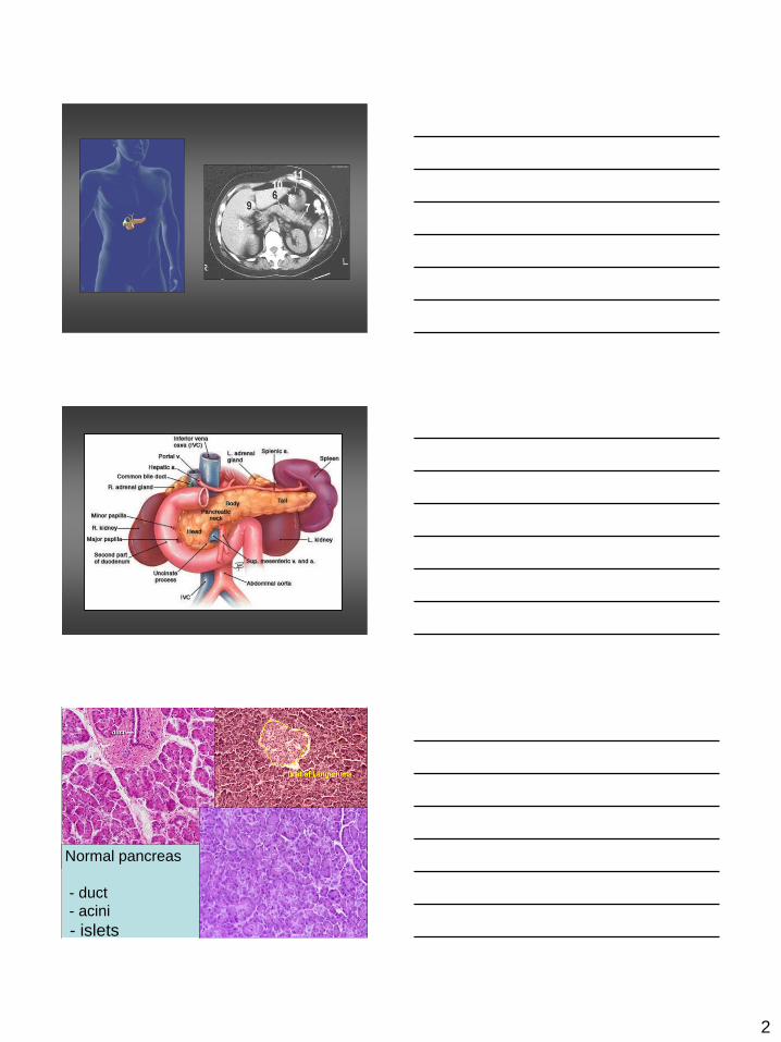
Normal pancreas
- duct
- acini
- islets
Page 3
3
Normal duct cells
Normal acinar cells
Benign ductal sheets - Giemsa
Duodeal sheet, goblet
cells
Gastric epithelium with mucus
Page 4
4
Duodenal epithelium with goblet cells
Pancreas: FNA procedures
• Intra-operative FNA (palpation)
• Transabdominal FNA
- ultrasound guidance
- CT guidance
ENDOSCOPIC ULTRASOUND-
GUIDED FNA (EUS-FNA)
Upper GI endoscopy
The Endoscope Ultrasound probe and needle
Page 5
5
CT scan performed for lung cancer surveillance CT scan performed for lung cancer surveillance
EBUS image of 2cm pancreatic tail cyst Same cyst at EUS
Page 6
6
Acute pancreatitis: Syndromes
• Acute edematous pancreatitis
Early treatment Low mortality
• Acute hemorrhagic pancreatitis
Pancreatic necrosis
Enzymatic digestion of pancreas
Peri-pancreatic enzyme leak
Medical emergency
High mortality
FNA plays no part in diagnosis of acute pancreatitis
Severe abdominal, back pain
High amylase, later lipase
Hemolysis, DIC and ARDS
Diffuse fat necrosis calcification
Peripheral vascular collapse
Acute onset renal failure
Shock, death
High power: Acute pancreatitis with loss of parenchyma,
Duct erosion, numerous polymorphonuclear leucocytes
Duct remnant
Massive inflammation L Loss of acini
Dilated vessels
Page 7
7
Chronic pancreatitis:
Morphology
Gross: Hard gland with dilated ducts, visible
calcification
Microscopic:
• Fibrosis of parenchyma
• Reduced number and size of acini
• Relative sparing of islets early
• Dilatation and /or fibrosis ducts
• Later loss of islets
Chronic pancreatitis
Massive fibrosis
Chronic pancreatitis
Calcification
Chronic pancreatitis:
Morphology
Loss of acini, sparing of islets, dense fibrosis, lymphocytes
Total loss of acini
Duct proliferation
Fibrosis
Islets of Langerhans remain
Page 8
8
Images of Chronic pancreatitis
FNA
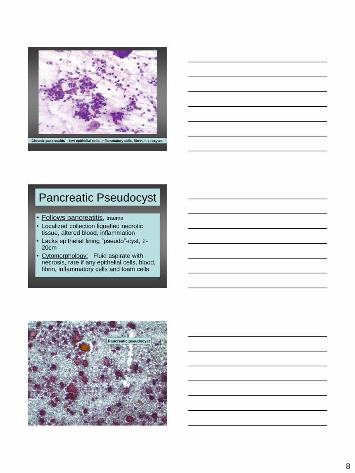
Chronic pancreatitis - few epithelial cells, inflammatory cells, fibrin, histiocytes
Pancreatic Pseudocyst
• Follows pancreatitis, trauma
• Localized collection liquefied necrotic tissue, altered blood, inflammation
• Lacks epithelial lining “pseudo”-cyst, 2-20cm
• Cytomorphology: Fluid aspirate with necrosis, rare if any epithelial cells, blood, fibrin, inflammatory cells and foam cells.
Pancreatic pseudocyst
Page 9
9
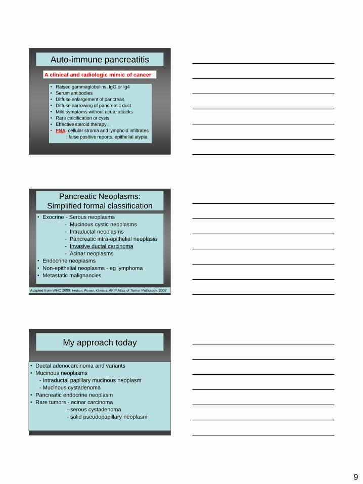
Auto-immune pancreatitis
• Raised gammaglobulins, IgG or Ig4
• Serum antibodies
• Diffuse enlargement of pancreas
• Diffuse narrowing of pancreatic duct
• Mild symptoms without acute attacks
• Rare calcification or cysts
• Effective steroid therapy
• FNA: cellular stroma and lymphoid infiltrates
: false positive reports, epithelial atypia
A clinical and radiologic mimic of cancer
Pancreatic Neoplasms:
Simplified formal classification
• Exocrine - Serous neoplasms
- Mucinous cystic neoplasms
- Intraductal neoplasms
- Pancreatic intra-epithelial neoplasia
- Invasive ductal carcinoma
- Acinar neoplasms
• Endocrine neoplasms
• Non-epithelial neoplasms - eg lymphoma
• Metastatic malignancies
Adapted from WHO 2000: Hruban, Pitman, Klimstra: AFIP Atlas of Tumor Pathology, 2007
My approach today
• Ductal adenocarcinoma and variants
• Mucinous neoplasms
- Intraductal papillary mucinous neoplasm
- Mucinous cystadenoma
• Pancreatic endocrine neoplasm
• Rare tumors - acinar carcinoma
- serous cystadenoma
- solid pseudopapillary neoplasm
Page 10
10
Invasive ductal cancer:
Incidence and survival
• Fourth highest incidence in USA (lung, breast, colon)
• M > F and B > W, middle/older ages
• National Cancer Institute 2008 figures New cases: 37,680 Deaths: 34,290
• Dismal prognosis in spite of therapy
• Five year survival minimal (<5%)
Invasive ductal cancer:
Clinical features
• 60% arise in head, fewer in body or tail
• Risks: alcohol, smoking chronic pancreatitis
• Asymptomatic >> late weight loss, anorexia, pain
• Jaundice if bile duct obstructed in head
• Trousseau’s sign, migratory thrombosis
• Most inoperable at diagnosis, 5% 5yr survival
• Surgery is Whipple’s or partial pancreatectomy
• Chemotherapy, radiation therapy, palliative
Cytomorphology: Adenocarcinoma
• Cellularity usually high
• Disorderly monolayer sheets
• Single columnar cells and small clusters
• Scant or mucinous cytoplasm
• Nuclear enlargement, pleomorphism
• Irregular nuclear outlines
• Chromatin granular, pale and marginated
• Nucleoli and mitotic figures
Page 11
11
Low power - high cellularity
Irregular sheet, cytoplasmic mucin,
irregular nucleoli
Crowded sheet, margination
Page 12
12
Giemsa – Crowding and nuclear pleomorphism
Nuclear convolutions, cytoplasmic vacuoles
Giemsa, mucus, single tumor cells
Page 13
13
Nuclear pallor and grooves
Mucinous adenocarcinoma
Intensive multinucleation
Page 14
14
Criteria: Well-Differentiated Ductal
Cancer Lin & Staerkel, MD Anderson, 71/291 cases
Cancer Cytopathology 2003; 99:44-50
1) Anisonucleosis (3-4x)
2) Nuclear membrane irregularity
3) Nuclear enlargement (2x RBCs)
4) Architecture - 2D aggregates, crowding, overlapping, loss of polarity
__________________________________________
Of NO use in WD tumors: Single cells, prominent nucleoli, chromatin disturbances, molding, mitotic figures, NC ratio, necrosis
Well differentiated adenocarcinoma
Well differentiated adenocarcinoma
Page 15
15
Well differentiated adenocarcinoma
Adenocarcinoma variants
• Adenosquamous carcinoma
• Signet ring adenocarcinoma
• Anaplastic carcinoma
- with osteoclastic giant cells
- with pleomorphic giant cells
- with both!
Adenosquamous carcinoma
Page 16
16
Fragment with osteoclastic giant cells
Single osteoclastic giant cell
Anaplastic carcinoma pancreas
Page 17
17
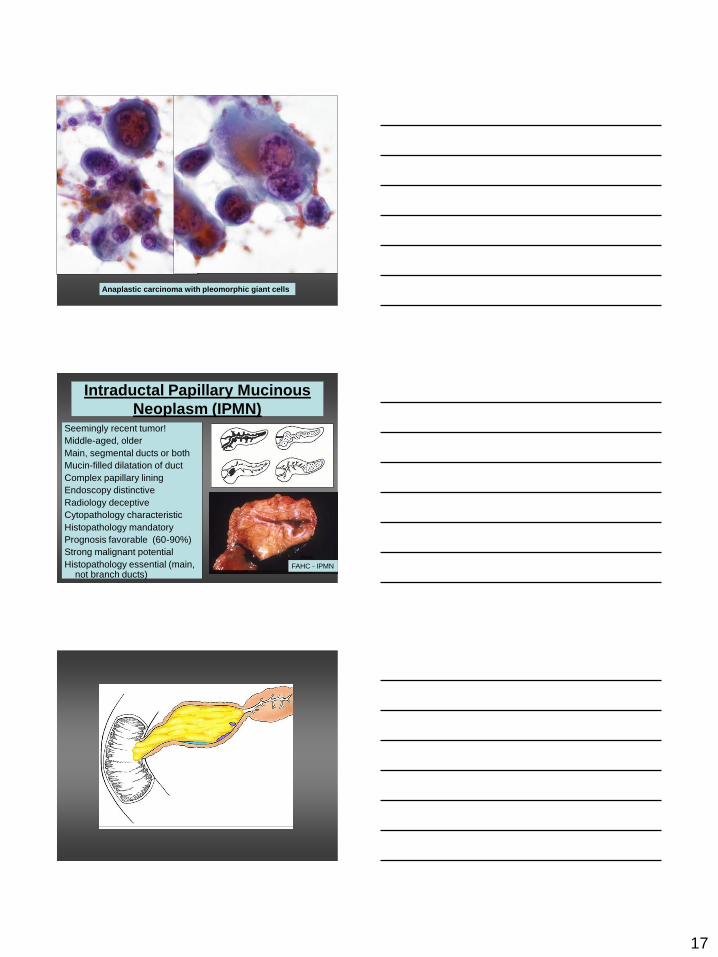
Anaplastic carcinoma with pleomorphic giant cells
Intraductal Papillary Mucinous
Neoplasm (IPMN) Seemingly recent tumor!
Middle-aged, older
Main, segmental ducts or both
Mucin-filled dilatation of duct
Complex papillary lining
Endoscopy distinctive
Radiology deceptive
Cytopathology characteristic
Histopathology mandatory
Prognosis favorable (60-90%)
Strong malignant potential
Histopathology essential (main, not branch ducts)
FAHC
Fa FAHC - IPMN
Page 18
18
Invasive IPMN
vs solid adenoca
Invasive IPMN
vs solid adenoca
- no nodes
Invasive IPMN
vs solid adenoca
- with nodes
Cancer 2010;116, 3369
Low power IPMN – thick papillary fragments
Intracellular and extracellular mucin
Page 19
19
High power IPMN – papilla and
Cytoplasmic mucin
Mucinous Cystadenoma
Middle-aged females, body and tail
No connection with pancreatic duct
Unilocular, multilocular, mucin-filled
Thick capsule, ovarian-type stroma
Smooth (~papillary) lining
Radiology/cytopathology distinctive
Prognosis intermediate (35-45%)
Adenoma-carcinoma sequence
Excision/histopathology essential
Page 20
20
Mucinous cystadenoma
Further notes: IPMN and MCA
• Spectrum of cytopathology from benign
through borderline to malignant
• Cytodiagnosis should reflect that – but
“Mucinous cystic lesion” permissible.
• Both entities have high malignant potential
• Histopathology essential in both
• Differential for both is benign gastric
epithelium!
Martha Bishop Pitman –
GI epithelium
Page 21
21
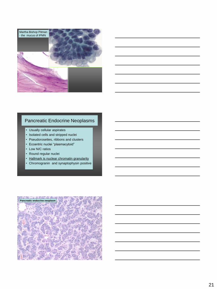
Martha Bishop Pitman
- the mucus of IPMN
Pancreatic Endocrine Neoplasms
• Usually cellular aspirates
• Isolated cells and stripped nuclei
• Pseudorosettes, ribbons and clusters
• Eccentric nuclei “plasmacytoid”
• Low N/C ratios
• Round regular nuclei
• Hallmark is nuclear chromatin granularity
• Chromogranin and synaptophysin positive
Pancreatic endocrine neoplasm
Page 22
22
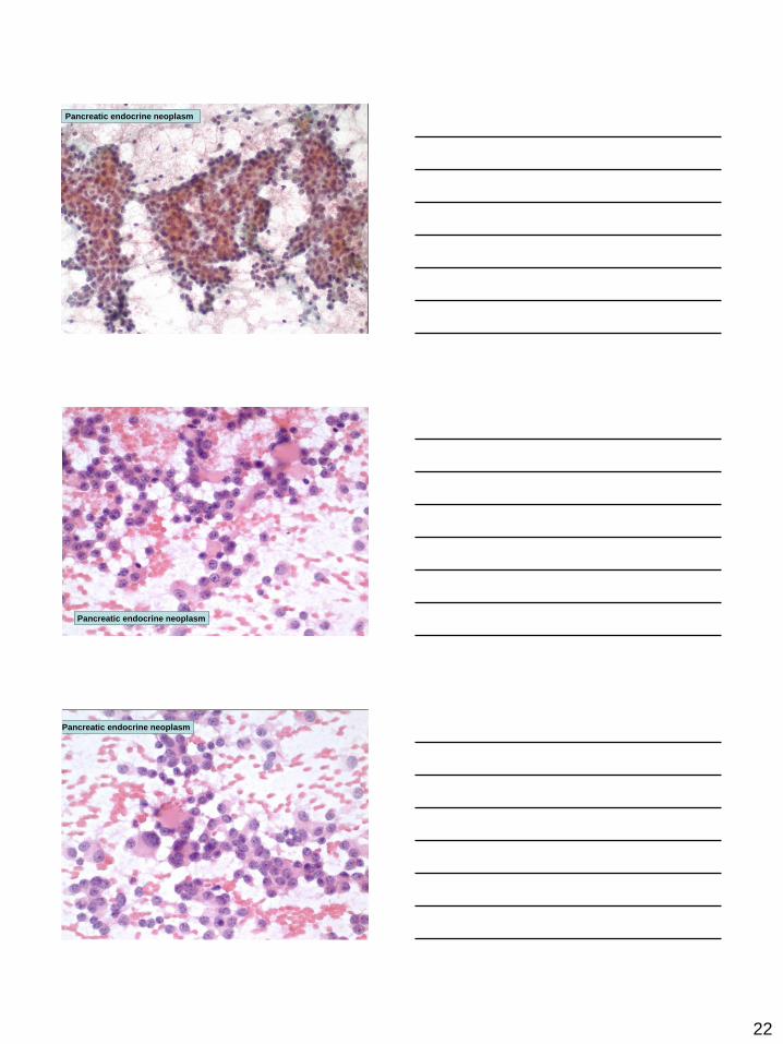
Pancreatic Endocrine neoplasm - LP
Pancreatic endocrine neoplasm
Pancreatic endocrine neoplasm
Pancreatic endocrine neoplasm
Page 23
23
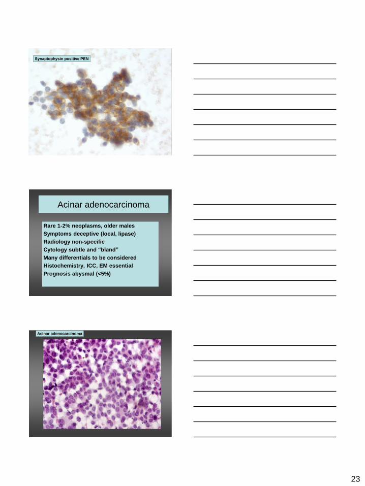
Synaptophysin positive PEN
Acinar adenocarcinoma
Rare 1-2% neoplasms, older males
Symptoms deceptive (local, lipase)
Radiology non-specific
Cytology subtle and “bland”
Many differentials to be considered
Histochemistry, ICC, EM essential
Prognosis abysmal (<5%)
Acinar adenocarcinoma
Page 24
24
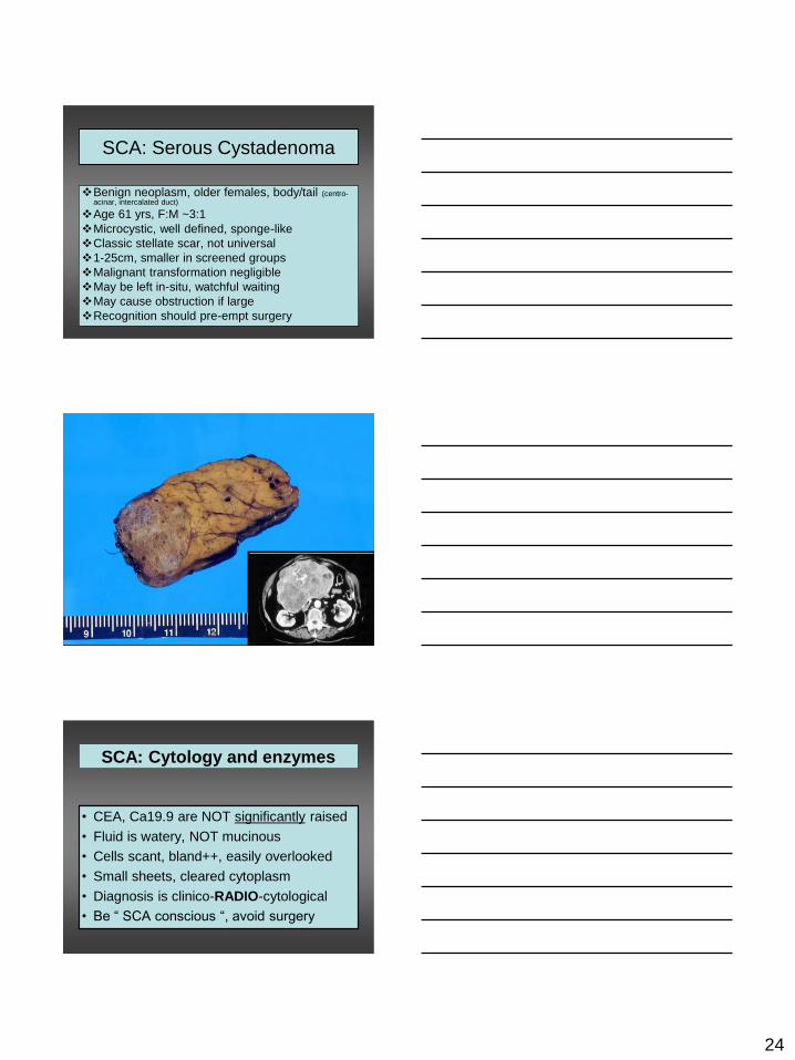
SCA: Serous Cystadenoma
Benign neoplasm, older females, body/tail (centro-acinar, intercalated duct)
Age 61 yrs, F:M ~3:1
Microcystic, well defined, sponge-like
Classic stellate scar, not universal
1-25cm, smaller in screened groups
Malignant transformation negligible
May be left in-situ, watchful waiting
May cause obstruction if large
Recognition should pre-empt surgery
SCA: Cytology and enzymes
• CEA, Ca19.9 are NOT significantly raised
• Fluid is watery, NOT mucinous
• Cells scant, bland++, easily overlooked
• Small sheets, cleared cytoplasm
• Diagnosis is clinico-RADIO-cytological
• Be “ SCA conscious “, avoid surgery
Page 25
25
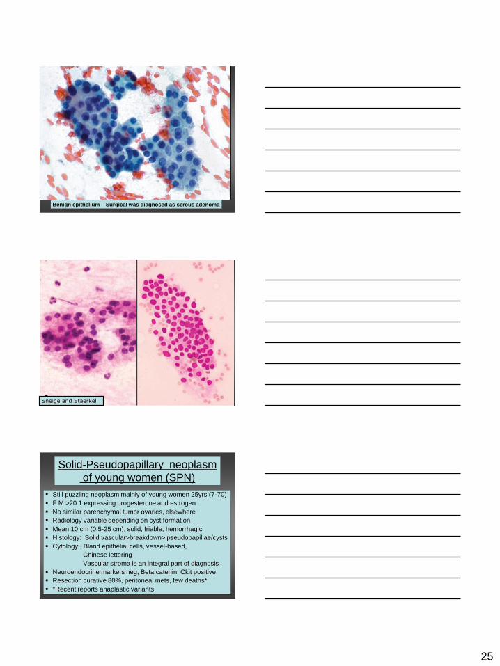
Benign epithelium – Surgical was diagnosed as serous adenoma
Sneige and Staerkel
Solid-Pseudopapillary neoplasm
of young women (SPN)
Still puzzling neoplasm mainly of young women 25yrs (7-70)
F:M >20:1 expressing progesterone and estrogen
No similar parenchymal tumor ovaries, elsewhere
Radiology variable depending on cyst formation
Mean 10 cm (0.5-25 cm), solid, friable, hemorrhagic
Histology: Solid vascular>breakdown> pseudopapillae/cysts
Cytology: Bland epithelial cells, vessel-based,
Chinese lettering
Vascular stroma is an integral part of diagnosis
Neuroendocrine markers neg, Beta catenin, Ckit positive
Resection curative 80%, peritoneal mets, few deaths*
*Recent reports anaplastic variants
Page 26
26
Initially solid growth
Later pseudo-papillary breakdown
SPN – Papanicolaou stain with faint central stromal cores
Page 27
27
Cover Diagnostic Cytopathology – Chinese lettering SPN
Beta-catenin
Page 28
28
Pancreatic non-Hodgkins
lymphoma –large cell B
Pancreatic lymphoma
• In adjacent nodes > or parenchymal
• Non-Hodgkins > Hodgkins
• Large cell B-lymphomas predominate
• MALT lymphomas well described
• All types have better prognosis cf cancer
• Biopsy often tricky / dangerous
• Flow cytometry and FNA diagnostic
The End