Page 1
PaO and the canola green seed problem
Research Area: Environmental Stress and Adaptation
Corresponding author: Donald R. Ort
Department of Plant Biology
190 ERML, 1201 W. Gregory Drive
University of Illinois
Urbana, IL 61801, USA
(tel) 217-333-2093
(fax) 217-244-0656
(e-mail) [email protected]
1
Plant Physiology Preview. Published on July 21, 2006, as DOI:10.1104/pp.106.084483
Copyright 2006 by the American Society of Plant Biologists
www.plantphysiol.orgon September 22, 2018 - Published by Downloaded from Copyright © 2006 American Society of Plant Biologists. All rights reserved.
Page 2
The Role of Pheophorbide A Oxygenase Expression and Activity in the Canola
Green Seed Problem1
Davyd W. Chung, Adriana Pružinská, Stefan Hörtensteiner, and Donald R. Ort*
Department of Plant Biology (D.W.C., D.R.O.), University of Illinois, Urbana, Illinois
61801; and Department of Biology, University of Bern, Altenbergrain 21, CH-3013 Bern,
Switzerland (A.P., S.H.); Photosynthesis Research Unit, USDA/ARS (D.R.O.) Urbana,
Illinois 61801
2 www.plantphysiol.orgon September 22, 2018 - Published by Downloaded from
Copyright © 2006 American Society of Plant Biologists. All rights reserved.
Page 3
1 This work was supported in part by the Integrative Photosynthesis Research Training
Grant from the Department of Energy (no. DEFGO2-92ER20095) funded under the
Program for Collaborative Research in Plant Biology and by a grant (no. 3100A0-
105389) from the Swiss National Science Foundation.
*Corresponding author; e-mail [email protected] ; fax 217-244-0656.
3 www.plantphysiol.orgon September 22, 2018 - Published by Downloaded from
Copyright © 2006 American Society of Plant Biologists. All rights reserved.
Page 4
Abstract
Under normal field growth conditions, canola (Brassica napus) seeds produce
chloroplasts during early seed development and then catabolize the photosynthetic
machinery during seed maturation, producing mature seeds at harvest that are essentially
free of chlorophyll. However, frost exposure early in canola seed development disrupts
the normal programming of chlorophyll degradation resulting in green seed at harvest
thereby significantly devaluing the crop. Pheophorbide a oxygenase (PaO), a key control
point in the overall regulation of chlorophyll degradation, was affected by freezing.
Pheophorbide a, the substrate of PaO, accumulated during late stages of maturation in
seeds that had been exposed to freezing during early seed development. Freezing
interfered with the induction of PaO activity that normally occurs in the later phases of
canola seed development when chlorophyll should be cleared from the seed. Moreover,
we found that the induction of PaO activity in canola seed was largely post-
translationally controlled and it was at this level that freezing interfered with PaO
activation. The increased accumulation of PaO transcript and protein levels during seed
development was not altered by the freezing episode and the increase in PaO protein was
small compared to the increase in PaO activity. We found that PaO could be
phosphorylated and that phosphorylation decreased with increasing activity implicating
PaO dephosphorylation as an important post-translational control mechanism for this
enzyme. Two PaO genes, BnPaO1 and BnPaO2, were identified in senescing canola
leaves and during early seed development but only BnPaO2 was expressed in maturing,
degreening seeds.
4 www.plantphysiol.orgon September 22, 2018 - Published by Downloaded from
Copyright © 2006 American Society of Plant Biologists. All rights reserved.
Page 5
Introduction
Brassica napus, canola, is an important oil seed crop grown extensively in North
America and northern Europe with annual yields exceeding seven million metric tons.
Canola is the world’s third most important vegetable oil crop, in significant part due to
the low levels of erucic acid and glucosinolates in canola oil (Levadoux et al., 1987;
Zhang et al., 2004). However, the chlorophyll (Chl) content is significantly higher than
that found in other major vegetable oils and is the biggest quality impediment in the
canola oil industry.
During the early stages of seed development, photosynthetically produced
carbohydrate is transferred from the leaves and silique walls to the seeds for the synthesis
of oil and other storage products. In canola seeds, the conversion of sugars to fatty acids
is the primary metabolic flux with more than 60% of carbon stored as oil (Schwender et
al., 2004a). There is evidence that developing embryos are capable of significant rates of
photosynthesis directly associated with fatty acid biosynthesis of the developing seed
(Eastmond et al., 1996; Willms et al., 1999; Ruuska et al., 2004). However, the low light
levels able to reach the seeds through the silique walls made it difficult to explain the
significance of photosynthesis in oil biosynthesis within the seed. This conundrum was
explained by the recent discovery that rubisco in developing B. napus embryos can use
the energy of photosynthesis while operating in a novel pathway that produces acetyl-
CoA with much greater efficiency than the previously described glycolytic pathway
(Schwender et al., 2004b). As the seed matures and the rate of oil synthesis declines, the
need for photosynthesis declines and chloroplasts are degraded resulting in seeds at
harvest free of Chl. Although canola is in general a cold hardy plant, a freezing episode
early in seed development can disrupt the normal program of Chl degradation, resulting
in a green seed at harvest and significantly devaluing the crop (Johnson-Flanagan and
Thiagarajah, 1990). This so-called “green seed” problem is a high priority seed quality
issue within the canola industry.
The key reactions in Chl degradation are catalyzed by chloroplast localized
enzymes (Matile et al., 1999; Hörtensteiner, 2006). Chl is removed from the Chl binding
proteins within the thylakoid membranes by a yet undescribed process. Once free from
the membrane, the initial step in Chl degradation is the removal of the phytol tail by
5 www.plantphysiol.orgon September 22, 2018 - Published by Downloaded from
Copyright © 2006 American Society of Plant Biologists. All rights reserved.
Page 6
chlorophyllase (Chlase), which catalyzes hydrolysis of the ester linkage of the phytol
chain to the porphyrin macrocycle (Matile et al., 1999; Tsuchiya et al., 1999; Jacob-Wilk
et al., 1999). In Arabidopsis thaliana (Arabidopsis), there are two known Chlase genes.
AtCLH1 appears to be induced in response to wounding and pathogen attack (Kariola et
al., 2005), whereas AtCLH2 expression is constitutive. Chlase activity is at least in some
cases latent prior to the onset of senescence (Benedetti and Arruda, 2002), but in other
cases, high Chlase activity has been associated with high rates of Chl synthesis (Roca and
Minguez-Mosquera, 2003) making it unlikely that Chlase is a central controlling step in
Chl degradation.
Mg-dechelatase, for which the gene and protein are yet to be identified, is
responsible for removing the magnesium ion from the tetrapyrrole producing the chlorin
molecule pheophorbide (Pheide) a (Shioi et al., 1996). Although the release of Mg2+
could in principle occur spontaneously, Mg-dechelatase activity has been demonstrated
with the artificial substrate chlorophyllin for which spontaneous Mg2+ removal is unlikely
(Shioi et al., 1996). Removal of the magnesium ion from the macrocycle prepares it for
pheophorbide a oxygenase (PaO), which opens the macrocycle of Pheide a resulting in
the final disappearance of the green color (Rodoni et al., 1997).
PaO, a nonheme iron monooxygenase localized to the inner envelope of maturing
gerontoplasts, opens the porphyrin macrocycle by adding two oxygen atoms (Matile and
Schellenberg., 1996). Pheide a has been shown to be an efficient substrate in PaO activity
measurements, whereas Pheide b acts as a competitive inhibitor (Hörtensteiner et al.,
1995; Pružinská et al., 2003). In Arabidopsis, AtPaO belongs to a five-member gene
family encoding nonheme iron oxygenases defined by the presence of a Rieske-type
domain in addition to a mononuclear iron-binding domain. This gene family also includes
Chl a oxygenase, choline monoxygenase, Tic55, and Ptc52 (Gray et al., 2004). The
Arabidopsis cell death mimic mutant accelerated cell death 1 (acd1) is an AtPaO mutant
allele (Pružinská et al., 2003) and is orthologous to lethal leaf spot 1 (lls1) of Zea mays
(maize) (Gray et al., 2004). The mutant leaves of both plants accumulate Pheide a
making them highly photosensitive thereby producing the cell death mimic phenotype.
The conversion of Pheide a to the colorless primary fluorescent Chl catabolite
(pFCC) is a complex step which involves not only PaO, but also red chlorophyll
6 www.plantphysiol.orgon September 22, 2018 - Published by Downloaded from
Copyright © 2006 American Society of Plant Biologists. All rights reserved.
Page 7
catabolite reductase (RCCR), a stromal protein, and is the causal gene in accelerated cell
death 2 (acd2) mutant of Arabidopsis. The opening of the Pheide a macrocycle by PaO
produces a red colored catabolite (RCC), an intermediary product, which in turn is
reduced by RCCR in a reaction requiring ferredoxin to form pFCC, a colorless compound
that is detected by its distinctive blue fluorescence (Wüthrich et al., 2000). The RCCR
gene is expressed in most tissues, even roots (Mach et al., 2001; Yao and Greenberg,
2006), and is constitutively active throughout leaf development including senescence.
Moreover, RCCR protein levels do not change during senescence or pathogen attack
(Mach et al., 2001) removing it from consideration as a significant control step in Chl
degradation. The final steps of Chl degradation involve the hydroxylation and
conjugation of the pFCC tetrapyrroles. FCCs (fluorescent Chl catabolites) are exported
from the gerontoplasts for further modification in the cytosol. The modified FCCs are
imported to the vacuole eventually leading to the formation of non-colored catabolites
(NCCs), which are not phototoxic and stored in the vacuoles (Oberhuber et al., 2003).
The major objective of this study was to identify those steps in Chl degradation in
maturing canola seeds that are disrupted by exposure to freezing temperatures early in
seed development. The results show that freezing interfered with the induction of PaO
activity that normally occurs in the later phases of canola seed development. Moreover,
we found that the regulation of PaO activity was largely post-translational and it was at
this level that freezing interfered with PaO activation in canola seeds. RESULTS
Non-lethal freezing exposure prevented the complete clearing of chlorophyll from
mature canola seeds.
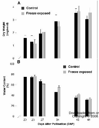
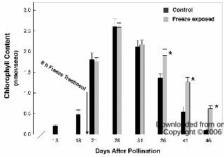
At 20 days after pollination (DAP), when seeds had attained about 45% of final
dry weight (Fig. 1A) and 60% of maximum Chl content (Fig. 2), canola plants were
cooled in the dark at 5°C / h until reaching –4° C, where the temperature was held for 6 h
followed by rewarming at 5°C / h back to the growth temperature. The Chl content of
seeds collected at intervals from 13 to 46 DAP was measured spectrophotometrically in
N, N-dimethylformamide using the specific absorption coefficients of Porra and Grimme
(1974) and expressed on the basis of seed fresh weight. The rate of Chl accumulation
increased over the first 26 DAP with the greatest increase occurring between 18 and 21
7 www.plantphysiol.orgon September 22, 2018 - Published by Downloaded from
Copyright © 2006 American Society of Plant Biologists. All rights reserved.
Page 8
DAP during which time there was nearly a 4-fold increase in Chl content per seed (Fig
2). The freeze exposure at 20 DAP had no significant affect on maximum Chl content
(Fig. 2) or the fresh (data not shown) or dry weight (Fig. 1A) of the developing seeds. Net
Chl degradation was initiated sometime after 26 DAP in both control and freeze-exposed
samples with Chl degraded to trace amounts by 46 DAP in control canola seeds. The
freezing-induced delay in Chl loss became evident at 36 DAP, and thereafter the rate of
net degradation in freeze-exposed seeds proceeded more slowly. Chl degradation stopped
when the seed moisture content dropped below ~ 40% (cv. Figs 1B and 2). The freezing-
induced delay in Chl degradation resulted in mature seeds with ~600% higher Chl
content than control (Fig. 2).
The effects of freeze exposure on Chl loss was direct and not mediated by
differential effects of freezing on seed moisture content.
Although the interference with developmentally programmed Chl degradation in
maturing canola seeds by freeze exposure is well established (Johnson-Flanagan and
Thiagarajah, 1990), it has been suggested that the effect is indirectly mediated by
acceleration of seed water loss from freeze-exposed plants (Green et al., 1998). Work was
done to attempt to separate the direct effects of freeze exposure from ancillary effects of
water loss on Chl degradation by maintaining high humidity during the freeze exposure
and recovery period. Seed water content was calculated as (fresh weight – dry weight) /
fresh weight and the dry weight of the seeds was determined following overnight
incubation at 75° C (Fig. 1A). The decline in water content was gradual over the period
of seed development (Fig. 1B) with greater than 50% water loss between 23 and 48 DAP.
Under the high humidity conditions, water content of seeds from plants exposed to the
freezing treatment was the same as controls, yet the freeze-exposed seeds retained higher
Chl levels. This result demonstrates that these two factors, freeze and water loss, affect
Chl degradation independently and can be separated experimentally.
Pheophorbide a accumulated in maturing canola seeds after freeze exposure.
In order to determine candidate steps in the Chl degradation of canola seed that
may be sensitive to freeze exposure, we investigated the effects of freeze exposure on
pools of Chl degradation catabolites during canola seed development (Fig. 3). The
catabolites were separated by HPLC based on their polarity in organic solvent and were
8 www.plantphysiol.orgon September 22, 2018 - Published by Downloaded from
Copyright © 2006 American Society of Plant Biologists. All rights reserved.
Page 9
quantified by fluorescence spectroscopy, which has the sensitivity required to detect Chl
catabolites in the trace amounts normally present. The concentration of each catabolite
was determined from the fluorescence intensity data using equations developed for the
quantification of tetrapyrrole moieties (Rebeiz, 2002).
Freeze exposure on 20 DAP had little effect on catabolite pool sizes until eight
days later. By 28 DAP there was a 3 to 4 fold freeze-induced increase in Pheide a levels.
The accumulation of Pheide a in the freeze-treated seeds became more exaggerated as
seed development progressed showing nearly a 10 fold increase compared to control by
46 DAP (Fig. 3). During the later stages of seed development, 35 DAP and after, freeze-
treatment induced chlorophyllide (Chlide) a accumulation and at seed maturity (i.e., 46
DAP) the percent increase of Chlide a exceeded that of Pheide a. The increased levels of
Pheide a, and eventually Chlide a, in freeze-exposed seeds suggested that freezing
interferes in some fashion with PaO function. That Pheide a accumulation preceded
Chlide a accumulation suggested that a progressive feedback within the degradation
pathway had developed.
In principle, a decrease in the products of PaO would also be anticipated if the
increase in Pheide a in freeze-treated seeds is due to a decrease in PaO activity. However
in senescing leaves, pFCC, the product of the PaO/RCCR reaction, is present in
exceedingly low and difficult to quantify amounts and FCCs and RCCs do not
accumulate to detectable levels. NCCs are the only products downstream of PaO that
accumulate in senescing leaves in canola (Pružinská et al. 2005). Neither FCCs nor RCCs
accumulated to detectable levels in either control or freeze-treated seeds (data not
shown). Thus, while we were unable to confirm that a decrease in product of the PaO
reaction accompanied the increase in substrate, the lack of accumulation of downstream
product is strong evidence that freeze-treatment had no significant effect on chlorophyll
degradation reactions downstream of PaO.
Brassica napus found to have two PaO genes with high homology to AtPaO.
The gene sequence of PaO from Arabidopsis was used to clone and identify the
orthologous genes in B. napus. Two different cDNA clones of PaO were isolated from
senescing canola leaves (Supplemental Fig. S1), which will be referred to as BnPaO1 and
BnPaO2. The codon derived amino acid sequences had 92% identity with AtPaO. In
9 www.plantphysiol.orgon September 22, 2018 - Published by Downloaded from
Copyright © 2006 American Society of Plant Biologists. All rights reserved.
Page 10
comparison to each other, the derived amino acid sequences of the two canola clones
were 98% identical. Like PaO from corn and Arabidopsis (Gray et al., 2004), the two
canola genes contain a conserved Rieske iron-sulfur domain and a mononuclear iron-
binding domain as well as two predicted transmembrane domains.
There are two notable differences in protein sequence at the N-terminal region of
BnPaO1, BnPaO2 and AtPaO, possibly due to an insertion/deletion event post-dating the
divergence of Arabidopsis and canola. (Supplemental Fig. S2). Alternatively, as canola is
an allotetraploid, BnPaO1 and BnPaO2 could be derived from the two ancestral genomes
of canola. BnPaO2 has an additional serine residue (S-29) when compared to BnPaO1
and AtPaO. In the same region, AtPaO has two threonine residues (T-27, T-28)
positioned where both BnPaO clones have an asparagine and a serine residue (N-27, S-
28). Both BnPaO clones have an extra alanine residue (A-30) not found in AtPaO.
Furthermore in the 75-78 region, BnPaO2 is completely missing a sequence of G-D-K-E
found in both AtPaO and BnPaO1. Since BnPaO2 has an additional serine residue (S-29)
and is missing G-75, D-76, K-77 and E-78, collectively this protein has 3 fewer amino
acids than BnPaO1. AtPaO has one less amino acid than BnPaO1 due to the missing
alanine residue (A-30).
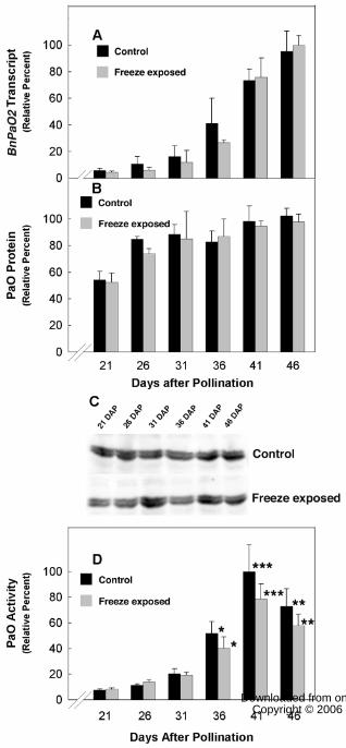
While the expression of BnPaO2 was measurable in seeds throughout seed
development, BnPaO1 transcripts were detectable only during early seed development.
At 8 to 10 DAP, BnPaO2 transcripts showed nearly 5.5 fold higher levels of expression
when compared to BnPaO1 transcripts and BnPaO2 transcripts at 8 to 10 DAP were
expressed at similar levels to 21 DAP canola seeds (data not shown). BnPaO2 transcripts
accumulated as seed development progressed with >10 fold increase from 21 to 41 DAP
and the accumulation was not affected by the 6 h freezing exposure on 20 DAP (Fig. 4A).
The expression of both BnPaO transcripts was readily detectable in senescing canola
leaves (data not shown).
Canola seed PaO shown to be regulated by a freezing-sensitive, post-translational
mechanism.
The amount of PaO protein from the membrane fraction of developing canola
seeds was measured by immunoblot analysis and quantified by infrared imaging. PaO
protein levels increased only about two fold (Fig. 4B) over the period of seed
10 www.plantphysiol.orgon September 22, 2018 - Published by Downloaded from
Copyright © 2006 American Society of Plant Biologists. All rights reserved.
Page 11
development in which BnPaO2 transcripts increased >10 fold (Fig. 4A). Freezing
exposure given on 20 DAP did not affect PaO protein content at any subsequent point
during seed development. The immunoreactive complexes of PaO protein from canola
seed resolved into a doublet on miniblots using 10% or lower acrylamide, differing in
apparent molecular mass by approximately 0.5 kD (Fig. 4C). Only the lighter, bottom
band showed increasing intensity as the seeds matured. No effect of freezing was evident
for either PaO band.
Whereas PaO protein levels only doubled between 21 and 41 DAP and were
unaffected by freezing exposure, PaO activity increased more than 10 fold over this
period and this induction was suppressed >20% by freezing (Fig. 4D). That the increase
in PaO activity was at least five times greater than the increase in PaO protein implies
strong post-translational regulation of PaO during seed maturation.
Evidence for dynamic phosphorylation of canola seed PaO.
Analysis of the BnPaO2 codon derived amino acid sequence revealed two
potential serine/threonine calcium-dependent protein kinase (CDPK) phosphorylation
recognition sites at S18 and T402 (Supplemental Fig. S1). These candidate
phosphorylation sites are fully conserved in BnPaO1 and AtPaO. To investigate if there
were changes in PaO phosphorylation corresponding to the changes we had observed in
PaO activity, we used immobilized metal affinity chromatography (IMAC), which
separates phosphorylated and nonphosphorylated proteins by binding the phosphorylated
form, followed by immunoblot analysis.
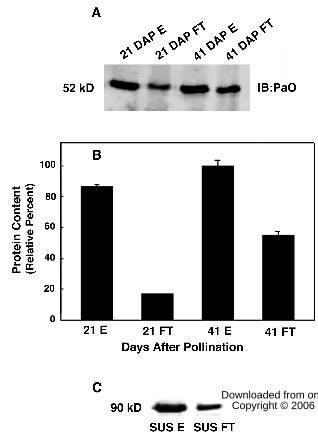
PaO phosphorylation was measured at 21 DAP when significant PaO protein was
present (Fig. 4B) but activity was low (Fig. 4D), and compared with 41 DAP when PaO
protein and activity levels were greatest. At 21 DAP, PaO was detected in both
phosphorylated and nonphosphorylated fractions, however, the phosphorylated fraction
contained nearly 5 fold higher amounts of PaO protein (Fig. 5A). At 41 DAP,
phosphorylated PaO proteins did not show a significant increase from 21 DAP, however,
the nonphosphorylated fraction increased by 3 fold showing that the increase in PaO
content between 21 and 41 DAP could be nearly accounted for by the nonphosphorylated
form. Neither antiphos-Thr nor antiphos-Ser antibodies (Zymed, Carlsbad, CA) reacted
with PaO from either fraction (data not shown).
11 www.plantphysiol.orgon September 22, 2018 - Published by Downloaded from
Copyright © 2006 American Society of Plant Biologists. All rights reserved.
Page 12
As validation of this approach to investigate PaO phosphorylation, we used IMAC
columns with sucrose synthase (SUS), which has two well-known phosphorylation sites
at S15 and S170 (Huber and Huber, 1996; Winter et al., 1997; Hardin et al., 2004).
Phosphorylated SUS was isolated from basal elongating maize leaf tissue and purified by
anion exchange chromatography. After separation on the phosphoprotein affinity column,
a SUS phosphospecific antibody detected phosphorylated SUS in both the fraction
initially immobilized and subsequently eluted from the column with phosphate as well as
in the flow-through fraction that did not bind to the column (Fig. 5C). The proportion of
phosphorylated SUS in the bound versus unbound fraction was 5.6 to 1 when 0.5 mg of
SUS was loaded on the column but only 2.45 to 1 when loading was increased to 1 mg.
This result is indicative of exceeding the phosphoprotein binding capacity of the column,
which is reported to have a maximum phosphoprotein binding capacity of 0.5 mg
(Qiagen PhosphoProtein Purification Handbook, 2002). Nevertheless, because the PaO
containing lysate loaded on the IMAC column contained only 10 to 20% of the total
phosphoprotein of the SUS samples, overloading, and thus contamination of the unbound
fraction with phospho-PaO, was unlikely.
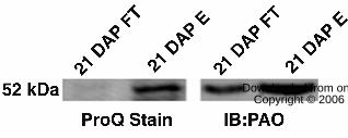
In order to further verify the phosphorylation of PaO, Pro-Q Diamond blot
staining (Invitrogen, Carlsbad, CA) was used to scrutinize the phosphorylated and
nonphosphorylated IMAC fractions collected at 21 DAP (Fig. 6). As expected, Pro-Q
analysis showed no bands representing phosphorylated proteins, including PaO, in the
IMAC flow through fraction, which should contain only nonphosphorylated proteins.
However, a 52 kD band corresponding to PaO (Western blot) was shown to align with a
Pro-Q staining protein of a similar running MW in the phosphorylated fraction eluted
from the IMAC column.
DISCUSSION
There have been numerous demonstrations that the inhibition of PaO activity
during leaf senescence leads to the accumulation of Pheide a and the inhibition of Chl
degradation. In pao1, the insertional knockout mutant of Arabidopsis, ~80% of the Chl is
retained during dark-induced leaf senescence, Chl that is degraded is largely accounted
for by Pheide a accumulation in the leaf as no further down stream products can be
12 www.plantphysiol.orgon September 22, 2018 - Published by Downloaded from
Copyright © 2006 American Society of Plant Biologists. All rights reserved.
Page 13
detected (Pružinská et al. 2005). Similarly, senescing leaves of stay-green mutants of
Festuca pratensis and Lolium temulentum accumulate Chlide and Pheide a, and reduced
PaO activity has been shown to be the biochemical defect in these mutants (Vicentini et
al., 1995; Roca et al. 2004). In freeze-exposed canola seeds, the induction of PaO activity
later in seed development is impaired but not obliterated (Fig. 4D) resulting in a “leaky”
phenotype where Pheide a accumulates (Fig. 3) and Chl degradation is slowed (Fig. 2)
but not stopped. It might be expected that the 20% lower maximum induction of PaO
activity in freeze-exposed maturing canola seeds (Fig. 4D) would prolong but not prevent
full clearing of Chl from the seeds. However, another factor that can ultimately limit Chl
clearing from the maturing seed is seed moisture content and we believe that the
intersection of these two control mechanisms is the cause of the “green seed” problem.
When the moisture content of canola seeds dips below approximately 40%, many aspects
of seed metabolism, including Chl degradation (Green et al., 1998), come to a halt. Under
field conditions, freeze exposure may enhance the rate of seed desiccation (Green et al.,
1998) further exacerbating the effects of the impaired induction of PaO activity on Chl
degradation that are evident even when accelerated desiccation is prevented (Fig. 1B &
4D).
In this work, as with all cases in which PaO activity has been shown to be
impaired (Pružinská et al. 2005, Roca et al., 2004, Pružinská et al. 2003, Hilditch et al,
1989, Bachmann et al. 1994, Tanaka et al. 2003), it is clear that a regulatory mechanism
limiting Chl metabolism is engaged that feeds back, ultimately preventing the removal of
Chl from the thylakoid membrane. That a strong feedback control on Chl degradation is
necessary (Hörtensteiner, 2006; Takamiya et al., 2000) is evident in the lesion-mimic
phenotype of PaO mutants such as acd1 in Arabidopsis (Greenberg and Ausubel, 1993)
and lls1 in maize (Gray et al. 1997) where accumulation of even small amounts of visible
light absorbing chlorophyll metabolites is extremely phototoxic. While no necrotic
patches were observed on freeze-treated canola seeds, this is likely because the seeds
were exposed to only low light intensities. Thus as previously observed in the leaves of
PaO mutants, it is apparent that the freeze treatment of canola seeds also indirectly affects
chlorophyll degradation processes up stream of PaO.
13 www.plantphysiol.orgon September 22, 2018 - Published by Downloaded from
Copyright © 2006 American Society of Plant Biologists. All rights reserved.
Page 14
The profiles of PaO transcript level, protein content, and activity all qualitatively
correlated with the progression of Chl degradation during seed development. However,
PaO protein increased only 2 fold over the measured period of seed development while
transcript levels indicated >10 fold increase in expression. Some of the apparent
discrepancy could be explained if PaO protein were highly stable compared to PaO
transcript. If so, the rate of newly synthesized PaO protein could track the transcript level
yet the amount of new protein would be small in comparison to the accumulated stable
pool. Indeed, PaO protein levels were half their maximum level at 21 DAP (Fig 4B) prior
to any measurable losses of Chl (Fig. 2) and when BnPaO transcript levels (Fig. 4A)
were low. Beyond the dependence of PaO expression on transcript abundance, it is
evident from our results that posttranslational control of the induction of PaO activity
accompanies the degreening of canola seed. Our data showed that PaO protein content
and activity have starkly different profiles during canola seed development. Whereas
freeze exposure caused a statistically significant >20% reduction in the induction of PaO
activity during 36 to 46 DAP freezing was without any significant effect on PaO
transcript or protein amounts.
Although the posttranslational regulatory mechanism is not yet known, it appears
likely that reversible protein phosphorylation is involved. Using both immobilized metal
affinity chromatography and Pro-Q Diamond blot staining we demonstrated a correlation
between PaO dephosphorylation and increasing PaO activity during seed maturation. The
stoichiometry of phosphorylated/dephosphorylated PaO decreased from 5:1 on 21 DAP
to 2:1 on 41 DAP with the dephosphorylated form of PaO increasing more than 3 fold
over this interval. The 10 fold increase in PaO activity (Fig. 4D) illustrates that the
observed change in PaO phosphorylation is large enough to have played a significant,
although perhaps not exclusive, role in the post-translational activation of this enzyme.
There are two CDPK recognition sites in BnPaO1, BnPaO2 and AtPaO protein
sequences (Supplemental Fig. S1). The first CDPK site is located within the putative
chloroplast target sequence, most likely cleaved once the protein is translocated, thus this
CDPK site is not likely to be involved in regulation of the enzyme in the chloroplast. The
common CDPK consensus phosphorylation site is ϕ-x-Basic-x-x-S/T, where the
underlined serine or threonine is phosphorylated, x is any residue, and ϕ is a hydrophobic
14 www.plantphysiol.orgon September 22, 2018 - Published by Downloaded from
Copyright © 2006 American Society of Plant Biologists. All rights reserved.
Page 15
residue (Huang and Huber, 2001). Although CDPKs are reported to associate with
various membranes within the cell (Harper et al., 2004), it is not known yet whether
CDPKs are found in the chloroplast, where it would presumably need to be located to
phosphorylate PaO, which is located on the inner chloroplast envelope. However,
ChloroP (http://www.cbs.dtu.dk/services/ChloroP/) searches indicate that 10 of the 34
putative CDPKs found in Arabidopsis contain hypothetical chloroplast targeting
sequences. Computer analysis of the codon derived PaO protein sequences revealed other
possible phosphorylation sites at various serine and threonine residues
(http://www.cbs.dtu.dk/services/NetPhos). It is interesting to note that the two major
differences near the N-terminus region of the two forms of BnPaO (Supplemental Fig.
S2) removes a nearby serine residue S29 in BnPaO1 as potential serine phosphorylation
sites as well as four amino acids (G-D-K-E).
That the freezing episode and any decrease in the observed rate of chlorophyll
degradation or PaO activity can be separated by more than a week indicates that freezing
does not interfere directly with the PaO protein but with the program controlling Chl
clearing from the seed. Since freezing appears to interfere the activation of PaO by
dephosphorylation, the delayed effect may be mediated at the level of PaO
phosphorylation/dephosphorylation. Cold stress may indirectly lead to changes in protein
phosphorylation and significant changes in CDPK activity by affecting the fluctuations in
cytosolic Ca2+ levels (Martin and Busconi, 2001; Cheng et al., 2002). It has been shown
in various species, including alfalfa and rice, that CDPK activity can be induced by low
temperature (Monroy and Dhindsa, 1995; Saijo et al., 1998), which in the case of PaO
would be expected to increase the ratio of phosphorylated/dephosphorylated PaO and
thereby decrease activity. The doublet seen in the immunoblots of PaO protein from canola seeds (Fig. 4C)
is most likely due to the presence of both BnPaO1 and BnPaO2 proteins in the sample.
The two distinct clones of PaO were isolated and identified from leaves of canola,
differing by 526 D, which would account for the difference between the two bands.
Although BnPaO1 transcript was expressed only early in seed development it seems
likely that its protein product persisted after BnPaO1 transcript disappeared. Only
BnPaO2 transcript was detected at later stages in seeds. Interestingly, the doublet is not
15 www.plantphysiol.orgon September 22, 2018 - Published by Downloaded from
Copyright © 2006 American Society of Plant Biologists. All rights reserved.
Page 16
seen when the protein has been isolated on IMAC columns (Fig. 5 and 6). The protein
retained on the IMAC column (i.e., phosphorylated form) corresponds to the lighter band
of the doublet and is the band that increases during seed development (Fig 4C). That the
doublet is not seen when the sample is run over the IMAC column (Fig. 5 and 6) could
support the notion that phosphorylation of BnPaO2 plays a role in the regulation of PaO
activity in canola seed. Another possibility is that the doublet is due to differential post-
translational modification, including phosphorylation. However, since a single
phosphorylation will add only 80 D it seems unlikely that even multiple sites of
reversible phosphorylation could alone account for the ~500 D difference estimated from
electrophoretic mobility on SDS-PAGE.
The extent to which posttranslational control of PaO operates in leaf senescence is
uncertain. Pružinská et al. (2005) showed a quantitative correlation among PaO activity,
transcript level, and protein level thereby demonstrating that, unlike the situation we
found in canola seeds, posttranslational control was not necessary to explain PaO
regulation during dark-induced senescence of Arabidopsis leaves. Indeed, the original
suggestion of the possible involvement of phosphorylation in PaO regulation (Pružinská
et al., 2003), the inhibition of PaO activity by phosphatase treatment, is the opposite
response that we would expect and was subsequently not reproduced (Pružinská et al.,
2005). While these observations do not eliminate a possible role for phosphorylation in
PaO regulation during Arabidopsis leaf senescence, they do indicate some important
differences in the overall regulation of Chl degradation in senescing leaves and maturing
seeds. Indeed, freezing does not interfere with the timing or extent of Chl degradation in
dark-induced leaf senescence system of either Arabidopsis or canola (data not shown).
Conclusion
Freezing exposure of developing canola seeds hinders the programmed
degreening of the seed by interfering with the post-translationally controlled induction of
PaO activity. Although the rate of Chl degradation is slowed only by ~20%, the inhibition
is sufficient to prevent Chl from fully clearing from the seed before seed moisture content
dips below the threshold at which seed metabolism is suspended. The mechanism of the
post-translational control is unknown but the increase in PaO activity during seed
16 www.plantphysiol.orgon September 22, 2018 - Published by Downloaded from
Copyright © 2006 American Society of Plant Biologists. All rights reserved.
Page 17
maturation corresponds to a decrease in the phosphorylation of the PaO enzyme. Canola
has two highly homologous PaO genes which contain two candidate CDPK
phosphorylation sites.
MATERIAL AND METHODS
Plant Material
Brassica napus L. (canola) cv. Westar seeds were germinated in moist
vermiculite. Two week old seedlings were transplanted to 12 in pots containing Sunshine
Mix LC1 soil (SunGro Horticulture, Inc., Quincy, MI) and grown in growth chambers at
12 h photoperiod of 450 µmol photons m-2s-1 and 22°C day/16°C night thermoperiod
with a relative humidity of 70%. Canola plants were fertilized weekly with 20-20-20
Peters Professional fertilizer (United Industries Corp, St. Louis, MO). A set of 10 to12
canola plants were grown for each of the control and freeze condition experiments. After
bolting and prior to flowering, inflorescences with similar maturity were chosen from
each canola plant for hand pollination. The tip of each flowering bud was cut open and
hand pollinated. Each pollination was marked on the stems of the flower bud. When
collecting samples, siliques were randomly chosen from different inflorescences of each
plant and pooled.
The freeze treatment was on whole plants in pots at 20 DAP in a darkened
controlled environment chamber initially set at 22°C with high humidity. The
temperature was decreased 5°C / h until reaching –4°C, where the temperature was held
for 6 h and then increased 5°C / h until reaching the initial temperature of 22°C. Chamber
conditions were then reset to normal growth conditions. Seeds were collected at preset
intervals throughout the completion of seed development.
RNA Isolation
Seeds harvested at various developmental stages were ground in liquid nitrogen
and total RNA was extracted using Trizol reagent (Invitrogen, Carlsbad, CA) according
to the manufacturer’s protocol. RNA quality was checked on 1% TAE agarose gel and
the absorbance at 260 nm was determined. RNA (20 µg) was mixed with RQ1 DNase
(Promega, Madison, WI) and buffer, RNasin, and H2O to a total volume of 50 µL and
DNase treated following the manufacturer’s protocol. 1-2 µg of DNase treated total RNA
17 www.plantphysiol.orgon September 22, 2018 - Published by Downloaded from
Copyright © 2006 American Society of Plant Biologists. All rights reserved.
Page 18
was used as a template for cDNA synthesis using manufacturer’s protocol (Invitrogen,
Carlsbad, CA).
PCR
First strand cDNA synthesis was performed using 1-2 µg DNase-treated RNA and
Oligo-dT18 primer according to manufacturer’s instructions (Invitrogen, Carlsbad, CA).
Quantitative real-time RT-PCR used QuantiTect SYBR Green PCR Kit (Qiagen,
Valencia, CA) with Cepheid SmartCycler according to the manufacturer’s suggestions.
Actin-3 was used as internal control. The primers used for amplifications were: BnPaO
(i.e., BnPaO1 and BnPaO2), forward – 5’-GAAGCTCGCGCTGTTAAATC-3’, reverse –
5’-CCCTTTGAATTGTCACCGTT-3’; BnPaO1, forward -5’-
ACGGCGGAGATAAGGAAGAA-3’, reverse – 5’-CTCGACCCAGGAGCTGAA-3’;
BnPaO2, forward -5’-GACGGAAACTTCTCGACAGC-3’, reverse – 5’-
TTGAACTCAGACCCTTCTTCG-3’; actin-3, forward – 5’-
ATGGTTAAGGCTGGTTTTGCT-3’, reverse -5’-ATCCTTCTGTCCCATTCCAAC-3’.
All primers used were within the optimal amplicon range between 100 to 200 bp. For
each gene, a range of six dilutions of genomic DNA of known concentration were
amplified under the same conditions as the cDNA samples, and then used as the standard
curve to determine the number of cDNA molecules present in the experimental samples.
At least four values were produced for each sample and repeated independently at least
twice.
Cloning of PaO from Canola
The primers for cloning PaO from canola leaves five days after darkening were
designed based on the open reading frame sequence of At3g44880: forward – 5’-
ATGTCAGTAGTTTTACTCTCTTCT-3’, reverse – 5’-
TCGATTTCAGAATGTACATAATCT-3’. PaO cDNA corresponding to the size of the
open reading frame, ~1600 bp, was cloned using a commercial cloning kit, pDrive
Cloning Vector (Qiagen, Valencia, CA). Multiple colonies were sequenced from both
directions with internal primers, M13 reverse and M13 forward (-20). The canola PaO
open reading frame was completely sequenced in both directions using an automated
DNA sequencing system, ABI 373A DNA sequencer (Applied Biosystems, Inc., Foster
City, CA). Sequencher 4.5 (Genecodes, Inc., Ann Arbor, MI) was used to align
18 www.plantphysiol.orgon September 22, 2018 - Published by Downloaded from
Copyright © 2006 American Society of Plant Biologists. All rights reserved.
Page 19
sequences, view chromatograms and edit sequences at the Biotechnology Center of
University of Illinois at Urbana-Champaign.
Isolation of Plastid (Gerontoplast) Membranes
Canola seeds were homogenized in 5 mL per g fresh weight of a medium
containing 400 mM sorbitol, 25 mM tricine-KOH (pH 8.0), 2 mM EDTA, 1 mM MgCl2,
0.1% BSA (w/v), 5 mM PEG 4,000, and 10 mM cysteamine-HCl using a chilled mortar
and pestle. After filtration through a layer of nylon membrane, the homogenate was
centrifuged at 10,000 x g for 4 min. The membrane pellet was resuspended with the
above medium without EDTA, MgCl2, and BSA, corresponding to 2 mL per g fresh
weight leaf tissue and centrifuged at 10,000 x g for 4 min. The supernatant was removed
and pellet frozen in liquid nitrogen and stored at -80°C.
Isolation of Phosphorylated PaO Membrane Fractions
Membrane fractions from chloroplasts were isolated as descrbied above with the
addition of the following: 1 μM E64 cysteine protease inhibitor, 0.1 μM Microcystin-LR ,
1 mM 4-(2-Aminoethyl)benzenesulphonyl fluoride, 1 mM p-aminobenzophenone, 5 mM
caproic acid, 10 μM leupeptin, 1 mM DTT, 1 mM NaF, 1 mM NaVO4, 1 mM EDTA, and
1 mM EGTA. The membrane pellet was resuspended in 2 μM E64 cysteine protease
inhibitor, 0.5 μM Microcystin-LR, 10 μM MG132 Mycoplasma genitalium proteasome
inhibitor, 1 mM 4-(2-Aminoethyl)benzenesulphonyl fluoride, 1 mM p-
aminobenzophenone, 5 mM caproic acid, 5 μM leupeptin, 10 mM DTT, 20 mM NaF, 1
mM NaVO4, 5 mM EDTA, 1 mM EGTA, 10 mM NaMO4, and 5 μg/μL SBT1 subtilisin-
like serine protease.
Extraction of Chlorophyll and Chlorophyll Catabolites
Chl was extracted from ground canola seeds with 500 μL of N, N’-
dimethylformamide in a microfuge tube using a mini plastic pestle. After three
subsequent washings with 300 μL N, N’-dimethylformamide, the homogenate was
centrifuged at 12,000 x g for 2 min at room temperature. The pellet was then extracted
further with 300 μL N, N’-dimethylformamide and the pooled supernatants adjusted to a
final volume of 2 mL. The Chl content of the seeds was determined
spectrophotometrically using the specific absorption coefficients for Chls a and b of
Porra and Grimme (1974).
19 www.plantphysiol.orgon September 22, 2018 - Published by Downloaded from
Copyright © 2006 American Society of Plant Biologists. All rights reserved.
Page 20
Chl catabolites were separated by HPLC based on their polarity in organic solvent
and were quantified by fluorescence spectroscopy according to published procedures
(Rebeiz, 2002; Pružinská et al. 2003, 2005). The concentration of each catabolite was
determined from the fluorescence intensity data using equations developed for the
quantification of tetrapyrrole moieties (Rebeiz, 2002).
Isolation of PaO and RCCR
The chloroplast membrane pellet (equivalent to 25 g fresh weight), isolated as
described above, was resuspended in 1.25 mL of 25 mM Tris-MES (pH 8.0) and
centrifuged twice at 12,000 x g for 5 min at 4°C to remove debris. The supernatant
containing RCCR was transferred to a new tube and stored at -80°C until used for PaO
assay.
Following the removal of soluble proteins, the membrane pellet was washed 3
times in 5 mL 25 mM Tris-MES (pH 8.0) and centrifuged at 12,000 x g for 5 min
followed by the removal of the supernatant. The washed membrane pellets were then
resuspended in 750 µL Tris-MES (pH 8.0) and mixed with Triton X-100 to a final
concentration of 1%. The membrane proteins were solublized by shaking for 30 min at
4°C and centrifuged at 10,000 x g for 5 min. The supernatant containing the solubilized
membrane proteins was used for PaO assay.
PaO Assays
PaO activity was assessed by using a coupled PaO/RCCR assay according to
established protocols (Hörtensteiner et al., 1995; Rodoni et al., 1998; Pružinská et al.,
2003). The assay contained 25 μL of enzyme preparation (PaO) and 10 μL RCCR,
supplemented with 2 mM Pheide a, 10 µg ferredoxin, 1 mM NADPH, 2 mM glucose-6-
phosphate, and 50 mU glucose-6-phosphate dehydrogenase in a total volume of 50 µL
(Rodoni et al., 1998). As a source of RCCR, either stromal protein isolates as described
above or Arabidopsis RCCR expressed in E. coli (Pružinská et al., 2005) was used. PaO
assays were stopped after 1 h by the addition of 80 μL of methanol followed by
centrifugation at 12,000 x g for 2 min to remove debris. The resulting supernatant was
applied to a Waters HPLC system (600E System Controller, 700 Satellite Wisp; Waters,
Millford, MA) using an isocratic gradient with 50 mM potassium phosphate (pH 7.0) /
methanol (1:2 v/v) as the solvent. Products were identified by retention time on a ODS
20 www.plantphysiol.orgon September 22, 2018 - Published by Downloaded from
Copyright © 2006 American Society of Plant Biologists. All rights reserved.
Page 21
Hypersil reverse phase column (250 mm x 4.6 mm, 5 μm particle size; Agilent
Technologies, Palo Alto, CA), and detected by fluorescence (excitation 320 nm, emission
450 nm) using a Hitachi Fluorescence Spectrophotometer (F-1260; Hitachi High
Technologies America, Inc., San Jose, CA) or UV absorption (320 nm) using a Waters
486 Tunable Absorbance Detector (Waters, Millford, MA).
Protein Isolation and Immunoblot Analysis
Membrane proteins were extracted from seeds (Pružinská et al., 2003) and
quantified by Bradford analysis. Proteins (10 µg) were separated on a 10% SDS-
polyacrylamide gel and blotted onto nitrocellulose membrane. The membranes were
blocked for 1 h at room temperature with blocking buffer (LI-COR, Lincoln, NB). The
membranes were incubated in primary antibody against monoclonal or polyclonal
antibodies from the maize LLS1 (PaO) protein. The antibodies recognize the PaO protein
in different monocot and dicot species, including Arabidopsis and canola. After washing
in phosphate buffered saline Tween-20, blots were incubated for 1 h at room temperature
with goat anti-mouse IgG or goat anti-rabbit IgG. The immunoreactive complexes were
visualized by fluorescence emission and quantified with a LI-COR Odyssey (LI-COR,
Lincoln, NB) infrared imaging system.
Phosphoprotein Detection Using Pro-Q Diamond Blot Staining and IMAC
Phosphoproteins were detected on PVDF membranes using Pro-Q Diamond blot
staining protocol (Invitrogen, Carlsbad, CA). A Peppermint Stick phosphoprotein
standard was used where 1 μL corresponded to 0.5 μg. Images were acquired on a
Typhoon 8600 Variable Mode Imager (Amersham Pharmacia Biotech, Piscataway, NJ)
following Pro-Q, with 532 nm lazer, 580 nm bandpass filter at normal sensitivity and a
PMT voltage of 300. IMAC separation of phosphorylated and nonphosphoryated PaO
was accomplished using Qiagen PhosphoProtein Purification Kit (Qiagen, Valencia, CA)
according to manufactor’s instructions.
Statistical Analysis
All data were analyzed by a mixed model ANOVA (PROC MIXED; SAS
Institute, 1996) with treatment as a fixed factor, time as a repeated factor, and a
compound symmetry covariance structure. Pre-planned comparisons of means for each
time point were analyzed with linear contrasts.
21 www.plantphysiol.orgon September 22, 2018 - Published by Downloaded from
Copyright © 2006 American Society of Plant Biologists. All rights reserved.
Page 22
Supplemental Material
Figure S1. The codon derived protein sequences of BnPaO2, BnPaO1 and AtPaO.
Figure S2. Comparison of codon derived protein sequence at the N-terminus region of
AtPaO, BnPaO1 and BnPaO2.
Accession numbers
The accession number for BnPaO1 is DQ388373 and BnPaO2 is DQ388372.
Acknowledgements
We are grateful to Dr. John Gray for providing antibodies for PaO and Dr. Steven
Huber for supplying SUS proteins and antibodies for SUS. We thank Dr. Adriana Ortiz-
Lopez for her contributions to the initial stages of this research. We acknowledge Kateri
Duncan, Dr. Shane Hardin, Dr. Aleel Grennan and Qingiu Gong for their contributions to
this research and Dr. Aleel Grennan for her expert help with the manuscript.
Literature Cited
Bachmann A, Fernandez-Lopez J, Ginsburg S, Thomas H, Bouwkamp JC, Solomos
T, Matile P (1994) Stay-green genotypes of Phaseolus vulgaris L.: Chloroplast
proteins and chlorophyll catabolites during foliar senescence. New Phytol 126:
593–600
Benedetti CE, Arruda P (2002) Altering the expression of the chlorophyllase gene
ATHCOR1 in transgenic Arabidopsis caused changes in the chlorophyll-to-
chlorophyllide ratio. Plant Physiol 128: 1255-1263
Cheng SH, Willmann MR, Chen HC, Sheen J (2002) Calcium signaling through
protein kinases. The Arabidopsis calcium-dependent protein kinase gene family.
Plant Physiol 129: 469-485
Eastmond PJ, Kolacna L, Rawsthorne S (1996) Photosynthesis by developing embryos
of oilseed rape (Brassica napus L.). J Exp Bot 47: 1763-1769
22 www.plantphysiol.orgon September 22, 2018 - Published by Downloaded from
Copyright © 2006 American Society of Plant Biologists. All rights reserved.
Page 23
Gray J, Close PS, Briggs SP, Johal GS (1997) A novel suppressor of cell death in
plants encoded by the Lls1 gene of maize. Cell 89: 25-31
Gray J, Wardzala E, Yang M, Reinbothe S, Haller S, Pauli F (2004) A small family
of LLS1-related non-heme oxygenases in plants with an origin amongst oxygenic
photosynthesizers. Plant Mol Biol 54: 39-54
Green BR, Singh S, Babic I, Bladen C, Johnson-Flanagan AM (1998) Relationship of
chlorophyll, seed moisture and ABA levels in the maturing Brassica napus seed
and effect of a mild freezing stress. Physiol Plant 104: 125-133
Greenberg JT, Ausubel FM (1993) Arabidopsis mutants compromised for the control of
cellular damage during pathogenesis and aging. Plant J 4: 327-341
Hardin SC, Winter H, Huber SC (2004) Phosphorylation of the amino terminus of
maize sucrose synthase in relation to membrane association and enzyme activity.
Plant Physiol 134: 1427-1438
Harper JE, Breton G, Harmon A (2004) Decoding Ca2+ signals through plant protein
kinases. Annu Rev Plant Biol 55: 263-288
Hilditch PI, Thomas H, Thomas BJ, Rogers LJ (1989) Leaf senescence in a non-
yellowing mutant of Festuca pratensis: Proteins of photosystem II. Planta 177:
265-272
Hörtensteiner S, Vicentini F, Matile P (1995) Chlorophyll breakdown in senescent
cotyledons of rape, Brassica napus L.: Enzymatic cleavage of pheophorbide a in
vitro. New Phytol 129: 237-246
Hörtensteiner S (2006) Chlorophyll degradation during senescence. Annu. Rev. Plant
Biol. 57: 55-77
23 www.plantphysiol.orgon September 22, 2018 - Published by Downloaded from
Copyright © 2006 American Society of Plant Biologists. All rights reserved.
Page 24
Huang JZ, Huber SC (2001) Phosphorylation of synthetic peptides by a CDPK and
plant SNF1-related protein kinase. Influence of proline and basic amino acid
residues at selected positions. Plant Cell Physiol 42(10): 1079-1087
Huber SC, Huber JL (1996) Role and regulation of sucrose-phosphate synthase in
higher plants. Annu Rev Plant Physiol Plant Mol Biol 47: 431-444
Jacob-Wilk D, Holland D, Goldschmidt EE, Riov J, Eyal Y (1999) Chlorophyll
breakdown by chlorophyllase: Isolation and functional expression of the Chlase1
gene from ethylene-treated Citrus fruit and its regulation during development.
Plant J 20: 653-661
Johnson-Flanagan AM, Thiagarajah MR (1990) Degreening in canola (Brassica napus
cv. Westar) embryos under optimum conditions. J Plant Physiol 136: 180-186
Kariola T, Brader G, Li J, Palva ET (2005) Chlorophyllase 1, a damage control
enzyme, affects the balance between defense pathways in plants. Plant Cell 17:
282-294
Levadoux WL, Kalmokoff M, Pickard M, GrootWassink J (1987) Pigment removal
from canola oil using chlorophyllase. J Am Oil Chem Soc 64: 139-144
Mach JM, Castillo AR, Hoogstraten R, Greenberg JT (2001) The Arabidopsis-
accelerated cell death gene ACD2 encodes red chlorophyll catabolite reductase
and suppresses the spread of disease symptoms. Proc Natl Acad Sci USA 98:
771-776
Martin ML, Busconi L (2001) A rice membrane-bound calcium-dependent protein
kinase is activated in response to low temperature. Plant Physiol 125: 1442-1449
24 www.plantphysiol.orgon September 22, 2018 - Published by Downloaded from
Copyright © 2006 American Society of Plant Biologists. All rights reserved.
Page 25
Matile P, Schellenberg M (1996) The cleavage of pheophorbide a is located in the
envelope of barley gerontoplasts. Plant Physiol Biochem 34: 55-59
Matile P, Hörtensteiner S, Thomas H (1999) Chlorophyll degradation. Annu Rev Plant
Physiol Plant Mol Biol 50: 67-95
Monroy AF, Dhindsa RS (1995) Low-temperature signal transduction: induction of cold
acclimation-specific genes of alfalfa by calcium at 25ºC. Plant Cell 7: 321-331
Oberhuber M, Berghold J, Breuker K, Hörtensteiner S, Kräutler B (2003)
Breakdown of chlorophyll: A nonenzymatic reaction accounts for the formation
of the colorless "nonfluorescent" chlorophyll catabolites. Proc Natl Acad Sci
USA 100: 6910-6915
Porra RJ, Grimme LH (1974) A new procedure for the determination of chlorophylls a
and b and its application to normal and regreening Chlorella. Anal Biochem 57:
255-267
Pružinská A, Tanner G, Anders I, Roca M, Hörtensteiner S (2003) Chlorophyll
breakdown: Pheophorbide a oxygenase is a Rieske-type iron-sulfur protein,
encoded by the accelerated cell death 1 gene. Proc Natl Acad Sci USA 100:
15259-15264
Pružinská A, Tanner G, Aubry S, Anders I, Moser S, Müller T, Ongania KH,
Kräutler B, Youn JY, Liljegren SJ, Hörtensteiner S (2005) Chlorophyll
breakdown in senescent Arabidopsis leaves. Characterization of chlorophyll
catabolites and of chlorophyll catabolic enzymes involved in the degreening
reaction. Plant Physiol 139: 52-63
Rebeiz CA (2002) Analysis of intermediates and end products of the chlorophyll
biosynthetic pathway. In: Heme, AG Smith and M Witty, eds, Chlorophyll, and
Bilins: Methods and protocols. Humana Press, Totowa, New Jersey, pp. 111-155
25 www.plantphysiol.orgon September 22, 2018 - Published by Downloaded from
Copyright © 2006 American Society of Plant Biologists. All rights reserved.
Page 26
Roca M, James C, Pružinská A, Hörtensteiner S, Thomas H, Ougham H (2004)
Analysis of the chlorophyll catabolism pathway in leaves of an introgression
senescence mutant of Lolium temulentum. Phytochemistry 65: 1231-1238
Roca M, Minguez-Mosquera MI (2003) Involvement of chlorophyllase in chlorophyll
metabolism in olive varieties with high and low chlorophyll content. Physiol
Plant 117: 459-466
Rodoni S, Mühlecker W, Anderl M, Kräutler B, Moser D, Thomas H, Matile P,
Hörtensteiner S (1997) Chlorophyll breakdown in senescent chloroplasts.
Cleavage of pheophorbide a in two enzymatic steps. Plant Physiol 115: 669-676
Rodoni S, Schellenberg M, Matile P (1998) Chlorophyll breakdown in senescing barley
leaves as correlated with pheophorbide a oxygenase activity. J Plant Physiol 152:
139-144
Ruuska SA, Schwender J, Ohlrogge JB (2004) The capacity of green oilseeds to utilize
photosynthesis to drive biosynthetic processes. Plant Physiol 136: 2700-2709
Saijo Y, Hato S, Izui K (1998) Characterization of a rice cold-stress-inducible calcium-
dependent protein kinase 475 (H1p18). Plant Cell Physiol Suppl 39: s127
Schwender J, Ohlrogge JB, Shachar-Hill Y (2004a) Understanding flux in plant
metabolic networks. Curr Opin Plant Biol 7: 309-317
Schwender J, Goffman F, Ohlrogge JB, Shachar-Hill Y (2004b) Rubisco without the
Calvin cycle improves the carbon efficiency of developing green seeds. Nature
432: 779-782
26 www.plantphysiol.orgon September 22, 2018 - Published by Downloaded from
Copyright © 2006 American Society of Plant Biologists. All rights reserved.
Page 27
Shioi Y, Tomita N, Tsuchiya T, Takamiya K (1996) Conversion of chlorophyllide to
pheophorbide by Mg-dechelating substance in extracts of Chenopodium album.
Plant Cell Physiol 34: 41-47
Takamiya K, Tsuchiya T, Ohta H (2000) Degradation pathway(s) of chlorophyll: What
has gene cloning revealed? Trends Plant Sci 5: 426-431
Tanaka R, Hirashima M, Satoh S, Tanaka A (2003) The Arabidopsis-accelerated cell
death gene ACD1 is involved in oxygenation of pheophorbide a: Inhibition of the
pheophorbide a oxygenase activity does not lead to the "stay-green" phenotype in
Arabidopsis. Plant Cell Physiol 44: 1266-1274
Thomas H, Bortlik K-H, Rentsch D, Schellenberg M, Matile P (1989) Catabolism of
chlorophyll in vivo: Significance of polar chlorophyll catabolites in a non-
yellowing senescence mutant of Festuca pratensis Huds. New Phytol 111: 3-8
Tsuchiya T, Ohta H, Okawa K, Iwamatsu A, Shimada H, Masuda T, Takamiya K
(1999) Cloning of chlorophyllase, the key enzyme in chlorophyll degradation:
Finding of a lipase motif and the induction by methyl jasmonate. Proc Natl Acad
Sci USA 96: 15362-15367
Vicentini F, Hörtensteiner S, Schellenberg M, Thomas H, Matile P (1995) Chlorophyll breakdown in senescent leaves: identification of the biochemical lesion in a stay-green genotype of Festuca pratensis Huds. New Phytol. 129: 247-252
Willms JR, Salon C, Layzell D (1999) Evidence for light-stimulated fatty acid synthesis
in soybean fruit. Plant Physiol 120: 1117-1127
Winter H, Huber JL, Huber SC (1997) Membrane association of sucrose synthase:
Changes during the graviresponse and possible control by protein
phosphorylation. FEBS Lett 420: 151-155
27 www.plantphysiol.orgon September 22, 2018 - Published by Downloaded from
Copyright © 2006 American Society of Plant Biologists. All rights reserved.
Page 28
Wüthrich KL, Bovet L, Hunziker PE, Donnison IS, Hörtensteiner S (2000)
Molecular cloning, functional expression and characterization of RCC reductase
involved in chlorophyll catabolism. Plant J 21: 189-198
Yao N, Greenberg JT (2006) Arabidopsis ACCELERATED CELL DEATH2 modulates
programmed cell death. Plant Cell 18: 397-411
Zhang HY, Vasanthan T, Wettasinghe M (2004) Dry matter, lipids, and proteins of
canola seeds as affected by germination and seedling growth under illuminated
and dark environments. J Agric Food Chem 52: 8001-8005
28 www.plantphysiol.orgon September 22, 2018 - Published by Downloaded from
Copyright © 2006 American Society of Plant Biologists. All rights reserved.
Page 29
Figure Legends
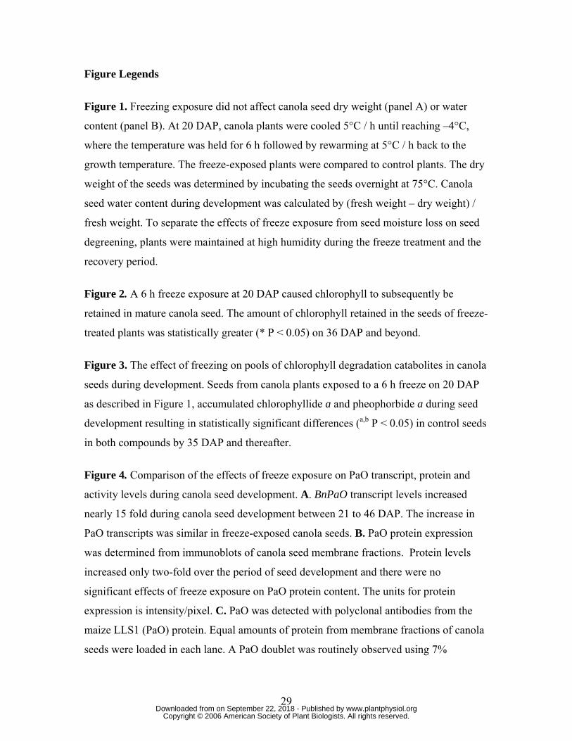
Figure 1. Freezing exposure did not affect canola seed dry weight (panel A) or water
content (panel B). At 20 DAP, canola plants were cooled 5°C / h until reaching –4°C,
where the temperature was held for 6 h followed by rewarming at 5°C / h back to the
growth temperature. The freeze-exposed plants were compared to control plants. The dry
weight of the seeds was determined by incubating the seeds overnight at 75°C. Canola
seed water content during development was calculated by (fresh weight – dry weight) /
fresh weight. To separate the effects of freeze exposure from seed moisture loss on seed
degreening, plants were maintained at high humidity during the freeze treatment and the
recovery period.
Figure 2. A 6 h freeze exposure at 20 DAP caused chlorophyll to subsequently be
retained in mature canola seed. The amount of chlorophyll retained in the seeds of freeze-
treated plants was statistically greater (* P < 0.05) on 36 DAP and beyond.
Figure 3. The effect of freezing on pools of chlorophyll degradation catabolites in canola
seeds during development. Seeds from canola plants exposed to a 6 h freeze on 20 DAP
as described in Figure 1, accumulated chlorophyllide a and pheophorbide a during seed
development resulting in statistically significant differences (a,b P < 0.05) in control seeds
in both compounds by 35 DAP and thereafter.
Figure 4. Comparison of the effects of freeze exposure on PaO transcript, protein and
activity levels during canola seed development. A. BnPaO transcript levels increased
nearly 15 fold during canola seed development between 21 to 46 DAP. The increase in
PaO transcripts was similar in freeze-exposed canola seeds. B. PaO protein expression
was determined from immunoblots of canola seed membrane fractions. Protein levels
increased only two-fold over the period of seed development and there were no
significant effects of freeze exposure on PaO protein content. The units for protein
expression is intensity/pixel. C. PaO was detected with polyclonal antibodies from the
maize LLS1 (PaO) protein. Equal amounts of protein from membrane fractions of canola
seeds were loaded in each lane. A PaO doublet was routinely observed using 7%
29 www.plantphysiol.orgon September 22, 2018 - Published by Downloaded from
Copyright © 2006 American Society of Plant Biologists. All rights reserved.
Page 30
polyacrylamide SDS-PAGE. D. PaO activity profile was induced about 10 fold during
seed maturation. The increase in PaO activity between 21 to 31 DAP was similar in
control and freeze-exposed seeds. In the later stages, induction of PaO activity was
impaired in freeze-exposed seeds compared to control seeds (* P < 0.01; ** P < 0.001;
*** P < 0.0001).
Figure 5. Immobilized metal affinity chromatography revealed dynamic PaO
phosphorylation during canola seed development. PaO phosphorylation was determined
at 21 DAP when PaO protein was half of maximum but activity very low and at 41 DAP
when both PaO protein and activity were highest. A PhosphoProtein Purification Kit
(Qiagen, Valencia, CA), was used to separate phosphorylated (E, elute) and
nonphosphorylated (FT, flow through) canola seed membrane proteins. The PaO
fractions were identified and quantified by immunoblot and infrared imaging. A.
Solubilized total membrane fractions containing PaO were isolated in combination with
protease and phosphatase inhibitors. Protein expression was measured based on isolated
membrane fractions of canola seeds. B. PaO protein levels increased over the period of
seed development. Quantification of the blot from “A” demonstrated that at 21 DAP, the
eluted phosphorylated fraction (21E) contained almost 5 fold higher amounts of PaO
protein than the flow through nonphosphorylated fraction (21FT). At 41 DAP, while
phosphorylated PaO proteins (41E) showed only a modest increase from 21 DAP, the
nonphosphorylated fraction (41FT) increased by >3 fold. PaO was detected with
polyclonal antibodies from the maize LLS1 (PaO) protein. Equal amounts of protein from
membrane fractions of canola seeds were loaded in each lane. The units for protein
expression is intensity/pixel. C. Separation of phosphorylated (SUS E) and
nonphosphorylated (SUS FT) maize leaf sucrose synthase.
Figure 6. Detection of dynamic PaO phosphorylation by Diamond Pro-Q staining.
Duplicates of the 21 DAP phosphorylated and nonphosphorylated fractions from
immobilized metal affinity chromatography were separated on SDS-PAGE and
transferred to PVDF membrane and the membrane was cut in half. One half was used for
an immunoblot with polyclonal antibodies for PaO (IB:PaO), and the other half was used
for Pro-Q Diamond staining (ProQ Stain). The two halves were realigned based on a
30 www.plantphysiol.orgon September 22, 2018 - Published by Downloaded from
Copyright © 2006 American Society of Plant Biologists. All rights reserved.
Page 31
ladder that was cut in middle between the two half blots. Pro-Q analysis showed no bands
representing phosphorylated proteins, including PaO, in the IMAC flow through fraction
containing nonphosphorylated proteins. However, a 52 kD band corresponding to PaO
was shown to align with a Pro-Q staining protein of a similar running MW in the
phosphorylated fraction eluted from the IMAC column.
Supplemental Figure S1. The codon derived protein sequences of BnPaO2, BnPaO1 and
AtPaO. The Arabidopsis PaO gene sequence was used to clone and identify the genes
from Brassica napus. Two cDNA clones of PaO were isolated from senescing canola
leaves. The Rieske-center domain and the mononuclear iron-binding domain are
completely conserved between canola and Arabidopsis. There are two predicted
transmembrane domains (TM1 & TM2) in all three PaO isoforms as well as two potential
CDPK phosphorylation sites. Region 1 and 2 indicate the major differences in protein
sequence among BnPaO1, BnPaO2 and AtPaO. The cleavage site of the predicted
chloroplast transit peptide (tp cleavage site) is also noted.
Supplemental Figure S2. Comparison of codon derived protein sequence at the N-
terminus region of AtPaO, BnPaO1 and BnPaO2. BnPaO2 has an additional serine
residue (S29) when compared to BnPaO1 (first box). In the same region, AtPaO has two
threonine residues (T27, T28) while both clones of BnPaO have an asparagine and serine
residue (N27, S28). Both clones of BnPaO have an extra alanine residue (A30) compared
to AtPaO. Another apparent difference in protein sequence is in the region of amino acids
75 to 78. While BnPaO1 has a protein sequence of G-D-K-E (second box), BnPaO2 is
completely missing these 4 amino acids at the same region.
31 www.plantphysiol.orgon September 22, 2018 - Published by Downloaded from
Copyright © 2006 American Society of Plant Biologists. All rights reserved.
Page 32
www.plantphysiol.orgon September 22, 2018 - Published by Downloaded from Copyright © 2006 American Society of Plant Biologists. All rights reserved.
Page 33
www.plantphysiol.orgon September 22, 2018 - Published by Downloaded from Copyright © 2006 American Society of Plant Biologists. All rights reserved.
Page 34
www.plantphysiol.orgon September 22, 2018 - Published by Downloaded from Copyright © 2006 American Society of Plant Biologists. All rights reserved.
Page 35
www.plantphysiol.orgon September 22, 2018 - Published by Downloaded from Copyright © 2006 American Society of Plant Biologists. All rights reserved.
Page 36
www.plantphysiol.orgon September 22, 2018 - Published by Downloaded from Copyright © 2006 American Society of Plant Biologists. All rights reserved.
Page 37
www.plantphysiol.orgon September 22, 2018 - Published by Downloaded from Copyright © 2006 American Society of Plant Biologists. All rights reserved.