Anim. Reprod., v.6, n.1, p.34-46, Jan./Mar. 2009 _________________________________________ 3 Corresponding author: [email protected]Phone: +44(0131)242-2687; Fax: +44(0131)242-2686 Paracrine regulation of luteal development and luteolysis in the primate W.C. Duncan 1,3 , M. Myers 1 , R.E. Dickinson 2 , S. van den Driesche 1 , H.M. Fraser 2 1 Obstetrics and Gynaecology, Division of Reproductive and Developmental Sciences, University of Edinburgh. 2 MRC Human Reproductive Sciences Unit, Centre for Reproductive Biology, The Queen’s Medical Research Institute, 47 Little France Crescent, Edinburgh EH16 4TJ, UK. Abstract The luteinizing hormone (LH) receptor is fundamental for the regulation of the corpus luteum (CL) in women and non-human primates. Its ligands, LH and human chorionic gonadotropin (hCG), have key roles in the regulation of tissue and vascular remodeling associated with luteal formation and regression. However this remodeling involves the regulation of cells that do not express LH receptors including endothelial cells, pericytes, fibroblasts and macrophages. We have taken a candidate molecule approach to identify important LH/hCG-regulated paracrine molecules and their receptors in CL and assess the effects of their manipulation in vivo and in vitro. Vascular endothelial growth factor (VEGF) acts on endothelial cells and is a major paracrine regulator of luteal angiogenesis and vasculature maintenance. Luteolysis is associated with increased SLIT/ROBO, connective tissue growth factor (CTGF) and matrix metalloproteinase (MMP) expression in luteal fibroblasts. Investigation of the inhibition of these changes by hCG has identified activin A as a novel paracrine luteolysin and locally generated cortisol as a novel paracrine luteotropin. The molecular regulation of luteal function in the primate is complex and the paracrine regulation of luteal function is still not fully understood. Locally, the luteolytic activities of SLIT/ROBO and activin-A are inhibited by hCG and the luteotropic activities of VEGF and cortisol are stimulated by hCG. Keywords: corpus luteum, paracrine, angiogenesis, pregnancy, luteinization, luteolysis. Introduction The role of the corpus luteum (CL) of all species is to synthesize large amounts of progesterone to prepare for, and support, pregnancy. Its formation, from the cells of the post-ovulatory follicle, is associated with marked tissue and vascular remodeling. Notably there is intense angiogenesis, with the development of a rich microvascular capillary network such that each steroidogenic cell is in direct contact with an endothelial cell. The mature CL is a large prominent vascular structure, clearly visible within the ovary. However its regression, when it is no longer required for maternal recognition of pregnancy and its maintenance, is relatively rapid and ultimately complete. Luteolysis involves tightly regulated and coordinated tissue remodeling and cell death that removes the CL from the ovary without scarring (Fig. 1). The CL of women and non-human primates is no different in these respects. It is therefore likely that observations about the molecular regulation of luteal development, function and luteolysis in other species, such as rodents or ruminants, will involve generic pathways that can inform us about how the primate CL may work. Indeed there are clear parallels between species with regards to the molecular pathways within the CL with regard to angiogenesis (Stocco et al., 2007) and extra-cellular matrix (ECM) remodeling (Curry and Osteen, 2003). However there are species differences in such fundamental aspects of CL biology that it is difficult to extrapolate findings in other species to the primate. As a consequence research into the human CL is challenging and suffers from limitations of tissue availability and access and animal models. This review will focus on research carried out on the molecular regulation of the human CL using non- human primate models and tissues and cells from women. There are four main approaches that have led to the current understanding of angiogenesis and luteolysis in the primate CL. 1. The observational approach where the composition and gene and protein expression in the forming CL is compared to the fully functioning CL and the regressing CL of the late luteal phase. 2. The manipulation of systemic regulatory molecules in vivo, prior to removal of the CL, to either induce luteolysis or rescue the CL from luteolysis by mimicking the hormonal changes of early pregnancy (Duncan, 2000). 3. The manipulation of signaling pathways and local regulatory molecules in vivo by local or systemic treatments prior to analysis of the CL. Unlike the other models, which have been carried out in women and in non-human primates, this approach has been restricted to non-human primates (Fraser and Duncan 2005, Xu and Stouffer, 2005). 4. The reductionist approach where different luteal cells are studied in vitro in culture or co-culture. These cells are obtained either from the luteinized follicle or from the CL itself. In order to draw robust conclusions about the molecular regulation of the primate CL most researchers utilize a combination of these approaches.
Paracrine regulation of luteal development and luteolysis in the primate
W.C. Duncan1,3, M. Myers1, R.E. Dickinson2, S. van den Driesche1, H.M. Fraser2
1Obstetrics and Gynaecology, Division of Reproductive and Developmental Sciences, University of Edinburgh. 2MRC Human Reproductive Sciences Unit, Centre for Reproductive Biology, The Queen’s Medical Research Institute, 47 Little
France Crescent, Edinburgh EH16 4TJ, UK.
Abstract
The luteinizing hormone (LH) receptor is fundamental for the regulation of the corpus luteum (CL) in women and non-human primates. Its ligands, LH and human chorionic gonadotropin (hCG), have key roles in the regulation of tissue and vascular remodeling associated with luteal formation and regression. However this remodeling involves the regulation of cells that do not express LH receptors including endothelial cells, pericytes, fibroblasts and macrophages. We have taken a candidate molecule approach to identify important LH/hCG-regulated paracrine molecules and their receptors in CL and assess the effects of their manipulation in vivo and in vitro. Vascular endothelial growth factor (VEGF) acts on endothelial cells and is a major paracrine regulator of luteal angiogenesis and vasculature maintenance. Luteolysis is associated with increased SLIT/ROBO, connective tissue growth factor (CTGF) and matrix metalloproteinase (MMP) expression in luteal fibroblasts. Investigation of the inhibition of these changes by hCG has identified activin A as a novel paracrine luteolysin and locally generated cortisol as a novel paracrine luteotropin. The molecular regulation of luteal function in the primate is complex and the paracrine regulation of luteal function is still not fully understood. Locally, the luteolytic activities of SLIT/ROBO and activin-A are inhibited by hCG and the luteotropic activities of VEGF and cortisol are stimulated by hCG.
Keywords: corpus luteum, paracrine, angiogenesis, pregnancy, luteinization, luteolysis.
Introduction
The role of the corpus luteum (CL) of all species is to synthesize large amounts of progesterone to prepare for, and support, pregnancy. Its formation, from the cells of the post-ovulatory follicle, is associated with marked tissue and vascular remodeling. Notably there is intense angiogenesis, with the development of a rich microvascular capillary network such that each steroidogenic cell is in direct contact with an endothelial cell. The mature CL is a large prominent vascular structure, clearly visible within the ovary. However its regression, when it is no longer required for maternal recognition of pregnancy and its maintenance, is relatively rapid and ultimately complete. Luteolysis
involves tightly regulated and coordinated tissue remodeling and cell death that removes the CL from the ovary without scarring (Fig. 1).
The CL of women and non-human primates is no different in these respects. It is therefore likely that observations about the molecular regulation of luteal development, function and luteolysis in other species, such as rodents or ruminants, will involve generic pathways that can inform us about how the primate CL may work. Indeed there are clear parallels between species with regards to the molecular pathways within the CL with regard to angiogenesis (Stocco et al., 2007) and extra-cellular matrix (ECM) remodeling (Curry and Osteen, 2003). However there are species differences in such fundamental aspects of CL biology that it is difficult to extrapolate findings in other species to the primate. As a consequence research into the human CL is challenging and suffers from limitations of tissue availability and access and animal models.
This review will focus on research carried out on the molecular regulation of the human CL using non-human primate models and tissues and cells from women. There are four main approaches that have led to the current understanding of angiogenesis and luteolysis in the primate CL.
1. The observational approach where the composition and gene and protein expression in the forming CL is compared to the fully functioning CL and the regressing CL of the late luteal phase.
2. The manipulation of systemic regulatory molecules in vivo, prior to removal of the CL, to either induce luteolysis or rescue the CL from luteolysis by mimicking the hormonal changes of early pregnancy (Duncan, 2000).
3. The manipulation of signaling pathways and local regulatory molecules in vivo by local or systemic treatments prior to analysis of the CL. Unlike the other models, which have been carried out in women and in non-human primates, this approach has been restricted to non-human primates (Fraser and Duncan 2005, Xu and Stouffer, 2005).
4. The reductionist approach where different luteal cells are studied in vitro in culture or co-culture. These cells are obtained either from the luteinized follicle or from the CL itself.
In order to draw robust conclusions about the molecular regulation of the primate CL most researchers utilize a combination of these approaches.
Figure 1. Transvaginal ovarian ultrasonography across the ovarian cycle in women: a) The preovulatory dominant follicle with 18 mm diameter is clearly visible (arrow); b) The mid-luteal CL measuring 16 mm in diameter is a major component of the ovary (arrow); c) After menstruation the ovary has several small antral follicles and the regressing CL is difficult to identify.
Luteal development The luteinizing hormone receptor
The molecular regulation of the primate CL is absolutely dependent on the luteinizing hormone (LH) receptor. The LH receptor is a seven transmembrane G-protein coupled receptor found on the surface of the steroidogenic cells of the CL (Duncan et al., 1996; Ascoli et al., 2002). There are two types of steroidogenic cells in the CL, the large granulosa-lutein cells and the surrounding smaller theca-lutein cells and they both express the LH receptor. However in this review the term steroidogenic cell is mainly used to describe the granulosa-lutein cell. This is because these cells are the most studied, the most abundant and the major source of progesterone secretion. The steroidogenic cell LH-receptor is activated by two ligands: LH from the pituitary gland and human chorionic gonadotrophin (hCG) secreted by the developing conceptus. LH and hCG are very similar in molecular structure apart from hCG having a unique carboxyterminal 29 amino acids that increase the number of associated carbohydrate residues and therefore its circulating half-life. They are believed to activate the same signaling pathways on binding to the LH receptor. Indeed the common marmoset monkey (Callithrix jacchus) uses the same CG molecule from the pituitary and the conceptus (Gromoll et al., 2003).
Formation of the CL is absolutely dependent on the LH surge. At ovulation the LH receptor is expressed on both the theca cells and the granulosa cells of the dominant follicle. The process of ovulation, which is LH-dependent, involves marked changes to the functional capacity of the steroidogenic cells, notably the granulosa cells. However as well as luteinization of these cells, ovulation is associated with the resumption of oocyte maturation and follicular rupture that involves marked tissue remodeling. However, as the oocyte and the immune cells, endothelial cells and fibroblasts whose function is regulated during follicular rupture do not express LH receptors the effects of LH on these cells is indirect. LH must therefore influence the expression of other paracrine effector molecules, and
inflammatory mediators, at ovulation by its action on the steroidogenic cells (Espey and Richards, 2006).
After follicular rupture, the transition of the collapsing follicle into the CL is associated with intense angiogenesis. This angiogenesis is LH-dependent as if LH is withdrawn at this stage the vascularization and viability of the CL is inhibited (Dickson and Fraser, 2000). However the invading endothelial cells and supporting pericytes and fibroblasts, involved in developing the extensive microvascular network, do not express LH receptors and must therefore be controlled by locally regulated paracrine molecules.
Luteal function, as measured by progesterone synthesis is absolutely dependent on continued LH secretion from the pituitary gland. Treatment with gonadotropin releasing hormone (GnRH) antagonists or anti-GnRH antibodies (Fraser et al., 1987), or stopping pulsatile GnRH administration, rapidly lowers circulating LH concentrations and inhibits progesterone secretion. In addition, replacement of LH or hCG after GnRH antagonist treatment fully restores progesterone secretion (Stouffer, 2003).
It is not just the function of the primate CL that is dependent on LH, its structural integrity also depends on LH action. Removal of LH support, using GnRH antagonists, initiates luteal cell death and facilitates structural luteolysis (Duncan et al., 1998a). LH therefore maintains the structural and functional integrity of the CL. It is attractive to speculate therefore that luteolysis in a natural cycle is initiated by LH withdrawal. However it is not clear how such functional LH withdrawal might occur. Luteolysis occurs in the presence of basal LH concentrations and when LH pulse frequency is maintained throughout the luteal phase (Hutchison et al., 1986). It also occurs in the presence of continued expression of LH receptors (Duncan et al., 1996). As luteolysis progresses, however, there seems to be a reduction in the expression of key elements of the steroidogenic pathway, such as steroidogenic acute regulatory protein (StAR) and 3ß-hydroxysteroid dehydrogenase (3ß-HSD; Duncan et al., 1998a, 1999, Bogan et al., 2008b). However, luteolysis is initiated, and progesterone secretion falls, in the presence of all
the elements of the steroidogenic pathway. One theory about how a functional LH withdrawal occurs is that the LH receptor in the CL becomes increasingly less responsive to LH (Brannian and Stouffer, 1991). How such increasing uncoupling from its intracellular cell signalling pathways occurs at a molecular level remains unclear.
What is clear is that luteolysis does not occur in the presence of increasing concentrations of hCG. During maternal recognition of pregnancy hCG, acting through the LH receptor, maintains the structural and functional integrity of the CL. The fact that it is needed in logarithmically increasing concentrations to maintain the CL suggests that the sensitivity of the LH receptor to its ligands is maximal in the mid-luteal phase and subsequently falls, even in a conception cycle. The response of the LH receptor to hCG however is fundamental for the establishment of pregnancy. Importantly, unlike in other species such as the rat, the LH receptor does not down-regulate in the presence of excess ligand (Duncan et al., 1996).
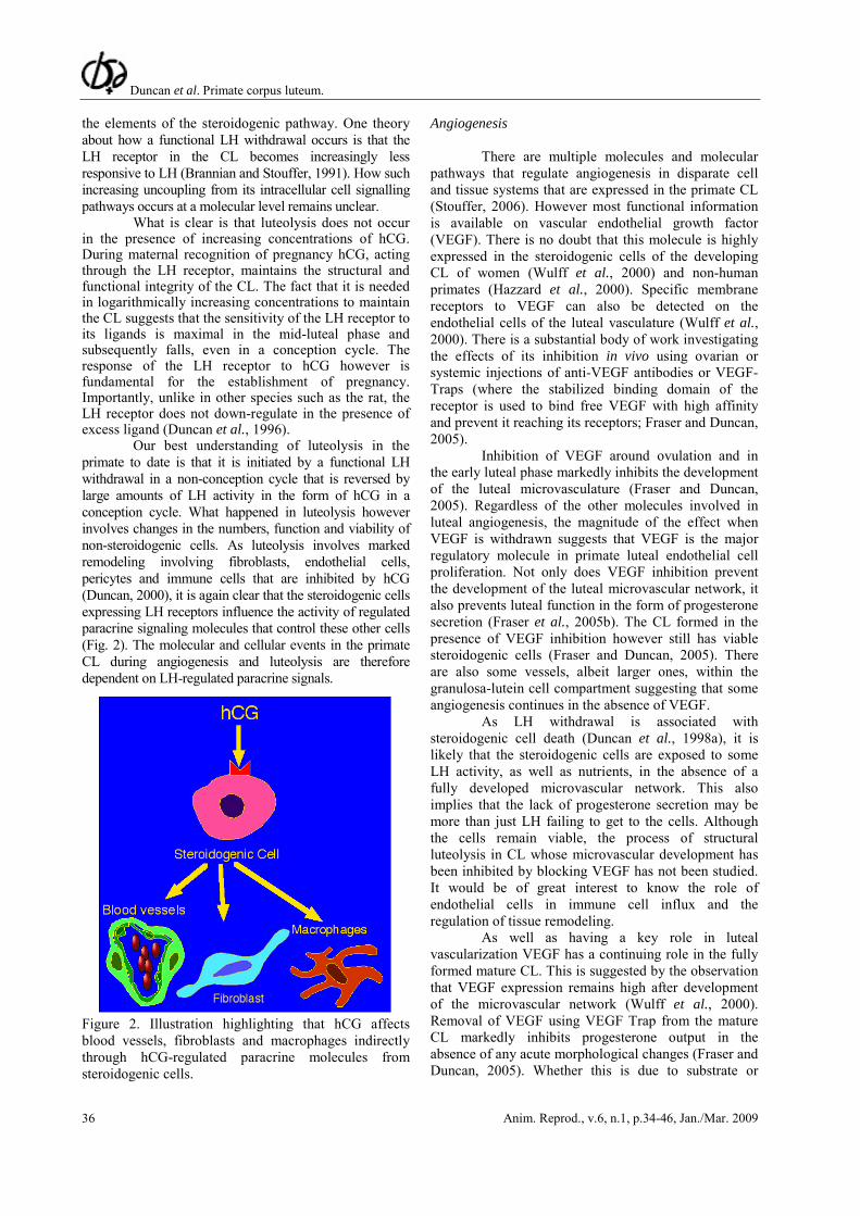
Our best understanding of luteolysis in the primate to date is that it is initiated by a functional LH withdrawal in a non-conception cycle that is reversed by large amounts of LH activity in the form of hCG in a conception cycle. What happened in luteolysis however involves changes in the numbers, function and viability of non-steroidogenic cells. As luteolysis involves marked remodeling involving fibroblasts, endothelial cells, pericytes and immune cells that are inhibited by hCG (Duncan, 2000), it is again clear that the steroidogenic cells expressing LH receptors influence the activity of regulated paracrine signaling molecules that control these other cells (Fig. 2). The molecular and cellular events in the primate CL during angiogenesis and luteolysis are therefore dependent on LH-regulated paracrine signals. Figure 2. Illustration highlighting that hCG affects blood vessels, fibroblasts and macrophages indirectly through hCG-regulated paracrine molecules from steroidogenic cells.
Angiogenesis
There are multiple molecules and molecular pathways that regulate angiogenesis in disparate cell and tissue systems that are expressed in the primate CL (Stouffer, 2006). However most functional information is available on vascular endothelial growth factor (VEGF). There is no doubt that this molecule is highly expressed in the steroidogenic cells of the developing CL of women (Wulff et al., 2000) and non-human primates (Hazzard et al., 2000). Specific membrane receptors to VEGF can also be detected on the endothelial cells of the luteal vasculature (Wulff et al., 2000). There is a substantial body of work investigating the effects of its inhibition in vivo using ovarian or systemic injections of anti-VEGF antibodies or VEGF-Traps (where the stabilized binding domain of the receptor is used to bind free VEGF with high affinity and prevent it reaching its receptors; Fraser and Duncan, 2005).
Inhibition of VEGF around ovulation and in the early luteal phase markedly inhibits the development of the luteal microvasculature (Fraser and Duncan, 2005). Regardless of the other molecules involved in luteal angiogenesis, the magnitude of the effect when VEGF is withdrawn suggests that VEGF is the major regulatory molecule in primate luteal endothelial cell proliferation. Not only does VEGF inhibition prevent the development of the luteal microvascular network, it also prevents luteal function in the form of progesterone secretion (Fraser et al., 2005b). The CL formed in the presence of VEGF inhibition however still has viable steroidogenic cells (Fraser and Duncan, 2005). There are also some vessels, albeit larger ones, within the granulosa-lutein cell compartment suggesting that some angiogenesis continues in the absence of VEGF.
As LH withdrawal is associated with steroidogenic cell death (Duncan et al., 1998a), it is likely that the steroidogenic cells are exposed to some LH activity, as well as nutrients, in the absence of a fully developed microvascular network. This also implies that the lack of progesterone secretion may be more than just LH failing to get to the cells. Although the cells remain viable, the process of structural luteolysis in CL whose microvascular development has been inhibited by blocking VEGF has not been studied. It would be of great interest to know the role of endothelial cells in immune cell influx and the regulation of tissue remodeling.
As well as having a key role in luteal vascularization VEGF has a continuing role in the fully formed mature CL. This is suggested by the observation that VEGF expression remains high after development of the microvascular network (Wulff et al., 2000). Removal of VEGF using VEGF Trap from the mature CL markedly inhibits progesterone output in the absence of any acute morphological changes (Fraser and Duncan, 2005). Whether this is due to substrate or
ligand availability, or changes in progesterone synthesis or secretion, is not clear. However, what is clear is that there is a likely change in vessel permeability. One of the actions of VEGF in vivo is to increase endothelial permeability, indeed it is this action that is thought to have a role in the development of ovarian hyperstimulation syndrome (OHSS) during assisted conception (Wang et al., 2002).
Tight junctional proteins are excellent candidate molecules for the effect of VEGF on endothelial cell permeability. In cell culture VEGF reduced the expression of occludin and claudin molecules on endothelial cell monolayers and this was associated with increased permeability (Villasante et al., 2007). In addition in vivo inhibition of VEGF in the mid-luteal phase is associated with an up-regulation of these proteins (Rodewald et al., 2007). Treatment with hCG in vivo, which is associated with increased VEGF concentrations, reduces the expression of endothelial and steroidogenic cell tight junctional proteins (Groten et al., 2006). This suggests that one endothelial function, in the form of permeability, is regulated by VEGF in the mature CL and this may be associated with progesterone secretion into the circulation.
As well as the function of endothelial cells in a formed vascular network, VEGF is also involved in maintaining the viability of endothelial cells (Fraser et al., 2006). Removal of VEGF from the fully formed CL resulted in a time-dependent increase in caspase-3 immunostaining and apoptosis of luteal endothelial cells. This was followed by increased caspase-3 immunostaining of the neighboring steroidogenic cells (Fraser et al., 2006).
VEGF expression is up-regulated in the CL of early pregnancy (Wulff et al., 2000). Indeed luteal rescue in women is associated with increased endothelial proliferation (Wulff et al., 2001). It is therefore likely that hCG increases the expression of luteal VEGF that in turn stimulates further endothelial cell division. This however has not been confirmed in the non-human primate because there is no additional endothelial proliferation in the CL of early pregnancy in the non-human primate (Christenson and Stouffer, 1996; Rowe et al., 2002). At all stages of the luteal phase however proliferation, function and survival of endothelial cells is influenced by VEGF.
There are some important issues about VEGF expression in the primate CL that require clarification. This may be because the expression of VEGF may differ when mRNA and protein concentrations are analyzed (Tesone et al., 2005). One unresolved issue is whether LH or hypoxia is the prime regulator of luteal VEGF expression. Certainly, in the highly vascular mature CL in women, hCG treatment in vivo increased VEGF and endothelial cell division. In addition there is a wealth of data that hCG increases VEGF expression in cultured luteinized granulosa cells (LGCs; Fraser et al., 2005a, van den Driesche et al., 2008a). Indeed
LH/hCG-dependent VEGF secretion is particularly implicated in the pathophysiology of OHSS clinically.
However there is evidence that dispersed luteal cells may not increase VEGF in response to LH while they do secrete more progesterone (Tesone et al., 2005). In these cells it was hypoxia that was able to increase VEGF expression. Hypoxia is certainly the major regulator of VEGF synthesis and secretion in many tissues, and there is a hypoxia inducible factor (HIF)-1α response element on the VEGF promoter. It may be that there is a role for both hypoxia and LH in the regulation of luteal VEGF expression. Indeed there is a relationship between HIF-1α expression and LH. Nuclear HIF-1α immunostaining is markedly up-regulated in primate granulosa cells at the time of ovulation (Duncan et al., 2008). It is therefore possible that HIF-1α expression is regulated by LH/hCG as well as hypoxia. In cell culture hCG increased the expression of both HIF-1α and VEGF (van den Driesche et al., 2008a). In addition the hCG-dependent increase in VEGF expression also occurred under hypoxic conditions. It is therefore likely that the regulation of VEGF in the CL has both ligand-induced and hypoxic elements that may be differentially expressed according to the stages of the luteal phase. However the mature CL expresses large amounts of VEGF in the absence of clear nuclear HIF-1α immunostaining (Duncan et al., 2008, van den Driesche et al., 2008a).
It is not clear if VEGF expression falls during luteolysis in primates. Data from monkeys and women suggest that it might be the case but any changes are certainly not marked (Hazzard et al., 2000; Wulff et al., 2000). However removal of VEGF clearly has effects on the integrity of the mature CL. There is a time-dependent up-regulation of nuclear HIF-1α suggesting increased hypoxia of the steroidogenic cells (Duncan et al., 2008). This might be secondary to vascular changes as there is an associated increase in endothelial cell death (Fraser et al., 2006). Whether this progression happens during natural luteolysis however remains to be established.
Although VEGF is particularly important in the regulation of luteal vasculature, other molecules have also been implicated in the regulation of the microvasculature network of the CL. The most studied of these, in the primate, are the angiopoietins. They are certainly expressed in the primate CL and their receptor, Tie-2, is exclusively expressed the luteal endothelial cells (Wulff et al., 2000). Angiopoietins have roles in the regulation of the integrity of the microvascular network. Angiopoietin-1 (Ang-1) expression is thought to play a crucial role in the interaction between endothelial cells and surrounding matrix during vessel maturation and stabilization. It could therefore be hypothesized that Ang-1 would be increased in the fully functioning CL of the mid-luteal phase with its mature vascular network. However, this was not particularly the case. Its expression in the human CL across the luteal
phase did not really change (Wulff et al., 2000; Sugino et al., 2005), and in non-human primates it tended to be higher in the late-luteal phase (Hazzard et al., 2000). In addition, manipulation of Ang-1 by injecting it into the preovulatory dominant follicle of the primate had little effect on ovulation and subsequent luteal function (Xu and Stouffer, 2005).
In contrast Ang-2 is thought to be involved in the destabilization and remodeling of blood vessels. It tends to be higher in the late luteal phase of women (Wulff et al., 2000; Sugino et al., 2005) and non-human primates (Hazzard et al., 2000) and has marked effects when injected into the preovulatory dominant follicle (Xu and Stouffer, 2005). Such local injection of Ang-2 inhibited ovulation, luteal formation and progesterone secretion. This implies that Ang-2 may have an important role in luteal vascularization. However its expression is not necessarily inhibitory to the integrity of the microvascular network. Luteal Ang-2 was increased in simulated early pregnancy in women (Wulff et al., 2000). Whether this is a direct effect of hCG is not clear as it was not regulated by hCG in cultures of primate LGCs in vitro (Hazzard et al., 1999) and not maintained later in pregnancy (Sugino et al., 2005). The importance of Ang-2 in luteal angiogenesis has not fully been clarified. Its expression however is consistent with continued remodeling of the luteal microvasculature as it matures and further develops during pregnancy.
Another vasoactive molecule that is abundantly expressed by the CL is prokineticin-1, which is also known as endocrine gland VEGF (EG-VEGF). It is localized to the steroidogenic granulosa-lutein cells and its expression is maximal in the late luteal phase. Although its expression pattern is different to that of VEGF, the apparent increase by hCG treatment in vitro (Fraser et al., 2005a) is similar to that observed with VEGF. It has been reported to have a role in the permeability regulation associated with angiogenesis. However, as yet there are no functional studies in the primate that inform us about its importance, at present its role remains unclear.
We have come some way to understanding how vascularization is regulated in the CL of women and non-human primates. However there are many facets that we still do not fully understand. More work needs to be done on the role of other molecules in endothelial proliferation, stabilization and in the dialogue with pericytes. The regression of the microvascular network and how this is regulated is also not fully understood.
Luteolysis
There are two main initial strategies that have informed us about molecules that are effectors of luteolysis and the removal of the primate CL from the ovary. The first is to investigate molecules whose
expression is increased in the late or very late luteal phase in comparison to the early or mid-luteal phases. Those whose expression is increased during functional and structural luteolysis are likely to be involved at some level. The second strategy is to examine the late-luteal CL in the presence or absence of hCG (Duncan, 2000). Important effector molecules involved in luteolysis would be expected to be highest in the late-luteal phase and to be inhibited by hCG. One caveat to these approaches however is that the cellular composition of the CL changes across the luteal phase and in response to hCG, and this may affect the abundance of both regulated and non-regulated genes.
That said these strategies have markedly increased our understanding of luteolysis in primates and how it can be inhibited by exposure to hCG during maternal recognition of pregnancy. Molecules identified can be further studied after manipulation in vivo or in primary cell cultures in vitro. Most researchers have taken a candidate gene approach to identify such molecules by investigating known regulators of tissue and vascular remodeling. These approaches have highlighted novel forms of cell death (Gaytán et al., 2008), macrophage influx (Duncan et al., 1998b) and increased expression of matrix metalloproteinases (MMPs; Duncan et al., 1998c) and connective tissue growth factor (CTGF; Duncan et al., 2005b) during luteolysis.
More recently important insights into the regulation of the primate CL have been obtained using a gene array approach (Yadav et al., 2004; Bogan et al., 2008b). The CL of the Macaque monkey has been studied across the luteal phase (Bogan et al., 2008b). Of the regulated genes that changed across the luteal phase there were four main families of genes that increased at the time of luteolysis:
1. Immune function; genes involved in immune function like some interleukin receptors suggesting a role for the immune system during luteolysis.
2. Hormone and growth factor signalling; genes like endothelin and carboxypeptidase suggest that locally produced factors are paracrine regulators of luteolysis.
3. Steroidogenesis; during luteolysis there is an alteration in the steroidogenic enzymes and notably 3ß-HSD is decreased.
4. Prostaglandin (PG) biosynthesis; the decrease in PGE synthesis and increase in PGF receptors suggest a local regulatory role for PGs in primate luteolysis.
Together, studies informed by candidate and genomic approaches continue to inform us about the pathways involved in luteolysis. Most information is available with regard to the immune system and growth factor and enzyme expression. The immune system
The role of the immune system in primate luteolysis has yet to be fully determined. It is very likely that various cytokines, chemokines, and leukocytes
increase in the CL during its regression (Stouffer, 2006). However most work has been done investigating macrophage influx into the human CL. In women numbers of macrophages increase in the CL from the mid to late luteal phase, reaching a maximum in the menstrual CL after functional luteolysis is complete (Duncan et al., 1998b). It is attractive to hypothesize that the macrophages are involved in remodeling the extracellular matrix (ECM) and in removing the cellular debris associated with luteolysis. Such a hypothesis is consistent with macrophages having a negative effect on luteal structure. In addition macrophage products could have an adverse functional effect on the local cells including the steroidogenic cells. Although positive and tropic effects of macrophage products have been reported on luteal cells, the prevention of the influx of macrophages by exposure to hCG in vivo confirms that macrophages are linked to the luteolytic process (Duncan et al., 1998b).
The regulation of macrophage influx during luteolysis is not clear. Although this influx is inhibited by hCG they do not seem to be directly regulated by LH/hCG or indeed by progesterone, as they do not express receptors to these molecules (Maybin and Duncan, 2004). The human CL expresses monocyte chemotactic protein (MCP)-1 and this is a potential candidate molecule involved in the regulation of macrophage influx. However, it is secreted by the perivascular cells (Senturk et al., 1999) that do not express LH receptors. Whether MCP-1, or other MCPs, is regulated by hCG and how this is achieved remains to be ascertained. What is clear is that LH/hCG-regulated paracrine molecules from the steroidogenic cells must be involved in the regulation of macrophage influx. Growth factors and enzymes
CTGF is a paracrine growth factor whose expression is increased during wound healing. MMPs are proteolytic enzymes involved in the digestion of the ECM during tissue remodeling (Curry and Osteen, 2003). Both are excellent candidates for effector molecules in the tissue remodeling associated with luteolysis. Indeed CTGF is maximally expressed in the late-luteal CL of women and is decreased after exposure to hCG in vivo (Duncan et al., 2005b). In addition expression and activity of MMP-2 is increased during luteolysis in the CL of women (Duncan et al., 1998c; Duncan, 2000) and non-human primates (Young and Stouffer, 2004). Their involvement is not surprising nor is their inhibition by hCG during maternal recognition of pregnancy. However both are primarily synthesized and secreted by luteal fibroblast-like cells that do not express LH receptors or respond directly to hCG in vitro. However when co-cultured with LGCs, hCG can inhibit their expression in fibroblasts (Duncan et al., 2005b; Myers et al., 2007a). This suggests that LH/hCG-regulated molecules from steroidogenic cells
are involved in regulating fibroblast function and the expression of CTGF and MMP-2 in particular.
It is therefore clear that locally produced factors from steroidogenic cells that are influenced by LH/hCG have major roles in the regulation of luteal function and luteolysis in particular (Fig. 2). Recently some additional insights have emerged as to the nature of these locally produced regulatory molecules. These studies have generally been informed by observational studies on whole CL and conducted in primary cell culture and co-culture experimental models. Classically paracrine regulatory molecules would be secreted from steroidogenic cells and their secretion would be regulated by LH/hCG and their receptors would be present on neighboring cells (Fig. 3). Such a paradigm is clearly evident in the VEGF regulation of endothelial cells. It is also evident in the paracrine regulation of fibroblast function.
Paracrine regulation Activin A
We believe that Activin A is a paracrine regulator of fibroblast function during luteolysis. Activins belong to the structurally related transforming growth factor (TGF)-β superfamily that includes inhibins and bone morphogenetic proteins (BMPs). Members of this family have been shown to have important paracrine regulatory roles in diverse physiological processes (Massague, 1998). Indeed, activin signaling has been shown to be essential in inflammation, cell proliferation, and apoptosis. Activins have important roles during follicular growth as activin A can stimulate the proliferation of granulosa cells, enhance their expression of FSH receptors and aromatase, and inhibit luteinization (Knight and Glister, 2006).
Activin A is made in the steroidogenic cells of the CL as these cells express the beta-A subunit. It can have actions on both the steroidogenic cells and neighboring fibroblasts as they express both the type I (ALK 2/4) and type II (A) activin receptors (Fig. 3) as well as components of the Smad (2, 3, and 4) signaling pathway that is induced by activin (Myers et al., 2007a). Indeed treatment of primary cultures of luteal fibroblast-like cells with physiological concentrations of activin A increased CTGF expression as well as MMP-2 expression and activity.
There is also good evidence that the expression and activity of activin A is regulated. Measurement of serum activin A concentrations across the menstrual cycle suggests that activin A increases towards the end of the luteal phase (Muttukrishna et al., 1996). Indeed, Smad 2 and 3 expression also tends to be highest in the late-luteal phase (Myers et al., 2007a). In addition, the expression and activity of activin A is regulated by hCG at multiple levels. HCG increases inhibin alpha subunit expression to create more inhibin A. Inhibin A can
functionally antagonize activin A as well as divert the beta-A subunit from activin A formation. HCG also increases follistatin secretion and follistatin binds to and neutralizes activin A. HCG will also inhibit the effects of activin A on the steroidogenic cells by increasing the beta-glycan inhibin receptor (Myers et al., 2007a). This is important as activin can act directly on steroidogenic
cells. Activin acts on steroidogenic cells to inhibit steroidogenesis at different levels, promote activin beta-A expression and inhibit beta-glycan expression. This serves to potentiate a feed-forward multiplication of its own synthesis and action (Fig. 4; Myers et al., 2008). Certainly in vitro, hCG and activin have opposing effects on human LGC function and gene expression.
Figure 3. Localization studies in the human CL: a) Haematoxylin and eosin (H&E) stained CL showing the steroidogenic cells (SC) surrounded by the stromal fibroblasts (St) and blood vessels (bv). Insert is mRNA in situ hybridization for the LH receptor showing localization to the SC and not St compartments; b) H&E stained CL with insert showing mRNA in situ hybridization for MMP-2 showing the primary localization is St and not SC; c) Fluorescent immunohistochemistry for 11β-HSD type 1 (red) showing expression in the SC; d) Immunohistochemistry (brown) for the beta-A subunit showing expression in the SC; e) Fluorescent immunohistochemistry showing nuclear glucocorticoid receptor (green) in St and bv (red); f) Immunohistochemistry showing both components of the activin receptor (brown) in the St.
Together these findings suggest that activin A increases in luteolysis and can stimulate local fibroblast CTGF and MMP-2 expression as well as inhibiting steroidogenesis and promoting its own synthesis (Fig. 4). HCG can inhibit activin A synthesis and activity. Indeed in cell co-culture models, fibroblast MMP-2 could be inhibited by hCG in the presence of LGCs and this could be replicated by the addition of follistatin. In addition in the mouse, where ovarian activin beta-A expression has been conditionally knocked out (Pangas et al., 2007), the ovary contains multiple CL when examined. We suspect that activin A is anti-luteal in nature. It is withdrawn at ovulation and increases during luteolysis in the absence of hCG facilitating increased tissue remodeling. Systemic injection of activin A was also luteolytic but the same was not seen functionally
when it was infused into the primate CL (Stouffer et al., 1994). This implies that further work needs to be done on the role of luteal activin production in vivo. Slit/Robo
Other potential luteolysins that we have identified include members of the SLIT/ROBO system. Slits are secreted membrane-associated glycoproteins that can be localized to various luteal cells including steroidogenic cells (Dickinson et al., 2008) and Robos are their transmembrane receptors that again are present on multiple cell types in the human CL (Dickinson et al., 2008). They have major roles in cell-cell communication during embryological development. In vertebrates three Slit (Slit1, Slit2, Slit3) and four Robo
(Robo1, Robo2, Robo3/Rig-1, Robo4/Magic Robo) genes have been identified (Dickson, 2002). They have been shown to have a pivotal and evolutionary conserved role in axon guidance by acting as a repulsive cue (Brose et al., 1999). Slit and Robo proteins are also expressed in a variety of non-neuronal tissues during development, including the heart, lungs and kidney. The effect seems to be negative with regards to cell migration, cell survival and angiogenesis (Wong et al., 2002).
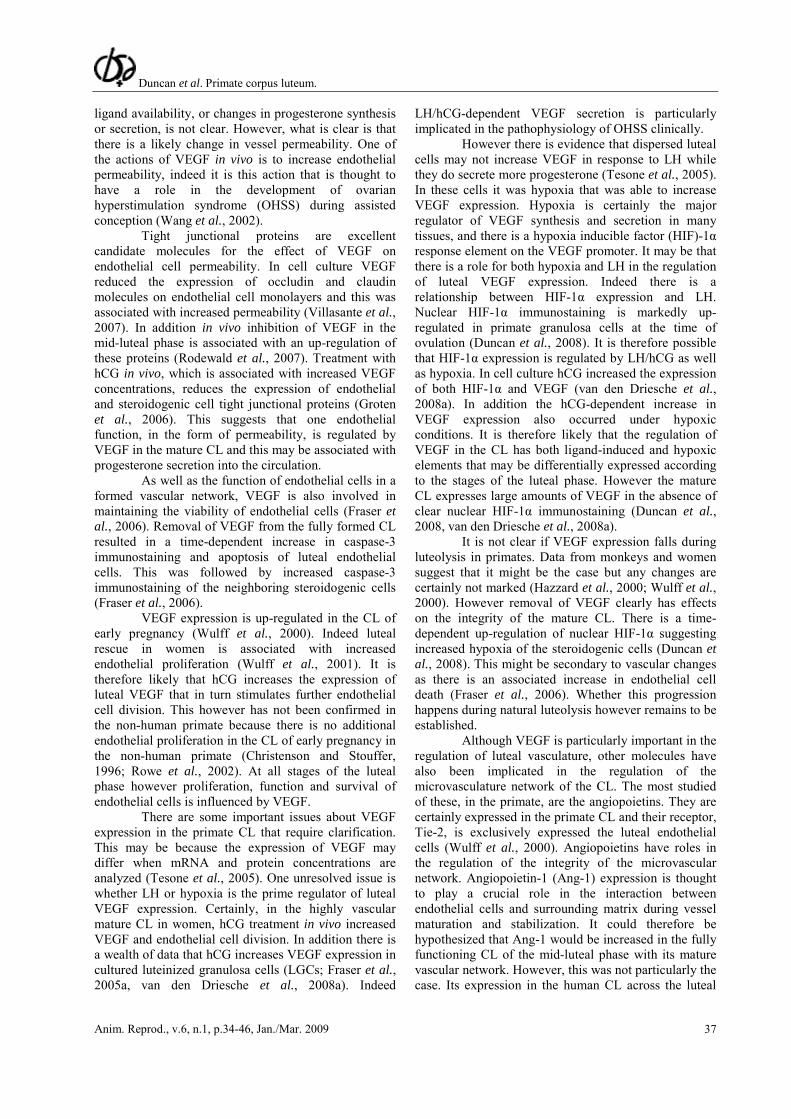
Figure 4. Schematic showing proposed activin A action and regulation in the human CL: a) During luteolysis LH effects are reduced and activin (ACT) from steroidogenic cells (SC) binds to receptors (AR) on neighboring fibroblasts (FIB) to increase MMP-2 and CTGF. ACT acts on SC to reduce LHR expression and steroidogenesis and stimulate further ACT synthesis; b) During luteal rescue hCG stimulates the LH receptor (LHR) to reduce ACT action by reducing secretion, increase follistatin (FS) to bind ACT and increase inhibin A (IHN) and beta-glycan (BG) to inhibit ACT action at AR. This promotes LHR expression and steroidogenesis and inhibits FIB MMP-2 and CTGF. The triangular arrow-heads represent stimulation and the square arrowheads represent inhibition.
Their negative roles on angiogenesis, cell
survival and cell migration suggests that they may inhibit tumor development in adult tissues (Chedotal et al., 2005; Legg et al., 2008). Indeed in cancer tissues ROBO1 (3p12), SLIT2 (4p15.2), SLIT3 (5q34-35) and, to a lesser extent, SLIT1 (10q23.3-24) are inactivated through deletions and hypermethylation of their promoter regions in a number of tumor types including breast and lung (Dickinson et al., 2004; Dallol et al., 2005). Re-expression of SLIT2 also suppressed breast tumor and glioma cell growth and induced apoptosis in colorectal and lung cancer cells (Dallol et al., 2003, 2005).
As the CL also undergoes extensive tissue and vascular remodeling, and luteolysis is associated with coordinated cell death, the SLIT/ROBO interaction was an excellent candidate physiological paracrine signaling system. They are present in multiple cells in the CL and their expression is regulated across the luteal phase. Expression of both ligands, notably SLIT2 and SLIT3, and receptor notably ROBO2 were elevated in the late luteal phase and inhibited by hCG both in vivo and in vitro (Dickinson et al., 2008). This suggests a role for the SLIT/ROBO interaction during luteolysis that is inhibited during maternal recognition of pregnancy.
The functional aspects of the SLIT/ROBO interaction were studied using primary cell culture models. Inhibition of SLIT in vitro reduced apoptosis and increased cell migration (Dickinson et al., 2008). This makes sense as their expression facilitates cell death and is anti-angiogenic. However it remains unclear why an anti-migration role would be required during luteolysis. However as activin A did not directly regulate the SLITs it appears that both pathways may have independent activities in luteolysis. Prostaglandins
Although other candidate compounds with likely roles in luteolysis in women have been reported one system that is certainly worth mentioning is the PG system. Prostaglandins have major regulatory roles in infra-primate species (Stouffer, 2006). Indeed it is uterine Prostaglandin F2α that is the clearly identified luteolysin in ruminants. Although uterine PGs are certainly not involved in luteolysis in women it is likely that PGs are effector molecules in luteolysis and that they are made in the CL. Indeed in the marmoset monkey luteolysis can be induced by a systemic injection of PGF2α (Duncan et al., 1998a). In addition in vitro PGE2 can have positive actions on steroidogenesis and PGF2α can inhibit it. Although the assessment and manipulation of PGs in the primate CL has been an intense area of research in the past, this research momentum has not been maintained. This is because the results were variable and good evidence for their role in vivo was lacking (Bogan et al., 2008a).
Recently the identification of the PG pathway as increased in the late luteal phase using genomic array
analysis has renewed interest (Bogan et al., 2008b). It is now possible to revisit luteal PGs in the primate CL using modern molecular investigative techniques. Peak level of proteins involved in PGE2 synthesis could be detected during periods of luteal development and maintenance and these generally reduced during functional regression. In addition the enzymes involved in PGF2α synthesis were there across the luteal phase and functional regression was associated with an up-regulation of PGF receptors (Bogan et al., 2008a). Although the steroidogenic cells seemed to be the main source of synthesis and action further work is required on their regulation and effects on non-steroidogenic cells. Steroids
Although the PGF2α, activin, and SLIT-ROBO pathways are excellent candidate paracrine regulatory systems that are up-regulated during luteolysis, the PG system introduces another concept: as PGE2 may be luteotropic there may be paracrine molecules that are up-regulated by hCG during luteal rescue. It is likely that there are cohorts of local regulatory molecules that fall during luteolysis and that are increased during maternal recognition of pregnancy. Steroid hormones are excellent candidate molecules to have a luteotropic effect. Firstly steroids, like progesterone and estradiol, are products of the luteal steroidogenic cells. In addition the primate CL expresses progesterone receptors (PR) and estradiol receptor-ß in multiple cell types (Duffy et al., 2000; Maybin and Duncan, 2004; van den Driesche et al., 2008b). Furthermore, their synthesis is LH-regulated such that they fall during functional luteolysis and are increased by hCG during maternal recognition of pregnancy.
Although estradiol may be luteotropic in rodents it is luteolytic in other species such as rabbits (Stocco et al., 2007). There is no good functional data on its effects in the primate CL although it certainly does not appear to be luteotropic (Duffy et al., 2000; van den Driesche et al., 2008b). Indeed hCG down regulates estradiol receptors in human LGCs (van den Driesche et al., 2008b). Progesterone is likely to have some direct luteotropic roles on cell survival and cell function but as yet these remain to be fully defined (Souffer, 2003). The addition of RU486 to cultures of LGC in vitro inhibits cell survival and function but replacement of progesterone is not restorative (Myers et al., 2007b). Treatment in vivo with RU486 or 3ß-HSD inhibitors have some negative effects on luteal function. However, manipulation of progesterone in vivo affects gonadotropin levels and this may have secondary effect on the CL. At present the local effects of progesterone in vivo remain uncertain. It is worth noting that although PR expression is down-regulated as the CL ages it is not up-regulated by hCG during maternal recognition of
pregnancy and PR expression remains low (Duncan et al., 2005a).
One steroid with possible effects on luteal function is cortisol. Cortisol does not seem to be made de novo in the CL but glucocorticoid receptors are expressed in multiple cell types (Myers et al., 2007b). In addition the steroidogenic cells of the CL express cortisol metabolizing enzymes in the form of 11ß-HSDs (Fig. 3). Type 1 11ß-HSD is primarily involved in shuttling inactive cortisone to active cortisol and type 2 11ß-HSD inactivates cortisol to cortisone. The expression of these isoforms changes across the luteal phase. There seems to be more 11ß-HSD type 2 during luteolysis suggesting a local inactivation of cortisol. In addition hCG tended to inhibit 11ß-HSD type 2 expression in vivo and in vitro (Myers et al., 2007b). However both in vivo and in vitro hCG increased 11ß-HSD type 1 expression and activity to promote local cortisol generation. In the presence of local large amounts of progesterone within the CL, cortisol binding protein is saturated and small changes in cortisol metabolism will result in important differences in local active free cortisol concentrations.
In women, there is a change in 11ß-HSD isoforms across the luteal phase that is associated with the functional state of the gland. Once the dominant follicle is exposed to the midcycle preovulatory gonadotropin surge, the predominant isoform switches from type 2 to type 1 11ß-HSD (Tetsuka et al., 1997). This phenomenon can also be seen by a rise of free cortisol that is 50 times higher in follicular fluid after the LH surge. As the changes in 11ß-HSD type 1 parallel this during luteal rescue the increased local free cortisol may have functional and luteotropic effects within the CL. Certainly the addition of cortisol can directly inhibit MMP-2 expression and activity (Myers et al., 2007b) as well as negatively regulate the SLIT/ROBO system (Dickinson et al., 2008). The effect of cortisol on prostaglandin synthesis and action within the primate CL has not been tested. However it is clear that cortisol inhibits PG synthesis as part of its anti-inflammatory action in other tissues.
In summary, there are many effector molecules in luteal cell survival and tissue and vascular remodeling in the primate and this review has highlighted the current candidates. However there are some important concepts that inform us about the molecular regulation of the primate CL. The first is that LH/hCG is of major importance and it acts on the CL to influence multiple cell types. As these do not all express LH receptors, paracrine regulatory molecules must be important. The nature of these is not clear but LH/hCG stimulates luteotropic paracrine molecules such as VEGF and cortisol and inhibits the more luteolytic molecules such as activin and the SLIT/ROBO pathway. It is also likely that local PG production is altered to favor luteal function (PGE2) over regression (PGF2α; Fig. 5).
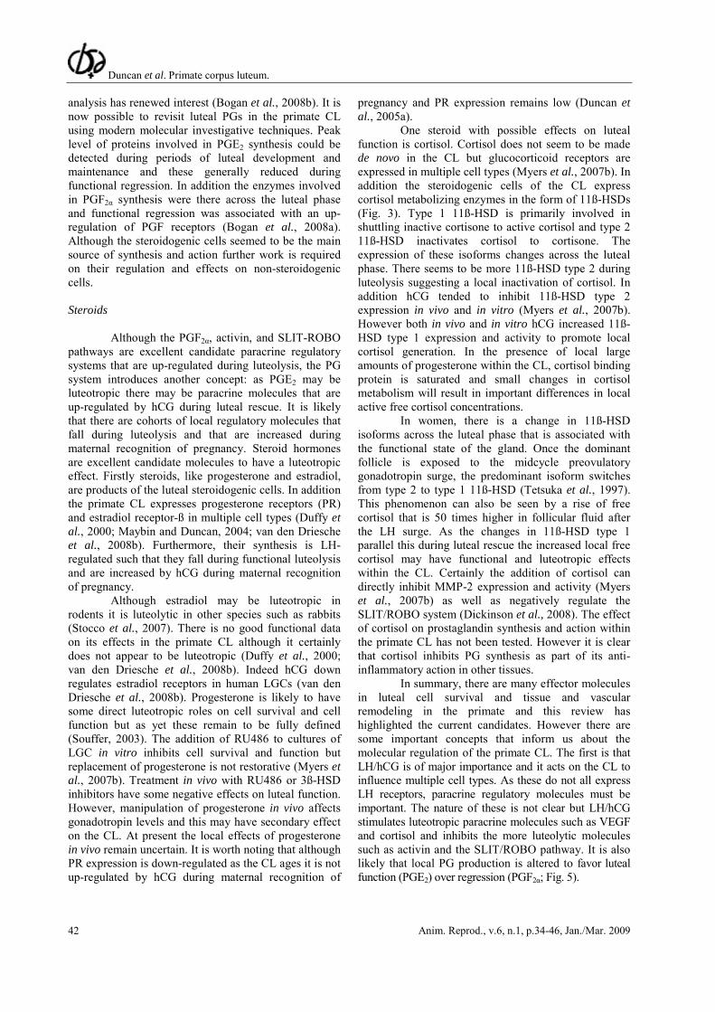
Figure 5. Illustration summarizing proposed paracrine regulatory molecules during luteolysis and luteal rescue by hCG: a), c) and e) show progesterone secretion on an axis from Day 14 to Day 28 of a 28 day menstrual cycle starting from ovulation. a) After ovulation the dominant follicle turns into the CL that secretes increasing amounts of progesterone to a maximum in the mid-luteal phase; b) The function of the steroidogenic cell is regulated by LH and this suppresses luteolytic molecules and promotes luteotropic molecules; c) As the CL matures progesterone secretion falls and the CL undergoes functional and structural luteolysis leaving a small avascular remnant on the ovary; d) LH action is reduced and the luteotropic paracrine molecules decrease and the luteolytic paracrine molecules increase; e) In the presence of hCG the CL is rescued and its functional and structural integrity is maintained; f) Large amounts of hCG maintain LH action on the steroidogenic cell to inhibit luteolytic paracrine molecules and increase luteotropic regulators.
References Ascoli M, Fanelli F, Segaloff DL. 2002. The lutropin/choriogonadotropin receptor, a 2002 perspective. Endocr Rev, 23:141-174. Brannian JD, Stouffer RL. 1991. Progesterone production by monkey luteal cell subpopulations at different stages of the menstrual cycle: changes in agonist responsiveness. Biol Reprod, 44:141-149. Bogan RL, Murphy MJ, Stouffer RL, Hennebold JD. 2008a. Prostaglandin synthesis, metabolism, and signaling potential in the rhesus macaque corpus luteum throughout the luteal phase of the menstrual cycle.
Endocrinology, 149:5861-5871. Bogan RL, Murphy MJ, Stouffer RL, Hennebold JD. 2008b. Systematic determination of differential gene expression in the primate corpus luteum during the luteal phase of the menstrual cycle. Mol Endocrinol, 22:1260-1273. Brose K, Bland KS, Wang KH, Arnott D, Henzel W, Goodman CS, Tessier-Lavigne M, Kidd T. 1999. Slit proteins bind Robo receptors and have an evolutionarily conserved role in repulsive axon guidance. Cell, 96:795-806. Chedotal A, Kerjan G, Moreau-Fauvarque C. 2005. The brain within the tumor: new roles for axon guidance
molecules in cancers. Cell Death Differ, 12:1044-1056. Christenson LK, Stouffer RL. 1996. Proliferation of microvascular endothelial cells in the primate corpus luteum during the menstrual cycle and simulated early pregnancy. Endocrinology, 137:367-374. Curry TE Jr, Osteen KG. 2003. The matrix metalloproteinase system: changes, regulation, and impact throughout the ovarian and uterine reproductive cycle. Endocr Rev, 24:428-465. Dallol A, Morton D, Maher ER, Latif F. 2003. SLIT2 axon guidance molecule is frequently inactivated in colorectal cancer and suppresses growth of colorectal carcinoma cells. Cancer Res, 63:1054-1058. Dallol A, Dickinson RE, Latif F. 2005. Epigenetic disruption of the SLIT-ROBO interactions in human cancer. In: Esteller M (Ed.). DNA Methylation, Epigenetics and Metastasis. Dordvecht: Springer Netherlands. pp. 191-214 Dickinson RE, Dallol A, Bieche I, Krex D, Morton D, Maher ER, Latif F. 2004. Epigenetic inactivation of SLIT3 and SLIT1 genes in human cancers. Br J Cancer, 91:2071-2078. Dickinson RE, Myers M, Duncan WC. 2008. Novel regulated expression of the SLIT/ROBO pathway in the ovary: possible role during luteolysis in women. Endocrinology. [Epub ahead of print]. Dickson BJ. 2002. Molecular mechanisms of axon guidance. Science, 298:1959-1964. Dickson SE, Fraser HM. 2000. Inhibition of early luteal angiogenesis by gonadotropin-releasing hormone antagonist treatment in the primate. J Clin Endocrinol Metab, 85:2339-23344. Duffy DM, Chaffin CL, Stouffer RL. 2000. Expression of estrogen receptor alpha and beta in the rhesus monkey corpus luteum during the menstrual cycle: regulation by luteinizing hormone and progesterone. Endocrinology, 141:1711-1717. Duncan WC, McNeilly AS, Fraser HM, Illingworth PJ. 1996. Luteinizing hormone receptor in the human corpus luteum: lack of down regulation during maternal recognition of pregnancy. Hum Reprod, 11:2291-2297. Duncan WC, Illingworth PJ, Young FM, Fraser HM. 1998a. Induced luteolysis in the primate: rapid loss of luteinising hormone (LH) receptors. Hum Reprod 13:2532-40. Duncan WC, McNeilly AS, Illingworth PJ. 1998b. The effect of luteal 'rescue' on the expression and localization of matrix metalloproteinases and their tissue inhibitors in the human corpus luteum. J Clin Endocrinol Metab, 83:2470-2478. Duncan WC, Rodger FE, Illingworth PJ. 1998c. The human corpus luteum: reduction in macrophages during maternal recognition of pregnancy. Hum Reprod, 13:2435-2442. Duncan WC, Cowen GM, Illingworth PJ. 1999. Steroidogenic enzyme expression in human corpora lutea in the absence and presence of exogenous human chorionic gonadotrophin. Mol Hum Reprod, 5:291-298.
Duncan WC. 2000. The human corpus luteum: remodelling during luteolysis and maternal recognition of pregnancy. Rev Reprod, 5:12-17. Duncan WC, Gay E, Maybin JA. 2005a. The effect of luteal rescue with human chorionic gonadotrophin (hCG) on human luteal-cell progesterone receptors. Reproduction, 130:83-93 Duncan WC, Hillier SG, Gay E, Bell J, Fraser HM. 2005b. Connective tissue growth factor (CTGF) expression in the human corpus luteum: paracrine regulation by human chorionic gonadotropin (hCG). J Clin Endocrinol Metab, 90:5366-5376. Duncan WC, van den Driesche S, Fraser HM. 2008. Inhibition of vascular endothelial growth factor in the primate ovary up-regulates hypoxia-inducible factor-1alpha in the follicle and corpus luteum. Endocrinology, 149:3313-3320. Espey LL, Richards JS. 2006. Ovulation. In: Neill JD (Ed.). Knobil and Neill’s Physiology of Reproduction. New York: Elsevier/Academic Press. Fraser HM, Nestor JJ Jr, Vickery BH. 1987. Suppression of luteal function by a luteinizing hormone-releasing hormone antagonist during the early luteal phase in the stumptailed macaque monkey and the effects of subsequent administration of human chorionic gonadotropin. Endocrinology, 121:612-618 Fraser HM, Duncan WC. 2005. Vascular morphogenesis in the primate ovary. Angiogenesis, 8:101-116. Fraser HM, Bell J, Wilson H, Taylor PD, Morgan K, Anderson RA, Duncan WC. 2005a. Localization and quantification of cyclic changes in the expression of endocrine gland vascular endothelial growth factor in the human corpus luteum. J Clin Endocrinol Metab, 90:427-34. Fraser HM, Wilson H, Morris KD, Swanston I, Wiegand SJ. 2005b. Vascular endothelial growth factor Trap suppresses ovarian function at all stages of the luteal phase in the macaque. J Clin Endocrinol Metab, 90:5811-5818. Fraser HM, Wilson H, Wulff C, Rudge JS, Wiegand SJ. 2006. Administration of vascular endothelial growth factor Trap during the 'post-angiogenic' period of the luteal phase causes rapid functional luteolysis and selective endothelial cell death in the marmoset. Reproduction, 132:589-600. Gaytán M, Morales C, Sánchez-Criado JE, Gaytán F. 2008. Immunolocalization of beclin 1, a bcl-2-binding, autophagy-related protein, in the human ovary: possible relation to life span of corpus luteum. Cell Tissue Res, 331:509-517. Gromoll J, Wistuba J, Terwort N, Godmann M, Müller T, Simoni M. 2003. A new subclass of the luteinizing hormone/chorionic gonadotropin receptor lacking exon 10 messenger RNA in the New World monkey (Platyrrhini) lineage. Biol Reprod, 69:75-80. Groten T, Fraser HM, Duncan WC, Konrad R, Kreienberg R, Wulff C. 2006. Cell junctional proteins
in the human corpus luteum: changes during the normal cycle and after HCG treatment. Hum Reprod, 21:3096-3102. Hazzard TM, Molskness TA, Chaffin CL, Stouffer RL. 1999. Vascular endothelial growth factor (VEGF) and angiopoietin regulation by gonadotrophin and steroids in macaque granulosa cells during the peri-ovulatory interval. Mol Hum Reprod, 5:1115-1121. Hazzard TM, Christenson LK, Stouffer RL. 2000. Changes in expression of vascular endothelial growth factor and angiopoietin-1 and -2 in the macaque corpus luteum during the menstrual cycle. Mol Hum Reprod, 6:993-8. Hutchison JS, Nelson PB, Zeleznik AJ. 1986. Effects of different gonadotropin pulse frequencies on corpus luteum function during the menstrual cycle of rhesus monkeys. Endocrinology, 119:1964-1971. Knight PG, Glister C. 2006. TGF-beta superfamily members and ovarian follicle development. Reproduction, 132:191-206. Legg JA, Herbert JM, Clissold P, Bicknell R. 2008. Slits and Roundabouts in cancer, tumour angiogenesis and endothelial cell migration. Angiogenesis, 11:13-21. Massague J. 1998. TGF-beta signal transduction. Annu Rev Biochem, 67:753-791. Maybin JA, Duncan WC. 2004. The human corpus luteum: which cells have progesterone receptors? Reproduction, 128:423-431. Muttukrishna S, Fowler PA, George L, Groome NP, Knight PG. 1996. Changes in peripheral serum levels of total activin A during the human menstrual cycle and pregnancy. J Clin Endocrinol Metab, 81:3328-3334. Myers M, Gay E, McNeilly AS, Fraser HM, Duncan WC. 2007a. In vitro evidence suggests activin-A may promote tissue remodeling associated with human luteolysis. Endocrinology, 148:3730-3739. Myers M, Lamont MC, van den Driesche S, Mary N, Thong KJ, Hillier SG, Duncan WC. 2007b. Role of luteal glucocorticoid metabolism during maternal recognition of pregnancy in women. Endocrinology, 148:5769-5779. Myers M, van den Driesche S, McNeilly AS, Duncan WC. 2008. Activin A reduces luteinisation of human luteinised granulosa cells and has opposing effects to human chorionic gonadotropin (hCG) in vitro. J Endocrinol. (in press). Pangas SA, Jorgez CJ, Tran M, Agno J, Li X, Brown CW. Kumar TR, Matzuk, M. 2007. Intraovarian activins are required for female fertility. Mol Endocrinol, 21:2458-2471. Rodewald M, Herr D, Fraser HM, Hack G, Kreienberg R, Wulff C. 2007. Regulation of tight junction proteins occludin and claudin 5 in the primate ovary during the ovulatory cycle and after inhibition of vascular endothelial growth factor. Mol Hum Reprod, 13:781-789. Rowe AJ, Morris KD, Bicknell R, Fraser HM. 2002. Angiogenesis in the corpus luteum of early pregnancy in
the marmoset and the effects of vascular endothelial growth factor immunoneutralization on establishment of pregnancy. Biol Reprod, 67:1180-1188. Senturk LM, Seli E, Gutierrez LS, Mor G, Zeyneloglu HB, Arici A. 1999. Monocyte chemotactic protein-1 expression in human corpus luteum. Mol Hum Reprod, 5:697-702. Stocco C, Telleria C, Gibori G. 2007. The molecular control of corpus luteum formation, function, and regression. Endocr Rev, 28:117-149. Stouffer RL, Dahl KD, Hess DL, Woodruff TK, Mather JP, Molskness TA. 1994. Systemic and intraluteal infusion of inhibin A or activin A in rhesus monkeys during the luteal phase of the menstrual cycle. Biol Reprod, 50:888-95. Stouffer RL. 2003. Progesterone as a mediator of gonadotrophin action in the corpus luteum: beyond steroidogenesis. Hum Reprod Update, 9:99-117. Stouffer RL. 2006. Structure, function, and regulation of the corpus luteum. In: Neill JD (Ed.). Knobil and Neill’s Physiology of Reproduction. New York: Elsevier Academic Press. Sugino N, Suzuki T, Sakata A, Miwa I, Asada H, Taketani T, Yamagata Y, Tamura H. 2005. Angiogenesis in the human corpus luteum: changes in expression of angiopoietins in the corpus luteum throughout the menstrual cycle and in early pregnancy. J Clin Endocrinol Metab, 90:6141-8. Tesone M, Stouffer RL, Borman SM, Hennebold JD, Molskness TA. 2005. Vascular endothelial growth factor (VEGF) production by the monkey corpus luteum during the menstrual cycle: isoform-selective messenger RNA expression in vivo and hypoxia-regulated protein secretion in vitro. Biol Reprod, 73:927-34. Tetsuka M, Thomas FJ, Thomas MJ, Anderson RA, Mason JI, Hillier SG. 1997. Differential expression of messenger ribonucleic acids encoding 11beta-hydroxysteroid dehydrogenase types 1 and 2 in human granulosa cells. J Clin Endocrinol Metab, 82:2006-9. van den Driesche S, Myers M, Gay E, Thong KJ, Duncan WC. 2008a. Human chorionic gonadotrophin up-regulates hypoxia inducible factor-1 alpha in luteinised granulosa cells: implications for the hormonal regulation of vascular endothelial growth factor A in the human corpus luteum. Mol Hum Reprod, 14:455-464. van den Driesche S, Smith VM, Myers M, Duncan WC. 2008b. Expression and regulation of oestrogen receptors in the human corpus luteum. Reproduction, 135:509-517. Villasante A, Pacheco A, Ruiz A, Pellicer A, Garcia-Velasco JA. 2007. Vascular endothelial cadherin regulates vascular permeability: implications for ovarian hyperstimulation syndrome. J Clin Endocrinol Metab, 92:314-321. Wang TH, Horng SG, Chang CL, Wu HM, Tsai YJ, Wang HS, Soong YK. 2002. Human chorionic gonadotropin-induced ovarian hyperstimulation syndrome is associated with up-regulation of vascular
endothelial growth factor. J Clin Endocrinol Metab, 87:3300-3308. Wong K, Park HT, Wu JY, Rao Y. 2002. Slit proteins: molecular guidance cues for cells ranging from neurons to leukocytes. Curr Opin Genet Dev, 12:583-591. Wulff C, Wilson H, Largue P, Duncan WC, Armstrong D, Fraser HM. 2000. Localisation and changes in angiopoietin messenger ribonucleic acid in the human corpus luteum. J Clin Endocrinol Metab, 85:4302-4309. Wulff C, Dickson SE, Duncan WC, Fraser HM. 2001. Angiogenesis in the human corpus luteum: simulated early pregnancy by HCG treatment is associated with both angiogenesis and vessel
stabilization. Hum Reprod, 16:2515-2524. Xu F, Stouffer RL. 2005. Local delivery of angiopoietin-2 into the preovulatory follicle terminates the menstrual cycle in rhesus monkeys. Biol Reprod, 72:1352-1358. Yadav VK, Muraly P, Medhamurthy R. 2004. Identification of novel genes regulated by LH in the primate corpus luteum: insight into their regulation during the late luteal phase. Mol Hum Reprod, 10:629-639. Young KA, Stouffer RL. 2004. Gonadotropin and steroid regulation of matrix metalloproteinases and their endogenous tissue inhibitors in the developed corpus luteum of the rhesus monkey during the menstrual cycle. Biol Reprod, 70:244-252.


![Paracrine Growth Stimulation of Androgen-responsive Shionogi … · (CANCER RESEARCH 50, 4979-4983. August 15. 1990] Paracrine Growth Stimulation of Androgen-responsive Shionogi Carcinoma](https://static.documents.pub/doc/80x56/5e73928322d4f1405956059e/paracrine-growth-stimulation-of-androgen-responsive-shionogi-cancer-research-50.jpg)


