+ All Categories
Home > Documents > Paralimbic structural abnormalities in psychopathy : a ...

Paralimbic structural abnormalities in psychopathy : a ...

Date post: 30-Oct-2021
Category:
Upload: others
View: 3 times
Download: 0 times
Share this document with a friend
60
University of New Mexico UNM Digital Repository Psychology ETDs Electronic eses and Dissertations 8-27-2009 Paralimbic structural abnormalities in psychopathy : a voxel-based morphometry study Lora Cope Follow this and additional works at: hps://digitalrepository.unm.edu/psy_etds is esis is brought to you for free and open access by the Electronic eses and Dissertations at UNM Digital Repository. It has been accepted for inclusion in Psychology ETDs by an authorized administrator of UNM Digital Repository. For more information, please contact [email protected]. Recommended Citation Cope, Lora. "Paralimbic structural abnormalities in psychopathy : a voxel-based morphometry study." (2009). hps://digitalrepository.unm.edu/psy_etds/27
Transcript

University of New MexicoUNM Digital Repository

Psychology ETDs Electronic Theses and Dissertations

8-27-2009

Paralimbic structural abnormalities in psychopathy: a voxel-based morphometry studyLora Cope

Follow this and additional works at: https://digitalrepository.unm.edu/psy_etds

This Thesis is brought to you for free and open access by the Electronic Theses and Dissertations at UNM Digital Repository. It has been accepted forinclusion in Psychology ETDs by an authorized administrator of UNM Digital Repository. For more information, please contact [email protected].

Recommended CitationCope, Lora. "Paralimbic structural abnormalities in psychopathy : a voxel-based morphometry study." (2009).https://digitalrepository.unm.edu/psy_etds/27

PARALIMBIC STRUCTURAL ABNORMALITIES IN

PSYCHOPATHY: A VOXEL-BASED MORPHOMETRY STUDY

BY

LORA M. COPE

BACHELOR OF SCIENCE

THESIS

Submitted in Partial Fulfillment of the Requirements for the Degree of

Master of Science

Psychology

The University of New Mexico Albuquerque, New Mexico

August, 2009

ACKNOWLEDGMENTS

I would like to thank Dr. Kent Kiehl, my advisor and thesis chair, for his help in

preparing this manuscript. I would also like to thank my committee members, Dr. Kent

Hutchison and Dr. Vince Clark. For data collection, thank you to Rachel Kahn, Amy

Byrd, Heidi Hansberry, and Mara Schreel. For help and guidance in data analysis, I

would like to thank Prashanth Nyalakanti, Judith Segall, Dr. Matt Shane, Dr. Vince

Calhoun, and Keith Harenski. And lastly, thank you to my parents, for the proofreading

and support along the way.

iii

PARALIMBIC STRUCTURAL ABNORMALITIES IN

PSYCHOPATHY: A VOXEL-BASED MORPHOMETRY STUDY

BY

LORA M. COPE

ABSTRACT OF THESIS

Submitted in Partial Fulfillment of the Requirements for the Degree of

Master of Science

Psychology

The University of New Mexico Albuquerque, New Mexico

August, 2009

v

PARALIMBIC STRUCTURAL ABNORMALITIES IN PSYCHOPATHY: A

VOXEL-BASED MORPHOMETRY STUDY

by

Lora M. Cope

B.S., Psychology, The Ohio State University, 2004

M.S., Psychology, University of New Mexico, 2009

ABSTRACT

Previous functional neuroimaging studies of psychopathy have demonstrated

abnormal functioning in several brain regions associated with emotion and decision-

making, including amygdala, orbital frontal cortex, insula, cingulate, anterior superior

temporal gyrus, and parahippocampal gyrus. However, investigations of structural

differences in these regions are sparse. The relation between structural abnormalities and

psychopathy was investigated using Voxel-Based Morphometry (VBM) and the

Psychopathy Checklist-Revised (PCL-R). Factor analysis of the PCL-R has revealed a

two factor model that was utilized here. Seventy-seven participants from a community

sample of substance users were scanned using high-resolution structural magnetic

resonance imaging and assessed for psychopathy using the PCL-R. VBM was used to

investigate morphometric differences correlated with PCL-R total score, factor one

(interpersonal/affective), and factor two (behavioral/antisocial). Results showed a

negative correlation between gray matter concentration and total PCL-R score in middle

and superior temporal areas, inferior parietal cortex, middle occipital gyrus, caudate, and

posterior cingulate. A similar result was found for factor two. There was a negative

correlation between gray matter concentration and factor one score in several regions,

including bilateral insula, bilateral middle temporal gyri, bilateral superior temporal gyri,

right temporal pole, bilateral amygdala/parahippocampal gyri, right orbital frontal cortex,

bilateral anterior and posterior cingulate, and thalamus. These results are consistent with

functional studies showing abnormal functioning in paralimbic regions.

vi

TABLE OF CONTENTS

Introduction........................................................................................................................1

History and Assessment.................................................................................................. 2

Factor Analysis ............................................................................................................... 4

Paralimbic Hypothesis .................................................................................................... 4

Clinical Examples ........................................................................................................... 5

Neuroimaging Studies of Affective Processing.............................................................. 7

Voxel-Based Morphometry ............................................................................................ 8

Structural Studies .......................................................................................................... 10

Methods.............................................................................................................................16

Participants.................................................................................................................... 16

Psychopathy Assessment .............................................................................................. 16

Substance Use Assessment ........................................................................................... 17

Clinical Assessment ...................................................................................................... 17

MRI Acquisition and Image Analysis........................................................................... 17

Choice of Covariates..................................................................................................... 18

Results ...............................................................................................................................20

Total, Factor One, and Factor Two............................................................................... 20

Facets One and Two...................................................................................................... 20

Facets Three and Four................................................................................................... 21

Substance and Alcohol Use .......................................................................................... 21

Discussion..........................................................................................................................23

Testing the Two Factor-Four Facet Model ................................................................... 25

vii

Substance and Alcohol Use .......................................................................................... 25

Future Directions .......................................................................................................... 27

References.........................................................................................................................45

1

Introduction

Stephen Stanko was an intelligent, seemingly polite, 39-year-old – described in

his high school yearbook as the “all-American boy.” In the fall of 2004, he met divorced

librarian and single mother Laura Ling in a small South Carolina town. Moved by his

kindness, intelligence, and quiet confidence, Laura allowed Stanko to move in with her

and her teenage daughter after just two months. Life seemed to be quite normal for the

new couple, and everyone got along just fine. Things changed, however, when during the

early morning hours of April 8, 2005, Stanko viciously murdered Laura and raped and

attempted to murder her daughter. Stanko then stole Laura’s car and used an ATM to

empty her bank account. He drove to a nearby town where Henry Turner, his friend and

business associate, lived. Stanko found Turner at his home and told him that Stanko’s

father had died. After talking the rest of the night and having breakfast the next morning,

Stanko shot Turner in the back and then in the chest. Stanko then ditched Laura’s car,

jumped into Turner’s, drove to Columbia, and went to happy hour.

During the trial, experts presented positron emission tomography (PET) scans of

Stanko’s brain allegedly showing abnormalities in the medial orbital frontal lobes. One

expert witness explained that “People with damage to that area of the brain become anti-

social. They're more likely to be impulsive. They're more likely to be aggressive and

violent.” In short, the witness said, “My diagnosis is Mr. Stanko is a psychopath”

(Schorn, 2007).

The story of Stephen Stanko, though perhaps less well-known than those of

infamous killers such as Ted Bundy, Kenneth Bianchi, and John Wayne Gacy,

nevertheless illustrates the type of characteristics common to approximately 25% of

2

incarcerated criminals – in short, Stanko was a manipulative, impulsive, lying

psychopath. Psychopathy is a complex personality disorder that is characterized by traits

such as superficial charm, pathological lying, impulsivity, need for stimulation, and lack

of guilt or remorse. Psychopaths are also parasitic, callous, and versatile criminals. In

general, psychopathy affects approximately 1% of the general population (Hare, 2003),

the same as schizophrenia and obsessive-compulsive disorder (Nesse, 2005). Compared

to non-psychopaths, psychopaths are responsible for a disproportionate amount of both

violent and repetitive crime (Hare, 1998). The average incarcerated psychopath has been

convicted of five serious crimes by the time he turns forty (Hemphill, Hare, and Wong,

1998) and costs society tens of millions of dollars in insurance, policing, prosecution, and

incarceration. Despite this significant negative impact on society, very little is known

about the etiology or neurobiological correlates of this serious disorder.

History and Assessment

The body of knowledge about psychopathy is far from complete, but nonetheless

it has progressed a great deal since the early nineteenth century. At that time French

psychiatrist Philippe Pinel used the phrase, “manie sans delire” (“insanity without

delirium”; Kinner, 2003) to describe individuals who exhibited antisocial behavior

without any signs of hallucinations or delusions. The term that is used today,

“psychopath,” was first coined in the late nineteenth century, when German psychiatrist

Emil Kraepelin used it in his influential psychiatry textbook (Kinner, 2003).

Great strides were made during the middle of the twentieth century, contributed

mainly by American psychiatrist Hervey Cleckley. Cleckley collected years of clinical

experience in psychiatric hospitals, and published The Mask of Sanity in 1941 based on

3

his countless interviews with the adult male “psychopaths” who were institutionalized

there. In the 1982 revised edition, Cleckley detailed the stories of thirteen of these

individuals, and concluded with a clinical profile as well as a section on “some questions

still without adequate answers,” including issues of legal competency and criminal

responsibility. The sixteen characteristics outlined in Table 1 emerged from his studies of

these individuals who are able to “know the words but not the music.” Despite there

being over 60 years since the first edition was published, Cleckley’s books remain as

some of the most influential and definitive writings on the topic, and have served as the

basis for Hare’s scales, to be discussed shortly.

During the next half century, psychopathy was associated with terms such as

“sociopathic personality” and “antisocial personality” (ASPD) in various editions of the

Diagnostic and Statistical Manual of Mental Disorders (DSM; American Psychiatric

Association, 1952; 1968; 1994), but these conceptualizations are not synonymous with

psychopathy. The diagnostic criteria for ASPD were originally intended to assess

psychopathy, but have been criticized for relying too heavily on antisocial behaviors that

are easier to identify than interpersonal and affective traits. Additionally, ASPD seems

not to be a good predictor in forensic samples, as 80-90% of inmates in a maximum-

security prison will meet criteria for ASPD. In contrast, just 15-25% of inmates will meet

criteria for psychopathy. Moreover, Tranel (1994) and others (Anderson et al., 1999)

have used the term “acquired sociopathy” to describe individuals who exhibit

psychopathic-like behaviors after sustaining damage to prefrontal areas. An inconsistency

of terms has hampered research in this area.

4

The current gold standard in psychopathy assessment has become Robert Hare’s

Psychopathy Checklist-Revised (PCL-R; Hare 1991; 2003), first introduced in 1991 as

the Psychopathy Checklist. Hare expanded and operationalized Cleckley’s criteria into a

20-item model that measures both the affective and behavioral characteristics of

psychopathy. The 20-item scale is scored based on institutional, psychiatric, and/or

medical files, as well as an in-depth semi-structured interview, in which school, family,

relationship, employment, substance use, and criminal histories are acquired. The

individual is then given a score of 0-2 on each item, where 0 = doesn’t apply, 1 = applies

somewhat, and 2 = definitely applies. The maximum score is 40, and the standard range

needed in order to be considered a psychopath is at least one standard deviation above the

mean, which is 30 and above (Hare, 1991).

Factor Analysis

The PCL-R has been subjected to multiple factor analysis studies (Harpur et al.,

1988, 1989; Cooke and Michie, 1997; Hare, 2003), from which there has emerged

multiple models. The current study focuses on the most recent model (Hare, 2003), which

yielded two factors and four facets. This model is outlined in Table 2.

Paralimbic Hypothesis

The psychopathy literature is a diverse collection of studies from multiple areas,

including behavioral studies of patients with focal brain damage, cognitive and affective

studies of psychopaths, and recent neuroimaging studies. Psychopathy is a relatively new

area of research, however, and the etiology remains unknown. Some studies have

suggested ventromedial/orbital frontal dysfunction (Blair, 2007), whereas others have

5

suggested amygdala dysfunction (Blair, 2007; Kiehl et al., 2001; Birbaumer et al., 2005;

Veit et al., 2002). Others still have found hippocampal and callosal abnormalities (Laakso

et al., 2001; Raine et al., 2003; Raine et al., 2004). One common feature among most of

these studies is that the specific regions reside within the paralimbic system – the network

of structures that neuroanatomists and cytoarchitectologists have identified as: anterior

superior temporal gyrus (temporal pole), anterior cingulate, orbital frontal cortex (OFC),

insula, parahippocampal region, and amygdala (Brodmann, 1909; Mesulam, 2000). These

regions are important for linking evolutionarily newer cortical regions responsible for

cognition with older structures responsible for emotion (Mesulam, 2000). Noting this

commonality, Kiehl (2006) has suggested that psychopathy may be characterized by

abnormalities across major components of the paralimbic system. Additional work will

be required to determine whether this explanation is the most accurate and parsimonious.

Clinical Examples

There is utility in studying patients with focal brain lesions who display

psychopathic-like traits. One might reason that areas in which these lesions occur are

related in some (likely complex) way to the manifestation of psychopathy. Perhaps the

most famous example of focal brain damage that led to psychopathic-like behavior is

Phineas Gage, the railroad worker who sustained a penetrating injury to the ventromedial

prefrontal cortex, including parts of the rostral anterior cingulate and anterior medial and

lateral temporal lobe. Because of the injury, Gage was transformed from a responsible

and hard-working individual to an impulsive, irresponsible, and sexually promiscuous

vagrant (Damasio et al., 1994). These behavioral characteristics are consistent with those

associated with psychopathy.

6

In another example, Anderson et al. (1999) studied two frontal lobe-damaged

patients. The first patient, who was 20 years old at the time of examination, had been hit

by a car at 15 months of age and sustained damage to her bilateral polar and ventromedial

prefrontal cortices. She appeared to recover within a few days, but behavioral problems

began to appear when she was about 3 years old. She became unresponsive to medication

and verbal or physical punishment, and was placed in a treatment facility at age 14.

Additionally, she was arrested multiple times, lied chronically, had few friends, and did

poorly in school, despite being described as intelligent and academically capable by her

teachers. This patient became pregnant at age 18 but was completely insensitive to her

baby’s needs. She lacked remorse and guilt and blamed her problems on others.

The second patient, who was 23 at the time of examination, underwent a resection

of a right frontal tumor at 3 months of age and sustained right medial and dorsal

prefrontal damage as a result of the surgery. Like the first patient, the second patient

appeared to have a normal recovery, and demonstrated only mild behavioral problems in

school. At age 9, however, he began to display a general lack of motivation, neutral

affect, limited social interaction, and anger outbursts. After graduation from high school,

he became obese, with terrible eating habits and filthy living quarters. He could not hold

a job, engaged in petty thievery and physical assault, and lied constantly and accrued

large debts. He, too, had no remorse or empathy.

Interestingly, both patients grew up in middle-class, otherwise normal families,

and had normal IQs. However, both patients’ moral reasoning was said to be equivalent

to that of a child. Researchers attributed the patients’ marked behavioral problems to

frontal lobe damage sustained when they were children. These individuals demonstrate

7

many of the same behavioral characteristics as psychopaths, but their conditions are not

identical. The term “acquired sociopathy” has been used to describe such cases, and

further research should investigate the relationship between seemingly acquired and

developmental etiologies.

Neuroimaging Studies of Affective Processing

Abnormalities in affective processing in psychopaths is well-documented. For

example, Kiehl et al. (2001) showed that compared to criminal non-psychopaths and non-

criminal controls, criminal psychopaths demonstrate less affect-related activity in the

amygdala, parahippocampal gyrus, ventral striatum, and anterior and posterior cingulate

during an affective verbal memory task. Additionally, they found that criminal

psychopaths show increased activity in bilateral fronto-temporal cortex when processing

affective stimuli. The authors interpreted these results as evidence for abnormal limbic

structure input in psychopaths.

Birbaumer et al. (2005) investigated the hypothesis that psychopaths are deficient

in fear conditioning, a process that has been shown to utilize amygdala, anterior

cingulate, insula, and sometimes prefrontal and cerebellar circuits (Buchel and Dolan,

2000; Buchel et al., 1998; Knight et al., 2004; Fischer et al., 2002). They tested 10

individuals scoring high on psychopathy (mean PCL-R = 24.89, SD = 5.23) and 10

healthy controls on an aversive differential delay conditioning task in which neutral faces

served as the conditioned stimulus and painful pressure served as the unconditioned

stimulus. Compared with healthy controls, psychopaths exhibited significantly less

engagement of left amygdala, left middle and right anterior insula, anterior cingulate,

OFC, and right secondary somatosensory cortex when the faces were followed by painful

8

pressure compared with when the faces were not followed by painful pressure. In spite of

this reduced engagement, psychopaths responded normally to the painful pressure,

indicating that sensation of the pain was intact. These results were interpreted as support

for a deficient amygdala-anterior cingulate-orbital frontal-parietal network in

psychopaths that is critical for assigning emotional significance to social stimuli. Veit et

al. (2002) also used a differential delay conditioning task in which neutral faces were

paired with painful pressure. Compared with healthy controls and individuals with social

phobia, psychopaths displayed hypoactive frontolimbic circuitry during the aversive

conditioning.

Müller et al. (2003) also found dysfunction in emotion-related regions in

psychopathy. Using positive, negative, and neutral pictures, they found that psychopaths

had increased engagement of orbital frontal and dorsolateral prefrontal regions, right

amygdala, and right insula in response to negative pictures, indicating an abnormal

emotional response by the psychopaths.

Voxel-Based Morphometry

Before reviewing the structural findings in psychopathy, it might first be helpful

to understand the method that underlies these studies: voxel-based morphometry. Voxel-

based morphometry (VBM) is a whole-brain unbiased technique for identifying

differences in the local composition of brain tissue after large differences in shape have

been discounted (Ashburner and Friston, 2000). It has been used in a variety of clinical

populations, including schizophrenia (Barkataki et al., 2006), alcoholism (Taki et al.,

2006), post-traumatic stress disorder (Jatzko et al., 2006), and antisocial personality

disorder and psychopathy (Raine et al., 2000; Laakso et al., 2001; Raine et al., 2003;

9

Yang et al., 2005; Müller et al., 2008; Oliveira-Souza et al., 2008). VBM is preferred

over other morphometric methods because it is capable of interrogating the whole brain

and is not confined to any one or two structures. Additionally, other methods require that

the brain regions be easily identified and defined (e.g. hippocampus or ventricles),

whereas VBM is a whole-brain technique.

The procedure for VBM is relatively simple. First, the images are spatially

normalized to the same stereotactic space (e.g. Montreal Neurological Institute template)

and segmented into gray matter, white matter, and cerebrospinal fluid. In practice, it must

first be determined manually that the non-normalized images are reasonably well aligned

to the template before proceeding with spatial normalization. In the statistical software

toolkit Statistical Parametric Mapping (SPM5; Wellcome Department of Imaging

Neuroscience, London, UK), normalization and segmentation are performed together in

an integrated step (Ashburner and Friston, 2005). (It is important to note that spatial

normalization does not match every cortical feature exactly, otherwise there would be no

differences to compare.) After the images have been normalized and segmented, the

segments are smoothed by convolving them with an isotropic Gaussian kernel

(Ashburner and Friston, 2000). Smoothing makes the subsequent voxel by voxel

comparison comparable to a region of interest approach, because each voxel in the

smoothed images contains the average local concentration of tissue from around the

voxel, the size of which is defined by the size of the smoothing kernel (Ashburner and

Friston, 2000). Smoothing also makes the data more normal (by the central limit

theorem), thereby increasing the validity of the parametric statistical tests. Additionally, it

makes the residuals more normally distributed and helps to compensate for inexact

10

normalization. After preprocessing has been completed, the General Linear Model is used

to identify regions of gray matter, white matter, or CSF that are related to the variables of

interest.

Structural Studies

Neuroimaging studies on psychopathy have thus far focused mainly on functional

abnormalities associated with the behavioral deficits that individuals with psychopathy

demonstrate. Research suggests that this disorder might also be associated with structural

abnormalities as well, though the literature here is sparse. Another limitation of previous

structural studies is that inconsistent samples have been used – habitually violent

offenders with ASPD and alcoholism who also score high on psychopathy (Laakso et al.,

2001), community psychiatric samples with high PCL Screening Version (PCL:SV)

scores (Oliveira-Souza et al., 2008), healthy controls who score high on impulsivity

measures (Matsuo et al., 2008), and “unsuccessful” and “successful” psychopaths (Raine

et al., 2004; Yang et al., 2005). In general, these studies have focused on brain areas

previously shown to have abnormal functioning in various antisocial and psychopathic

samples, including the ventromedial prefrontal cortex (vmPFC), amygdala, medial

temporal cortex, hippocampus, and corpus callosum. Because of the aforementioned

limitations of previous studies, the current study is valuable in that it will help fill a void

in this line of research and utilize a sample of individuals who have been assessed using

the PCL-R.

One of the earliest studies to assess structural abnormalities in psychopathy was

conducted by Laakso and colleagues (2001), and investigated the correlation between

regional volumes of the hippocampus along the anteroposterior axis and degree of

11

psychopathy, as assessed using the PCL-R. Participants were 18 habitually violent

offenders with a diagnosis of ASPD and type two alcoholism. Laakso et al. found that

posterior bilateral hippocampal volume was negatively correlated with PCL-R score. The

authors concluded that these results support previous studies showing that dorsal

hippocampus lesions are associated with impairment of conditioned fear acquisition, and

noted that this impairment is thought to be present in individuals with psychopathy. In

another study by Laakso et al. (2002), total prefrontal, prefrontal white, and cortical

prefrontal volumes were measured in a sample of 24 males with ASPD and alcoholism

who were also assessed for psychopathy, and 33 age-matched healthy controls. Prefrontal

differences were found in the ASPD group, but were attributed to duration of alcoholism,

and no significant correlations between prefrontal volume and psychopathy were found.

Raine and colleagues (2003) investigated corpus callosum abnormalities in a

sample of 15 men with both ASPD and high psychopathy (PCL-R) scores and 25

matched controls. Using MRI and VBM, they measured callosal volume, thickness, and

length. Compared to healthy controls, subjects with high psychopathy scores showed a

22.6% increase in callosal volume, a 6.9% increase in callosal length, a 15.3% decrease

in callosal thickness, and an increase in overall functional connectivity. Additionally,

Raine et al. assessed whether callosal abnormalities correlated with

affective/interpersonal (PCL-R factor one) or social deviance (PCL-R factor two) scores.

They found that factor one was associated with larger callosal volume, low autonomic

stress reactivity, and low spatial ability. Factor two was independent of any callosal

abnormalities. Raine et al. concluded that these results support a neurodevelopmental

12

etiology of psychopathy, which could include an arrest of early axonal pruning and/or

increased white matter myelination.

Raine et al. (2004) also investigated hippocampal structure, this time in a sample

of incarcerated (“unsuccessful”) psychopaths, community (“successful”) psychopaths,

and healthy controls. In this first study comparing successful and unsuccessful

psychopaths, Raine et al. tested whether a prior finding of functional hippocampal

asymmetry in caught violent offenders would generalize both to the structural domain

and to unsuccessful psychopaths. They found that unsuccessful psychopaths, relative to

successful psychopaths and healthy controls, showed exaggerated hippocampal

asymmetry (right > left) in the anterior region. They concluded that these results suggest

that psychopathy is associated with a neurodevelopmental abnormality that disrupts

hippocampal-prefrontal networks, which results in poor affect regulation, abnormal

contextual fear conditioning, and insensitivity to cues predicting capture.

Yang et al. (2005) used the same sample of unsuccessful and successful

psychopaths and tested the hypothesis that psychopathy (as manifested in unsuccessful

psychopaths only) is associated with a reduction in prefrontal gray matter volume. Using

structural MRI, they measured prefrontal gray matter and white matter volume. The

authors found that higher psychopathy scores were related to lower prefrontal gray

volume, and that unsuccessful psychopaths had a 22.3% reduction in prefrontal gray

matter compared to healthy controls. They concluded that these results support a

prefrontal theory of psychopathy, and highlight important differences between successful

and unsuccessful psychopaths.

13

Oliveira-Souza et al. (2008) investigated gray matter abnormalities in brain

regions associated with moral conduct using optimized VBM and the PCL:SV.

Participants were 15 community psychiatric patients with high PCL:SV scores and 15

healthy controls. They found gray matter reductions in frontopolar, orbital frontal, and

anterior temporal cortices, superior temporal sulcus, and insula. Additionally, they found

that the degree of abnormality was related to PCL factor one (interpersonal/affective) but

not factor two (lifestyle/antisocial). The authors concluded that the regions of interest

where reductions were found are part of a fronto-temporal network that is important for

moral sensibility and moral-guided behavior, which are two characteristics found to be

deficient in psychopaths.

Matsuo et al. (2008) used VBM to investigate frontal gray matter correlates of

impulsivity in a sample of healthy subjects. They used the Barratt Impulsiveness Scale

(BIS) and optimized VBM to investigate the relationship between gray and white matter

volumes in the amygdala and vmPFC (including orbital frontal, anterior cingulate, and

medial prefrontal cortices) and impulsivity. They found a negative correlation between

gray matter volumes of bilateral OFC and BIS score and a negative correlation between

left ACC gray matter volume and BIS score. Additionally, they found that right OFC

gray matter volume is inversely correlated with BIS nonplanning impulsivity, and left

OFC gray matter volume is inversely correlated with BIS motor impulsivity. No

significant white matter correlations were found. The authors concluded therefore that

smaller OFC gray matter volume is related to high impulsivity. Additionally, these results

extend the prior finding that the vmPFC is involved in modulating impulsivity, which is

an important (deficient) feature of psychopathy.

14

Tiihonen et al. (2008) examined both white and gray matter in a sample of 26

persistently violent offenders and 25 healthy controls. Violent offenders showed

increased white matter in bilateral occipital and parietal lobes, but after adding several

different covariates to the model, these differences were not found to be associated with

psychopathy score, substance abuse, psychotropic medication, or IQ. Offenders also

showed decreased gray matter in frontopolar and orbital frontal areas compared with the

healthy controls, and these differences were greatest in those offenders with high

psychopathy scores.

Lastly, Müller et al. (2008) attempted to resolve the discrepant findings in

prefrontal and temporal areas by investigating regional gray matter abnormalities in

psychopathy using the PCL-R. They compared 17 male forensic patients with PCL-R

scores of 29 and above (mean = 33.3) with 17 male controls with PCL-R scores of less

than 10 (mean = 0.5), and found reduced gray matter in bilateral superior temporal gyri,

bilateral prefrontal cortex, and right cingulate in the high psychopathy group. The authors

concluded that abnormalities in cortical and subcortical frontotemporal brain regions that

are part of the paralimbic network are structurally altered in individuals with

psychopathy.

Indeed, the psychopathy morphometric literature is inconsistent and is fraught

with methodological heterogeneity. The current study was undertaken in an attempt to

clarify some of these discrepancies. In order to investigate gray matter structural

abnormalities in psychopathy, voxel-based morphometry was performed on a sample of

community substance users assessed for psychopathy using the PCL-R. A regression

analysis with psychopathy score (coded as a continuous variable) as the main predictor

15

was carried out. It was hypothesized that compared with those scoring low on the PCL-R,

those classified as psychopaths would demonstrate reduced gray matter concentration in

paralimbic regions (anterior superior temporal gyrus, anterior and posterior cingulate,

orbital frontal cortex, insula, parahippocampal region, and amygdala) based on previous

functional and structural findings.

16

Methods

Participants

Seventy-seven right-handed adults (32 female) were recruited from a community

sample of substance users (mean age = 36.7 years; SD = 8.1). Four subjects were

excluded for excessive head motion in the scanner, leaving 73 participants. Consistent

with standard MRI research, subjects were excluded if they were over 55 years of age,

were pregnant, had a history of seizures, epilepsy, or psychosis, had experienced a loss of

consciousness exceeding 30 minutes, or had a pacemaker or metal implants. In addition,

all participants were required to show fluency in English at or above a grade four reading

level. Ethnic groups and their distributions are presented in Table 3. Full scale IQ was

estimated using the Hopkins Adult Reading Test (HART; mean = 97.7, SD = 10.0). All

subjects gave written, informed consent, according to the standards of the Institutional

Review Board. Participants were compensated for their time.

Psychopathy Assessment

Participants were assessed for psychopathy using the Psychopathy Checklist-

Revised (PCL-R; Hare, 1991, 2003). Trained research personnel conducted each

interview and scored individuals based on information obtained from the interview as

well as collateral file information. Official criminal background files were obtained for

those recruited from local probation or parole offices. These files contained detailed

information about criminal history, including number of arrests, explanation of crimes

and sentences received, as well as information related to school, work, social, emotional,

and substance use history. For those recruited through drug and alcohol treatment

17

facilities, participants agreed to have credit and background checks completed

(performed by SSC Inc., Hartford, Connecticut), which provided relevant information

including chronological history of all criminal charges, driving record, and credit history.

Psychopathy scores in this sample ranged from 2.1 to 36 (mean = 18.7; SD = 8.0).

Substance Use Assessment

The comorbidity between psychopathy and substance use has been consistently

high (Alterman, Cacciola, and Rutherford, 1993; Hemphill, Hart, and Hare, 1994). In

order to address this issue, substance use information was obtained using a customized

substance use questionnaire. Years of regular use (3 or more times per week) of the major

drugs classes – stimulants (i.e. cocaine, crack, and methamphetamine), cannabis, opioids

(mainly heroin), and hallucinogens – were summed to obtain a measure of drug use

severity (mean = 27.86; SD = 19.24). Years of regular alcohol use was summed

separately (mean = 14.14; SD = 9.52).

Clinical Assessment

Participants were administered the Structured Clinical Interview (SCID) for Axis

I and Axis II disorders (First, Spitzer, Gibbon, and Williams, 2002) in order to identify

any mental or personality disorders (including substance use disorders). Rates of

diagnosis are presented in Tables 4 (Axis I) and 5 (Axis II).

MRI Acquisition and Image Analysis

High-resolution T1-weighted structural MRI scans were acquired on a Siemens

3T Allegra scanner, using an MPRAGE pulse sequence (TR = 2500 ms, TE = 2.74 ms, TI

18

= 900 ms, flip angle = 8°, slice thickness = 1 mm, matrix size = 176 × 256) yielding 256

sagittal slices with an in-plane resolution of 1 mm × 1 mm.

Data were pre-processed and analyzed using SPM5. T1 images were inspected

manually by an operator blind to subject identity and reoriented if improper spatial

normalization was likely due to gross misalignment. Images were then spatially

normalized to the SPM5 T1 (MNI) template and segmented into gray matter (GM), white

matter (WM), and cerebrospinal fluid (CSF). No modulation of the normalized

segmented images was performed, due to the potential for atrophy associated with severe

and chronic substance use (Eckert et al., 2006). Lastly, segmented images were smoothed

with a 10 mm full-width at half-maximum (FWHM) Gaussian kernel.

Group analyses were performed on a voxel-by-voxel basis using the General

Linear Model, controlling for total intracranial volume (TIV). A whole-brain multiple

regression analysis was performed in order to determine which regions were correlated

with PCL-R total, factor one, factor two, facet one, facet two, facet three, and facet four

scores.

Choice of Covariates

For any VBM study, there is a long list of potential covariates that could be

included in the correlational analysis. This list includes age, gender, TIV, IQ, and

substance use severity, to name a few. In VBM studies of gray matter, it has been shown

that of the three most common covariates (age, gender, and TIV), the optimal strategy is

to use TIV only (Pell et al., 2008). TIV is preferable over gender because of the high

degree of multi-collinearity between the two variables, and the continuous nature of TIV

is preferable over the categorical nature of gender. Age and IQ should also not be

19

included because they account for very little variance over and above the TIV-dependent

structural variance, and statistical over-control should be avoided. Substance use severity

was included in this study in a secondary analysis.

20

Results

Using voxel-based morphometry, scores on the PCL-R (total, factor one, and

factor two, and facet one, facet two, facet three, and facet four) were used to correlate

psychopathy with gray matter (GM) concentration in a sample of community substance

users. GM concentration was negatively correlated with all seven scores. Positive

correlations were far less numerous, and can be found in Table 6.

Total, Factor One, and Factor Two

All regions were significant at the uncorrected level of p < .01. PCL-R factor one

score was negatively correlated with GM concentration in the following paralimbic

regions: bilateral insula, right amygdala, bilateral parahippocampal gyri, right orbital

frontal areas, bilateral anterior and posterior cingulate/precuneus, and right temporal pole

(see Figure 1). Factor two PCL-R score was negatively correlated with GM concentration

in left posterior cingulate (see Figure 2). Total PCL-R score was also negatively

correlated with GM concentration in left posterior cingulate (see Figure 3). Whole-brain

results can be found in Tables 7-9, respectively.

Facets One and Two

All regions were significant at the uncorrected level of p < .01. PCL-R facet one

score was negatively correlated with GM concentration in the following paralimbic

regions: bilateral insula, right amygdala, bilateral parahippocampal gyri, right orbital

frontal areas, right anterior and left posterior cingulate/precuneus, and bilateral temporal

pole (see Figure 4). PCL-R facet two score was negatively correlated with GM

concentration in right temporal pole, bilateral insula, bilateral parahippocampal gyri, right

21

orbital frontal cortex, bilateral anterior and posterior cingulate/precuneus, and right

amygdala (see Figure 5). Whole-brain results can be found in Tables 10 and 11,

respectively.

Facets Three and Four

All regions were significant at the uncorrected level of p < .01. PCL-R facet three

score was negatively correlated with GM concentration in left superior and middle

temporal gyrus, bilateral supramarginal gyrus, right lingual gyrus, left middle occipital

gyrus, and left posterior cingulate (see Figure 6, Table 12). PCL-R facet four score was

negatively correlated with GM concentration in left inferior parietal cortex, left middle

temporal gyrus, and right cuneus (see Table 13).

Substance and Alcohol Use

Previous studies have found high correlations between drug and alcohol use and

psychopathy (Alterman, Cacciola, and Rutherford, 1993; Hemphill, Hart, and Hare,

1994), and it was therefore necessary to assess this relationship in the current study. Here,

PCL-R total score was significantly positively correlated with a measure of regular

substance use (r = .365; p = .003), but not regular alcohol use (r = .177; p = .156). PCL-R

factor one score was not significantly correlated with either substance use (r = .226; p

=.069) or alcohol use (r = .191; p = .124), but PCL-R factor two score was significantly

positively correlated with substance use (r = .394; p = .001) but not alcohol use (r = .148;

p = .237). Total gray matter volume was significantly negatively correlated with both

substance use (r = -.285; p = .02) and alcohol use (r = -.319; p = .009). In order to control

22

for this high degree of correlation, substance and alcohol use information was used in a

secondary analysis for PCL-R total, factor one, and factor two scores.

PCL-R score was the main predictor variable, and TIV, substance use, and

alcohol use were entered as covariates. For PCL-R total score, there was a significant

negative correlation with gray matter concentration in middle temporal gyrus, middle

occipital gyrus, and cuneus. For PCL-R factor one score, only one region remained

significantly negatively correlated with gray matter concentration: middle temporal

gyrus. Lastly, for PCL-R factor two score, there remained a significant negative

correlation with gray matter concentration in middle temporal gyrus, cuneus, and

supramarginal gyrus. Removing outliers in substance use severity from the analysis did

not change the results.

23

Discussion

Despite the growing interest in the neurobiological correlates of psychopathy, no

definitive answer has emerged from the functional and structural imaging literature. This

discrepancy may be due to a number of inconsistencies among studies, such as methods

or participant characteristics. The current study was performed in order to better

characterize the structural differences related to psychopathy.

PCL-R total, factor one, factor two, facet one, facet two, facet three, and facet

four were analyzed separately in order to identify the relationship between specific gray

matter regions and distinct aspects of psychopathy.

Factor one scores were shown to be negatively related to a number of regions,

including several previously identified as being functionally abnormal in psychopaths.

These regions include insula, amygdala, parahippocampal gyri, orbital frontal cortex, and

cingulate. Factor one consists of interpersonal and affective items, including

conning/manipulative, lack of remorse or guilt, shallow affect, and callous/lack of

empathy. Indeed, the finding that these regions are related to factor one but not factor two

scores is consistent with other studies in which significant regions associated with

emotion and morality were negatively related to factor one but not factor two (Oliveira-

Souza et al., 2008). This makes sense, given the affective characteristic of factor one and

the affective functions of amygdala, parahippocampal gyri, and cingulate (Mesulam,

2000).

The results for total score and factor two were similar to each other. Both total

score and factor two were found to be negatively correlated with GM concentration in

mainly posterior regions, including posterior cingulate, posterior temporal areas, middle

24

occipital, and inferior parietal regions. Why might this be the case? One possibility is that

factor two was more highly correlated with total score (r = .917) than was factor one (r =

.833). Additionally, mean factor two score was significantly higher than mean factor one

score (t72 = -14.59, p = .000) indicating that in this sample, factor two contributed more to

total score than did factor one.

Factor two consists of behavioral and lifestyle items such as impulsivity,

irresponsibility, juvenile delinquency, and criminal versatility. These aspects of

psychopathy are closely related to ASPD, a DSM construct that is present in

approximately 80-90% of incarcerated individuals. Previous studies have suggested that

the vmPFC, including OFC, anterior cingulate, and medial prefrontal cortex, as well as

the amygdala, are important for modulating impulsivity and aggression (Bechara et al.,

1999; Berlin et al., 2004). Studies of ASPD have also found prefrontal (dorsolateral,

orbital frontal, and medial frontal) gray matter differences in violent individuals with

ASPD and alcoholism, but these differences did not remain when education and duration

of alcoholism were added to the model (Laakso et al., 2002). Tiihonen et al. (2008) found

smaller gray matter volumes in postcentral gyri, frontopolar cortex, and orbital frontal

cortex in violent offenders with ASPD. In contrast, previous psychopathy studies have

found no differences in brain structure related to PCL-R factor two (Raine et al., 2003;

Oliveira-Souza et al., 2008) and the results of the present study are more consistent with

these latter findings. However, a larger sample size and the inclusion of individuals

scoring over 30 in the present study could explain the discrepancies between the present

study and other studies in which no factor two differences were found.

25

Testing the Two Factor-Four Facet Model

In the current study, the two factor-four facet model of psychopathy was tested.

Facets one and two (which comprise factor one) were similar to factor one and to each

other, but not identical, and facets three and four (which comprise factor two) were

likewise similar to each other and to factor two, but not identical. These findings, though

very preliminary, support the presence of two factors and four facets of psychopathy.

However, it is likely that a psychometric approach is not the proper tool for the job in this

particular case. One potential future approach is to utilize a brain-coupled factor analysis

(e.g. independent components analysis fusion), which would provide a more direct link

between PCL-R factor structure and brain structure and function.

Substance and Alcohol Use

In addition to investigating the relationship between gray matter concentration

and PCL-R score, substance and alcohol use were included in a secondary analysis. This

was done for three main reasons. The first is that substance and alcohol use have

traditionally been highly correlated with psychopathy (Alterman, Cacciola, and

Rutherford, 1993; Hemphill, Hart, and Hare, 1994).

The second reason is that previous neuroimaging work has demonstrated a

significant impact of heavy substance and alcohol use on brain structure. For example,

Fein et al. (2006) demonstrated that long-term abstinent alcoholics (individuals who met

a lifetime criteria for alcohol dependence, had a lifetime average of at least 100 drinks per

month for men and 80 drinks per month for women, and had been abstinent for at least 6

months) show a bilateral reduction of gray matter in the amygdala. Tanabe et al. (2009)

found reduced gray matter in bilateral medial OFC in substance dependent individuals

26

(indicated by DSM-IV dependence on one or more illicit substances) compared to healthy

controls. Franklin et al. (2002) reported decreased gray matter in cocaine dependent

subjects in ventromedial, orbital frontal, and superior temporal cortices, as well as

anterior cingulate and insula. Several of these areas overlap with regions implicated in

psychopathy, again underscoring the importance of assessing psychopathy and substance

and alcohol use separately.

Lastly, given the present sample of community substance users, it seemed

necessary to include this variable in an analysis of brain structure. Alcohol and substance

use severity were measured by summing years of regular use of alcohol and the major

classes of drugs, respectively. These variables were entered into the regression analysis

with PCL-R score and TIV. Including substance and alcohol use in the model had a

significant impact on all three analyses (total, factor one, and factor two), raising the

possibility that substance and alcohol use, not PCL-R score, drove the obtained results.

This effect has been documented previously, as Müller et al. (2008) found no significant

gray matter differences between the psychopathy group and healthy control group when

drug intake was entered as a nuisance variable into an ANCOVA.

It is important to note that the correlation between total score and substance use (r

= .365) and between factor two and substance use (r = .394) were significant, and there

was a trend for factor one and substance use (r = .226). This indicates that there was

overlap between the substance and alcohol use measures and PCL-R scores. Thus, one

cannot assume a causal effect, and it is not possible to know whether these results are

related to psychopathy, a chronic, antisocial way of life, or some other factor. Individuals

in this sample had extremely high measures of substance use, and it might be necessary to

27

utilize a sample with less extreme histories of abuse in the future. Another possibility

might be to exclude individuals with extremely high severity scores in order to see if the

present effects remain. This issue will continue to be difficult to handle given the high

comorbidity between psychopathy and substance use. Furthermore, it will be difficult to

find an independent measure of substance use in psychopathic samples.

Future Directions

This study remains as one of the few investigations of brain structure and

psychopathy, and avenues for future investigations are plentiful. For example, others

have proposed a PCL-R factor structure (Cooke and Michie, 1997) that is different from

the one used in this study (Hare, 2003). It could be fruitful to analyze the present data

using a three factor model as opposed to the two factor-four facet model that was utilized

here. On a similar note, there is an ongoing debate in the field about whether psychopathy

is truly a continuous or categorical construct. Future studies could address this question

by doing both a regression analysis as well as a group comparison, where those scoring

low (≤20) are compared with high scorers (≥30) on the PCL-R.

Future studies could also investigate white matter differences in psychopathy

using diffusion tensor imaging (DTI), based on evidence that psychopaths exhibit

abnormal functional interhemispheric connectivity (Kosson, 1996, 1998; Raine et al.,

2003; Hiatt and Newman, 2007). Another potential avenue for future studies is to dig

deeper into the relationships among psychopathy, substance and alcohol use, and brain

structure. It is well known that chronic substance and alcohol use affects both gray and

white matter integrity (Mechtcheriakov et al., 2007), but how this effect interacts with

psychopathy is unknown. One possibility would be to use substance and/or alcohol use

28

severity as the main predictor in a VBM analysis. Additionally, animal work in substance

use could be informative about the causal effects of chronic substance and alcohol use

without the confound of psychopathy.

In conclusion, the preponderance of the evidence suggests that the etiology of

psychopathy is at least partly neurodevelopmental. Dysfunctional axonal pruning

potentially could be to blame for any increases in regional size, whereas deficient growth

could be to blame for regional decreases in size. This theory would be consistent with

what is currently known about psychopathy, including its early behavioral manifestation,

its resistance to traditional therapies and treatments, and its genetic component. A recent

comment by Oliveira-Souza et al. (2008) perhaps best summarizes a complicated process:

Although we might suppose that regions showing anatomical changes are dysfunctional and that such impairment is causally related to at least some core symptoms of psychopathy, it is not yet possible to tell if that impairment interferes with all functions of these areas more or less equally, or if some degree of selectivity is in order.

Indeed, additional research will be needed in order to better characterize the causal link

between the demonstrated anatomical abnormalities and functional deficits in

psychopaths.

29

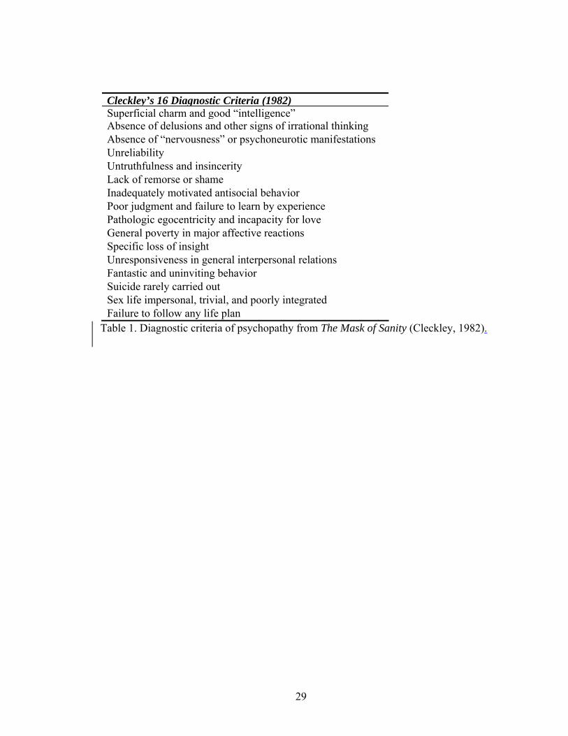
Cleckley’s 16 Diagnostic Criteria (1982)Superficial charm and good “intelligence”Absence of delusions and other signs of irrational thinking Absence of “nervousness” or psychoneurotic manifestations Unreliability Untruthfulness and insincerity Lack of remorse or shame Inadequately motivated antisocial behavior Poor judgment and failure to learn by experience Pathologic egocentricity and incapacity for love General poverty in major affective reactions Specific loss of insight Unresponsiveness in general interpersonal relations Fantastic and uninviting behavior Suicide rarely carried out Sex life impersonal, trivial, and poorly integrated Failure to follow any life plan

Table 1. Diagnostic criteria of psychopathy from The Mask of Sanity (Cleckley, 1982).

30

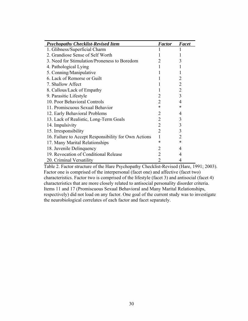
Psychopathy Checklist-Revised Item Factor Facet 1. Glibness/Superficial Charm 1 1 2. Grandiose Sense of Self Worth 1 1 3. Need for Stimulation/Proneness to Boredom 2 3 4. Pathological Lying 1 1 5. Conning/Manipulative 1 1 6. Lack of Remorse or Guilt 1 2 7. Shallow Affect 1 2 8. Callous/Lack of Empathy 1 2 9. Parasitic Lifestyle 2 3 10. Poor Behavioral Controls 2 4 11. Promiscuous Sexual Behavior * * 12. Early Behavioral Problems 2 4 13. Lack of Realistic, Long-Term Goals 2 3 14. Impulsivity 2 3 15. Irresponsibility 2 3 16. Failure to Accept Responsibility for Own Actions 1 2 17. Many Marital Relationships * * 18. Juvenile Delinquency 2 4 19. Revocation of Conditional Release 2 4 20. Criminal Versatility 2 4

Table 2. Factor structure of the Hare Psychopathy Checklist-Revised (Hare, 1991; 2003). Factor one is comprised of the interpersonal (facet one) and affective (facet two) characteristics. Factor two is comprised of the lifestyle (facet 3) and antisocial (facet 4) characteristics that are more closely related to antisocial personality disorder criteria. Items 11 and 17 (Promiscuous Sexual Behavioral and Many Marital Relationships, respectively) did not load on any factor. One goal of the current study was to investigate the neurobiological correlates of each factor and facet separately.

31

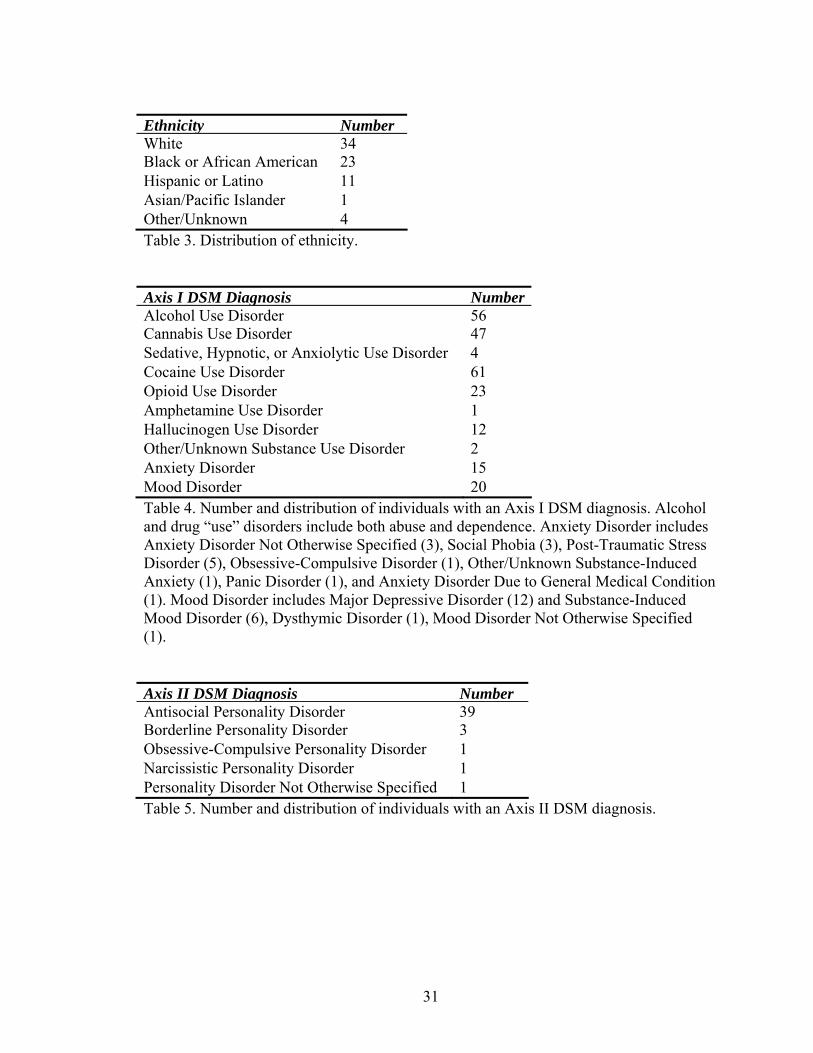
Ethnicity NumberWhite 34Black or African American 23 Hispanic or Latino 11 Asian/Pacific Islander 1 Other/Unknown 4 Table 3. Distribution of ethnicity. Axis I DSM Diagnosis NumberAlcohol Use Disorder 56Cannabis Use Disorder 47 Sedative, Hypnotic, or Anxiolytic Use Disorder 4 Cocaine Use Disorder 61 Opioid Use Disorder 23 Amphetamine Use Disorder 1 Hallucinogen Use Disorder 12 Other/Unknown Substance Use Disorder 2 Anxiety Disorder 15 Mood Disorder 20 Table 4. Number and distribution of individuals with an Axis I DSM diagnosis. Alcohol and drug “use” disorders include both abuse and dependence. Anxiety Disorder includes Anxiety Disorder Not Otherwise Specified (3), Social Phobia (3), Post-Traumatic Stress Disorder (5), Obsessive-Compulsive Disorder (1), Other/Unknown Substance-Induced Anxiety (1), Panic Disorder (1), and Anxiety Disorder Due to General Medical Condition (1). Mood Disorder includes Major Depressive Disorder (12) and Substance-Induced Mood Disorder (6), Dysthymic Disorder (1), Mood Disorder Not Otherwise Specified (1). Axis II DSM Diagnosis NumberAntisocial Personality Disorder 39Borderline Personality Disorder 3 Obsessive-Compulsive Personality Disorder 1 Narcissistic Personality Disorder 1 Personality Disorder Not Otherwise Specified 1 Table 5. Number and distribution of individuals with an Axis II DSM diagnosis.

32

Factor/Region MNI Coordinates t-value Factor One Lentiform nucleus 12 -2 -2 3.29 Facet two Lentiform nucleus 10 -2 -2 3.34 Thalamus -14 -8 0 2.84 Facet three Postcentral gyrus -28 -36 44 2.59 Facet four Postcentral gyrus 30 -40 72 2.58

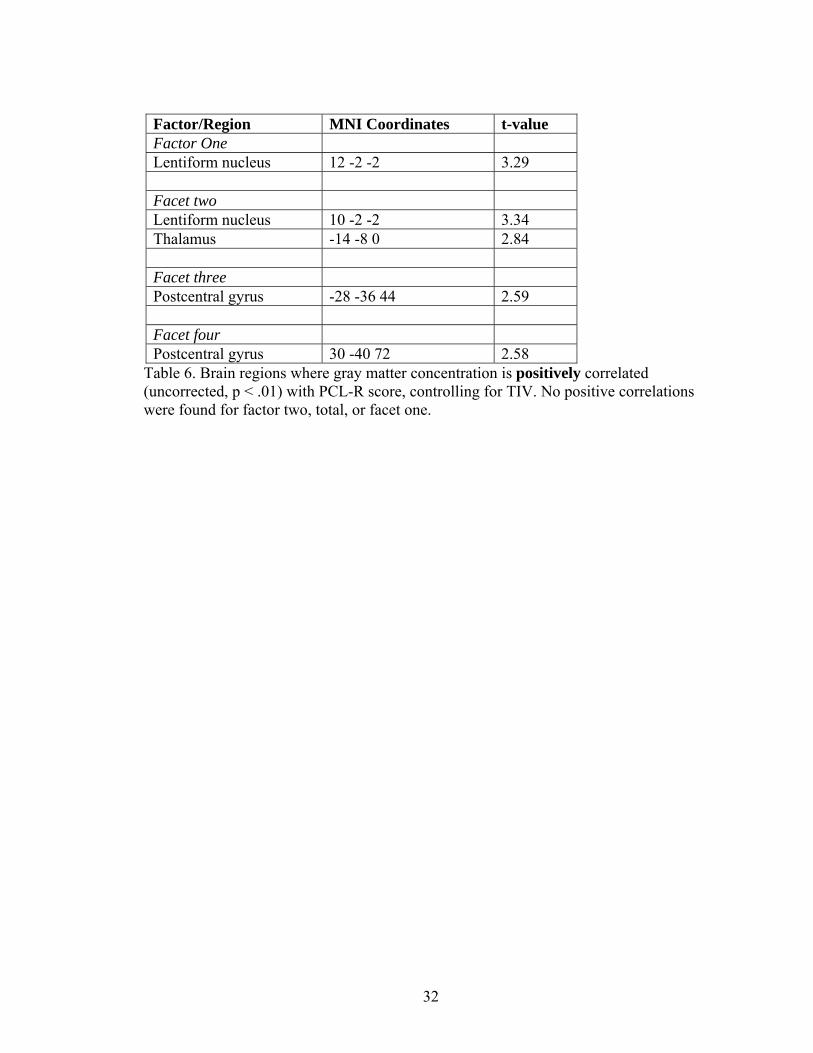
Table 6. Brain regions where gray matter concentration is positively correlated (uncorrected, p < .01) with PCL-R score, controlling for TIV. No positive correlations were found for factor two, total, or facet one.

33

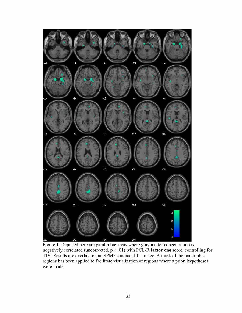
Figure 1. Depicted here are paralimbic areas where gray matter concentration is negatively correlated (uncorrected, p < .01) with PCL-R factor one score, controlling for TIV. Results are overlaid on an SPM5 canonical T1 image. A mask of the paralimbic regions has been applied to facilitate visualization of regions where a priori hypotheses were made.

34

Region MNI Coordinates t-value Region MNI Coordinates t-value Temporal Regions Parietal Regions Middle temporal gyrus -58 -42 -2 * 3.77 Post-central gyrus -62 -14 24 2.56 -62 -58 2 2.60 Inferior parietal -48 -34 38 3.01 46 -64 22 2.97 40 -46 40 2.78 68 -24 -14 2.49 Superior temporal gyrus 56 -48 16 2.89 Occipital Regions 50 -34 2 2.86 Cuneus 12 -78 12 3.18 -56 -12 4 2.96 -22 -86 22 3.17 Inferior temporal gyrus 60 -46 -14 2.53 -40 -86 8 2.57 68 -46 -10 2.45 -36 -90 14 2.52 -46 -70 -6 2.66 Inferior occipital 34 -96 -2 2.52 66 -16 -20 2.86 Lingual gyrus 26 -72 -2 3.32 -62 -18 -22 2.72 16 -78 -10 3.24 62 -6 -28 2.63 -28 -80 -12 2.93 Insula -36 -10 -8 * 3.43 -46 -26 16 3.02 Cingulate -56 -38 20 2.68 Mid cingulate 8 -38 42 2.93 38 12 8 2.49 12 -32 46 2.93 38 -10 6 2.46 -6 -4 50 2.68 34 20 6 2.45 2 -8 40 2.53 32 22 -8 2.65 Anterior cingulate -8 34 28 3.00 Temporal pole 38 6 -38 2.98 -6 20 38 2.53 Parahippocampal gyrus -16 0 -24 3.25 12 48 10 2.98

28 -28 -22 2.58 Posterior cingulate / precuneus -8 -68 26 2.50

Parahippocampal gyrus / amygdala 20 0 -20 3.15 10 -62 26 2.45

Hippocampus 32 -12 -20 3.18 Fusiform 20 -50 -14 2.69 Subcortical 30 -64 -18 2.58 Thalamus 10 -28 8 2.41 -42 -44 -20 2.57 -26 -12 -40 2.82 Cerebellum 28 -86 -28 2.68 32 -68 -40 * 3.29 Frontal Regions -22 -70 -34 3.06 Orbital frontal 22 26 -20 2.69 -44 -60 -30 2.69 30 38 -10 2.65 -24 -48 -18 2.60 50 38 -14 2.56 18 -58 -22 2.49 Superior medial frontal -10 58 4 2.61 24 -48 -50 3.02 Superior frontal gyrus 28 44 18 2.95 -18 -54 -44 2.73 22 -10 56 2.76 Middle frontal gyrus -26 10 50 2.93 -38 10 34 2.61 Inferior frontal gyrus 42 28 8 2.45 -48 18 12 2.86 38 10 26 2.75 Precentral gyrus -50 -6 30 2.88 -6 -26 54 2.84

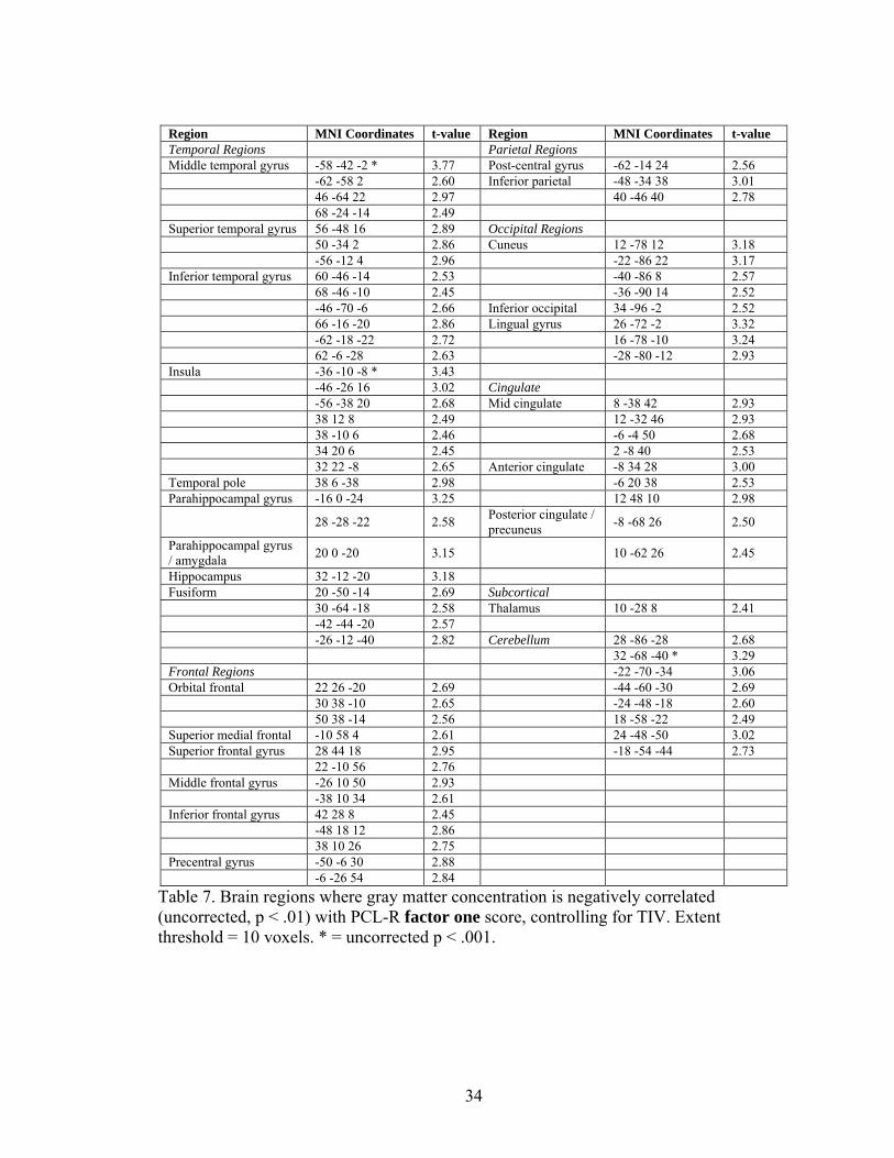
Table 7. Brain regions where gray matter concentration is negatively correlated (uncorrected, p < .01) with PCL-R factor one score, controlling for TIV. Extent threshold = 10 voxels. * = uncorrected p < .001.

35

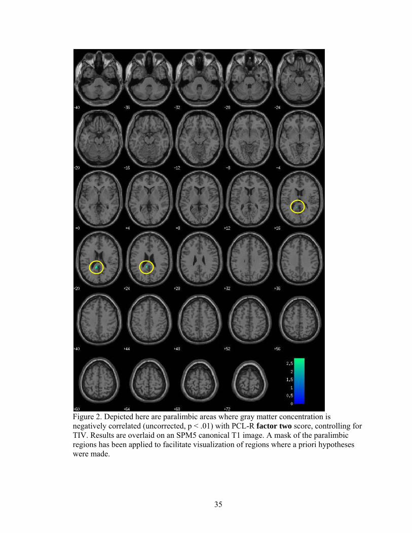
Figure 2. Depicted here are paralimbic areas where gray matter concentration is negatively correlated (uncorrected, p < .01) with PCL-R factor two score, controlling for TIV. Results are overlaid on an SPM5 canonical T1 image. A mask of the paralimbic regions has been applied to facilitate visualization of regions where a priori hypotheses were made.

36

Region MNI Coordinates t-value Middle temporal gyrus -56 -42 -6 3.50 Superior temporal gyrus -48 -26 2 2.78 Angular/supramarginal gyrus 42 -44 34 3.03 Posterior cingulate -10 -44 20 2.83 Lingual gyrus -12 -78 0 2.72 24 -72 0 2.65 Middle occipital gyrus -32 -70 14 2.79 Inferior parietal cortex -48 -38 36 3.15

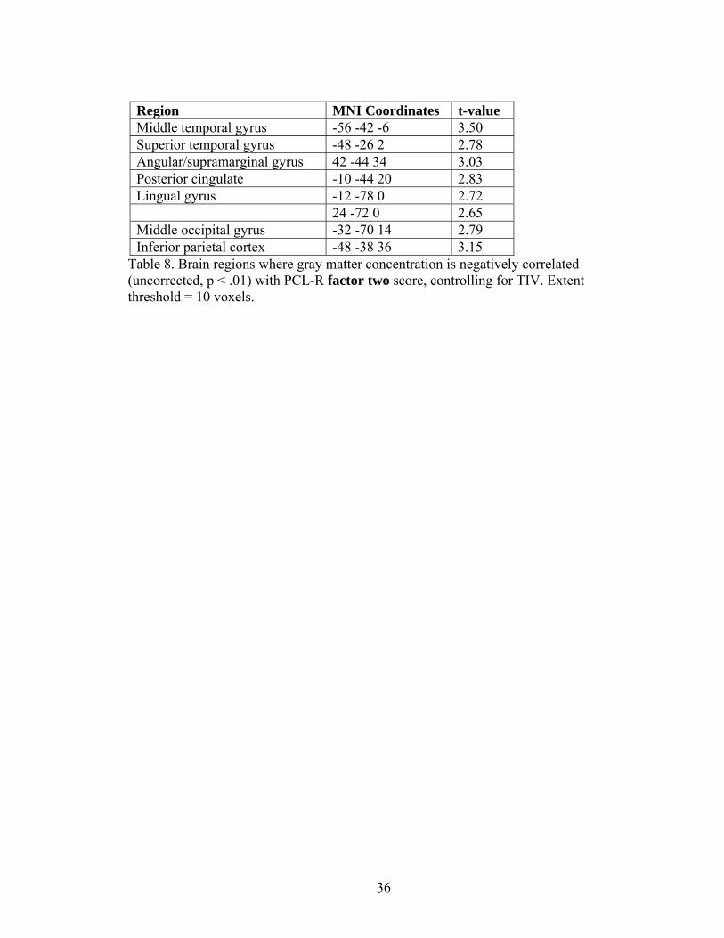
Table 8. Brain regions where gray matter concentration is negatively correlated (uncorrected, p < .01) with PCL-R factor two score, controlling for TIV. Extent threshold = 10 voxels.

37

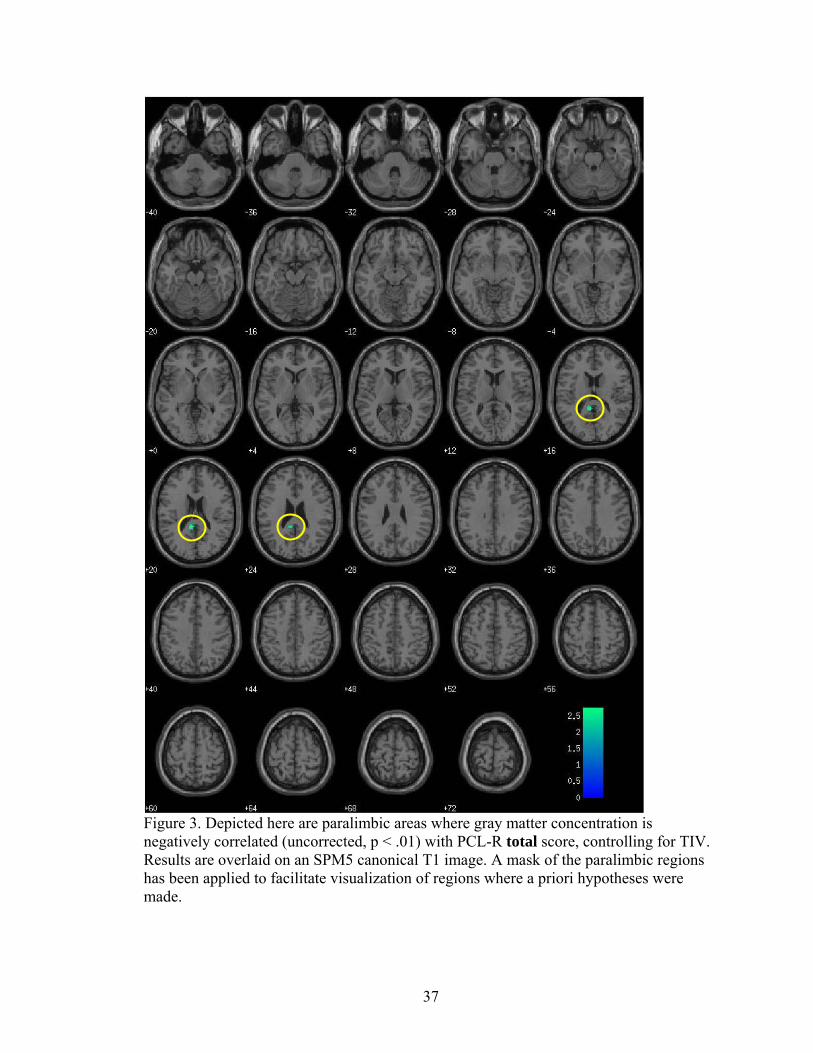
Figure 3. Depicted here are paralimbic areas where gray matter concentration is negatively correlated (uncorrected, p < .01) with PCL-R total score, controlling for TIV. Results are overlaid on an SPM5 canonical T1 image. A mask of the paralimbic regions has been applied to facilitate visualization of regions where a priori hypotheses were made.

38

Region MNI Coordinates t-value Middle temporal gyrus -56 -42 -4 * 3.65 Superior temporal gyrus -48 -26 2 2.62 Supramarginal gyrus 40 -44 36 3.00 Lingual gyrus 24 -72 0 3.12 -12 -78 0 2.68 Posterior cingulate -8 -42 20 2.76 Middle occipital gyrus -32 -70 14 2.88 Caudate 20 -32 14 2.66 Inferior parietal cortex -48 -36 36 2.91 Cerebellum 22 -68 -40 2.55 30 -64 -42 2.43

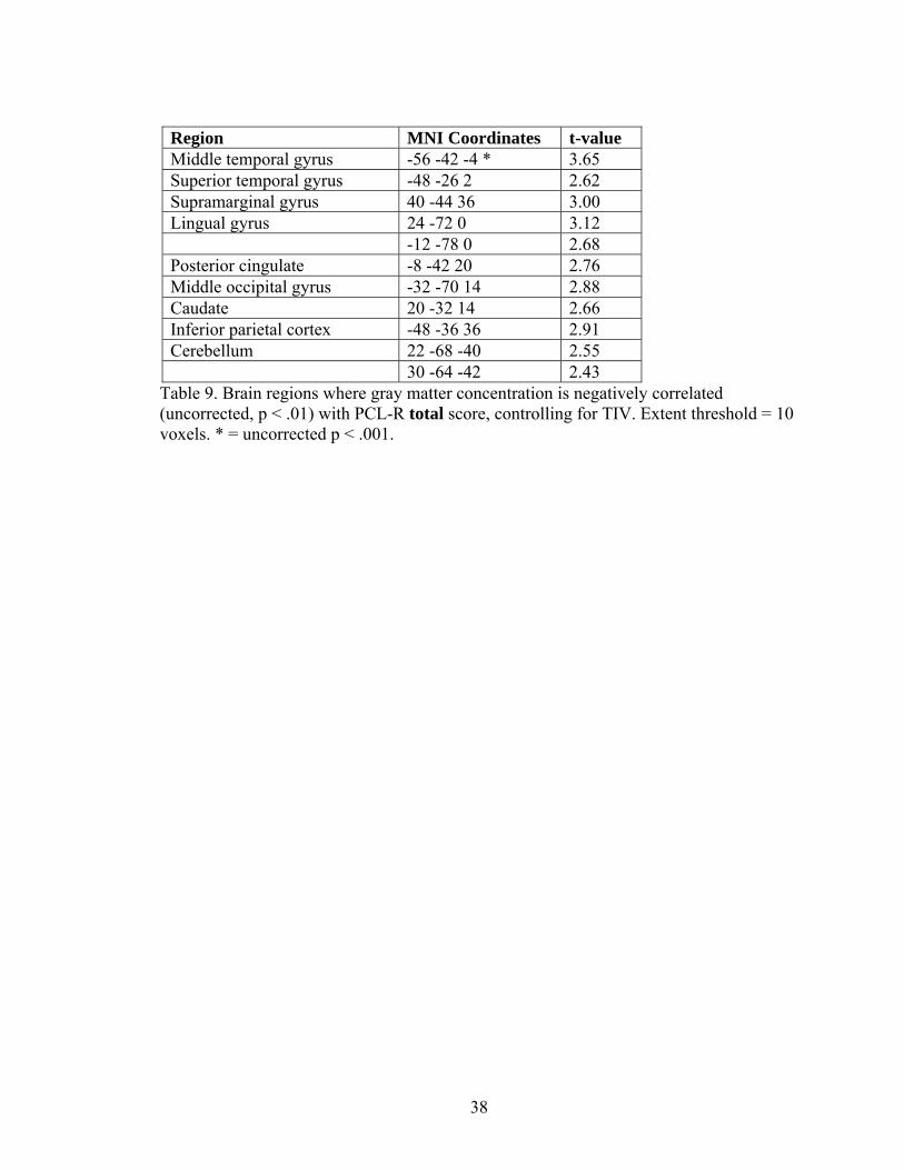
Table 9. Brain regions where gray matter concentration is negatively correlated (uncorrected, p < .01) with PCL-R total score, controlling for TIV. Extent threshold = 10 voxels. * = uncorrected p < .001.

39

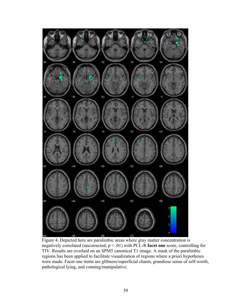
Figure 4. Depicted here are paralimbic areas where gray matter concentration is negatively correlated (uncorrected, p < .01) with PCL-R facet one score, controlling for TIV. Results are overlaid on an SPM5 canonical T1 image. A mask of the paralimbic regions has been applied to facilitate visualization of regions where a priori hypotheses were made. Facet one items are glibness/superficial charm, grandiose sense of self-worth, pathological lying, and conning/manipulative.

40

Region MNI Coordinates t-value Region MNI Coordinates t-value Temporal Regions Parietal Regions Middle temporal gyrus 64 -6 -28 3.00 Precuneus 12 -60 26 2.64 66 -16 -20 2.71 Postcentral gyrus -64 -8 20 2.46 68 -24 -14 2.58 -62 -60 -2 2.57 Occipital Regions Superior temporal gyrus -58 -44 -2 * 3.57 Lingual gyrus 24 -72 -2 * 3.60 -56 -12 2 3.22 -24 -84 -8 2.89 -56 -44 18 2.70 -10 -80 -2 2.65 Inferior temporal gyrus -64 -16 -26 * 2.88 Cuneus 14 -78 12 2.77 Insula -36 -10 -8 3.44 Middle occipital 32 -82 20 3.25 32 10 -18 2.68 46 -66 24 2.74 Supramarginal gyrus -52 -24 20 3.11 Parahippocampal gyrus -20 -30 -14 2.47 Cingulate -14 0 -26 2.63 Anterior cingulate -8 34 30 2.83 Parahippocampal gyrus / amygdala 20 2 -20 3.14 -8 22 38 2.47

Hippocampus 30 -12 -20 3.02 Mid cingulate -8 -2 50 2.92 Fusiform 20 -52 -14 2.88 12 -32 46 2.78 -44 -42 -20 2.64 Temporal pole -24 18 -38 2.63 Cerebellum -18 -54 -44 3.10 56 8 -34 2.64 24 -50 -48 3.28 -52 16 -34 2.73 34 -66 -42 3.16 34 -84 -30 2.86 Frontal Regions -20 -50 -16 2.58 Orbital frontal 26 28 -24 2.87 -20 -72 -34 2.45 18 34 -26 2.58 Superior frontal gyrus 28 44 18 3.00 Middle frontal gyrus 42 2 62 2.81 Inferior frontal gyrus 38 8 26 2.56 50 24 4 2.55 -58 24 2 2.51 40 2 -36 2.69 Superior medial frontal -10 40 24 2.63 -8 34 50 2.71 Precentral gyrus -8 -26 52 2.85

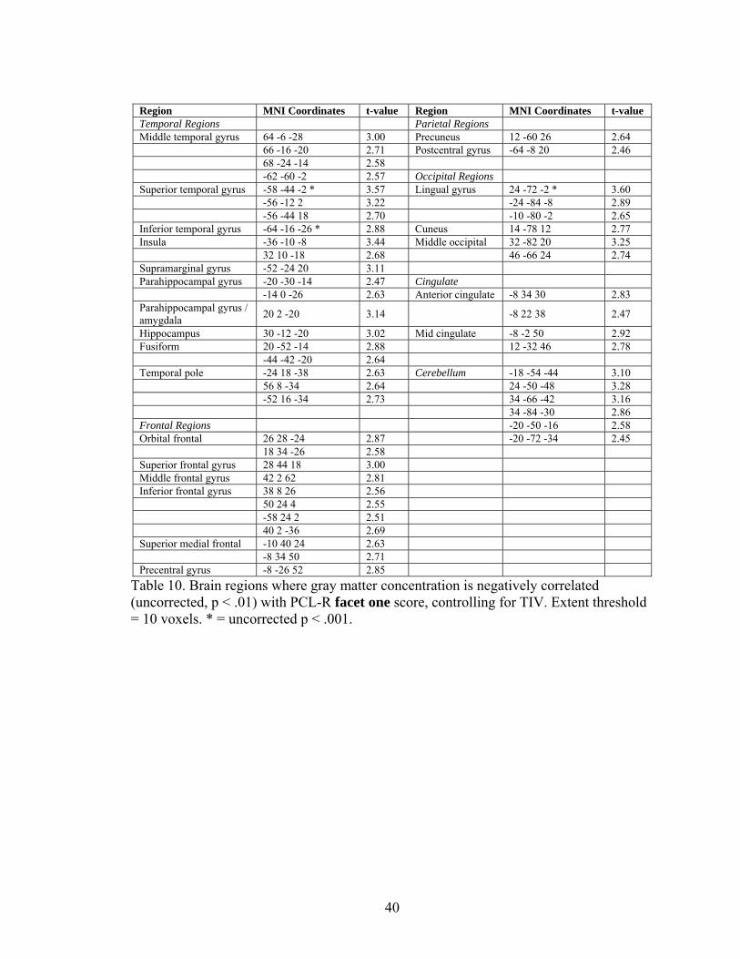
Table 10. Brain regions where gray matter concentration is negatively correlated (uncorrected, p < .01) with PCL-R facet one score, controlling for TIV. Extent threshold = 10 voxels. * = uncorrected p < .001.

41

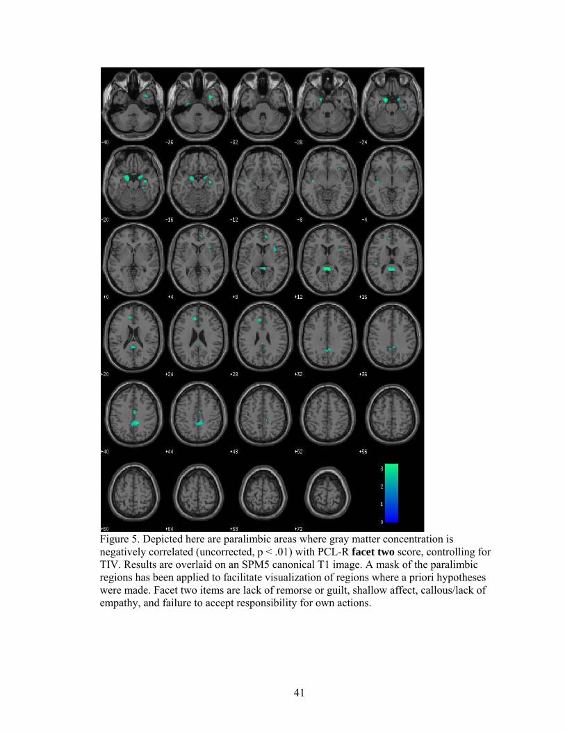
Figure 5. Depicted here are paralimbic areas where gray matter concentration is negatively correlated (uncorrected, p < .01) with PCL-R facet two score, controlling for TIV. Results are overlaid on an SPM5 canonical T1 image. A mask of the paralimbic regions has been applied to facilitate visualization of regions where a priori hypotheses were made. Facet two items are lack of remorse or guilt, shallow affect, callous/lack of empathy, and failure to accept responsibility for own actions.

42

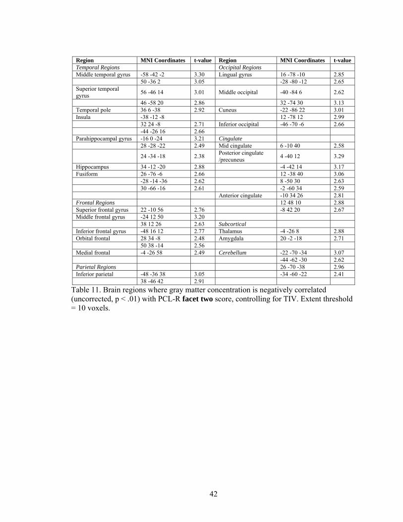
Region MNI Coordinates t-value Region MNI Coordinates t-value Temporal Regions Occipital Regions Middle temporal gyrus -58 -42 -2 3.30 Lingual gyrus 16 -78 -10 2.85 50 -36 2 3.05 -28 -80 -12 2.65 Superior temporal gyrus 56 -46 14 3.01 Middle occipital -40 -84 6 2.62

46 -58 20 2.86 32 -74 30 3.13 Temporal pole 36 6 -38 2.92 Cuneus -22 -86 22 3.01 Insula -38 -12 -8 12 -78 12 2.99 32 24 -8 2.71 Inferior occipital -46 -70 -6 2.66 -44 -26 16 2.66 Parahippocampal gyrus -16 0 -24 3.21 Cingulate 28 -28 -22 2.49 Mid cingulate 6 -10 40 2.58

24 -34 -18 2.38 Posterior cingulate /precuneus 4 -40 12 3.29

Hippocampus 34 -12 -20 2.88 -4 -42 14 3.17 Fusiform 26 -76 -6 2.66 12 -38 40 3.06 -28 -14 -36 2.62 8 -50 30 2.63 30 -66 -16 2.61 -2 -60 34 2.59 Anterior cingulate -10 34 26 2.81 Frontal Regions 12 48 10 2.88 Superior frontal gyrus 22 -10 56 2.76 -8 42 20 2.67 Middle frontal gyrus -24 12 50 3.20 38 12 26 2.63 Subcortical Inferior frontal gyrus -48 16 12 2.77 Thalamus -4 -26 8 2.88 Orbital frontal 28 34 -8 2.48 Amygdala 20 -2 -18 2.71 50 38 -14 2.56 Medial frontal -4 -26 58 2.49 Cerebellum -22 -70 -34 3.07 -44 -62 -30 2.62 Parietal Regions 26 -70 -38 2.96 Inferior parietal -48 -36 38 3.05 -34 -60 -22 2.41 38 -46 42 2.91

Table 11. Brain regions where gray matter concentration is negatively correlated (uncorrected, p < .01) with PCL-R facet two score, controlling for TIV. Extent threshold = 10 voxels.

43

Figure 6. Depicted here are paralimbic areas where gray matter concentration is negatively correlated (uncorrected, p < .01) with PCL-R facet three score, controlling for TIV. Results are overlaid on an SPM5 canonical T1 image. A mask of the paralimbic regions has been applied to facilitate visualization of regions where a priori hypotheses were made. Facet three items are need for stimulation/proneness to boredom, parasitic lifestyle, lack of realistic long term goals, impulsivity, and irresponsibility.

44

Region MNI Coordinates t-value Superior temporal gyrus -50 -28 2 2.71 Middle temporal gyrus -56 -42 -6 3.20 Posterior cingulate -10 -44 22 3.00 Supramarginal gyrus 44 -46 34 2.91 -46 -44 34 2.71 Lingual gyrus 20 -72 0 3.17 Middle occipital gyrus -32 -70 14 3.22

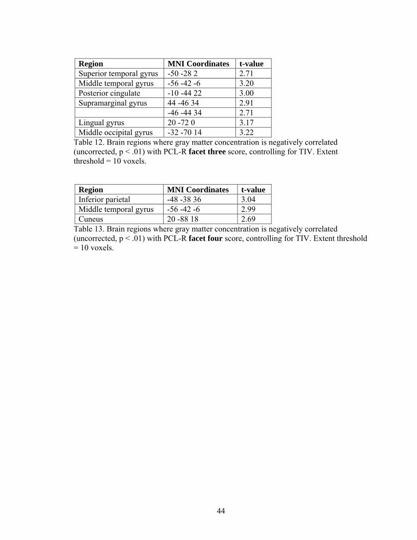
Table 12. Brain regions where gray matter concentration is negatively correlated (uncorrected, p < .01) with PCL-R facet three score, controlling for TIV. Extent threshold = 10 voxels.

Region MNI Coordinates t-value Inferior parietal -48 -38 36 3.04 Middle temporal gyrus -56 -42 -6 2.99 Cuneus 20 -88 18 2.69

Table 13. Brain regions where gray matter concentration is negatively correlated (uncorrected, p < .01) with PCL-R facet four score, controlling for TIV. Extent threshold = 10 voxels.

45

References

Alterman, A. I., Cacciola, J. S., & Rutherford, M. J. (1993). Reliability of the Revised

Psychopathy Checklist in substance abuse patients. Psychological Assessment, 5,

442-448.

American Psychiatric Association. (1952). Diagnostic and statistical manual of mental

disorders (1st ed.). Washington, DC: American Psychiatric Association.

American Psychiatric Association. (1968). Diagnostic and statistical manual of mental

disorders – Second edition. Washington, DC: American Psychiatric Association.

American Psychiatric Association. (1994). Diagnostic and statistical manual of mental

disorders – Fourth edition. Washington, D.C.: American Psychiatric Association.

Anderson, S. W., Bechara, A., Damasio, H., Tranel, D., & Damasio, A. R. (1999).

Impairment of social and moral behavior related to early damage in human

prefrontal cortex. Nature Neuroscience, 2, 1032-1037.

Ashburner, J. & Friston, K. J. (2000). Voxel-based morphometry – The methods.

Neuroimage, 11, 805-21.

Ashburner, J. & Friston, K. J. (2005). Unified segmentation. Neuroimage, 26, 839-851.

Barkataki, I., Kumari, V., Das, M., Taylor, P., & Sharma, T. (2006). Volumetric

structural brain abnormalities in men with schizophrenia or antisocial personality

disorder. Behavioural Brain Research, 169, 239-247.

Bechara, A., Damasio, H., Damasio, A. R., & Lee, G. P. (1999). Different contributions

of the human amygdala and ventromedial prefrontal cortex to decision-making.

Journal of Neuroscience, 19, 5473-5481.

46

Berlin, H. A., Rolls, E. T., & Kischka, U. (2004). Impulsivity, time perception, emotion

and reinforcement sensitivity in patients with orbitofrontal cortex lesions. Brain,

127, 1108-1126.

Birbaumer, N., Veit, R., Lotze, M., Erb, M., Hermann, C., Grodd, W., et al. (2005).

Deficient fear conditioning in psychopathy. Archives of General Psychiatry, 62,

799-805.

Blair, R. J. R. (2007). The amygdala and ventromedial prefrontal cortex in morality and

psychopathy. Trends in Cognitive Sciences, 11, 387-392.

Brodmann, K. (1909). Vergleichende Lokalisationlehre der Grosshirnrinde in ihren

Prinzipien dargestellt auf Grund des Zellenbaues. Leipzig, Barth, J.A.

Büchel, C. & Dolan, R. J. (2000). Classical fear conditioning in functional neuroimaging.

Current Opinion in Neurobiology, 10, 219-223.

Büchel, C., Morris, J., Dolan, R. J., & Friston, K. (1998). Brain systems mediating

aversive conditioning: An event-related fMRI study. Neuron, 20, 947-957.

Cleckley, H. (1982). The Mask of Sanity. New York: Mosby.

Cooke, D. J. & Michie, C. (1997). An item response theory analysis of the Hare

Psychopathy Checklist-Revised. Psychological Assessment, 9, 3-14.

Damasio, H., Grabowski, T., Frank, R., Galaburda, A. M., & Damasio, A. R. (1994). The

return of Phineas Gage: Clues about the brain from the skull of a famous patient.

Science, 264, 1102-5.

Eckert, M. A., Tenforde, A., Galaburda, A. M., Bellugi, U., Korenberg, J. R., Mills, D., et

al. (2006). To modulate or not to modulate: Differing results in uniquely shaped

Williams syndrome brains. Neuroimage, 32, 1001-1007.

47

Fein, G., Landman, B., Tran, H., McGillivray, S., Finn, P., Barakos, J., et al. (2006).

Brain atrophy in long-term abstinent alcoholics who demonstrate impairment on a

simulated gambling task. Neuroimage, 32, 1465-1471.

First, M. B., Spitzer, R. L., Gibbon, M., Williams, J. B. W. (2002). Structured Clinical

Interview for DSM-IV-TR Axis I Disorders – Patient Edition (SCID-I/P).

Washington, D. C.: American Psychiatric Press.

Fischer, H., Andersson, J. L., Furmack, T., Wik, G., & Fredrikson, M. (2002). Right-

sided human prefrontal brain activation during acquisition of conditioned fear.

Emotion, 2, 233-241.

Franklin, T. R., Acton, P. D., Maldjian, J. A., Gray, J. D., Croft, J. R., Dackis, C. A., et al.

Decreased gray matter concentration in the insular, orbitofrontal, cingulate, and

temporal cortices of cocaine patients. Biological Psychiatry, 51, 134-142.

Hare, R. D. (1991). Manual for the Hare Psychopathy Checklist-Revised. Toronto: Multi

Health Systems.

Hare, R. D. (1998). Psychopaths and their nature: Implications for the mental health and

criminal justice systems. In T. Millon, E. Simonsen, et al. (Eds.), Psychopathy:

Antisocial, criminal, and violent behavior (pp. 188-212). New York: Guilford

Press.

Hare, R. D. (2003). Manual for the Hare Psychopathy Checklist-Revised, 2nd ed.

Toronto: Multi-Health Systems.

Harpur, T. J., Hakstian, A. R., & Hare, R. D. (1988). Factor structure of the Psychopathy

Checklist. Journal of Consulting and Clinical Psychology, 56, 741-747.

48

Harpur, T. J., Hare, R. D., & Hakstian, A. R. (1989). Two-factor conceptualization of

psychopathy: Construct validity and assessment implications. Psychological

Assessment, 1, 6-17.

Hemphill, J. F., Hare, R. D., & Wong, S. (1998). Psychopathy and recidivism: A review.

Legal & Criminological Psychology, 3, 139-170.

Hemphill, J. F., Hart, S. D., & Hare, R. D. (1994). Psychopathy and substance use.

Journal of Personality Disorders, 8, 169-180.

Hiatt, K. D., & Newman, J. P. (2007). Behavioral evidence of prolonged interhemispheric

transfer time among psychopathic offenders. Neuropsychology, 21, 313-318.

Jatzko, A., Rothenhoefer, S., Schmitt, A., Gaser, C., Demirakca, T., Weber-Fahr, W., et

al. (2006). Hippocampal volume in chronic posttraumatic stress disorder (PTSD):

MRI study using two different evaluation methods. Journal of Affective

Disorders, 94, 121-126.

Kiehl, K. A. (2006). A cognitive neuroscience perspective on psychopathy: Evidence for

paralimbic system dysfunction. Psychiatry Research, 142, 107-128.

Kiehl, K. A., Smith A. M., Hare R. D., Mendrek A., Forster B. B., Brink J., et al. (2001).

Limbic abnormalities in affective processing by criminal psychopaths as revealed

by functional magnetic resonance imaging. Biological Psychiatry, 50, 677-84.

Kinner, S. (2003). Psychopathy as an adaptation: Implications for society and social

policy. In R. W. Bloom & N. Dess (Eds.), Evolutionary psychology and violence:

A primer for policymakers and public policy advocates (pp. 57-81). Westport, CT:

Praeger.

49

Knight, D. C., Cheng, D. T., Smith, C. N., Stein E. A., Helmstetter, F. J. (2004). Neural

substrates mediating human delay and trace fear conditioning. Journal of

Neuroscience, 24, 218-228.

Kosson, D. S. (1996). Psychopathy and dual task performance under focusing conditions.

Journal of Abnormal Psychology, 105, 391-400.

Kosson, D. S. (1998). Divided visual attention in psychopathic and non-psychopathic

offenders. Personality and Individual Differences, 24, 373-391.

Laakso, M. P., Vaurio, O., Koivisto, E., Savolainen, L., Eronen, M., Aronen, H. J., et al.

(2001). Psychopathy and the posterior hippocampus. Behavioural Brain

Research, 118, 187-93.

Laakso, M. P., Gunning-Dixon, F., Vaurio, O., Repo-Tiihonen, E., Soininen, H., &

Tiihonen, J. (2002). Prefrontal volumes in habitually violent subjects with

antisocial personality disorder and type 2 alcoholism. Psychiatry Research:

Neuroimaging, 114, 95-102.

Matsuo, K., Nicoletti, M., Nemoto, K., Hatch, J. P., Peluso, M. A. M., Nery, F. G., et al.

A voxel-based morphometry study of frontal gray matter correlates of

impulsivity. Human Brain Mapping, 30, 1188-1195.

Mechtcheriakov, S., Brenneis, C., Egger, K., Koppelstaetter, F., Schocke, M., &

Marksteiner, J. (2007). A widespread distinct pattern of cerebral atrophy in

patients with alcohol addiction revealed by voxel-based morphometry. Journal of

Neurology, Neurosurgery, and Psychiatry, 78, 610-614.

Mesulam, M. M., Ed. (2000). Principles of behavioral and cognitive neurology. New

York: Oxford University Press.

50

Müller J. L., Sommer, M., Wagner, V., Lange, K., Taschler, H., & Roeder, C. H. (2003).

Abnormalities in emotion processing within cortical and subcortical regions in

criminal psychopaths: Evidence from a functional magnetic resonance imaging

study using pictures with emotional content. Biological Psychiatry, 54, 152-162.

Müller, J. L., Gänβbauer, S., Sommer, M., Döhnel, K., Weber, T., Schmidt-Wilcke, T., et

al. (2008). Gray matter changes in right superior temporal gyrus in criminal

psychopaths. Evidence from voxel-based morphometry. Psychiatry Research:

Neuroimaging, 163, 213-222.

Nesse, R. M. (2005). Evolutionary Psychology and Mental Health. In D. M. Buss (Ed.),

The handbook of evolutionary psychology (pp. 903-27). Hoboken, NJ: Wiley.

Oliveira-Souza, R., Hare, R. D., Bramati, I. E., Garrido, G. J., Ignacio, F. A., Tovar-Moll,

F., et al. (2008). Psychopathy as a disorder of the moral brain: Fronto-temporo-

limbic grey matter reductions demonstrated by voxel-based morphometry.

NeuroImage, 40, 1202-13.

Pell, G. S., Briellmann, R. S., Chan, C. H., Pardoe, H., Abbott, D. F., & Jackson, G. D.

(2008). Selection of the control group for VBM analysis: Influence of covariates,

matching and sample size. Neuroimage, 41, 1324-1335.

Raine, A., Ishikawa, S. S., Arce, E., Lencz, T., Knuth, K. H., Bihrle, S., et al. (2004).

Hippocampal structural asymmetry in unsuccessful psychopaths. Biological

Psychiatry, 55, 185-91.

Raine, A., Lencz, T., Bihrle, S., LaCasse, L., & Colletti, P. (2000). Reduced prefrontal

gray matter volume and reduced autonomic activity in antisocial personality

disorder. Archives of General Psychiatry, 57, 119-127.

51

Raine, A., Lencz, T., Taylor, K., Hellige, J. B., Bihrle, S., LaCasse, L., et al. (2003).

Corpus callosum abnormalities in psychopathic antisocial individuals. Archives of

General Psychiatry, 60, 1134-42.

Schorn, D. (2007, January 13). Murder on his mind: Can scientific images show what's in

the mind of a murderer? Retrieved June 3, 2008, from http://www.cbsnews.com/

stories/2007/01/11/48hours/main2352668.shtml.

Taki, Y., Kinomura, S., Sato, K., Goto, R., Inoue, K., Okada, K., et al. (2006). Both

global gray matter volume and regional gray matter volume negatively correlate

with lifetime alcohol intake in non-alcohol-dependent Japanese men: A

volumetric analysis and a voxel-based morphometry. Alcoholism: Clinical and

Experimental Research, 30, 1045-1050.

Tanabe, J., Tregellas, J. R., Dalwani, M., Thompson, L., Owens, E., Crowley, T., et al.

(2009). Medial orbitofrontal cortex gray matter is reduced in abstinent substance-

dependent individuals. Biological Psychiatry, 65, 160-164.

Tiihonen, J., Rossi, R., Laakso, M. P., Hodgins, S., Testa, C., Perez, J., et al. (2008).

Brain anatomy of persistent violent offenders: More rather than less. Psychiatry

Research: Neuroimaging, 163, 201-212.

Tranel, D. (1994). ‘Acquired sociopathy’: The development of sociopathic behavior

following focal brain damage. Progress in Experimental Personality and

Psychopathology Research, 285-311.

Veit, R., Flor, H., Erb, M., Hermann, C., Lotze, M., Grodd, W., et al. (2002). Brain

circuits involved in emotional learning in antisocial behavior and social phobia in

humans. Neuroscience Letters, 328, 233-236.

52

Yang, Y., Raine, A., Lencz., T., Bihrle, S., LaCasse, L., & Colletti, P. (2005). Volume

reduction in prefrontal gray matter in unsuccessful criminal psychopaths.

Biological Psychiatry, 57, 1103-08.


Recommended