2014 28 September -- Paul Fatt. 13 January 1924 Jonathan F. Ashmore published online June 1, 2016 , 167-186, published 1 June 2016 originally 62 2016 Biogr. Mems Fell. R. Soc. Supplementary data /23/rsbm.2016.0005.DC2 http://rsbm.royalsocietypublishing.org/content/suppl/2017/08 "Data Supplement" /31/rsbm.2016.0005.DC1 http://rsbm.royalsocietypublishing.org/content/suppl/2016/05 "Data Supplement" Email alerting service here or click sign up in the box at the top right-hand corner of the article Receive free email alerts when new articles cite this article - http://rsbm.royalsocietypublishing.org/subscriptions , go to: Biogr. Mems Fell. R. Soc. To subscribe to on August 3, 2018 http://rsbm.royalsocietypublishing.org/ Downloaded from on August 3, 2018 http://rsbm.royalsocietypublishing.org/ Downloaded from
Transcript
2014 28 September−−Paul Fatt. 13 January 1924
Jonathan F. Ashmore
published online June 1, 2016, 167-186, published 1 June 2016 originally622016 Biogr. Mems Fell. R. Soc.
Department of Neuroscience, Physiology and Pharmacology, University College London, London WC1E 6BT, UK
Paul Fatt made discoveries that are fundamental to our understanding of synaptic transmission in the nervous system. He grew up in the USA and saw service in World War II, but came to London in 1948 as a research student supported by the GI Bill. His seminal work with Bernard Katz at University College London (UCL), John Eccles in Canberra, and Bernard Ginsborg at UCL was carried out during an intense period between 1950 and 1960. His work with Katz demonstrated for the first time that neurotransmitter is released in small packets, or ‘quanta’. His work with Eccles (and Katz) provided an understanding of the mechanism underlying synaptic inhibition, and his work with Ginsborg identified voltage-gated calcium currents for the first time. Furthermore, in the early 1960s his electrical measurements of the muscle transverse tubule system contributed to the early models of excitation–contraction coupling in muscle. The final period of his research career was spent working on phototransduction in the visual system.
Early life
Paul Fatt was born on 13 January 1924 in Chicago, USA, and spent his early youth there. He was the second of three sons of parents who had emigrated to the USA in the early twentieth century. His father, David (ca. 1880–1968), had come from Eastern Slovakia, which was then part of the Austro-Hungarian empire. It seems that his name was changed from ‘Fett’ (listed as such on the passenger ship Sabraus arriving in New York in 1899) to ‘Fatt’ by the filing clerk on Ellis Island. David Fatt worked in the leather business in Chicago. Paul’s mother, Annie (née Arkin; d. 1974), was originally from Lithuania. Both were orthodox Jews and strictly kosher. Neither parent had a scientific training, but all three brothers had scientific careers.
This memoir originally appeared in Biographical Memoirs of the US National Academy of Sciences and is reprinted, with slight modifications, with permission.
Paul’s elder brother, Irving (1920–96), was trained at California Institute of Technology as a chemical engineer and later, becoming known for his work on the diffusion of oxygen through contact lenses, became Dean of Optometry at the University of California (UC), Berkeley. The younger brother, Milton (1929–2011), became a mathematician, taking a PhD with Beno Eckmann at Eidgenössische Technische Hochschule in Zurich, and then taught at the California State University at Long Beach for many years. Paul had an early interest in biology and palaeontology that was fostered by regular visits to the museums of Chicago while a young boy at Grant’s School in Chicago. He then spent just one term at the age of 14 years at Robert Emmet’s High School, a school that he was later proud to acknowledge because it was named after an Irish nationalist and rebel leader.
In 1938 the family moved to Los Angeles and settled in the Fairfax district, made up mostly of orthodox Jews. The father ran the leather supply business until the late 1950s when a fire destroyed his stock that was kept in his garage. The move, however, allowed Irving to go to California Institute of Technology. Paul attended a junior school, Mount Vernon High School, before going to Fairfax High School and then on to Los Angeles City College. He was persuaded to specialize in civil engineering but always really wanted to study biology. He confessed that this was what had really interested him, and that his Chicago museum visits as a teenager had served to encourage this interest. Nevertheless, it was his mathematical training as an engineer that resurfaced in his wartime and subsequent scientific career.
When the USA entered World War II in 1941 after the bombing of Pearl Harbor, Paul joined as a reservist. This, at the time, permitted him to continue at City College for a short period. He was eventually called up in 1942 and underwent basic infantry training before being transferred to the eastern USA. Somehow this allowed him still to continue civil engineering in a course at the University of Kentucky in Lexington. By 1943 he was assigned to 291st Field Artillery Battalion, and with his mathematical knowledge he was put to the task of sound ranging to identify enemy guns by triangulating the sources (a method originally introduced into the British Army during World War I by William Lawrence (later Sir Lawrence) Bragg (FRS 1921)).
Paul, by then with the rank of a T4 sergeant, was sent to Europe in 1944. He arrived in France in October, landing on Omaha Beach, having travelled via Liverpool and London, where he acquired a taste for England. He moved with his battalion along with the Allied front and reached Aachen after the Battle of the Bulge on New Year’s Day 1945, where he narrowly avoided being hit in a Luftwaffe attack. Towards the end of his life he recalled this traumatic episode as though it had only just happened. After seeing further action, Paul moved on with the advance, eventually crossing the Rhine and along the Lippe Canal by March 1945 and into Dortmund on the north side of the Ruhr valley. His battalion met the end of the war at the Elbe river. The summer was spent in the region until he was demobilized after Japan surrendered; he later admitted to some relief that he did not have to fight in the Pacific war.
Although already enamoured of England by 1945, Paul found himself back in the USA by the end of the year and applied to study biochemistry at the UC Berkeley, under the GI Bill. Having achieved his degree in two years, he joined the biophysics programme, also at UC Berkeley, thinking he would like to put his physics and mathematics to use rather than employ his degree to enter medical school like so many of his contemporaries. During this year he met Vernon Brooks, who was then a zoologist but later turned to neuroscience, and it was Brooks who encouraged him to write to A. V. Hill FRS at University College London, requesting to be
on August 3, 2018http://rsbm.royalsocietypublishing.org/Downloaded from
taken on as a research student. Because he had another two years of his own GI Bill funding and because it simplified, then as now, any bureaucratic problems, Hill agreed to accept him into the Biophysics Research Unit as a research student.
Paul therefore found himself back in London in August 1948, in time to watch the London Olympics. Much to his delight, entry to the Games was free at Wembley. Hill suggested that, because there was nobody around at that time of summer, he should go down to the Marine Biological Association Laboratories at Plymouth, where Alan (later Sir Alan) Hodgkin FRS (PRS 1970–75), Andrew (later Sir Andrew) Huxley (FRS 1955; PRS 1980–85) and Bernard (later Sir Bernard) Katz (FRS 1952) were working on the action potential mechanism in the squid axon. This was Paul’s introduction to using electrical methods to understand basic biological mechanisms.
The Biophysics Research Unit, University College London
The Biophysics Research Unit, under A. V. Hill, at University College London had been newly re-established in 1946, and by 1948 Katz was working on muscle spindles in his laboratory there (Katz 1950). Back in London from Plymouth, he took on Paul as a PhD student, suggesting as a topic that he should look at the depolarization of striated muscle by acetylcholine (ACh). At the time, although some work had been done by Robert Conway at University College Dublin, who had reported that the endplate region of muscle was depolarized by ACh, there was very little understanding of the detailed mechanisms of how nerves controlled muscle, or even what the mechanism of electrical signalling entailed. It was not really even understood what factors determined the resting potential of excitable cells. Most recordings of electrical activity were carried out using extracellular recording of the potentials that surrounded active nerve cells. Indeed, Paul’s first publication used an ingenious liquid electrode chamber using extracellular measurements to show that ACh depolarized the muscle endplate region (1)*. By 1948 Hodgkin and Huxley had started to use the voltage clamp, based on a technique originally devised by Kenneth (‘Kacy’) Cole and George Marmont at Woods Hole Laboratories, to measure currents that flowed across the membrane of the giant axon of the squid, and so initiated the modern era of membrane biophysics. The work led to the award of a Nobel Prize for Hodgkin and Huxley (together with Sir John Eccles FRS) in 1963. Instead of using extracellular electrodes, they inserted a wire into the axoplasm as a way of gaining electrical access to the inner membrane surface (Hodgkin et al. 1952). The idea of inserting a fine glass tube filled with a conducting saline (a ‘microelectrode’) into a cell had recently been introduced to the Cambridge physiology group by a visiting research fellow, William A. Nastuk, who had learnt the technique from the work of Gilbert Ling and Ralph Gerrard at the University of Chicago. Nastuk paid a visit to London and showed Paul how to make microelectrodes by using a small flame to melt a thin glass tube and pull it out to a fine tip. The electrode was then filled by boiling these electrodes under reduced pressure in a solution of 3 M potassium chloride.
Fatt and Katz started to record membrane potentials from the sartorius muscle of the frog. Eccles, Katz and Stephen Kuffler (ForMemFRS 1971) had previously looked at the endplate potential (EPP) of cat muscle when Katz was in Sydney, Australia (Eccles et al. 1942) and so
* Numbers in this form refer to the bibliography at the end of the text.
on August 3, 2018http://rsbm.royalsocietypublishing.org/Downloaded from
the existence of these potentials was already known. Paul graphically described a typical day working with Katz during his PhD:
Leaving Katz at the entrance to the underground station I would go off for a meal at a cheap restaurant nearby and then return to the lab to prepare electrodes for the next day. We had the idea that electrodes had to be fairly fresh—no more than a day or two old, after which they would become brittle from soaking in the KCl filling solution and then break in use … .
Katz would do the dissection and I would make the microelectrodes, then I would manipulate the microelectrodes while Katz ground his teeth as it wouldn’t go into the muscle fibre where we wanted it to. And then he did the photographs. We knew about endplate potentials. And so how do you go from endplate potential to the whole thing? And there of course was a big problem because you get a contraction, you know, the thing would jump off the electrodes.
Their work showed that the EPP involved an increased ionic permeability depolarizing the postsynaptic membrane towards zero, the first clear demonstration that synaptic receptors were chemically gated ion channels (3). The paper, the substance of Paul’s PhD thesis, characterized for the first time the basic properties of the resultant intracellular EPP. However, the most startling result to come out of this work was the discovery of the existence of miniature EPPs (2, 4). These are small fluctuations of the membrane potential, of relatively uniform size, and have come to underpin our understanding of how synapses function: they are the result of neurotransmitter being released in packets, or ‘quanta’ (figure 1). This finding came out of a chance observation in the spring of 1950: when observed at high gain, the endplate region of the muscle was the site of spontaneous ongoing electrical activity. Although the observation was published in Nature (as ‘Some observations of biological noise’, with a Dr P. Fatt and Dr B. Katz as authors (2), although Paul had yet to be awarded a PhD), the origin of ‘minEPPs’ was unclear. They toyed with the notion that the events were non-propagated action potentials from the fine nerve branches, induced by thermal fluctuations. When the full paper came out in Journal of Physiology (with the same figure as in Nature), these events, dubbed ‘miniature endplate potentials’ (minEPPs) were unequivocally ascribed to the discharge of multimolecular ‘quanta’ of ACh from the nerve terminal. In a series of elegant experiments
Figure 1. Miniature endplate potentials. The left panel shows small events about 1 mV in amplitude recorded at the muscle endplate of a frog sartorius muscle; in the right panel the microelectrode has been moved 2 mm away. (Reproduced from (4), with permission.)
on August 3, 2018http://rsbm.royalsocietypublishing.org/Downloaded from
appearing in Journal of Physiology in 1952 (4), they had demonstrated that minEPPs (later more universally referred to as MEPPs), represented the ‘basic coin’ of chemical synaptic transmission—and that the full-sized EPP triggered by a nerve impulse represented the superposition of a large number of synchronously occurring MEPPs. They went on to characterize these quantal events in detail, paving the way for Katz’s later studies with José del Castillo, Stephen Thesleff and Ricardo Miledi (FRS 1970). The EPP, and its constituent ‘miniatures’—or ‘minis’—became the fundamental model for explaining neurotransmission at other chemical synapses, including those in the brain.
In the original Nature letter they considered, but eliminated, the possibility that the events could be some sort of thermal ‘noise’ due to ACh colliding with receptors. Some 20 years later ACh receptor ‘noise’ at the neuromuscular endplate was observed by Katz and Miledi (Katz & Miledi 1970) and became the key method to analyse single-receptor channels, later growing into the full analysis of single-ionic channels by means of the patch-clamp recording methods devised by Erwin Neher (ForMemFRS 1994) and Bert Sakmann (ForMemFRS 1994) and for which they were awarded a Nobel Prize in 1991.
Marine Biological Association, Plymouth, and crustacean muscle neurotransmission
In addition to their groundbreaking work on the frog endplate, Paul and Katz decamped during the summer months of 1949–51 to the Marine Biological Association Laboratories, Plymouth, to work on the readily available crustacean nerve-muscle preparation. Hodgkin and Huxley worked nearby carrying out their famous squid axon studies during the seasonal availability of the squid. It must have been an unusually fruitful environment for a graduate student. The experiments on the crustacean neuromuscular junction provided not only an elegant description of the electrical properties of invertebrate muscle but also the first real insight into synaptic inhibition, establishing that it arose from a simple shunt of membrane conductance, thereby reducing the excitatory response (5).
The Australian National University and synaptic transmission in the spinal cord
In the summer of 1952, after the completion of his PhD and already appointed to a position at UCL from which he resigned, Paul left UCL for the John Curtin School in Canberra to work with John (‘Jack’) Eccles, who had just been appointed to the chair there. He took a surface route back through the USA, visiting laboratories, and a 20-day boat voyage across the Pacific which allowed him plenty of time to read the reprints of papers that he had collected on the way.
The work he undertook with Eccles greatly extended the concepts explaining postsynaptic membrane changes that underlie excitatory and inhibitory transmission in the central nervous system. Eccles was at the time in favour of electrical transmission across nerve endings. Paul’s work changed that view profoundly: ‘He [Eccles] was really loud and he would shout at me abuse and then I would laugh at him and he would laugh too’, Paul later recalled. This was an extremely fruitful period, although Paul remarked that Eccles’s style of writing a paper—lay out all the traces on a big table and then fill in the text afterwards—was not to his taste.
on August 3, 2018http://rsbm.royalsocietypublishing.org/Downloaded from
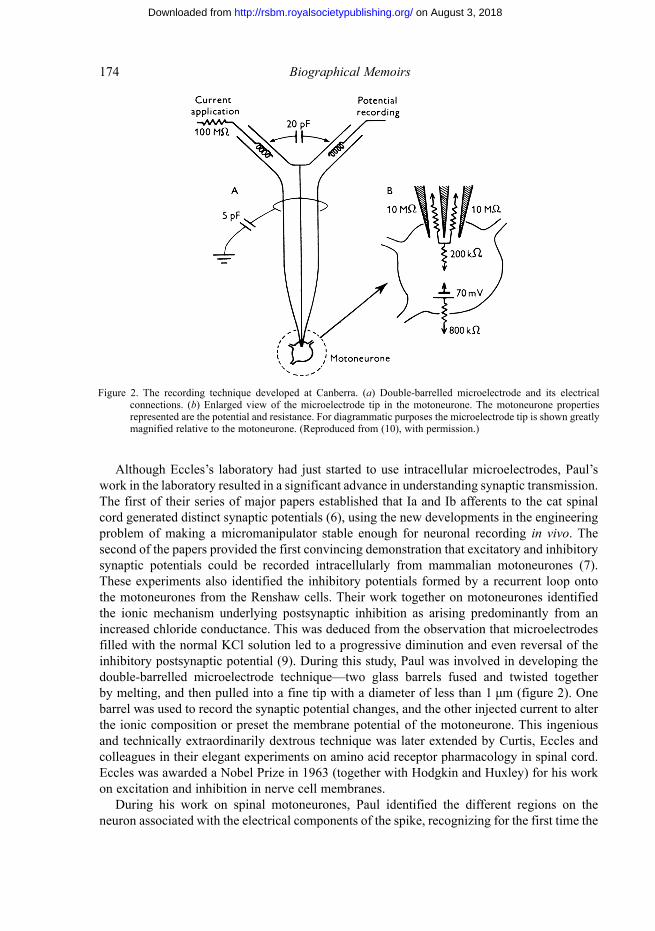
Although Eccles’s laboratory had just started to use intracellular microelectrodes, Paul’s work in the laboratory resulted in a significant advance in understanding synaptic transmission. The first of their series of major papers established that Ia and Ib afferents to the cat spinal cord generated distinct synaptic potentials (6), using the new developments in the engineering problem of making a micromanipulator stable enough for neuronal recording in vivo. The second of the papers provided the first convincing demonstration that excitatory and inhibitory synaptic potentials could be recorded intracellularly from mammalian motoneurones (7). These experiments also identified the inhibitory potentials formed by a recurrent loop onto the motoneurones from the Renshaw cells. Their work together on motoneurones identified the ionic mechanism underlying postsynaptic inhibition as arising predominantly from an increased chloride conductance. This was deduced from the observation that microelectrodes filled with the normal KCl solution led to a progressive diminution and even reversal of the inhibitory postsynaptic potential (9). During this study, Paul was involved in developing the double-barrelled microelectrode technique—two glass barrels fused and twisted together by melting, and then pulled into a fine tip with a diameter of less than 1 μm (figure 2). One barrel was used to record the synaptic potential changes, and the other injected current to alter the ionic composition or preset the membrane potential of the motoneurone. This ingenious and technically extraordinarily dextrous technique was later extended by Curtis, Eccles and colleagues in their elegant experiments on amino acid receptor pharmacology in spinal cord. Eccles was awarded a Nobel Prize in 1963 (together with Hodgkin and Huxley) for his work on excitation and inhibition in nerve cell membranes.
During his work on spinal motoneurones, Paul identified the different regions on the neuron associated with the electrical components of the spike, recognizing for the first time the
Figure 2. The recording technique developed at Canberra. (a) Double-barrelled microelectrode and its electrical connections. (b) Enlarged view of the microelectrode tip in the motoneurone. The motoneurone properties represented are the potential and resistance. For diagrammatic purposes the microelectrode tip is shown greatly magnified relative to the motoneurone. (Reproduced from (10), with permission.)
on August 3, 2018http://rsbm.royalsocietypublishing.org/Downloaded from
ways in which the initial segment and soma dendrite determine the action potential properties of central neurons (10–12). In his investigations on the active properties of motoneurone dendrites, Paul obtained some of the first evidence, running against the conventional views of the day, for active impulse invasion of dendrites. With some difficulty he published this work after his return to London, using data he had collected in Canberra (13, 14). He has also been widely credited with the prediction (in a highly influential paper in Physiological Reviews (8)) that, on theoretical grounds, transmission could be mediated electrically rather than chemically but only at certain synapses, something confirmed soon afterwards in a classic study by Ed Furshpan and David Potter in the Biophysics Department at UCL (Furshpan & Potter 1959).
Inhibition at the crustacean neuromuscular junction
Paul returned to London in 1956 to take up the position of Reader in UCL’s Biophysics Department, as it now was called, to which he had been appointed in October 1955. The department was headed by Bernard Katz after A. V. Hill’s retirement in 1952. Katz wanted Paul to continue working on the cat spinal cord because this was would be a natural extension of work in the department. Paul decided that the organization of a mammalian spinal cord laboratory would be too great a burden—in Canberra, Eccles had had the resources of a large laboratory to support the effort—and instead decided to take up his work again on the more accessible synapses of the crayfish neuromuscular junction.
The work with Katz before he left for Australia had examined which ions determined the crustacean muscle membrane potential and excitability. The interest in this muscle, as noted by Katz in his earlier work with Kuffler, was that the EPP could be inhibited. Paul and Katz had previously suggested that the inhibition might arise either from competition between the excitatory and inhibitory transmitters for the same postsynaptic receptor, or as a result of an induced membrane permeability increase mainly to potassium (5). The former explanation was not completely satisfactory: it was found that stimulating the inhibitory fibre alone could sometimes produce a change in the membrane potential. With Bernard Ginsborg, a research fellow with a PhD in physics but who had decided to move into physiology instead, Paul revisited the crustacean muscle (15, 16). Rather surprisingly for the time, they found that it was not only sodium that could support all-or-none action potentials, but they also demonstrated for the first time the existence of calcium-mediated action potentials and hence of one of the most important currents, the calcium current, present in biological tissues. All the techniques and the ionic substitutions used in the paper look completely contemporary to a modern reader. The range of compounds tested for substitution experiments, some of which are still used, some superseded, is really surprising. Perhaps most tantalizing of all is a small observation tucked away in a Methods section (17) of the observation by Fatt and Ginsborg that glutamate could not be used as a substitute for chloride in ion replacement experiments because it ‘caused a large increase in membrane conductance, possibly through the removal of ionized calcium’. Thus Paul and Ginsborg narrowly missed identifying glutamate as a neurotransmitter for excitation at the crayfish neuromuscular junction. It is now realized to be the major neurotransmitter of the central nervous system. That honour was left to the Takeuchis, again using the crayfish, some six years later (Takeuchi & Takeuchi 1964).
on August 3, 2018http://rsbm.royalsocietypublishing.org/Downloaded from
There was one final experiment to carry out. Using his experience from Canberra and the development of new recording techniques, Paul, together with Boistel, a British Council Scholar, used two separate intracellular microelectrodes, one to pass current and the other to measure membrane potential, and found that during synaptic inhibition crayfish muscle showed an increased permeability to chloride ions (17). At least as remarkably, they provided direct evidence that the mechanism of inhibition was likely to involve γ-amino butyric acid (GABA), a newly described compound, because both GABA and activation of the inhibitory fibres increased the synaptic chloride permeability in a similar way.
Muscle T-tubule system
Established in the Biophysics Department at UCL (figure 3), Paul turned his attention to another problem that had remained unsolved. From their earliest intracellular recordings he and Katz had noticed that the membrane capacitance of a skeletal muscle, in this case the frog sartorius muscle, was much larger than expected from the apparent surface area, given that the specific capacitance of cell membrane is about 1 μF cm −2. Instead, the membrane capacitance was about 7–8 times greater. The problem originated with work of Cole and colleagues in the 1930s in the USA, who had measured the electrical impedance of nerve fibres at frequencies between 1 kHz and 2.5 MHz and found that any attempt to determine the impedance of the muscle fibre membrane gave a different value depending on the frequency used (Curtis & Cole 1936).
To explore this apparent paradox, Paul resorted to measurements of the sort used by Cole. The experiments were designed to use current passed extracellularly across all the fibres in a muscle. The measurement depended on a formula of Rayleigh’s that had first been introduced
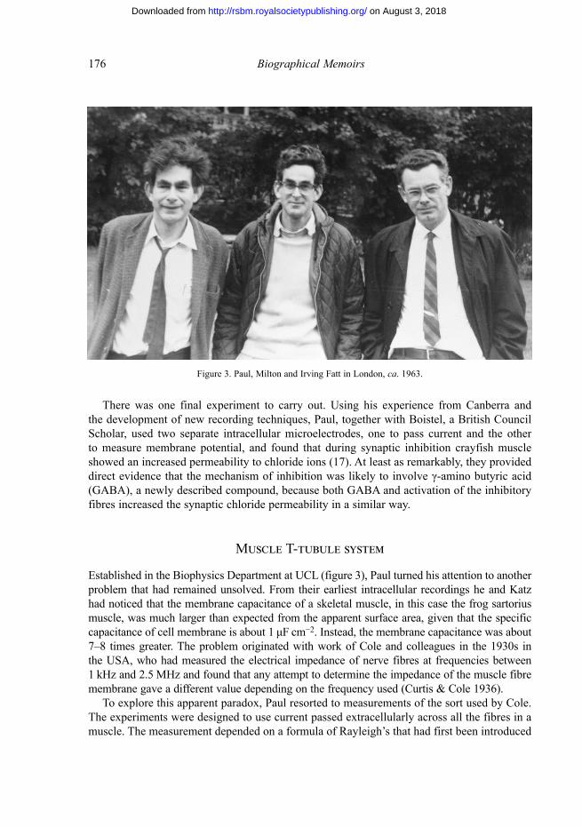
Figure 3. Paul, Milton and Irving Fatt in London, ca. 1963.
on August 3, 2018http://rsbm.royalsocietypublishing.org/Downloaded from
for biophysical measurements in 1913. The specific impedance, Z, of a system of conducting rods, each with transverse impedance Zj, is found to be
where Ro is the resistivity of the bathing medium and p is the volume fraction occupied by the rods. The ‘rods’ in this case are the individual muscle fibres. The advantage of the approach is that, without the bandwidth limitations imposed by using small glass microelectrodes, the impedance can be studied with great precision at frequencies well over 100 kHz, and in some cases over 1 MHz.
Figure 4 The impedance locus (imaginary part, X, versus real part, R) of a frog muscle as the measuring frequency is progressively increased. The large arc is due to the impedance of the sarcolemma, and the current passing through the branch Cm, Ri of the proposed equivalent circuit for Zj shown to the right. The smaller arc is due to current through the branch Cx,Rx and was ascribed to the T-tubule system. As external sodium chloride was progressively replaced by sucrose (top to bottom), so increasing the specific resistance of the bathing medium, the relative contributions of each branch changed. (Reproduced from (18).)
on August 3, 2018http://rsbm.royalsocietypublishing.org/Downloaded from
At these high frequencies the surface membrane capacitance can have a vanishingly small impedance and the effect of electrically resistive structures inside the cell becomes evident (figure 4). The problem, however, is that the interpretation of the data depends on a degree of mathematical sophistication that does not immediately appeal to the biologically trained. The inversion of the formula to determine Zj depends very much on the adopted electrical model of the cell. The chosen model (figure 4) predicts that, as the measurement frequency changes, the real and imaginary components of Z change in a way that would be expected if the current could flow through two parallel arms of the circuit, one describing the surface membrane and the other any membrane system continuous with the sarcolemma (18).
It therefore is something of a surprise that Paul, having become one of the leading exponents of microelectrode recording, should resort to extracellular current measurements in his subsequent scientific work. Although teaching the techniques of recording from the neuromuscular junction to generations of students at UCL in a very popular biophysics laboratory course, he never again used microelectrode recording in his published work. That was left to his scientific collaborator, Gertrude Falk.
The careful study of the T-tubule system using extracellular impedance measurements was linked to a subsequent paper published together with Falk in the same issue of Proceedings of the Royal Society B. The latter used intracellular microelectrodes to explore the membrane impedance, but frequencies were limited to less than 10 kHz (19). The two papers came firmly to the conclusion that the skeletal muscle membrane needed to be represented by two conducting paths, one corresponding to the circumferential muscle membrane and the other to a system of intracellular conducting tubules, continuous with the surface, which subsequently became referred to as the T-tubule system of muscle.
The electrical measurements carried out by Paul and Falk therefore complement the direct observation by electron microscopy of the connection of the T-tubule system to the plasma membrane but are not often recognized for their painstaking effort and technical virtuosity.
A series of internal muscle membranes had been described in the electron micrographs by Porter & Palade (1957) and with hindsight can be identified in much earlier light microscopic studies (Huxley 1980). It was only with the introduction of glutaraldehyde fixation in the early 1960s that it became more widely appreciated that there was a system of transverse and highly convoluted tubules that were separate from the sarcoplasmic reticulum, the store from which calcium is released to ensure contraction. There had been earlier work by Andrew Huxley and Robert Taylor (Huxley & Taylor 1958) identifying that the activation of muscle spreads inwards from ‘hotspots’ on the surface muscle membrane, but evidence for the continuity of the T-tubule system with the surface membrane of muscle remained incomplete until it was shown by the electrical measurements of Paul and Falk and from the electron microscopy by Hugh Huxley FRS (Huxley 1964) and Sally Page (Page 1964), who showed that ferritin had access to the interior of the T-tubule system from the extracellular fluid. The precise molecular mechanism, of how excitation spreading along the T-tubule could couple to the calcium release mechanism in the sarcoplasmic reticulum, had to wait another 25 years for elucidation.
Visual transduction
The completion of these two projects in London after Paul’s return from Australia really marks both an end of unfinished business remaining from his collaboration with Katz and a
on August 3, 2018http://rsbm.royalsocietypublishing.org/Downloaded from
watershed in Paul’s scientific career. The final 20 years of his work at UCL concerned visual transduction.
‘I liked the idea of being able to excite something from a distance with the light’, he said. At the time this was more of a revolutionary statement than it might now seem. From around 1963 onwards, Paul’s work revolved around the mechanisms by which rhodopsin in photoreceptors could absorb light and then signal to the synapse to trigger activity in the retinal neurons. It had been known from the psychophysical experiments of Selig Hecht, Simon Schlaer and Maurice Pirenne in 1942 that rod photoreceptors could detect the absorption of a single photon. The biochemistry of rhodopsin had been extensively explored by George Wald and his colleagues at Harvard (Hubbard & Wald 1952) and eventually led to the award of a Nobel Prize to Wald in 1967. One of those colleagues was Ruth Hubbard, Wald’s wife, who had also been at UCL during the period when Paul started his PhD. The emphasis in Wald’s rhodopsin work had been on the early mechanisms of absorption of light and on the several photochemical steps that followed. How the absorption of a single quantum of light could be signalled to the retinal ganglion cells was completely unknown. Hypotheses abounded. The attraction to Paul of tackling one of the big problems of visual biophysics must have been irresistible. Unfortunately, he was just a little too soon: the techniques and the biochemical concepts were not yet available to solve this complex problem.
Paul’s first reported experiments with Falk in vision concerned the movement of protons in suspension of rod outer segments (20). Frog rod outer segments (ROSs) are cylinders about 6 μm in diameter and 50 μm in length, and the observation that protons could be taken up in outer segments tallied with measurements of the photochemistry of extracted rhodopsin. It is likely that the ease with which such ROS could be prepared led him to believe that isolated outer segments could be used to understand visual transduction. In part this was true but it became apparent only in the next decade, through the work of W. A. (‘Bill’) Hagins, Mike Fuortes, Alan Hodgkin and Denis Baylor (ForMemRS 2003), that there was a circulating current from the outer to the inner segments of the photoreceptors and that the ionic gradient dissipated rapidly in the absence of an internal ATP energy source. However, in the mid 1960s Paul was using techniques similar to that which he had employed for studying muscle, except that the stimulating current covered a range of frequencies up to 1 MHz. Although there were clearly changes in the impedance of the ROS suspension on illumination with bright light, Paul and Falk with characteristic honesty were careful to point out that changes in impedance could be ascribed to the light-induced volume changes of the ROS rather than to permeability changes in the plasma membrane itself. The approach was thus less promising than expected (21).
The first convincing reports that photoreceptors signalled electrically appeared in 1965 when Tsuneo Tomita, using intracellular electrodes, reported that cones hyperpolarized when illuminated (Tomita 1965). These data were followed by results from many other groups who showed ever more convincingly that there had to be some sort of intracellular message communicating between the rhodopsin and the photoreceptor membrane conductance. From the early 1970s onwards, Paul’s enthusiasm for solving the vertebrate transduction problem, although not completely extinguished, definitely waned. The nature of the visual excitation was eventually solved in 1985 by a then unheard-of group from the USSR Academy of Sciences in Moscow (Fesenko et al. 1985), who seemed to have stumbled on the solution almost by mistake: they found, using the new patch-clamp techniques, that the internal messenger was cyclic guanosine monophosphate (cGMP) but controlled by a cascade involving guanine
on August 3, 2018http://rsbm.royalsocietypublishing.org/Downloaded from
nucleotide-binding proteins (G-proteins), all undiscovered in 1970. For a while, influenced by results of Penn and Hagins (Penn & Hagins 1969), Paul had nursed the idea that rhodopsin was a calcium channel, and that light gated calcium out of the ROS internal lamellae. But although there was some circumstantial evidence for this idea (in particular that a low external calcium concentration around the ROS mimicked the effect of light), by 1979 even he was beginning to have doubts about whether calcium really was the long-sought visual second messenger (23).
A short theoretical note by Paul and Falk in 1974 did, however, have a significant effect on the direction of visual physiology (22). It had been noted that the b-wave of the electroretinogram, thought to be an indicator of the second-order neuronal activity in the retina, was a much more sensitive indicator of light than the extracellular recordings of photoreceptor activity. Their note pointed out that this might arise if the so-called ON bipolar cells had a synapse with the rod photoreceptors that amplified considerably because the action of transmitter release from the rods was to close transmitter-gated channels. Knowing of other synapses for which a transmitter had a similar (unconventional) action, Paul had identified a soluble problem but one to which the conclusion was unexpected. This, as always, was an original and creative idea for the field. Gertrude Falk and I did show subsequently that there was amplification at this synapse (Ashmore & Falk 1980), but it took another 12 years for the mechanisms to be identified. The result, discovered by Falk at UCL (Shiells & Falk 1992) and by Craig Jahr in the USA (Nawy & Jahr 1991) was that, as in photo-transduction itself, ON-bipolar cell synaptic transmission amplified because it was another G-protein-modulated cGMP pathway, which this time determined the postsynaptic response.
In the early 1980s Paul had moved his laboratory and activities to the Biochemistry Department of the Royal Free Hospital. The department was part of ‘greater UCL’ and conveniently close to his home in Hampstead. There he found a more sympathetic attitude to his search for an oligomeric structure for rhodopsin. The summary of 10 years’ work on the search for rhodopsin clusters was published as a final note in 1985 (24).
Within UCL there had been considerable and growing university pressure on the Biophysics Department after Katz’s retirement in 1978, because it was seen as doing insufficient teaching and its rationale as a solely research department was less in line with the spirit of the times. Ricardo Miledi had taken over as head of the department but relations between him and Paul were strained. With the departure of Miledi in 1983, the writing was on the wall for Biophysics and it was reabsorbed by the Physiology Department, from which it had originally blossomed. Paul, perhaps with some sense of relief, stopped his scientific work in 1985 when the cGMP hypothesis came out, and he retired from UCL in 1989 at the age of 65 years. Apart from speaking movingly at a symposium in memory of Bernard Katz in 2003, his connections with UCL remained severed.
Personal and later life
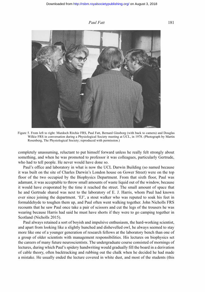
Paul eschewed honours. Although he was elected to the Fellowship of the Royal Society in 1969 (he had become a naturalized British citizen in 1965) he did not seek any further nominations or recognition of his achievements. Although it was clear that he had a large circle of influential scientific friends, when he retired all that became a separate episode in his life (figure 5). His entry in Who’s who was a model of brevity. He was made a Professor of Biophysics at UCL at what many viewed as a surprisingly late stage, in May 1976. He was
on August 3, 2018http://rsbm.royalsocietypublishing.org/Downloaded from
completely unassuming, reluctant to put himself forward unless he really felt strongly about something, and when he was promoted to professor it was colleagues, particularly Gertrude, who had to tell people. He never would have done so.
Paul’s office and laboratory in what is now the UCL Darwin Building (so named because it was built on the site of Charles Darwin’s London house on Gower Street) were on the top floor of the two occupied by the Biophysics Department. From that sixth floor, Paul was adamant, it was acceptable to throw small amounts of waste liquid out of the window, because it would have evaporated by the time it reached the street. The small amount of space that he and Gertrude shared was next to the laboratory of E. J. Harris, whom Paul had known ever since joining the department. ‘EJ’, a stout walker who was reputed to soak his feet in formaldehyde to toughen them up, and Paul often went walking together. John Nicholls FRS recounts that he saw Paul once take a pair of scissors and cut the legs of the trousers he was wearing because Harris had said he must have shorts if they were to go camping together in Scotland (Nicholls 2015).
Paul always retained a sort of boyish and impulsive enthusiasm, the hard-working scientist, and apart from looking like a slightly hunched and dishevelled owl, he always seemed to stay more like one of a younger generation of research fellows at the laboratory bench than one of a group of older scientists with management responsibilities. His lectures on biophysics set the careers of many future neuroscientists. The undergraduate course consisted of mornings of lectures, during which Paul’s spidery handwriting would gradually fill the board in a derivation of cable theory, often backtracking and rubbing out the chalk when he decided he had made a mistake. He usually ended the lecture covered in white dust, and most of the students (this
Figure 5. From left to right: Murdoch Ritchie FRS, Paul Fatt, Bernard Ginsborg (with back to camera) and Douglas Wilkie FRS in conversation during a Physiological Society meeting at UCL, in 1978. (Photograph by Martin Rosenberg, The Physiological Society; reproduced with permission.)
on August 3, 2018http://rsbm.royalsocietypublishing.org/Downloaded from
was meant to be an undergraduate course) were left slightly mystified by his explanations. The rest of the day was spent in the class laboratory, during which everyone gradually did the Fatt and Katz experiments until they got the equipment to work and data to show for it. For well-primed students it was the best possible introduction to biophysics.
Paul Fatt was married three times. While in Australia he married Ione Copplestone (1926–2015) and had a son Michael (b. 1954), an epidemiologist and presently a full-time specialist GP, and two daughters, Laura (b. 1955) and Harriet (b. 1957). The Australian family changed the surname back the original name Fett after Ione and Paul divorced. When at University College London he married Gertrude Falk (1925–2008), a physiologist trained at Rochester University with an expertise in microelectrode recording who had come in 1961 on a Guggenheim Fellowship to work with Paul. They had met at a conference in Seattle at which Paul was reporting on his new work on the effects of GABA. Paul and Gertrude had a daughter Ilsa (b. 1963), now a jewellery designer. After separating from Gertrude, he married Carla Wartenberg, a freelance translator, in 1985. Paul had met her through the Labour Party. For the last 30 years of his life they lived in Hampstead in a flat overlooking Parliament Hill Fields, allowing Paul to accompany Carla across to the Ladies’ Swimming Pool each morning, waiting for her with hot tea after her swim. Hampstead was where he first decided he wanted to live when he came to London in 1948.
Carla Wartenberg has written of her time with Paul. She describes him best as a passionate secular humanist who sometimes could not accept the realities of the world around him. His heroes were mostly rebels and so-called heretics, fighters against what they saw as injustice or obscurantism and who often failed or seemed to have failed. His list included Akhnaton, the Egyptian pharaoh who wished to replace a pantheon of ancient gods by one supreme impersonal deity, the Sun; Giordano Bruno, burned at the stake for refusing to hide his belief in an infinite universe; and Shelley in his aspect as a revolutionary poet. Paul even ordered a small bust of the poet, which he kept on his desk.
Paul was always independent and could be fiercely outspoken at times. He joined the Labour Party and for many years remained an active member (his idea of a good after-lunch exercise for his guests was to go out canvassing for the party) but he resigned in 2003 after the Iraq invasion, in the belief that Labour was abandoning the socialist–humanist ideals that had attracted him to it 50 years earlier. Paul joined Pugwash, the Campaign for Nuclear Disarmament and the World Disarmament campaign, went on marches and demonstrations and gave fiery talks about the horrors of nuclear war to audiences in church halls and civic centres. Paul was outraged when Margaret Thatcher was elected under Statute 12 of the Society and organized a memorandum, which several Fellows signed. Although Paul was not successful (it was pointed out that a repeal of the Statute so soon after her re-election in 1983 might be embarrassing to the Society and to Fellows who had signed) it is conceivable that ultimately he did succeed, because no other sitting prime minister has since been put up for election.
Paul and Carla travelled extensively both abroad and in Britain. Paul was a determined walker and at different times and in different stages they walked almost the whole of the South West Coast Path around from Dorset to Somerset, walked the Pennine Way and spent months staying in Scotland, mainly in the Hebrides. They were energetic travellers around most of the Mediterranean, visiting many countries independently, including Turkey, Syria and, shortly after the massacre at Luxor in 1997, Egypt, finding themselves not surrounded by all the usual tourist crowds. The attraction of being able to explore all the sites that they wanted on just a
on August 3, 2018http://rsbm.royalsocietypublishing.org/Downloaded from
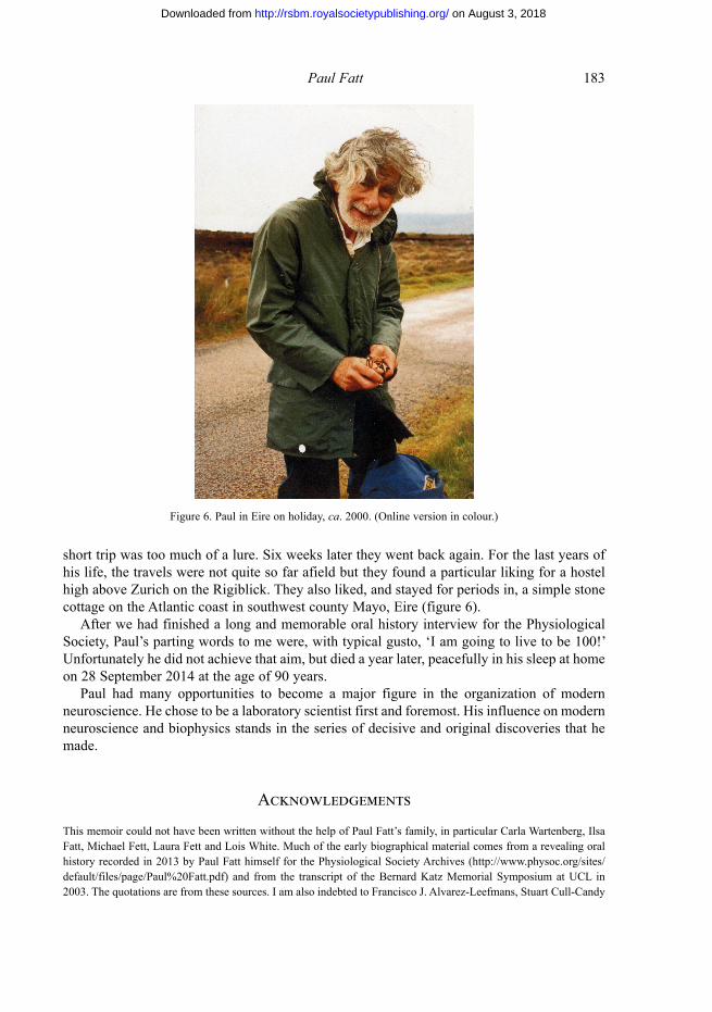
short trip was too much of a lure. Six weeks later they went back again. For the last years of his life, the travels were not quite so far afield but they found a particular liking for a hostel high above Zurich on the Rigiblick. They also liked, and stayed for periods in, a simple stone cottage on the Atlantic coast in southwest county Mayo, Eire (figure 6).
After we had finished a long and memorable oral history interview for the Physiological Society, Paul’s parting words to me were, with typical gusto, ‘I am going to live to be 100!’ Unfortunately he did not achieve that aim, but died a year later, peacefully in his sleep at home on 28 September 2014 at the age of 90 years.
Paul had many opportunities to become a major figure in the organization of modern neuroscience. He chose to be a laboratory scientist first and foremost. His influence on modern neuroscience and biophysics stands in the series of decisive and original discoveries that he made.
Acknowledgements
This memoir could not have been written without the help of Paul Fatt’s family, in particular Carla Wartenberg, Ilsa Fatt, Michael Fett, Laura Fett and Lois White. Much of the early biographical material comes from a revealing oral history recorded in 2013 by Paul Fatt himself for the Physiological Society Archives (http://www.physoc.org/sites/default/files/page/Paul%20Fatt.pdf) and from the transcript of the Bernard Katz Memorial Symposium at UCL in 2003. The quotations are from these sources. I am also indebted to Francisco J. Alvarez-Leefmans, Stuart Cull-Candy
Figure 6. Paul in Eire on holiday, ca. 2000. (Online version in colour.)
on August 3, 2018http://rsbm.royalsocietypublishing.org/Downloaded from
FRS, Bernard Ginsborg and Sally Page, all former members of the UCL Biophysics Department, for much helpful information, additions to and corrections of my many errors. Aspects of Paul’s later life are adapted from a memoir written by Carla Wartenberg.
The frontispiece is a photograph of Paul Fatt in his laboratory in 1979 taken by Francisco J. Alvarez-Leefmans, and is reproduced with permission.
Author profile
Jonathan AshmoreJonathan Ashmore is Bernard Katz Professor of Biophysics at University College London. He completed a PhD in theoretical physics under Tom Kibble FRS and a short postdoctoral fellowship with Abdus Salam FRS at the International Centre for Theoretical Physics in Trieste, Italy, before retraining as a physiologist at UCL. He worked with Paul Fatt and Gertrude Falk between 1974 and 1977 in the Biophysics Department, UCL, on a project identifying high-gain synaptic transmission in the retina. His work since 1980 has been on hearing mechanisms, carried out first at the University of Sussex and then at the University of Bristol before returning to UCL in 1996. He was elected a Fellow of the Royal Society in the same
year and was President of the Physiological Society from 2012 to 2014.
References to other authors
Ashmore, J. F. & Falk, G. 1980 Responses of rod bipolar cells in the dark-adapted retina of the dogfish, Scyliorhinus canicula. J. Physiol. 300, 115–150.
Curtis, H. J. & Cole, K. S. 1938 Transverse electric impedance of the squid axon. J. Gen. Physiol. 21, 757–765.Eccles, J. C., Katz, B. & Kuffler, S. W. 1942 Effect of eserine on neuromuscular transmission. J. Neurophysiol. 5,
211–230.Fesenko, E. E., Kolesnikov, S. S. & Lyubarsky, A. L. 1985 Induction by cyclic GMP of cationic conductance in
plasma membrane of retinal rod outer segment. Nature 313, 310–313.Furshpan, E. J. & Potter, D. D. 1959 Transmission at the giant motor synapses of the crayfish. J. Physiol. 145,
289–325.Hodgkin, A. L., Huxley, A. F. & Katz, B. 1952 Measurement of current–voltage relations in the membrane of the giant
axon of Loligo. J. Physiol. 116, 424–448.Hubbard, R. & Wald, G. 1952 Cis–trans isomers of vitamin A and retinene in the rhodopsin system. J. Gen. Physiol.
36, 269–315.Huxley, A. F. 1980 Reflections on muscle (The Sherrington Lectures, vol. 14). Liverpool University Press.Huxley, A. F. & Taylor, R. E. 1958 Local activation of striated muscle fibres. J. Physiol. 144, 426–441.Huxley, H. E. 1964 Evidence for continuity between the central elements of the triads and extracellular space in frog
sartorius muscle. Nature 202, 1067–1071.Katz, B. 1950 Depolarization of sensory terminals and the initiation of impulses in the muscle spindle. J. Physiol.
111, 261–282.Katz, B. & Miledi, R. 1970 Membrane noise produced by acetylcholine. Nature 226, 962–963.Nawy, S. & Jahr, C. E. 1991 cGMP-gated conductance in retinal bipolar cells is suppressed by the photoreceptor
transmitter. Neuron 7, 677–683.Nicholls, J. 2015 Pioneers of neurobiology: my brilliant eccentric heroes. Sunderland, MA: Sinauer Associates, Inc.Page, S. G. 1964 The organisation of the sarcoplasmic reticulum in frog muscle. J. Physiol. 175, 10P–11P.
on August 3, 2018http://rsbm.royalsocietypublishing.org/Downloaded from
Penn, R. D. & Hagins, W. A. 1969 Signal transmission along retinal rods and the origin of the electroretinographic a-wave. Nature 223, 201–204.
Porter, K. R. & Palade, G. E. 1957 Studies on the endoplasmic reticulum. III. Its form and distribution in striated muscle cells. J. Biophys. Biochem. Cytol. 3, 269–300.
Shiells, R. A. & Falk, G. 1992 The glutamate-receptor linked cGMP cascade of retinal on-bipolar cells is pertussis and cholera toxin-sensitive. Proc. Biol. Sci. 247, 17–20.
Takeuchi, A. & Takeuchi, N. 1964 The effect on crayfish muscle of iontophoretically applied glutamate. J. Physiol. 170, 296–317.
Tomita, T. 1965 Electrophysiological study of the mechanisms subserving color coding in the fish retina. Cold Spring Harb. Symp. Quant. Biol. 30, 559–566.
Bibliography
The following publications are those referred to directly in the text. A full bibliography is available as electronic supplementary material at http://dx.doi.org/10.1098/rsbm.2016.0005 or via http://rsbm.royalsocietypublishing.org.(1) 1949 The depolarizing action of acetylcholine on muscle. J. Physiol. 109, 10P.(2) 1950 (With B. Katz) Some observations on biological noise. Nature 166, 597–598.(3) 1951 (With B. Katz) An analysis of the end-plate potential recorded with an intracellular electrode. J.
Physiol. 115, 320–370.(4) 1952 (With B. Katz) Spontaneous subthreshold activity at motor nerve endings. J. Physiol. 117, 109–128.(5) 1953 (With B. Katz) The effect of inhibitory nerve impulses on a crustacean muscle fibre. J. Physiol. 121,
374–389.(6) 1954 (With J. C. Eccles, S. Landgren & G. J. Winsbury) Spinal cord potentials generated by volleys in the
large muscle afferents. J. Physiol. 125, 590–606.(7) (With J. C. Eccles & K. Koketsu) Cholinergic and inhibitory synapses in a pathway from motor-axon
collaterals to motoneurones. J. Physiol. 126, 524–562.(8) Biophysics of junctional transmission. Physiol. Rev. 34, 674–710.(9) 1955 (With J. S. Coombs & J. C. Eccles) The electrical properties of the motoneurone membrane. J. Physiol.
130, 291–325.(10) (With J. S. Coombs & J. C. Eccles) The specific ionic conductances and the ionic movements across
the motoneuronal membrane that produce the inhibitory post-synaptic potential. J. Physiol. 130, 326–374.
(11) (With J. S. Coombs & J. C. Eccles) The inhibitory suppression of reflex discharges from motoneurones. J. Physiol. 130, 396–413.
(12) (With J. S. Coombs & J. C. Eccles) Excitatory synaptic action in motoneurones. J. Physiol. 130, 374–395.
(13) 1957 Electric potentials occurring around a neurone during its antidromic activation. J. Neurophysiol. 20, 27–60.
(14) Sequence of events in synaptic activation of a motoneurone. J. Neurophysiol. 20, 61–80.(15) 1958 (With B. L. Ginsborg) The ionic requirements for the production of action potentials in crustacean
muscle fibres. J. Physiol. 142, 516–543.(16) (With B. L. Ginsborg) The production of regenerative responses in crayfish muscle fibres by the action
of calcium, strontium and barium. J. Physiol. 140, 59P–60P.(17) (With J. Boistel) Membrane permeability change during inhibitory transmitter action in crustacean
muscle. J. Physiol. 144, 176–191.(18) 1964 An analysis of the transverse electrical impedance of striated muscle. Proc. R. Soc. Lond. B 159,
606–651.(19) (With G. Falk) Linear electrical properties of striated muscle fibres observed with intracellular
electrodes. Proc. R. Soc. Lond. B 160, 69–123.
on August 3, 2018http://rsbm.royalsocietypublishing.org/Downloaded from
(20) 1966 (With G. Falk) Rapid hydrogen ion uptake of rod outer segments and rhodopsin solutions on illumination. J. Physiol. 183, 211–224.
(21) 1968 (With G. Falk) Conductance changes produced by light in rod outer segments. J. Physiol. 198, 647–699.
(22) 1974 (With G. Falk) The dynamic voltage-transfer function for rod–bipolar cell transmission. Vision Res. 14, 739–741.
(23) 1979 Decline of the calcium hypothesis of visual transduction. Nature 280, 355–356.(24) 1985 Relation of the different forms of frog rhodopsin observed by isoelectric focusing and electrophoresis
to a functional model of rhodopsin clusters in the disc membrane. Vision Res. 25, 1865–1867.
on August 3, 2018http://rsbm.royalsocietypublishing.org/Downloaded from