352 | april 2012 | volume 42 | number 4 | journal of orthopaedic & sports physical therapy [ RESEARCH REPORT ] D espite the high prevalence of low back pain in the population, options regarding effective treatment strategies are still limited, possibly due to the lack of knowledge of the underlying mechanisms. 9 Trunk stabilization and postural trunk control may play an important role in the etiology of low back pain. 9 In turn, the function of the diaphragm may affect how the trunk is stabilized, especially during postural activity. 11,15,22 Various studies have shown that the pelvic girdle and lumbar spine are reflexively stabilized and braced prior to the initiation of extrem- ity movements. 3,13,17,22 The central nervous system must be able to anticipate move- ment and stabilize the entire core mus- culature automatically to provide a stable base from which the muscles performing the movement can pull. Trunk bracing maintains all spinal segments in a biomechanically neutral position during the course of any move- ment. Segmental movement (eg, hip joint movement) is therefore related to the synergistic activity of the spinal ex- tensors and all the muscles modulating intra-abdominal pressure (ie, abdominal muscles, the diaphragm, and the pelvic floor). The diaphragm is the muscle that contributes the intra-abdominal pressure modulation and plays an important role in spinal stability. 7,16,18,27,31 Insufficient function and poor coordi- nation of postural, or stabilizing, muscles are considered to be important etiological factors in spinal disorders associated with low back pain, such as deformational spondyloarthrosis (with or without spinal disc herniation), spinal disc protrusion, and/or spondylolisthesis. 5,12,21,25 However, a study designed specifically to test the dynamics of the diaphragm in chronic spinal disorders is lacking. We aimed T T STUDY DESIGN: A case-control study. T T OBJECTIVES: To examine the function of the diaphragm during postural limb activities in patients with chronic low back pain and healthy controls. T T BACKGROUND: Abnormal stabilizing function of the diaphragm may be an etiological factor in spinal disorders. However, a study designed specifically to test the dynamics of the diaphragm in chronic spinal disorders is lacking. T T METHODS: Eighteen patients with chronic low back pain due to chronic overloading, as ascertained via clinical assessment and magnetic resonance imaging, and 29 healthy subjects were examined. Both groups presented with normal pulmonary function test results. A dynamic mag- netic resonance imaging system and specialized spirometric readings were used with subjects in the supine position. Measurements during tidal breathing (TB) and isometric flexion of the upper and lower extremities against external resistance with TB were performed. Standard pulmonary function tests, including respiratory muscle drive (PI max and PE max ), were also assessed. T T RESULTS: Using multivariate analysis of covari- ance, smaller diaphragm excursions and higher diaphragm position were found in the patient group (P<.05) during the upper extremity TB and lower extremity TB conditions. Maximum changes were found in costal and middle points of the diaphragm. A 1-way analysis of covariance showed a steeper slope in the middle-posterior diaphragm in the patient group both in the upper extremity TB and lower extremity TB conditions (P<.05). T T CONCLUSION: Patients with chronic low back pain appear to have both abnormal position and a steeper slope of the diaphragm, which may contribute to the etiology of the disorder. J Orthop Sports Phys Ther 2012;42(4):352-362, Epub 21 December 2011. doi:10.2519/jospt.2012.3830 T T KEY WORDS: dynamic magnetic resonance imaging, lung function, spinal disorders, stabilizing function, thorax 1 Associate Professor, Department of Rehabilitation and Sport Medicine, Second Medical Faculty, Charles University and University Hospital Motol, Prague, Czech Republic. 2 Lecturer, Department of Rehabilitation and Sport Medicine, Second Medical Faculty, Charles University and University Hospital Motol, Prague, Czech Republic. 3 Lecturer, Department of Radiology, Second Medical Faculty, Charles University and University Hospital Motol, Prague, Czech Republic. 4 Associate Professor, School of Aging Studies, University of South Florida, Tampa, FL; International Clinical Research Center, St Anne’s University Hospital Brno, Brno, Czech Republic. 5 Associate Professor, Department of Physical Therapy, Azusa Pacific University, Azusa, CA. This study was supported by the foundation Movement without Help, Prague, Czech Republic. This study was approved by the Second Medical Faculty, Ethical Committee, Charles University and University Hospital Motol, Prague, Czech Republic. Address correspondence to Dr Alena Kobesová, Department of Rehabilitation and Sport Medicine, Second Medical Faculty, Charles University and University Hospital Motol, V Úvalu 84, Prague 5, 15006, Czech Republic. E-mail: [email protected]PAVEL KOLÁR, PaedDr, PhD 1 • JAN ŠULC, MD, PhD 2 • MARTIN KYNCL, MD 3 • JAN ŠANDA, Ing 3 • ONDREJ CAKRT, MSc 2 ROSS ANDEL, PhD 4 • KATHRYN KUMAGAI, DPT 5 • ALENA KOBESOVÁ, MD, PhD 2 Postural Function of the Diaphragm in Persons With and Without Chronic Low Back Pain ˇ ˇ ˇ ˇ
Transcript
352 | april 2012 | volume 42 | number 4 | journal of orthopaedic & sports physical therapy
[ research report ]
Despite the high prevalence of low back pain in the population, options regarding effective treatment strategies are still limited, possibly due to the lack of knowledge of the underlying mechanisms.9 Trunk stabilization and postural trunk control
may play an important role in the etiology of low back pain.9 In turn, the function of the diaphragm may affect how the trunk is stabilized, especially during postural activity.11,15,22 Various studies have shown that the pelvic girdle and lumbar spine are reflexively stabilized and
braced prior to the initiation of extrem-ity movements.3,13,17,22 The central nervous system must be able to anticipate move-ment and stabilize the entire core mus-culature automatically to provide a stable base from which the muscles performing the movement can pull.
Trunk bracing maintains all spinal segments in a biomechanically neutral position during the course of any move-ment. Segmental movement (eg, hip joint movement) is therefore related to the synergistic activity of the spinal ex-tensors and all the muscles modulating intra-abdominal pressure (ie, abdominal muscles, the diaphragm, and the pelvic floor). The diaphragm is the muscle that contributes the intra-abdominal pressure modulation and plays an important role in spinal stability.7,16,18,27,31
Insufficient function and poor coordi-nation of postural, or stabilizing, muscles are considered to be important etiological factors in spinal disorders associated with low back pain, such as deformational spondyloarthrosis (with or without spinal disc herniation), spinal disc protrusion, and/or spondylolisthesis.5,12,21,25 However, a study designed specifically to test the dynamics of the diaphragm in chronic spinal disorders is lacking. We aimed
TT STUDY DESIGN: A case-control study.
TT OBJECTIVES: To examine the function of the diaphragm during postural limb activities in patients with chronic low back pain and healthy controls.
TT BACKGROUND: Abnormal stabilizing function of the diaphragm may be an etiological factor in spinal disorders. However, a study designed specifically to test the dynamics of the diaphragm in chronic spinal disorders is lacking.
TT METHODS: Eighteen patients with chronic low back pain due to chronic overloading, as ascertained via clinical assessment and magnetic resonance imaging, and 29 healthy subjects were examined. Both groups presented with normal pulmonary function test results. A dynamic mag-netic resonance imaging system and specialized spirometric readings were used with subjects in the supine position. Measurements during tidal breathing (TB) and isometric flexion of the upper and lower extremities against external resistance with TB were performed. Standard pulmonary
function tests, including respiratory muscle drive (PImax and PEmax), were also assessed.
TT RESULTS: Using multivariate analysis of covari-ance, smaller diaphragm excursions and higher diaphragm position were found in the patient group (P<.05) during the upper extremity TB and lower extremity TB conditions. Maximum changes were found in costal and middle points of the diaphragm. A 1-way analysis of covariance showed a steeper slope in the middle-posterior diaphragm in the patient group both in the upper extremity TB and lower extremity TB conditions (P<.05).
TT CONCLUSION: Patients with chronic low back pain appear to have both abnormal position and a steeper slope of the diaphragm, which may contribute to the etiology of the disorder. J Orthop Sports Phys Ther 2012;42(4):352-362, Epub 21 December 2011. doi:10.2519/jospt.2012.3830
1Associate Professor, Department of Rehabilitation and Sport Medicine, Second Medical Faculty, Charles University and University Hospital Motol, Prague, Czech Republic. 2Lecturer, Department of Rehabilitation and Sport Medicine, Second Medical Faculty, Charles University and University Hospital Motol, Prague, Czech Republic. 3Lecturer, Department of Radiology, Second Medical Faculty, Charles University and University Hospital Motol, Prague, Czech Republic. 4Associate Professor, School of Aging Studies, University of South Florida, Tampa, FL; International Clinical Research Center, St Anne’s University Hospital Brno, Brno, Czech Republic. 5Associate Professor, Department of Physical Therapy, Azusa Pacific University, Azusa, CA. This study was supported by the foundation Movement without Help, Prague, Czech Republic. This study was approved by the Second Medical Faculty, Ethical Committee, Charles University and University Hospital Motol, Prague, Czech Republic. Address correspondence to Dr Alena Kobesová, Department of Rehabilitation and Sport Medicine, Second Medical Faculty, Charles University and University Hospital Motol, V Úvalu 84, Prague 5, 15006, Czech Republic. E-mail: [email protected]
PAVEL KOLÁR, PaedDr, PhD1 • JAN ŠULC, MD, PhD2 • MARTIN KYNCL, MD3 • JAN ŠANDA, Ing3 • ONDREJ CAKRT, MSc2
journal of orthopaedic & sports physical therapy | volume 42 | number 4 | april 2012 | 353
to examine diaphragm excursions and inspiratory/expiratory positions during normal tidal breathing (TB) and during postural tasks in patients with chronic low back pain and healthy volunteers. We hypothesized that the diaphragm excursions would be reduced in the pa-tient group and that the inspiratory and expiratory positions of the diaphragm during postural tasks would be more cranial, reflecting abnormal diaphragm function. In addition, we expected that the recruitment manner of diaphragm sections during inspiration would show an altered contraction pattern in the patient group compared to healthy con-trols, indicating abnormal coordination. We assessed diaphragm positions using dynamic magnetic resonance imaging (MRI), with synchronized respiratory as-sessment during normal TB and postural limb activities, and analyzed respiratory muscle drive (maximum inspiratory/ex-piratory occlusion pressures).
METHODS
Participants
Participant recruitment oc-curred in 2 phases. All participants were recruited at the University
Hospital Motol in Prague, Czech Re-public, by a single clinician (P.K.). In the first phase, a sample of 30 healthy participants was recruited as part of a previously published study of the stabi-lizing function of the diaphragm.27 One participant was excluded due to insuffi-cient data required for the present study. In the second phase, available volunteers with chronic low back pain due to verte-brogenic disorders were assessed (n = 18).
The inclusion criteria of the patient group included (a) low back pain that was not due to a specific injury and that could be classified as chronic (lasting at least 6 months), (b) a lack of systemic disease that would contribute to low back pain or morphological changes, and (c) observation of morphological changes of chronic character in the lumbar spine due to chronic overloading (morphol-
ogy of the spinal disorder determined by MRI). Specifically, 7 patients were diagnosed with spondylosis and spinal stenosis, 2 patients with spondylosis and spondylolisthesis, 5 patients with spon-dylosis and disk hernia, and 4 patients with failed back surgery syndrome (pa-tients operated for advanced spondylosis, spinal stenosis, and disk hernia not due to an injury). None of these conditions were a result of a spine or pelvic trau-matic injury, or a chronic systemic or res-piratory disease. Rather, we determined via clinical and MRI assessment that the morphological findings were consistent with abnormal stress to the structural elements of the lumbar spine. None of the patients suffered from chronic respi-ratory disease or such symptoms at the time of the study. Finally, participants who underwent a surgery for low back pain, listing injury as the primary cause, and patients in whom injury or a cause other than overuse or misuse contributed substantially to morphological changes in the lumbar spine were excluded.
The healthy participants (controls) included 4 males (14%) and 25 females (86%), with a mean SD age of 29 7 years and body mass index (BMI) of 22.3 2.5 kg/m2 (height, 168 8 cm; weight, 64 8 kg). The 18 patients with chronic low back pain included 11 males (61%) and 7 females (39%), with a mean age SD of 48.6 13 years and BMI of 26.8 3.6 kg/m2 (height, 177 11 cm; weight, 84 16 kg).
ProceduresThis study was approved by The Insti-tutional Ethical Committee. All partici-pants underwent an in-person interview to ensure that they met the inclusion criteria of the study. All testing proce-dures were thoroughly explained to the participants with a detailed description of the dynamic MRI and spirometry as-sessments. All subjects reported that they understood the test procedures and gave their informed consent. Subjects were also instructed to fast at least 4 hours be-fore each assessment procedure.
Participants were evaluated by dy-namic MRI, with simultaneous respi-ratory recordings. Diaphragm activity, measured by excursion of the diaphragm, was evaluated by dynamic MRI, with sub-jects in the supine position, their heads supported 5 cm above the MRI plinth. Volumetric changes during the breathing cycle were recorded with a specially de-signed spirometer and specialized com-puter software. The subjects wore nose clips to prevent any air exchange through the nostrils. A mouthpiece connected to a pneumotachograph was placed in the subject’s mouth, and the subjects were allowed to practice normal breathing through the mouthpiece. After the sub-jects were trained in normal breathing with the mouthpiece for 2 minutes, mea-surements were taken during TB at rest and again with isometric limb contrac-tions of the upper and lower extremities. To ensure consistency during the testing procedures, the same physical therapist (P.K.) performed all assessments. Data collection time was 20 seconds in each condition per subject to record standard MRI measurements together with the re-spiratory readings.
Diaphragm activity was assessed un-der the following conditions (FIGURE 1). In all conditions, the subject was in the supine position with arms and legs re-laxed along the torso. The subject was instructed to breathe normally through-out the assessment. The measurements of diaphragm movement and respiratory readings were recorded throughout the 20-second data collection period.Tidal Breathing After the initial synchro-nization between respiratory and MRI recordings, simultaneous synchronized respiratory and MRI recordings were taken.Isometric Flexion of Upper Extremity (UE) The clinician placed his hands on the dorsal surface of the subject’s fore-arms, which were at rest. The subject was then instructed to keep the elbows straight and flex both shoulders, attempt-ing to lift the arms against the clinician’s resistance, maintaining an isometric con-
42-04 Kolar.indd 353 3/21/2012 4:38:17 PM
354 | april 2012 | volume 42 | number 4 | journal of orthopaedic & sports physical therapy
[ research report ]
traction (FIGURE 1C). The muscle power generated by the subject corresponded to a grade 4 manual muscle test.26
Isometric Flexion of Lower Extremity (LE) The clinician placed his hands on the anterior surface of the subject’s thighs while the subject remained at rest. The subject was then instructed to perform bilateral hip flexion against the clinician’s resistance, maintaining an isometric con-traction (FIGURE 1B). The muscle power generated by the subject corresponded to a grade 4 manual muscle test.26
All subjects were able to achieve grade 4 force, without substantial pain. As the clarity of MRI images is negated by invol-untary and/or excessive movement, the inevitable movement caused by substan-tial pain during the performed postural activities would have resulted in images that could not be interpreted.
MRI AssessmentsMRI scans and MRI analysis of dia-phragm movement were performed us-ing the approach described previously.27 The diaphragm was imaged in the sagit-tal plane with the subject in supine, us-
ing a body coil. The imaging plane was placed sagittally in the axial topogram directed paravertebrally to the right, mid-way between the center of the vertebral body and the edge of the thoracic wall. Slice thickness was 33 mm. Sequence duration was 20 seconds. Each subject had 3 markers (10-mL syringes of wa-ter) affixed to the skin surface and placed at (1) the midclavicular line, level of the jugular opening, (2) the inferior ventral costal margin, midclavicular line, and (3) the thoracolumbar junction in the dorsal axillar line.
The MRI image files were converted to Analyze format with MRIcro soft-ware. In each 20-second sequence, the baseline position of the diaphragm was determined for TB and postural activity conditions. The most caudal baseline po-sition of the diaphragm was subtracted from the position of the other images in the sequence to determine the position changes of the diaphragm throughout the 20-second collection period. FIGURES
2A and 2B provide examples of the cres-cent-shaped image of diaphragm excur-sion, contrasting the most caudal and
cranial diaphragm positions during TB in a healthy control (FIGURE 2A) and a patient with chronic low back pain (FIGURE 2B).
The diaphragm excursion images were converted to binary images to calculate their area in pixels. The bottom edge of the diaphragm excursion represents the most caudal baseline diaphragm posi-tion during inspiration. The top edge of the diaphragm excursion represents the diaphragm in its most cranial position during expiration. Successive images with the next highest pixel count were analyzed in order as the excursion of the diaphragm changed during the breathing cycle.
The next analysis was completed on the subtracted maximal crescent area of each image, where the horizontal, ante-rior/posterior alignment (perpendicular to the body axis), used as baseline, was represented in the front (point A in FIGURE
2C) by the inferior ventral costal margin, midclavicular line, and in the back (point E in FIGURE 2C) by the thoracolumbar junction in the dorsal axillar line. Mark-ers designating the baseline were placed on each subject’s body (the total ante-rior/posterior distance was linked with the dotted line from point A to point E). The total horizontal distance was divided into 6 equal sections, demarcating 5 equi-distant points, with point C marking the midpoint of the line from points A to E (FIGURE 2C). The upper and lower edges of diaphragm excursion were determined as average change from the baseline at each of the 3 middle points (B, C, and D) across all breaths taken within the 20-second interval. The distance at each point from the horizontal baseline was calculated to determine the difference in inspiratory position compared to the expiratory position of the diaphragm in millimeters (B1-B2, C1-C2, and D1-D2) (FIGURE 2C).
Synchronized Respiratory MeasurementsSynchronized respiratory recordings and their processing were performed using the approach described previous-ly.27 Tidal volumes were recorded with
FIGURE 1. Schematic illustration of postural task performance during MRI assessment. (A) Initial resting supine condition. (B) Isometric flexion of lower extremity against the clinician’s resistance: the arrow marked “F” corresponds with the direction of the clinician’s resistance. The blue arrow represents the direction of required movement of the subject. (C) Isometric flexion of upper extremity against the clinician’s resistance corresponding with the MRI assessment utilized in this study. Abbreviation: MRI, magnetic resonance imaging.
42-04 Kolar.indd 354 3/21/2012 4:38:18 PM
journal of orthopaedic & sports physical therapy | volume 42 | number 4 | april 2012 | 355
a spirometer (MasterScope Jaeger Ver-sion 4.67; Jaeger, VIASYS, Würzburg, Germany) by a specially designed pneu-motachograph with a plastic isoresistive membrane. This device allowed safe and reliable respiratory recording while in a strong magnetic field. A specialized reading and recording BreathRecorder software (J. Volejník, Kurka-Jaeger Ser-vis, Ltd, Czech Republic) was developed for the purposes of this study. The flow signal measured the airflow, which was then converted and digitally integrated to yield the measurement of volume using an analog-to-digital converter and saved on a hard disk. Prior to respiratory mea-surements, every subject was familiarized with the mouthpiece in a supine position for a 2-minute period, during which no recordings were performed. The record-ing system was calibrated to each subject using a 1-L calibration pump prior to data collection.
The respiratory data were processed using Software Grapher (J. Volejník, Kurka-Jaeger Servis, Ltd, Czech Repub-lic). From the 20 seconds of recorded data in each condition, 4 to 7 respiratory cycles were used to calculate the tidal volume.Synchronization of Respiratory Mea-surements and MRI Sequence The respi-
ratory measurements were synchronized at the beginning of the 20-second MRI sequence within the initial 200 to 300 milliseconds by an electronic marker imprinted simultaneously on both re-cordings. The individually marked re-spiratory recordings were converted to DICOM format and synchronized with the dynamic MRI sequence of diaphragm movement images. The synchronized progression of the trace volume-time respiratory curve and the corresponding diaphragm movement were monitored using RADinfo Scan View System soft-ware (Radiology Information Systems, Inc, Sterling, VA).
Pulmonary Function TestsStandardized spirometric recordings29 of pulmonary function tests (PFTs) were performed on the same day for all subjects with a MasterScope Jaeger spi-rometer (Version 4.5; Jaeger, VIASYS, Würzburg, Germany), with a special module for the assessment of respira-tory muscles (drive). This is a widely used method that has been described elsewhere.2 In brief, the patients were instructed to maintain maximum in-spiratory and expiratory pressure for at least 1.5 seconds, so that the maximum pressure sustained for 1 second could be
recorded. The measured airway opening pressure for both maximum static inspi-ratory (PImax) and maximum static expi-ratory pressure (PEmax) indicates global respiratory muscle output.2 All subjects were properly instructed and coached by an experienced technician during all PFTs. Proper procedures for quality assurance, based on the criteria of the American Thoracic Society,29 were used for these measurements. The following spirometric parameters were measured: forced expiratory volume in 1 second (FEV1), forced vital capacity (FVC), and FEV1/FVC. Concomitantly, assessments of respiratory muscles were performed. The following parameters used for these measurements were PImax and PEmax. Procedures and quality criteria of the American Thoracic Society were used for these measurements.2 The PFT results were compared to established reference values33 using a regression equation that included age, sex, and height, which cor-related most strongly with the respective normative values, along with the pub-lished regression coefficients as predic-tors of the observed values. The results are presented as percentages of the pre-dicted values. The standard deviation of the residual of the predicted values (ie, the difference between the observed and
FIGURE 2. (A) Subtracted image of the diaphragm excursions in the most caudal (inspiratory) and cranial (expiratory) diaphragm positions during tidal breathing in a healthy control. (B) A subtracted image of the diaphragm excursions in the most caudal (inspiratory) and cranial (expiratory) diaphragm positions during tidal breathing in a patient with chronic low back pain. (C) Schematic description of 3 diaphragmatic points (B, C, and D) used for diaphragm excursion calculations. The following 6 distances (in mm) were obtained by measuring the distance between the horizontal baseline in both expiratory and inspiratory diaphragm positions. Diaphragm excursion points: B1 to D1 were derived from the inspiratory diaphragm positions obtained from MRI images; B2 to D2 were derived from expiratory diaphragm positions obtained from respective MRI images. The inspiratory diaphragm position is designated by points B1, C1, and D1. The expiratory diaphragm position is designated by points B2, C2, and D2. Total diaphragm excursion is designated by the distance from the lower to the upper curve along points B1 to B2, C1 to C2, and D1 to D2. Adapted from Kolar et al.27
42-04 Kolar.indd 355 3/21/2012 4:38:20 PM
356 | april 2012 | volume 42 | number 4 | journal of orthopaedic & sports physical therapy
[ research report ]
predicted values) was used to assess nor-mality of the PFT results.
Pulmonary function tests yielded a predicted mean SD FEV1 of 105.4% 9.6%, FVC of 109.7% 12.0%, and FEV1/FVC of 99.5% 8.3% for the healthy controls, and FEV1 of 106.1% 14.2%, FVC of 113.8% 16.0%, and FEV1/FVC of 95.3% 11.6% for the patients with chronic low back pain. All observed val-ues fell within 1 standard deviation of the residual of the predicted values, deeming them within the normal range.33
Statistical AnalysisAll analyses were performed using com-mercial software SPSS Version 15 (SPSS Inc, Chicago, IL). First, we used multi-
variate analyses of covariance (MAN-COVAs) within the general linear model statistical framework35 to assess group differences in diaphragm function. The MANCOVA yields an overall (multivari-ate) main effect, commonly represented by the F value associated with Wilks’ lambda criterion. This statistic reflects the covariate-adjusted effect across the dependent variables, while taking into account the common variance shared by the dependent variables. The MANCOVA also yields covariate-adjusted main ef-fects linked to the individual dependent variables, which we interpreted when the multivariate effect was statistically significant.
Group (patient group versus control
group) served as the independent vari-able across the models. The dependent variables were diaphragm excursions at each point of measurement, estimated separately for the TB, upper extrem-ity TB (UETB), and lower extremity TB (LETB) conditions. In subsequent analy-ses, we examined group differences in the individual inspiratory or expiratory diaphragm positions by entering as the dependent variables the 3 points of mea-surement on the diaphragm during the TB, UETB, or LETB conditions.
We also tested whether coordina-tion of the individual sections of the diaphragm (the recruitment manner) would be reduced during inspiration in patients with chronic low back pain. We
TABLE Inspiratory and Expiratory Positions of the Diaphragm (mm)
Abbreviations: CI, confidence interval.*Standard deviation from the mean.†Differences in means: lower and upper limits for the 95% CI around the difference in means; a result is significant when the CI does not cross zero.‡Points B, C, and D refer to the anterior, middle, and posterior parts of the diaphragm, as illustrated in FIGURE 2.
Mean SD* Mean SD* Mean Difference 95% CI†
Tidal breathing, inspiration
Point B‡ 94.1 29.4 92.8 26.1 1.3 –15.3, 18.0
Point C‡ 94.8 30.9 92.9 29.3 1.9 –16.3, 20.0
Point D‡ 77.9 33.5 71.0 31.4 7.0 –12.6, 26.5
Tidal breathing, expiration
Point B 119.1 23.4 115.6 23.3 3.5 –10.7, 17.7
Point C 127.8 24.0 125.8 23.6 2.0 –12.5, 16.4
Point D 118.2 24.3 112.8 23.8 5.4 –9.2, 20.0
Upper extremity resistance, inspiration
Point B 90.1 29.7 123.3 50.4 –33.1 –60.1, –6.1
Point C 88.3 31.3 121.3 52.8 –33.0 –61.3, –4.7
Point D 67.6 34.9 89.6 50.9 –22.0 –49.9, 6.0
Upper extremity resistance, expiration
Point B 119.8 23.2 126.5 24.5 –6.7 –21.3, 7.9
Point C 127.6 23.8 139.2 27.2 –11.7 –27.5, 4.2
Point D 116.2 25.1 130.8 34.8 –14.6 –33.9, 4.7
Lower extremity resistance, inspiration
Point B 85.7 32.6 120.1 46.1 –34.3 –57.5, –11.2
Point C 81.7 34.4 115.7 47.2 –34.0 –60.2, –7.7
Point D 59.7 38.2 82.8 46.8 –23.1 –49.9, 2.1
Lower extremity resistance, expiration
Point B 116.5 25.4 125.2 25.3 –8.6 –24.0, 6.7
Point C 122.1 25.7 136.5 29.1 –14.5 –30.8, 1.9
Point D 109.0 28.1 125.3 36.7 –16.3 –36.9, 4.4
Controls Cases
42-04 Kolar.indd 356 3/21/2012 4:38:21 PM
journal of orthopaedic & sports physical therapy | volume 42 | number 4 | april 2012 | 357
calculated a slope of the middle-posterior diaphragm for each participant by sub-tracting values for the middle and poste-rior inspiratory positions and compared mean values between groups using a 1-way analysis of covariance (ANCOVA), in which the slope of the middle-poste-rior diaphragm was the dependent vari-able, group (patient versus control) was the independent variable, and age and gender were the covariates.
To test coordination between the dia-phragm and other respiratory muscles, we calculated Pearson correlation co-efficients for the relationship between
MRI-derived total diaphragm excursion, specified as the sum of diaphragm excur-sion at points B, C, and D for each pos-tural task (TB, UETB, and LETB), and the 2 indices of respiratory muscle drive (PImax and PEmax) separately in the control and patient groups.
Statistical significance was assessed at a 2-tailed .05 level across all analyses.
RESULTS
The TABLE presents the unadjust-ed means and standard deviations for diaphragm position during the 3
tasks. FIGURE 3 illustrates inspiratory and expiratory diaphragm positions during normal TB (FIGURE 3A), UETB (FIGURE 3B), and LETB (FIGURE 3C).
The groups differed in age, gender, and body mass index (BMI). In an in-dependent t test, with equal variances not assumed, the patient group was on average older (t[24] = 6.04, P<.001) and had higher BMI (t[27] = 4.70, P<.001) than the control group. There were more women than men in the patient group (χ2 = 11.40, P<.001). Subsequently, we also found that age and BMI were highly cor-related (r = .75, P<.001) and, in a 1-way analysis of covariance, BMI was not sta-tistically different in the patient group compared to the control group when age was used as a covariate (F45 = 1.07, P = .31), indicating that the higher BMI in the patient group was attributable to the higher age in this group. To use a parsimonious model more amenable to the relatively small sample size, only age and gender were used as covariates in the MANCOVAs.
Diaphragm Excursions and Inspiratory/Expiratory Diaphragm Positions During TBDuring TB without postural movement, the MANCOVAs yielded no significant multivariate effects for the differences between the patient and control groups in diaphragm excursions, as well as dia-phragm inspiratory and expiratory posi-tions (P>.05) (FIGURE 3A).
Diaphragm Excursions and Inspiratory/Expiratory Diaphragm Positions During Postural ActivitiesDifferences between the patient and control groups emerged when postural tasks were applied during TB (FIGURES
3B and 3C). For diaphragm excursions, the MANCOVA-based multivariate ef-fects for group were significant in both the UETB (F1,41 = 3.69, P = .020) and the LETB (F1,41 = 4.76, P = .006) conditions, indicating that diaphragm excursions were significantly smaller in the patient group compared to the control group un-der both conditions. In the UETB con-dition, the significant overall effect for group was attributable primarily to the group differences at point B (P = .016), whereas the differences at points C (P = .059) and D (P = .601) were not signifi-cant. In the LETB condition, the smaller diaphragm excursion in the patient group existed at points B (P<.001), C (P<.001), and D (P = .023).
We then assessed group differences in inspiratory and expiratory diaphragm positions individually. During the UETB condition, the multivariate main effect for group across the 3 points of measurement (B, C, and D) was significant during in-spiration (F1,41 = 4.11, P = .012), indicating a higher (more cranial) position of the di-aphragm in the patient group. This effect could not be attributed to any single point on the diaphragm (P>.30), although the group differences in diaphragm position appeared more pronounced at points B and C (FIGURE 3B). The multivariate main effect for group was not significant during expiration (P = .336).
During the LETB condition, the mul-tivariate main effect for group was again significant during inspiration (F1,41 = 3.49, P = .024), with the diaphragm po-sitioned higher in the patient group than the control group. As in the UETB con-dition, no single point represented this difference in diaphragm inspiratory po-sition, with the group difference slightly larger at points B (P = .132) and C (P = .141) than D (P = .361). The multivariate main effect for group during expiration
0
B C D
20406080
100120140
Mea
n Po
sitio
n of
the
Diap
hrag
m, m
m
0
B C D
20406080
100120140
Mea
n Po
sitio
n of
the
Diap
hrag
m, m
m
0
B C D
20406080
100120140
Mea
n Po
sitio
n of
the
Diap
hrag
m, m
m
Controls expiratory
Patients expiratory
Controls inspiratory
Patients inspiratory
A
B
C
FIGURE 3. (A) Inspiratory and expiratory positions of the diaphragm during tidal breathing for the patient and control groups. (B) Inspiratory and expiratory positions of the diaphragm during tidal breathing with isometric flexion of the upper extremity in the patient and control groups. (C) Inspiratory and expiratory positions of the diaphragm during tidal breathing with isometric flexion of the lower extremity in the patient and control groups.
42-04 Kolar.indd 357 3/21/2012 4:38:23 PM
358 | april 2012 | volume 42 | number 4 | journal of orthopaedic & sports physical therapy
[ research report ]
was not significant (P = .40), although a post hoc analysis indicated that this effect was significant (P = .022) when age and gender were not controlled.
The Recruitment Manner of the DiaphragmIn this step, we examined the hypoth-esis that diaphragm contraction during inspiration occurs more unevenly in the middle-posterior portion in the patient group. Given previous evidence and the angle variation illustrated in FIGURE 3, we tested whether poor coordination in the patient group was represented specifi-cally by altered diaphragm contraction in the middle-posterior (crural) portion of the diaphragm, denoted as points C and D. The unadjusted means on which this analysis was based are presented in the TABLE. In an analysis of covariance exam-
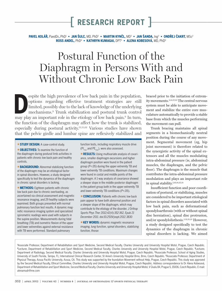
ining group differences in the slope of the middle-posterior diaphragm controlling for age and gender, the main effect for group was not significant during the TB condition (P>.05). However, there was a significant main effect for group during both the UETB (F1,43 = 10.07, P = .003) and the LETB (F1,43 = 5.49, P = .024) con-ditions, indicating that the contraction of the diaphragm followed a substantially steeper recruitment pattern in the pa-tient group relative to the control group. For better illustration of this situation, FIGURE 4 shows the slope of the middle-posterior diaphragm during inspiration in the UETB condition.
Total Diaphragm Excursion and Respiratory Muscle DriveNo significant correlations emerged be-tween total diaphragm excursion and
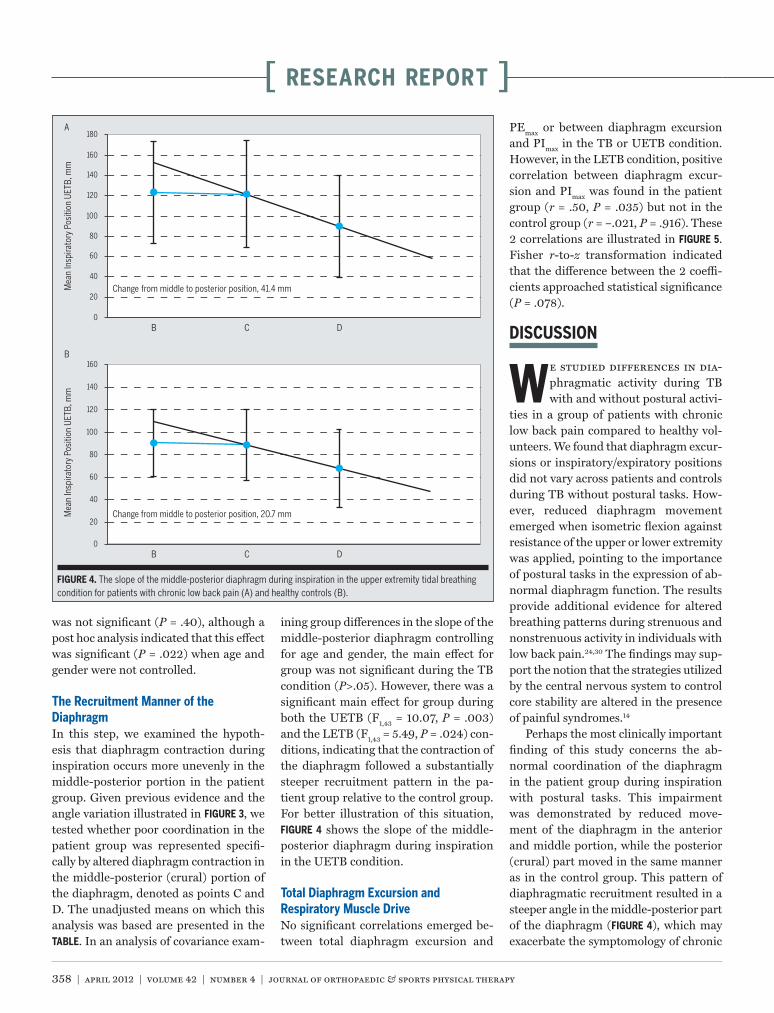
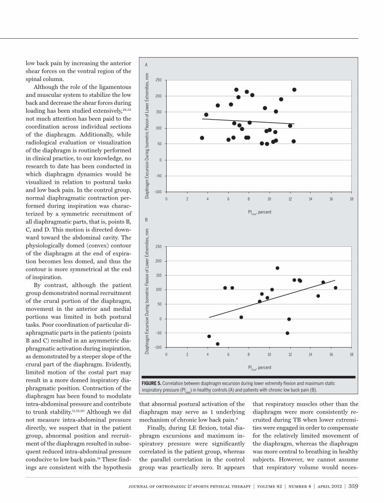
PEmax or between diaphragm excursion and PImax in the TB or UETB condition. However, in the LETB condition, positive correlation between diaphragm excur-sion and PImax was found in the patient group (r = .50, P = .035) but not in the control group (r = –.021, P = .916). These 2 correlations are illustrated in FIGURE 5. Fisher r-to-z transformation indicated that the difference between the 2 coeffi-cients approached statistical significance (P = .078).
DISCUSSION
We studied differences in dia-phragmatic activity during TB with and without postural activi-
ties in a group of patients with chronic low back pain compared to healthy vol-unteers. We found that diaphragm excur-sions or inspiratory/expiratory positions did not vary across patients and controls during TB without postural tasks. How-ever, reduced diaphragm movement emerged when isometric flexion against resistance of the upper or lower extremity was applied, pointing to the importance of postural tasks in the expression of ab-normal diaphragm function. The results provide additional evidence for altered breathing patterns during strenuous and nonstrenuous activity in individuals with low back pain.24,30 The findings may sup-port the notion that the strategies utilized by the central nervous system to control core stability are altered in the presence of painful syndromes.14
Perhaps the most clinically important finding of this study concerns the ab-normal coordination of the diaphragm in the patient group during inspiration with postural tasks. This impairment was demonstrated by reduced move-ment of the diaphragm in the anterior and middle portion, while the posterior (crural) part moved in the same manner as in the control group. This pattern of diaphragmatic recruitment resulted in a steeper angle in the middle-posterior part of the diaphragm (FIGURE 4), which may exacerbate the symptomology of chronic
A
B
0
20
40
60
80
100
120
140
160
180
Mea
n In
spira
tory
Pos
ition
UET
B, m
m
B C D
0
20
40
60
80
100
120
140
160
Mea
n In
spira
tory
Pos
ition
UET
B, m
m
B C D
Change from middle to posterior position, 41.4 mm
Change from middle to posterior position, 20.7 mm
FIGURE 4. The slope of the middle-posterior diaphragm during inspiration in the upper extremity tidal breathing condition for patients with chronic low back pain (A) and healthy controls (B).
42-04 Kolar.indd 358 3/21/2012 4:38:24 PM
journal of orthopaedic & sports physical therapy | volume 42 | number 4 | april 2012 | 359
low back pain by increasing the anterior shear forces on the ventral region of the spinal column.
Although the role of the ligamentous and muscular system to stabilize the low back and decrease the shear forces during loading has been studied extensively,28,32 not much attention has been paid to the coordination across individual sections of the diaphragm. Additionally, while radiological evaluation or visualization of the diaphragm is routinely performed in clinical practice, to our knowledge, no research to date has been conducted in which diaphragm dynamics would be visualized in relation to postural tasks and low back pain. In the control group, normal diaphragmatic contraction per-formed during inspiration was charac-terized by a symmetric recruitment of all diaphragmatic parts, that is, points B, C, and D. This motion is directed down-ward toward the abdominal cavity. The physiologically domed (convex) contour of the diaphragm at the end of expira-tion becomes less domed, and thus the contour is more symmetrical at the end of inspiration.
By contrast, although the patient group demonstrated normal recruitment of the crural portion of the diaphragm, movement in the anterior and medial portions was limited in both postural tasks. Poor coordination of particular di-aphragmatic parts in the patients (points B and C) resulted in an asymmetric dia-phragmatic activation during inspiration, as demonstrated by a steeper slope of the crural part of the diaphragm. Evidently, limited motion of the costal part may result in a more domed inspiratory dia-phragmatic position. Contraction of the diaphragm has been found to modulate intra-abdominal pressure and contribute to trunk stability.11,19,20 Although we did not measure intra-abdominal pressure directly, we suspect that in the patient group, abnormal position and recruit-ment of the diaphragm resulted in subse-quent reduced intra-abdominal pressure conducive to low back pain.18 These find-ings are consistent with the hypothesis
that abnormal postural activation of the diaphragm may serve as 1 underlying mechanism of chronic low back pain.9
Finally, during LE flexion, total dia-phragm excursions and maximum in-spiratory pressure were significantly correlated in the patient group, whereas the parallel correlation in the control group was practically zero. It appears
that respiratory muscles other than the diaphragm were more consistently re-cruited during TB when lower extremi-ties were engaged in order to compensate for the relatively limited movement of the diaphragm, whereas the diaphragm was more central to breathing in healthy subjects. However, we cannot assume that respiratory volume would neces-
–100
–50
0
0 2 4 6 8 10 12 14 16 18
50
100
150
200
250
PImax, percent
Diap
hrag
m E
xcur
sion
Dur
ing
Isom
etric
Fle
xion
of L
ower
Ext
rem
ities
, mm
–100
–50
0
0 2 4 6 8 10 12 14 16 18
50
100
150
200
250
PImax, percent
Diap
hrag
m E
xcur
sion
Dur
ing
Isom
etric
Fle
xion
of L
ower
Ext
rem
ities
, mm
A
B
FIGURE 5. Correlation between diaphragm excursion during lower extremity flexion and maximum static inspiratory pressure (PImax) in healthy controls (A) and patients with chronic low back pain (B).
42-04 Kolar.indd 359 3/21/2012 4:38:25 PM
360 | april 2012 | volume 42 | number 4 | journal of orthopaedic & sports physical therapy
[ research report ]sarily be reduced in the patient group. Previous research suggests that inhaled lung volume may even be greater in pa-tients with low back pain,10 although this research did not include any measure-ment of diaphragm movement. Finally, we found pulmonary function within a normal range for both groups, further suggesting that compensation for the limited diaphragm movement might have occurred. Taken together, we speculate that an altered mode of activation, indic-ative of poor activation of the diaphragm during TB with postural tasks and more consistent activation of other respiratory muscles, may be typical in patients with chronic low back pain.
In healthy subjects, the diaphragm is able to perform the dual task (trunk stability and respiration) when trunk stability is challenged.19 Generally, dur-ing any body movement, with activation of the extremities during weight-bearing or weight-lifting activities and transi-tional movements, there is simultaneous spinal bracing and transdiaphragmatic pressure elevation.11,22 Intra-abdominal pressure increases, with a simultaneous decrease of intrapleural pressure, during a contraction of both the posterior (cru-ral) and anterior (costal) portions of the diaphragm.7 This coordination may be compromised in patients with chronic low back pain.
Stabilizing postural activation of the diaphragm has been reported during, for example, weight lifting20 and isomet-ric activation of the extremities.27 Simi-larly, higher inspiratory pressures and hypertrophic changes in the diaphragm have been demonstrated during exer-cise.1,6 It can be assumed that, in cases of postural instability, the insufficient stabilizers must be compensated by other muscles. A significant decrease in strength of trunk muscles, especially the extensors, in patients with low back pain has been established,8,23,34,37,38 suggesting that strengthening exercises of the trunk muscles may be an optimal rehabilitation strategy.
One possibility is that the lack of pos-
tural diaphragmatic activation is sub-stituted by excessive activation of the superficial lumbar paraspinal muscles, which may lead to hypertrophy and, eventually, result in lumbar hyperlor-dosis and/or anterior pelvic tilt. Future research should study this mechanism as possibly contributing to or even un-derlying the etiology of low back pain symptoms. Furthermore, long-lasting ef-fects and pain alleviation may be aided by achieving balanced agonist-antagonist postural activation (that is, balanced ac-tivation across the sections of the dia-phragm, pelvic floor, and the abdominal wall and extensors). Research is needed to investigate the possibility that work-ing to correct the altered function of the diaphragm specifically may contribute to alleviating low back pain symptoms by improving spinal stability.
There are several limitations to this study. First, we used a convenience sample in which the patient and control groups differed in size and some demo-graphic characteristics. Although we used statistical methods appropriate for unbal-anced designs and controlled for these differences statistically, we cannot fully exclude the possibility that these differ-ences had some influence on the results. Second, ideally, the entire rib cage, includ-ing the whole range of diaphragm excur-sions, should have been imaged. Only an isolated analysis of the diaphragm was performed, which focused on the dia-phragm excursions, due to the limited size of the field of view.36 Although we have limited the diaphragm excursion mea-surements to 3 points, this is similar to studies conducted by other authors36 and can be considered sufficient for this type of study. In addition, the diaphragm ex-cursions alone may not be sufficient to un-derstand all mechanical actions of the rib cage and related musculature. For exam-ple, individual breathing patterns may be considered along with diaphragm excur-sions in future research. Third, although external pressure to generate grade 4 force was applied by the same clinician (P.K.) and standardized requirements of
current MRI methodology4 were followed in order to reduce variation in diaphragm motion and function, force and direction were not formally assessed. Therefore, we cannot exclude the possibility that the re-sistance varied across the subjects.
Fourth, we did not assess the length or duration of low back pain in the pa-tient group, only that the pain had lasted at least 6 months. Nor did we assess pain during postural tasks. However, all par-ticipants exhibited the ability to perform grade 4 force without substantial pain. Had pain been present during postural tasks, we could not have obtained read-able MRI images. It could also be argued that the 4 patients who underwent a failed back surgery might have differed from the rest of the patient sample in their outcomes. Excluding these 4 pa-tients, however, would not have substan-tially altered the results, although group differences in diaphragm excursion dur-ing lower and upper extremity postural tasks were somewhat (not substantially) greater, as were group differences in the correlation between total diaphragm excursion and respiratory muscle drive. Finally, poor postural function of the dia-phragm may result in symptoms of low back pain and lead to chronic vertebro-genic dysfunction. However, considering all possibilities, we cannot exclude the reverse order of events. Low back pain symptoms may be indicative of an initial pathogenic insult resulting in secondary quantitative as well as qualitative adap-tive changes in diaphragmatic function.
CONCLUSION
We found reduced diaphragm movement when isometric flex-ion against resistance of the up-
per or lower extremities was applied. The combined, more cranial position in the anterior and middle portions of the diaphragm and, particularly, the steeper slope between the middle and crural por-tions of the diaphragm in patients with chronic low back pain may contribute to low back pain symptoms. However,
42-04 Kolar.indd 360 3/21/2012 4:38:26 PM
journal of orthopaedic & sports physical therapy | volume 42 | number 4 | april 2012 | 361
given that the results are based on cross-sectional analysis, we cannot exclude the possibility of reverse causation. Still, the results support the theory that patients with low back pain complaints present with compromised diaphragm function, which may play an important role in pos-tural stability. t
KEY POINTSFINDINGS: We found reduced diaphragm movement in patients with chronic low back pain compared to healthy controls when isometric flexion against resis-tance of the upper or lower extremity was applied, mainly in the anterior and middle portions. This pattern of diaphragmatic recruitment resulted in a steeper angle in the middle-posterior part of the diaphragm and likely a great-er strain during activity on the ventral region of the spinal column.IMPLICATIONS: Abnormal postural activa-tion of the diaphragm during the pos-tural task of isometric resistance to the extremities may serve as 1 underlying mechanism of chronic low back pain.CAUTION: Only an isolated analysis of the diaphragm excursion was performed, due to the limited field of view. In ad-dition, the diaphragm excursion alone may not be sufficient to understand all mechanical actions of the rib cage and related musculature. We used a con-venience sample in which the patient and control groups differed in size and certain demographic characteristics. Because our study was cross-sectional in nature, we cannot exclude the possibil-ity that low back pain symptoms may be indicative of an initial pathogenic insult resulting in secondary quantitative as well as qualitative adaptive changes in diaphragmatic function.
REFERENCES
1. Al-Bilbeisi F, McCool FD. Diaphragm recruitment during nonrespiratory activities. Am J Respir Crit Care Med. 2000;162:456-459.
2. American Thoracic Society. ATS/ERS statement on respiratory muscle testing. Am J Respir Crit
Care Med. 2002;166:518-624. http://dx.doi.org/10.1164/rccm.166.4.518
3. Aruin AS, Latash ML. Directional specificity of postural muscles in feed-forward postural reac-tions during fast voluntary arm movements. Exp Brain Res. 1995;103:323-332.
4. Chu WC, Li AM, Ng BK, et al. Dynamic mag-netic resonance imaging in assessing lung volumes, chest wall, and diaphragm motions in adolescent idiopathic scoliosis versus normal controls. Spine (Phila Pa 1976). 2006;31:2243-2249. http://dx.doi.org/10.1097/01.brs.0000232822.74349.32
5. Cresswell AG, Oddsson L, Thorstensson A. The influence of sudden perturbations on trunk muscle activity and intra-abdominal pressure while standing. Exp Brain Res. 1994;98:336-341.
6. DePalo VA, Parker AL, Al-Bilbeisi F, McCool FD. Respiratory muscle strength training with nonrespiratory maneuvers. J Appl Physiol. 2004;96:731-734. http://dx.doi.org/10.1152/japplphysiol.00511.2003
7. De Troyer A, Loring SH. Action of the respiratory muscles. In: Macklem PT, Mead J, eds. Hand-book of Physiology: Section 3: The Respiratory System Volume III, Parts 1 & 2: Mechanics of Breathing. New York, NY: Oxford University Press; 1986:443-462.
8. Dvir Z, Keating JL. Trunk extension effort in pa-tients with chronic low back dysfunction. Spine (Phila Pa 1976). 2003;28:685-692. http://dx.doi.org/10.1097/01.BRS.0000051917.04731.A4
9. Ebenbichler GR, Oddsson LI, Kollmitzer J, Erim Z. Sensory-motor control of the lower back: implications for rehabilitation. Med Sci Sports Exerc. 2001;33:1889-1898.
10. Hagins M, Lamberg EM. Individuals with low back pain breathe differently than healthy in-dividuals during a lifting task. J Orthop Sports Phys Ther. 2011;41:141-148. http://dx.doi.org/10.2519/jospt.2011.3437
11. Hemborg B, Moritz U, Lowing H. Intra-abdominal pressure and trunk muscle activity during lifting. IV. The causal factors of the intra-abdominal pressure rise. Scand J Rehabil Med. 1985;17:25-38.
12. Hides JA, Stokes MJ, Saide M, Jull GA, Cooper DH. Evidence of lumbar multifidus muscle wasting ipsilateral to symptoms in patients with acute/subacute low back pain. Spine (Phila Pa 1976). 1994;19:165-172.
13. Hodges P. Lumbopelvic stability: a functional model of biomechanics and motor control. In: Richardson C, Hodges P, Hides J, eds. Thera-peutic Exercise for Lumbopelvic Stabilization. 2nd ed. Sydney, Australia: Churchill Livingstone; 2004:13-28.
14. Hodges PW. The role of the motor system in spinal pain: implications for rehabilitation of the athlete following lower back pain. J Sci Med Sport. 2000;3:243-253.
15. Hodges PW, Butler JE, McKenzie DK, Gandevia SC. Contraction of the human diaphragm during rapid postural adjustments. J Physiol. 1997;505 pt 2:539-548.
16. Hodges PW, Cresswell AG, Daggfeldt K, Thor-stensson A. In vivo measurement of the effect of intra-abdominal pressure on the human spine. J Biomech. 2001;34:347-353.
19. Hodges PW, Gandevia SC. Activation of the human diaphragm during a repetitive postural task. J Physiol. 2000;522 pt 1:165-175.
20. Hodges PW, Gandevia SC. Changes in intra-ab-dominal pressure during postural and respira-tory activation of the human diaphragm. J Appl Physiol. 2000;89:967-976.
21. Hodges PW, Richardson CA. Altered trunk muscle recruitment in people with low back pain with upper limb movement at different speeds. Arch Phys Med Rehabil. 1999;80:1005-1012.
22. Hodges PW, Richardson CA. Relationship between limb movement speed and as-sociated contraction of the trunk muscles. Ergonomics. 1997;40:1220-1230. http://dx.doi.org/10.1080/001401397187469
23. Iwai K, Nakazato K, Irie K, Fujimoto H, Nakajima H. Trunk muscle strength and disability level of low back pain in collegiate wrestlers. Med Sci Sports Exerc. 2004;36:1296-1300.
24. Janssens L, Brumagne S, Polspoel K, Troosters T, McConnell A. The effect of inspiratory muscles fatigue on postural control in people with and without recurrent low back pain. Spine (Phila Pa 1976). 2010;35:1088-1094. http://dx.doi.org/10.1097/BRS.0b013e3181bee5c3
25. Kalpakcioglu B, Altinbilek T, Senel K. Determina-tion of spondylolisthesis in low back pain by clinical evaluation. J Back Musculoskelet Reha-bil. 2009;22:27-32. http://dx.doi.org/10.3233/BMR-2009-0212
26. Kendall FP, McCreary EK, Provance PG, Rodgers MM, Romani WA. Muscles: Testing and Function with Posture and Pain. 5th ed. Baltimore, MD: Lippincott Williams & Wilkins; 2005.
27. Kolar P, Sulc J, Kyncl M, et al. Stabilizing function of the diaphragm: dynamic MRI and synchronized spirometric assessment. J Appl Physiol. 2010;109:1064-1071. http://dx.doi.org/10.1152/japplphysiol.01216.2009
28. McGill SM. Low back stability: from formal description to issues for performance and reha-bilitation. Exerc Sport Sci Rev. 2001;29:26-31.
29. Miller MR, Hankinson J, Brusasco V, et al. Standardisation of spirometry. Eur Respir J. 2005;26:319-338. http://dx.doi.org/10.1183/09031936.05.00034805
30. O’Sullivan PB, Beales DJ, Beetham JA, et al. Altered motor control strategies in subjects with sacroiliac joint pain during the active straight-leg-raise test. Spine (Phila Pa 1976). 2002;27:E1-8.
362 | april 2012 | volume 42 | number 4 | journal of orthopaedic & sports physical therapy
[ research report ]
MORE INFORMATIONWWW.JOSPT.ORG@
31. Panjabi MM. The stabilizing system of the spine. Part I. Function, dysfunction, adaptation, and enhancement. J Spinal Disord. 1992;5:383-389; discussion 397.
32. Pel JJ, Spoor CW, Goossens RH, Pool-Goudz-waard AL. Biomechanical model study of pelvic belt influence on muscle and ligament forces. J Biomech. 2008;41:1878-1884. http://dx.doi.org/10.1016/j.jbiomech.2008.04.002
33. Quanjer PH, Tammeling GJ, Cotes JE, Pedersen OF, Peslin R, Yernault JC. Lung volumes and forced ventilatory flows. Report Working Party Standardization of Lung Function Tests, Euro-pean Community for Steel and Coal. Official Statement of the European Respiratory Society.
36. Takazakura R, Takahashi M, Nitta N, Murata K. Diaphragmatic motion in the sitting and supine positions: healthy subject study using a verti-cally open magnetic resonance system. J Magn Reson Imaging. 2004;19:605-609. http://dx.doi.org/10.1002/jmri.20051
37. Tsai YS, Sell TC, Smoliga JM, Myers JB, Lear-man KE, Lephart SM. A comparison of physical
characteristics and swing mechanics between golfers with and without a history of low back pain. J Orthop Sports Phys Ther. 2010;40:430-438. http://dx.doi.org/10.2519/jospt.2010.3152
38. Yahia A, Jribi S, Ghroubi S, Elleuch M, Baklouti S, Habib Elleuch M. Evaluation of the posture and muscular strength of the trunk and inferior members of patients with chronic lumbar pain. Joint Bone Spine. 2011;78:291-297. http://dx.doi.org/10.1016/j.jbspin.2010.09.008
EARN CEUs With JOSPT’s Read for Credit Program
JOSPT’s Read for Credit (RFC) program invites Journal readers to study and analyze selected JOSPT articles and successfully complete online quizzes about them for continuing education credit. To participate in the program:
1. Go to www.jospt.org and click on “Read for Credit” in the left-hand navigation column that runs throughout the site or on the link in the “Read for Credit” box in the right-hand column of the home page. 2. Choose an article to study and when ready, click “Take Exam” for that article. 3. Login and pay for the quiz by credit card. 4. Take the quiz. 5. Evaluate the RFC experience and receive a personalized certificate of continuing education credits.
The RFC program o�ers you 2 opportunities to pass the quiz. You may review all of your answers—including the questions you missed. You receive 0.2 CEUs, or 2 contact hours, for each quiz passed. The Journal website maintains a history of the quizzes you have taken and the credits and certificates you have been awarded in the “My CEUs” section of your “My JOSPT” account.