• Other metabolic substrates that fetus needs diffuse into blood in same manner as oxygen
• Glucose in fetal blood about 20 to 30 percent lower than maternal blood
• Results in rapid diffusion of glucose to fetus
• Transports other substrates; fatty acids, potassium, sodium, and chloride
• Actively absorbs some nutrients from maternal blood
19
Placenta
• Responsible for five functions
– Excretion of wastes
• Diffuse from fetal blood into maternal blood
• Examples are urea, uric acid, and creatinine
• Excreted with waste products of mother
• Transfer from fetal circulation to maternal circulation moving osmotically from higher concentration to lower concentration; same manner as carbon dioxide
20
Placenta
• Responsible for five functions
– Hormone production
• Placenta becomes temporary endocrine gland
• Secretes estrogen and progesterone
• By third month of development, corpus luteum on the ovary no longer is needed to sustain pregnancy
• Estrogen, progesterone, and other hormones maintain uterine lining, prevent occurrence of menses
• Stimulate changes in pregnant woman's breasts, vagina, cervix, and pelvis
• Most of this blood is deoxygenated blood from head of fetus
• Blood is pumped by right ventricle into pulmonary artery
• Deoxygenated blood passes from pulmonary artery, through ductus arteriosus, into descending aorta, through two umbilical arteries, and into placenta for oxygenation
• At birth, various arteriovenous shunts close in most infants
28
29
Amniotic Sac and Fluid
• Completely surrounds embryo
• Contains fluid primarily produced by fetal urine and placenta
• Ductus venosus, ductus arteriosus, and foramen ovale allow blood flow to bypass immature liver and lungs of the developing fetus
• Blood flow through placenta ceases at birth, resultant increase in systemic vascular resistance and increase in pressure in aorta, left ventricle, and left atrium
40
Infant Adaptations After Birth
• Pulmonary vascular resistance decreases greatly because of lung expansion
– Reduces pulmonary arterial, right ventricular, and right atrial pressures
• Arteriovenous shunts close normally within few hours after birth
• Eventually close completely and covered with growth of fibrous tissue
41
Do fetal heart tones sound normal if you auscultate them immediately
• Morning sickness and nausea may occur at any time– Usually begin by 6th and abate by 14th week – Cause is unknown but may be related to high serum levels of chorionic gonadotropin in early pregnancy
• Enlarging uterus displaces the mother’s stomach and intestines upward and laterally– May cause indigestion and gastroesophageal reflux (GERD)
– May increase risk for aspiration in unconscious patients
52
Gastrointestinal System
• Liver is displaced backward, upward, and to right
• Tone and motility of gastrointestinal tract decrease
– Leading to prolonged gastric emptying and relaxation of pyloric sphincter
– Heartburn and constipation are common
53
What problems are associated with these GI changes for the
unconscious pregnant woman who has sustained trauma?
• Fetal heart sounds– Can be auscultated beginning at 12 weeks gestation
• May be difficult to hear in noisy environment
– Can be auscultated by use of stethoscope, fetoscope, or Doppler probe
– Purpose of checking heart tones is to assess fetal well‐being
– Monitor fetal heart rate and maternal vital signs every 5 to 10 minutes
76
77
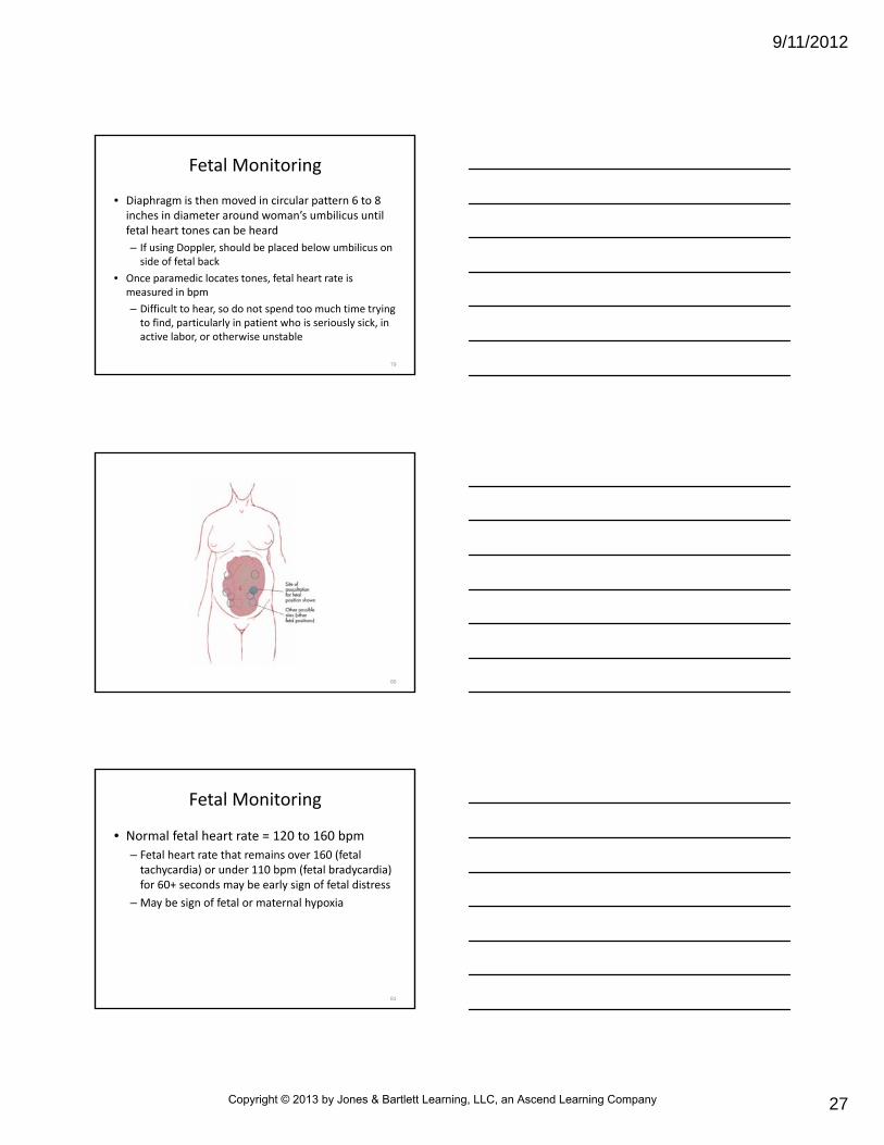
Fetal Monitoring
• When auscultating fetal heart rate (FHR), position high‐intensity diaphragm of stethoscope (bell of fetoscope or microphone of Doppler probe) firmly on mother’s abdominal wall
– If more than 20 weeks gestation, palpate for fetal back (defined by structure that is firm and hard vs. small body parts)
• Diaphragm is then moved in circular pattern 6 to 8 inches in diameter around woman’s umbilicus until fetal heart tones can be heard
– If using Doppler, should be placed below umbilicus on side of fetal back
• Once paramedic locates tones, fetal heart rate is measured in bpm
– Difficult to hear, so do not spend too much time trying to find, particularly in patient who is seriously sick, in active labor, or otherwise unstable
79
80
Fetal Monitoring
• Normal fetal heart rate = 120 to 160 bpm
– Fetal heart rate that remains over 160 (fetal tachycardia) or under 110 bpm (fetal bradycardia) for 60+ seconds may be early sign of fetal distress
• Intermittent, short‐term increases or decreases in fetal heart rate usually are normal– Variation can occur at any time and is sign of fetal suckling
– Short‐term periodic changes in fetal heart rate are common during
• Fetal sleep
• Fetal movement
• Contractions associated with labor and delivery
82
General Management of theObstetrical Patient
• If birth is not imminent, care for healthy patient should be limited to
– Basic treatment modalities (airway, ventilatory, and circulatory support)
– Transport
• In absence of distress or injury, transport in comfortable position
• Usually left lateral recumbant
83
General Management of the Obstetrical Patient
• May need to– Monitor ECG
– Administer oxygen (5 to 7 L/min)
– Monitor fetus based on patient assessment and vital sign determinations
– Medical direction may advise IV access in some patients
– Most drugs usually are inappropriate as they can mask symptoms of worsening condition
• 1 in every 12 pregnancies is complicated by physical trauma
– When pregnant woman is severely injured, fetus at high risk for death
– Anatomical and physiological changes of pregnancy can alter woman’s response to injury
• May necessitate modified assessment, treatment, and transportation strategies
85
Maternal Injury
• Causes of maternal injury in decreasing order of frequency are
– Vehicular crashes
– Falls
– Penetrating objects
• Injuries can result in trauma to gravid uterus, maternal bladder, liver, spleen
– Injury that results in pelvic fracture can produce massive hemorrhage and damage to fetal skull
– Severity of injury depends on many factors and may involve multiple organ systems
86
Maternal Injury
• During pregnancy, fetus is well protected within uterus
– Amniotic fluid surrounds fetus
– Fluid serves as excellent shock absorber
– Because of this protection, fetus rarely experiences physical trauma except as result of direct penetrating wounds or extensive blunt trauma to maternal abdomen
• Direct life‐threatening fetal injury uncommon in blunt trauma
– In penetrating trauma, direct injury to fetus can cause fetal death, even if mother’s injuries are not life threatening
91
Assessment and Management
• Priorities in assessing and managing pregnant trauma patient are same as for nonpregnant patient – Adequate airway, ventilatory, and circulatory support with spinal precautions
– Hemorrhage control
– Rapid assessment
– Stabilization
– Rapid transport
92
Assessment and Management
• Resuscitating mother is key to survival of mother and fetus
– During first stages of assessment and management, mother’s status should be focus
– Despite severity, all pregnant trauma patients should be given high‐concentration oxygen and transported for physician evaluation
– Must detect, identify, manage injuries that contribute to hypovolemia or hypoxia
– With normal increase in maternal blood volume, mother can tolerate more blood loss before showing signs and symptoms of shock
94
• 30 to 35 percent reduction in blood volume can produce minimal changes in BP but reduce uterine blood flow by 10 to 20 percent– Mother may maintain adequate BP at expense of fetus
• True amount of blood loss may be difficult to detect– Fetal monitoring is best available indicator of fetal well‐being after trauma
– Patient transport should never be delayed to assess fetal heart rate
Assessment and Management
95
Assessment and Management
• Accelerations of fetal heart rate above baseline are associated with fetal movement and contractions
– May be early sign of fetal distress
– Decreased fetal movement and increased fetal heart rate can indicate maternal shock
• If cardiac arrest occurs, institute CPR with few modifications– Relieve pressure on aorta and inferior vena cava
• Can be done by placing hands on abdomen to move uterus to patient's right or left side
• Displacement can also be accomplished by placing patient in left lateral tilt of 20 to 30 degrees, using blankets, pillows, or wedge to support pelvis
• Generally perform chest compressions higher on sternum (ensures palpable pulse wave) to adjust for shifting of pelvic and abdominal contents toward head
Special Management Considerations
112
113
• Other patient management considerations unique to pregnant patient in cardiac arrest– Establish IV access above diaphragm to enhance systemic circulation of fluids and drugs
– Manage maternal hypotension (systolic BP less than 100 mm Hg or less than 80 percent of baseline) to avoid reduced placental perfusion
– Anticipate a difficult airway because of changes in airway mucosa that occur during pregnancy
– Standard drug doses and defibrillation therapies are recommended
• Women who are sensitized will need careful monitoring and serial blood testing to measure antibody levels during their pregnancy– Doppler studies and amniocentesis may be performed to monitor fetus
– If fetal anemia is severe, baby may need blood transfusions before birth (intrauterine transfusion) and immediately after birth
– Early caesarean delivery is common in these cases
124
Rh Disease
• An infant born with Rh disease may have no symptoms of illness
– Other newborns can have a serious and life‐threatening blood disorder known as erythroblastosis fetalis
125
Rh Disease
• Symptoms– Anemia
– Jaundice
– Edema
– Enlarged liver or spleen
– Hydrops fetalis (accumulation of fluid throughout body tissues, including lungs, heart, abdominal organs)
– Calls for initial resuscitation measures and rapid transport for surgical intervention
– May become unstable quickly
– If paramedic suspects an ectopic pregnancy, patient should be managed like any victim of hemorrhagic shock, with airway, ventilatory, and circulatory support and IV fluid resuscitation
166
Third‐trimester Bleeding
• Third‐trimester bleeding occurs in 4 percent of all pregnancies and is never normal
– About half of bleeding episodes are result of
• Abruptio placentae
• Placenta previa
• Uterine rupture
167
Abruptio Placentae
• Partial or full detachment of normally implanted placenta at more than 20 weeks gestation
– Occurs in about 1 percent of all pregnancies
– Severe enough to result in fetal death in about 15 percent of cases of abruption
• Presenting part of fetus (usually head) emerges from vaginal opening
– Known as crowning, indicates delivery is imminent
– Usually lasts 1 to 2 hours in nullipara mother
– Usually lasts 30 minutes or less in multiparamother
199
Third Stage of Labor
• Begins with delivery of infant and ends when placenta is expelled and uterus has contracted
– Length of this stage varies from 5 to 60 minutes, regardless of parity
200
Signs and Symptoms of Imminent Delivery
• Following signs and symptoms indicate delivery is imminent; prepare for childbirth at scene– Regular contractions lasting 45 to 60 seconds at 1‐to 2‐minute intervals
• Intervals are measured from beginning of one contraction to beginning of next
• If contractions are more than 5 minutes apart, generally is time to transport to receiving hospital
– Mother has urge to bear down or has sensation of bowel movement
• Following signs and symptoms indicate delivery is imminent, prepare for childbirth at scene
– Large amount of bloody show
– Crowning occurs
– Mother believes that delivery is imminent
Signs and Symptoms of Imminent Delivery
202
• With exception of cord presentation, do not try to delay delivery
– If complications are anticipated or abnormal delivery occurs, medical direction may recommend expedited transport of patient to medical facility
Signs and Symptoms of Imminent Delivery
203
Preparation for Delivery
• When preparing for delivery, try to provide area of privacy– Mother should be positioned on a bed, stretcher, or table
– Surface should be long enough to project beyond mother’s vagina
– Delivery area should be as clean as possible• Should be covered with absorbent material to guard against staining and contamination by blood and fecal material
• Mother should be placed on her back– Knees should be flexed and widely separated (or in another position preferred by mother)
– Vaginal area should be draped appropriately
– If delivery occurs in car, mother should be instructed to lie on her back across seat with one leg flexed on seat and the other leg resting on floorboard
– Pillow or blanket, if available, should be placed beneath mother’s buttocks
205
Preparation for Delivery
• Will aid in delivery of infant’s head
– Evaluate mother’s vital signs for baseline measurements
– Monitor fetal heart for signs of fetal distress
– Per protocol and medical direction, consider maternal oxygen administration and IV access for fluid administration or postdelivery administration of oxytocin if needed
206
Preparation for Delivery
• Mother should be coached to bear down and push during contractions and to rest between contractions to conserve strength
– If mother finds it difficult to refrain from pushing, should be encouraged to breathe deeply or “pant” through her mouth between contractions
– Deep breathing and panting help decrease force of bearing down and promote rest
• In most cases paramedic only assists in natural events of childbirth
– Chief duties of the EMS crew are to prevent uncontrolled delivery and protect infant from cold and stress after birth
211
Assistance with Delivery
• Following are steps to be taken in assisting mother with normal delivery
– Observe standard precautions
– When crowning occurs, apply gentle palm counterpressure to infant’s head to prevent explosive delivery and tearing of mother’s perineum
• If membranes are still intact, tear sac with finger pressure to allow escape of amniotic fluid
212
Assistance with Delivery
• Following are steps to be taken in assisting mother with normal delivery– After delivery of head, examine infant’s neck for looped (nuchal) umbilical cord
• If cord is looped around neck, gently slip it over infant’s head
– Suction infant’s mouth and nose with bulb syringe to clear airway
• Perform suction after head appears but before next contraction
• Following are steps to be taken in assisting mother with normal delivery
– Support infant’s head as it rotates for shoulder presentation
• Most infants present face down
• Infant usually rotates to left or right so shoulders present in an anterior‐posterior position
214
Assistance with Delivery
• Following are steps to be taken in assisting mother with normal delivery
– If shoulders do not spontaneously deliver with next contraction, using gentle pressure, guide infant’s head downward to deliver anterior shoulder and then upward to release posterior shoulder
• Rest of infant is delivered quickly by smooth uterine contraction
215
Assistance with Delivery
• Following are steps to be taken in assisting mother with normal delivery
– Be careful to grasp and support infant as he or she emerges, using dry towel or clean piece of clothing
• Hold infant with his or her head dependent to aid drainage of secretions
• Place infant on mother's abdomen if she is able to hold her infant
• Take following measures to encourage uterine contraction
– Massage the uterus
• Palpate uterus for firmness or loss of tone
• If uterus does not feel firm, apply fundal pressure by supporting lower uterine segment with edge of one hand just above symphysis and massaging fundus with other hand
• Continue massaging until uterus feels firm
• Reevaluate patient every 10 minutes
• Note location of fundus in relation to level of umbilicus, degree of firmness, vaginal flow
241
Management
• Take following measures to encourage uterine contraction
– Encourage infant to breast‐feed
• If mother and infant are stable and mother is agreeable, place newborn to her breast to encourage breast‐feeding
• Stimulation of breasts may promote uterine contraction
242
Management
• Take following measures to encourage uterine contraction– Administer oxytocin
• Per medical direction and after ensuring that second fetus is not present in uterus, add 10 units of oxytocinto 1000 mL lactated Ringer’s solution
• Infuse at 20 to 30 drops/min via microdrip tubing (titrated to severity of hemorrhage and uterine response or as ordered by medical direction)
• Continue with fluid resuscitation as indicated by patient’s vital signs
• If head does not deliver immediately, action must be taken to prevent suffocation of infant– Maintain fetal head in flexed position by placing index and middle fingers on either side of infant's nose (the Mauriceau maneuver)
• Fetal body should be supported in neutral position, using care not to overextend baby's neck– During this maneuver second rescuer should apply suprapubic pressure
256
Management
• If head does not deliver quickly, chance for good fetal outcome is poor
– Mother should be transported rapidly
257
Shoulder Dystocia
• Occurs when fetal shoulders are wedged against maternal symphysis pubis
– Blocks shoulder delivery
– In this presentation, head delivers normally but then pulls back tightly against maternal perineum (the turtle sign)
• Common condition in pregnancy, occurring in 1 in 300 deliveries
– Complications
• Brachial plexus damage
• Fractured clavicle
• Fetal anoxia from cord compression
– 50 percent occur in women without risk factors
259
Management
• Calls for dislodging one shoulder and then rotating fetal shoulder girdle at angle into wider part of pelvic opening– Because shoulder is pressing against pelvis, there is potential for cord compression
• Paramedic should deliver anterior shoulder immediately after head– Several maneuvers can help paramedic successfully deliver infant when shoulder dystociaarises
260
Management
• Following steps represent one approach to shoulder dystocia
– Position mother on her left side in dorsal‐knee‐chest position
• Increases diameter of pelvis
– Try to guide infant’s head downward to allow anterior shoulder to slip under symphysis pubis
• Following steps represent one approach to shoulder dystocia– Gently rotate fetal shoulder girdle at angle to wider pelvic opening
• Posterior shoulder usually delivers without resistance
• Medical direction may recommend that paramedic try to deliver posterior shoulder first by rotating posterior shoulder downward and into left posterior quadrant
• Anterior shoulder usually follows
– After delivery, continue with resuscitative measures as needed
262
Shoulder Presentation
• Results when long axis of fetus lies perpendicular to that of mother
– Position usually results in fetal shoulder lying over pelvic opening
– Fetal arm or hand may be presenting part
– Occurs in only 0.3 percent of deliveries but occurs in 10 percent of second twins
263
Management
• Normal delivery of a presentation is not possible
– Provide mother with adequate oxygen, ventilatoryand circulatory support, and rapid transport
– Cesarean delivery is required whether fetus is viable or not
• After delivery, prehospital management for premature infant includes
– Keep infant warm
• Dry infant, wrap in warm blanket, place infant on mother’s abdomen, and cover mother and infant
• If transport time is delayed, very small (under 1500 g) infants should be wrapped in food‐grade heat‐resistant plastic wrap and placed under radiant heat in addition to other warming methods
274
Premature Birth
• After delivery, prehospital management for premature infant includes
– Frequently suction secretions from infant’s mouth and nares
– Carefully monitor cut end of umbilical cord for oozing
• If bleeding present, manage as described before
275
Premature Birth
• After delivery, prehospital management for premature infant includes– Administer humidified free‐flow oxygen through makeshift oxygen tent
• Aim oxygen flow toward top of tent
• Do not allow it to flow directly into infant’s face
– Protect infant from contamination• Don mask and gown and minimize family member and bystander contact with infant
Premature Birth• Note that tocolytic agents (drugs used to inhibit labor) are used widely today by some mothers who are at risk for premature birth– May be administered in home setting– Include
• First‐twin delivery is identical to single delivery with same presentation
– Up to 50 percent of second‐twin deliveries are not in normal presentation position
– Fetuses are smaller in multiple births
283
Delivery Procedure
• After delivery of first twin, cut and clamp (or tie) umbilical cord as described earlier– Within 5 to 10 minutes after delivery of first twin, labor begins again
– Delivery of second twin usually occurs within 30 to 45 minutes
– Medical direction may recommend transport before delivery of second twin
– Usually both twins are born before delivery of placenta
284
Delivery Procedure
• Infants in multiple births often are smaller than infants in single term births
– Give special attention to keeping these infants warm, well oxygenated, and free from unnecessary contamination as described for premature infants
• If paramedic expects a precipitous delivery, attempts should be made to prevent explosive one– Can be done by providing gentle counterpressureto infant’s head
• Do not attempt to detain fetal head descent
• After delivery, infant should be kept dry and warm to prevent heat loss
• Mother should be examined for perineal tears that often accompany rapid birth
289
Uterine Inversion
• Infrequent complication of childbirth where uterus turns “inside out”
– Thought to occur in about 1 in 2000 deliveries
– Serious condition
– Resultant postpartum hemorrhage is associated with maternal mortality rate of around 15 percent
290
Uterine Inversion
• May occur suddenly after contraction or with increased abdominal pressure caused by coughing or sneezing
– More often is caused by medical personnel or medical procedure (iatrogenic), secondary to excessive pulling on umbilical cord and fundalmassage
• Most often seen in multiparous women late in first stage of labor
– Other conditions that can increase incidence
• Placenta previa
• Abruptio placentae
• Intrauterine fetal death
– Maternal mortality rate high
304
Amniotic Fluid Embolism
• Signs and symptoms of amniotic fluid embolism are same as those for pulmonary embolism
– May include cardiopulmonary arrest
• Treatment
– Airway, ventilatory, and circulatory support
– Fluid resuscitation
– Rapid transportation
305
Summary
• Cultural differences may influence a woman’s response to pregnancy and childbirth– Paramedic should be sensitive to these cultural beliefs
• Fertilization of an ovum by a sperm forms a zygote that divides as it passes through fallopian tube to become a morula– Trophoblast cells of the morula implant within 7 days after
fertilization and transform into the life support systems of the embryo
• Patient history should include obstetrical history; presence of pain; presence, quantity, and character of vaginal bleeding; presence of abnormal vaginal discharge; presence of “bloody show”; current general health and prenatal care; allergies and medicines taken; and maternal urge to bear down
310
Summary
• Goal in examining an obstetrical patient is to rapidly identify acute life‐threatening conditions
– Part of this involves recognizing imminent delivery
– Then paramedic must take the proper management steps
• In addition to the routine physical examination, the paramedic should assess the abdomen, uterine size, and fetal heart sounds
311
Summary
• If birth is not imminent, paramedic should limit prehospital care for healthy patient
– Limited to basic treatment modalities
– Include transport for physician evaluation
• Causes of fetal death from maternal trauma include death of mother, separation of the placenta, maternal shock, uterine rupture, and fetal head injury
• Gestational diabetes mellitus is diabetes caused by pregnancy
• Infection during pregnancy can place the mother and fetus at risk
– TORCH is an acronym for infections mother can pass to fetus that cause fetal death or complication
316
Summary
• Vaginal bleeding during pregnancy can result from abortion (miscarriage), ectopic pregnancy, abruptio placentae, placenta previa, uterine rupture, or postpartum hemorrhage– Abortion is termination of pregnancy from any cause before 20 weeks gestation
– Ectopic pregnancy occurs when a fertilized ovum implants anywhere other than the uterus
– Abruptio placentae is partial or complete detachment of the placenta at more than 20 weeks gestation
317
Summary
• Placenta previa is placental implantation in the lower uterine segment partially or completely covering the cervical opening
• Uterine rupture is a spontaneous or traumatic rupture of the uterine wall