47
Pediatric Hepatobiliary, Pancreatic & Splenic US Susan J. Back, MD Department of Radiology, The Children’s Hospital of Philadelphia

Pediatric Hepatobiliary, Pancreatic & Splenic US
Susan J. Back, MD
Department of Radiology, The Children’s Hospital of Philadelphia
No Disclosures
Objectives
•Normal
•Abnormal: cases and US advances

Objectives
•Normal
•Abnormal: cases and US advances
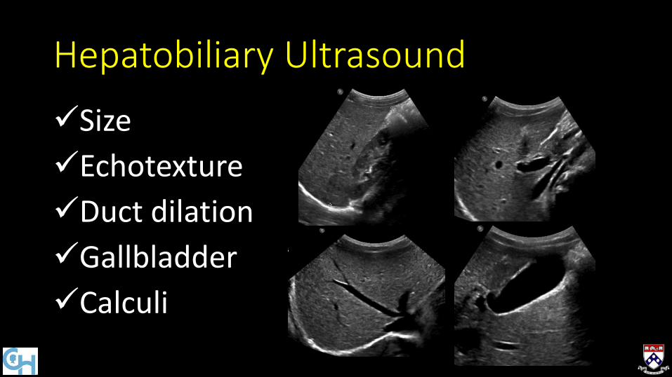
Hepatobiliary Ultrasound
Size
Echotexture
Duct dilation
Gallbladder
Calculi

Term 307 children
5 days to 16 years
Genders equal
Height best correlate
Hepatobiliary: Normal Liver Size

Term 307 children
5 days to 16 years
Genders equal
Height best correlate
Preterm 498 infants
24-36 wk GA
Girls smaller
Weight best correlate
Hepatobiliary: Normal Liver Size
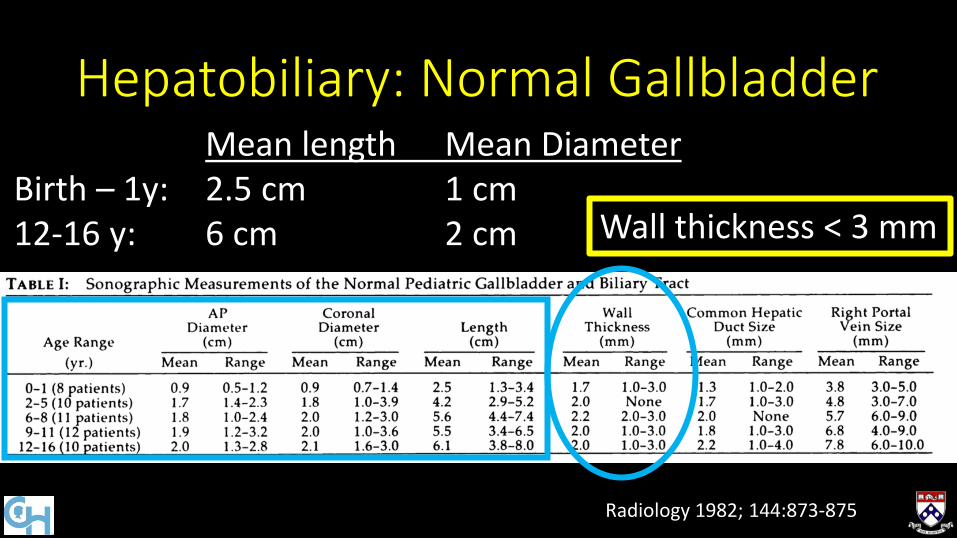
Hepatobiliary: Normal Gallbladder
Radiology 1982; 144:873-875
Mean length Mean Diameter Birth – 1y: 2.5 cm 1 cm 12-16 y: 6 cm 2 cm Wall thickness < 3 mm

Hepatobiliary: Normal CHD
Common hepatic duct little variation with age
Always < 4 mm
Radiology 1982; 144:873-875

Hepatobiliary: Echotexture
Homogeneous
No sound attenuation deep lobe
Portal vein/duct wall interfaces

Hepatobiliary: Echotexture
Homogeneous
No sound attenuation deep lobe
Portal vein/duct wall interfaces
Echogenicity > right kidney

Spleen Ultrasound
Shape: cleft, lobules
Location: wandering
Number: polysplenia or asplenia
Size: splenomegaly or atrophy
Radiographics 1999; 19:1465-1485

Spleen Ultrasound
Lesions Solitary Multiple Diffuse
Radiographics 1999; 19:1465-1485

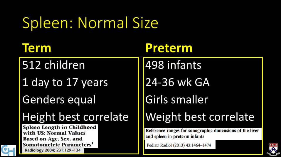
Term 512 children
1 day to 17 years
Genders equal
Height best correlate*
Spleen: Normal Size
*Others weight best AJR 1991; 157:119-121 AJR 1993; 160:1107-1109
Term 512 children
1 day to 17 years
Genders equal
Height best correlate
Preterm 498 infants
24-36 wk GA
Girls smaller
Weight best correlate
Spleen: Normal Size
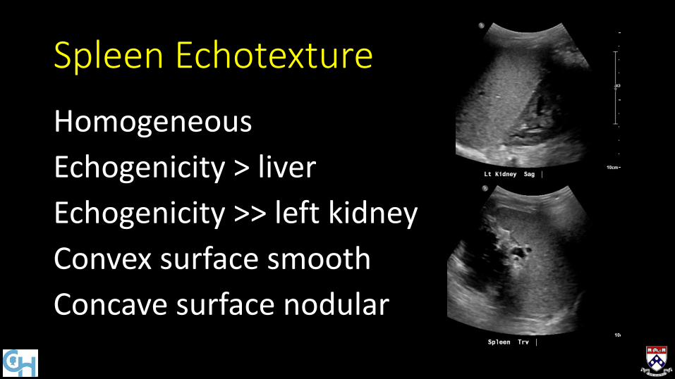
Spleen Echotexture
Homogeneous
Echogenicity > liver
Echogenicity >> left kidney
Convex surface smooth
Concave surface nodular
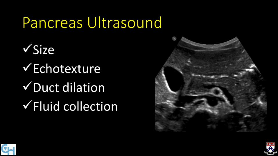
Pancreas Ultrasound
Size
Echotexture
Duct dilation
Fluid collection

Pancreas US Technique
• Sonographic window
• Left hepatic lobe
• Left kidney or spleen
• Stomach with water ingestion
Ultrasound Clin 2013;8:299-321
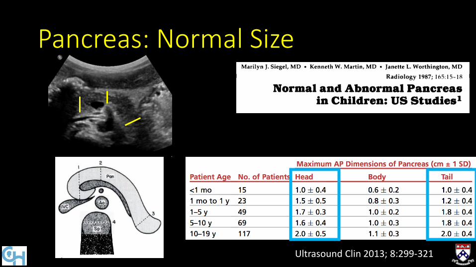
Pancreas: Normal Size
Ultrasound Clin 2013; 8:299-321

Pancreas: Normal Duct Size
Mean 1.65 +/- 0.45 mm
J Ultrasound Med 2000; 19:757-763
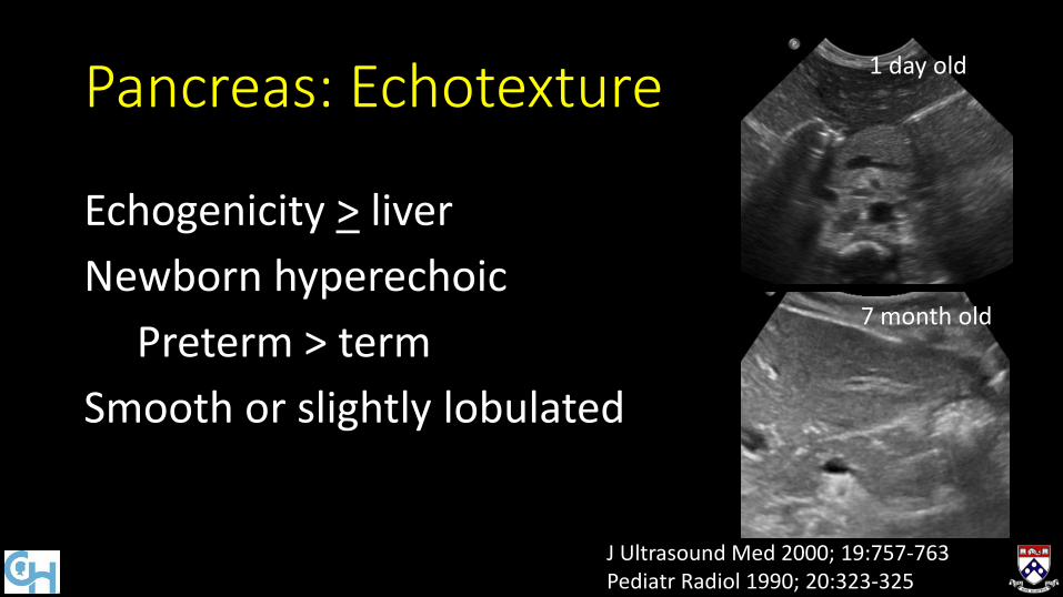
Pancreas: Echotexture
Echogenicity > liver
Newborn hyperechoic
Preterm > term
Smooth or slightly lobulated
J Ultrasound Med 2000; 19:757-763 Pediatr Radiol 1990; 20:323-325
1 day old
7 month old

Objectives
•Normal
•Abnormal: cases and US advances
Inherited and Congenital
• Fibropolycystic Disease
• Biliary Atresia
• Hyperinsulinism
• Cystic Fibrosis
• Sickle cell disease

3 yo girl with progressive abdominal distension, thrombocytopenia
Size: large Echotexture: coarse Duct dilation: variable
10.2 cm (95%)

Size: large Echotexture: coarse Duct dilation: variable
Fibrous enlargement of bile ducts and portal tracts Ducts are present, not paucity
Congenital Hepatic Fibrosis (CHF)
10.2 cm (95%)
Duct Size Fibropolycystic disease
Small CHF, biliary hamartoma
Medium ADPLD
Large Choledochal cyst, Caroli disease

Size: large Echotexture: coarse Duct dilation: variable
Cirrhotic morphology Portal hypertension Splenomegaly
Additional 11.4 cm
Congenital Hepatic Fibrosis (CHF)
10.2 cm (95%)
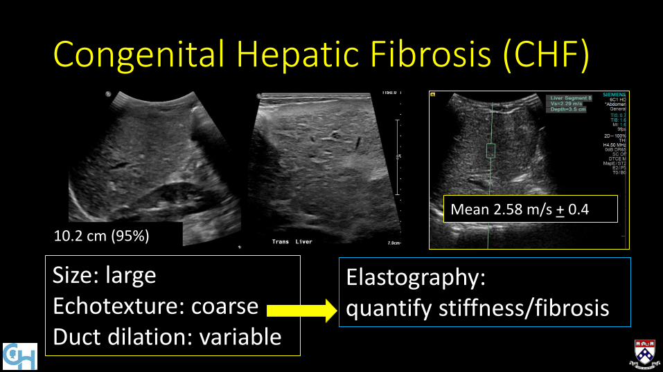
Size: large Echotexture: coarse Duct dilation: variable
Elastography: quantify stiffness/fibrosis
10.2 cm (95%)
Mean 2.58 m/s + 0.4
Congenital Hepatic Fibrosis (CHF)

4 week old girl with conjugated hyperbilirubinemia

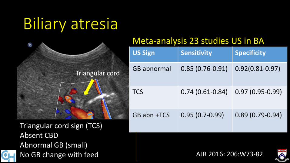
Biliary atresia
Triangular cord sign (TCS) Absent CBD Abnormal GB (small) No GB change with feed
Triangular cord
Gallbladder
Biliary atresia
Triangular cord sign (TCS) Absent CBD Abnormal GB (small) No GB change with feed
Triangular cord
Gallbladder Meta-analysis 23 studies US in BA US Sign Sensitivity Specificity
GB abnormal 0.85 (0.76-0.91) 0.92(0.81-0.97)
TCS 0.74 (0.61-0.84) 0.97 (0.95-0.99)
GB abn +TCS 0.95 (0.7-0.99) 0.89 (0.79-0.94)
AJR 2016: 206:W73-82

Biliary atresia
Images at 1, 4, 6.5 hours no bowel activity, GB not visualized
Triangular cord sign (TCS) Absent CBD Abnormal GB (small) No GB change with feed
Triangular cord

Biliary atresia
Triangular cord sign (TCS) Absent CBD Abnormal GB (small) No GB change with feed
Enlarged hepatic artery (HA) Increased ratio HA to portal vein
Additional
Triangular cord
Gallbladder
Elastography: BA vs other liver disease
Pediatr Radiol (2015) 45:366–375
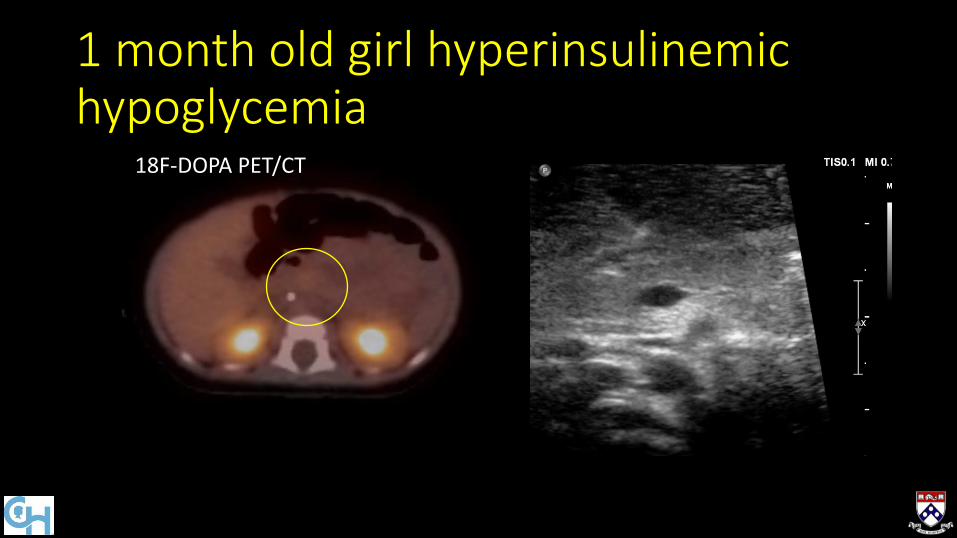
1 month old girl hyperinsulinemic hypoglycemia
18F-DOPA PET/CT

Congenital Hyperinsulinism
18F-DOPA PET/CT
Hyperfunctioning β cells
Focal or diffuse
Unregulated release of insulin
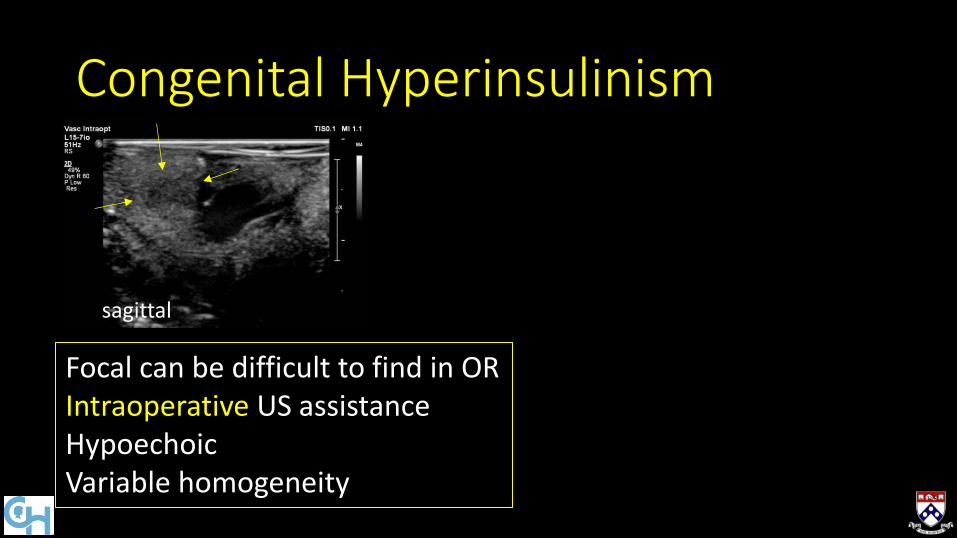
Congenital Hyperinsulinism
Focal can be difficult to find in OR Intraoperative US assistance Hypoechoic Variable homogeneity
sagittal

Congenital Hyperinsulinism
Focal can be difficult to find in OR Intraoperative US assistance Hypoechoic Variable homogeneity
sagittal
0.3 mm duct
50 MHz
15 MHz
4.2 x 4.2 mm

12 yo boy with Cystic Fibrosis

Cystic Fibrosis liver disease
Echotexture: Hyperechoic, homogeneous
Heterogeneous Cirrhotic morphology Portal hypertension
Additional
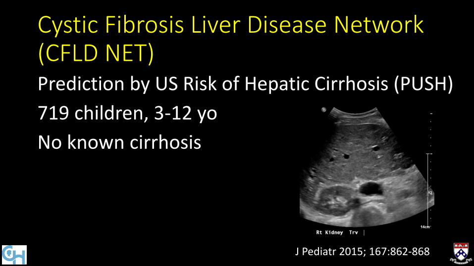
Prediction by US Risk of Hepatic Cirrhosis (PUSH)
719 children, 3-12 yo
No known cirrhosis
Cystic Fibrosis Liver Disease Network (CFLD NET)
J Pediatr 2015; 167:862-868
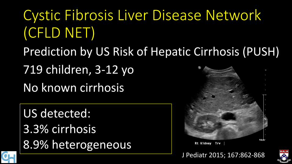
Prediction by US Risk of Hepatic Cirrhosis (PUSH)
719 children, 3-12 yo
No known cirrhosis
Cystic Fibrosis Liver Disease Network (CFLD NET)
J Pediatr 2015; 167:862-868
US detected: 3.3% cirrhosis 8.9% heterogeneous

Focal biliary cirrhosis
ARFI elastography
Cystic Fibrosis liver disease
Radiol med 2012; 117:1408-1418

Cystic Fibrosis- Pancreas
Size: normal or atrophy Echotexture: increased Calcifications, cysts, cystosis

Cystic Fibrosis- Pancreas
Size: normal or atrophy Echotexture: increased Calcifications, cysts, cystosis
ARFI Elastography Softer in pancreatic insufficiency Insufficiency 0.88 m/s + 0.66 No insufficiency 1.07 m/s + 0.31 Normal 1.22 m/s + 0.32*
J Cystic Fibrosis 2013;12:431-439 *Eur J Radiol 2011;80:e226–30

8 yo boy Hb SS and abdominal pain

8 yo boy Hb SS and abdominal pain
Number: present, one Size: small Echotexture: increased Lesions: multiple
Previously autosplenectomy by 5y
J Pediatr 2012 160:281-285 J Ultrasound Med 2016; 35:1735–1745
Transfusion & Hydroxyurea Splenomegaly Echogenic parenchyma Nodules Hemosiderosis
Regenerative nodules Extramedullary hematopoiesis

8 yo boy Hb SS and abdominal pain
Number: present, one Size: small Echotexture: increased Lesions: multiple
Absent Heterogeneous echotexture Calcification Abscess
Additional
Summary
•Normal
•Abnormal: cases and US advances


