Pediatric Major Trauma: An Approach to Evaluation and Management Jahn T. Avarello, MD, FAAP a, * , Richard M. Cantor, MD, FAAP, FACEP a,b a Department of Emergency Medicine, SUNY Upstate Medical University, 750 East Adams Street, Syracuse, NY 13210, USA b Central New York Poison Center, 750 East Adams Street, Syracuse, NY 13210, USA More than 45% of all deaths in children from 1 to 14 years are the result of trauma. Over 5000 traumatic deaths per year occur within this age group; 80% of these mortalities were unintentional and 47% directly related to motor vehicle collisions (MVCs). Injury accounts for approximately 5% of infant deaths as well [1,2]. Nationwide estimates of mortality for children hospitalized after injury are uniformly low; however, most fatalities occur in the field before arrival at a health care facility. This contributes to an un- derestimation of the magnitude of overall mortality figures. The most common single organ system injury associated with death in injured children is head trauma [3,4]. Rates of 80% have been reported in patients with combined thoracoabdominal injuries [1,5]. Because multiple injury is common in children, the emergency physician (EP) must evaluate all organ systems in any injured child, regardless of the actual mechanism of injury. Within the subset of MVC, death rates begin to climb steeply in children 13 years of age and beyond. MVC mortality statistics demonstrate that the youngest occupant in the vehicle is the most vulnerable to injury. Within the school-age group of 5 to 9 years old, pedestrian injuries and bicycle crashes predominate. Submersion injury accounts for 10% to 15% of injury, burns 5% to 10%, and falls from heights approximately 2% [5–7]. Nationwide, the number of children who are victims of violent acts has decreased by 39% from 1994 to 2004. Even with this significant decline, 13% of all traumatic deaths in the age group of children 1 to 14 years old were a result of * Corresponding author. E-mail address: [email protected](J.T. Avarello). 0733-8627/07/$ - see front matter Ó 2007 Elsevier Inc. All rights reserved. doi:10.1016/j.emc.2007.06.013 emed.theclinics.com Emerg Med Clin N Am 25 (2007) 803–836
Transcript
Emerg Med Clin N Am 25 (2007) 803–836
Pediatric Major Trauma: An Approachto Evaluation and Management
Jahn T. Avarello, MD, FAAPa,*,Richard M. Cantor, MD, FAAP, FACEPa,b
aDepartment of Emergency Medicine, SUNY Upstate Medical University,
750 East Adams Street, Syracuse, NY 13210, USAbCentral New York Poison Center, 750 East Adams Street, Syracuse, NY 13210, USA
More than 45% of all deaths in children from 1 to 14 years are the resultof trauma. Over 5000 traumatic deaths per year occur within this age group;80% of these mortalities were unintentional and 47% directly related tomotor vehicle collisions (MVCs). Injury accounts for approximately 5%of infant deaths as well [1,2]. Nationwide estimates of mortality for childrenhospitalized after injury are uniformly low; however, most fatalities occur inthe field before arrival at a health care facility. This contributes to an un-derestimation of the magnitude of overall mortality figures.
The most common single organ system injury associated with death ininjured children is head trauma [3,4]. Rates of 80% have been reported inpatients with combined thoracoabdominal injuries [1,5]. Because multipleinjury is common in children, the emergency physician (EP) must evaluateall organ systems in any injured child, regardless of the actual mechanismof injury.
Within the subset of MVC, death rates begin to climb steeply in children13 years of age and beyond. MVC mortality statistics demonstrate that theyoungest occupant in the vehicle is the most vulnerable to injury. Within theschool-age group of 5 to 9 years old, pedestrian injuries and bicycle crashespredominate. Submersion injury accounts for 10% to 15% of injury, burns5% to 10%, and falls from heights approximately 2% [5–7]. Nationwide, thenumber of children who are victims of violent acts has decreased by 39%from 1994 to 2004. Even with this significant decline, 13% of all traumaticdeaths in the age group of children 1 to 14 years old were a result of
homicide in 2004 [2,8]. The leading causes of traumatic death in the UnitedStates are listed in Table 1.
Principles of disease
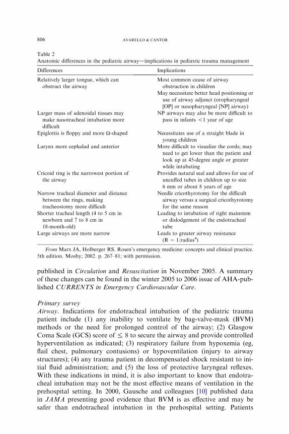
There are major anatomic, physiologic, and psychological differences inthe pediatric and adult patients that play a significant role in the evaluationand management of the pediatric trauma patient. A summary of overall an-atomic differences and their implications in the pediatric trauma patient arelisted in Box 1. Anatomic differences in the pediatric and adult airway andtheir implication in the pediatric trauma patient are listed in Table 2.
Compared with adults, in children, any given force is more widely distrib-uted though the body, making multiple injuries significantly more likely tooccur. The proportionately large surface area of an infant or child relativeto weight predisposes them to greater amounts of heat loss as a result ofevaporation. Maintenance requirements for free water, trace metals, andminerals are therefore magnified to a greater degree. All these factors con-tribute to a significantly higher energy and caloric requirement for theinjured child when compared with an injured adult. Physiologically, childrenrespond to injury quite differently than adults, depending on the age andmaturation of the child and the severity of the injury. Unlike adults, childrenhave a great capacity to maintain blood pressure despite significant acuteblood losses (25% to 30%). Subtle changes in heart rate, blood pressure,and extremity perfusion may indicate impending cardiorespiratory failureand should not be overlooked. Finally, children may not cope well outsideof their usual environment. They are often disproportionately irritablerelative to their degree of injury, making assessment all the more difficult.Recent evidence suggests that 25% of children involved in road traffic acci-dents will show signs of posttraumatic stress disorder after discharge [9].They need to be approached in a calming and sometimes unconventionalmanner so as to lessen their anxiety.
Table 1
Leading causes of traumatic deaths in children 1 to 14 years of age in the United Statesd2004
Etiology No. %
Motor Vehicle Accidents 2,026 38.2
Homicide 706 13.3
Drowninga 699 13.2
Fire/Burna 484 9.1
Suicide 285 5.4
Suffocationa 238 4.5
Other 860 16.3
a Unintentional.
805PEDIATRIC MAJOR TRAUMA
Clinical features
Initial assessment priorities/primary survey
The highest priority in the approach to the injured child is ruling out thepresence of life- or limb-threatening injury. Treatment of these injuries mustoccur before proceeding with the rest of the physical examination. This initialassessment (the primary survey) and necessary initial resuscitation effortsmust occur simultaneously. In general, they should be addressed within thefirst 5 to 10 minutes of evaluation.
Any infant or child with a potentially serious or unstable injury requirescontinual reassessment. Vital signs should be repeated every 5 minutes dur-ing the primary survey and every 15 minutes thereafter until the patient isconsidered stable.
The International Liaison Committee on Resuscitation (ILCOR) pediat-ric task force is a multinational team of expert reviewers that was assignedto review resuscitation science and develop an evidence-based consensus toguide resuscitation practices. In January of 2005, the ILCOR presented theevidence at the International Consensus Conference on CardiopulmonaryResuscitation and Emergency Cardiovascular Care Science With TreatmentRecommendations, hosted by the American Heart Association (AHA). Bythe end of the conference some major changes to the Pediatric AdvancedLife Support (PALS) guidelines were established and simultaneously
Box 1. Anatomic difference in adults and childrendimplicationsfor pediatric trauma management
� The child’s body size allows for a greater distribution oftraumatic injuries, therefore multiple trauma is common.
� The child’s greater relative body surface area also causesgreater heat loss.
� The child’s internal organs are more susceptible to injurybased on more anterior placement of liver and spleen and lessprotective musculature and subcutaneous tissue mass.
� The child’s kidney is less well protected and more mobile,making it very susceptible to deceleration injury.
� The child’s growth plates are not yet closed, leading toSalter-type fractures with possible limb-length abnormalitieswith healing.
� The child’s head-to-body ratio is greater, the brain lessmyelinated, and cranial bones thinner, resulting in moreserious head injury.
From Marx JA, Holberger RS. Rosen’s emergency medicine: concepts and clin-ical practice. 5th edition. Mosby; 2002. p. 267–81; with permission.
806 AVARELLO & CANTOR
published in Circulation and Resuscitation in November 2005. A summaryof these changes can be found in the winter 2005 to 2006 issue of AHA-pub-lished CURRENTS in Emergency Cardiovascular Care.
Primary surveyAirway. Indications for endotracheal intubation of the pediatric traumapatient include (1) any inability to ventilate by bag-valve-mask (BVM)methods or the need for prolonged control of the airway; (2) GlasgowComa Scale (GCS) score of % 8 to secure the airway and provide controlledhyperventilation as indicated; (3) respiratory failure from hypoxemia (eg,flail chest, pulmonary contusions) or hypoventilation (injury to airwaystructures); (4) any trauma patient in decompensated shock resistant to ini-tial fluid administration; and (5) the loss of protective laryngeal reflexes.With these indications in mind, it is also important to know that endotra-cheal intubation may not be the most effective means of ventilation in theprehospital setting. In 2000, Gausche and colleagues [10] published datain JAMA presenting good evidence that BVM is as effective and may besafer than endotracheal intubation in the prehospital setting. Patients
Table 2
Anatomic differences in the pediatric airwaydimplications in pediatric trauma management
Differences Implications
Relatively larger tongue, which can
obstruct the airway
Most common cause of airway
obstruction in children
May necessitate better head positioning or
use of airway adjunct (oropharyngeal
[OP] or nasopharyngeal [NP] airway)
Larger mass of adenoidal tissues may
make nasotracheal intubation more
difficult
NP airways may also be more difficult to
pass in infants !1 year of age
Epiglottis is floppy and more U-shaped Necessitates use of a straight blade in
young children
Larynx more cephalad and anterior More difficult to visualize the cords; may
need to get lower than the patient and
look up at 45-degree angle or greater
while intubating
Cricoid ring is the narrowest portion of
the airway
Provides natural seal and allows for use of
uncuffed tubes in children up to size
6 mm or about 8 years of age
Narrow tracheal diameter and distance
between the rings, making
tracheostomy more difficult
Needle cricothyrotomy for the difficult
airway versus a surgical cricothyrotomy
for the same reason
Shorter tracheal length (4 to 5 cm in
newborn and 7 to 8 cm in
18-month-old)
Leading to intubation of right mainstem
or dislodgement of the endotracheal
tube
Large airways are more narrow Leads to greater airway resistance
(R ¼ 1/radius4)
From Marx JA, Holberger RS. Rosen’s emergency medicine: concepts and clinical practice.
5th edition. Mosby; 2002. p. 267–81; with permission.
807PEDIATRIC MAJOR TRAUMA
were randomized to receive either BVM or endotracheal intubation. Of the830 pediatric patients enrolled, there was a 4% better survival rate and a 3%better neurologic outcome rate in the BVM group with even more signifi-cance in the subgroups of respiratory arrest and child maltreatment. TheAHA included these findings in their PALS updates stating to ‘‘ventilateand oxygenate infants and children with a bag-mask devise, especially iftransport time is short’’ [10].
Intubation of the pediatric patient involves special considerations. Cuffedendotracheal tubes have recently been considered as safe as uncuffed tubesfor infants beyond the newborn period and in children when in the hospitalsetting. The 2005 update by the AHA states ‘‘In certain circumstances (eg,poor lung compliance, high airway resistance, or a large glottic air leak)a cuffed tube may be preferable provided that attention is paid to endotra-cheal tube size, position, and cuff inflation pressure’’ [11]. On the contrary,in patients younger than 8 years who do not fit those circumstances, uncuf-fed tubes should be used, as the narrowest portion of the pediatric airway inthis age group is at the level of the cricoid cartilage (Table 2). For children1 to 10 years of age, the following formulas may be used for estimation ofproper endotracheal tube size (ID, internal diameter):
� Uncuffed endotracheal tube size (mm ID) ¼ (age in years þ 16)/4� Cuffed endotracheal tube size (mm ID) ¼ (age in years þ 12)/4
In general, the orotracheal approach is recommended. Problems associ-ated with nasotracheal intubation include inherent difficulties in children,impairment of tube passage by the acute angle of the posterior pharynx,and the probability of causing or worsening bleeding within the oral cavityand causing increases in intracranial pressure (ICP) with insertion.
Breathing/ventilation. Assess for adequacy of chest rise. In a young childthis will occur in the lower chest and upper abdomen. Also assess respira-tory rate. Rates that are too fast or slow can indicate impending respira-tory failure. Treatment is assisted ventilation. If ventilation is necessary,a BVM device is recommended initially. Be careful to provide only the vol-ume necessary to cause the chest to rise, because excessive volume or rateof ventilation can increase the likelihood of gastric distention and furtherimpair ventilation. Once an advanced airway is placed, ventilations shouldbe given at a rate of 1 breath every 6 to 8 seconds (8 to 10 breaths perminute). Cricoid pressure may be useful to decrease the amount of airentering the esophagus during positive-pressure ventilation. With regardto assessment of ventilation, early monitoring with pulse oximetry isvery useful; however, pulse oximetry will measure adequacy of oxygena-tion only. There are many limiting factors that compromise ventilatoryfunction in the injured child, including depressed sensorium, occlusion ofthe airway itself, painful restriction of lung expansion, and direct pulmo-nary injury. Determination of adequate ventilation is possible only in
808 AVARELLO & CANTOR
the face of airway patency and adequate air exchange. The diaphragmplays a special role in the maintenance of proper ventilatory status in chil-dren. It is easily fatigued in the young child and is often displaced by anyprocess that promotes distention of the stomach. In this regard, it is advis-able to consider early placement of a nasogastric tube to facilitate decom-pression of the stomach.
Circulation and hemorrhage control. Assessment of circulation in a child in-volves a combination of factors, namely the pulse, skin color, and capillaryrefill time. In a child, maintenance of systolic blood pressure does not ensurethat the patient is not in shock, a direct effect of the ability of the pediatricvasculature to constrict and increase systemic vascular resistance in anattempt to maintain perfusion. Therefore skin signs such as cool distal ex-tremities, decreases in peripheral versus central pulse quality, and delayedcapillary refill time are signs of pediatric shock, even when blood pressureis maintained. The lowest acceptable blood pressures for age (5th percentile)are as follows [11]:
� 60 mm Hg in term neonates (0 to 28 days)� 70 mm Hg in infants (1 month to 12 months)� 70 mm Hg þ (2 � age in years) in children 1 to 10 years of age� 90 mm HG in children R 10 years of age
In general, a palpable peripheral pulse correlates with a systolic bloodpressure greater than 80 mm Hg and a palpable central pulse with a pressuregreater than 50 to 60 mm Hg [12]. Normal capillary refill times are less than2 seconds. Alteration in a child’s response to the environment or interactionwith caregivers may also indicate respiratory failure or shock. External hem-orrhage should be sought for and controlled with direct pressure.
All pediatric patients involved in major trauma should be placed on a car-diac monitor, receive supplemental oxygen, and have constant reassessmentof vital signs and oximetry. Vascular access is best obtained by accessing theupper extremity for the establishment of two large-bore intravenous (IV)lines. In the absence of available upper extremity peripheral sites, lowerextremity sites could be used, and many clinicians favor the femoral veinas a safe site for insertion of a central line, by use of a guide wire technique.If cutdowns are necessary, the antecubital or saphenous sites will suffice.If access is needed emergently or if there is no success in the first 5 minutesof an urgent scenario, intraosseous access may be obtained at the proximalmedial tibia 1 cm below and medial to the tibial tuberosity, the distal tibia1 to 2 cm proximal to the medial malleolus, or the lower third of the femurat the midline 3 cm above the lateral condyle (Fig. 1) [13]. The intraosseousroute serves as an appropriate venous access site; however, delivery rate oflarge amounts of crystalloid solutions is limited based on maximum flowrates of approximately 25 mL/min [14–16]. Intraosseous placement in a frac-tured extremity is contraindicated.
809PEDIATRIC MAJOR TRAUMA
Most hypovolemic pediatric trauma patients respond to infusions of 20mL/kg of isotonic crystalloid solutions. If 40 mL/kg has already been givenand a third bolus is still required to reverse systemic signs of hypoperfusion,an infusion of packed blood cells at 10 mL/kg should be considered [17]. Inpatients who present in decompensated shock or cardiopulmonary failure,and occult bleeding is a potential cause for the shock, administer crystalloidand blood products simultaneously.
In contrast to the adult, cardiogenic shock is a rare event in the face ofchildhood injury [3,18]. However, any degree of chest trauma associatedwith the presence of shock must alert the clinician to the possibility of con-comitant myocardial contusion or rupture. The classic presentation of neu-rogenic shock, involving hypotension without an increase in heart rate orcompensatory vasoconstriction, should be considered in patients withhead or neck injuries.
With regard to cardiopulmonary resuscitation (CPR) in the infant andchild, the guidelines have been recently updated. The chest compression-ventilation ratio for one lay rescuer and one lone health care provider isnow 30:2, and 15:2 for health care providers performing two-rescuerCPR. Although these recommendations come mostly from theoreticaldata and rational conjecture, the thought is that, besides less fatigue ofthe rescuer(s), more interruptions in chest compressions will prolong theduration of low coronary perfusion pressures reducing the likelihood of areturn of spontaneous circulation. High-dose epinephrine has been removedfrom the recommendation with the exceptions being if it is administered bythe endotracheal route or in exceptional circumstances such as b-blockeroverdose.
Fig. 1. (A–C) Preferred locations for intraosseous needle placement. (Reprinted from
Fleisher GR, Ludwig S, Henretig FM. Textbook of pediatric emergency medicine. 5th edition.
Philadelphia: Lippincott Williams and Wilkins; 2006. p. 1879; with permission.)
810 AVARELLO & CANTOR
Disability assessment (thorough neurologic examination). To assess patientdisability, there is need for a rapid neurologic evaluation. The GCS (Boxes2 and 3) and the AVPU System (Box 4) are used for a rapid neurologicalevaluation. It is important to rule out hypoglycemia in any patient withaltered mental status. This is particularly important in younger childrensince their glycogen stores are easily depleted, predisposing them tohypoglycemia.
Exposure. The final component of the primary survey involves fully undress-ing the patient to assess for hidden injury.Maintenance of normothermia is ofparamount importance in the toddler and infant during the exposure phasebecause metabolic needs are greatly increased by hypothermia.
Family. In the management of children, the family could be added to theprimary survey. Rapidly informing the family of what has happened andthe evaluation that is proceeding will help lessen the stress of the caregivers.Allowing family presence during resuscitations is acceptable and often pre-ferred by families. Some caregivers will choose not to be present, but thatchoice should be given to them. If a caregiver is present, it is advisable toassign a staff member to be with them during the trauma resuscitation toexplain the process [19].
Box 2. Glasgow Coma Scale
Eye opening response (1–4 points)4. Spontaneous3. To verbal stimuli2. To painful stimuli1. None
Motor response (1–6 points)6. Normal spontaneous movements5. Localizes painful stimuli4. Withdraws from painful stimuli3. Abnormal flexion (decorticate rigidity)2. Abnormal extension (decerebrate rigidity)1. None
811PEDIATRIC MAJOR TRAUMA
Secondary surveyThe secondary survey is designed to assess the patient and treat addi-
tional injury not found on the primary survey and also to obtain a morecomplete and detailed history. Features of the detailed history that needto be obtained can be remembered by the mnemonic AMPLE (Box 5). Dur-ing the secondary survey the EP should attend to the tasks outlined in Box 6.
Specifics of the head examination include pupillary size and reactivity,funduscopic examination, and palpation of the skull itself. Assessment ofthe cervical spine must be done carefully, with the patient in full c-spineimmobilization.
Assessment of the chest and internal structures involves inspection forwounds and flail segments, palpation for tenderness and crepitance, and
Box 4. The AVPU system
A = AlertV = Responds to Verbal stimuliP = Responds to Painful stimuliU = Unresponsive
Box 3. Modified GCS
Modified GCS for infantsEye opening response (1–4 points)4. Spontaneous3. To verbal stimuli2. To painful stimuli1. None
Verbal response (1–5 points)5. Coos and/or babbles4. Irritable and continuous crying3. Cries to painful stimuli2. Moans to painful stimuli1. None
Motor response (1–6 points)6. Spontaneous purposeful movements5. Withdraws to touch4. Withdraws to painful stimuli3. Abnormal flexion (decorticate rigidity)2. Abnormal extension (decerebrate rigidity)1. None
812 AVARELLO & CANTOR
auscultation for asymmetry or poorly transmitted breath sounds or cardiacimpulses.
Examination of the pediatric abdomen is most reliable when performedon a cooperative patient and should be considered an insensitive screeningprocess for the presence of an injury when the patient has an associatedhead injury or a GCS % 13 [20].
A rectal examination provides information concerning sphincter tone,prostatic position, and the presence of blood in the stool. Although urethralinjury is rare in children, all trauma patients should be assessed for a perinealor lower abdominal hematoma and blood from the urethral meatus. Exam-ination of the extremities is directed toward the evaluation of any defor-mities, penetrations, and interruptions of perfusion. Most fracture sitesmay be stabilized with splinting until surgical intervention can be per-formed. Early orthopedic consultation is advisable.
As mentioned previously, the pediatric patient is at great risk for the de-velopment of hypothermia. This is based on the large amount of surfacearea relative to body weight. Careful attention to core temperatures in thesevulnerable patients is required, with early intervention as needed, with sup-plemental external warming techniques.
An overview of recommended interventions for the multiply traumatizedchild is contained in Tables 3–8.
Box 5. The AMPLE history
A, AllergiesM, MedicationsP, Past medical historyL, Last mealE, Environments and events
Box 6. Tasks to be completed after the secondary survey
� Complete head-to-toe examination� Appropriate tetanus immunization� Antibiotics as indicated� Continued monitoring of vital signs� Ensure urine output of 1 mL/kg/hr
From Marx JA, Holberger RS. Rosen’s emergency medicine: concepts and clin-ical practice. 5th edition. Mosby; 2002. p. 267–81; with permission.
813PEDIATRIC MAJOR TRAUMA
LaboratoryBlood sampling for the pediatric trauma patient is no different from that
of the adult trauma patient; however, use of smaller blood collection tubesand microtechnique by laboratory staff may be necessary in infants andsmall children. All older children and adolescent trauma patients shouldbe assessed for the possible use of drugs or alcohol as contributing factorsto the traumatic event. In patients with hypovolemic shock, the hemoglobinalone is not sensitive because equilibration may not have occurred upon pre-sentation to the emergency department (ED) [21,22].
RadiologyThe most important ‘‘traditional’’ radiographs to obtain on a moderately
to severely injured child are of the chest and pelvis to assess for sites ofblood loss or potential causes of shock. In stable, alert children without
Table 4
Breathing: assessment and treatment
Assessment priorities Interventions
Respiratory rate Oxygen 100% by nonrebreather mask or intubate if in
respiratory failure; fast rates may indicate shock (fluid
resuscitation) or pain (parenteral analgesics)
Chest wall movements For pneumo- or hemothorax: place chest tube
Transfer to operating room if initial drainage O20 mL/kg or
output O2 mL/kg/hr
Percussion note Open pneumothorax: seal with occlusive dressing (vaseline
gauze) followed by tube thoracostomy
Paradoxical breathing Contusion/flail chest: intubate if tachypneic or Po2 ! 50 mm or
Pco2 O 50 mm Hg
Tracheal deviation Tension pneumothorax: needle decompression at second
intercostal space, midclavicular line, followed by placement
of chest tube
Flail segments Oxygen by nonrebreather mask or intubate if in respiratory
failure
Open wounds Compress bleeding sites and cover as indicated
From Marx JA, Holberger RS. Rosen’s emergency medicine: concepts and clinical practice.
5th edition. Mosby; 2002. p. 267–81; with permission.
Level of consciousness Cervical spine immobilization
Maxillofacial injury Apply 100% O2 by mask
Stridor or cyanosis Intubate for:
Glasgow Coma Scale % 8 or absent
gag reflex or Po2 ! 50 or Pco2 O 50
Needle cricothyrotomy if intubation
impossible
814 AVARELLO & CANTOR
distracting injuries, the pelvic film may be eliminated. The radiographs ofthe cervical spine may be delayed until after further diagnostic studies areobtained depending on the clinical presentation of the patient.
The diagnostic test of choice to assess intra-abdominal injury in stabletrauma patients is rapid abdominal CT scanning [23]. The role of diagnosticperitoneal lavage (DPL) and Focused Abdominal Sonography for Trauma(FAST) is somewhat more limited although the FAST exam is gaining moreacceptance [24,25]. As with all of these tests, the finding of intraperitonealhemorrhage alone is not an indication for surgery in the pediatric patient(Box 7).
Other radiographs will be obtained based on the physical examination.For patients sustaining minor trauma, radiographs may not be needed. Fi-nally, children younger than 2 years of age with injuries consistent with childabuse will need a skeletal survey including skull, chest, abdomen, and long-bone radiographs.
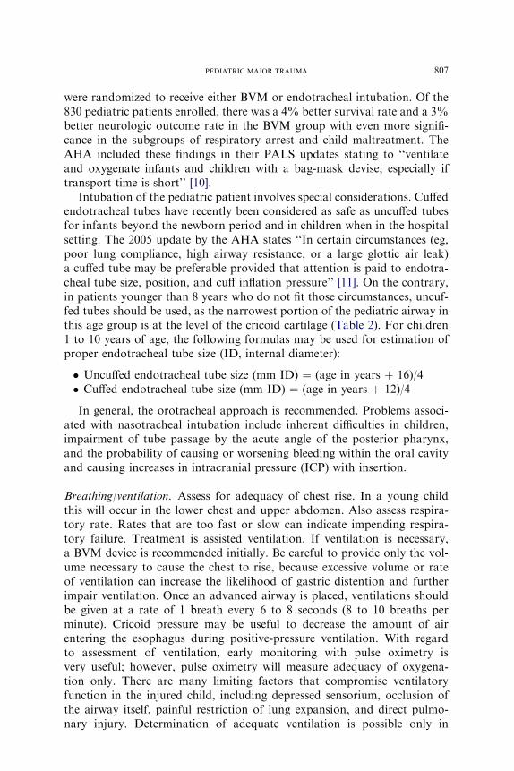
Table 6
Disability: assessment and treatment
Assessment priorities Interventions
Level of consciousness Maintain blood pressure and oxygenation and
ventilation
AVPU system or GCS If head injury with GCS % 8: RSI and intubate; head
CT, neurosurgical consult
If normotensive, consider mannitol 0.25–1 g/kg
Pupil size and reactivity Hyperventilate Pco2 to 30–35 mm Hg with signs of
herniation
Extremity movement and tone Stabilize spinal column
If blunt cord trauma: methylprednisolone (See
Table 9 for dosing recommendations)
Posturing Hyperventilate Pco2 to 30–35 mm Hg
Reflexes Assess for signs of respiratory failure
Abbreviation: RSI, rapid sequence intubation.
Table 5
Circulation: assessment and treatment
Assessment priorities Interventions
Open wounds Compress bleeding sites and cover as indicated
Capillary refill Oximeter and cardiac monitor, oxygen and fluid resuscitation
20 mL/kg
Heart rate Monitor vital signs every 5 minutes
Peripheral pulses Two large-bore intravenous sites
Sensorium Bolus with 20 mL/kg lactated Ringer’s or normal saline
solution (warm all intravenous fluids)
Pulse pressure Repeat fluid bolus 2 times if necessary
Skin condition/perfusion Packed red blood cells, 10–20 mL/kg for decompensated shock
secondary to blood loss
815PEDIATRIC MAJOR TRAUMA
Specific disorders/injuries
Head injury
PerspectiveHead trauma is the leading cause of death among injured children and is
responsible for 80% of all trauma deaths [3]. Each year, 29,000 childrenyounger than 19 years old suffer permanent disability from traumatic braininjury. In children younger than 14 years old, there are 475,000 hospitalvisits, 37,000 hospitalizations, and nearly 3000 deaths each year related totraumatic brain injury [4,26].
Falls account for 39%of pediatric head injuries, andMVCs are responsiblefor 11% [4,27,28]. In addition, pedestrian injuries account for 17% and fallsfrom bicycles 10%. On an age-related basis, infants and toddlers are moreprone to falls from their own height, school-age children are involved in sportsinjuries and MVCs, and all ages are subject to the sequelae of abuse [26].
Clinical featuresIn most cases it is important to establish whether there was loss of con-
sciousness at the time of the injury event. With playground trauma, the his-tory may be vague and the interpretation of any change in consciousness ofthe child may be regarded as an actual loss of consciousness. The behaviorof the child after the event should include questions related to the presenceor absence of irritability, lethargy, abnormal gait, or alterations in behavior.Most importantly, establishment of a timeline from the point of insult ishelpful in determining whether there have been changes in the mental statusof the patient. A caretaker present to establish the child’s baseline behavior,mental status, and neurologic function is very useful.
The prognostic significance of vomiting after pediatric head trauma is un-certain [26,29]. There is no adequate study defining an acceptable time framein which vomiting after head injury is benign in nature. The development ofseizures after head trauma, in contrast to vomiting, has been well studied[30]. A brief seizure that occurs immediately after the insult (with rapid re-turn of normal level of consciousness) is commonly called ‘‘an impact sei-zure’’ and is usually unassociated with intracranial parenchymal injury
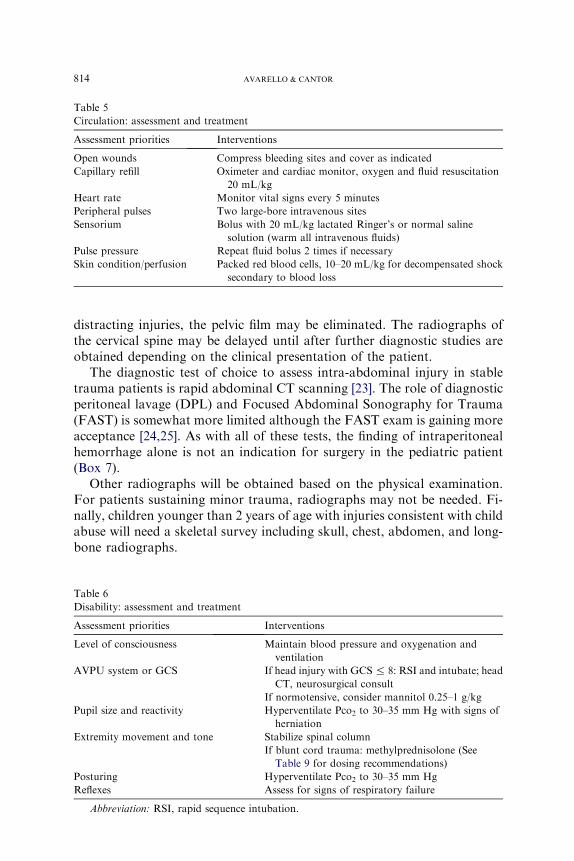
Table 7
Exposure: assessment and treatment
Assessment priorities Interventions
Undress
Look under collar and splints
Log roll and examine back
Trauma examination including rectal examination
Keep patient warm:
� Remove wet clothes
� Place under heater
� External warming devices
� Warm blanket
� Warm fluids
816 AVARELLO & CANTOR
and in no way mandates the institution of anticonvulsant therapy. Seizuresthat occur later (longer than 20 minutes after the insult) portend the greaterpossibility of both internal injury and the development of seizures at a laterdate. Patients who experience seizures later in the course of the posttrau-matic event are best evaluated by the neurosurgical service. As in all in-stances of trauma, a careful history related to the possibility of substanceabuse must be obtained.
Table 8
Emergent management of increased intracranial pressure
Therapy Dose Mechanism of action
Head evaluation
(30 degrees)
Lowers intracranial venous
pressure
Head in midline Prevents jugular vein
compression
Hyperventilation Reduces Paco2 to
30–35 mm Hg
Promptly decreases cerebral
blood volume and thus
intracranial pressure
Caution: too much
ventilation can Y cerebral
blood flow
Lidocaine 1–2 mg/kg IV Y airway reflex’s that may [intracranial pressure
Mannitol 0.25–1 g/kg IV Yblood viscosity /improved cerebral blood
flow
Contraindicated in
hemodynamically
unstable patients
Thiopental 3–5 mg/kg IV Decreases cerebral
metabolism reducing
cerebral ischemic risk
Caution with hypotensive
patients
Etomidate 0.3 mg/kg IV Decreases cerebral
metabolism reducing
cerebral ischemic risk
Emergent burr hole: Only in
the rapidly deteriorating
patient when all medical
interventions have failed,
a neurosurgeon is
unavailable, and the
patient will not likely
survive transport
Temporary decompression
to ameliorate herniation
Hypothermia (27�C–31�C) Thought to decrease
cerebral blood flow and
metabolic rate; can cause
cardiac dysrhythmias
From Marx JA, Holberger RS. Rosen’s emergency medicine: concepts and clinical practice.
5th edition. Mosby; 2002. p. 267–81; with permission.
817PEDIATRIC MAJOR TRAUMA
There are several methods for evaluating head-injured patients. Theseinclude AVPU and the GCS. The GCS has been modified for pediatricpatients as illustrated in Box 3.
The GCS more reliably predicts outcome of traumatic brain injury inchildren than in adults. In a study involving 80 children with traumatic braininjuries admitted to an intensive care unit (ICU), initial GCS scores werecompared with eventual outcome. Both ICU lengths of stay and time to cog-nition relative to GCS scores indicated that scores greater than or equal to 6were associated with favorable outcomes and neurologic status. While thenumbers in this study are small, the important message is that no matter
Table 9
Corticosteroid treatment recommendations in acute spinal trauma
Indications Acute spinal cord injury presenting within 8 hours of injury
(6) Receiving maintenance steroids for other reasons
(7) !13 years of age (relative contraindication)
Treatment Methylprednisolone 30 mg/kg IV over 15 minutes then a 45-minute
pause, then
(1) 5.4 mg/kg/hr IV over 23 hrs if !3 hrs since injury
OR
(2) 5.4 mg/kg/hr IV over 47 hrs if 3–8 hrs since injury
Data from Refs. [17,65,81].
Box 7. Potential uses for the FAST examination
� Rapidly identify the source of hypotension in thehemodynamically unstable patient
� Assist in decision making in the child with head and abdominaltrauma (head and abdominal CT versus laparotomy and/orcraniotomy)
� Evaluate the stable and alert trauma patient without finding onphysical exam who would not routinely undergo radiologyimaging
� Help prioritize imaging studies in the multiple-trauma patient� Help avoid additional imaging in a child with an already low
likelihood of intra-abdominal injury
From Marx JA, Holberger RS. Rosen’s emergency medicine: concepts and clin-ical practice. 5th edition. Mosby; 2002. p. 267–81; with permission.
818 AVARELLO & CANTOR
how the patient presents neurologically, all efforts should be generated toensure survival and maintain stable neurologic status within the ED [31].
Examining the brain-injured child involves cranial nerve testing andmotor and sensory testing. The evaluation of cranial nerve function is essen-tially no different from that of the adult. The most important aspect of bothmotor and cranial nerve evaluation involves ruling out the presence of in-creased ICP. Common symptoms and signs of increased ICP in infantsand children are described as follows:
Infants� Full fontanel� Split sutures� Altered states of consciousness� Paradoxical irritability� Persistent emesis� The ‘‘sun-setting’’ sign (inability to open eyes fully)Children� Headache� Stiff neck� Photophobia� Altered states of consciousness� Persistent emesis� Cranial nerve involvement� Papilledema� Cushing’s triad (Hypertension, bradycardia and irregular respirations)� Decorticate or decerebrate posturing
Minor injury to the scalp of infants and children involves the developmentof three possible injury complexes: caput succedaneum, injury to the connec-tive tissue itself; subgaleal hematoma, injury to the tissue surrounding theskull; and cephalohematoma, a collection of blood under the periosteum.
Skull fractures in children occur in many different configurations[27,30,32]. Linear fractures, the most common type of skull fracture, rarelyrequire therapy and are often associated with good outcomes. Factors favor-ing a poor outcome include the presence of the fracture overlying a vascularchannel, a diastatic fracture, or a fracture that extends over the area of themiddle meningeal artery. Diastatic fractures, or defects extending throughsuture lines, are unlike linear fractures, in that leptomeningeal cysts (growingfractures) may develop at these sites. Fractures of the basilar portions of theoccipital, temporal, sphenoid, or ethmoid bones commonly occur in children[33]. The presence of cerebrospinal fluid (CSF), rhinorrhea, and otorrheahas been associated with these injuries. Signs of basilar skull fractures in chil-dren are similar to adults and include hemotympanum, seventh and eighthnerve dysfunction, posterior auricular ecchymosis, or Battle’s sign, and rac-coon eyes, or the presence of periorbital subcutaneous bleeding [17,26]. Asa basic rule, serial examinations are the most reliable indicators of clinical
819PEDIATRIC MAJOR TRAUMA
deterioration [26,29,30,32]. The presence of focality is a reliable indicator ofa localized insult, whereas the absence of focality may be misleading. Al-though there may be few or subtle signs and symptoms of intracranial injury,the following independent predictors have been identified [29]:
� Altered mental status� Focal neurologic deficits� Signs of a basilar skull fracture� Seizure� Skull fracture
ConcussionStrictly speaking, concussion is defined as a brain insult with transient
impairment of consciousness. Amnesia is often involved and patients whosuffer from concussive insults frequently demonstrate anorexia, vomiting,or pallor soon after the insult. This transitional period is followed by rapidrecovery to baseline and if a CT scan of the head is obtained, it is most oftennormal. Following the Second International Symposium on Concussion inSport November 2004, concussion was reclassified as simple or complex [34].
� Simple concussions are those where loss of consciousness (if any) was lessthan 1 minute, and symptoms resolve without complication within 7 to10 days. In such cases, apart from limiting playing and other activitieswhile symptomatic, no further intervention is required during the periodof recovery.� Complex concussions include those where there is prolonged loss of con-sciousness, symptoms or cognitive impairment last longer than 7 to 10days, or there is a history of multiple concussions. In these cases, returnto play should be managed by a multidisciplinary team, and formal neu-ropsychological testing (testing of attention, memory, and so forth)should be considered.
Since Saunders andHarbaugh [35] first described it in 1984, second-impactsyndrome has gained much attention and has led to strict ‘‘return-to-play’’guidelines for athletes who have suffered a head injury. Second-impact syn-drome has been defined as an injury in which ‘‘an athlete who has sustainedan initial head injury, most often a concussion, sustains a second head injurybefore symptoms associated with the first have fully cleared’’ [36]. Given thatthis can ultimately lead to death, the return-to-play guidelines require strictadherence. When a player shows ANY symptoms or signs of a concussion:
1. The player should NOT be allowed to return to play in the current gameor practice.
2. The player should not be left alone, and regular monitoring for deteri-oration is essential over the initial few hours following injury.
3. The player should be medically evaluated following the injury.4. Return to play must follow a medically supervised stepwise process.
820 AVARELLO & CANTOR
In contrast to concussions, contusions are often the result of coup and con-trecoup forces atwork. Theymay not be associatedwith any loss of conscious-ness at the timeof insult. Patients often presentwith associated symptoms suchas altered level of consciousness, severe headache, vomiting, or focal deficitson neurologic assessment. These injuries are clearly demonstrable on CT.
Traditional teaching regarding the development of epidural hematomas in-volves the typical triad of head injury followed by a lucid interval, followed byrapid deterioration as intracranial hemorrhage worsens. Unfortunately, incontrast to the adult, pediatric epidural hematomas may be the result of ve-nous bleeding, which predisposes them to a subtle and more subacute presen-tation over days. In any event, epidural hematomas are associated with a highincidence of overlying skull fractures (60% to 80% of cases).
Special attention to the infant or toddler should be made to rule out thepresence of subdural hematomas [26,30]. This clinical scenario is most oftensecondary to rupture of bridging veins and is rarely associated with the pres-ence of overlying fractures (!30%). Subdural hematomas most commonlyoccur in patients younger than 2 years of age, with 93% of cases involvingchildren younger than 1 year. Chronic subdural hematomas are most oftenencountered in patients who have been subjected to what has been named‘‘shaken baby syndrome.’’ This clinical complex involves forcible shakingof the child with acceleratory and deceleratory forces impacting the cranialvault. Twenty-two percent of abused children have central nervous system(CNS) injuries. Patients will present with nonspecific findings, such as vom-iting, failure to thrive, change in level of consciousness, or seizures. Retinalhemorrhages are present in 75% of cases, and all patients should have care-ful funduscopic examinations to rule out the presence of these pathogno-monic findings. Left to their own development, the worst cases mayactually present with signs of increased ICP. Retinal hemorrhages are notobserved in children with mild-to-moderate trauma from other causes andare not associated with a prior history of CPR; the presence of retinal hem-orrhages suggests child abuse.
RadiologySkull films. With the advent of multislice CT scanners, the number of skullradiographs being done to rule out skull fractures in children has decreaseddramatically. That being said, the presence of a skull fracture on a plain ra-diograph is one of the strongest predictors for intracranial injury in childrenyounger than 2 [29,37]. Because of this, skull films still may play a role in theevaluation of head trauma in the infant and young child. This is not to saythat skull radiography should take the place of CT scanning, but that underthe right circumstances the higher radiation and sedation complications thatcome with CT scans could be avoided without compromising patient out-come [37]. Brenner and colleagues [38] estimated that for every 1500 headCTs done in children younger than 2, one lethal malignancy is directlyrelated to the radiation from the CT. Previous studies have evaluated the
821PEDIATRIC MAJOR TRAUMA
utility of skull radiography versus CT scanning in the evaluation of pediatrichead injury. It is universally understood that in any patient with signs and/or symptoms of intracranial injury, CT is the test of choice. In 2001, it wasproposed that in a child younger than 2 without symptoms of brain injurybut at some risk for skull fracture (eg, presence of scalp swelling), skullradiography may be of some utility [37]. Most clinicians agree that at pres-ent, firm indications for skull films alone include the skeletal survey involvedwith the evaluation of child abuse, establishment of a functioning ventricu-lar peritoneal shunt, penetrating wounds of the scalp, the suspicion of for-eign bodies underlying scalp lacerations, or for screening a child youngerthan 2 thought to have a fracture in the absence of signs or symptoms ofbrain injury.
CT of the head. There has been a considerable amount of research on theindications and relative value of CT scanning in the pediatric head-injuredpatient. A large study evaluated children between the ages of 2 and 17 yearswith loss of consciousness and GCS scores of 15 after mild head injury [39].The children were grouped according to physical examination findings,neurologic status, and whether the head injury was isolated or nonisolated.Patients with obvious skull fractures were excluded. Two variables werehighly associated with the presence of intracranial hemorrhage: the present-ing neurologic status and presence of multiple injuries. None of the 49 neu-rologically normal children with isolated head injury had intracranialhemorrhages. All patients with intracranial hemorrhages were noted tohave other traumatic insults on physical examination. The authorsconcluded that after isolated head injury with loss of consciousness, childrenolder than 2 years who were neurologically normal may be discharged with-out a CT scan after careful physical examination alone. Other studies con-tradict these findings, establishing a clear association with parenchymalinjury and loss of consciousness [29,37,40,41]. At present, recommendationsfor CT scanning include altered mental status, focal neurologic deficit, signsof a basilar skull fracture, seizure, and a skull fracture or injury patterns thatare the result of major forcible insults [29,37]. Special consideration must betaken with children younger than 1 year. These patients challenge the clini-cian since their neurologic milestones are harder to evaluate. Within this agegroup any loss of consciousness, protracted vomiting, irritability or poorfeeding, or suspicion of abuse would mandate CT scanning.
The infant with minor closed head injury has recently been studied. Ina series of 608 children younger than 2 years who underwent CT scanning,a subset of 92 infants younger than 2 months was further scrutinized. Thepresence of a significant scalp hematoma was highly correlative (77% ofsubjects with intracranial injury) with underlying parenchymal brain injury.The authors recommend that radiographic imaging not only be directed atasymptomatic infants, but also at those asymptomatic infants with signifi-cant scalp hematomas [42].
822 AVARELLO & CANTOR
Recently, the Pediatric Emergency Care Applied Research Networkprospectively enrolled 42,495 children who had head trauma and a GCSof 14-15 [43]. The goal was to derive and validate clinical decision rulesfor obtaining head CT in children less than 2 years old and children greater2 years with mild head trauma. The results should be available by late2007–2008.
Cervical spine injury
PerspectiveIn the United States more than 1100 children sustain spinal injury annu-
ally, leading to an annual cost exceeding $4 billion [44,45]. Cervical injurypatterns vary with the age of the patient. It has been reported that 1% to10% of all spine injuries are in the pediatric age group and that 60% to80% of all pediatric vertebral injuries are in the cervical spine [46–48]. Frac-tures below the C3 level account for only 30% of spinal lesions among chil-dren younger than 8 years, dramatically different from those patterns seen inthe adult population. Likewise, spinal cord injury without radiographicabnormality (SCIWORA) has been found in 30% to 40% of spinal cordinjuries in this same age group [48–51].
Imaging the pediatric cervical spineA prospective study done in 3065 patients younger than 18 proved that
when using their criteria for obtaining cervical spine imaging in pediatric blunttrauma, there was a 100% sensitivity and a 100% negative predictive value.Criteria for obtaining imaging included midline cervical tenderness, alteredlevel of alertness, evidence of intoxication, neurologic abnormality, and thepresence of a painful distracting injury. The authors urged caution in usingthe criteria in children younger than 2 years old, as this age group was notwell represented (88 of 3065) [52]. Caution should also be used in patientswith congenital or acquired abnormalities (eg, Down syndrome, juvenilerheumatoid arthritis, prior fracture) [50]. When indicated, radiographic eval-uation should routinely consist of three views: a cross-table lateral view, an an-teroposterior view, and an open-mouth view to help visualize the odontoidprocess of C1.With these three plain film views of the cervical region, the sen-sitivity for detecting cervical fractures is 89%and the negative predictive valueof these three views adequately done is nearly 100% [53]. Interpretation ofplain cervical spine films in children may be challenging because of the ana-tomic changes that occur with growth (Box 8). In addition, pseudosubluxa-tion of C2 on C3 is common in children up to adolescence, occurring inapproximately 40% of patients [54]. The EP distinguishes between pseudosu-bluxation and true subluxation by the posterior cervical line or spinolaminarline, also known as the line of Swischuk.A line is drawn from the anterior cor-tical margin of the spinous process of C1 down through the anterior corticalmargin of C3. If this line at C2 crosses the anterior cortical margin of the
823PEDIATRIC MAJOR TRAUMA
spinous process at C2 or is off by less than 2 mm and no fractures are visual-ized, then the patient has pseudosubluxation versus true subluxation of theligaments at that level (Fig. 2) [55,56].
An important criterion for radiographic clearing of the cervical spine iscomplete visualization of all seven cervical vertebral bodies down to, andincluding, the C7 to T1 interface. The predental space should not exceed4 to 5 mm in children younger than 10 years, and the prevertebral soft-tissuespace should not be greater than normal. The 4 cervical radiographic linesshould be evaluated, and the atlanto-occipital alignment should be assessedfor dislocation in this region (see Fig. 2).
Other imaging modalities that can be used to delineate cervical fracturesinclude thin-section CT and MRI.
Cardiothoracic injury
PerspectiveThe vast majority of serious chest injuries in children (83%) are the result
of blunt trauma [8,57]. Most are the result of MVCs with the remainder sec-ondary to bicycle crashes. Isolated chest injury is a relatively infrequent oc-currence when considering the typical mechanisms of blunt trauma in thepediatric patient. The presence of significant chest injury greatly enhances
Box 8. Anatomic differences in the pediatric cervical spine
� Relatively larger head size, resulting in greater flexion andextension injuries
� Smaller neck muscle mass with ligamentous injuries morecommon than fractures
� Increased flexibility of interspinous ligaments� Infantile bony column can lengthen significantly without
rupture� Flatter facet joints with a more horizontal orientation� Incomplete ossification making interpretation of bony
alignment difficult� Basilar odontoid synchondrosis fuse at 3 to 7 years of age� Apical odontoid epiphysis fuse at 5 to 7 years of age� Posterior arch of C1 fuses at 4 years of age� Anterior arch fuses at 7 to 10 years of age� Epiphyses of spinous process tips may mimic fractures� Increased preodontoid space up to 4 to 5 mm (3 mm in an
adult)� Pseudosubluxation of C2 on C3 seen in 40% of children� Prevertebral space size may change because of variations with
respiration
824 AVARELLO & CANTOR
the potential for multisystem trauma mortality by a factor of 10. Sequelae ofblunt injury include rib fractures and pulmonary contusion (50%), pneumo-thorax (20%), and hemothorax (10%).
Overall mortality is nearly equivalent for blunt versus penetratingtrauma. Children subjected to penetrating trauma, unlike the injuriesassociated with blunt trauma, will often die from the primary insult itself.Penetrating trauma accounts for only 15% of thoracic insults in children[58–60]. Our nationwide fascination with the use of firearms has resultedin an increasing incidence of penetrating trauma with the child as victim.Specific clinical patterns should alert the clinician to the potential forconcurrent abdominal and thoracic injury. Any patient with penetratingtrauma at or below the level of the sixth rib will fall into this category.
Principles of diseaseIt is important to understand the physiology of pediatric respiration
when considering the potential for early decompensation. Infants and youngchildren are preferential diaphragm breathers and any impairment of dia-phragmatic mobility will compromise ventilation. The presence of gastricdistention will elevate the diaphragm and severely lessen the vital capacityof a child. In addition, the particular types of muscle fibers involved inthe diaphragm of infants and young children predispose to the suddendevelopment of apnea when fatigued. Most importantly, the presence ofadequate oxygenation in the pediatric patient does not always ensure suffi-ciency of ventilation. It is important to rely upon auscultatory and otherphysical findings rather than simple measurements of oximetry.
Fig. 2. (A) The spinolaminar line (Swischuk’s line) used to determine the presence of pseudo-
subluxation of C2 on C3. (B) Normal relationships in the lateral aspect of the cervical spine.
1 ¼ spinous processes, 2 ¼ spinolaminar line, 3 ¼ posterior vertebral body line, and 4 ¼ anterior
vertebral body line. (Reprinted from Copley LA, Dormans JP. Cervical spine disorders in
infants and children. J Am Acad Orthop Surg 1998;6:207; with permission.)
825PEDIATRIC MAJOR TRAUMA
Infants and children are anatomically protected against blunt thoracic cagetrauma because of the compliance of the rib cage [61]. Compressibility of therib cage will dissipate the force of impact, which will lessen the likelihood ofbony injury. These protective mechanisms may also mask fairly complex pe-diatric thoracic insults. The compliance of the rib cage will allow significantinjury to occur with little apparent external signs of trauma.Multiple rib frac-tures are amarker of serious injury in children, with child abuse being themostlikely etiology and themortality rate exceeding 40%. In addition, the pediatricmediastinum ismobile, which favors the development of rapid ventilatory andcirculatory collapse in the presence of a tension pneumothorax.
PneumothoraxThe development of a traumatic pneumothorax is commonly associated
with significant pulmonary injury [58]. In contrast to spontaneous pneumo-thoraces, these insults will not resolve spontaneously and are often associ-ated with the presence of a hemothorax.
Signs and symptoms include external evidence of chest trauma such asabrasion, contusion, or ecchymosis; tachypnea; respiratory distress; hypox-emia; and chest pain. Decreased breath sounds may not be appreciated inchildren with pneumothoraces because of the wide transmission of breathsounds in the chest and upper abdomen.
Management of a simple pneumothorax noted on chest radiograph in-cludes the placement of a large-caliber, laterally placed chest tube or closeobservation. Chest tube size can be estimated as 4 times the endotrachealtube size or can be found on a length-based resuscitation tape. Also, any pa-tient with a pneumothorax who will be undergoing mechanical ventilationrequires the placement of a chest tube. In the most conservative of scenarios,small (!20%), simple pneumothoraces that are not under tension, and inchildren who will not be mechanically ventilated, may be observed carefullyfor extended periods, with treatment of 100% oxygen supplementation andreassessment by repeat chest radiographs at selected intervals.
Tension pneumothoraxThe development of pulmonary air leaks that occur in a one-way valve
arrangement favors the development of a tension pneumothorax. Increasingamounts of free air within the pleural cavity will cause the mediastinal struc-tures to shift toward the opposite side, compromising cardiac output. Thefinal common pathway involves hypoxia, hypotension, and refractive car-diogenic shock. Most patients will present with severe respiratory distress,decreased breath sounds, and a shift in the point of maximal cardiac im-pulse. In the worst scenario, the mediastinum shift will force contralateraltracheal deviation and distention of the neck veins. In pediatric patients,signs of tension pneumothorax are often subtle. A short neck and increasedsoft tissue may make detection of tracheal deviation difficult. Therefore, theevaluation of pediatric patients for tension pneumothorax should consider
826 AVARELLO & CANTOR
skin signs and profound tachycardia, suggesting shock. The EP shouldconsider the diagnosis of tension pneumothorax, and if detected or stronglysuspected, should immediately treat the patient. Without adequate decom-pression, circulatory collapse and hypotension will occur.
The treatment for a tension pneumothorax involves rapid evacuation ofthe trapped air. In the out of hospital setting treatment includes needle thor-acostomy placed in the second intercostal space within the midclavicular lineor possibly in the fourth intercostal space anterior to the axillary line (nippleline). The needle should be placed above the rib margin to avoid injuring theintercostal vessels. In the ED, definitive treatment involves the use ofa large-caliber thoracostomy tube that will favor drainage of the tensionpneumothorax and any accompanying hemothorax.
HemothoraxSignificant bleeding may occur when injury is directed toward the inter-
costal, internal mammary vessels or lung parenchyma. Without an uprightchest film, it is difficult to quantify the degree of bleeding on plain radio-graphs. Development of a massive hemothorax is rare in children and ismost associated with severe impact, such as seen in high-velocity MVCs,falls from extreme heights, or the use of high-powered firearms. Theseinjuries must undergo rapid evaluation and treatment. Clinically, patientswill present with decreased breath sounds and dullness to percussion onthe affected side. A pneumothorax may coexist with a hemothorax. The pe-diatric patient may present with early or late signs of hypovolemic shock.
Any alteration in cardiovascular sufficiency should be treated with rapidreplacement of fluids with isotonic crystalloid solutions. One must also pre-pare for transfusion with the institution of red blood cell (RBC) replacementas necessary. Patients who present with profound shock may receive eithertype-specific or O-negative blood, while cross-matched blood may be usedfor more stable patients. The amount of blood that is salvaged from thechest tube should be quantified to help determine the need for RBC replace-ment. Many centers have the capability to salvage blood from hemothoracesand reinfuse, using an autotransfuser. As in all cases of trauma, initial mea-surement of the hemoglobin is often unreliable for estimating the amount ofblood loss, since an adequate time interval for equilibration may not yethave passed.
The treatment of hemothorax includes a tube thoracostomy. The tubeneeds to be large enough to occupy most of the intercostal space and shouldbe placed laterally and directed posteriorly. As in all interventions, repeatchest radiographs should be obtained to confirm positioning and documentimprovement in lung expansion. Indications for thoracotomy include evac-uated blood volumes exceeding 10 to 15 mL/kg of blood, blood loss thatexceeds 2 to 4 mL/kg/hr, or continued air leak. ED thoracotomy is reservedfor patients suffering penetrating trauma who deteriorate to cardiopulmo-nary failure despite maximal resuscitation in the out of hospital setting or
827PEDIATRIC MAJOR TRAUMA
ED. The EP will often be able to stabilize the patient with RBC replacementuntil surgical intervention is achieved [59–61].
Cardiac and vascular injuries
Fortunately, injuries to the heart and large vessels are uncommon in chil-dren [62–65]. The most common traumatic cardiovascular injury sustainedby children is myocardial contusion. Patients will often present with chestwall tenderness or may have a complaint of generalized chest pain. Tachy-cardia is the most common finding. Elevation of myocardial enzymes maybe diagnostic. Patients with myocardial contusions should be monitoredclosely for the development of both dysrhythmias and impaired myocardialfunction; however, in most cases of myocardial contusion, there are no long-term sequelae. The most life-threatening scenario involving the cardiacstructures is the development of cardiac tamponade. Extravasated bloodwill fill the pericardial space and impair cardiac filling during diastole. Tam-ponade is most often the result of a penetrating wound. Firearm insult willoften cause sudden death, and blunt trauma rarely results in the develop-ment of cardiac tamponade. Clinically, patients will present with tachycar-dia, distant heart sounds, narrow pulse pressure, jugular venous distention,and pulsus paradoxus. In the scenario of profound hypovolemia, venousdistention will be absent. The final common pathway involves the develop-ment of pulseless electrical activity (PEA).
In cardiac and vascular injuries, the ECG may demonstrate anythingfrom tachycardia with low voltage (pericardial tamponade) to findingsconsistent with acute myocardial infarction (ST elevation). In the subacutescenario, echocardiography will often make the diagnosis. Bedside transtho-racic echocardiography will clearly define the degree of pericardial effusionpresent, as well as the significance of any diastolic dysfunction present.A simple single subxyphoid view during the FAST exam provides the EPwith an excellent view of the pericardial sac and heart. Pericardiocentesismay be both diagnostic and therapeutic. Definitive treatment involves drain-age of the fluid from the pericardial sac. In certain situations, the amount ofpericardial blood and clot will necessitate the performance of a thoracotomyto adequately evacuate the pericardium.
Commotio cordisCommotio cordis is a disorder described in the pediatric population that
results from sudden impact to the anterior chest wall (such as seen in base-ball injuries) that causes cessation of normal cardiac function [57,65,66]. Thepatient may have an immediate dysrhythmia or ventricular fibrillation thatis refractory to resuscitation efforts.
Significant morbidity and mortality are associated with this disorder, and,although most recover completely, some patients require extended treatmentwith antiarrhythmic agents, cardiac pacemaker placement, inotropic agents,
828 AVARELLO & CANTOR
or intra-aortic balloon pumps. Cardiogenic shock and death are often fre-quent outcomes of patients, despitemaximal therapeutic interventions [57,66].
Abdominal injuries
Serious abdominal injury accounts for approximately 8% of admissionsto pediatric trauma centers [67]. Abdominal trauma is the third leadingcause of pediatric traumatic death after head and thoracic injuries and isthe most common cause of unrecognized fatal injury in children. Pediatricabdominal trauma results from blunt causes in 85% of cases, and penetrat-ing trauma accounts for the remaining 15%. Of patients presenting primar-ily for other associated injuries, 9% die from abdominal trauma associatedwith these injuries.
Blunt trauma related to MVCs causes more than 50% of the abdominalinjuries in children and is also the most lethal. ‘‘Lap-belt’’ injury includingsmall bowel injury and Chance fractures occurs in approximately 5% to10% of restrained children involved in MVCs [68–70]. Another commoncause of abdominal injury involves bicycle crashes. Handlebar injuriesrepresent a serious cause of injury and subsequent hospitalization for thepediatric population, with those requiring admission having a mean hospitalstay exceeding 3 weeks. Often the effects of bicycle injuries may not be seenon initial presentation, with the mean elapsed time to onset of symptomsbeing nearly 24 hours.
Sports-related injuries are another common cause of pediatric abdominaltrauma. Sports-related injuries are most commonly associated with isolatedorgan injury as a result of a blow to the abdomen. At particular risk are thespleen, kidney, and intestinal tract in children [71]. Finally, significantabdominal injury occurs in only about 5% of child abuse cases, but it isthe second most common cause of death in these cases.
The anatomy of the child lends special protection from some abdominalinjury patterns and predisposes the patient to other types of injuries in bothblunt and penetrating abdominal trauma [71]. Children have proportionallylarger solid organs, less subcutaneous fat, and less protective abdominalmusculature than adults and therefore relatively more solid organ injuryfrom both blunt and penetrating mechanisms. Children also have relativelylarger kidneys with fetal lobulations that predispose them to renal injury.The child has a fairly flexible cartilaginous ribcage that allows for significantexcursion of the lower chest wall, permitting compression of the internalorgans. The combination of these factors provides the basis for the differ-ences in abdominal injury patterns seen between children and adults.
Blunt abdominal trauma often presents as a part of the multitraumatizedpediatric patient. In the child, history is often limited, traditional signs ofdecompensation seen in adults are often not as evident, and physical examina-tion can be difficult [71]. Therefore, subtle, early abdominal findings may beoverlooked, leading to significant morbidity and mortality. The history and
829PEDIATRIC MAJOR TRAUMA
examination of young children who have suffered trauma is challenging, as itmay be difficult to know if the child hurts ‘‘all over’’ or has focal findings. TheEPmay use distraction with toys, lights, or keys to get the child’s mind off theexaminer and onto the distraction; in this way, areas of tenderness may belocated.
Signs and symptoms of abdominal injury in children include tachypneafrom impaired diaphragmatic excursion, abdominal tenderness, ecchymosis,and signs of shock. Abdominal distension is a common nonspecific laterfinding, often the result of air swallowing subsequent to a painful event.Children with hepatic and splenic injuries may have trouble localizing theirpain. Thus, any abdominal tenderness on examination should prompt eval-uation of the abdomen. Vomiting is usually a late sign or one associatedwith duodenal hematoma or traumatic pancreatitis. Signs of small bowelinjury may be delayed and only noted clinically with serial examinations.Pelvic bone stability and a rectal examination looking for signs of urethralinjury (rare) in boys or blood in the stool (both girls and boys) needs to beperformed in all cases of serious trauma.
Even minor falls can result in significant splenic injury, but with onlyminimal findings on examination. Repeated examination, prolonged obser-vation, and close attention to vital signs are warranted. Any child with a clin-ically suspicious abdominal examination should be evaluated further withadditional radiologic and laboratory studies.
In patients with suspect abdominal injury or with mechanisms of possibleinjury, management and resuscitation must be rapid. Children, because offear and pain, can compound the difficulties in the management of seriouspenetrating or blunt abdominal trauma. Children tend to distend the stomachgreatly with ingested air that can then decrease the diaphragmatic excursionrelated to overdistention of the abdomen. This can compromise respiratoryefforts, and therefore early decompression via nasogastric or orogastrictube insertion should be considered. Children with a stable pelvis and whoare not at risk of urethral trauma should have a urinary catheter insertedto decompress the bladder, evaluate for the presence of urinary retention,and examine for the presence of blood in the urine [72]. Also, before anyinvasive evaluation of the abdomen, such as DPL, the bladder should bedecompressed to prevent accidental laceration during the procedure.
RadiologyBecause pediatric patients suffer more from injury to the spleen, liver,
kidneys, and the gastrointestinal tract, CT of the abdomen can providehigh sensitivity and specificity for identification of these injuries while beingrelatively noninvasive [23]. That being said, there are adverse effects relatedto CT in pediatric patients. Because of delays in evaluation, difficulty withadministration, and risk for aspiration, the necessity of using oral contrastmedia in CT is often criticized. The radiation that accompanies CT is alsoof great concern. Brenner and colleagues [38] estimated that the total
830 AVARELLO & CANTOR
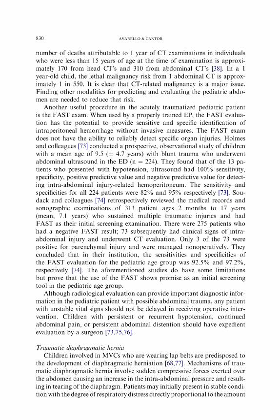
number of deaths attributable to 1 year of CT examinations in individualswho were less than 15 years of age at the time of examination is approxi-mately 170 from head CT’s and 310 from abdominal CT’s [38]. In a 1year-old child, the lethal malignancy risk from 1 abdominal CT is approx-imately 1 in 550. It is clear that CT-related malignancy is a major issue.Finding other modalities for predicting and evaluating the pediatric abdo-men are needed to reduce that risk.
Another useful procedure in the acutely traumatized pediatric patientis the FAST exam. When used by a properly trained EP, the FAST evalua-tion has the potential to provide sensitive and specific identification ofintraperitoneal hemorrhage without invasive measures. The FAST examdoes not have the ability to reliably detect specific organ injuries. Holmesand colleagues [73] conducted a prospective, observational study of childrenwith a mean age of 9.5 (� 4.7 years) with blunt trauma who underwentabdominal ultrasound in the ED (n ¼ 224). They found that of the 13 pa-tients who presented with hypotension, ultrasound had 100% sensitivity,specificity, positive predictive value and negative predictive value for detect-ing intra-abdominal injury-related hemoperitoneum. The sensitivity andspecificities for all 224 patients were 82% and 95% respectively [73]. Sou-dack and colleagues [74] retrospectively reviewed the medical records andsonographic examinations of 313 patient ages 2 months to 17 years(mean, 7.1 years) who sustained multiple traumatic injuries and hadFAST as their initial screening examination. There were 275 patients whohad a negative FAST result; 73 subsequently had clinical signs of intra-abdominal injury and underwent CT evaluation. Only 3 of the 73 werepositive for parenchymal injury and were managed nonoperatively. Theyconcluded that in their institution, the sensitivities and specificities ofthe FAST evaluation for the pediatric age group was 92.5% and 97.2%,respectively [74]. The aforementioned studies do have some limitationsbut prove that the use of the FAST shows promise as an initial screeningtool in the pediatric age group.
Although radiological evaluation can provide important diagnostic infor-mation in the pediatric patient with possible abdominal trauma, any patientwith unstable vital signs should not be delayed in receiving operative inter-vention. Children with persistent or recurrent hypotension, continuedabdominal pain, or persistent abdominal distention should have expedientevaluation by a surgeon [73,75,76].
Traumatic diaphragmatic herniaChildren involved in MVCs who are wearing lap belts are predisposed to
the development of diaphragmatic herniation [68,77]. Mechanisms of trau-matic diaphragmatic hernia involve sudden compressive forces exerted overthe abdomen causing an increase in the intra-abdominal pressure and result-ing in tearing of the diaphragm. Patients may initially present in stable condi-tionwith the degree of respiratory distress directly proportional to the amount
831PEDIATRIC MAJOR TRAUMA
of abdominal contents that protrude into the pulmonary space. Presence ofecchymosis across the abdomen and flanks, which may be secondary to lapbelt compression, should alert the clinician to the possibility of diaphragmatichernia, other intra-abdominal injuries (small bowel injury), and the possibilityof associated thoracic spinal insults, such as Chance fractures [68,70]. Unfor-tunately, traumatic diaphragmatic herniation is one of the most commonlymissed injuries and has been reportedly missed in 12% to -66% of cases [78].
Diagnosis is often difficult and although various imaging modalities areavailable to assist in making the diagnosis, there is no one highly sensitivetest. In a retrospective chart review of a mostly adult population, Mihosand colleagues [78] reported an 80% sensitivity in diagnoses using CT alone.That same article reported that the sensitivity of CT ranges from 33% to83%. This injury can be missed even after undergoing a laparotomy,stressing the difficulty in making the diagnosis.
Initial management for these patients involves placement of a nasogastrictube to decompress the stomach and augment diagnostic imaging. In casesof severe respiratory distress, intubation is indicated. Surgery will berequired for repair of the injury.
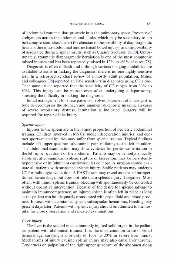
Splenic injuryInjuries to the spleen are in the largest proportion of pediatric abdominal
trauma. Children involved in MVCs, sudden deceleration injuries, and con-tact sports-related injuries may suffer from splenic trauma. Typical findingsinclude left upper quadrant abdominal pain radiating to the left shoulder.The abdominal examination may show evidence for peritoneal irritation inthe left upper quadrant of the abdomen. Patients may be hemodynamicallystable or, after significant splenic rupture or laceration, may be persistentlyhypotensive or in fulminant cardiovascular collapse. A surgeon should eval-uate all patients with suspected splenic injury. Stable patients may undergoCT for radiologic evaluation. A FAST exam may reveal associated intraper-itoneal hemorrhage, but does not rule out a splenic injury if negative. Mostoften, with minor splenic trauma, bleeding will spontaneously be controlledwithout operative intervention. Because of the desire for splenic salvage tomaintain immuncompetency, an injured spleen is often left in place as longas the patient can be adequately resuscitated with crystalloid and blood prod-ucts. In cases with a contained splenic subcapsular hematoma, bleeding maypresent days later. Patients with splenic injury should be admitted to the hos-pital for close observation and repeated examinations.
Liver injuryThe liver is the second most commonly injured solid organ in the pediat-
ric patient with abdominal trauma. It is the most common cause of lethalhemorrhage, carrying a mortality of 10% to 20% in severe liver injury.Mechanisms of injury causing splenic injury may also cause liver trauma.Tenderness on palpation of the right upper quadrant of the abdomen along
832 AVARELLO & CANTOR
with the complaint of abdominal pain in this region or in the right shoulderare signs of possible liver injury. Patients managed conservatively often dowell; however, those that are initially treated as such and then go on torequire delayed laparotomy often have significant morbidity and mortality.Therefore, close observation in the hospital, serial abdominal examinations,and serial hemoglobin should be performed.
Renal injuryThe kidney is less susceptible to trauma from forces applied to the
anterior abdomen, but is often injured in the multitrauma pediatric patient[7]. Because this organ is retroperitoneal, complaints of abdominal painare often less obvious and are more diffuse. Often dull back pain, ecchy-mosis in the costovertebral region, or hematuria are the only clues to renalinjury. Renal ultrasound and CT may be used in the stable patient toassess the degree of renal involvement [41,79,80]. Other organs such asthe pancreas and gastrointestinal tract are less frequently injured in thepediatric patient.
Penetrating injuryPenetrating wounds to the abdomen usually require rapid evaluation
by a surgeon and in some cases, operative intervention. The role ofDPL in the management of pediatric trauma is controversial and seemsto be fading out of practice as the FAST exam continues to gain accep-tance. DPL provides a rapid, nonspecific evaluation of possible intraper-itoneal hemorrhage. Certainly patients who remain unstable despite fluidresuscitation may be candidates for DPL if they are too unstable for CTand there are multiple potential sites of blood loss. An important role forDPL is in the setting of an underlying small bowel injury (SBI). In somepatients with SBI, CT findings of free fluid may be improperly ascribedto underlying splenic bleeding. Finally, DPL may be considered in theoperating room for patients undergoing emergent craniotomy, whenadequate evaluation of the abdomen cannot take place because of thetime urgency required for intervention for head injury. If the woundappears superficial and does not appear to travel below the abdominalmusculoaponeurotic layer, local exploration by an experienced surgeonmay be useful [17].
Disposition
Finally, the EP must decide to admit the pediatric trauma patient,transfer the patient to a tertiary care facility, or discharge the patient. Thedecision for admission should be based on consultation with the surgeonand the patient’s primary care physician. Infants and children who are mod-erately to severely injured have improved outcomes in a pediatric intensivecare unit (PICU) versus an adult ICU; therefore, the primary role of the EP
833PEDIATRIC MAJOR TRAUMA
would be to evaluate and stabilize the patient before admission to a PICU orbefore transfer to a tertiary care facility. Before transport, it is vital that thechild be appropriately stabilized and that the EP communicates directly withthe accepting physician at the transfer facility.
Indications for admission are many, but the main principle is to admitpatients requiring ongoing monitoring for deterioration or complicationsof their injuries. Indications for surgery are outlined in (Box 9). Whateverthe surgical preference within a health care facility, it is most importantto establish a protocol for approaching these challenging patients. Ofcourse, patients who remain hypotensive after adequate crystalloid infusionare candidates for early operative exploration. In addition, children withsuspected physical injury from child abuse may be admitted for their protec-tion as well as for medical treatment.
Summary
Trauma remains the leading cause of death in children nationwide.Proper management of the pediatric trauma patient involves most of thecomponents of standard trauma protocols. By paying strict attention tothe anatomic and physiologic differences in children, the clinician will beensured the best patient outcomes.
References
[1] Jaffe D. Emergency management of blunt trauma in children. N Engl J Med 1991;324:
1477–82.
[2] Minino A, Heron M, Smith B, et al. Deaths: final data for 2004. National vital statistics
reports. Hyattsville (MD). National Center for Health Statistics.
[3] King DR. Trauma in infancy and childhood: initial evaluation and management. Pediatr
Clin North Am 1985;32:1299–310.
Box 9. Indications for surgery
� Hemodynamic instability despite maximal resuscitative efforts� Transfusion of greater than 50% of total blood volume� Radiographic evidence of pneumoperitoneum, intraperitoneal
bladder rupture, grade V renovascular injury� Gunshot would to the abdomen� Evisceration of intraperitoneal or stomach contents� Signs of peritonitis� Evidence of fecal or bowel contamination on diagnostic
peritoneal lavage
834 AVARELLO & CANTOR
[4] Langlois JA, Rutland-Brown W, Thomas KE. Traumatic brain injury in the United
States: emergency department visits, hospitalizations, and deaths. Atlanta (GA): Centers
for Disease Control and Prevention, National Center for Injury Prevention and Control;
2006.
[5] Peclet MH, Newman KD, Eichelberger MR, et al. Patterns of injury in children. J Pediatr
Surg 1990;25:85–90.
[6] Sieben RL, Leavitt JD, French JH. Falls as childhood accidents: an increasing urban risk.
Pediatrics 1971;47:886.
[7] Ablu-Jaude WA. Indicators of genitourinary tract injury or anomaly in cases of pediatric
blunt trauma. J Pediatr Surg 1996;31:86.
[8] Corey TS. Infant deaths due to unintentional injury: an 11-year autopsy review. Am J Dis
Child 1992;146:968–71.
[9] Schafer I, Barkmann C, Riedesser P, et al. Posttraumatic syndromes in children and
adolescents after road traffic accidentsda prospective cohort study. Psychopathology
2006;39(4):159–64.
[10] GauscheM, Lewis RJ, Stratton SJ, et al. Effect of out-of-hospital pediatric endotracheal in-
tubation on survival and neurological outcome: a controlled clinical trial. JAMA 2000;283:
783–9.
[11] Part 12: Pediatric Advanced Life Support Circulation 112: IV-167-187IV-; published online
before print as doi:10.1161/CIRCULATIONAHA.105.166573.
[12] Waltzman ML, Mooney DP. Major trauma. In: Fleisher GR, Ludwig S, Henretig FM,
editors. Textbook of pediatric emergency medicine. 5th edition. Philadelphia: Lippincott