Pediatric Syndromes of Head and Neck Murtaza Z. Kharodawala, MD Faculty Advisor: Matthew Ryan, MD The University of Texas Medical Branch Department of Otolaryngology Grand Rounds Presentation, November 17, 2004
Transcript
Pediatric Syndromes
of Head and Neck Murtaza Z. Kharodawala, MD
Faculty Advisor: Matthew Ryan, MD
The University of Texas Medical Branch
Department of Otolaryngology
Grand Rounds Presentation,
November 17, 2004
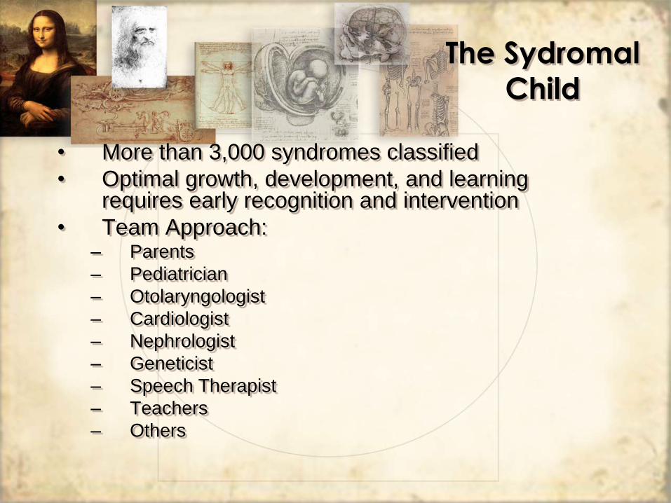
• More than 3,000 syndromes classified
• Optimal growth, development, and learning requires early recognition and intervention
• Team Approach: – Parents
– Pediatrician
– Otolaryngologist
– Cardiologist
– Nephrologist
– Geneticist
– Speech Therapist
– Teachers
– Others
The Sydromal
Child
The Sydromal
Child
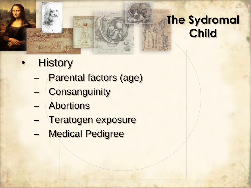
• History
– Parental factors (age)
– Consanguinity
– Abortions
– Teratogen exposure
– Medical Pedigree
• Physical Exam
– Major and Minor Anomalies
• Airway
• Skull
• Ears
• Facial skeleton
– Comparison to Family Members
– Reference Material
The Sydromal
Child
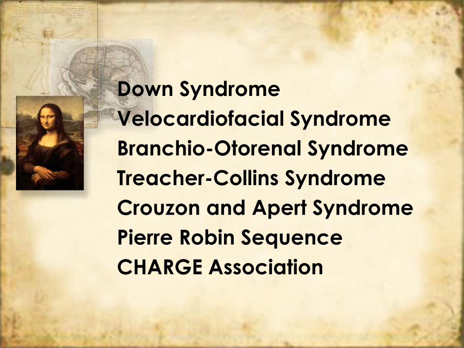
Down Syndrome
Velocardiofacial Syndrome
Branchio-Otorenal Syndrome
Treacher-Collins Syndrome
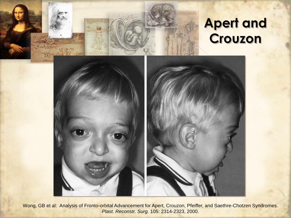
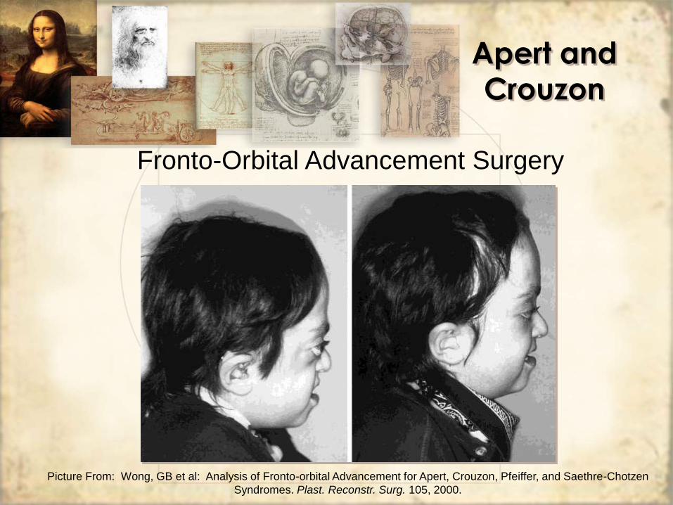
Crouzon and Apert Syndrome
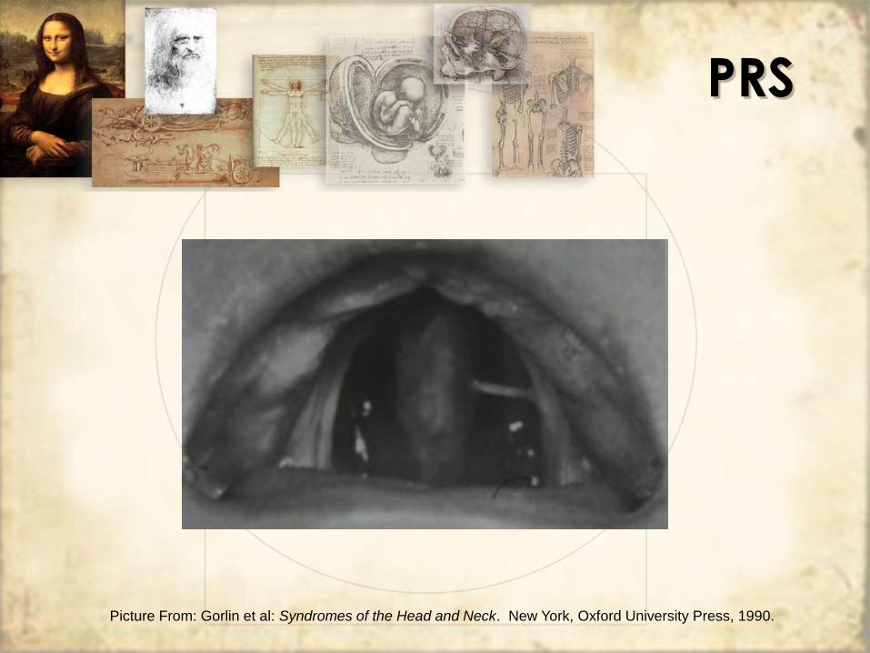
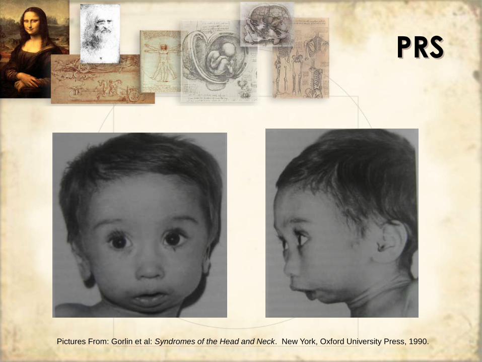
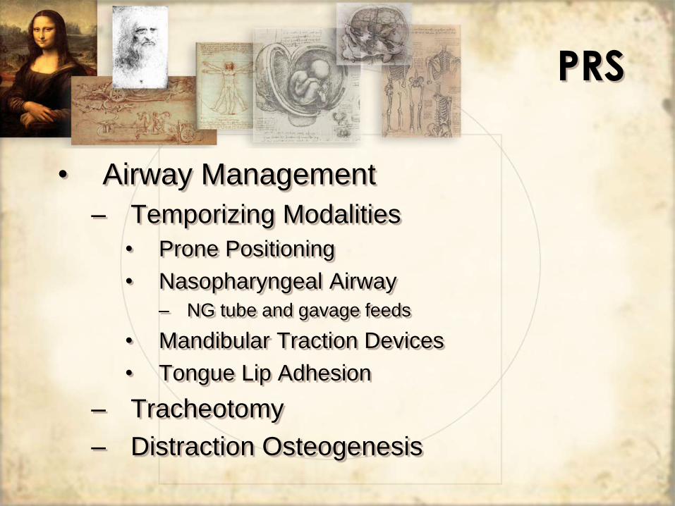
Pierre Robin Sequence
CHARGE Association
Down
Syndrome
• Described by John Landon Down in
1866
• Etiology: nondisjuction mutation
resulting in Trisomy 21
• Prevalence 1:700
– Most common chromosomal anomaly
• Associated with Maternal age > 35
Down
Syndrome
• Facial Characteristics – Macroglossia
– Micrognathia
– Midface hypoplasia
– Flat occiput
– Flat nasal bridge
– Epicanthal folds
– Up-slanting palpebral fissures
– Progressive enlargement of lips
Down
Syndrome
Down
Syndrome
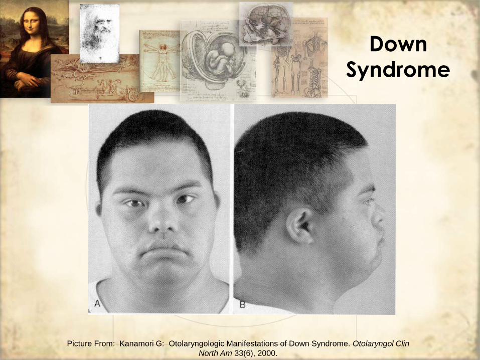
Picture From: Kanamori G: Otolaryngologic Manifestations of Down Syndrome. Otolaryngol Clin
North Am 33(6), 2000.
• Airway Concerns
– Due to midface hypoplasia, the
nasopharynx and oropharynx dimensions
are smaller
• Slight adenoid hypertrophy can cause upper
airway obstruction
– Congenital mild-moderate subglottic
narrowing not uncommon
• Post-extubation stridor
Down
Syndrome
• Obstructive Sleep Apnea
– Prevalence 54-100% in DS patients
– Combination of anatomic and functional
mechanisms
• Midface hypoplasia, macroglossia, etc
• Hypotonia of pharyngeal muscles
Down
Syndrome
• Obstructive Sleep Apnea
– Management:
• Polysomnography to confirm
• Medical interventions:
– CPAP
– Weight Loss
– Medications to stimulate respiratory drive
Down
Syndrome
• Obstructive Sleep Apnea
– Management:
• Surgical
– Adenoidectomy and Tonsillectomy
» Controversial
– UPPP
– Partial tongue resection
– Tracheotomy
Down
Syndrome
• Otologic Concerns
– Small pinna, Stenotic EAC
• Cerumen impaction
– CHL
• ETD: PE tubes
• Ossicular fixation: surgical correction
– SNHL
• Progressive ossification along outflow
pathway of basal spiral tract
Down
Syndrome
• Cardiovascular anomalies (40%)
– ASD, VSD, Tetralogy of Fallot, PDA
• GI anomalies (10-18%)
– Pyloric stenosis, duodenal atresia, TE
fistula
• Malignancy
– 20 fold higher incidence of ALL
– Gonadal tumors
Down
Syndrome
Velocardiofacial
Syndrome
• First described by Shprintzen et al. in 1978
• Not uncommon
– Prevalence 1 in every 4,000 newborns
– 8% of all cleft palate patients
• Autosomal Dominant inheritance
– Hemizygous microdeletion shared with DiGeorge Sequence at 22q11.2 locus
• Features
– Cleft palate
– Congenital heart disease
– Characteristic facies
– Hypernasal speech
– Learning disablities
VCFS
• Oropharyngeal Findings:
– Apparent cleft palate (10-35%)
– Submucous cleft (33%)
– Submucous cleft and velar paresis (33%)
– Tonsils small or aplastic (50%)
– Adenoids small or aplastic (85%)
– Malocclusion
– Hypernasal speech
VCFS
• Airway Obstruction is common
– 50% of neonates with VCFS have OSA
– Adenotonsillectomy should be avoided if
not indicated
– Oral airway needed in urgent setting
– Cleft palate repair required
VCFS
VCFS
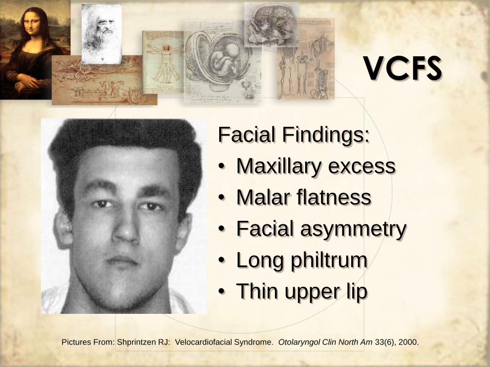
Facial Findings:
• Maxillary excess
• Malar flatness
• Facial asymmetry
• Long philtrum
• Thin upper lip
Pictures From: Shprintzen RJ: Velocardiofacial Syndrome. Otolaryngol Clin North Am 33(6), 2000.
Nasal Findings:
• Prominent nasal root
• Large tip
• Pinched, hypoplastic alar
base
VCFS
Pictures From: Shprintzen RJ: Velocardiofacial Syndrome. Otolaryngol Clin North Am 33(6), 2000.
• Ear findings
– Small auricles (48%)
– CHL secondary to serous effusions and
ETD (75%)
• PE tubes effective
– SNHL (8%)
• Amplification devices
VCFS
• Cardiovascular Findings
– 75-80% with cardiac anomalies
– 10% of patients with VCFS die in early
infancy due to these anomalies
– VSD (65%)
– Right sided aortic arch (35%)
– Tetralogy of Fallot (20%)
– Aberrant subclavian artery (20%)
VCFS
VCFS
MRA:
Tortuous and
medially deviated
internal carotid
artery
Pictures From: Shprintzen RJ. Velocardiofacial Syndrome. Otolaryngol Clin North Am 33(6), 2000.
• Growth and mental retardation
• Flat affect and poor social interaction
with impulsive behavior
• Renal anomalies in 35%
• T cell dysfuction in 10% with
hypocalcemia
VCFS
Branchio-Otorenal
Syndrome
BORS
• First termed by Melnick et al in 1975
• 1 in every 40,000 births
• Autosomal dominant inheritance – Isolated to 8q13.3 locus
• Characteristics: – Branchial cleft cysts or fistulas
– Preauricular pits
– Malformed auricles
– Hearing loss
– Renal anomalies
BORS
• Branchial cleft cysts and fistulas
– Present in 50-60% of cases
– Usually bilateral
– Found in lower third of neck
– Fistulas may connect to tonsillar fossa
• Facial nerve paralysis (10%)
• Aplasia or stenosis of lacrimal duct
(25%)
BORS
• External ear anomalies
– Auricular malformation (30-60%) or abnormal position • Minor aberration of anatomy to severe microtia
– Helical or preauricular pits (70-80%)
• Middle ear anomalies
– Malformation and/or fixation of ossicles
– Abnormal size/structure of the tympanic cavity
BORS
Picture From: Gorlin et al: Syndromes of the Head and Neck. New York, Oxford University Press, 1990
BORS
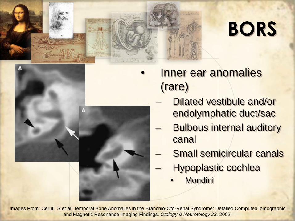
• Inner ear anomalies
(rare)
– Dilated vestibule and/or
endolymphatic duct/sac
– Bulbous internal auditory
canal
– Small semicircular canals
– Hypoplastic cochlea
• Mondini
Images From: Ceruti, S et al: Temporal Bone Anomalies in the Branchio-Oto-Renal Syndrome: Detailed ComputedTomographic
and Magnetic Resonance Imaging Findings. Otology & Neurotology 23, 2002.
BORS
• Hearing loss (75-95%)
– CHL (30%)
– SNHL (20%)
– MHL (50%)
BORS
• Renal anomalies (12-20%)
– Likely underreported when a disease
process not involved
– Renal agenesis or hypoplasia
– Structural anomalies of renal pelvis or
ureters
BORS
• Diagnosis and Treatment
– History and Physical Examination
– Audiogram, CT temporal bones
– CT neck
– Renal Ultrasound, IVP
BORS
• Diagnosis and Treatment
– Surgical excision of branchial cleft cyst,
sinus, or fistula
– Otoplasty
– Excision of pits
– Possible ossicular chain reconstruction
– Hearing aids
– Urology consultation for renal anomalies
Treacher
Collins
Syndrome
TCS
• First described by Thomson and Toynbee in 1846-7 – Later, essential components described by
Treacher Collins in 1960
• Autosomal dominant inheritance – TCOF1, mapped to 5q32-33.1
• 60% are from new mutation – Associated with increased paternal age
• Prevalence of 1 in 50,000
• a.k.a. Mandibulofacial dysostosis
TCS
• Characteristics – Likely due to abnormal migration of neural crest cells into
first and second branchial arch structures
– Usually bilateral and symmetric
– Malar and supraorbital hypoplasia
– Non-fused zygomatic arches
– Cleft palate in 35%
– Hypoplastic paranasal sinuses
– Downward slanting palpebral fissures
– Mandibular hypoplasia with increased angulation
– Coloboma of lower eyelid with absent cilia
– Malformed pinna
– Normal intelligence
TCS
Picture From: Cummings, CW: Otolaryngology: Head and Neck Surgery. St Louis, Mosby, 1998
TCS
• OP/Airway concerns
– Cleft palate
– Choanal atresia may be present
• Respiratory distress in newborn
• Oral airway, McGovern nipple
– Obstructive sleep apnea is the most common
airway dysfunction
• Mandibular hypoplasia results in retrodisplacement of
tongue into oropharynx
• Oral airway, tracheotomy
• Distraction osteogenesis vs. free fibular transfer
TCS
• Otologic concerns
– Malpositioned auricles
– Malformed pinna
– EAC atresia
– Ossicular abnormalities
– Conductive hearing loss is common
• Hearing aids are effective
– Normal intelligence
TCS
Picture From: Acosta, HL et al: Vertical Mesenchymal Distraction and Bilateral Free Fibula Transfer for Severe Treacher
• Mouth breathing is a learned response, developed at 4-6 weeks
• Bilateral CA presents at birth with respiratory distress and cyanosis, relieved with crying
• Unilateral CA usually presents later in life with chronic nasal discharge
CA
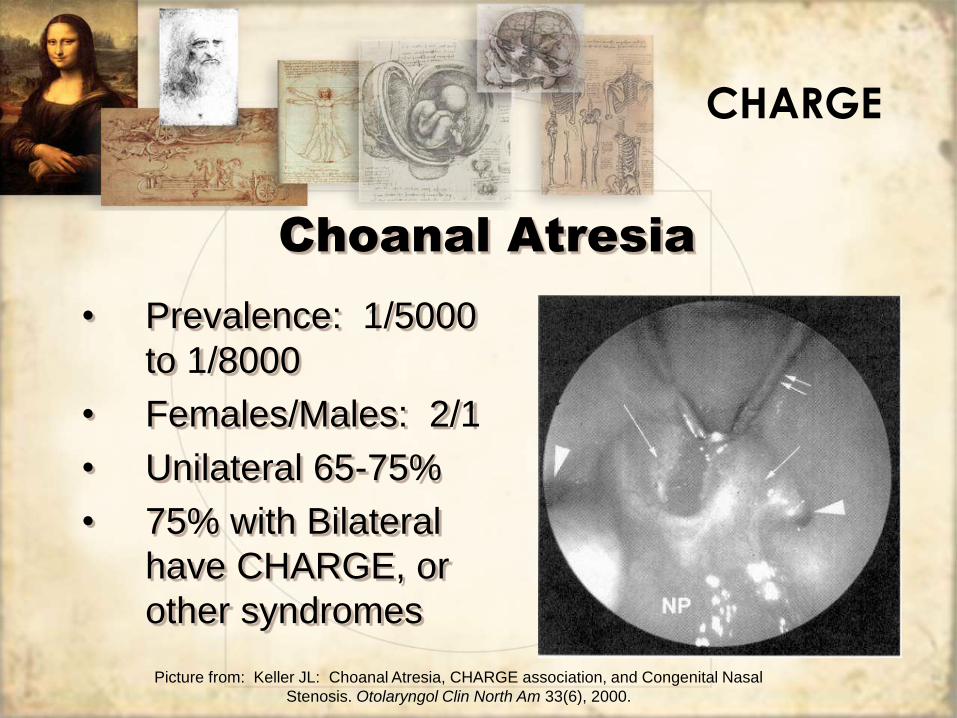
Choanal Atresia
• Diagnosis:
– 6 French catheter
– Nasal endoscopy
– Bell of Stethoscope
– Mirror
• Radiology
– CT (preferred method)
CA
Choanal Atresia
• Treatment:
– Unilateral CA does not require immediate
correction
• May be delayed until starting school
– Bilateral CA requires immediate interventions:
• Oral Airway
• McGovern Nipple
• Intubation
• Tracheostomy
CA
Choanal Atresia
• Surgical Correction:
– Transnasal
– Transpalatal
– Laser
– +/- Stenting
– +/- Mitomycin-C Topical (0.3 mg/cc)
CHARGE
Choanal Atresia
Bibliography
• Gorlin, RJ et al: Syndromes of the Head and Neck. New York, Oxford University Press, 1990.
• Bluestone CD et al. Pediatric Otolaryngology. Philadelphia, Saunders, 2003.
• Chang, J: Reconstruction of the Hand in Apert Syndrome: A Simplified Approach. Plast. Reconstr. Surg. 109, 2002.
• Wong, GB et al: Analysis of Fronto-orbital Advancement for Apert, Crouzon, Pfeiffer, and Saethre-Chotzen Syndromes. Plast. Reconstr. Surg. 105, 2000.
• Acosta, HL et al: Vertical Mesenchymal Distraction and Bilateral Free Fibula Transfer for Severe Treacher Collins Syndrome. Plastic & Reconstructive Surgery, 113(4), 2004.
• Levin AV: Congenital Eye Abnormalities. Pediatr Clin North Am 50(1), 2003.
• Ceruti, S et al: Temporal Bone Anomalies in the Branchio-Oto-Renal Syndrome: Detailed ComputedTomographic and Magnetic Resonance Imaging Findings. Otology & Neurotology 23, 2002.
• Keller JL: Choanal Atresia, CHARGE association, and Congenital Nasal Stenosis. Otolaryngol Clin North Am 33(6), 2000.
• Kanamori G: Otolaryngologic Manifestations of Down Syndrome. Otolaryngol Clin North Am 33(6), 2000. Shprintzen RJ. Velocardiofacial Syndrome. Otolaryngol Clin North Am 33(6), 2000.
• Weintraub AS: Neonatal Care of Infants with Head and Neck Anomalies. Otolaryngol Clin North Am 33(6), 2000.