Peptidyl-prolyl Isomerase Pin1 Controls Down-regulation ofConventional Protein Kinase C Isozymes*□S
Received for publication, February 3, 2012 Published, JBC Papers in Press, February 8, 2012, DOI 10.1074/jbc.M112.349753
Hilde Abrahamsen‡1, Audrey K. O’Neill‡§, Natarajan Kannan‡¶2, Nicole Kruse¶, Susan S. Taylor‡¶,Patricia A. Jennings¶, and Alexandra C. Newton‡3
From the Departments of ‡Pharmacology and ¶Chemistry and Biochemistry and §Biomedical Sciences Graduate Program,University of California, San Diego, La Jolla, California 92093
Background:Conventional PKC isozymes have a putative Pin1 isomerization sequence at their turnmotif phosphorylationsite.Results: Pin1 binds conventional PKCs and promotes their activation-induced down-regulation.Conclusion: Pin1 isomerizes the phosphorylated turn motif of conventional PKC isozymes, priming them for subsequentdown-regulation.Significance: Pin1 provides a switch regulating the lifetime of conventional PKCs.
The down-regulation or cellular depletion of protein kinase C(PKC) attendant to prolonged activation by phorbol esters is awidely described property of this key family of signalingenzymes. However, neither the mechanism of down-regulationnor whether this mechanism occurs following stimulation byphysiological agonists is known.Herewe show that the peptidyl-prolyl isomerase Pin1 provides a timer for the lifetime of con-ventional PKC isozymes, converting the enzymes into a speciesthat can be dephosphorylated and ubiquitinated following acti-vation induced by either phorbol esters or natural agonists. Theregulation by Pin1 requires both the catalytic activity of theisomerase and the presence of a Pro immediately followingthe phosphorylated Thr of the turn motif phosphorylation site,one of two C-terminal sites that is phosphorylated during thematuration of PKC isozymes. Furthermore, the secondC-termi-nal phosphorylation site, the hydrophobic motif, docks Pin1 toPKC. Our data are consistent with a model in which Pin1 bindsthe hydrophobic motif of conventional PKC isozymes to cata-lyze the isomerization of the phospho-Thr-Pro peptide bond atthe turnmotif, thus converting these PKC isozymes into speciesthat can be efficiently down-regulated following activation.
The peptidyl-prolyl cis-trans isomerase Pin1 is emerging asan important regulator of signal transduction pathways (1).
Pin1-catalyzed isomerization plays a key role in the control ofnormal cellular functions, most notably proliferation wherePin1 is essential for cell cycle progression (2). Pin1 belongs tothe Parvulin family of peptidyl-prolyl cis-trans isomerases andis the only member that specifically isomerizes phospho-(Ser/Thr)-Pro ((Ser(P)/Thr(P))-Pro) motifs (3): the enzyme displaysan �1000-fold selectivity for peptides phosphorylated on theSer/Thr preceding the Pro compared with unphosphorylatedpeptides (3).Pin1-induced conformational changes in target proteins
affect a variety of protein properties from folding to regulationof activity and stability. As a consequence, deregulation ofphosphorylation steps and their attendant conformationalchanges often lead to disease (4). For example, Pin1 is down-regulated in degenerating neurons from Alzheimer diseasepatients, correlating with age-dependent neurodegeneration(5). Pin1 has also been implicated in cancer progression: levelsof this protein are increased inmany cancers, including those ofthe breast, prostate, brain, lung, and colon (6–9). Thus, Pin1has been proposed to function as a catalyst for oncogenic path-ways (10). The molecular mechanisms that lead to disease pro-gression most likely involve postphosphorylation conforma-tional changes catalyzed by Pin1 that are required fordownstream effects.Members of the protein kinase C (PKC) family of Ser/Thr
kinases transduce an abundance of diverse signals that mediateprocesses such as cell cycle progression (11, 12), apoptosis (13),and immune responses (14). The PKC family consists of 10isozymes that all possess an N-terminal regulatory domain, aconserved C-terminal catalytic core, and an autoinhibitorypseudosubstrate sequence (for reviews, see Refs. 15 and 16).The PKC family is subdivided into three subclasses based on thecofactor dependence of their regulatory domains: conventional(�,�, and �; activated by diacylglycerol andCa2�), novel (�, �, �,and �; activated by diacylglycerol), and atypical (� and ; insen-sitive to diacylglycerol or Ca2�) isozymes. Before conventionalPKC isozymes can be activated by second messengers, theyundergo a series of ordered phosphorylations (17, 18) and con-formational transitions. Newly synthesized, unphosphorylated
* This work was supported, in whole or in part, by National Institutes of HealthGrants P01 DK54441 (to A. C. N. and S. S. T.) and GM43154 (to A. C. N.) andTraining Grant T32 GM007752 from the NIGMS (to A. O. through the Uni-versity of California, San Diego Graduate Training Program in Cellular andMolecular Pharmacology). This work was also supported by The Norwe-gian Research Council (to H. A.).
□S This article contains supplemental Fig. 1.1 Present address: Dept. of Biochemistry, Inst. for Cancer Research, the Nor-
wegian Radium Hospital, Oslo University Hospital and Centre for CancerBiomedicine, Faculty of Medicine, University of Oslo, Montebello, N-0310Oslo, Norway.
2 Present address: Dept. of Biochemistry and Molecular Biology, University ofGeorgia, Athens, GA 30602.
3 To whom correspondence should be addressed: Dept. of Pharmacology,University of California, San Diego, 9500 Gilman Dr., La Jolla, CA 92093-0721. Tel.: 858-534-4527; Fax: 858-822-5888; E-mail: [email protected].
conventional PKC isozymes are loosely tethered at the mem-brane (19) with an exposed pseudosubstrate and an accessibleC-terminal tail (20). The upstream kinase, phosphoinositide-dependent kinase 1 (PDK-1),4 docks onto the C-terminal tail ofthis newly synthesized conventional PKC (21), allowing effi-cient phosphorylation of the activation loop site (Thr500; num-bering according to rat PKC�II) (17, 18, 22). This initial phos-phorylation triggers two sequential phosphorylation events onthe C-terminal tail that have recently been shown to depend onthemammalian target of rapamycin complex 2 (mTORC2) pro-tein complex (23, 24). These sites are the turn motif (Thr641;numbering according to rat PKC�II) and the hydrophobicmotif (Ser660; numbering according to rat PKC�II). The role ofmTORC2 in these phosphorylations on PKC remains to beclarified. In the case of Akt, mTORC2 phosphorylates the turnmotif site co-translationally (25). This is not the case with PKCbecause phosphorylation at the turn motif occurs after biosyn-thesis; the half-time of phosphorylation of newly synthesizedPKC is on the order of 15min (20). Once phosphorylated on theturn motif, PKC becomes phosphorylated at the hydrophobicmotif via an intramolecular autophosphorylation (26). The fullyphosphorylated conventional PKC then localizes to the cytosolwhere it is maintained in an inactive and phosphatase-resistantconformation (27, 28). This form is the major species of con-ventional PKC found in unstimulated cells. The phosphoryla-tions at the PDK-1 site (activation loop) and at the turn andhydrophobic motifs are essential for PKC function; however,once PKC is matured by phosphorylation, phosphate on theactivation loop (but not turn motif) becomes dispensable (19,27).Natural agonist-induced acute signaling by conventional
PKC is terminated following removal of the secondmessengers(diacylglycerol andCa2�), relocalizing conventional PKC to thecytosol in the closed, autoinhibited conformation. Chronicactivation of conventional PKC, however, eventually results inthe complete dephosphorylation and degradation of theenzyme by a ubiquitin/proteasome-dependent mechanismreferred to as down-regulation (29–32). The classic trigger forchronic activation and subsequent down-regulation of conven-tional and novel PKC isozymes is phorbol ester treatment ofcells (33). These potent analogues of diacylglycerol are notmetabolized and thus cause sustained recruitment of PKC tomembranes. Here, PKC ismaintained in an open conformationthat has a 2-orders of magnitude increased sensitivity to phos-phatases (34). The recently discovered protein phosphatasePHLPP (pleckstrin homology (PH) domain leucine-rich repeatprotein phosphatase (35)) catalyzes the first dephosphorylationevent of PKC, which occurs on the hydrophobic motif andshunts PKC to the detergent-insoluble fraction of cells (36).Protein phosphatase 2A, which can dephosphorylate the acti-vation loop and the hydrophobic motif in vitro (37), also con-tributes to the dephosphorylation of PKC in cells (38). Dephos-
phorylationhas traditionally been considered to be the first stepin phorbol ester-mediated down-regulation (39), although fullyphosphorylated PKC� has been reported to be degraded in onestudy (40). Dephosphorylated PKC can also be rescued byrephosphorylation in a manner that depends on the chaperoneprotein heat shock protein 70 (Hsp70), which specifically bindsthe dephosphorylated turn motif (41, 42).Here we report that Pin1 is required for the efficient down-
regulation of conventional PKC isozymes that is triggered byeither phorbol ester or natural agonist stimulation. Experi-ments using cells lacking Pin1, pharmacological inhibition ofPin1, constructs of PKC unable to bind or be isomerized byPin1, and peptide binding arrays revealed that Pin1 binds theC termini of the conventional PKC isozymes PKC� andPKC�II, converting them into a species that can be readilydephosphorylated and ubiquitinated. Our data are consistentwith a model in which Pin1 catalyzes a cis/trans isomerizationof the phospho-Thr-Pro peptide bond of the turn motif, thusconverting PKC into a species that is down-regulation-sensi-tive. Thus, Pin1-mediated isomerization provides a molecularsignal that primes conventional PKC isozymes for agonist-evoked down-regulation.
EXPERIMENTAL PROCEDURES
Plasmids—Rat PKC�II in pcDNA3, PKC���-T641AAA,PKC���-K371R (27), PKC���-T660A (43), PKC� (44), andMyc-PDK-1 (22), have been described previously. Myc-taggedrat Xpress-tagged PKC� and PKC� were gifts from Alex Toker,PKC� was from Peter Blumberg, and PKC� was a gift fromYusuf Hannun. GST-tagged PKCbII C-terminal constructswere generated as described (21). GST-Pin1 for bacterialexpression was a gift from Joseph P. Noel, and GST-Pin1 andHA-Pin1 mammalian expression vectors were generated byPCR and subsequent cloning into the BamHI and NotI sites ofpEBG vector (a gift from Bruce Mayer) or into the EcoRI andNotI sites 3� to the HA epitope in pcDNA3-HA, respectively.PKC�II mutants were generated using QuikChange (Strat-agene). 3HA-ubiquitin constructs were a kind gift from VishvaM. Dixit.Antibodies and Materials—Antibodies for immunoblotting
PKC� (sc-208), PKC�� (sc-209), PKC�II (sc-210), PKC� (sc-937), PKC� (sc-214), and PKC� (sc-216) were purchased fromSanta Cruz Biotechnology, Santa Cruz, CA. PKC�was detectedusing Omni-probe (Xpress tag) (Santa Cruz Biotechnology,sc-7270). HA-tagged ubiquitin was detected using a high affin-ity rat HA antibody (RocheApplied Science, 1 867 423). Endog-enous ubiquitination was detected using a ubiquitin antibody(Covance, MMs-258R), and Myc-tagged PDK-1 was detectedwith a Myc antibody (Covance, PRB-150P). Immunoprecipita-tion of PKC� and PKC�II was performed using an antibodygenerated against a region common to PKC� and PKC�II (BDTransduction Laboratories, 610108). A monoclonal antibodydirected toward Hsp70 was also obtained from BD Transduc-tion Laboratories (610607). The monoclonal Pin1 antibodyused was a gift fromKun Ping Lu. The polyclonal Pin1 antibody(3722) and the antibody detecting phosphorylated PKC�II/�(Ser660/638; 9371) were from Cell Signaling Technology, Inc.The �-tubulin (T6074) and �-actin (A2066) antibodies were
4 The abbreviations used are: PDK-1, phosphoinositide-dependent kinase 1;PDBu, phorbol 12,13-dibutyrate; LPA, 1-oleoyl lysophosphatidic acid;Hsp70, heat shock protein 70; MEFs, mouse embryonic fibroblasts; PiB, die-thyl-1,3,6,8-tetrahydro-1,3,6,8-tetraoxobenzo[lmn][3,8]phenanthroline-2,7-diacetate; mTORC2, mammalian target of rapamycin complex 2; TM,turn motif.
Pin1 Controls Down-regulation of Conventional PKC
APRIL 13, 2012 • VOLUME 287 • NUMBER 16 JOURNAL OF BIOLOGICAL CHEMISTRY 13263
from Sigma. Phorbol 12,13-dibutyrate (PDBu; 524390),MG-132 (474790), and diethyl-1,3,6,8-tetrahydro-1,3,6,8-tetraoxobenzo[lmn][3,8]phenanthroline-2,7-diacetate (PiB;529627) were obtained from Calbiochem. Protein A/G beadswere purchased from Pierce (53133), and glutathione-Sephar-ose beads were from Amersham Biosciences (17-0756-01).EasyTag Expre35S35S (1000 Ci/mmol) protein labeling mixturewas purchased from PerkinElmer Life Sciences. N-Ethylma-leimide (E1271) and bombesin (B-126) were purchased fromSigma. 1-Oleoyl lysophosphatidic acid (LPA) was obtainedfrom Cayman Chemical (62215).Cell Culture and Transfection—COS7 cells, HeLa cells, 293T
cells, Pin1�/�, and Pin1�/� mouse embryonic fibroblasts(MEFs; a gift from Kun-Ping Lu) were cultured in DMEM con-taining 10% fetal bovine serum and 1% penicillin/streptomycinat 37 °C in 5% CO2. HT1080 cells (a gift from Roger Y. Tsien)were grown with the same supplements but in RPMI 1640medium. Cells were plated in 6-well plates 1 day prior to trans-fection and transfected with FuGENE 6 (Roche AppliedScience, 11-814-443-001) (COS7 and 293T) for transient trans-fection (18–24 h) or with Lipofectamine 2000 (Invitrogen, 11668-019) (HeLa and HT1080) for siRNA transfection (72 h; 100 nMsiRNA) following themanufacturers’ recommendations.Cell Lysis, Immunoprecipitations, GST Pulldowns, and
Immunoblotting—Prior to lysis, cells were washed in PBS andplaced on ice. Cells were then lysed in lysis buffer (50 mM Tris,pH 7.4, 100 mM NaCl, 5 mM EDTA, 1 mM sodium orthovana-date, 1 mM PMSF, 10 mM sodium pyrophosphate, and 50 mM
sodium fluoride). After clearing the lysates by centrifugation at13,000 � g for 10 min (except for the PDBu-stimulated down-regulation experiments for which whole cell lysates were used),proteins were resolved by SDS-PAGE, transferred to PVDF,and visualized by immunoblotting. For immunoprecipitation, 1g of antibody and 30 l of protein A/G beads were added tothe cleared lysates. Immune complexes were then allowed toform by incubating with rotation overnight at 4 °C. Complexeswere thereafter washed three times in 1 ml of lysis buffer, pel-leting each time by centrifugation at 300 � g for 3 min at 4 °C.GST pulldowns were performed in a similar way. However,instead of adding antibodies, 30 l of washed GST-Sepharosebeads (50:50 slurry) were added to the lysates followed by incu-bation overnight. Complexes were washed as described above.Cell Stimulation and in Vivo Ubiquitination Assay—Cells
were stimulated using 200 nM PDBu for the indicated times toinduce ubiquitination or down-regulation and lysed in lysisbuffer as described above. For ubiquitination assays using exog-enous ubiquitin, COS7 cells in 6-well plates were transfectedwith 0.5 g of cDNA encoding the indicated PKC isozyme and1.5 g of cDNA 3HA-ubiquitin (Lys48-ubiquitin or Lys63-ubiq-uitin). For detection of endogenous ubiquitination, COS7 cellswere transfected with the PKC isozyme of interest. Sixteen to24 h post-transfection, cells were stimulatedwith bombesin (10nM) or LPA (10M) in the presence of the proteasome inhibitorMG-132 (10 M). Cells were washed in ice-cold PBS and lysedon ice for 15 min in lysis buffer supplemented withN-ethylma-leimide (10 mM). Relevant proteins were immunoprecipitatedfrom cleared lysates as described above. For ubiquitin experi-ments in the presence of the Pin1 inhibitor, cells were first
pretreated with PiB (10 M) for 2 h and thereafter treated withbombesin (10 nM) or LPA (10 M) and MG-132 (10 M) asdescribed above. For detection of endogenous ubiquitination inMEFs, confluent 10-cm dishes containing either Pin1�/�MEFs or control MEFs were used. Cells were stimulated withPDBu (200 nM) or LPA (10 M) for 1 h in the presence ofMG-132 (10 M) and lysed in lysis buffer supplemented withN-ethylmaleimide (10 mM). Thereafter, PKC was immunopre-cipitated as described above.Autospot Peptide Array—Peptide arrays were synthesized on
nitrocellulosemembranes using aMultiPep automated peptidesynthesizer (INTAVIS Bioanalytical Instruments AG) asdescribed (45). The peptide-containing membranes were acti-vated inmethanol andwashed in distilledwater and then in PBSwith 0.05% Triton. Membranes were blocked in 5% milk andoverlaid with purified His-Pin1 (1 M). Bound Pin1 was subse-quently detected using a horseradish peroxidase-conjugatedanti-His antibody.siRNA—Nineteen-nucleotide siRNAs targeting Pin1 (NCBI
Reference Sequence NM_006221.3) were designed based onthe algorithm developed by Amarzguioui and Prydz (46). Thetwo 19-nucleotide sequences scoring best (siRNAs 1 and 2) foreach sequence were ordered as preannealed duplexes fromDharmacon and carried 3�-dTdT overhangs. Duplex 3 wasordered from IntegratedDNATechnologies and contained a 3�overhang similar to the RNA region it was targeted against. Thesequences for the sense strand of the siRNAs are as follows: 1,5�-GGCUACAUCCAGAAGAUCA-3�; 2, 5�-GCCUCACAA-UUCAGCGACU-3�; 3, 5�-UCAGGCCGAGUGUACUACU-3�. The non-targeting control duplex was from Dharmacon(D-001210-01-20).Pulse-Chase and Kinase Assay—For pulse-chase assays, cells
were first incubated in Met/Cys-free DMEM for 30 min andthen labeled with [35S]Met/Cys (0.1 mCi/ml) for 7 min. Cellswere then chased in unlabeled Met/Cys for the times indicatedin the figure legends. PKC was immunoprecipitated overnightand analyzed by SDS-PAGE and autoradiography as described(41). PKC activity assays were performed as described previ-ously (43). Briefly, whole cell lysates (containing endogenousPKC) were diluted in buffer containing 20mMHepes, 0.1% Tri-ton, 2 mM DTT, and 1 mM PMSF and incubated with 500 M
[�-32P]ATP (0.1 mCi/mol), 25 mM MgCl2, and 500 g/ml pep-tide substrate (Ac-FKKSFKL-NH2) in the presence or absenceof phosphatidylserine/diacylglycerol vesicles (140 M/3.8 M)and 0.1mMCaCl2 at 30 °C for 5min. TheCa2�/lipid-dependentactivity was normalized to PKC� levels determined byWesternblot analysis of the lysates.RT-PCR—Total RNA was extracted from Pin1�/� and
Pin1�/� MEFs stimulated for 24 h with PDBu using a QiagenRNEasy kit according to the manufacturer’s instructions. TheRNA concentration was measured on a Nanodrop ND-1000spectrophotometer (Thermo), and equal amounts of RNAwereused in RT-PCRs using a QiagenOneStep RT-PCR kit (accord-ing to the manufacturer’s instructions), an annealing tempera-ture of 55 °C, 25 cycles, and the following primers: mousePKC� 1 forward, 5�-TGAAAGACCACAAATTCATCGCC-3�;mouse PKC� 1 reverse, 5�-ACGAACTCATGGCACCTCTTA-T-3�; mouse PKC� 2 forward, 5�-AGAGGTGCCATGAGTT-
Pin1 Controls Down-regulation of Conventional PKC
13264 JOURNAL OF BIOLOGICAL CHEMISTRY VOLUME 287 • NUMBER 16 • APRIL 13, 2012
CGTTA-3�; mouse PKC� 2 reverse, 5�-GGCTTCCGTATGT-GTGGATTTT-3�; mouse hypoxanthine-guanine phosphor-ibosyltransferase forward, 5�-GATTAGCGATGATGAACCA-GGTTATGACCTAGATTTG-3�; mouse hypoxanthine-gua-nine phosphoribosyltransferase reverse, 5�-CAATGTGATGG-CCTCCCATCTCC-3�. PCR products were run on 2% agarosegels and were of the expected sizes.Modeling of PKC—Modeling studies were performed using
the Insight II software package. The turn motif Pro (Pro642 inFig. 8A) was modeled in cis conformation using the crystalstructure of PKC�II (Protein Data Bank code 2I0E) as the tem-plate. The flanking regions of the turn motif were energy-min-imized after rotating the peptide bond. The structures wererendered using PyMOL.Quantification and Statistics—Western blot and RT-PCR gel
signals were quantified using Scion Image or AlphaView soft-ware (Alpha-Innotech, San Leandro, CA). PKC� protein andmRNA levels were normalized to �-actin and hypoxanthine-guanine phosphoribosyltransferase, respectively. Differencesbetween conditions were assessed using one-sided t testsassuming unequal variances; significance was set at p � 0.05.
RESULTS
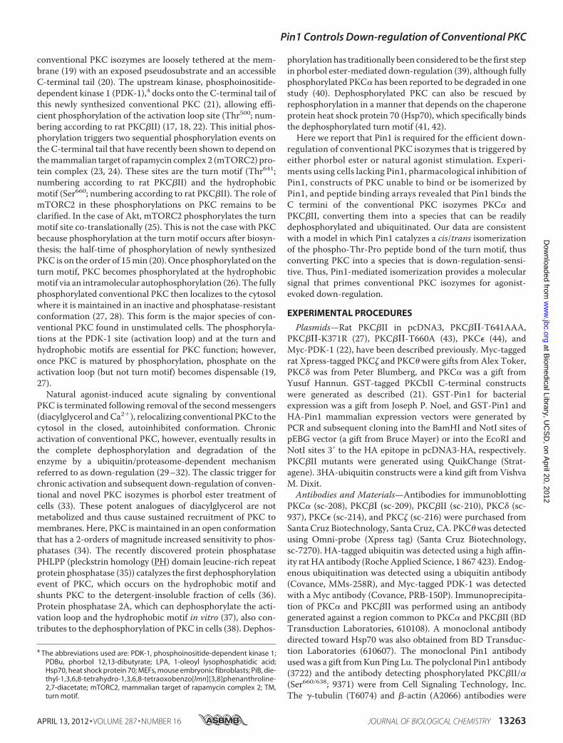
C Termini of Conventional and Atypical PKCs Contain Con-served Pin1 Isomerization Site—Wehave shownpreviously thatthe phosphorylation state of the two C-terminal phosphoryla-tion sites in PKC���, the turnmotif (Thr641) and the hydropho-bic motif (Ser660), function as determinants allowing specificprotein interactions that regulate the maturation, stability, andfunction of PKC��� (21, 41). Curiously, we found that C-termi-nal binding partners discriminated between PKC that hadnever been phosphorylated (newly synthesized) and PKC thathadmatured and subsequently been dephosphorylated. Specif-ically, PDK-1 preferentially binds the C terminus of a PKC spe-cies that has never been phosphorylated (21), whereas Hsp70preferentially binds the C terminus of a PKC species that hasbeen dephosphorylated (41). Therefore, we reasoned that theconformation of the C terminus changes following phosphory-lation/dephosphorylation. We noted that the conserved turnphosphorylation motif of all PKC isozymes except the novelisozymes �, �, and � contains a Thr followed by a Pro (Fig. 1,residues marked in light blue and yellow, respectively). Given
the selectivity of the peptidyl-prolyl cis-trans isomerase Pin1for phosphorylated (Ser/Thr)-Pro motifs (3, 47), we hypothe-sized that Pin1 could regulate the transition between the vari-ous conformations that PKC��� is known to adopt during itslife cycle.Pin1 Binds C Termini of Conventional PKC Isozymes—To
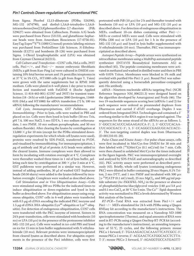
test our hypothesis that Pin1 could account for the finding thatthe C termini of never-phosphorylated and dephosphorylatedPKC differ, we explored whether Pin1 could recognize the Ctermini of PKC isozymes that have a Pro immediately followingthe phosphoacceptor Thr at the turn motif (Fig. 1). Wedesigned and synthesized a peptide array containing overlap-ping 18-residue peptides covering the entire C-terminaldomain of conventional PKC�II or novel PKC�. One set of pep-tides had a non-phosphorylated residue at the turn motif (TM-Thr strip) and a second set had a phosphorylated residue (TM-P-Thr strip). Overlay of the peptide array with bacteriallyexpressed His-Pin1 revealed strong interactions with peptidescovering a region between the turn motif and hydrophobicmotif of PKC�II (residues 640–663; peptides marked in red),but no detectable interaction with corresponding peptidesfrom theC-terminal tail of PKC�was observed (Fig. 2,A andB).Interestingly, these results indicate that Pin1 might bind to anunconventional site in PKC rather than the typical (Ser(P)/Thr(P))-Pro described in previous reports. The entireuncropped array is presented in supplemental Fig. 1. Consistentwith earlier publications on the binding specificity of Pin1, pep-tides from the PKC�II sequence with phosphate on the turnmotif Thr (TM-P-Thr strip) bound Pin1 more strongly (pep-tides marked in green) compared with the same unphosphory-lated peptides (TM-Thr strip), supporting the notion that Pin1indeed binds better to PKC when it is phosphorylated at theturn motif. The interaction of the isolated C-terminal segmentof PKC�II with Pin1 was verified by co-precipitation of HA-Pin1 with a GST-tagged C-terminal segment of PKC�II (resi-dues 628–673; Fig. 2C). To further delineate specific residuesin the Pin1-binding sequences that mediate the interactionwith Pin1, we synthesized a new array consisting of the 25-amino acid sequence shown to contain the core �II C-terminalPin1 interaction motif (VLT641PPDQEVIRNIDQSEFEGFS660-FVN) and additional peptides with Ala replacements at each
FIGURE 1. C termini of conventional and atypical PKCs contain conserved Pin1 isomerization site. A sequence alignment of the C-terminal segments ofprotein kinase C isozymes is shown. NCBI Reference Sequences for human PKC isozymes are from the University of California, Santa Cruz genome browser (�,NM_002737; ��/���, NP_002738; �, NM_002739; , NM_002740; �, NM_002744; �, NM_006255; �, NM_005400; �, NM_006254; �, NM_006257). Proteinsequences were aligned using ClustalW. The conserved turn motif and hydrophobic motif phosphorylation sites are indicated in light blue, and the turn motifprolines are indicated in yellow.
Pin1 Controls Down-regulation of Conventional PKC
APRIL 13, 2012 • VOLUME 287 • NUMBER 16 JOURNAL OF BIOLOGICAL CHEMISTRY 13265
consecutive position in the peptide (“Ala scan”; Fig. 2D). Thetop peptide in the scan is the wild-type sequence and repro-duces the binding of Pin1 to the C-terminal sequences in the
original array (Fig. 2A). The interaction with the wild-type pep-tide was abolished upon Ala replacement of a number of keyresidues (residues boxed in light gray; also marked with a red
Pin1 Controls Down-regulation of Conventional PKC
13266 JOURNAL OF BIOLOGICAL CHEMISTRY VOLUME 287 • NUMBER 16 • APRIL 13, 2012
asterisk on the left side of the blot) or significantly reduced byAla replacement of another set of residues (boxed in yellow;indicated with a gray asterisk on the left side of the blot). Moststrikingly, replacement of any of the 8 underlined residues inthe 11-residue sequence IDQSEFEGFS660F abolished Pin1binding. Interestingly, this sequences includes the characteris-tic hydrophobic motif FXXF(S/T)X(F/Y) (43) where the under-lined residue is the Ser or Thr phosphorylated in conventionaland novel PKCs (i.e. residue Ser660 in PKC�II). Altogether,these data reveal that the region upstream of the hydrophobicmotif is a key novel recognition determinant on PKC�II forPin1. To test the relative importance of the hydrophobic andturn motifs for Pin1 binding, we mutated PKC�II at either thepotentially isomerizable turnmotif Pro (P642A) or the two Pheresidues immediately upstream of the hydrophobic motif(F656A/F659A) and assessed binding of the mutants to GST-Pin1; both constructs were phosphorylated on the hydrophobicmotif (data not shown). Mutation of the turn motif Prodecreased binding, whereas the F656A/F659A mutation abol-ished binding to Pin1 (Fig. 2E). These data reveal that Pin1selectively binds the C terminus of PKC�II via two determi-nants (listed in order of interaction strength): 1) a segmentimmediately C-terminal to the turn motif and 2) the phos-phorylated turn motif.Many of the residues that support Pin1 binding (e.g. the Glu/
Gln immediately upstream of the FXXF(S/T)X(F/Y) as well asthe turn motif Pro) are conserved in most of the conventionaland atypical PKC isozymes but absent in one or more of thenovel PKCs (�, �, and �). Furthermore, as noted above, Pin1binds peptides corresponding to the C terminus of PKC�II butnot PKC� (Fig. 2, A and B). We therefore tested binding offull-length PKC proteins to GST-Pin1 and found that Pin1binds the conventional isozymes PKC� and PKC�I and theatypical PKC� but not the novel PKC isozymes �, �, and � (Fig. 2,F–H), consistent with the lack of key Pin1-binding residues inthe latter set of enzymes.Pin1Affects Interactions between PKC��� and PDK-1 but Not
Hsp70—To determine whether Pin1 helps to control the con-formational changes that occur as PKC matures, we assessedwhether it affects the interactions of PKCwith binding partnersthat discriminate based on its conformation. PDK-1 interactswith unphosphorylated, newly synthesized PKC, whereasHsp70 prefers dephosphorylated PKC. Thus, we investigatedwhether Pin1-mediated isomerization could provide the
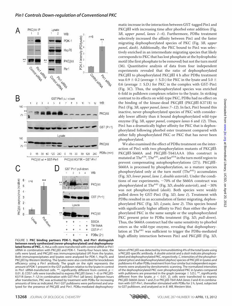
“molecular memory” that controls the history-dependent asso-ciation of PDK-1 and Hsp70 with PKC. To address this, wetested whether depletion of Pin1 affected the binding of thesetwo proteins to PKC. We expressed PKC��� and Myc-PDK-1together with control siRNA or Pin1-specific siRNA in HeLacells, immunoprecipitated PKC��� from the lysate, and ana-lyzed its interaction with PDK-1 or Hsp70 by Western blot. Incontrol cells expressing endogenous Pin1, we observed basalinteractions among PKC���, PDK-1, and Hsp70 (Fig. 3A, lane1). Knockdown of Pin1 caused an �8-fold increase in theamount of PDK-1 bound to PKC��� (Fig. 3A, PDK-1 blot, lane2). In contrast, knockdown of Pin1 had no significant effect onthe interaction between Hsp70 and PKC��� (Fig. 3A, Hsp70blot, lane 2). Because Hsp70 itself has Pro isomerase activity(48), it may not discriminate between cis and trans conformersof the turn motif. However, our data clearly establish that thebinding of PDK-1 to PKC is dramatically increased in cells lack-ing Pin1, consistent with the model that isomerization of theturn motif by Pin1 provides the molecular memory that allowsPDK-1 to discriminate between never-phosphorylated anddephosphorylated PKC.Pin1 Distinguishes between Mature and Immature PKCs—
Given that Pin1 controls interactions that are dependent on theconformational state of PKC, we asked whether the PKC-Pin1interaction was itself conformation-dependent. To discrimi-nate between phosphorylated, dephosphorylated, and unphos-phorylated (i.e. never-phosphorylated) PKC, we used wild-typePKC��� (phosphorylated and dephosphorylated) and kinase-dead (K371R) PKC��� (unphosphorylated). Phorbol esters trig-ger the dephosphorylation of mature, fully phosphorylatedPKC���. On the other hand, kinase-dead PKC��� (K371R) isnever phosphorylated and hence not dephosphorylated afterPDBu treatment. COS7 cells were co-transfected with GST-Pin1 and either wild-type PKC��� or PKC���-K371R, and theinteraction between Pin1 and the kinase wasmonitored follow-ing phorbol ester treatment of cells (Fig. 3B). In the absence ofPDBu, wild-type PKC��� migrated as a single band on SDS-PAGE corresponding to the migration position of kinase quan-titatively phosphorylated at the two C-terminal sites (Fig. 3B,lower panel, lane 1; the fully phosphorylated species is indicatedwith a double asterisk). PDBu treatment resulted in the accu-mulation of a faster migrating species corresponding to PKCdephosphorylated at bothC-terminal sites (Fig. 3B, lower panel,lanes 1–6) (37). GST pulldown experiments revealed a dra-
FIGURE 2. Conventional PKC and Pin1 interact via determinants in PKC C-terminal hydrophobic motif. A and B, His-Pin1 was overlaid on an array of 18-merpeptides derived from the C-terminal tail of rat PKC�II (A) or PKC� (B) with either a non-phosphorylated residue at the turn motif (Thr641 or Ser643; indicated byan asterisk) or a phosphorylated residue at the turn motif (Thr(P)641 or Ser(P)643), and binding was detected as described under “Experimental Procedures.” Thepeptide sequences used in the peptide array are indicated to the right of the blot; each peptide shares 16 amino acids in common with the next peptide in thearray. Sequences in red indicate peptides that bound strongly to Pin1; sequences in green indicated peptides that only displayed significant binding to Pin1when the turn motif Thr was phosphorylated. C, GST pulldowns and lysates from COS7 cells expressing GST alone (lane 1) or the GST-tagged C-terminal regionof PKC�II (lane 2) in combination with HA-Pin1 were analyzed by Western blotting (WB) using GST and HA antibodies. D, an “Ala scan array” comprising theregion in PKC�II found to interact with Pin1 (from A; residues 639 – 663 in rat �II with Ala substitutions at each position) was incubated with pure His-Pin1, andthe amount of binding was determined using the chemiluminescent signal from an HRP-conjugated anti-His antibody. The residue in the original peptide (ontop) replaced with Ala in the scan is indicated in red. Residues required for the interaction with Pin1 are indicated in light gray boxes in the sequence on the rightand with red asterisks on the left of the strip; those that influence the interaction but are not strictly required are boxed in yellow and indicated with a gray asteriskon the left of the strip. E, HeLa cells were transfected with wild-type PKC�II and GST (lane 1), wild-type PKC�II and GST-Pin1 (lane 2), the TP motif mutantPKC�II-P642A and GST-Pin1 (lane 3), or the hydrophobic motif mutant PKC�II-F656A/F659A and GST-Pin1 (lane 4). Cells were lysed 24 h post-transfection, andthe resulting cleared lysates were subjected to PKC�II immunoprecipitation (IP). Pin1 that co-immunoprecipitated with PKC�II was detected using a GSTantibody. F–H, COS7 cells were transfected with conventional (F), atypical (G), and novel (H) PKC isozymes in combination with GST or GST-Pin1 as indicated.Lysates and GST pulldowns were subsequently analyzed for the presence of GST and the various PKC isozymes as described under “Experimental Procedures.”
Pin1 Controls Down-regulation of Conventional PKC
APRIL 13, 2012 • VOLUME 287 • NUMBER 16 JOURNAL OF BIOLOGICAL CHEMISTRY 13267
matic increase in the interaction betweenGST-tagged Pin1 andPKC��� with increasing time after phorbol ester addition (Fig.3B, upper panel, lanes 1–6). Furthermore, PDBu treatmentselectively increased the affinity between Pin1 and the fastermigrating dephosphorylated species of PKC (Fig. 3B, upperpanel, dash). Additionally, the PKC bound to Pin1 was selec-tively enriched in an intermediate migrating species that likelycorresponds to PKC that has lost phosphate at the hydrophobicmotif (the first phosphate to be removed) but not the turnmotif(36). Quantitative analysis of data from four independentexperiments revealed that the ratio of dephosphorylatedPKC��� to phosphorylated PKC��� 4 h after PDBu treatmentwas 0.9 � 0.2 (average � S.D.) for PKC in the lysate and 5.0 �0.6 (average � S.D.) for PKC in the complex with GST-Pin1(Fig. 3C). Thus, the unphosphorylated species was enriched6-fold in pulldown complexes relative to the lysate. In strikingcontrast to its effects on wild-type PKC, PDBu had no effect onthe binding of the kinase-dead PKC��� (PKC���-K371R) toPin1 (Fig. 3B, upper panel, lanes 7–12). In fact, Pin1 bound thisinactive, never-phosphorylated species of PKC with consider-ably lower affinity than it bound dephosphorylated wild-typeenzyme (Fig. 3B, upper panel, compare lanes 6 and 12). Thus,Pin1 has a dramatically higher affinity for PKC that is dephos-phorylated following phorbol ester treatment compared witheither fully phosphorylated PKC or PKC that has never beenphosphorylated.We also examined the effect of PDBu treatment on the inter-
action of Pin1 with two phosphorylation mutants of PKC���:PKC���-S660A and PKC���-T641AAA (this construct ismutated atThr634, Thr641, and Ser654 in the turnmotif region toprevent compensating autophosphorylations (27)). PKC���-S660A is processed by phosphorylation, so a mature speciesphosphorylated only at the turn motif (Thr641) accumulates(Fig. 3D, lower panel, lane 1, double asterisk). Under the condi-tions of our experiments, �70% of the S660A construct wasphosphorylated at Thr641 (Fig. 3D, double asterisk), and �30%was not phosphorylated (dash). Both species were weaklypulled down by GST-Pin1 (Fig. 3D, lane 1). Treatment withPDBu resulted in an accumulation of faster migrating, dephos-phorylated PKC (Fig. 3D, Lysate, lane 2). This species boundwith significantly higher affinity to Pin1 than either the phos-phorylated PKC in the same sample or the unphosphorylatedPKC present prior to PDBu treatment (Fig. 3D, pull-down).Thus, the S660A construct had the same sensitivity to phorbolesters as the wild-type enzyme, revealing that dephosphory-lation at Thr641 was sufficient to trigger the PDBu-mediatedhigh affinity interaction between Pin1 and PKC��� (Fig. 3D,
FIGURE 3. PKC binding partners PDK-1, Hsp70, and Pin1 distinguishbetween newly synthesized (never-phosphorylated) and dephosphory-lated forms of PKC. A, HeLa cells were transfected with control siRNA or Pin1siRNA in combination with PKC��� and PDK-1. Twenty-four hours later, thecells were lysed, and PKC��� was immunoprecipitated (IP) from the lysates.Both immunoprecipitates and lysates were analyzed for PDK-1, Hsp70, andPKC��� by Western blotting. The lysates were also controlled for knockdownefficiency using a Pin1 antibody. The graph on the right represents theamount of PDK-1 present in the GST pulldown relative to the lysate in controlin Pin1 siRNA-transfected cells. **, significantly different from control, p �0.01. B, COS7 cells were transfected to express PKC��� (lanes 1– 6) or PKC���-K371R (lanes 7–12) in combination with GST-Pin1 (all lanes). Eighteen hoursafter transfection, PKC was activated by treatment with PDBu for increasingamounts of time as indicated. Pin1 GST pulldowns were performed and ana-lyzed for the presence of PKC��� and Pin1. PDBu-mediated dephosphory-
lation of PKC��� was detected by immunoblotting 4% of the total lysate usinga PKC���-specific antibody. A double asterisk and a dash indicate phosphory-lated and dephosphorylated PKC, respectively. C, intensities of the phosphor-ylated (phos) and dephosphorylated (dephos) species of PKC��� in lysates andpulldowns 4 h after PDBu treatment from four similar but independent exper-iments were analyzed by densitometric scanning. The normalized intensitiesof the dephosphorylated PKC over phosphorylated PKC in lysates comparedwith pulldowns are presented in the graph (average � S.D.). **, significantlydifferent from the lysate, p � 0.01. D, cells were transfected to expressPKC���-S660A (lanes 1 and 2) or PKC���-T641AAA (lanes 3 and 4) in combina-tion with GST-Pin1, thereafter stimulated with PDBu for 2 h, lysed, subjectedto GST pulldown, and analyzed as in B. WB, Western blot.
Pin1 Controls Down-regulation of Conventional PKC
13268 JOURNAL OF BIOLOGICAL CHEMISTRY VOLUME 287 • NUMBER 16 • APRIL 13, 2012
pull-down, PKC��� blot). In contrast, analysis of GST pull-downs from cells expressing PKC���-T641AAA showed nodramatic change in the interaction with Pin1 after PDBu treat-ment (Fig. 3D, lanes 3 and 4). This result is consistent with ourprevious observation that Pin1 selectively binds PKC��� thathas been dephosphorylated compared with PKC that has neverbeen phosphorylated (Fig. 3B). We also noted that overexpo-sure of blots from such experiments revealed a clear laddering/smearing of PKC following phorbol ester treatment (data notshown), suggesting that the PKC interacting with Pin1 afterPDBu treatment may be ubiquitinated.Pin1 Controls Ubiquitination of PKC� and PKC�II but not
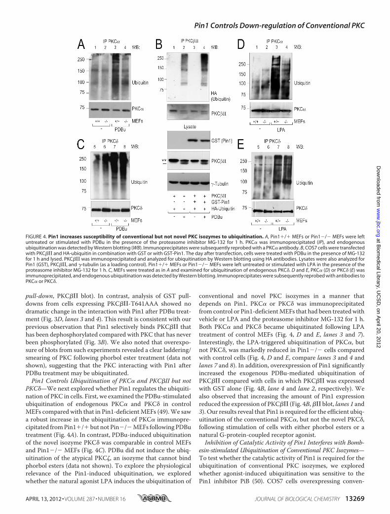
PKC�—We next explored whether Pin1 regulates the ubiquiti-nation of PKC in cells. First, we examined the PDBu-stimulatedubiquitination of endogenous PKC� and PKC� in controlMEFs compared with that in Pin1-deficientMEFs (49).We sawa robust increase in the ubiquitination of PKC� immunopre-cipitated fromPin1�/�but not Pin�/�MEFs followingPDButreatment (Fig. 4A). In contrast, PDBu-induced ubiquitinationof the novel isozyme PKC� was comparable in control MEFsand Pin1�/� MEFs (Fig. 4C). PDBu did not induce the ubiq-uitination of the atypical PKC�, an isozyme that cannot bindphorbol esters (data not shown). To explore the physiologicalrelevance of the Pin1-induced ubiquitination, we exploredwhether the natural agonist LPA induces the ubiquitination of
conventional and novel PKC isozymes in a manner thatdepends on Pin1. PKC� or PKC� was immunoprecipitatedfrom control or Pin1-deficientMEFs that had been treatedwithvehicle or LPA and the proteasome inhibitor MG-132 for 1 h.Both PKC� and PKC� became ubiquitinated following LPAtreatment of control MEFs (Fig. 4, D and E, lanes 3 and 7).Interestingly, the LPA-triggered ubiquitination of PKC�, butnot PKC�, was markedly reduced in Pin1�/� cells comparedwith control cells (Fig. 4, D and E, compare lanes 3 and 4 andlanes 7 and 8). In addition, overexpression of Pin1 significantlyincreased the exogenous PDBu-mediated ubiquitination ofPKC��� compared with cells in which PKC��� was expressedwith GST alone (Fig. 4B, lane 4 and lane 2, respectively). Wealso observed that increasing the amount of Pin1 expressionreduced the expression of PKC��� (Fig. 4B,��� blot, lanes 1 and3). Our results reveal that Pin1 is required for the efficient ubiq-uitination of the conventional PKC�, but not the novel PKC�,following stimulation of cells with either phorbol esters or anatural G-protein-coupled receptor agonist.Inhibition of Catalytic Activity of Pin1 Interferes with Bomb-
esin-stimulated Ubiquitination of Conventional PKC Isozymes—To test whether the catalytic activity of Pin1 is required for theubiquitination of conventional PKC isozymes, we exploredwhether agonist-induced ubiquitination was sensitive to thePin1 inhibitor PiB (50). COS7 cells overexpressing conven-
FIGURE 4. Pin1 increases susceptibility of conventional but not novel PKC isozymes to ubiquitination. A, Pin1�/� MEFs or Pin1�/� MEFs were leftuntreated or stimulated with PDBu in the presence of the proteasome inhibitor MG-132 for 1 h. PKC� was immunoprecipitated (IP), and endogenousubiquitination was detected by Western blotting (WB). Immunoprecipitates were subsequently reprobed with a PKC� antibody. B, COS7 cells were transfectedwith PKC��� and HA-ubiquitin in combination with GST or with GST-Pin1. The day after transfection, cells were treated with PDBu in the presence of MG-132for 1 h and lysed. PKC��� was immunoprecipitated and analyzed for ubiquitination by Western blotting using HA antibodies. Lysates were also analyzed forPin1 (GST), PKC���, and �-tubulin (as a loading control). Pin1�/� MEFs or Pin1�/� MEFs were left untreated or stimulated with LPA in the presence of theproteasome inhibitor MG-132 for 1 h. C, MEFs were treated as in A and examined for ubiquitination of endogenous PKC�. D and E, PKC� (D) or PKC� (E) wasimmunoprecipitated, and endogenous ubiquitination was detected by Western blotting. Immunoprecipitates were subsequently reprobed with antibodies toPKC� or PKC�.
Pin1 Controls Down-regulation of Conventional PKC
APRIL 13, 2012 • VOLUME 287 • NUMBER 16 JOURNAL OF BIOLOGICAL CHEMISTRY 13269
tional PKC� or PKC�II or novel PKC� as a control were pre-treated with vehicle or with a Pin1 inhibitor (PiB) for 2 hand then stimulated with the natural agonist bombesin or LPA(in the presence of MG-132) for 30 min or 1 h. Subsequently,the specific PKC isozyme was immunoprecipitated and ana-lyzed for ubiquitination byWestern blotting. Bombesin5 treat-ment of COS7 cells caused a readily detectable increase in theubiquitination of PKC isozymes, whereas the LPA effects werevariable in these cells. Therefore, we used bombesin to induceubiquitination of PKC in COS7 cells. Bombesin triggered arobust ubiquitination of PKC� (Fig. 5A, lanes 1–3) that wasabolished in the presence of the Pin1 inhibitor (lanes 4–6).Similar results were observed for PKC�II (see Fig. 5C). In con-trast, the bombesin-triggered ubiquitination of the novelisozyme PKC� (Fig. 5B) was unaffected by Pin1 inhibition.
These data reveal that the prolyl isomerase activity of Pin1 isrequired to convert the conventional PKC isozymes � and �IIinto readily ubiquitinated species. In contrast, ubiquitination ofPKC�, which lacks Pin1 binding and the residues necessary forisomerization, occurs independently of Pin1.Next, we mutated residues in PKC�II identified as essential
for Pin1 binding or isomerization and asked how mutation ofthese residues affected agonist-evoked ubiquitination of PKC.Specifically, we tested the effect of Pin1 inhibition on the bomb-esin-evoked ubiquitination of wild-type PKC�II, PKC�II-P642A (loss of isomerizable residue), and PKC�II-F656A/F659A (loss of Pin1 binding). PiB strongly inhibited thebombesin-triggered ubiquitination of wild-type PKC�II (Fig.5C) but had little effect on the ubiquitination of the P642Amutant (Fig. 5D). Bombesin triggered significant ubiquitinationof PKC�II-F656A/F659A as well, but importantly, this ubiq-uitination was relatively insensitive to Pin1 inhibition (Fig. 5E)compared with that of the wild-type enzyme. These data are
5 Note that bombesin treatment of MEFs did not induce ubiquitination of anyPKC isoforms; it is not known whether fibroblasts express bombesin receptors.
FIGURE 5. Inhibition of catalytic activity of Pin1 interferes with bombesin-stimulated ubiquitination of conventional but not novel PKC isozymes. Aand B, COS7 cells transfected with PKC� (A) or PKC� (B) were pretreated with vehicle or the Pin1 inhibitor PiB for 2 h and thereafter stimulated with bombesinin the presence of MG-132 for 0, 30, or 60 min. The overexpressed PKC isozyme was immunoprecipitated (IP), and the amount of the isozyme and degree ofubiquitination were analyzed by Western blot (WB). C–E, COS7 cells were transfected to express wild-type PKC�II (C), PKC�II-P642A (D), or PKC�II-F656A/F659A(E). Approximately 24 h after transfection, the cells were pretreated with vehicle or PiB for 2 h and then stimulated with bombesin in the presence of MG-132for 0, 30, or 60 min (�) to induce ubiquitination. Each PKC mutant was immunoprecipitated and analyzed for endogenous ubiquitination and total PKC�II levels.F, COS7 cells overexpressing PKC� and either HA-Lys48-ubiquitin or HA-Lys63-ubiquitin were stimulated with bombesin for the indicated times, immunopre-cipitated, and analyzed for ubiquitination by probing Western blots with an anti-HA antibody. G, COS7 cells were co-transfected with PKC� and HA-Lys48-ubiquitin. Transfected cells were pretreated with PiB for 2 h and then stimulated with bombesin for the indicated times. Subsequently, PKC� was immuno-precipitated and analyzed for ubiquitination by Western blotting. H, COS7 cells were co-transfected with PKC� and HA-Lys63-ubiquitin (Ubiq) and analyzed asin G.
Pin1 Controls Down-regulation of Conventional PKC
13270 JOURNAL OF BIOLOGICAL CHEMISTRY VOLUME 287 • NUMBER 16 • APRIL 13, 2012
consistent with Pin1 binding to the hydrophobic motif beingessential for isomerization of the turn motif. Furthermore, dis-ruption of the interaction between Pin1 and PKC abolishes thesensitivity of agonist-evoked ubiquitination to Pin1 inhibition.Note that the high agonist-evoked ubiquitination of the P642Aand F656A/F659A mutants likely results from decreased pro-tein stability of these constructs.We next addressed the nature of the bombesin-induced
ubiquitin linkage on conventional PKC isozymes. The func-tional outcome of polyubiquitination of proteins depends onthe lysine utilized in the formation of the ubiquitin chain (51).Typically, substrates destined for degradation through theproteasomal pathway are targeted with a ubiquitin chain inwhich at least four successive ubiquitins are linked togetherthrough an isopeptide bond between Lys48 in the last ubiquitinin the chain and theC-terminal Gly (Gly76) in the newubiquitinmolecule (52). Ubiquitin chains can also be formed throughconjugation to Lys63 in the ubiquitin molecule. It has been sug-gested that the latter type of ubiquitin chain is not a target forproteasomal degradation but rather acts as a signal in severalnon-degradative processes in a cell (53). It is currently notknown what type(s) of ubiquitin chain becomes attached toconventional PKC isozymes during activation-induced down-regulation; therefore, to determine this, we transfected COS7cells with PKC� in combination with either HA-tagged ubiqui-tin inwhich only Lys48 is available for conjugation (Fig. 5F, lanes1–3, K48-Ubiq) or HA-ubiquitin where only Lys63 is available(lanes 4–6, K63-Ubiq). To prevent endogenous ubiquitin fromcompeting with the modified transfected ubiquitin, we trans-fected a 3-fold excess of ubiquitin cDNA compared with kinasecDNAand analyzed immunoprecipitated PKC� usingHA anti-bodies detecting only the transfected ubiquitin. The Westernblot in Fig. 5F reveals that bombesin induced similar incorpo-ration of Lys48-linked and Lys63-linked ubiquitin chains onPKC�. Comparable results were obtained using PKC�II (datanot shown). Inclusion of the Pin1 inhibitor PiB in these exper-iments revealed that the Lys48-linked (Fig. 5G) but not Lys63-linked (Fig. 5H) ubiquitination of PKC� was controlled by thecatalytic activity of Pin1. These data reveal that Pin1 selectivelycontrols the Lys48-linked ubiquitination of conventional PKCthat leads to proteasomal degradation.Pin1 Decreases Basal Protein Stability of Conventional PKC
Isozymes—Because Pin1 regulates the ubiquitination of PKC�and PKC��� in response to natural agonists, we investigatedwhether Pin1 affects the rate of degradation of these isozymes.Specifically, we examined the effect of Pin1 knockdown on thelevels of PKC�, PKC���, PKC�, and PKC� following cyclohex-imide treatment. HeLa cells were treatedwith control siRNAorPin1 siRNA for 72 h to induce efficient Pin1 knockdown.Thereafter, cells were treated with cycloheximide to preventnew protein synthesis and harvested at the time points indi-cated in Fig. 6A. Under conditions of efficient knockdown ofPin1 (Fig. 6A, top two panels), we observed the following effectson PKC isozymes: PKC�, PKC���, and PKC�were considerablyless stable in Pin1-expressing cells compared with Pin1 knock-down cells, whereas PKC� was equally stable in the presence orabsence of Pin1. Quantitation of Western blots from Fig. 6A(see graphs) revealed that PKC degradation was�3-fold slower
in Pin1 knockdown cells compared with control cells: about50% of both PKC� and PKC��� was degraded in 3 h in controlcells comparedwith 9 h in Pin1 knockdown cells, and PKC�wasdegraded with a half-life of�7 h in control cells compared with12 h in knockdown cells. In striking contrast, Pin1 knockdownhad no detectable effect on the rate of degradation of PKC�: thehalf-life of PKC� was 6 h in control and knockdown cells. Thisresult is consistent with the finding that Pin1 does not bindPKC� (Fig. 2H) or alter its ubiquitination state (Fig. 4,A and B).Wenext examinedwhether Pin1 affects the synthesis ormat-
uration of conventional PKCs using cells depleted of Pin1.Newly synthesized conventional PKCs are processed by a seriesof ordered phosphorylations that can be visualized by a mobil-ity shift in pulse-chase experiments. The first phosphorylationbyPDK-1doesnotcauseamobility shift,whereas thephosphor-ylations at the two C-terminal positions do (37). To address theeffects of Pin1 on the maturation of PKC���, cells were pulsedwith 35S-labeled Met/Cys for 7 min to label the unphosphoryl-ated, newly synthesized pool of PKC. After this, cells werechased in medium containing unlabeled Met/Cys for 10, 30, or60 min to monitor the mobility shifts accompanying C-termi-nal autophosphorylation of the newly synthesized PKC. Theautoradiogram showing immunoprecipitated PKC from suchpulse-chase experiments revealed that endogenous PKC��� inHT1080 cells was processed at a comparable rate in controlcells (Fig. 6C, lanes 1–4) and in cells where Pin1 was knockeddown (24 h after transfection) (Fig. 6C, lanes 5–8): the ratio ofphosphorylated (double asterisk) to unphosphorylated PKC(dash) was similar at all chase points (Fig. 6C, compare e.g. lanes3 and 7) with a half-time of processing on the order of 30 min.Given that Pin1 depletion decreases the rate of conventional
PKC degradation without affecting synthesis, we hypothesizedthat the steady-state levels of conventional PKCs would beincreased in these cells. Indeed, in HeLa cells depleted of Pin1,the levels of PKC� and PKC�II were increased by�50% at 24 hafter transfection (Fig. 6B). We validated the decrease in con-ventional PKC expression induced by Pin1 by overexpressingincreasing amounts of GST-tagged Pin1 in COS7 cells andexamining the effects on PKC��� levels. At the highest ratio ofGST-Pin1 to PKC��� (Fig. 6D, lane 5), Pin1 reduced the proteinlevels of PKC��� 5-fold to 21 � 15% (Fig. 6E; average � S.D.,n 7) of the levels in control cells transfected with PKC��� andGST alone. Taken together, these data suggest that Pin1decreases the steady-state levels of conventional PKC isozymesby increasing their rate of degradation.Finally, we assessed the effects of Pin1 depletion on the
intrinsic catalytic activity of PKCby analyzing the specific activ-ity of PKC in Pin1-deficient cells. Lipid-dependent activity wasmeasured in lysates from control cells or Pin1 knockdown cells.Pin1 knockdown by siRNA did not significantly affect the lipid-dependent specific activity of PKC (Fig. 6F). In all kinase assays,activity in the lysate was normalized to the expression of PKC�.The specific activity of PKC from control MEFs and Pin1�/�MEFs was also the same (data not shown). These data revealthat Pin1 regulates the amount of PKC in cells and that thespecific activity of this PKC is unaffected by the presence orabsence of Pin1.
Pin1 Controls Down-regulation of Conventional PKC
APRIL 13, 2012 • VOLUME 287 • NUMBER 16 JOURNAL OF BIOLOGICAL CHEMISTRY 13271
FIGURE 6. Pin1 decreases basal protein stability of conventional PKC isozymes. A, control or Pin1 siRNA-transfected HeLa cells were treated with cyclo-heximide (2 M) for 0, 3, 6, 9, or 12 h to block the synthesis of new proteins. Lysates were analyzed for the loss of Pin1 (knockdown) and �-tubulin (for equalloading) as well as PKC�, PKC���, PKC�, and PKC�. Densitometric analysis of the Western blots (WB) is presented in the graphs to the right. B, HeLa cells weretransfected with control or Pin1 siRNA. Seventy-two hours after transfection, cell lysates were analyzed for Pin1 expression and concomitantly PKC� andPKC��� levels. PKC� and PKC��� intensities were analyzed by densitometric scanning and are presented as the average � S.D. (n 3). *, significantly differentfrom control, p � 0.05. Data represent the average S.E. C, HT1080 cells were transfected with control siRNA or Pin1 siRNA for 24 h after which they weremetabolically labeled with [35S]Met/Cys for 7 min and chased for 0, 10, 30, or 60 min. PKC��� was immunoprecipitated (IP) from the lysates and separated bySDS-PAGE. Differentially phosphorylated species of PKC��� were detected by autoradiogram. A double asterisk indicates phosphorylated PKC (mature),whereas a dash indicates the newly synthesized, unphosphorylated PKC. Data represent the average S.E. D and E, cells were transfected with equal amounts ofPKC��� and increasing amounts of GST-Pin1. Lysates were separated by SDS-PAGE, and PKC��� and Pin1 (GST) were detected by Western blotting. A graphicalrepresentation of total PKC��� expression in cells when co-expressed with vector or the maximum amount of Pin1 (average � S.D., n 7) is shown in E. F,lipid-dependent PKC activity was measured in control and Pin1 knockdown cells. The data are presented as lipid-dependent kinase activity relative to totalPKC� in the lysate (average � S.D., n 3). Data represent the average S.E.
Pin1 Controls Down-regulation of Conventional PKC
13272 JOURNAL OF BIOLOGICAL CHEMISTRY VOLUME 287 • NUMBER 16 • APRIL 13, 2012
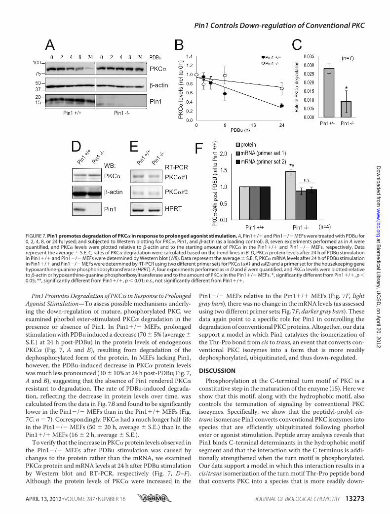
Pin1 PromotesDegradation of PKC� in Response to ProlongedAgonist Stimulation—To assess possible mechanisms underly-ing the down-regulation of mature, phosphorylated PKC, weexamined phorbol ester-stimulated PKC� degradation in thepresence or absence of Pin1. In Pin1�/� MEFs, prolongedstimulationwith PDBu induced a decrease (70� 5% (average�S.E.) at 24 h post-PDBu) in the protein levels of endogenousPKC� (Fig. 7, A and B), resulting from degradation of thedephosphorylated form of the protein. In MEFs lacking Pin1,however, the PDBu-induced decrease in PKC� protein levelswasmuch less pronounced (30� 10% at 24 h post-PDBu; Fig. 7,A and B), suggesting that the absence of Pin1 rendered PKC�resistant to degradation. The rate of PDBu-induced degrada-tion, reflecting the decrease in protein levels over time, wascalculated from the data in Fig. 7B and found to be significantlylower in the Pin1�/� MEFs than in the Pin1�/� MEFs (Fig.7C; n 7). Correspondingly, PKC� had amuch longer half-lifein the Pin1�/� MEFs (50 � 20 h, average � S.E.) than in thePin1�/� MEFs (16 � 2 h, average � S.E.).To verify that the increase in PKC� protein levels observed in
the Pin1�/� MEFs after PDBu stimulation was caused bychanges to the protein rather than the mRNA, we examinedPKC� protein andmRNA levels at 24 h after PDBu stimulationby Western blot and RT-PCR, respectively (Fig. 7, D–F).Although the protein levels of PKC� were increased in the
Pin1�/� MEFs relative to the Pin1�/� MEFs (Fig. 7F, lightgray bars), there was no change in themRNA levels (as assessedusing two different primer sets; Fig. 7F, darker gray bars). Thesedata again point to a specific role for Pin1 in controlling thedegradation of conventional PKCproteins.Altogether, our datasupport a model in which Pin1 catalyzes the isomerization ofthe Thr-Pro bond from cis to trans, an event that converts con-ventional PKC isozymes into a form that is more readilydephosphorylated, ubiquitinated, and thus down-regulated.
DISCUSSION
Phosphorylation at the C-terminal turn motif of PKC is aconstitutive step in thematuration of the enzyme (15). Here weshow that this motif, along with the hydrophobic motif, alsocontrols the termination of signaling by conventional PKCisozymes. Specifically, we show that the peptidyl-prolyl cis-trans isomerase Pin1 converts conventional PKC isozymes intospecies that are efficiently ubiquitinated following phorbolester or agonist stimulation. Peptide array analysis reveals thatPin1 binds C-terminal determinants in the hydrophobic motifsegment and that the interaction with the C terminus is addi-tionally strengthened when the turn motif is phosphorylated.Our data support a model in which this interaction results in acis/trans isomerization of the turn motif Thr-Pro peptide bondthat converts PKC into a species that is more readily down-
FIGURE 7. Pin1 promotes degradation of PKC� in response to prolonged agonist stimulation. A, Pin1�/� and Pin1�/� MEFs were treated with PDBu for0, 2, 4, 8, or 24 h; lysed; and subjected to Western blotting for PKC�, Pin1, and �-actin (as a loading control). B, seven experiments performed as in A werequantified, and PKC� levels were plotted relative to �-actin and to the starting amount of PKC� in the Pin1�/� and Pin1�/� MEFs, respectively. Datarepresent the average � S.E. C, rates of PKC� degradation were calculated based on the trend lines in B. D, PKC� protein levels after 24 h of PDBu stimulationin Pin1�/� and Pin1�/� MEFs were determined by Western blot (WB). Data represent the average � S.E. E, PKC� mRNA levels after 24 h of PDBu stimulationin Pin1�/� and Pin1�/� MEFs were determined by RT-PCR using two different primer sets for PKC� (�#1 and �#2) and a primer set for the housekeeping genehypoxanthine-guanine phosphoribosyltransferase (HPRT). F, four experiments performed as in D and E were quantified, and PKC� levels were plotted relativeto �-actin or hypoxanthine-guanine phosphoribosyltransferase and to the amount of PKC� in the Pin1�/� MEFs. *, significantly different from Pin1�/�, p �0.05; **, significantly different from Pin1�/�, p � 0.01; n.s., not significantly different from Pin1�/�.
Pin1 Controls Down-regulation of Conventional PKC
APRIL 13, 2012 • VOLUME 287 • NUMBER 16 JOURNAL OF BIOLOGICAL CHEMISTRY 13273
regulated by dephosphorylation and ubiquitination: locking theThr-Pro peptidyl bond in trans by replacing the turn motif Prowith Ala locks PKC into a readily down-regulated species,whereas inhibition of Pin1 catalytic activity retains PKC in aconformation that is not readily down-regulated. Thus, ourdata unveil a new model in which Pin1 regulates the conven-tional PKC isozymes by controlling a conformation-dependentswitch in the C-terminal tail that terminates the lifetime of theenzyme.Consensus Thr-Pro of Turn Motif Is Pin1-controlled Timing
Switch—Biochemical studies have revealed that phosphoryla-tion at the turnmotif locks the conventional PKC� and PKC�IIinto catalytically competent, thermally stable species that arerelatively resistant to dephosphorylation at all three processingsites (27, 54). Lack of phosphate at the turn motif shunts con-ventional PKC to the detergent-insoluble fraction where it iseventually degraded. We have also shown that the molecularchaperone Hsp70 can rescue PKC from phorbol ester-directeddegradation: it specifically binds the dephosphorylated turnmotif with data supporting a model in which it allows PKC tobecome rephosphorylated and re-enter the pool of signaling-competent enzyme (41, 42). Thus, the turn motif both controlsthe processing of conventional PKC by phosphorylation andfollowing dephosphorylation recruits Hsp70 to sustain the sig-naling lifetime of the enzyme.Our data unveil an added level of complexity to regulation by
turn motif phosphorylation: Pin1-catalyzed cis/trans isomer-ization converts conventional PKC into a species that is effi-ciently ubiquitinated and degraded following activation. Pin1interacts with and isomerizes proteins phosphorylated at Ser orThr residues preceding a Pro (55). Consistent with this, ourdata reveal that Pin1 controls the ubiquitination of wild-typePKC�II whose turn motif phosphorylation site comprises theconsensus TP sequence but not the construct in which the Prois mutated to Ala (P642A) nor the novel isozyme PKC� whoseturn motif phosphoacceptor site is not followed by Pro.Although the consensus binding site for Pin1 has been shown tocomprise a (Ser(P)/Thr(P))-Pro motif, we show here that thephosphorylated PKC�II TPmotif is not the only determinant ofthe interaction between Pin1 and PKC. Additional interactionswith the segment around the hydrophobic motif control bind-ing, revealing an unconventional, novel consensus for Pin1interaction. Peptide array analysis identified a specific segmentsurrounding the hydrophobic phosphorylation motif thatmediates binding to Pin1. Note that although many of the keyresidues in this segment identified in the array are conservedamong conventional PKC isozymes a few are not. It is possiblethat weakening of one interaction is compensated by strength-ening in another elsewhere in the segment.The conversion of conventional PKC into a species that can
be ubiquitinated also requires the catalytic activity of Pin1.First, we show that the Pin1 inhibitor PiB abolishes the agonist-induced ubiquitination of the Pin1-dependent isozymes PKC�and PKC�II but has no effect on the agonist-induced ubiquiti-nation of the novel isozyme PKC� or the Pin1-independentPKC�II mutants P642A and F656A/F659A (Fig. 5, A–E). Thus,the effects of Pin1 depend on the intrinsic isomerase activity ofthe enzyme and the integrity of both the hydrophobic motif
and the phospho-Thr-Pro consensus sequence at the turnmotif.Analysis of the solved structure of the PKC�II kinase core
suggests a structural basis for how Pin1-catalyzed isomeriza-tion of the Thr-Pro peptide bond of the turn motif can convertPKC into a species that is readily ubiquitinated (56). In thesolved structure, the Thr-Pro bond is in trans, resulting in thephospho-Thr at position 641 being relatively solvent-exposed.Consistent with exposure, this site is sensitive to dephosphory-lation when mature PKC�II is in the active, membrane-bound,and “open” conformation (34). Ubiquitination follows dephos-phorylation of PKC that has been activated. Molecular model-ing of the Thr-Pro bond in cis reveals that the phosphate nowpivots into the core of the protein, potentially forming an elec-trostatic interaction with Arg415 (Fig. 8A). Importantly, Arg415is invariant among all PKC isozymes that have the Thr-Promotif: it is present in conventional PKC� and PKC� but is notpresent in novel PKC�, which lacks theThr-Pro sequence at theturn motif. This residue forms part of a basic binding pocketrecently identified by Frödin and co-workers (57) as promotinga “zipper-like” association of the C-terminal tail turn motif andhydrophobic motif of AGC kinases with the upper lobe of thekinase core. Our data reveal that this “zipper”-like association isloosened by Pin1 through isomerization, exposing the phos-phorylated C-terminal sites to dephosphorylation.Functional Effects of Pin1 on PKC—Pin1 does not affect the
rate of processing of conventional PKC by phosphorylation nordoes it affect the catalytic activity of conventional PKC. Rather,Pin1 accelerates the agonist-evoked down-regulation of con-ventional PKC isozymes.The novel isozyme PKC� does not contain the TP consensus
motif and lacks several key residues in the hydrophobic motifimportant for PKC�II-Pin1 interaction. Consistent with themodel that the TPmotif is required for Pin1 to control the turnmotif, Pin1 did not interact with PKC� nor was the agonist-induced ubiquitination of PKC� sensitive to Pin1 inhibition.Because novel isozymes lack the TPmolecular timer, we wouldpredict that they are synthesized in a conformation that isalready degradation-sensitive. Consistent with this, Blumbergand co-workers (58) have shown that the rate of phorbol ester-mediated down-regulation of the novel PKC isozymes � and � is�5 times faster than that of the conventional PKC�.
There is precedent for Pin1 destabilizing signaling proteinsas we report here for conventional PKC isozymes. The stabilityof c-Myc is also increased in cells lacking Pin1: protein expres-sion and the half-life of c-Myc are increased in a manner anal-ogous to our finding for conventional PKC (59). In contrast,Pin1 stabilizes several other proteins. p65 (RelA) levels are up-regulated, and the protein has increased nuclear accumulationwhen Pin1 levels are elevated (60). In addition, genotoxin-me-diated accumulation of p73 (61) and p53 (62, 63) is impaired inPin1-deficient cells. Thus, Pin1 controls conformationalswitches that regulate the lifetime of signaling molecules byboth positive and negative mechanisms.Pin1 has also been proposed to control the mitochondrial
accumulation of the adapter protein p66Shc following phos-phorylation by PKC�II (64). Furthermore, it was recentlyshown that Pin1 interacts with PKC� and that PKC� can phos-
Pin1 Controls Down-regulation of Conventional PKC
13274 JOURNAL OF BIOLOGICAL CHEMISTRY VOLUME 287 • NUMBER 16 • APRIL 13, 2012
phorylate Pin1 (65). Thus, Pin1 is emerging as a novel regulatorof PKC signaling function both through directly controlling thelifetime of conventional PKC isozymes and by controlling thefunction of downstream targets of PKC.Pin1 Is aMolecular Timer in Life Cycle of Conventional PKC—
Fig. 8B presents a model for how cis/trans isomerization cata-
lyzed by Pin1 controls the lifetime of conventional PKCisozymes. Our data are consistent with a model in which thepeptidyl-prolyl bond at the turn motif of newly synthesizedconventional PKC isozymes is in a cis conformation (Fig. 8B,Species 1). PDK-1 docks onto the C-terminal tail of newly syn-thesized PKC and phosphorylates the activation loop (Thr500),
FIGURE 8. Cis/trans isomerization of TP motif acts as switch to control down-regulation of conventional PKC isozymes. A, conformational changes in theturn motif that may occur upon cis/trans isomerization of phospho-Thr641-Pro642 peptide bond in conventional PKC�II are shown. In the trans state (left panel),Thr(P)641 is relatively solvent-exposed, whereas in the cis state (right panel) Thr(P)641 points toward key charged residues (Arg415, Lys355, and Lys374) inthe catalytic domain. Arg415 may play an important role in this conformational transition because it hydrogen bonds to the backbone of Thr(P)641 in thetrans state but appears to coordinate the phosphate group of Thr641 in the cis state. Key regions of the catalytic domain and the C-terminal tail are shownin a schematic representation. The C terminus is shown in red, and the catalytic domain is shown in gray. Turn motif (TM) and hydrophobic motif (HM)phosphorylation sites are indicated in red. Residues are shown as sticks, and a hydrogen bond is shown as a dotted line. B, a model depicts the role of Pin1in controlling the life cycle of conventional PKC. PKC is shown as a blue circle with the C-terminal tail indicated by a blue line. The first and required stepin the maturation of conventional PKC is binding of PDK-1 to the C-terminal tail, which is in cis (Species 1; kink in C-terminal tail); PDK-1 then phosphor-ylates the activation loop (Thr500 in PKC�II). This event triggers the phosphorylation of the turn motif (Thr641 in PKC�II) followed immediately byintramolecular autophosphorylation at the hydrophobic motif (Ser660 in PKC�II), resulting in the formation of Species 2. The fully phosphorylated,mature, and catalytically competent conventional PKC localizes to the cytosol in a phosphatase-resistant conformation and is able to interact with Pin1(Species 2). PKC activation does not require Pin1. Pin1 catalyzes a cis/trans isomerization of the phospho-Thr-Pro peptidyl bond in the C-terminal tail(Species 3), which facilitates the dephosphorylation of conventional PKC (Species 4), resulting in its ubiquitination and degradation. Note that binding ofHsp70 to the dephosphorylated turn motif provides a mechanism to recycle PKC into the signaling-competent pool of enzyme presumably byfacilitating the reisomerization (from trans to cis) and phosphorylation of PKC.
Pin1 Controls Down-regulation of Conventional PKC
APRIL 13, 2012 • VOLUME 287 • NUMBER 16 JOURNAL OF BIOLOGICAL CHEMISTRY 13275
rendering PKC catalytically competent (17, 18, 22). Upon dis-engagement of PDK-1 from the C terminus, PKC becomes rap-idly phosphorylated on the turnmotif, an event that depends onmTORC2 (23) and on the intrinsic catalytic activity of PKC.Phosphorylation of the turn motif is required to process func-tional PKC, and it is also the rate-limiting step in the matura-tion of PKC (19). Phosphorylation on the turnmotif is followedimmediately by intramolecular autophosphorylation of thehydrophobic motif (26) to yield the mature, activatable form ofPKC.We suggest that Pin1 binds this fully phosphorylated andcatalytically competent species via determinants surroundingthe hydrophobic motif and via the phosphorylated Thr of theturn motif (Fig. 8B, Species 2). Upon PKC activation, Pin1 cat-alyzes a cis to trans isomerization of the phospho-Thr-Pro bondof the turn motif, unmasking the phospho-Thr (see the mod-eled structure in Fig. 8A and see Fig. 8B, Species 3). Thisunmasking renders PKC much more sensitive to dephosphor-ylation (Fig. 8B, Species 4) following agonist activation, result-ing in the ubiquitination and degradation of PKC. PKC can,however, be rescued from degradation: binding of Hsp70 to thedephosphorylated turn motif recycles PKC into the pool of sig-naling-competent enzyme presumably by permitting the rei-somerization and phosphorylation of PKC (41, 42). Thus,isomerization of conventional PKC isozymes by Pin1 provides aconformation-dependent degradation switch: in the absence ofPin1, conventional PKC isozymes are trapped in a conforma-tion that cannot be efficiently ubiquitinated and down-regu-lated following agonist stimulation. This switch can bebypassed by locking the peptide bond after the phosphorylatedturn motif Thr in trans as occurs in novel PKC isozymes wherethe Pro is absent or by mutagenesis of the PKC�II Pro to Ala asshown in this report.Peptide array data revealed that Pin1 has a higher affinity for
C-terminal peptides with phospho-Thr versus unphosphory-lated Thr at the turn motif, but co-immunoprecipitation stud-ies revealed that Pin1 preferentially binds dephosphorylatedPKC in cells. One possible explanation is that the C-terminaltail of full-length PKC is in amore exposed conformation whenthe turn motif and hydrophobic motif are dephosphorylated,thus favoring Pin1 binding. Similar results occur with the bind-ing of PDK-1: although it binds constructs of the C terminusthat are phosphorylated at the hydrophobic motif with muchhigher affinity than unphosphorylated constructs, in the con-text of the full-length protein, it preferentially binds dephos-phorylated enzyme (21).The Pin1-controlled timing switch on the turn motif of PKC
provides a new level of complexity to the life cycle of theenzymewhere down-regulation of conventional (but not novel)PKC isozymes requires a priming step by Pin1. It remains to beestablished whether the Pin1 step is itself regulated or part ofthe constitutive processing of PKC. Interestingly, isomerizationof theTPmotif provides amolecularmechanism to increase thedegradation of PKC that is unique to the conventional PKCisozymes. Precise control of the amplitude of the PKC signal iscritical to normal physiology, and dysregulation of the levels ofconventional PKC isozymes accompany many diseased states,most notably cancer (66). Whether dysregulation of the Pin1-
mediated down-regulation switch accounts for the elevated lev-els of PKC in these disease states also remains to be explored.
Acknowledgments—We thank Christine Gould, Robert Romero, andCharles C. King, for helpful discussions and Laura A. Braughton fortechnical assistance. We thank Kun Ping Lu for Pin1 constructs formammalian expression and the Pin1�/� MEFs.
REFERENCES1. Wulf, G., Finn, G., Suizu, F., and Lu, K. P. (2005) Phosphorylation-specific
prolyl isomerization: is there an underlying theme? Nat. Cell Biol. 7,435–441
2. Lu, K. P., Hanes, S. D., and Hunter, T. (1996) A human peptidyl-prolylisomerase essential for regulation of mitosis. Nature 380, 544–547
3. Yaffe, M. B., Schutkowski, M., Shen, M., Zhou, X. Z., Stukenberg, P. T.,Rahfeld, J. U., Xu, J., Kuang, J., Kirschner,M.W., Fischer, G., Cantley, L. C.,and Lu, K. P. (1997) Sequence-specific and phosphorylation-dependentproline isomerization: a potential mitotic regulatory mechanism. Science278, 1957–1960
4. Lu, K. P. (2004) Pinning down cell signaling, cancer and Alzheimer’s dis-ease. Trends Biochem. Sci. 29, 200–209
5. Liou, Y. C., Sun, A., Ryo, A., Zhou, X. Z., Yu, Z. X., Huang, H. K., Uchida,T., Bronson, R., Bing, G., Li, X., Hunter, T., and Lu, K. P. (2003) Role of theprolyl isomerase Pin1 in protecting against age-dependent neurodegen-eration. Nature 424, 556–561
6. Wulf, G. M., Ryo, A., Wulf, G. G., Lee, S. W., Niu, T., Petkova, V., and Lu,K. P. (2001) Pin1 is overexpressed in breast cancer and cooperateswithRassignaling in increasing the transcriptional activity of c-Jun towards cyclinD1. EMBO J. 20, 3459–3472
7. Ryo, A., Nakamura, M., Wulf, G., Liou, Y. C., and Lu, K. P. (2001) Pin1regulates turnover and subcellular localization of �-catenin by inhibitingits interaction with APC. Nat. Cell Biol. 3, 793–801
8. Ayala, G., Wang, D., Wulf, G., Frolov, A., Li, R., Sowadski, J., Wheeler,T. M., Lu, K. P., and Bao, L. (2003) The prolyl isomerase Pin1 is a novelprognostic marker in human prostate cancer. Cancer Res. 63,6244–6251
9. Bao, L., Kimzey, A., Sauter, G., Sowadski, J. M., Lu, K. P., andWang, D. G.(2004) Prevalent overexpression of prolyl isomerase Pin1 in human can-cers. Am. J. Pathol. 164, 1727–1737
10. Yeh, E. S., and Means, A. R. (2007) PIN1, the cell cycle and cancer. Nat.Rev. Cancer 7, 381–388
11. Chen, D., Purohit, A., Halilovic, E., Doxsey, S. J., and Newton, A. C. (2004)Centrosomal anchoring of protein kinase C �II by pericentrin controlsmicrotubule organization, spindle function, and cytokinesis. J. Biol. Chem.279, 4829–4839
12. Levin, D. E., Fields, F. O., Kunisawa, R., Bishop, J. M., and Thorner, J.(1990) A candidate protein kinase C gene, PKC1, is required for the S.cerevisiae cell cycle. Cell 62, 213–224
13. Yang, C., and Kazanietz, M. G. (2003) Divergence and complexities inDAG signaling: looking beyond PKC.Trends Pharmacol. Sci. 24, 602–608
14. Sedwick, C. E., and Altman, A. (2004) Perspectives on PKC� in T cellactivation.Mol. Immunol. 41, 675–686
15. Newton, A. C. (2003) Regulation of the ABC kinases by phosphorylation:protein kinase C as a paradigm. Biochem. J. 370, 361–371
16. Newton, A. C. (2010) Protein kinase C: poised to signal. Am. J. Physiol.Endocrinol. Metab. 298, E395–E402
17. Dutil, E. M., Toker, A., and Newton, A. C. (1998) Regulation of conven-tional protein kinase C isozymes by phosphoinositide-dependent kinase 1(PDK-1). Curr. Biol. 8, 1366–1375
18. Le Good, J. A., Ziegler, W. H., Parekh, D. B., Alessi, D. R., Cohen, P., andParker, P. J. (1998) Protein kinase C isotypes controlled by phosphoinosit-ide 3-kinase through the protein kinase PDK1. Science 281, 2042–2045
19. Sonnenburg, E. D., Gao, T., and Newton, A. C. (2001) The phosphoinosit-ide-dependent kinase, PDK-1, phosphorylates conventional protein ki-nase C isozymes by a mechanism that is independent of phosphoinositide3-kinase. J. Biol. Chem. 276, 45289–45297
Pin1 Controls Down-regulation of Conventional PKC
13276 JOURNAL OF BIOLOGICAL CHEMISTRY VOLUME 287 • NUMBER 16 • APRIL 13, 2012
20. Dutil, E. M., and Newton, A. C. (2000) Dual role of pseudosubstrate in thecoordinated regulation of protein kinase C by phosphorylation and dia-cylglycerol. J. Biol. Chem. 275, 10697–10701
21. Gao, T., Toker, A., and Newton, A. C. (2001) The carboxyl terminus ofprotein kinase C provides a switch to regulate its interaction with thephosphoinositide-dependent kinase, PDK-1. J. Biol. Chem. 276,19588–19596
22. Chou, M.M., Hou,W., Johnson, J., Graham, L. K., Lee, M. H., Chen, C. S.,Newton, A. C., Schaffhausen, B. S., and Toker, A. (1998) Regulation ofprotein kinase C� by PI 3-kinase and PDK-1. Curr. Biol. 8, 1069–1077
23. Facchinetti, V., Ouyang, W., Wei, H., Soto, N., Lazorchak, A., Gould, C.,Lowry, C., Newton, A. C.,Mao, Y.,Miao, R.Q., Sessa,W.C.,Qin, J., Zhang,P., Su, B., and Jacinto, E. (2008) The mammalian target of rapamycincomplex 2 controls folding and stability of Akt and protein kinase C.EMBO J. 27, 1932–1943
24. Ikenoue, T., Inoki, K., Yang, Q., Zhou, X., and Guan, K. L. (2008) Essentialfunction of TORC2 in PKC and Akt turn motif phosphorylation, matura-tion and signalling. EMBO J. 27, 1919–1931
25. Oh, W. J., Wu, C. C., Kim, S. J., Facchinetti, V., Julien, L. A., Finlan, M.,Roux, P. P., Su, B., and Jacinto, E. (2010) mTORC2 can associate withribosomes to promote cotranslational phosphorylation and stability ofnascent Akt polypeptide. EMBO J. 29, 3939–3951
26. Behn-Krappa, A., and Newton, A. C. (1999) The hydrophobic phospho-rylation motif of conventional protein kinase C is regulated by autophos-phorylation. Curr. Biol. 9, 728–737
27. Edwards, A. S., Faux, M. C., Scott, J. D., and Newton, A. C. (1999) Carbox-yl-terminal phosphorylation regulates the function and subcellular local-ization of protein kinase C �II. J. Biol. Chem. 274, 6461–6468
28. Bornancin, F., and Parker, P. J. (1996) Phosphorylation of threonine 638critically controls the dephosphorylation and inactivation of protein ki-nase C�. Curr. Biol. 6, 1114–1123
29. Goode, N. T., Hajibagheri, M. A., and Parker, P. J. (1995) Protein kinase C(PKC)-induced PKC down-regulation. Association with up-regulation ofvesicle traffic. J. Biol. Chem. 270, 2669–2673
30. Lee, H. W., Smith, L., Pettit, G. R., and Bingham Smith, J. (1996) Dephos-phorylation of activated protein kinase C contributes to downregulationby bryostatin. Am. J. Physiol. Cell Physiol. 271, C304–C311
31. Lee, H. W., Smith, L., Pettit, G. R., and Smith, J. B. (1997) Bryostatin 1and phorbol ester down-modulate protein kinase C-� and -� via theubiquitin/proteasome pathway in human fibroblasts. Mol. Pharmacol.51, 439–447
32. Lu, Z., Liu, D., Hornia, A., Devonish, W., Pagano, M., and Foster, D. A.(1998) Activation of protein kinase C triggers its ubiquitination and deg-radation.Mol. Cell Biol. 18, 839–845
33. Jaken, S., Tashjian, A. H., Jr., and Blumberg, P. M. (1981) Characterizationof phorbol ester receptors and their down-modulation in GH4C1 rat pi-tuitary cells. Cancer Res. 41, 2175–2181
34. Dutil, E. M., Keranen, L. M., DePaoli-Roach, A. A., and Newton, A. C.(1994) In vivo regulation of protein kinase C by trans-phosphorylationfollowed by autophosphorylation. J. Biol. Chem. 269, 29359–29362
35. Warfel, N. A., and Newton, A. C. (2012) Pleckstrin homology domainleucine-rich repeat protein phosphatase (PHLPP): a new player in cellsignaling. J. Biol. Chem. 287, 3610–3616
36. Gao, T., Brognard, J., and Newton, A. C. (2008) The phosphatase PHLPPcontrols the cellular levels of protein kinase C. J. Biol. Chem. 283,6300–6311
37. Keranen, L. M., Dutil, E. M., and Newton, A. C. (1995) Protein kinase C isregulated in vivo by three functionally distinct phosphorylations. Curr.Biol. 5, 1394–1403
38. Hansra, G., Bornancin, F., Whelan, R., Hemmings, B. A., and Parker, P. J.(1996) 12-O-Tetradecanoylphorbol-13-acetate-induced dephosphory-lation of protein kinase C� correlates with the presence of a membrane-associated protein phosphatase 2A heterotrimer. J. Biol. Chem. 271,32785–32788
39. Hansra,G., Garcia-Paramio, P., Prevostel, C.,Whelan, R.D., Bornancin, F.,and Parker, P. J. (1999)Multisite dephosphorylation and desensitization ofconventional protein kinase C isotypes. Biochem. J. 342, 337–344
40. Leontieva, O. V., and Black, J. D. (2004) Identification of two distinct
pathways of protein kinase C� down-regulation in intestinal epithelialcells. J. Biol. Chem. 279, 5788–5801
41. Gao, T., and Newton, A. C. (2002) The turn motif is a phosphorylationswitch that regulates the binding of Hsp70 to protein kinase C. J. Biol.Chem. 277, 31585–31592
42. Gao, T., and Newton, A. C. (2006) Invariant Leu preceding turn motifphosphorylation site controls the interaction of protein kinase C withHsp70. J. Biol. Chem. 281, 32461–32468
43. Edwards, A. S., and Newton, A. C. (1997) Phosphorylation at conservedcarboxyl-terminal hydrophobic motif regulates the catalytic and regula-tory domains of protein kinase C. J. Biol. Chem. 272, 18382–18390
44. Cenni, V., Dppler, H., Sonnenburg, E. D., Maraldi, N., Newton, A. C., andToker, A. (2002) Regulation of novel protein kinase C� by phosphoryla-tion. Biochem. J. 363, 537–545
45. Kramer, A., and Schneider-Mergener, J. (1998) Synthesis and screening ofpeptide libraries on continuous cellulose membrane supports. MethodsMol. Biol. 87, 25–39
46. Amarzguioui, M., and Prydz, H. (2004) An algorithm for selection of func-tional siRNA sequences. Biochem. Biophys. Res. Commun. 316, 1050–1058
47. Ranganathan, R., Lu, K. P., Hunter, T., andNoel, J. P. (1997) Structural andfunctional analysis of the mitotic rotamase Pin1 suggests substrate recog-nition is phosphorylation dependent. Cell 89, 875–886
48. Schiene-Fischer, C.,Habazettl, J., Schmid, F. X., and Fischer,G. (2002)Thehsp70 chaperone DnaK is a secondary amide peptide bond cis-transisomerase. Nat. Struct. Biol. 9, 419–424
49. Liou, Y. C., Ryo, A., Huang, H. K., Lu, P. J., Bronson, R., Fujimori, F.,Uchida, T., Hunter, T., and Lu, K. P. (2002) Loss of Pin1 function in themouse causes phenotypes resembling cyclin D1-null phenotypes. Proc.Natl. Acad. Sci. U.S.A. 99, 1335–1340
51. Pickart, C. M. (2001) Mechanisms underlying ubiquitination. Annu. Rev.Biochem. 70, 503–533
52. Chau, V., Tobias, J. W., Bachmair, A., Marriott, D., Ecker, D. J., Gonda,D. K., and Varshavsky, A. (1989) A multiubiquitin chain is confined tospecific lysine in a targeted short-lived protein. Science 243, 1576–1583
53. Pickart, C. M., and Fushman, D. (2004) Polyubiquitin chains: polymericprotein signals. Curr. Opin. Chem. Biol. 8, 610–616
54. Zhang, J., Wang, L., Schwartz, J., Bond, R. W., and Bishop, W. R. (1994)Phosphorylation of Thr642 is an early event in the processing of newlysynthesized protein kinase C �1 and is essential for its activation. J. Biol.Chem. 269, 19578–19584
55. Lu, K. P., Liou, Y. C., and Zhou, X. Z. (2002) Pinning down proline-di-rected phosphorylation signaling. Trends Cell Biol. 12, 164–172
56. Grodsky, N., Li, Y., Bouzida, D., Love, R., Jensen, J., Nodes, B., Nonomiya,J., andGrant, S. (2006) Structure of the catalytic domain of human proteinkinase C �II complexed with a bisindolylmaleimide inhibitor. Biochemis-try 45, 13970–13981
57. Hauge, C., Antal, T. L., Hirschberg, D., Doehn, U., Thorup, K., Idrissova,L., Hansen, K., Jensen, O. N., Jørgensen, T. J., Biondi, R.M., and Frödin,M.(2007) Mechanism for activation of the growth factor-activated AGC ki-nases by turn motif phosphorylation. EMBO J. 26, 2251–2261
58. Szallasi, Z., Smith, C. B., and Blumberg, P. M. (1994) Dissociation of phor-bol esters leads to immediate redistribution to the cytosol of protein ki-nases C� andC� inmouse keratinocytes. J. Biol. Chem. 269, 27159–27162
59. Yeh, E., Cunningham, M., Arnold, H., Chasse, D., Monteith, T., Ivaldi, G.,Hahn,W. C., Stukenberg, P. T., Shenolikar, S., Uchida, T., Counter, C.M.,Nevins, J. R., Means, A. R., and Sears, R. (2004) A signalling pathwaycontrolling c-Myc degradation that impacts oncogenic transformation ofhuman cells. Nat. Cell Biol. 6, 308–318
60. Ryo, A., Suizu, F., Yoshida, Y., Perrem, K., Liou, Y. C., Wulf, G., Rottapel,R., Yamaoka, S., and Lu, K. P. (2003) Regulation of NF-�B signaling byPin1-dependent prolyl isomerization and ubiquitin-mediated proteolysisof p65/RelA.Mol. Cell 12, 1413–1426
61. Mantovani, F., Piazza, S., Gostissa, M., Strano, S., Zacchi, P., Mantovani,R., Blandino,G., andDel Sal, G. (2004) Pin1 links the activities of c-Abl and
Pin1 Controls Down-regulation of Conventional PKC
APRIL 13, 2012 • VOLUME 287 • NUMBER 16 JOURNAL OF BIOLOGICAL CHEMISTRY 13277
p300 in regulating p73 function.Mol. Cell 14, 625–63662. Zheng, H., You, H., Zhou, X. Z.,Murray, S. A., Uchida, T.,Wulf, G., Gu, L.,
Tang, X., Lu, K. P., and Xiao, Z. X. (2002) The prolyl isomerase Pin1 is aregulator of p53 in genotoxic response. Nature 419, 849–853
63. Zacchi, P., Gostissa, M., Uchida, T., Salvagno, C., Avolio, F., Volinia, S.,Ronai, Z., Blandino, G., Schneider, C., and Del Sal, G. (2002) The prolylisomerase Pin1 reveals a mechanism to control p53 functions after geno-toxic insults. Nature 419, 853–857
64. Pinton, P., Rimessi, A., Marchi, S., Orsini, F., Migliaccio, E., Giorgio, M.,Contursi, C., Minucci, S., Mantovani, F., Wieckowski, M. R., Del Sal, G.,
Pelicci, P. G., and Rizzuto, R. (2007) Protein kinase C� and prolyl isomer-ase 1 regulate mitochondrial effects of the life-span determinant p66Shc.Science 315, 659–663
65. Shen, Z. J., Esnault, S., Rosenthal, L. A., Szakaly, R. J., Sorkness, R. L.,Westmark, P. R., Sandor, M., and Malter, J. S. (2008) Pin1 regulatesTGF-�1 production by activated human and murine eosinophils andcontributes to allergic lung fibrosis. J. Clin. Investig. 118, 479–490
66. Lahn,M., Kohler, G., Sundell, K., Su, C., Li, S., Paterson, B.M., and Bumol,T. F. (2004) Protein kinase C� expression in breast and ovarian cancer.Oncology 67, 1–10
Pin1 Controls Down-regulation of Conventional PKC
13278 JOURNAL OF BIOLOGICAL CHEMISTRY VOLUME 287 • NUMBER 16 • APRIL 13, 2012