PERITONITIS IN THE NEONATAL PERIOD* BY P. P. RICKHAM From Alder Hey Children's Hospital, Liverpool It has generally been assumed that peritonitis occurs but rarely in newborn infants. In the 1952 Charles West lecture (Moncrieff, 1953) on infection in newborn babies the condition was not mentioned. Thelander, who wrote a classical paper on perfora- tions of the gastro-intestinal tract in newborn infants in 1939, stated that most paediatricians never saw a case, and in 1949 Low, Cooper and Cosby could only find 100 instances in the literature since 1761, when Morgagni first reported the condition in De Sedibus et Causis Morborum. During the last five years, however, many cases of peritonitis in infants have been described and it has become clear that a condition which was formerly thought to be a patho- logical curiosity, is in fact a not uncommon cause of disease and death in the neonatal period. The pathology and aetiology are, as yet, little understood and recent publications have caused confusion by the synonymous use of terms, such as meconium peritonitis, intra-uterine peritonitis, foetal peri- tonitis, and peritonitis in the newborn. This paper is presented in order to describe our experience with neonates suffering from peritonitis, to evaluate the incidence of the disease, and to * A paper given at the inaugural meeting of the British Association of Paediatric Surgeons in London in July, 1954. classify the various types of the condition. It is based upon the case histories of 17 newborn infants seen by us during the last three and a half years. During this period we admitted about 250 babies to the Neonatal Surgical Unit at Alder Hey Children's Hospital; 98 of these had intestinal obstruction (excluding pyloric stenosis and imper- forate anus). It appears, therefore, that about 7 % of all newborn infants admitted for urgent surgical conditions and 17 % of all cases of neonatal intestinal obstruction are suffering from peritonitis. Of the 17 cases under discussion, seven were classified as meconium peritonitis, and 10 as acute bacterial peritonitis. There was a marked prepon- derance of girls, 12, or 70%. This is surprising as it is generally stated (Table 1) that perforation of the intestinal tract during the newborn period affects boys three times more commonly than girls (Thelander, 1939). Physiological and Pathological Considerations Meconium Peritonitis. It is generally accepted that meconium starts to accumulate in the intestine of the 4-month-old embryo, but there is some evidence that the 3-month-old foetus is already swallowing amniotic fluid and passing it along the TABLE I RESULTS IN PRESENT SERIES Finding at Operation Operative Procedure Result Meconium Peritonitis: Zena 10 hrs. I 6 lb. 9 oz. Volvulus of jejunum Resection and anastomosis Recovery John 3 days 5 lb. 2 oz. Ileal atresia ,. ,, Died 14 days later Hilary 2 days 6 lb. Meconium ileus Died 12 hr. later Linda 3 days 5 lb. 2 oz. Perforation of ileum Died three days later Brian 3 weeks 9 lb. 12 oz. Ileal obstruction due to bands Division of bands Recovery Sarah 3 weeks 6 lb. Ileal obstruction due to bands ,, ,, Recovery Joan 2 days 5 lb. 6 oz. Jejunal atresia Resection and anastomosis Died four days later Acute Bacterial Peritonitis: Mary 2 weeks 6 lb. 9 oz. Abscess round terminal ileum Drainage and ileocolostomy Recovery Nancy 2 weeks 5 lb. 14 oz. Abscess in R. iliac fossa Drainage of abscess Recovery Stephen 8 days 3 lb. Perforation of stomach Closure of perforation Died four days later Jean 1 day 5 lb. 7 oz. Gangrenous volvulus of ileum Resection and anastomosis Recovery Lesley 5 days 5 lb. 3 oz. Perforation of ileum ,, Recovery Loraine 3 weeks 6 lb. 8 oz. Pelvic abscess Drainage of abscess Recovery Doreen 3 days 2 lb. Perforation of Meckel's diverticulum Resection and anastomosis Died three days later Alexander 3 weeks 7 lb. 9 oz. Abscess in R. iliac fossa Drainage of abscess Recovery Angela I week 6 lb. Perforated duplication of ileum Resection and anastomosis Died four weeks later Michael 4 weeks 7 lb. 9 oz. Perforated ileum Suture of perforation Recovery 23 Name Age Weight copyright. on April 2, 2020 by guest. Protected by http://adc.bmj.com/ Arch Dis Child: first published as 10.1136/adc.30.149.23 on 1 February 1955. Downloaded from
Transcript
PERITONITIS IN THE NEONATAL PERIOD*BY
P. P. RICKHAMFrom Alder Hey Children's Hospital, Liverpool
It has generally been assumed that peritonitisoccurs but rarely in newborn infants. In the 1952Charles West lecture (Moncrieff, 1953) on infectionin newborn babies the condition was not mentioned.Thelander, who wrote a classical paper on perfora-tions of the gastro-intestinal tract in newborninfants in 1939, stated that most paediatricians neversaw a case, and in 1949 Low, Cooper and Cosby couldonly find 100 instances in the literature since 1761,when Morgagni first reported the condition in DeSedibus et Causis Morborum. During the last fiveyears, however, many cases of peritonitis in infantshave been described and it has become clear that acondition which was formerly thought to be a patho-logical curiosity, is in fact a not uncommon cause ofdisease and death in the neonatal period. Thepathology and aetiology are, as yet, little understoodand recent publications have caused confusion bythe synonymous use of terms, such as meconiumperitonitis, intra-uterine peritonitis, foetal peri-tonitis, and peritonitis in the newborn.
This paper is presented in order to describe ourexperience with neonates suffering from peritonitis,to evaluate the incidence of the disease, and to
* A paper given at the inaugural meeting of the British Associationof Paediatric Surgeons in London in July, 1954.
classify the various types of the condition. It isbased upon the case histories of 17 newborn infantsseen by us during the last three and a half years.During this period we admitted about 250 babiesto the Neonatal Surgical Unit at Alder HeyChildren's Hospital; 98 of these had intestinalobstruction (excluding pyloric stenosis and imper-forate anus). It appears, therefore, that about 7%of all newborn infants admitted for urgent surgicalconditions and 17% of all cases of neonatalintestinal obstruction are suffering from peritonitis.Of the 17 cases under discussion, seven were
classified as meconium peritonitis, and 10 as acutebacterial peritonitis. There was a marked prepon-derance of girls, 12, or 70%. This is surprisingas it is generally stated (Table 1) that perforationof the intestinal tract during the newborn periodaffects boys three times more commonly than girls(Thelander, 1939).
Physiological and Pathological ConsiderationsMeconium Peritonitis. It is generally accepted
that meconium starts to accumulate in the intestineof the 4-month-old embryo, but there is someevidence that the 3-month-old foetus is alreadyswallowing amniotic fluid and passing it along the
TABLE IRESULTS IN PRESENT SERIES
Finding at Operation Operative Procedure Result
Meconium Peritonitis:Zena 10 hrs. I 6 lb. 9 oz. Volvulus of jejunum Resection and anastomosis RecoveryJohn 3 days 5 lb. 2 oz. Ileal atresia ,. ,, Died 14 days laterHilary 2 days 6 lb. Meconium ileus Died 12 hr. laterLinda 3 days 5 lb. 2 oz. Perforation of ileum Died three days laterBrian 3 weeks 9 lb. 12 oz. Ileal obstruction due to bands Division of bands RecoverySarah 3 weeks 6 lb. Ileal obstruction due to bands ,, ,, RecoveryJoan 2 days 5 lb. 6 oz. Jejunal atresia Resection and anastomosis Died four days later
Acute Bacterial Peritonitis:Mary 2 weeks 6 lb. 9 oz. Abscess round terminal ileum Drainage and ileocolostomy RecoveryNancy 2 weeks 5 lb. 14 oz. Abscess in R. iliac fossa Drainage of abscess RecoveryStephen 8 days 3 lb. Perforation of stomach Closure of perforation Died four days laterJean 1 day 5 lb. 7 oz. Gangrenous volvulus of ileum Resection and anastomosis RecoveryLesley 5 days 5 lb. 3 oz. Perforation of ileum ,, RecoveryLoraine 3 weeks 6 lb. 8 oz. Pelvic abscess Drainage of abscess RecoveryDoreen 3 days 2 lb. Perforation of Meckel's diverticulum Resection and anastomosis Died three days laterAlexander 3 weeks 7 lb. 9 oz. Abscess in R. iliac fossa Drainage of abscess RecoveryAngela I week 6 lb. Perforated duplication of ileum Resection and anastomosis Died four weeks laterMichael 4 weeks 7 lb. 9 oz. Perforated ileum Suture of perforation Recovery
23
Name Age Weight
copyright. on A
pril 2, 2020 by guest. Protected by
http://adc.bmj.com
/A
rch Dis C
hild: first published as 10.1136/adc.30.149.23 on 1 February 1955. D
gastro-intestinal tract (Davies and Potter, 1946).According to Keith (1933), it reaches the ileo-colicjunction at four months and the rectum at five monthsof intra-uterine life. Meconium peritonitis cantherefore theoretically occur during the last sixmonths of pregnancy and Rudnew (1915) reportedthe condition in a 6-month-old foetus. Oncemeconium escapes into the peritoneal cavity itproduces a chemical peritonitis. Numerous ad-hesions form, which, on microscopy containmeconium nodules and meconium-laden giantcells.Two years ago we reported three cases of meco-
nium peritonitis (Forshall, Hall and Rickham, 1952).We then agreed with Boikan (1930) in restricting theterm 'meconium peritonitis' to those cases where'meconium, calcified meconium, mucous droplets,foreign body giant cells, cells from the site of per-foration, fibrous and fibrinous adhesions, andrarely lanugo hair, can be demonstrated in theperitoneal cavity'. Bendel and Michel (1953)defined meconium peritonitis rather similarly as a'non-bacterial foreign body and chemical peri-tonitis occurring during intra-uterine or early neo-natal life as a result of an abnormal communicationbetween the bowel contents and the peritonealcavity.' If the abnormal communication is stillpatent after birth the original sterile chemicalperitonitis will soon become infected and a secondarypyogenic peritonitis will result. We do not think thatthese cases should be included under the title'meconium peritonitis' as the clinical picture and theoperative and necropsy findings are those of acutebacterial peritonitis, the presence of meconium beingincidental.Meconium peritonitis can be divided into two
groups on an aetiological basis (Ramos, 1950).Group 1 includes all cases with intestinal obstruc-tion. First, the cause of the obstruction can be inthe lumen of the gut, i.e., meconium ileus, as in ourCase 2, and in the cases described by Kornblith andAtani (1929) and Porter and Weeks (1915). Secondly,it can be in the wall of the gut, either an intestinalatresia, as our Cases 2 and 7, and numerous othersreported in the literature (Neuhauser, 1944; Lowet al., 1949; Chandler, 1949) or, less commonly, anintestinal stenosis (Butler et al., 1945). Thirdly,the cause of the obstruction may be outside the gut,for example, a volvulus as in our Case 1, and in casesdescribed by Abt (1931) and Sturzenegger (1927),or it can be due to extrinsic congenital bands(Farr and Brunkow, 1925).Group 2 comprises all those cases where there is
perforation of the bowel wall, without distalobstruction. We have seen three such cases (Cases
4, 5 and 6) and numerous other examples can befound in the literature (Franklin and Hosford, 1952).There has been speculation as to the cause of intes-tinal perforation in the absence of obstruction.Davis and Poynter (1922) pointed out that perfora-tion is by no means common, even in cases withintestinal atresia; the back pressure due to theintestinal block does, however, offer some explana-tion for a rupture of the thinned wall of the proximalintestine. In cases without intestinal obstructionone has to assume that there is an abnormality inthe wall of the gut. Aplasia of the muscularismucosa has been suggested as the cause of theseperforations by von Sury (1912) and Moretti (1949),while Helbing (1908) and Paltauf (1888) postulateda primary vascular insufficiency of the affectedintestine. Finally, Boikan (1930), Lattes (1943),and Maguire and Moore (1950) have broughtforward evidence that a marked hypertrophy of theglands of Lieberkuhn, coupled with thinness of theintestinal wall and lymphoid hyperplasia, may be apredisposing cause of perforation. This wouldexplain why the lower ileum is the common site ofperforation as in our Case 4.
In many of the reported cases of meconium ileusno sign of a perforation can be found at operationand one has to assume that the perforation sealsitself off during intra-uterine life (Peiser, 1908;Davis and Poynter, 1922; Flesch, 1925). In six ofour seven cases no perforation could be demon-strated. We were, however, able to show by serialsection that there were deep ulcers with completedestruction of the muscular layers in the proximaldistended segment of the gut, and that in Case 1,which had a volvulus, the walls of the involved gutshowed incomplete layers of musculature whichwere replaced by granulation tissue containing giantcells and calcified meconium. It is thereforereasonable to assume that in many instances there isa perforation in utero which closes before birth.The question of trauma as an aetiological factor
in the causation of meconium peritonitis has beendiscussed very fully in the literature. Rupture ofthe gut due to trauma usually occurs during andafter birth, and although some meconium mayexude into the peritoneal cavity, bacterial peritonitiswill quickly supervene. We have seen only onecase where trauma was the cause of peritonitis inthe newborn, and nowadays such cases of mis-management are rare. Russell (1928) reviewed thecases reported to be caused by trauma. Most ofthem were described in the nineteenth century byGerman workers (Zillner, 1884; Paltauf, 1888) andat that time the medico-legal aspect of the conditionwas widely discussed. Lee and MacMillan (1950)
24
copyright. on A
pril 2, 2020 by guest. Protected by
http://adc.bmj.com
/A
rch Dis C
hild: first published as 10.1136/adc.30.149.23 on 1 February 1955. D
in a critical review of the published cases doubtedif trauma were an aetiological factor in many ofthem.Whatever the cause of the entrance of meconium
into the peritoneal cavity, the effect is always thesame, namely, a plastic peritonitis with numerousadhesions binding down the intestine. Theseadhesions may cause kinking of the intestine andhence obstruction, as shown in two of our patients(Cases 5 and 6).
Acute Bacterial Peritonitis. Under this headingwe have included all those cases where a gastro-intestinal perforation occurred during or afterbirth and where no meconium was found in theperitoneal cavity. It is well known that acuteperitonitis can occur in early infancy without gastro-intestinal perforation. This is usually due to blood-stream infections with such organisms as strepto-coccus, gonococcus or pneumococcus (GubernSalisachs, 1951). Peritonitis due to Bact. coliis very rarely seen as a complication of gastro-enteritis. Such cases have become uncommon andonly rarely come to the surgeon's notice.Four of our nine cases came first to our notice
when a large intra-peritoneal abscess had formedin the right iliac fossa or the pelvis (Cases 8, 9, 13and 15). At operation, no attempt was made todemonstrate the site of perforation, and as all theinfants survived and subsequent barium follow-through studies did not reveal any abnormality,we are not certain if any part of the gut ruptured.It might have been a perforated lower ileum,a Meckel's diverticulum, or an appendix, or due toblood-stream infection, although the age, clinicalpicture and localization of the abscesses are ratheragainst this hypothesis. In the other six cases, thesite of rupture was seen at operation. We founda rupture of the lesser curvature of the stomach,a perforation of a gangrenous volvulus, two rup-tures of the lower ileum, a perforated Meckel'sdiverticulum and a perforation in an ileal duplicationrespectively.
In discussing acute bacterial peritonitis in thenewborn, the following aetiological factors haveto be considered:PERFORATED PEPTIC ULCER. Perforation of peptic
ulcers have not infrequently been encountered duringthe newbom period. Bird, Limper and Mayer (1941)collected from the literature 42 cases of peptic ulcersin newborn babies. Duodenal ulcers were twice asfrequent as gastric ones. The presenting symptomwas either bleeding or perforation. In 1953 Greeneand Gose collected 20 reported cases of perforatedulcers and added two more which had been under
their care. The great Cruveilhier is often creditedwith having described the first case in 1829, butSiebold (1825) reported a perforated peptic ulcer ina newborn infant three years previously. The firstsuccessful operation for this condition was performedby Leger, Ricard, Leonard and Piette in 1950.Since then Kellogg, Abelson and Cornwell (1951),Brink and Keyser (1952), and Moncrief (1954) havereported further successes. Perforation of a gastriculcer in utero was described by Lee and Wells (1923).Several theories about the aetiology of pepticperforations have been advanced: Wright andScott (1950) thought that ulcers might developbecause of the extraordinary high acidity of thestomach contents during the first few days of life.The gastric acidity reaches adult values during thisperiod (Miller, 1941). Herbut (1943) postulated adefect in the gastric musculature and Russell (1940)considered trauma to be an aetiological factor.In Case 10 of our series there was a large rent in thelesser curvature which had caused gross pneumo-peritoneum (Fig. 1). The obstetric history of this3 lb. premature baby is of interest. There was aprolapse of the cord and the child was delivered asa difficult breech delivery at home. No cause forthe perforation was found at operation and the defectwas closed by sutures. The baby died four daysafter operation and at necropsy an extensive tearin the falx and massive extradural haemorrhage
Fio. 1.-Case 10: radiograph of abdomen, child erect, showinggross pneumoperitoneum.
25
copyright. on A
pril 2, 2020 by guest. Protected by
http://adc.bmj.com
/A
rch Dis C
hild: first published as 10.1136/adc.30.149.23 on 1 February 1955. D
were found. The immediate cause of death was athrombosis of the inferior vena cava causing in-farction of the liver, spleen, stomach and duo-denum. It is likely that the original tear in thelesser curvature was due to the same vascularcatastrophe. Our experience with this baby is ofinterest in view of Guthrie's (1942) theory that theanoxia associated with prolonged labour leads todevitalization of the duodenal mucosa. Our casecertainly had a difficult and prolonged delivery.Intracranial haemorrhage in association with per-foration of a gastric ulcer has also previously beenreported by Gottlieb, Chu and Sharlin (1950).ACUTE APPENDICITIS. Acute appendicitis is a
great rarity in this age group. In 1945 Etherington-Wilson could only discover five cases in the litera-ture and reported a sixth. He overlooked a casereported by Ch'eng and K'ang (1937). All thesechildren died shortly after birth. Since then onemore fatal case (Creery, 1953) and one case survivingoperation (Meigher and Lucas, 1952) have beenreported.PERFORATED MECKEL's DIVERTICULUM. Perfora-
tion of Meckel's diverticulum in the neonatal periodhas occasionally been described. Ungari andValiani (1952) had a fatal case where the perforationwas due to ectopic gastric mucosa. Another fatalcase was described by Rosza and Gross (1953).
FIG. 2. -Case 17: rupture of small omphalocoele. The protrudedsmall intestine is strangulated and black.
In Case 14 of our series there was a small gan-grenous area near the base of the diverticulum whichhad perforated. Normal intestinal mucosa wasfound on section.PERFORATED INTESTINE. Perforation of the small
or large intestine is the cause of the majority ofcases with peritonitis in the newborn. As mentionedbefore, the lower ileum is the site most frequentlyinvolved. In our Cases 11 and 17 such a perforationwas found and in Cases 8, 9, 13 and 15 it is possiblethat the peritonitis was due to a similar cause.In considering the aetiology of these ruptures,Gross and Ferguson (1952) have pointed out that thegastro-intestinal tract in newborn infants shows agreater development of its secretory and absorbingsurfaces than of its musculature. In Case 11 therewas a marked thinning of the intestinal wall aroundthe site of perforation. Defects in the muscularcoat of the intestine have been considered as thecause of some neonatal intestinal perforations,especially when associated with increased intestinalpressure due to partial obstruction (Qvigstad, 1950).Trauma as a cause of perforation of the intestine
has already been discussed under meconium peri-tonitis. In Case 17 of our series trauma may havebeen an aetiological factor.
This baby was perfectly well until he was 8 daysof age, when the shrivelled remnants of the umbilicalcord dropped off. It was then noticed that a loopof bowel protruded through a small defect at theumbilicus. By the time the child was admitted toour unit, the whole- of the small intestine hadprolapsed. It was black and appeared not viable(Fig. 2). As it was obviously impossible to resectthe whole length of intestine, it was returned to theabdomen and the defect was repaired. To oursurprise, the child made an uninterrupted recoveryand was discharged 10 days after operation. Twoand a half weeks later the child was readmitted asan emergency with a history of vomiting for 12 hours.On examination he was very shocked and cyanosed,the respiration was grunting and the abdomen wasenormously distended and silent. Some mucuswas passed per rectum. A radiograph of the abdo-men revealed free gas under the diaphragm. Atoperation the abdominal cavity was found to befilled with faeces and pus and numerous denseadhesions had formed between the coils of intestine.There was a rent, half an inch long, in the wall of thelower ileum. The rent was sutured transversely andthe abdomen closed. A biopsy from the edge ofthe intestinal defect revealed no evidence of specificinfection or thrombosis on microscopy. The childmade a good recovery.
In retrospect, it seems possible that after the first
26
copyright. on A
pril 2, 2020 by guest. Protected by
http://adc.bmj.com
/A
rch Dis C
hild: first published as 10.1136/adc.30.149.23 on 1 February 1955. D
FIG. 3.-Case 16: perforation in a duplication of the ileum.The probe points to the site of perforation.
operation the partially devitalized area of gutbecame surrounded by adhesions. Before thesecond operation these adhesions may have beenruptured by minimal trauma, such as lifting thechild or compressing the abdomen.
Perforation of a duplication of the intestinal tractis another rare cause of acute bacterial peritonitis.Only one of the 68 cases of duplication reportedfrom the Boston Children's Hospital had perforated.
FIG 4 Case 16 the mesentery of the ileum and its duplication
have been divided. The perforation is marked by the probe.
FIG. 5.-Case 16: the excised segment of ileum with duplication.The perforation can be seen on the right side.
Resection was performed and the child died (Gross,1953). We have quite recently seen a case with aduplication of the lower ileum about 1 ft. in length.The proximal end of the duplication ended blindly,while the distal end opened into the main lumen ofthe ileum. There was a round perforation of about3 mm. in diameter (Figs. 3 to 6). The child, a girlweighing 5 lb. 9 oz., had been quite well for oneweek after birth. She then started vomiting and
FIG. 6.-Case 16: the anastomosis between the proximal anddistal part of the ileum has been completed.
27
copyright. on A
pril 2, 2020 by guest. Protected by
http://adc.bmj.com
/A
rch Dis C
hild: first published as 10.1136/adc.30.149.23 on 1 February 1955. D
ARCHIVES OF DISEASE IN CHILDHOODsix hours later collapsed; she was resuscitated.Eighteen hours after the onset of the illness straightradiographs of the abdomen showed multiple fluidlevels. There was no gas under the diaphragm.She was admitted to our unit 24 hours after thefirst vomiting. On admission she was very shockedand toxic with a high temperature. The abdomenwas distended and there was definite tenderness in theright iliac fossa. A provisional diagnosis of bac-terial peritonitis, probably due to intestinal perfora-tion, was made. At operation, the whole length ofthe ileum with its duplication was resected and anend-to-end anastomosis was performed.PYOGENIC PEIuToNITIs. Finally, pyogenic peri-
tonitis may occur secondary to gangrene of thebowel as in our Case 10, in which there was agangrenous volvulus of the mid-ileum; a similarcase was described by Arnheim (1945).
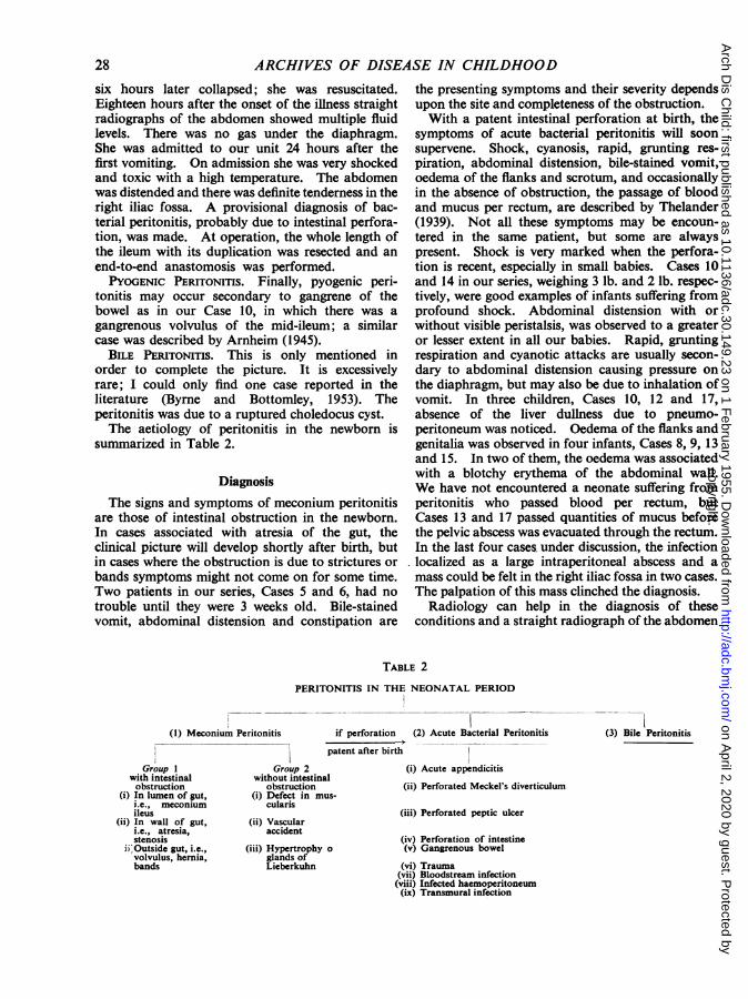
BILE PERITONITIS. This is only mentioned inorder to complete the picture. It is excessivelyrare; I could only find one case reported in theliterature (Byrne and Bottomley, 1953). Theperitonitis was due to a ruptured choledocus cyst.The aetiology of peritonitis in the newborn is
summarized in Table 2.
DiagnosisThe signs and symptoms of meconium peritonitis
are those of intestinal obstruction in the newborn.In cases associated with atresia of the gut, theclinical picture will develop shortly after birth, butin cases where the obstruction is due to strictures orbands symptoms might not come on for some time.Two patients in our series, Cases 5 and 6, had notrouble until they were 3 weeks old. Bile-stainedvomit, abdominal distension and constipation are
the presenting symptoms and their severity dependsupon the site and completeness of the obstruction.With a patent intestinal perforation at birth, the
symptoms of acute bacterial peritonitis will soonsupervene. Shock, cyanosis, rapid, grunting res-piration, abdominal distension, bile-stained vomit,oedema of the flanks and scrotum, and occasionallyin the absence of obstruction, the passage of bloodand mucus per rectum, are described by Thelander(1939). Not all these symptoms may be encoun-tered in the same patient, but some are alwayspresent. Shock is very marked when the perfora-tion is recent, especially in small babies. Cases 10and 14 in our series, weighing 3 lb. and 2 lb. respec-tively, were good examples of infants suffering fromprofound shock. Abdominal distension with orwithout visible peristalsis, was observed to a greateror lesser extent in all our babies. Rapid, gruntingrespiration and cyanotic attacks are usually secon-dary to abdominal distension causing pressure onthe diaphragm, but may also be due to inhalation ofvomit. In three children, Cases 10, 12 and 17,absence of the liver dullness due to pneumo-peritoneum was noticed. Oedema of the flanks andgenitalia was observed in four infants, Cases 8, 9, 13and 15. In two of them, the oedema was associatedwith a blotchy erythema of the abdominal wall.We have not encountered a neonate suffering fromperitonitis who passed blood per rectum, butCases 13 and 17 passed quantities of mucus beforethe pelvic abscess was evacuated through the rectum.In the last four cases under discussion, the infectionlocalized as a large intraperitoneal abscess and amass could be felt in the right iliac fossa in two cases.The palpation of this mass clinched the diagnosis.
Radiology can help in the diagnosis of theseconditions and a straight radiograph of the abdomen
BLE 2
PERITONITIS IN THE NEONATAL PERIOD
(1) Meconium Peritonitis if perforation (2) Acute Bacterial Peritonitis (3) Bile Peritonitis
Rarely, intra-abdominal calcification is discoveredaccidentally in babies not suffering from intestinalobstruction. Olnick and Hatcher (1953) reportthe case of a baby of 2 months of age whose radio-graphs showed calcification in the abdomen andscrotum. The diagnosis was confirmed by scrotalbiopsy.
Calcification may occasionally occur in meconiumMk
within the lumen of the bowel, the peritoneum beingunaffected (Camp and Roberts, 1949). The differ-ential diagnosis can only be made at operation.A straight radiograph of the abdomen may also
be helpful in those cases where a mass can be felton palpating the abdomen. In Case 15 of ourseries there was some doubt if a palpable mass wasan enlarged, displaced kidney. An intramuscularpyelogram showed normally functioning kidneysand a soft tissue shadow displacing the gas-filledintestine (Fig. 10).
TreatmentThe treatment of peritonitis in the newbor is the
treatment of the underlying condition. This is notthe place to discuss treatment in any detail, as itwould involve discussing the management of alltypes of intestinal obstruction in the neonatal
FIG. 7.-Case 12: perforation of the ileum. The radiograph inthe erect position shows air under the diaphragm.
often reveals the nature of the condition. Kirchhoffin 1932 was the first to describe a pneumoperi-toneum when radiographing a newborn baby. It isobvious that only cases with a patent intestinalperforation will show free gas in the abdomen.Three of our cases were diagnosed on this radio-logical finding (Figs. 7 and 8).
In meconium peritonitis the extruded meconiumfrequently calcifies within the peritoneum. Litten(quoted by Boikan, 1930) stated that this calcifica-tion can occur within 24 to 48 hours. This pheno-menon can be reproduced experimentally byintroducing meconium into the peritoneal cavity ofrats (Rubovits, Taft and Neuwelt, 1938). Calcifiedmasses can occasionally be observed in radiographsof the abdomen (Fig. 9). This diagnostic radio-logical sign was first recognized by Neuhauser in1944. As we have previously pointed out (Forshall,Hall and Rickham, 1952), calcification is often soslight that it cannot be seen on a straight abdominalradiograph, although it can be demonstrated in FIG. 8.-Case 14: perforated Meckel's diverticulum. Theradiograph of the abdomen in the erect position shows a smallx-ray films of the excised tissues. air bubble between the liver and the diaphragm.
29
copyright. on A
pril 2, 2020 by guest. Protected by
http://adc.bmj.com
/A
rch Dis C
hild: first published as 10.1136/adc.30.149.23 on 1 February 1955. D
did not attempt to separate these bands at operationas it was felt that this would greatly increase opera-ting time, produce a considerable amount of bleedingand shock, and lead to the formation of newadhesions. We have usually thought it wiser to-close the abdomen after resection and anastomosis.had been completed. In those cases surviving
. _ . F . '.:.:i3] operation, no further obstruction developed.Where there is a large intraperitoneal abscess, this.
should be drained by the most convenient route.The abscess wall should not be disturbed and webelieve it is unwise to search for the site of perfora-tion. There is, of course, danger of a faecal fistuladeveloping subsequently if this treatment is employ-ed. One of our four cases of abscess developedsuch a fistula post-operatively; this closed spon-taneously after a week. The three cases whichwere treated according to this principle had a verysatisfactory convalescence. In Case 8 no mass was.felt pre-operatively and the baby was operated uponfor intestinal obstruction of unknown origin. Atoperation, an abscess surrounded by coils of lowerileum was found in the right iliac fossa. The
FIG. 9.-Case 7: Straight radiograph of the abdomen showingintra-abdominal islands of calcification.
period. It is hardly necessary to stress that shock,dehydration and electrolyte disturbances must becorrected if these small infants are to survive opera-tion. Most of them need intravenous therapy andcontinuous gastric suction for some hours pre-operatively until their blood chemistry has come backto normal and their general condition has improved.At operation, bands or adh sions should be dividedif they cause demonstrable obstruction. Gastricperforations should be closed by suturing the defect,but when encountering intestinal perforations, weprefer sezection of the affected loop of gut andend-to-end anastomosis. Intestinal atresia, orstenosis, should also be treated by wide resection ofthe affected segment and end-to-end anastomosis.The numerous intraperitoneal adhesions may pre- FIG. 10.-Case 15: radiograph of the abdomen showing a constant
vent a thorough inspection of the distal intestine and shadow in the right iliac fossa which displaces the gas-filledintestine. It was found at operation to be an intra-peritoneal
produce subsequent intestinal obstruction, but we abscess in the right iliac fossa.
30
copyright. on A
pril 2, 2020 by guest. Protected by
http://adc.bmj.com
/A
rch Dis C
hild: first published as 10.1136/adc.30.149.23 on 1 February 1955. D
PERITONITIS IN THE NEONATAL PERIOD 31abscess was drained, but the densely adherentloops of gut were still obstructed. Rather thandissect out the adhesions, it was felt that a short-circuiting side-to-side ileo-transverse colostomyoffered the best chances for survival. The childmade an uninterrupted recovery. She has beenfollowed up for over three years and has no furthersymptoms.
Post-operatively, a prolonged period of ileus is tobe expected. Intravenous therapy, continuous low-pressure gastric suction and vigilant control of anychanges in the blood chemistry will, in our experi-ence, overcome this dreaded complication.
MortalityIn this series of 17 cases there were seven deaths,
a mortality of 41 2%. This high figure comparesunfavourably with a mortality of 19 8 % in 81 casesof intestinal obstruction without peritonitis innewborn babies operated upon during the sameperiod of three and a half years. It appears, there-fore, that peritonitis in the newborn carries an extrarisk and is, in our experience, next to meconiumileus, the most fatal of the intra-abdominal catas-trophes in this age group.The causes of death were as follows: three children
died of bronchopneumonia. One of them had, inaddition, a congenital malformation of the heartand another was very premature, weighing only2 lb. The causes of death in the other four weregastro-enteritis, overwhelming toxaemia, thrombosisof the inferior vena cava and intestinal fistulaproducing inanition. In retrospect, two of thesedeaths might have been prevented, if at the timewe had known more about the correct post-operativemanagement of these infants.
In conclusion, we should again like to stress thatperitonitis in the newborn is not a very rare con-dition. It is still frequently not diagnosed duringlife and only discovered at necropsy. In a survey ofneonatal deaths in Liverpool during 1949 severalrecords of such cases were found (Rickham, 1952).Once diagnosed, the condition is curable by opera-tion and should be associated with a reasonablechance of survival.
I should like to thank the paediatricians of theLiverpool region who referred these cases to us andwhose vigilance in detecting surgical emergencies in new-born babies has played such a big part in lowering ouroperative mortality.
I should like to acknowledge Miss I. Forshall's helpin preparing this paper. We collaborated closely in
treating these infants and Miss Forshall performed halfthe operations.
REFERENCESAbt, I. A. (1931). Med. Clin. N. Amer., 15, 611.Arnheim, E. E. (1945). Amer. J. Dis. Child., 69, 108.Bendel, W. L. and Michel, M. L. (1953). Surgery, 34, 320.Bird, C. E., Limper, M. A. and Mayer, J. M. (1941). Ann. Surg.,
114, 526.Boikan, W. S. (1930). Arch. Path., Chicago, 9, 1164.Brink, E. ten and Keyzer, J. L. (1952). Maandschr. Kindergeneesk.,
20, 108.Butler, A. M. (1945). New Engl. J. Med., 233, 257.Byrne, J. J. and Bottomley, G. T. (1953). Amer. J. Dis. Child.,
85, 694.Camp, R. and Roberts, M. H. (1949). Ibid., 78, 393.Chandler, L. R. (1949). Discussion of paper by C. W. Brunkow
et al. West. J. Surg., 57, 424.Ch'eng, Y. H. and K'ang, H. J. (1937). Chin. med. J., 52, 876.Creery, R. D. G. (1953). Brit. med. J., 1, 871.Cruveilhier, J. (1829-42). Anatomie Pathologique du Corps Humain.
Paris.Davies, M. E. and Potter, E. L. (1946). J. Amer. med. Ass., 131,
1194.Davis, D. L. and Poynter, C. W. M. (1922). Surg. Gynec. Obstet.,
34, 35.Etherington-Wilson, W. (1945). Proc. roy. Soc. Med., 38, 186.Farr, R. E. and Brunkow, C. W. (1925). Arch. Surg., Chicago,
11, 417.Flesch, H. (1925). Jb. Kinderheilk., 108, 366.Forshall, 1., Hall, E. G. and Rickham, P. P. (1952). Brit. J. Surg.,
40, 31.Franklin, A. W. and Hosford, J. P. (1952). Brit. med. J., 2, 257.Greene, W. W. and Gose, D. F. (1953). Amer. J. Dis. Child., 85, 47.Gross, R. E. (1953). Surgery of Infancy and Childhood. Phila-
delphia and London.and Ferguson, C. C. (1952). Surg. Gynec. Obstet., 95, 631.
Gottlieb, C., Chu, F. and Sharlin, H. S. (1950). Radiology, 54, 595.Gubern Salisachs, L. (1951). Acta pedid. esp., 9, 27.Guthrie, K. J. (1942). Archives of Disease in Childhood, 17, 82.Helbing, T. (1908). Ueberfoetale Peritonitis, nebst einem casuistischen
Beitrag aus der Universitats. Frauenklinik zu Freiburg. Inaug.Diss., Freiburg.
Herbut, P. A. (1943). Arch. Path., Chicago, 36, 91.Keith, A. (1933). Human Embryology and Morphologv. 5th ed.
London.Kellogg, H. G., Abelson, S. M. and Cornwell, F. A. (1951). J.
Pediat., 39, 357.Kirchhoff, H. (1932). Rontgenpraxis, 4, 233.Kornblith, B. A. and Atani, S. (1929). Amer. J. Path., 3, 249.Lattes, R. (1943). Amer. J. Obstet. Gynec., 46, 149.Lee, C. M. and MacMillan, B. G. (1950). Surgery, 28, 48.Lee, W. E. and Wells, J. R. (1923). Ann. Surg., 78, 36.Leger, J. L., Ricard, P. M.. Leonard, C. and Piette, J. (1950). Un.
*med. Can., 79, 1277.Low, J. R., Cooper, G. and Cosby, L. (1949). Surgery, 26, 223.Maguire, C. H. and Moore. W. R. (1950). Surgery. 28, 568.Meigher, S. C. and Lucas, A. W. (1952). Ann. Surg., 136, 1044.Miller, R. A. (1941). Archives of Disease in Childhood, 16, 22.Moncrieff, A. (1953). Brit. med. J., 1, 1.Moncrief, W. H. (1954). Ann. Surg., 139, 99.Moretti, I. (1949). Minerva pediat., Torino, 1, 239.Morgagni, J. B. (1761). De Sedibus et Causis Morborum. Typo-
graphia Remondiniana, Venice.Neuhauser, E. B. D. (1944). Amer. J. Roentgenol., 51, 421.Olnick, H. M. and Hatcher, M. B. (1953). J. Amer. med. Ass., 152,
582.Paltauf, A. (1888). Virchows Arch. path. Anat., 111, 461.Porter, L. and Weeks, A. (1915). Amer. J. Dis. Child., 9, 283.Peiser, A. (1908). Bruns' Beitr. klin. Chir., 60, 168.Qvigstad, I. (1950). Nord. Med., 43, 504.Ramos, J. F. (1950). J. nat. med. Ass., 42, 105.Rickham, P. P. (1952). Lancet, 1, 332.Rosza, S. and Gross, R. J. (1953). Amer. J. Roentgenol., 69, 944.Rubovits, W. H., Taft, E. and Neuwelt, F. (1938). Amer. J. Obstet.
Gynec., 36, 501.Rudnew, W. (1915). Ueber die spontanen Darmrupturen bei Foeten
und Neugeborenen. Inaug. Diss., Basel.Russell, T. H. (1928). J. Amer. med. Ass., 90, 1931.- (1940). Trans. New Engl. Surg. Soc. (1939), 22, 286.Siebold, A. E. von (1825). J. Geburtsh., 5, 3.Sturzenegger, E. (1927). Beitr. path. Anat., 78, 85.Sury, K. von (1912). Vjschr. gerichtl. Med., 43, II Supplement-Heft,
91.Thelander, H. E. (1939). Amer. J. Dis. Child., 58, 371.Ungari, C. and Valiani, A. (1952). Clin. Ostet. Ginec., 54, 147.Wright, L. T. and Scott, B. E. (1950). J. Pediat., 37, 905.Zillner, E. (1884). Virchows. Arch. path. Anat., 96, 307.
copyright. on A
pril 2, 2020 by guest. Protected by
http://adc.bmj.com
/A
rch Dis C
hild: first published as 10.1136/adc.30.149.23 on 1 February 1955. D