121
PRINCIPLES OF PHARMACOLOGY OpenCourseWare MIT

PRINCIPLES OF PHARMACOLOGY
OpenCourseWareMIT

MIT
Principles of Pharmacology
OpenCourseWare

This text is disseminated via the Open Education Resource (OER) LibreTexts Project (https://LibreTexts.org) and like thehundreds of other texts available within this powerful platform, it is freely available for reading, printing and"consuming." Most, but not all, pages in the library have licenses that may allow individuals to make changes, save, andprint this book. Carefully consult the applicable license(s) before pursuing such effects.
Instructors can adopt existing LibreTexts texts or Remix them to quickly build course-specific resources to meet the needsof their students. Unlike traditional textbooks, LibreTexts’ web based origins allow powerful integration of advancedfeatures and new technologies to support learning.
The LibreTexts mission is to unite students, faculty and scholars in a cooperative effort to develop an easy-to-use onlineplatform for the construction, customization, and dissemination of OER content to reduce the burdens of unreasonabletextbook costs to our students and society. The LibreTexts project is a multi-institutional collaborative venture to developthe next generation of open-access texts to improve postsecondary education at all levels of higher learning by developingan Open Access Resource environment. The project currently consists of 14 independently operating and interconnectedlibraries that are constantly being optimized by students, faculty, and outside experts to supplant conventional paper-basedbooks. These free textbook alternatives are organized within a central environment that is both vertically (from advance tobasic level) and horizontally (across different fields) integrated.
The LibreTexts libraries are Powered by MindTouch and are supported by the Department of Education Open TextbookPilot Project, the UC Davis Office of the Provost, the UC Davis Library, the California State University AffordableLearning Solutions Program, and Merlot. This material is based upon work supported by the National Science Foundationunder Grant No. 1246120, 1525057, and 1413739. Unless otherwise noted, LibreTexts content is licensed by CC BY-NC-SA 3.0.
Any opinions, findings, and conclusions or recommendations expressed in this material are those of the author(s) and donot necessarily reflect the views of the National Science Foundation nor the US Department of Education.
Have questions or comments? For information about adoptions or adaptions contact [email protected]. Moreinformation on our activities can be found via Facebook (https://facebook.com/Libretexts), Twitter(https://twitter.com/libretexts), or our blog (http://Blog.Libretexts.org).
This text was compiled on 01/12/2022
®

1 1/12/2022
TABLE OF CONTENTSThe object of this text is to teach students an approach to the study of pharmacologic agents. The focus is on the basic principles ofbiophysics, biochemistry and physiology, as related to the mechanisms of drug action, biodistribution and metabolism. The courseconsists of lectures and student-led case discussions.
1: CHAPTERS1.1: IMMUNOSUPPRESSION FOR SOLID ORGAN TRANSPLANTATION1.2: INTRODUCTION TO PHARMACOLOGY1.3: PHARMACOKINETICS I1.4: PHARMACOKINETICS II - DOSING1.5: CASE STUDY - ANTICHOLINESTERASE1.6: AUTONOMIC PHARMACOLOGY1.7: LOCAL ANESTHETICS1.8: ANTIINFLAMMATORY DRUGS1.9: VASOACTIVE DRUGS I1.10: VASOACTIVE DRUGS II - HEART FAILURE1.11: LIPID LOWERING DRUGS - HYPERLIPIDEMIA AND ATHEROSCLEROSIS1.12: NEUROPHARMACOLOGY I - DRUGS FOR MOVEMENT DISORDERS1.13: NITRIC OXIDE1.14: NEUROPHARMACOLOGY II - ANXIOLYTICS AND ANTIDEPRESSANTS1.15: NEUROPHARMACOLOGY III - ANTICONVULSANTS1.16: ANTIMICROBIALS I AND II1.17: CHEMOTHERAPY1.18: OPIOID PHARMACOLOGY
BACK MATTERINDEXGLOSSARYGLOSSARY

1 1/12/2022
CHAPTER OVERVIEW1: CHAPTERS
1.1: IMMUNOSUPPRESSION FOR SOLID ORGAN TRANSPLANTATIONThe success of solid organ and bone marrow transplantation (BMT) has correlated with improvements in selectiveimmunosuppression. Immunosuppression decreases both the incidence of acute and chronic organ graft and bone marrow rejection,and a potentially life threatening complication of BMT known as graft-vs-host disease (GVHD). Selective immunosuppression targetsspecific pathways of immune signaling and activation, and minimizes the incidence of deleterious side effects.
1.2: INTRODUCTION TO PHARMACOLOGYPharmacology (Gr. pharmakon - a drug or poison, logos - word or discourse) is the science dealing with actions of drugs on the body(pharmacodynamics) and the fate of drugs in the body (pharmacokinetics). It overlaps with pharmacy, the science of preparation ofdrugs; much of it deals with therapeutics, the treatment of disease (by whatever means). Toxicology is the branch of pharmacologydealing with the "undesirable" effects of drugs on biological processes.
1.3: PHARMACOKINETICS IDescribe the physicochemical and physiological factors that influence the absorption of drugs from enteral and parenteral routes ofadministration, their distribution within the body, and their routes and mechanisms of elimination. Explain how dose, bioavailability,rate of absorption, apparent volume of distribution, total clearance, and elimination half-life affect the plasma concentrations of a drugafter administration of a single dose.
1.4: PHARMACOKINETICS II - DOSING1.5: CASE STUDY - ANTICHOLINESTERASEReversible versus irreversible inhibition of AchE causes build up of Ach at synapse Toxicity associated with AchE inhibitors (patientcase!) include global nicotinic, muscarinic, & CNS effects (DUMBBELLS) Treatment for Exposure to Irreversible InhibitorsAtropine – counteract ACh agonism 2-Pralidoxime – prevent aging
1.6: AUTONOMIC PHARMACOLOGYAs you will see throughout the text, the autonomic nervous system (ANS) is a very important topic for two reasons: First,manipulation of ANS function is the basis for treating a great deal of cardiovascular, pulmonary, gastrointestinal and renal disease;second, there is hardly a drug worth mentioning without some major autonomic side effects (cf. antihistamines).
1.7: LOCAL ANESTHETICSLocal anesthesia is the selective numbing of a particular, circumscribed region of the body by a controlled, reversible procedure.Drugs called local anesthetics (LA) are usually employed for these procedures, although directly applied pressure, cooling, or evenheating will also produce numbness. The general strategy is to inhibit the propagation or generation of impulses in nerves from adefined anatomical region.
1.8: ANTIINFLAMMATORY DRUGSInflammation is mediated in part by prostaglandins produced by the cyclooxygenase pathway. NSAIDs inhibit this pathway and serveas combined anti-inflammatory, anti pyretics, and analgesics. Because NSAIDs are generally nonspecific and exert numerous sideeffects, there is great interest in more specific therapeutics such as selective COX-2 inhibitors and anti-cytokine agents.
1.9: VASOACTIVE DRUGS IHypertensive emergencies (malignant hypertension) are defined as severe hypertension coupled with acute end-stage organ damage.
1.10: VASOACTIVE DRUGS II - HEART FAILURECongestive heart failure simply means that the pulmonary blood volume is expanded and, therefore, the pulmonary circulation iscongested with blood. The congestion arises because of elevated left ventricular end-diastolic pressure (LVEDP). An elevated LVEDPis a hallmark of uncompensated congestive heart failure. Common symptoms include shortness of breath, fatigue, orthopnea andparoxysmal nocturnal dyspnea (PND).
1.11: LIPID LOWERING DRUGS - HYPERLIPIDEMIA AND ATHEROSCLEROSISAtherosclerosis is a chronic inflammatory disease characterized by enzymatic destruction of the normal arterial skeleton (largelyelastin, collagen and smooth muscle), and replacement by disorganized collagen and elastin, cholesterol, and foam cells.
1.12: NEUROPHARMACOLOGY I - DRUGS FOR MOVEMENT DISORDERSThese are a diverse group of neurologic disorders in which the normal functions of the motor system are impaired. Parkinson’sdisease is by far the most common disorder of movement, affecting >3% of individuals over the age of 65.

2 1/12/2022
1.13: NITRIC OXIDEThe objective of this lecture is to describe the effects of inhaling low levels of nitric oxide (NO) on the hemodynamic and gasexchange function of both the normal and diseased lung. Considerable attention will be paid to safety and hazards of inhaled NOtherapy. Progress has been made in understanding the NO guanylate cyclase signal transduction system. NO was given considerableclinical investigation in pulmonary artery hypertension and adult respiratory distress syndrome (ARDS) patients.
1.14: NEUROPHARMACOLOGY II - ANXIOLYTICS AND ANTIDEPRESSANTSDepression is a frequent problem, affecting up to 5% of the population. Common presentations include low mood, loss of energy,disinterest in activities. May also include weight loss, sleep disturbance, or psychosis. Should be considered in patients with atypicaldementia and chronic pain
1.15: NEUROPHARMACOLOGY III - ANTICONVULSANTSSeizures are episodes of neurologic dysfunction arising from abnormal synchronous activity of neurons. Alterations of consciousnessand abnormal motor activity are the most common manifestations. Epilepsy (recurring seizures without a clear precipitant) iscommon, affecting about 1% of the population. Pharmacological treatment is very successful in the majority of cases, but requiresaccurate diagnosis and classification of seizures.
1.16: ANTIMICROBIALS I AND IISuccessful antimicrobial therapy occurs when an effective concentration of drug is delivered to the site of infection for a sufficientperiod of time. Minimum effective concentrations are those needed to inhibit growth (bacteriostatic concentration, MIC) or kill(bacteriocidal concentration,MBC) the pathogen in question.
1.17: CHEMOTHERAPYChemotherapy is a type of cancer treatment that uses one or more anti-cancer drugs (chemotherapeutic agents) as part of astandardized chemotherapy regimen. Chemotherapy may be given with a curative intent (which almost always involves combinationsof drugs), or it may aim to prolong life or to reduce symptoms (palliative chemotherapy). Chemotherapy is one of the majorcategories of the medical discipline specifically devoted to pharmacotherapy for cancer, which is called medical oncology.
1.18: OPIOID PHARMACOLOGYOpium – a mixture of alkaloids from Papaver somniferum. An opiate is a naturally occurring alkaloid, i.e., morphine or codeine, andan opioid is any natural or synthetic compound, which has morphine-like properties. Hundreds of opioid alkaloids and peptides havebeen synthesized, but all clinically available opioid analgesics are alkaloids.

1.1.1 12/15/2021 https://med.libretexts.org/@go/page/10633
1.1: Immunosuppression for Solid Organ TransplantationThe success of solid organ and bone marrow transplantation (BMT) has correlated with improvements in selectiveimmunosuppression. Immunosuppression decreases both the incidence of acute and chronic organ graft and bone marrowrejection, and a potentially life threatening complication of BMT known as graft-vs-host disease (GVHD). Selectiveimmunosuppression targets specific pathways of immune signaling and activation, and minimizes the incidence ofdeleterious side effects.
History1954: First successful human kidney transplant1960s: Introduction of effective immunosuppressive drugs. Steroids, ATG, azathioprine1968: Successful bone marrow transplants for congenital immunodeficiency syndromes1970s: Cyclosporine introduced1980s: OKT3, tacrolimus, mycophenolate mofetil introducedIn 1988 1 year renal cadaver graft survival was 76% and 1 year renal living donor graft survival was 89%▫ By 1995, graft survival rates improved to 87% and 93% respectively1980s: The addition of cyclosporine to GVHD prophylaxis regimens halved the incidence of severe disease andimproved survival post -transplant1990s: Leflunomide, TNF antagonists, and selective mAbs introduced; additional mAb therapies expected in future
Balancing Benefits and Risks of Immunosuppression
Benefits: Immunosuppression decreases risks of both acute and chronic organ graft and bone marrow rejection, and GVHD
Risks: Immunosuppression poses risk of several types of side effects to the patient:
Acute effects: gastrointestinal upsetOpportunistic infection because patient is immunocompromised: CMV, Candida,Pneumocystis carinii, etc.Malignancies (lymphomas, skin cancer, etc.)Toxicities specific to particular immunosuppressive agent: steroids, etc.
Types of Organ Graft Rejection
Hyperacute: Occurs within minutes after transplant. Mediated by preformed anti- donor antibodies in recipient.Involves small vessel thrombosis and graft infarction.Acute: Occurs weeks after transplant. Delayed-type hypersensitivity / Cell mediated response of cytotoxic Tlymphocytes reacting against the foreign MHC molecules of the graft. Histologically characterized by mononuclearinfiltrate, hemorrhage, and edema in graft. Reversible with immunosuppressive therapy.Chronic: Occurs months to years post transplant. Results from antibody mediated vascular damage (fibrinoid necrosis)and is irreversible. Vascular damage results in vascular cell wall proliferation which may occlude vessel lumenresulting in graft ischemia and fibrosis. Can progress insidiously despite increased immunosuppressive therapy.
Graft Rejection and GVHD Following Bone Marrow Transplantation
Graft rejection occurs uncommonly (<1%) after conventional myeloablative bone marrow/stem cell transplantation,with increased incidence (1-15%) after HLA- mismatched BMT/cord blood transplantation.The rate of graft rejection is higher after nonmyeloablative preparative therapy for BMTAcute GVHD, which is usually evident before day 100 post-transplant, occurs in 1/3 of HLA matched transplants and2/3 of HLA-mismatched transplants.Chronic GVHD (>day 100) occurs in approximately 1⁄2 of transplantsAcute GVHD affects predominantly skin, the GI tract, and liver. Tissue injury involves effector cells (initiated by T-cells), particularly of the TH1 subset, and cytokines (e.g. TNF-alpha, interferon-gamma, and interleukin-1).Chronic GVHD may affect almost any organ/tissue and often mimics a collagen vascular disease in its clinicalpresentation.
Molecular Basis of Immune Response and Immunosuppression

1.1.2 12/15/2021 https://med.libretexts.org/@go/page/10633
The immune response involves both humoral and cellular responses: Humoral: The humoral response involves recognitionof foreign antigens which causes the differentiation of B-cells into memory cells and plasma cells. Plasma cells secreteantibodies into circulation. Cellular: Macrophages ingest and present antigen via the major histocompatibility (MHC) IImolecule. The macrophage is a type of antigen presenting cell which binds to CD4 T lymphocytes cells via the MHC II –T cell receptor (TCR) interaction. CD3 is a necessary accessory molecule to the MHC II – TCR interaction. Theinteraction induces proliferation of the CD4 T-cell (see diagram page 4), and release of IL-2, which promotes activation ofcytotoxic CD8 T-cells. CD8 T cells bind to MHC I molecules. When CD8 cells recognize an antigen presented on an MHCI molecule (which indicates the presenting cell is foreign, or, for example, a tumor cell or virally infected cell) the CD8 cellinduces the death of the target.
In the context of an MHC mismatched organ or bone marrow transplant, the MHC molecules of the cells of the organ (orbone marrow) graft are recognized by the host
TCR not as self-MHC molecules, but rather in the same manner as a self-MHC plus the foreign peptide it is presenting.The immunologic response to the foreign MHC molecules is a major cause of most graft rejection. Conversely the donorimmune system may recognize disparate MHC antigens in the host and initiate an immunologic response in the form ofGVHD.
MHC molecules are divided into class I and class II antigens; inheritance involves multiple alleles, and all loci are foundon chromosome 6. Class I antigens include HLA A, HLA-B, and HLA-C alleles . Class II antigens include HLA-DR,HLA-DQ, and HLA-DP alleles. Minimization of the antigenic differences between donor and recipient, by matching theirMHC alleles, has decreased rejection and GVHD and improved graft survival. Identical twins have identical MHC genes(6/6 loci), siblings have half similarity (3/6), and the match of unrelated persons must be determined by tissue typing.
As most bone marrow/stem cell transplants are from HLA matched (or only minor mismatched) related or unrelateddonors, minor histocompatibility antigens play a large role in eliciting an immune response (GVHD). Polymorphisms ofminor histompatibility antigens have been identified as risk factors for GVHD in the HLA-matched setting.
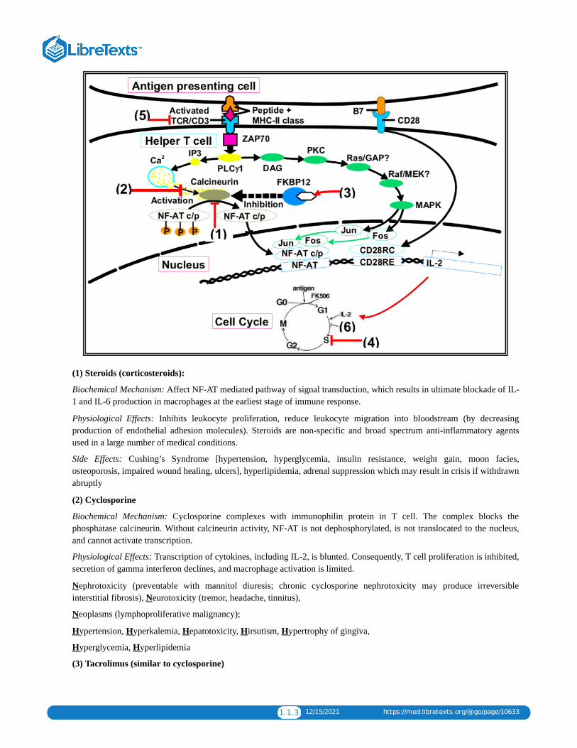
Types of Immunosuppressive Agents and Sites of ActionThe schematic on page 4 shows the molecular pathways that are activated in a helper T cell after it contacts an antigenpresenting cell which presents an antigen on its MHC II molecule. Superimposed on the pathways are sites ofpharmacologic intervention which will inhibit signaling and thus can be used as immunosuppressive agents.
Affect NFAT pathway: Steroids (1)Affect Calcineurin: Cyclosporine (2); Tacrolimus (3)Affect Cell Cycle by inhibiting DNA synthesis (4): azathioprine, MMF, leflunomide, cyclophosphamideAnti-TCR/CD3 antibody agents (5): Polyclonal agents are ATG, ALG. OKT3 is a monoclonal agent.Inhibit IL-2 dependent signaling: Sirolimus (6)
1.1.3 12/15/2021 https://med.libretexts.org/@go/page/10633
(1) Steroids (corticosteroids):
Biochemical Mechanism: Affect NF-AT mediated pathway of signal transduction, which results in ultimate blockade of IL-1 and IL-6 production in macrophages at the earliest stage of immune response.
Physiological Effects: Inhibits leukocyte proliferation, reduce leukocyte migration into bloodstream (by decreasingproduction of endothelial adhesion molecules). Steroids are non-specific and broad spectrum anti-inflammatory agentsused in a large number of medical conditions.
Side Effects: Cushing’s Syndrome [hypertension, hyperglycemia, insulin resistance, weight gain, moon facies,osteoporosis, impaired wound healing, ulcers], hyperlipidemia, adrenal suppression which may result in crisis if withdrawnabruptly
(2) Cyclosporine
Biochemical Mechanism: Cyclosporine complexes with immunophilin protein in T cell. The complex blocks thephosphatase calcineurin. Without calcineurin activity, NF-AT is not dephosphorylated, is not translocated to the nucleus,and cannot activate transcription.
Physiological Effects: Transcription of cytokines, including IL-2, is blunted. Consequently, T cell proliferation is inhibited,secretion of gamma interferon declines, and macrophage activation is limited.
Nephrotoxicity (preventable with mannitol diuresis; chronic cyclosporine nephrotoxicity may produce irreversibleinterstitial fibrosis), Neurotoxicity (tremor, headache, tinnitus),
Neoplasms (lymphoproliferative malignancy);
Hypertension, Hyperkalemia, Hepatotoxicity, Hirsutism, Hypertrophy of gingiva,
Hyperglycemia, Hyperlipidemia
(3) Tacrolimus (similar to cyclosporine)

1.1.4 12/15/2021 https://med.libretexts.org/@go/page/10633
Biochemical Mechanism: Binds to FK506 Binding Protein (FKBP). The Tacrolimus- FKBP complex inhibits calcineurin.The mechanism is therefore the same as that of cyclosporine.
Physiological Effects: 10-100 times more potent than cyclosporine, but similar mechanism: decreased production of IL-2
Side Effects: Similar to cyclosporine. Do not use with cyclosporine!
(4) Cell Cycle Inhibitors
Azathioprine: Azathioprine is metabolized in vivo to 6-mercaptopurine (6MP). 6MP is a purine anti-metabolite thatprevents DNA and RNA synthesis which inhibits proliferation of lymphocytes. Side effects include pancytopenias,gastrointestinal symptoms, and hepatic dysfunction (hepatitis).
Mycophenolate mofetil (MMF): MMF is a prodrug of mycophenolic acid (MPA), which is an inhibitor of inosinemonophosphate dehydrogenase (IMPDH). Inhibition of IMPDH limits production of guanosine nucleotides required fornucleic acid synthesis, and thus exerts a potent, selective cytostatic effect on B- and T- lymphocytes (decreased antibodyproduction and generation of cytotoxic T-cells). Side effects include pancytopenias, infections and malignancies, andgastrointestinal symptoms.
Leflunomide: Leflunomide inhibits dihydroorotate dehydrogenase (an enzyme involved in de novo pyrimidine synthesis);as a result, nucleic acid synthesis and lymphocyte proliferation are inhibited. Side effects include pancytopenias,hepatotoxicity, risk of lymphoproliferative disorders, and rare cases of Stevens- Johnson syndrome and toxic epidermalnecrolysis.
Cyclophosphamide: Cyclophosphamide is an extremely potent alkylating agent which destroys proliferating lymphoidcells. Cyclophosphamide causes pancytopenias, hemorrhagic cystitis, alopecia, and infertility.
(5) Anti-TCR/CD3 antibody agents:
anti-lymphocyte globulin (ALG) and anti-thymocyte globulin (ATG) both inactivate peripheral lymphocytes andimpair cellular immunity. These antibodies are used for induction of immunosuppression, treating initial rejection, andtreating steroid resistant rejection. Side effects include anaphylactic response (because these antibodies are foreignproteins), serum sickness, antigen-antibody induced glomerulonephritis, reactivation of latent viral infections, posttransplant lymphoproliferative disease, and development of human anti(mouse) antibodies.
OKT3: OKT3 is a murine monoclonal antibody that blocks the binding of the TCR to antigen, thus downregulating theactivity of the entire TCR/CD3 receptor complex. Compared to ALG and ATG, there are fewer serum proteins associatedwith OKT3 preparations than with those polyclonal antibody preparations (which may result in a more specific effect andfewer side effects). Side effects of OKT3 include fever, myalgias, arthralgias, CNS symptoms, GI irritation, and B-celllymphoproliferative disorders.
(6) Sirolimus (rapamycin, a macrolide antbiotic)
Biochemical Mechanism: Inhibits IL-2 mediated signaling by inhibiting Target of Rapamycin (TOR), an enzyme active inIL-2 cascades in proliferating lymphocytes.
Physiological Effects: Cell cycle progression from G1 to S is blocked; T- cell and B- cell proliferation is limited, and B-cell antibody production is inhibited.
Side Effects: Hyperlipidemia, Hypertension, Hypokalemia, Pancytopenias, decreased GFR / increased Serum Creatinine,metallic taste in mouth
Newer agents:
Anti-TNF agents include etanercept and infliximab (see Dr. Weinblatt’s lecture).Etanercept is a form of soluble TNFreceptor. Infliximab is a chimeric IgG1 monoclonal with a human Fc and murine Fab. By limiting TNF activity, thegeneration of pro- inflammatory cytokines IL-1 and IL-6 are diminished. In addition, anakinra is recombinant version ofthe human IL-1 receptor antagonist that inhibits IL-1 by blocking its receptor. Soluble IL-1 receptor antagonists are beingdeveloped.
Daclizumab is a humanized monoclonal murine IgG1 antibody that binds to a subunit of the IL-2 (CD25) receptor on thesurface of activated lymphocytes. Daclizumab thereby functions as an IL-2 inhibitor. Side effects include hypersensitivity,

1.1.5 12/15/2021 https://med.libretexts.org/@go/page/10633
cellulitis, and wound infection.
Basiliximab is a chimeric murine/human monoclonal IgG1 antibody that blocks the alpha chain of the IL-2 (CD25)receptor on the surface of activated T-cell lymphocytes. Basiliximab is indicated for the prophylaxis of acute organrejection in renal transplantation. Side effects include hypersensitivity and gastrointestinal disorders.
ALG/ATG can deplete the lymphocyte population. Since daclizumab and basiliximab preferentially affect activated Tcells, they are less apt to cause lymphocyte depletion.
Relationship Between Immunosuppressive and Cancer Chemotherapy.
Many cytotoxic drugs are used for both immunotherapy and cancer chemotherapy, but the therapeutic goals are tied toimportant differences in the cellular targets.
1. Cancer cells undergo largely unregulated, asynchronous proliferation, while immune cells proliferate in a burst ofactivity when antigen (i.e. transplant) is presented. This means immunosuppressant therapy can be timed for maximaleffect on the smallest population of immune cells.
2. An antigen stimulates proliferation by specific clones of precursor cells, so a cytotoxic drug will have greatly enhancedeffects on those clones (i.e. the drug will be relatively selective for rapidly dividing cells). In cancer chemotherapy,selectivity of cell cycle active drugs depends upon the innate growth characteristics of the tumor relative to normaltissue (see Dr. Kufe’s lecture) and is much harder to achieve.
3. Immunosuppressants are usually given in low, continuous dose regimens in order to maintain suppression of cellularand inflammatory responses to a persistent antigenic stimulus. In cancer chemotherapy high-dose pulse administrationis more common because it allows for recovery of immune function and re-growth of normal cell populations.
1.2.1 1/5/2022 https://med.libretexts.org/@go/page/10623
1.2: Introduction to PharmacologyA drug is a chemical agent which can affect living processes. For purposes of this course we will mainly be talking aboutsmall molecules which affect cellular processes. Most of these are Xenobiotics (Gr. xenos - stranger) chemicals that are notsynthesized by the body, but introduced into it from outside. There is inevitably a certain amount of ambiguity in thisdefinition: Is oxygen or water a drug? How about Vitamin C in a glass of orange juice? How about an injection of VitaminC to treat scurvy?
Pharmacology (Gr. pharmakon - a drug or poison, logos - word or discourse) is the science dealing with actions of drugson the body (pharmacodynamics) and the fate of drugs in the body (pharmacokinetics). It overlaps with pharmacy, thescience of preparation of drugs; much of it deals with therapeutics, the treatment of disease (by whatever means).Toxicology is the branch of pharmacology dealing with the "undesirable" effects of drugs on biological processes (in thecase of a nerve gas the bad effect may be a desired one).
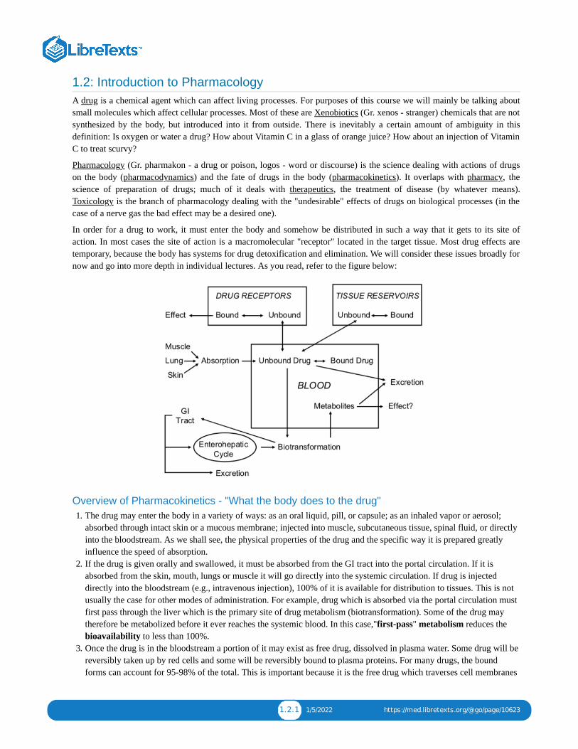
In order for a drug to work, it must enter the body and somehow be distributed in such a way that it gets to its site ofaction. In most cases the site of action is a macromolecular "receptor" located in the target tissue. Most drug effects aretemporary, because the body has systems for drug detoxification and elimination. We will consider these issues broadly fornow and go into more depth in individual lectures. As you read, refer to the figure below:
Overview of Pharmacokinetics - "What the body does to the drug"1. The drug may enter the body in a variety of ways: as an oral liquid, pill, or capsule; as an inhaled vapor or aerosol;
absorbed through intact skin or a mucous membrane; injected into muscle, subcutaneous tissue, spinal fluid, or directlyinto the bloodstream. As we shall see, the physical properties of the drug and the specific way it is prepared greatlyinfluence the speed of absorption.
2. If the drug is given orally and swallowed, it must be absorbed from the GI tract into the portal circulation. If it isabsorbed from the skin, mouth, lungs or muscle it will go directly into the systemic circulation. If drug is injecteddirectly into the bloodstream (e.g., intravenous injection), 100% of it is available for distribution to tissues. This is notusually the case for other modes of administration. For example, drug which is absorbed via the portal circulation mustfirst pass through the liver which is the primary site of drug metabolism (biotransformation). Some of the drug maytherefore be metabolized before it ever reaches the systemic blood. In this case,"first-pass" metabolism reduces thebioavailability to less than 100%.
3. Once the drug is in the bloodstream a portion of it may exist as free drug, dissolved in plasma water. Some drug will bereversibly taken up by red cells and some will be reversibly bound to plasma proteins. For many drugs, the boundforms can account for 95-98% of the total. This is important because it is the free drug which traverses cell membranes

1.2.2 1/5/2022 https://med.libretexts.org/@go/page/10623
and produces the effect. It is also important because protein-bound drug can act as a reservoir which releases drugslowly and thus prolongs its action.
4. The unbound drug may then follow its concentration gradient and distribute into peripheral tissues. In some cases, thetissue contains the target site and in others the tissue is not affected by the drug. Sites of non-specific binding act asfurther reservoirs for the drug. This total volume of distribution determines the equilibrium concentration of drug aftera specified dose.
5. Tissue-bound drug eventually reenters the bloodstream where it perfuses the liver and kidneys. The liver metabolizesmost drugs into inactive or less active compounds which are more readily excreted. These metabolites and some of theparent compound may be excreted in the bile and eventually may pass out of the body in the feces. Alternatively, someof the drug may be reabsorbed again, farther down the GI tract (the so-called enterohepatic cycle). Any biotransformeddrug which is not excreted in bile passes back into the systemic circulation.
6. Parent drug and metabolites in the bloodstream may then be excreted: most are filtered by the kidney, where a portionundergoes reabsorption, and the remainder is excreted in the urine. Some drugs are actively secreted into the renaltubule. Another route of excretion is the lung: Drugs like alcohol and the anesthetic gases are eliminated by this route.Smaller amounts of drug are eliminated in the sweat, tears and breast milk.
7. Biotransformation may sometimes produce metabolites with a great deal of activity. Occasionally, we administer aparent drug which is inactive (a pro-drug) and only the metabolite has activity. [How might this be useful?]
Overview of Pharmacodynamics - "What the drug does to the body"As stated above, the majority of drugs bind to specific receptors on the surface or interior of cells, but there are many othercellular components and non-specific sites which can serve as sites of drug action.
1. Water can be a target. Osmotic diuretics like mannitol are not reabsorbed by the kidney, and the osmotic load theycreate in the renal tubule obligates the loss of water. Laxatives like magnesium sulfate work in the intestine by the sameprinciple.
2. Hydrogen ions can be targets. Ammonium chloride is sometimes used to acidify the urine. When it is taken orally, theliver metabolizes ammonium ion to urea, while the chloride is excreted in the urine. The loss of Cl- obligates the lossof H+ in the urine, thus the pH is lowered.
3. Metal ions can be targets. Chelating agents like EDTA may be used to bind divalent cations like Pb++. Metal ions aremost frequently drug targets in cases of poisoning.
4. Enzymes are targets of many therapeutically useful drugs. Drugs may inhibit enzymes by competitive, non-competitive, or irreversible blockade at a substrate or cofactor binding site. Digitalis glycosides increase myocardialcontractility by inhibiting the membrane enzyme, Na+-K+ - ATPase. Antimicrobial and antineoplastic drugs commonlywork by inhibiting enzymes which are critical to the functioning of the cell. In order to be effective, these drugs musthave at least someselective toxicity toward bacterial or tumor cells. This usually means that there is a unique metabolicpathway in these cells or some difference in enzyme selectivity for a common metabolic pathway. An example of this isthe inhibition of folate synthesis by sulfonamides. These drugs are effective antibacterial agents because the bacteriadepend upon folate synthesis, while the host doesn't. This example will be covered in detail in one of our casediscussions.
5. Nucleic acids are targets for antimetabolites and some antibiotics. In the case of 5- fluorouracil, the compound acts as acounterfeit substitute for uracil and becomes incorporated into a faulty mRNA. Antisense oligonucleotides are anothervery specific way to interfere with a restricted part of the genome.
6. Some drugs, like general anesthetics, appear to act by non-specific binding to a macromolecular receptor target.These drugs are thought to alter the function of membrane proteins, in part, by disordering the structure of thesurrounding lipid membranes. Their lack of specificity is reflected in very low chemical structural requirements. Thegeneral anesthetics include compounds as chemically diverse as nitrogen, xenon, halogenated ethers, and steroids. Theyexhibit very little stereoselectivity, that is, there are not marked differences in anesthetic activity between enantiomers.
7. Finally, we have the drugs which act by binding to specific receptors. As you will see in lectures 2 and 6, these drugshave both high structural specificity and stereoselectivity, i.e. relatively small changes in chemical structure canradically alter the activity of these drugs.
Let us finish with some important definitions. These are concepts which we will return to repeatedly throughout thecourse.
1.2.3 1/5/2022 https://med.libretexts.org/@go/page/10623
Agonist is a drug which binds to its "receptor" and produces its characteristic effect. A drug may be a full agonist orpartial agonist, depending on the maximal effect it produces. An antagonist binds to the receptor without causing aneffect, thereby preventing an active substance from gaining access. Antagonists, like enzyme inhibitors, may becompetitive, non-competitive or irreversible.Dose-Response. The sine qua non of drug effect. Simply put, as the dose of drug increases, the response shouldincrease. [What if the response increases, then decreases as the dose is raised?] The curve generated is usuallysigmoidal when effect is plotted against log dose (Dr. Strichartz will discuss the theoretical basis for this). Effect maybe measured as a graded variable (change in blood pressure, force of contraction) or as a quantal variable (numberdead/alive). The slope of the curve is characteristic of the particular drug-receptor interaction. When two drugs act bythe same receptor mechanism, we expect to see two parallel log-dose response curves.ED . The median effective dose, or the dose which produces a response in 50% of subjects. If the response is death(lethality) we call it the LD . The EC50 refers to concentration rather than dose. Similar abbreviations are used forother response levels: ED , LD , etc.Potency. A terribly misused word – the lay public uses it to mean “effectiveness.” The potency of a drug refers to thedose (actually the molar concentration) required to produce a specific intensity of effect. [We usually specify the ED50,why?] If the ED50of drug A and B are 5 and 10 mg, respectively, the Relative Potency of A is twice that of B. Relativepotency specifically applies to the comparison of drugs which act by the same mechanism, and therefore have paralleldose-response curves.Efficacy. Also called Maximal Efficacy or Intrinsic Activity. This is the maximum effect of which the drug iscapable. A potent drug may have a low efficacy, and a highly efficacious drug may have a low potency. For theclinician, efficacy is much more important than potency (within limits). Who cares if the pill contains 5 or 10 mg ofdrug?Affinity. This refers to the strength of binding between drug and receptor. It is quantified by the dissociation constantkD (covered in the next lecture).Selectivity. This refers to the separation between desired and undesired effects of a drug. In the ideal case, a drug iscompletely specific, and an effective dose does not elicit any undesired effect. Penicillin is an example of a highlyselective drug, since it works specifically by inhibiting cell wall synthesis, and (other than allergic responses) it hasvery little effect on human cells at normal doses. Unfortunately, many therapeutic agents, like digoxin and theophylline,produce dose-related side effects near their therapeutic dose range. For some drugs like cancer chemotherapeuticagents, their selectivity is their dose-limiting property, i.e., they are given to kill tumor cells until they produce toxicityin normal cells as well.Therapeutic Window. For every drug, there exists some concentration which is just barely effective (the EffectiveConcentration) and some dose which is just barely toxic (the Toxic Concentration). Between them is the therapeuticwindow where most safe and effective treatment will occur.Therapeutic Index. This is the ratio of toxic to effective doses at the level of 50% response: TD /ED . In animaltoxicology studies, it is usually the LD /ED . Another measure sometimes utilized is the Certain Safety Factor,which is TD /ED
50
50
99 1
50 50
50 50
1 99.

1.3.1 12/15/2021 https://med.libretexts.org/@go/page/10624
1.3: Pharmacokinetics I
Describe the physicochemical and physiological factors that influence the absorption of drugs from enteral andparenteral routes of administration, their distribution within the body, and their routes and mechanisms ofelimination.Explain how dose, bioavailability, rate of absorption, apparent volume of distribution, total clearance, andelimination half-life affect the plasma concentrations of a drug after administration of a single dose.Describe the factors which determine the time-course of systemic accumulation of a drug administered by infusionor multiple doses.
Absorption of DrugsA. Transport Across Cell Membranes
1. Passive diffusiona. Passage through lipid cell membrane by dissolution in membrane; rate dependent on concentration gradient and
lipid:water partition coefficient of drug; rate markedly higher for unionized form of weak electrolyte because ofits higher lipophilicity than the ionized form; obeys first-order kinetics (rate of transport is proportional toconcentration gradient at transport site).
2. Filtration through aqueous channels within membranes and between cells.2. Active transport
a. Passage facilitated by an energy-dependent membrane carrier mechanism such that transport can occur against aconcentration gradient; transporters include the family of ATP-dependent proteins, such as
the multidrug resistance p-glycoprotein (amphipathic cationic and neutral substrates, 170 kD, mdr geneproduct, verapamil sensitive)the multidrug resistance-associated proteins (MRP1-6, organic anion substrates, 190 kD, probenecidsensitive).
b. Exhibits structural selectivity, saturability, competition between structural analogues and genetic variants.3. Sites for drugs in intestinal mucosa (cell to lumen), capillary endothelium of brain and testis (cell to blood),
choroid plexus (CSF to blood), proximal renal tubular cell (blood to urine), hepatocyte (blood to bile), tumorcells (efflux pump).
4. Obeys Michaelis-Menten kinetics: if drug concentration is high enough to saturate carrier mechanism, kineticsare zero-order (rate of transport is constant).
3. Endocytosisa. Passage into cell within membrane invagination.b. Important mechanism for particulates and high molecule weight compounds, such as proteins.
B. Routes of Drug Administration
1. General determinants of absorption ratea. Dissolution into aqueous fluids at absorption site, lipid solubility, concentration gradient, blood flow at
absorption site, surface area of absorption site.2. Importance of rate-limiting process
2. Oral (p.o.) Ingestiona. Convenient route for administration of solid as well as liquid formulations.2. Additional variables which may influence rate and extent of absorption include disintegration and dissolution of
solids, acidity of gastric contents, gastric emptying rate, intraluminal and mucosal biotransformation by host orbacterial enzymes, dietary contents, and presence of other drugs.
3. First-pass effect: absorbed drug passes via portal circulation through liver which may clear substantial fractionand thus decrease bioavailability (percent of dose which reaches the systemic circulation).
3. Parenteral Injection
Learning Objectives
1.3.2 12/15/2021 https://med.libretexts.org/@go/page/10624
1. Subcutaneous (s.c.) and intramuscular (i.m.) administration: more extensive absorption of high molecularweight, polar molecules than by oral route, via lymphatic circulation; absorption rate can be manipulated byformulation, e.g. rapid from aqueous solution, slow from suspension or solid pellet.
2. Intravenous (i.v.) injection: complete bioavailability; drugs only given in sterile solution; important whenimmediate effect required; increased risk of toxicity.
4. Pulmonary Inhalation1. Rapid absorption of drugs in gaseous, vaporized or aerosol form.2. Absorption of particulates/aerosols depends on particle/droplet size which influences depth of entry in
pulmonary tree; 1-5 uM particles reach alveolus
5. Topical Application1. Usually for local effect; patch formulations for systemic effect2. Absorption through mucous membrane may be rapid.3. Absorption through skin generally slow; enhanced by increased lipophilicity, by damage to stratum corneum,
and by increased blood flow.C. Distribution of Drugs
A. Tissue differences in rates of uptake of drugs.1. Blood flow: distribution occurs most rapidly into tissues with high blood flow (lungs, kidneys, liver, brain) and
least rapidly in tissues with low flow (fat).2. Capillary permeability: permeability of capillaries is tissue dependent;distribution rates relatively slower into
CNS because of tight junction between capillary endothelial cells, insignificant aqueous membrane pores,juxtaposed glial cells around endothelium and efflux transporters in vascular endothelium ("blood-brainbarrier"); capillaries of liver and kidney more porous.
B. Differences in tissue/blood ratios at equilibrium1. Dissolution of lipid-soluble drugs in adipose tissue2. Binding of drugs to intracellular sites3. Plasma protein binding; many drugs reversibly bind to albumin, α1-acidglycoprotein or other proteins in plasma;
extent of binding dependent on affinity, number of binding sites, and drug concentrations; drug bound toalbumin is not filtered by renal glomerulus but may be cleared by proximal renal tubule and liver; bindingreduces free drug available for distribution into tissue; many drug interactions based on displacement frombinding sites.
C. Apparent Volume of Distribution (V )1. Fluid compartments of 70-kg subject in liters and as percent of body weight: plasma 3 l (4%), extracellular
water 12 l (17%), total body water 41 l (58%).2. Estimation of Vd from extrapolated plasma concentration at "zero-time" (Co) after intravenous administration:
3. Prediction of Vd from chemical characteristics of drug, e.g. high lipid solubility, high V4. The plasma half-life of a drug (the time to reduce the concentration by one- half) is directly proportional to Vd,
and inversely proportional to total clearance (Cl ); for a given Cl , the higher the V , the longer the t :
Elimination of DrugsA. Total Clearance (Cl )
1. Volume of plasma completely cleared of drug per unit time by all routes and mechanisms.2. Summation of clearance values for each route, generally:
d
=Vd
Dose
Co
(1.3.1)
d
T T d 1/2
=t 1
2
ln2( )Vd
ClT(1.3.2)
T
= +CLT Clrenal Clhepatic (1.3.3)

1.3.3 12/15/2021 https://med.libretexts.org/@go/page/10624
3. If intrinsic capacity of an organ to clear drug is high and exceeds plasma flow to that organ, then the clearanceequals plasma flow and is altered by changes in plasma flow.
4. The plasma half-life of a drug is inversely proportional to total clearance, and directly proportional to Vd; for agiven Vd, the higher the total clearance, the shorter the half-life.
B. Biotransformation1. Elimination of drug by chemical modification of the molecule by spontaneous or (more usually) enzymatically
catalyzed reaction. Drug may be biotransformed by reactions at several sites on the molecule.2. Product(s) may have greater, lesser or qualitatively different pharmacologic activity from parent compound. A
prodrug is inactive and is biotransformed to a therapeutic agent. Highly reactive products such as quinones orepoxides may cause tissue necrosis or DNA damage.
3. Reaction rate dependent on chemical structure and obeys Michaelis-Menten kinetics (usually first-order attherapeutic drug concentrations).
4. Enzymatic activity generally highest in liver; enzymes in target organ may be responsible for conversion of drug totherapeutic or toxic metabolite; enzymes in intestinal bacteria may facilitate enterohepatic circulation of drugconjugates excreted in bile.
5. Sources of individual variation in rates of biotransformation: chemical exposures (drugs, dietary constituents andsupplements, smoke); genetics; age; disease
6. Major pathways of hepatic biotransformation
a. Phase I: often first step in biotransformation with formation of product susceptible to phase II conjugativereaction
b. Phase II: Coupling of drug or its oxidized metabolite to endogenous conjugating agent derived formcarbohydrate, protein or sulfur sources; generally products more water-soluble and more readily excreted inurine or bile.
C. Excretion1. Elimination of drug by excretion unchanged in body fluid or breath.2. Routes of excretion
a. Urine: quantitatively most important excretory route for nonvolatile drugs and their metabolites; excretion ratedepends on rate of glomerular filtration (drug not bound to plasma proteins), proximal tubular active secretion,and passive reabsorption
1) Determination of renal clearance (ClR), the volume of plasma completely cleared of drug per unit time(ml/min).
Measure the amount of drug excreted in the urine during a time interval t to t2. Find the plasma concentrationof the drug at the midpoint of the time interval, (t + t )/2, by interpolating on the ln C vs. t plot.
2) Mechanism of renal excretion can be inferred by comparison of Cl to that of an indicator of glomerularfiltration (creatinine), i.e., greater than 120 ml/min in 70-kg subject indicates tubular secretion and less than thatindicates net reabsorption (if no plasma binding); maximum renal clearance = renal plasma flow (e.g. para-aminohippuric acid, 650 ml/min in 70-kg subject).
3) Factors modifying Cl : extent of plasma protein binding (displacement enhances glomerular filtration),urinary pH (reabsorption of drugs with ionizable group is dependent on urinary pH; raising the pH promotesexcretion of acids, impairs excretion of bases), renal disease (creatinine clearance or its estimate from serumcreatinine provides a useful clinical indicator of impaired renal function and is approximately proportional todrug renal clearance; the effect of renal impairment on the total clearance of a drug can be estimated from theCl and the nonrenal clearance).
=ClRexcreation rate iurine
plasma concentration(1.3.4)
1
1 2 p
= [ ]ClRamount excreted from to t1 t2
( − )t2 t1
atCp( + )t1 t2
2
(1.3.5)
R
R
CR

1.3.4 12/15/2021 https://med.libretexts.org/@go/page/10624
b. Bile: quantitatively important excretory route for drugs and their metabolites which are actively transported byhepatocyte; once in small intestine, compounds with sufficient lipophilicity are reabsorbed and cleared again byliver (enterohepatic circulation), more polar substances may be biotransformed by bacteria (e.g. hydrolysis ofdrug conjugates) and products reabsorbed; unabsorbed drugs and metabolites are excreted in feces.
c. Minor routes: sweat, tears, reproductive fluids, milk; generally pH- dependent passive diffusion of lipophilicdrugs; can be of toxicologic significance e.g. exposure of infants to drugs in milk.
Time Course of Plasma ConcentrationsA. Relationship between plasma concentration and drug effect: minimum effective concentration, latency, duration of
effect, time and magnitude of peak effectB. Time-course of plasma concentrations for a single dose
1. Case with Highly Rapid Absorption Relative to Eliminationa. Single compartment model
1) First -order elimination: drug assumed to rapidly equilibrate into volume of distribution; plasmaconcentrations decline according to first-order kinetics; elimination rate from plasma is proportional to plasmaconcentration, fraction eliminated per unit time is elimination rate constant (k ).
Determination of elimination rate constant and elimination half-life:
Plot of ln C vs. t is a straight line with slope of -k . Plasma half-life (t =.693/k ) is constant and independentof dose.
Determination of apparent volume of distribution:
Extrapolation to time zero of the line of best fit for ln C vs t data; antilog of drug concentration at time 0designated as C . Then,
Determination of total clearance:
el
= −dCp
dtkelCp (1.3.6)
=Cp C0e− tkel (1.3.7)
= ln − tlnCp C0 kel (1.3.8)
p el 1/2 el
p
0
(in mls or liters) =VdTotal Dose
C0
(1.3.9)

1.3.5 12/15/2021 https://med.libretexts.org/@go/page/10624
According to definitions above, total clearance is the mass of drug (Cp Vd) eliminated per unit time divided bythe plasma concentration; therefore,
Determination of nonrenal clearance (ClNR):
If total clearance and renal clearance are determined from plasma and urine samples as described above, thenclearance by nonrenal routes (which includes biotransformation) can be estimated from
2) Kinetics of zero-order elimination: elimination rate is constant, t is dose-dependent (example: ethanol).
3)
b. Multicompartment model
Non-instantaneous distribution from blood to tissue resulting in multiexponential plasma concentration curve,initial phase reflects distribution out of central compartment into total Vd, terminal phase reflects elimination.
Where α and β are hybrid rate constants describing the 2 slopes.
2. Case with Non-Instantaneous Absorptiona. Kinetics of first-order absorption and elimination: determination of absorption and elimination half-lives
Note that the terminal slope may be either the elimination rate constant, the absorption rate constant, or a hybrid
See Katzung, Basic & Clinical Pharmacology, 2001, p. 42
b. Peak plasma concentration is dependent on absorption and elimination half-lives, volume of distribution, dose(D), and fraction of dose absorbed (F)
c. Area under plasma concentration vs. time curve (AUC) is dependent on dose (D), fraction of dose absorbed (F)and total clearance Cl
= = ( )( ) = [ ]( )ClT( )( ⋅ )kel Cp Vd
Cp
kel Vd0.693
t1/2Vd (1.3.10)
= −ClNR ClT ClR (1.3.11)
1/2
= − tCp C0 k0 (1.3.12)
= A +BCp e−αt e−βt (1.3.13)
= [ − ]Cp
FDka
( − )Vd ka kele− tkel e− tka (1.3.14)
T

1.3.6 12/15/2021 https://med.libretexts.org/@go/page/10624
Fraction of dose absorbed into systemic circulation (F ) is the bioavailability of the drug product; determinedexperimentally by measuring AUC of dosage form of drug given by one route and comparing it to AUC of samedose of drug under conditions of complete absorption, i.e. given i.v.
C. Effect of infusions or multiple dosing on time-course of plasma concentrations
1. Infusion Kinetics
One approach to maintaining a desired therapeutic level of a drug is to administer the agent by intravenous infusion.Drug delivery may be controlled by gravity- regulated drip of the agent into i.v. tubing or by use of an infusionpump.
a. When a drug is administered at a constant dosing rate (DR) and its elimination follows first-order kinetics, theconcentration of drug in the plasma rises exponentially and reaches a steady-state or plateau level (C ).
b. At steady-state the INPUT RATE = OUTPUT RATE. The input rate is DR, which may be expressed as the totaldose (D) divided by the length of the infusion (T). The output rate in the case of first-order elimination is thetotal amount of drug in the body (C Vd) times the elimination rate constant (k ).
Therefore, the plasma concentration at steady-state can be predicted as follows:
Remember that total clearance equals the elimination rate constant (k ) times the volume of distribution.Therefore, the plasma concentration at steady-state (C ) is directly proportional to the input rate (DR) of thedrug and inversely proportional to its total plasma clearance (Cl ).
c. The rate of achieving steady-state is dependent only on the elimination half-life of the drug. Half the C level isachieved in one t , and about 94% of C in four t .
d. Because of the lag in achieving steady-state when a constant infusion rate is administered, a loading dose maybe given to achieve the desired therapeutic effect more quickly. The loading dose may be chosen to produce the
AUC =F ⋅D
ClT(1.3.15)
ss
(t) = (1 − )Cp Css e t−kel (1.3.16)
ss el
DR = ⋅ ⋅Css Vd kel (1.3.17)
= DRCss
Vdkel(1.3.18)
el
ss
T
=Css
DR
ClT(1.3.19)
ss
1/2 ss 1/2
1.3.7 12/15/2021 https://med.libretexts.org/@go/page/10624
amount of drug in the body that would eventually be reached by the infusion alone.
At least on a theoretical basis, the plasma concentration will instantaneously reach the therapeutic level and thatlevel will be maintained. Note that the steady-state level achieved with a continuous infusion is determined bythe infusion rate and is not affected by the size of the loading dose.
2. Multiple Dosing Kineticsa. Commonly, drugs are administered repeatedly in order to maintain their therapeutic effects. In the simplest case,
a maintenance dose (D) is given at a constant dosing interval ( ) – [note that this is not the same as the timeconstant, ]. Since the route of administration may not be i.v., the amount of drug which reaches the systemiccirculation may be some fraction (F) of the dose. If elimination is by first-order kinetics, a steady-state iseventually reached. The “average” Css at steady-state equals the fraction absorbed times dosing rate divided bytotal clearance, analogous to the C from an infusion (see above).
\[ {C}_ss\ average={({F\cdot D\over\tau})\over {Cl}_T}$
b. However, in the case of repetitive dosing, unlike an infusion, plasma concentrations of drug fluctuate during thedosing interval, depending on the kinetics of absorption and elimination. The degree of fluctuation in the plasmaconcentration during a dosing interval increases with increasing dose, dosing interval, clearance, and absorptionrate.
c. If a drug is administered i.v. (or where absorption is rapid and complete), the peak plasma concentration atsteady-state (C ) relative to the peak after
the first dose (C ) depends on the ratio of the elimination half-life and the dosing interval (t ).
d. $$ {C}_{ {mass}_{ss}}={ {C}_0\over1-f}\]
f is the fraction of drug remaining at the end of a dosing interval.
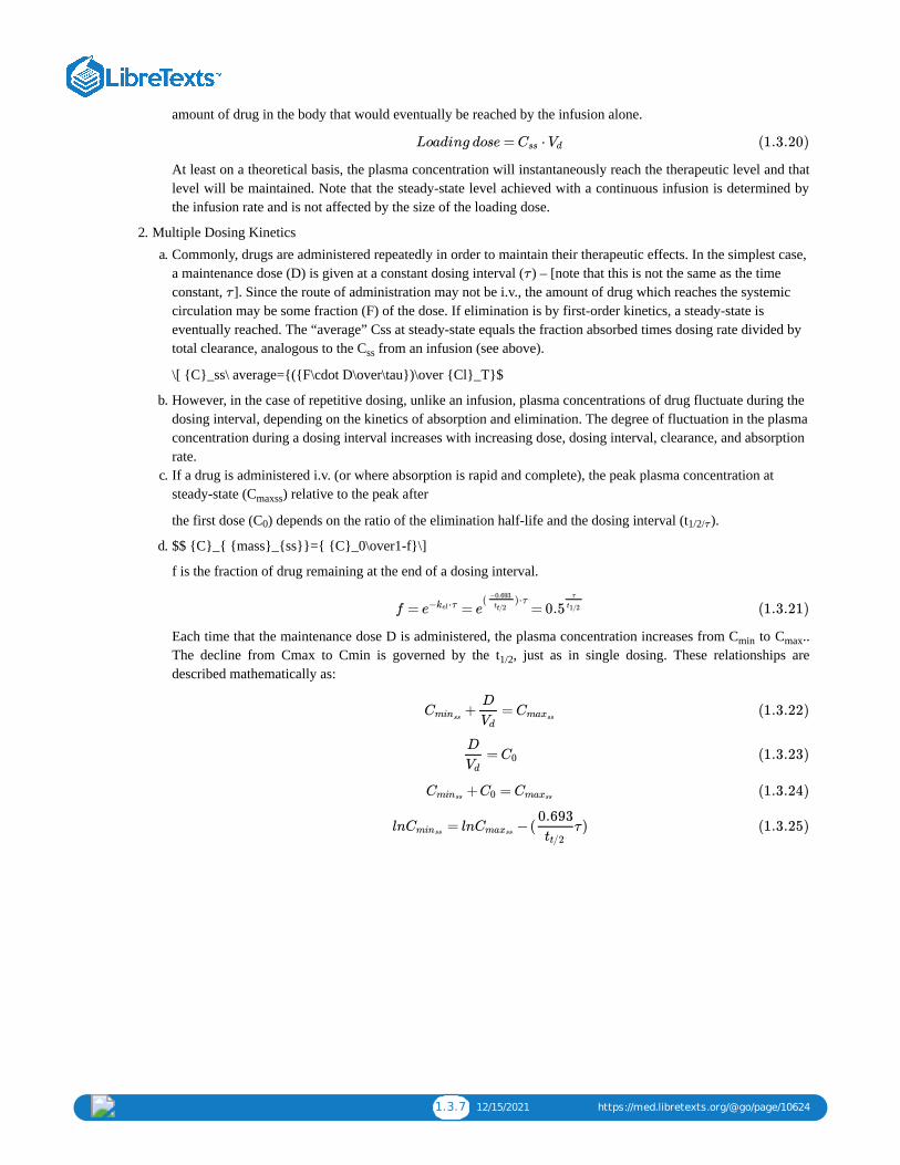
Each time that the maintenance dose D is administered, the plasma concentration increases from C to C ..The decline from Cmax to Cmin is governed by the t , just as in single dosing. These relationships aredescribed mathematically as:
Loading dose = ⋅Css Vd (1.3.20)
τ
τ
ss
maxss
0 1/2/τ
f = = =e ⋅τ−kel e( )⋅τ
−0.693
tt/2 0.5τ
t1/2 (1.3.21)
min max
1/2
+ =Cminss
D
VdCmaxss
(1.3.22)
=D
VdC0 (1.3.23)
+ =Cminss C0 Cmaxss (1.3.24)
= −( τ)lnCminss lnCmaxss
0.693
tt/2(1.3.25)
1.3.8 12/15/2021 https://med.libretexts.org/@go/page/10624
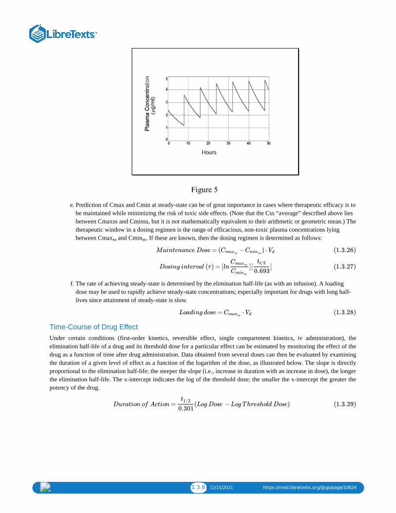
e. Prediction of Cmax and Cmin at steady-state can be of great importance in cases where therapeutic efficacy is tobe maintained while minimizing the risk of toxic side effects. (Note that the Css “average” described above liesbetween Cmaxss and Cminss, but it is not mathematically equivalent to their arithmetic or geometric mean.) Thetherapeutic window in a dosing regimen is the range of efficacious, non-toxic plasma concentrations lyingbetween Cmax and Cmin . If these are known, then the dosing regimen is determined as follows:
f. The rate of achieving steady-state is determined by the elimination half-life (as with an infusion). A loadingdose may be used to rapidly achieve steady-state concentrations; especially important for drugs with long half-lives since attainment of steady-state is slow.
Time-Course of Drug EffectUnder certain conditions (first-order kinetics, reversible effect, single compartment kinetics, iv administration), theelimination half-life of a drug and its threshold dose for a particular effect can be estimated by monitoring the effect of thedrug as a function of time after drug administration. Data obtained from several doses can then be evaluated by examiningthe duration of a given level of effect as a function of the logarithm of the dose, as illustrated below. The slope is directlyproportional to the elimination half-life; the steeper the slope (i.e., increase in duration with an increase in dose), the longerthe elimination half-life. The x-intercept indicates the log of the threshold dose; the smaller the x-intercept the greater thepotency of the drug.
ss ss
Maintenance Dose = ( − ) ⋅CmaxssCminss
Vd (1.3.26)
Dosing interval (τ) = [ln ][ ]Cmaxss
Cminss
tt/2
0.693(1.3.27)
Loading dose = ⋅CmaxssVd (1.3.28)
Duration of Action = (Log Dose −Log Threshold Dose)t1/2
0.301(1.3.29)

1.3.9 12/15/2021 https://med.libretexts.org/@go/page/10624

1.4.1 12/31/2021 https://med.libretexts.org/@go/page/10625
1.4: Pharmacokinetics II - DosingUSE OF PHARMACOKINETIC PARAMETERS TO ESTIMATE DOSING REGIMENS
You have decided to prescribe a new drug GOOD-4U to your patient, Ms. H.S.T., who weighs 70 kg and has normal renalfunction. The population average pharmacokinetic parameters for GOOD-4U are: Vd = 0.6 l/kg (about total body water),ClT = 60.6 ml/min. Therapeutic efficacy generally occurs at Cp of 2.38 μg/ml; side effects begin to occur with Cp of 5.0μg/ml.
You decide to administer a single dose of 100 mg by iv injection.
1. Assuming rapid distribution in the Vd, are you expecting to produce side effects (hint: what is the initial C0)?
No, assuming a single compartment system, the 100 mg will distribute in 42 liters to achieve an initial Cp of 2.38μg/ml. See Fig. 1.
2. How long before 94% of the dose is eliminated (hint: what is the half-life)?
The half-life computed from the total clearance and Vd is 8 hours; 94% of the dose is eliminated in about 4 half-lives, 32 hours.
3. A complete urine collection from the time of dosing until 16 hr later contains 37.5 mg of the drug. To what extentis the renal function of Ms. H.S.T. of importance to the total clearance of this drug?
Computation of the renal clearance indicates that it is about 50% of the total clearance. At 16 hr, which is 2 half-lives, 75 mg should have been eliminated by all clearance mechanisms. Half of that is appearing in the urinesuggesting the renal clearance is 30 ml/min. The drug must be extensively bound to plasma proteins and/or issubstantially reabsorbed after glomerular filtration. It is reasonable to predict that reduction of the patient’screatinine clearance by 50% will reduce total clearance by at least 25%.
One week later you decide to administer GOOD-4U by constant iv infusion to achieve the therapeutic effect.
4. What loading dose would you administer?
The minimum loading dose would be (2.38.μg/ml)(42 liters) or 100 mg.
5. What infusion rate would you prescribe?
To achieve a Css of 2.38 μg/ml, given a total clearance of 60.6 ml/min, the infusion rate should be 144.2 μg/min.See Fig. 4.
If instead you had administered 100 mg by iv injection every 8 hours:
6. At steady-state what would be the Cmax?
The drug is given repeatedly at a dosing interval which in this case equals the elimination half-life. The drug willaccumulate to twice the initial C0, ie. 4.76
μg/ml. You can prove that from the equation provided (cf. Figure 5).
7. At steady-state would the Cmin be sufficient to achieve continuous therapeutic efficacy throughout the regimen?
Yes, since Cmin will be 2.38 μg/ml. At steady-state the input from each dose equals the output over the dosinginterval. Since each dose adds 2.38 μg/ml, the Cmax,ss drops by 2.38 μg/ml to a Cmin of 2.38 μg/ml. Orapproached another way, the dosing interval equals one half-life so Cmin will be 50% of Cmax! See Fig. 5.
®
®
=Cmaxss
C0
1 − f(1.4.1)

1.5.1 12/22/2021 https://med.libretexts.org/@go/page/10626
1.5: Case Study - Anticholinesterase
Case 1: AnticholinesteraseFebruary 3, 20051. Cholinergic Pharmacology2. Anticholinesterase inhibitors3. Therapeutic use4. Managing toxicity
Case: Organophosphate Poisoning
A 55 yr old crop duster calls because he has lost control over his chronic twitch, and he is now beginning to have problemswith blurry vision andcontrol of his bowels and bladder. He wants to go back to the airfield to finish his crop dusting, buthis supervisor makes him call you first.
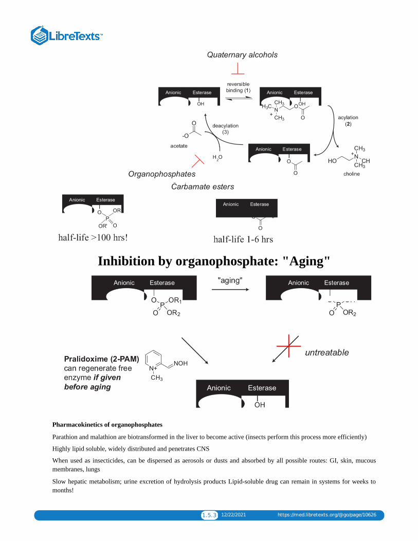
Synthesized from acetyl-CoA and choline by cholineacetyltransferase (ChAT).
Poor absorption and low lipophilicity due to charge onquaternary ammonium.
Multiple systemic effects, esp autonomic pathways and at theneuromuscular junction (NMJ).
Acetylcholinesterase (AChE)
Clears Ach from site of action (also degraded by plasma
butyrylcholinesterase)
Bound on post-synaptic membrane
Rate = 400,000 per min
Inhibition of AchE results in build up of Ach at muscarinic and nicotinic synapses!
Step 1: Binding
Step 2: Formation of covalent intermediate and release choline
Step 3: Hydrolysis of acyl-enzyme intermediate
Direct-acting agonists

1.5.2 12/22/2021 https://med.libretexts.org/@go/page/10626
Mimics acetylcholine by binding Achreceptor and activating downstreamsignaling
Examples: methacholine, carbachol,bethanechol, pilocarpine
Indirect agonists
Inhibits AchE from breaking downacetylcholine at synapse
Quaternary alcohols
- competes w/ AChfor binding toAChE (step 1)
Examples:edrophonium
Carbamate esters
- formation ofcarbamylatedenzymeintermediate (step2)
Examples:neostigmine,
pryidostigmine
Organophosphates
- formation ofphosphorylated enzymeintermediate (step 2)
Examples: parathion, malathion are insecticides soman, sarin are nerve agents
AchE inhibitors: reversible versus irreversible
1.5.3 12/22/2021 https://med.libretexts.org/@go/page/10626
Inhibition by organophosphate: "Aging"
Pharmacokinetics of organophosphates
Parathion and malathion are biotransformed in the liver to become active (insects perform this process more efficiently)
Highly lipid soluble, widely distributed and penetrates CNS
When used as insecticides, can be dispersed as aerosols or dusts and absorbed by all possible routes: GI, skin, mucousmembranes, lungs
Slow hepatic metabolism; urine excretion of hydrolysis products Lipid-soluble drug can remain in systems for weeks tomonths!
1.5.4 12/22/2021 https://med.libretexts.org/@go/page/10626
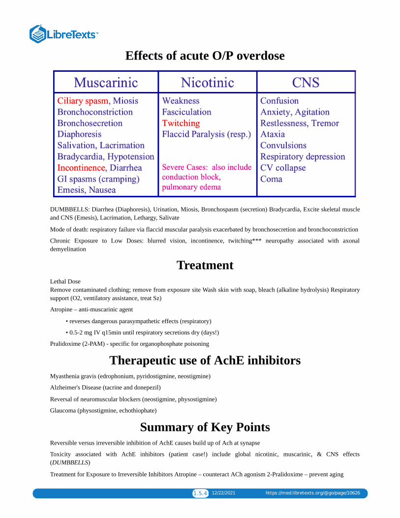
Effects of acute O/P overdose
DUMBBELLS: Diarrhea (Diaphoresis), Urination, Miosis, Bronchospasm (secretion) Bradycardia, Excite skeletal muscleand CNS (Emesis), Lacrimation, Lethargy, Salivate
Mode of death: respiratory failure via flaccid muscular paralysis exacerbated by bronchosecretion and bronchoconstriction
Chronic Exposure to Low Doses: blurred vision, incontinence, twitching*** neuropathy associated with axonaldemyelination
TreatmentLethal Dose Remove contaminated clothing; remove from exposure site Wash skin with soap, bleach (alkaline hydrolysis) Respiratorysupport (O2, ventilatory assistance, treat Sz)
Atropine – anti-muscarinic agent
• reverses dangerous parasympathetic effects (respiratory)
• 0.5-2 mg IV q15min until respiratory secretions dry (days!)
Pralidoxime (2-PAM) - specific for organophosphate poisoning
Therapeutic use of AchE inhibitors Myasthenia gravis (edrophonium, pyridostigmine, neostigmine)
Alzheimer's Disease (tacrine and donepezil)
Reversal of neuromuscular blockers (neostigmine, physostigmine)
Glaucoma (physostigmine, echothiophate)
Summary of Key Points
Reversible versus irreversible inhibition of AchE causes build up of Ach at synapse
Toxicity associated with AchE inhibitors (patient case!) include global nicotinic, muscarinic, & CNS effects(DUMBBELLS)
Treatment for Exposure to Irreversible Inhibitors Atropine – counteract ACh agonism 2-Pralidoxime – prevent aging
1.6.1 1/5/2022 https://med.libretexts.org/@go/page/10627
1.6: Autonomic PharmacologyAs you will see throughout the course, the autonomic nervous system (ANS) is a very important topic for two reasons:First, manipulation of ANS function is the basis for treating a great deal of cardiovascular, pulmonary, gastrointestinal andrenal disease; second, there is hardly a drug worth mentioning without some major autonomic side effects (cf.antihistamines). You have already heard something about the ANS and its wiring diagram in the lecture by Dr. Strichartzon cholinergic receptors, and it is certainly not my intent to reproduce these pictures or the various diagrams in your text. Ihope to give you a slightly different presentation which highlights the important points in this rather long textbookassignment.
You have already heard about nicotinic cholinergic receptors and the somatic nervous system (SNS) control of voluntarystriated muscle. The ANS, simply put, controls everything else: smooth muscle, cardiac muscle, glands, and otherinvoluntary functions. We usually think about the ANS as a motor system -- although it does have sensory nerves, there isnothing particularly distinctive about them.
Anatomy
The sympathetic division of the ANS is called THORACOLUMBAR, but it has input from higher brain centers likehypothalamus, limbic cortex, etc. The preganglionic sympathetic nerves have cell bodies in the intermediolateral columnof the spinal cord from about T1 to L3. The efferent fibers exit with the ventral roots of the spinal nerves and then leave ina white ramus which leads to a GANGLION (i.e., a collection of cell bodies of postganglionic neurons). The preganglionicnerves may stimulate several postganglionic nerves which rejoin the spinal nerve by way of a grey ramus. The ganglia arelocated in several places:
1. Paravertebral: 22 pairs located on either side of the vertebral column. The uppermost ganglia are fused to form thesuperior and middle cervical ganglia and the stellate ganglion, which is located at about C6. The preganglionic neuronmay travel up or down several dermatomal levels before synapsing with one or more postganglionic neurons.
2. Prevertebral: The celiac, superior mesenteric and inferior mesenteric ganglia. Sometimes called collateral ganglia.3. Adrenal Medulla: This is also derived from neural crest tissue and functions in much the same way as a ganglion,
although the output is circulating epinephrine and norepinephrine.
The parasympathetic or CRANIOSACRAL division has its origin in the nuclei of cranial nerves III, VII, IX, and X as wellas the S2-4 nerve roots. The preganglionic fibers travel almost to the end-organ before synapsing in the ganglion:
1. III goes from the Edinger Westphal nucleus to the ciliary ganglion, and the postganglionic nerves continue to the eye.2. VII innervates the pterygopalatine and submandibular ganglia which control lacrimal and salivary glands, respectively.3. IX innervates the otic ganglion which controls the parotid4. X innervates the heart, lung, GI tract, and other splanchnic viscera. The postganglionic cell bodies are contained in
specialized tissue within the heart (e.g. AV nodal tissue), GI tract (e.g. Auerbach's plexus).5. S2-4 preganglionic nerves originate in the sacral parasympathetic nucleus and leave the cord by way of the pelvic
splanchnic and pudendal nerves. They innervate the distal GI tract, bladder, and genitalia.
We will not specifically discuss the enteric nervous system – often treated as a third division of the ANS. It consists ofcomplex networks of interconnected ganglia and nerve fibers, largely contained with the myenteric (Auerbach’s) andsubmucosal (Meissner’s) plexuses. This system exerts local control over GI secretion, motility, blood vessel tone,, andfluid transport. It is subject to control by sympathetic, parasympathetic, and CNS iinputs.
If we look at the sympathetic and parasympathetic divisions schematically, it is easy to see how the sympathetic division issuited to "flight or fright" responses. The stimulation of one preganglionic neuron can lead to widespread activation ofpostganglionic neurons and to the liberation of stress hormones like epinephrine. The parasympathetic division is oftencalled a "vegetative" system, and it is well suited to controlling discrete parts of the body.

1.6.2 1/5/2022 https://med.libretexts.org/@go/page/10627
Screen Shot 2019-01-14 at 11.40.23 AM.png
There are some differences between the somatic and autonomic systems that are worth remembering.
AUTONOMIC SOMATIC
Synapses in peripheryNerve plexusesOrgans, glands, sm. muscle have activity without nervesSymp and Parasymp afferent and efferent nerves overlap interminal retinaculumSm. muscle has protoplasmic bridges, so stimulating one candepolarize 100 others.
Synapses in CNSNo plexusesSkeletal muscle atrophies without nerveNerves end in discrete motor end plates on muscle fibersMuscle fiber depolarized discretely
Cholinergic neurotransmission
Fig 6-3 and 6-4 in Katzung schematize the cholinergic and adrenergic nerve terminals. Cholinergic receptors are generallycategorized as follows:
Nicotinic motor end plate autonomic ganglia
Muscarinic autonomic ganglia parasympathetic postganglionic
All nicotinic receptors are, by definition, stimulated by the alkaloid nicotine. We know that the two types of nicotinicreceptor differ because they are differentially affected by various agonists and antagonists
Agonists Antagonists
Motor End Plate phenyltrimethylammonium (PTMA) decamethonium bungarotoxin
Ganglion dimethylphenylpiperazinium (DMPP) hexamethonium
Muscarinic receptors are those stimulated by the alkaloid muscarine, which comes from the mushroom Amanita muscaria.At this writing there are 5 postulated subtypes of muscarinic receptors (see table) although not much is known about thelast two.
Muscarinic Receptor Subtypes

1.6.3 1/5/2022 https://med.libretexts.org/@go/page/10627
Cholinergic Signal Transduction
The nicotinic response of skeletal muscle has been discussed in detail. ACh causes depolarization by a sudden increase inNa+ conductance. Repolarization depends upon the outward flow of K+.
The transduction of muscarinic responses depends upon the tissue. For example ACh causes hyperpolarization of cardiacconducting tissue by increasing K+ conductance (M2). In G.I. smooth muscle it causes partial depolarization by increasingNa+ and Ca2+ conductance (M3).
Muscarinic Responses
Ganglionic transmission is a very complex system (cf. Strichartz lecture). An initial nicotinic effect leads to an increase inNa+ conductance and a fast excitatory post-synaptic potential (EPSP). This is modulated by a muscarinic (M1) slow EPSP,and an inhibitory postsynaptic potential (IPSP) which may be muscarinic (M2) or may involve adrenergic transmissionfrom a SIF (small, intensely fluorescent) interneuron.

1.6.4 1/5/2022 https://med.libretexts.org/@go/page/10627
Adrenergic Neurotransmission
Neurotransmission from almost all sympathetic postganglionic nerves is adrenergic, that is, it involves noradrenaline (NE,norepinephrine, levarterenol). Adrenergic receptors were first divided into α and β by Ahlquist in 1948. The pattern ofresponses he defined was based on the relative potencies of agonists:
α-- epinephrine > norepinephrine > phenylephrine >> isoproterenol
β -- isoproterenol > epinephrine ≥ norepinephrine >> phenylephrine
More subtle potency differences and selective antagonists allowed us to subtype these receptors.

1.6.5 1/5/2022 https://med.libretexts.org/@go/page/10627
Selective Adrenergic Agonists and Antagonists
Subtypes of α receptors (α1a, α1b, α1d; α2a, α2b, α2c) have been cloned and localized in different tissues, but theirphysiologic functions are not known. A β-3 response has been described which mediates lipolysis in adipocytes. There areno truly selective agonists/antagonists for the β-3 receptor, and the response is not blocked by most β antagonists. Theselectivity of the drugs in the table is only relative.

1.6.6 1/5/2022 https://med.libretexts.org/@go/page/10627
Signal transduction of adrenergic receptors has been studied extensively. The α-2 and β receptors depend on G-proteinmediated inhibition or activation of adenylyl cyclase, while α-1 works by activating phospholipases to hydrolyzephosphoinositides.
How can we use drugs to promote or inhibit cholinergic or noradrenergic neurotransmission? I recommend that each ofyou draw a schematic of the two types of nerve terminals (cf Katzung 6-3 and 6-4) and try to incorporate the informationfrom Table 6-5. We'll go through this briefly in class. In both cases, there are toxins or approved/experimental drugs whichcan affect
1. The synthesis, transport or storage of transmitter2. The release of transmitter3. The effect of transmitter at receptor sites4. The inactivation or metabolism of transmitter
[It is also possible to modify sympathetic neurotransmission at the level of signal transduction--Can you think of anexample?]
It is important to realize that the responses to most autonomic drugs are not static over time. The magnitude of theresponse depends enormously on baseline tone, and responses may change with repeated drug administration or alterationsin patient physiology. When a drug loses its effect we say that the patient becomes tolerant -- i.e. the same dose producesless effect (or it takes a lot more to produce the same effect). This phenomenon is best described in the case of adrenergicdrugs:
"Tachyphylaxis" Certain drugs like ephedrine act by releasing intraneuronal stores of preformed norepinephrine. Afterrepeated dosing, the supply of neurotransmitter may be temporarily exhausted.Receptor Down Regulation This may be a decrease in the number of available receptors or a decrease in ligand affinityfor the receptor. A good example is the decrease in β1 receptors in the myocardium of patients with congestive failure.Desensitization Reversible uncoupling of receptor occupancy and cellular response by receptor phosphorylation andinternalization. This is best worked out for the β adrenergic receptor.
Effects on End-Organs
Table 6-3 in Katzung (or Table 5-1 in G & G) lists the effects of stimulating α and β or muscarinic cholinergic receptors. Inmost cases you will notice that parasympathetic stimulation produces effects which promote normal "vegetative" functionslike urination, defecation, production of saliva, accommodation of the eye for near vision, etc. Sympathetic activation doesthose things deemed necessary for "fight or flight" like increasing cardiac output, increasing metabolic rate, relaxing theciliary muscle for distance vision, etc. Often the two systems function in opposition, but this is not always the case. In thecase of blood vessels, the predominant tone is α−adrenergic, and there is relatively little β or muscarinic tone. [Why doesthis make sense?]
In a healthy, young individual at rest the predominant tone in most tissues is parasympathetic: for example, there ispredominant vagal tone in the heart, peristalsis in the GI and GU tract, and the pupils are small. In a subject under physicalstress (or a critically ill patient) there may be maximal sympathetic tone. This is important for two reasons:
1. A drug which acts by blocking any autonomic activity will have much more effect if there is a great deal of activity tobegin with.
2. It follows that a drug which blocks ganglionic transmission (i.e., both sympathetic and parasympathetic function) willhave its greatest effect on the system which predominates. [So, are the predominant effects of ganglionic blockers
1.6.7 1/5/2022 https://med.libretexts.org/@go/page/10627
sympatholytic or parasympatholytic? What are they used for?]
Clinical Applications of the Various Drug Classes
Cholinergic Agonists
The use of these agents is rather limited, in part because they have such widespread effects. Nicotinic agonists occupy asmall but important place in therapeutics and toxicology. We cannot use acetylcholine itself effectively [why?].
Nicotine and succinylcholine are the two most commonly used nicotinic "agonists," but paradoxically both of thesedrugs cause such persistent membrane depolarization that they inactivate Na channels and block neuromusculartransmission (cf. Dr. Strichartz comments on "depolarizing" muscle relaxants). Nicotine is used as an insecticide andself-administered (in low doses) by those who smoke and those who are trying to quit. Succinylcholine is usedclinically to produce neuromuscular blockade.
The most common “agonists” are those that act indirectly by inhibiting acetylcholinesterase (ChE). The reversibleinhibitors like neostigmine and pyridostigmine are used by anesthesiologists to reverse the effects of nicotinic antagonistslike curare. They are also given to patients who suffer from myasthenia gravis and have circulating antibody to their ownnicotinic receptors. In both cases, the drugs are working by increasing the concentration of acetylcholine available at themotor end-plate. Centrally acting ChE inhibitors are also being used in Alzheimer’s dementia.
The irreversible ChE inhibitors like the organophosphates permanently inactivate the enzyme. These drugs were presentedin Case Discussion 1 --they are used as insecticides and as "nerve gases." One of them, echothiophate, is used clinically toproduce long-lasting miosis (pupil constriction) for refractory glaucoma.
Muscarinic agonists like methacholine, pilocarpine, bethanecol and carbachol are used to produce miosis (in cases ofglaucoma or eye surgery) and to increase the activity of GI and GU smooth muscle. The latter effects are sometimes usefulin cases of diabetic gastroparesis or bladder dysfunction.
Cholinergic Antagonists
Nicotinic Antagonists like d-tubocurarine (curare) and pancuronium are used to produce muscle relaxation for surgery andsometimes to facilitate ventilation in critically ill patients. These so-called "non-depolarizing" relaxants act as competitiveantagonists at the nicotinic receptor. The selectivity of these older relaxants is not very good: curare is a weak ganglionicblocker, and pancuronium can produce substantial muscarinic block. [What will their side effects be, when used for musclerelaxation?]
Botulinum toxin is a bacterial toxin which blocks acetylcholine release by binding to docking proteins and inhibitingexocytosis. It is now used in some focal dystonias, movement disorders and strabismus (crossed eyes). The drug is givenby direct injection into the muscle and produces partial denervation for 3-5 months.
Muscarinic antagonists are still fairly widely used. The prototypes are the belladonna alkaloids, atropine and scopolamine.There are also a fairly large number of synthetic atropine-like drugs. These are all competitive, reversible antagonistswhich produce mydriasis (pupil dilation), dry mouth, tachycardia, sedation, and decreased tone of bronchial, GI and GUsmooth muscle. Logically enough, they are used to treat bradycardia, diarrhea and bladder spasms. Atropinics are also usedas sedatives, to dilate the pupils, to reduce secretions, and to produce bronchodilation. Frequently, atropine is used tocounteract the muscarinic effects of drugs like the ChE inhibitors.
It is very important to remember that many useful drugs from completely different categories have pronouncedanticholinergic side-effects-- examples of these include antihistamines (Benadryl), tricyclic antidepressants (Elavil),phenothiazines (Mellaril). All of these drugs can produce sedation, rapid heartbeat, constipation and urinary retention.
Adrenergic Agonists
This class includes the endogenous agonists norepinephrine (NE), epinephrine (E), and dopamine (DA). These drugs arenot terribly selective for the various receptors, and their relative α vs β (or δ, in the case of DA) activity depends a greatdeal on the dose administered. Most of the agonists act directly at pre- or postsynaptic receptors, but a few (ephedrine,amphetamine, tyramine) act by releasing NE from terminals.

1.6.8 1/5/2022 https://med.libretexts.org/@go/page/10627
Please review Table 9-4 and Figure 9-6 in Katzung which describe the different cardiovascular responses to phenylephrine,isoproterenol, and epinephrine (an α, β, and mixed sympathomimetic drug, respectively). Epinephrine (in low doses) andisoproterenol stimulate β1 and β2 receptors to increase heart rate and contractility but decrease peripheral vascularresistance. Norepinephrine has very little effect on vascular β2 receptors, so it produces peripheral constriction (G & G p.205 ):
α-1 agonists like phenylephrine are primarily used for their effects on vascular tone. By local vasoconstriction they can actas nasal decongestants and retard the absorption of other drugs like local anesthetics. Of course, they are used to treathypotension or shock, especially when there is inappropriate vasodilation (e.g. gram-negative sepsis). These drugs are alsoused frequently as mydriatics
α-2 agonists. Clonidine, methyldopa, guanfacine, and guanabenz reduce blood pressure through a central reduction insympathetic tone. All are used for the outpatient treatment of hypertension. Stimulation of postsynaptic α-2 receptors caninitially cause vasoconstriction and increase peripheral resistance. The antihypertensive effects of clonidine and otherimidazolines are thought to be mediated, in part, by non- adrenergic “imidazoline” receptors (cf. Katzung, p. 159).Clonidine and dexmedetomidine are also used in man for their sedative and analgesic properties.
β-1 agonists like dobutamine or non-selective β agonists like isoproterenol are used to increase myocardial contractility,and sometimes to increase heart rate or automaticity. The fact that these agents may actually decrease afterload is anadvantage in patients with congestive failure; the propensity of β agonists to cause tachyarrhythmias is not.
β-2 agonists like albuterol, metaproterenol or terbutaline are extremely useful for their bronchodilator properties.Asthmatics commonly take them by inhalation. β-2 agonists can also stabilize mast cell membranes and abort acutehypersensitivity reactions like anaphylaxis. The drug of choice for this indication is epinephrine. β-2 agonists can relaxuterine smooth muscle, and drugs like ritodrine are sometimes used to delay premature labor.
Adrenergic Antagonists
α-1 antagonists like prazosin are used for the outpatient treatment of hypertension. The older non-selective a blockers likephentolamine were much more likely to cause tachycardia [why?]. Phenoxybenzamine, a non-competitive α blocker, hasbeen used for the therapy of pheochromocytoma.
α-2 antagonists like atipamezole are not marketed, but have been tested in man. Theoretically, they could be useful toreverse the effects of drugs like clonidine and dexmedetomidine.
β antagonists like propranolol, nadolol, and timolol are non-selective drugs which are widely used. Metoprolol, esmololand atenolol are relatively selective for β-1 receptors. [What is the theoretical advantage of this?] Labetalol has both α andβ antagonist activity. These drugs are of major importance for the treatment of hypertension, ischemic heart disease,obstructive cardiomyopathy, and tachyarrhythmias. They may increase survival after myocardial infarction. Propranol hasalso been used to treat thyroid storm. Unlike some β blockers, propranolol has substantial CNS activity and has provenuseful for migraine headache and benign essential tremor. [How might the metabolic effects of β-blockers pose aproblem?]
Miscellaneous sympathoplegic drugs. These drugs are older treatments for hypertension and have largely been supercededby drugs like angiotensin converting enzyme inhibitors, calcium channel blockers, as well as α and β blockers. Reserpine,which depletes nerve terminals of NE, is described (Katzung, p.163) as an acceptable treatment for hypertension. In myopinion, the drug is of mainly historical interest. Guanethidine, prevents the release of NE -- another kind of chemicalsympathectomy. Its side effects, especially postural hypotension and diarrhea, can be severe, so it has traditionally beenreserved for cases of hypertension refractory to other treatments. Metyrosine (α-methyl p- tyrosine) inhibits tyrosinehydroxylase, the rate limiting step in the biosynthesis of catecholamines. It has had some use in the adjunctive treatment ofpheochromocytoma.
1. What are the important differences between the somatic and autonomic nervous systems?2. How is the anatomy of the sympathetic/parasympathetic system suited to its physiologic role?3. How can we distinguish nicotinic receptors at the motor end-plate from those in autonomic ganglia?
Questions to be covered in Autonomic Nervous System lectures

1.6.9 1/5/2022 https://med.libretexts.org/@go/page/10627
4. What is the basis for subtyping α and β adrenergic receptors? What are the supposed advantages of therapeuticagents which are specific for one subtype of receptor?
5. By what mechanisms may drugs modify adrenergic and cholinergic transmisssion?
Problem Set -- Autonomic Nervous System
Pindolol is a partial agonist at β receptors. How can it act as an antagonist? What is the putative advantage of this drug?
What are the possible reasons for the development of tolerance to a βagonist drug (in general terms, please)? If we suspectthat there has been receptor down-regulation, how might we investigate it? How could we determine whether there arefewer receptors or the receptor has decreased affinity for the agonist? Hint: See Scatchard Plot in notes by Dr. Strichartz.
We have accepted the fact that NE and ACh are neurotransmitters for postganglionic sympathetic and parasympatheticneurons, respectively. What kind of evidence was necessary to come to this conclusion?
A patient receiving the MAO inhibitor, pargyline, can have a hypertensive crisis after eating cheese or drinking Chianti,yet this drug was originally marketed to lower blood pressure. How can we explain these apparently opposite effects?

1.7.1 1/4/2022 https://med.libretexts.org/@go/page/10628
1.7: Local Anesthetics
To describe the mechanisms of local anesthesia as well as some relevant clinical pharmacology of local anesthetics.
Local anesthesia is the selective numbing of a particular, circumscribed region of the body by a controlled, reversibleprocedure. Drugs called local anesthetics (LA) are usually employed for these procedures, although directly appliedpressure, cooling, or even heating will also produce numbness. The general strategy is to inhibit the propagation orgeneration of impulses in nerves from a defined anatomical region.
Knowledge of the structure of local anesthetic drugs is essential for an understanding of their mechanism of action,potencies and pharmacokinetics. The general structure of a local anesthetic is:
The aim of this lecture is to describe the mechanisms of local anesthesia as well as some relevant clinical pharmacology oflocal anesthetics. (Public Domain; User:Edgar181)
Structures and properties of drugs used clinically are listed in Table 1, along with one experimental derivative, QX-314.
The aromatic group sometimes contains a para-amino group (-NH ) at R3 (procaine) and additional alkyl groups attachedto this amino (tetracaine), or at R , R (lidocaine, and other amides).
Amide or ester bonds connect the aromatic moiety to a tertiary (3°-) amine which can have alkyl groups of lengths from -CH to -C H attached to it. The absolute potency of LA increases with increasing alkyl length substituents on botharomatic and 3°-amine groups. Physico-chemical analysis reveals a monotonic increase of absolute potency withincreasing hydrophobicity for all compounds. Since the mechanisms of action are complex (see below), the exactrelationships between structure, pKa, and membrane distribution are still not known.
Mechanism of ActionLAs block nerve impulses by interfering with the sodium permeability increase (PNa) which subserves the depolarizingphase of action potentials. The mechanistic details depend on the LA molecule being used.
A. Active species: (3°)-amine local anesthetics (pKa = 7.8-10) exist as equilibrium mixtures of protonated cation andneutral base at physiological pH.
The ionization reactions at neutral pH are quite rapid (-10 sec).
Evidence to answer these questions comes from:
Conclusion: Both neutral and protonated species of LA can inhibit Na channels and block impulses. In general,however, the protonated form appears to be more potent.
1. Which form of the LA module blocks P ?2. Where does it act: inside or outside the cell or on the membrane?
Skills to Develop
2
1 2
3 4 9
-3
Na

1.7.2 1/4/2022 https://med.libretexts.org/@go/page/10628
3. Quaternary (4 )-amine derivatives (permanent cations, e.g., QX-314 which do not permeate the membrane,block sodium channels (P ), but only when applied in the cytoplasm.
4. The observed impulse-blocking potency of benzocaine and of lidocaine derivatives where -OH replaces -NR(both permanently neutral molecules). These drugs act identically whether applied externally orcytoplasmically.
Molecular Mechanisms1. The block of sodium current (I ) or of impulses by 4 -amine LAs increases in extent with repetitive opening of
sodium channels ("use-dependent" block) (Figure 2). Use-dependent block is reversed when stimulation stops.2. With benzocaine (and some alcohols) and with 3 -amine anesthetics at alkaline pH, resting nerve block reveals more
"inactivated" sodium channels (Fig. 3) but use-dependent block is very weak. 3o-amine LAs show much more use-dependent block at neutral or slightly acid pH than at alkaline pH (external). Internal pH has surprisingly little effect.
3. Inhibition of ionic Na+ current by benzocaine is paralleled by a proportional reduction of "gating current", themovement of charge which results directly from conformational changes of Na channels during activation (Figure 4).
The sodium channel itself appears to be a receptor for local anesthetics. Intentional mutation of part of the channel'sinner pore region changes resting and use- dependent pharmacology of various local anesthetic molecules. In addition,in normal channels the membrane potential changes the channel conformations, which in turn have different anestheticaffinities. This is collectively referred to as the "modulated receptor hypothesis" (Figure 5). In addition, there is a non-receptor mediated action of local anesthetic agents, which may occur through a disruption of normal membranestructure.
4. Calcium ions may antagonize the blocking action of some local anesthetics, but this probably is mediated throughchanges in channel structure and is not necessarily evidence for direct steric competition between Ca and LA binding.
5. LAs also have been shown to inhibit K+ channels, Ca channels, and the nicotinic acetylcholine-activatedconductance, the substance P receptor and even the G-protein modulation of certain channels. These alternative actionsmay contribute to spinal (intrathecal) anesthesia and to some aspects of toxicity.
6. Modes of Administration and PharmacokineticsA. A. Injection--minor, to block small regions via peripheral nerve; major (includes iv), to block whole limbs via
peripheral nerve.
Clinically, local anesthetics are usually injected as 0.25-1 % (w:v) solutions, equivalent to 10-40 mM, where 1/40-1/100 of those concentrations provide a 50% absolute block of impulses in an isolated, desheathed nerve.Interestingly, less than 10% of the dose of injected drug actually reaches the nerve to provide complete functionalblock.
B. Infiltration--usually at skin or other superficial surfaces, e.g. scalp, oral mucosa.C. Topical--superficial application, on skin, tracheal (pre-intubation) to reduce irritation and gag reflex.D. Central injections--at spinal cord:
1. epidural--blocks roots, but LA also enters cord, CSF.2. intrathecal--"spinal":
a. potent block of many dermatomesb. drug is often dissolved in a hypo- or hyperbaric solution to control spread.c. positioning of patient may also be adjusted to control anatomical distribution of block.
E. Removal--LAs are removed from site of injection by local tissue uptake and local circulation.1. removal by circulation is often reduced by co- injection of epinephrine, but this in not true for all LAs at all
locations.2. complications arising:
a. epinephrine itself may have subliminal blocking action.b. epinephrine is usually packaged with anti- oxidant and at acid pH. Antioxidant can be neurotoxic, and low
pH renders LA less penetrating, therefore less effective.F. Metabolism--little intact LA is eliminated from the body.
1. esters--hydrolyzed by tissue and serum cholinesterases (non-specific).
o
Na
2
Nao
o
2+
2+

1.7.3 1/4/2022 https://med.libretexts.org/@go/page/10628
2. amides--oxidized by mixed-function oxidase system, of hepatic ER.
7. Differential Fiber Blockade: Early papers and most pharmacology texts report that smaller nerve fibers are blocked"before" larger diameter fibers by LA drugs. "Before" almost certainly means earlier during the development of theblock, but when a steady- state (absolute) block has been achieved, single impulses in the larger fibers are often moreinhibited than those in the smaller ones (Figure 6).
It is unlikely that an absolute differential block, short of a total one, is ever reached under these "clinical" conditions.`During onset of block of a nerve containing many fiber types, we observe functional activities being lost in aconsistent sequence: pain, temperature, touch, proprioception, and skeletal muscle tone and voluntary tension. Sinceboth sensory and motor functions are dependent on frequencies of trains of impulses, the modulation of AP frequencyrather than the absolute loss of impulses may correspond to the functional deficits observed clinically.
Sensations from more proximal regions are blocked earlier and recover later than those from more distal segments.This reflects the diffusion of anesthetic through the somatotopically organized peripheral nerve.
References1. Ritchie JM and Greene NM. Chapter 15. Local Anesthetics. In: The Pharmacological Basis of Therapeutics 8th ed.
Macmillan, N.Y., pp. 311-331. A concise yet thorough summary by one of the pioneers (JMR) on mechanisms of LA.2. Bromage P. (1978) Epidural Anesthesia, Saunders, Philadelphia, PA. A scholarly treatise ranging from basic
neurophysiology and anatomy to clinical complications. Chapters 2-4 are particularly pertinent to these lectures.
3. Dripps, RD, Eckenhoff JE, and Vandam LD (1983) Introduction to Anesthesia. The Principles of Safe Practice.Saunders, Philadelphia, PA. The best introductory text for anesthesiology, this book places basic science firmly in thecontext of clinical practice. Emphasize chapter 17; read chapters 18-20 for more clinical information.
4. Butterworth, JF and Strichartz G. (1990) Molecular mechanisms of local anesthesia: A Review. Anesthesiology72:711-734. The state of knowledge as of 1989, in excessive detail.
1.8.1 12/29/2021 https://med.libretexts.org/@go/page/10629
1.8: Antiinflammatory DrugsInflammation is mediated in part by prostaglandins produced by the cyclooxygenase pathway. NSAIDs inhibit thispathway and serve as combined anti-inflammatory, anti pyretics, and analgesics. Because NSAIDs are generallynonspecific and exert numerous side effects, there is great interest in more specific therapeutics such as selective COX-2inhibitors and anti-cytokine agents.
Prostaglandins: Physiologic and Pathologic FunctionsAll cells in the body have the capacity to synthesize prostaglandins. In response to inflammatory stimuli arachidonic acid(AA) is separated from plasma phospholipids by phospholipase A2. Cyclooxygenase metabolizes AA to thecycloendoperoxide prostaglandin H2 (PGH2), which is then converted to either PGD2, PGE2, PGF2α, PGI2(prostacyclin)or TXA2 (thromboxane) by appropriate enzymes (i.e. thromboxane synthase in platelets, prostacyclin synthase inendothelial cells).
The prostaglandins exert numerous physiologic and pathophysiologic functions :
Physiologic: temperature homeostasis, bronchial tone, cytoprotection (gastric and renal mucosa), intestinal mobility,myometrial tone, semen viability (some prostaglandins like PGE1 have anti-inflammatory effects), renin secretionƒ Pathologic: fever (aberrant hypothalamic thermoregulation), asthma (airway responsiveness and immunehyperreactivity), ulcers (loss of cytoprotection), diarrhea (intestinal mobility), dysmenorrhea (myometrial tone),inflammation, bone erosion, pain (thought to be caused by PGD2)
Specific functions of prostaglandins in the context of inflammation include:
ƒ PGI2: inhibits platelet aggregation, vasodilatation, vascular permeability (edema)ƒ PGE2: pain, hyperalgesia, heat, vasodilatation, bronchoconstriction, synergistically act with other pro-inflammatorymediators (histamine, complement, LTB4)ƒ TXA2: promotes platelet aggregation, vasoconstriction, bronchoconstriction
Cyclooxygenase
There are two forms of cyclooxygenase (COX) enzymes: COX-1 and COX-2. Though COX-1 and COX-2 catalyze thesame reaction, their expression, functions, and properties are markedly different.
COX-1 produces PGE2, PGI2, and TXA2 in platelets, GI mucosa, vascular endothelium, and the kidney. Thehousekeeping functions of these prostaglandins include maintaining renal and gastrointestinal blood flow (cytoprotection),regulation of vascular homeostasis, renal function, intestinal mucosal proliferation, and platelet function.
Pro-inflammatory functions of COX-2 produced prostaglandins include pain, fever, leukocyte proliferation, andinflammation. COX-2 produces prostaglandins at sites of inflammation (in macrophages, in synovial tissue of rheumatoidarthritis joint). Mitogenic functions of COX-2 produced prostaglandin include renal genesis and reproduction.
The goal of pharmacologic anti-inflammatory therapy has been to inhibit COX-2 produced prostaglandins. Non-specificinhibition of COX-1 results in gastrointestinal and platelet side effects. Recent data on the toxicity of COX-2 selectivensaids illustrate that this is an overly simplistic view. The magnitude of the COX-2 problem is still unclear at this writing,but it will be considered at various points in this discussion.

1.8.2 12/29/2021 https://med.libretexts.org/@go/page/10629
There is an entire additional pathway of arachidonic acid metabolism by enzymes called lipoxygenases. 5-lipoxygenase is not present in all tissues but is limited to neutrophils, eosinophils, monocytes, and certain mast cellpopulations. Lipoxygenases produce leukotrienes (e.g. LTB4, LTD4), which are potent bronchoconstrictors andchemotactic agents. Leukotrienes have important roles in asthma, glomerulonephritis and inflammatory bowel disease.(Refer to the Asthma case.
NSAIDs (Non-steroidal anti-inflammatory drugs)Most NSAIDs are polycyclic carboxylic acid derivatives with relatively low pKa values. NSAIDs are often classified onthe basis of their chemical structure (see Figure 1).
ƒ Salicylates: aspirin; diflunisal, 5-aminosalicylate, sodium salicylate, magnesium salicylate, sulfasalazine, olasalzineƒ Acetic acids: indomethacin, diclofenac, sulindac, etodolac, ketorolac, tolmetinƒ Propionic acids: ibuprofen, naproxen, fenoprofen, ketoprofen, flurbiprofen, oxaprozinƒ Fenamic acids: meclofenamate, mefenamateƒ Enolic acids (oxicam class): piroxicamƒ Ketones: nabumetone (converted to 6-naphthylacetic acid in liver)
NSAID General Pharmacodynamics
All NSAIDs (except aspirin) act as reversible, competitive cyclooxygenase inhibitors. They block the hydrophobic channelby which the substrate arachidonic acid accesses the enzyme active site. Aspirin covalently modifies and destroys thecyclooxygenase enzyme.
The ultimate function of the NSAID is to inhibit COX-2, preventing generation of proinflammatory eicosanoids, and thuslimiting the extent of inflammation and adverse signs and symptoms.
ƒ All NSAIDs have a ratio of inhibition of COX-2 / inhibition of COX-1. The higher the ratio, the more specific thetherapeutic effect and fewer GI or platelet effects.
− NSAIDs with high ratio (100:1 to 1000:1) are COX-2 Selective (Coxib)ƒ
Despite the benefits of NSAIDs, they only provide symptomatic relief, as the underlying pathophysiology or injurygenerally is unaffected.
NSAIDs have three primary therapeutic effects:
ƒ Analgesiaƒ Anti-pyrexia (decreasing hypothalamic PGE2)ƒAnti-inflammatory
NSAIDs are also used as anti-thrombotics. Since they impair platelet aggregation, they prolong bleeding time, and functionas anticoagulants. The COX-2 specific inhibitors do not exert anti-thrombotic effects. Other functions of NSAIDs includeinhibition of:
Superoxide generationƒ Lysosomal enzyme releaseƒ Neutrophil aggregation / adhesionLymphocyte functionƒ Cytokine release (IL-6)
Indications for Specific NSAIDs
Please refer to Table 1 to find indications common to each structural class of NSAID. See below for COX-2 selectivedrugs.
Non-selective NSAIDs are used as analgesics for moderate pain of musculoskeletal and inflammatory origin (headaches,dysmenorrhea, osteoarthritis, rheumatoid arthritis, gout, surgical pain, tendonitis, and bursitis). NSAIDs also function asanti-inflammatory agents in many of these conditions and ulcerative colitis. Aspirin anti-platelet effects are used for MI
lipoxygenases

1.8.3 12/29/2021 https://med.libretexts.org/@go/page/10629
and stroke prophylaxis. Acetaminophen (technically not an NSAID) has no anti-inflammatory activity but is widely usedas an analgesic and anti-pyretic.
NSAID Pharmacokinetics
As stated previously, NSAIDs are weak organic acids. They generally haveƒ
Efficient GI absorption (nearly complete)ƒLow first pass hepatic metabolismSmall volumes of distribution but extensive protein binding (>95%) which slows the rate at which these drugs cross thecapillary wall and penetrate tissue.ƒ Accumulation in cells at sites of inflammation (acidic NSAIDs are preferentially sequestered in inflamed synovialtissues)ƒ Efficient enterohepatic and renal excretionƒ Variable half lives (the lower the pKa generally the shorter the half-life)
Plasma Elimination Half Lives: Another method to classify NSAIDs (besides structure)
Short Half Life (< 6 hours): more rapid effect and clearance Aspirin (0.25-0.33 hrs), Diclofenac (1.1 ± 0.2 hrs),Ketoprofen (1.8 ± 0.4 hrs), Ibuprofen (2.1 ± 0.3 hrs), Indomethacin (4.6 ± 0.7 hrs)ƒ Long Half Life (> 10 hours): slower onset of effect and slower clearance Naproxen (14 ± 2 hrs), Sulindac (14 ± 8hrs),Namebutone (26 ± 5 hrs), Piroxicam (57 ± 22 hrs) (also COX-2 Selective Inhibitors)
Important Drug Interactions:
Displace other drugs from plasma protein binding sites:Anti-coagulants (warfarin): Bleeding risk greatly increasedPhenytoin: (increased CNS toxicity, difficulty dosing)Oral Hypoglycemics: (increased hypoglycemic risk)Methotrexate: (increased toxicity)
Anti-Hypertensives (diuretics, beta blockers, ACE inhibitors): NSAIDs may blunt the anti-hypertensive effects andcause renal decompensation or renal failure in patients receiving these drugsMethotrexate, digoxin, aminoglycosides, lithium: NSAIDs inhibit clearanceƒProbenecid: renal clearance of NSAIDs reduced by probenecidƒ Antacids: absorption of some NSAIDs inhibited by antacidsƒ Aspirin: may lower levels of other NSAIDs, but side effects are additive
NSAID Toxicity
NSAIDs affect the gastrointestinal, CNS, hepatic, renal, hematologic, and skin systems. NSAIDs also cause allergicphenomena.
Gastrointestinal Toxicity of NSAIDs
Prostaglandins suppress gastric acid secretion and help maintain gastric mucosal barrier, thus providing gastrointestinalprotection. Because of their suppression of prostaglandin synthesis NSAIDs tend to cause gastric irritation, exacerbatepeptic ulcer disease, cause mucosal lesions (superficial to penetrating ulcers), and may induce bleeding.
NSAID induced gastropathy typically includes gastritis, gastric bleeding, mucosal and subepithelial damage, and erosions,which may progress to ulcerations and perforations.
ƒ Occult blood loss may occur and massive GI bleeding may also develop.ƒ Symptoms including pain, dyspepsia, nausea, vomiting are frequentƒ Overall, there is poor correlation of these symptoms with endoscopic findings.ƒNSAID induced gastric toxicity causes great morbidity, requiring annual care expenditures of $4 billion, and causes7500 deaths per year.FDA estimates that ulcers, bleeding or perforation occur in 1 to 2 % of patients using NSAIDs for three months and 2to 5% of those using them for one year.

1.8.4 12/29/2021 https://med.libretexts.org/@go/page/10629
ƒSpecific risk factors for NSAID induced GI toxicity include: higher NSAID doses, older age, concurrent steroid use,history of peptic ulcer disease
Treatment of NSAID Induced GI Toxicity
ƒ Discontinuation / Avoidance of NSAIDs / Use “Gastroprotective” NSAIDsƒTake medication with mealƒPharmacologic
H2 Receptor Antagonists (high doses of ranitidine)Proton Pump Inhibitors (omeprazole)Misoprostol (PGE1 analog which restores cytoprotective effects)Sucralfate
COX-2 Specific NSAIDs – use now called into question (see discussion below)Reduce risks of ulceration, bleeding, perforation vs. nonselective NSAIDs
CNS Toxicity:
CNS toxicity includes headache, confusion, tinnitus (aspirin), dizziness, mood alteration and depression, and asepticmeningitis (particularly in SLE patients). Aspirin is linked toReye’s Syndrome (below).
Hepatic Toxicity:
NSAIDs may cause asymptomatic elevations of liver enzyme, or transaminitis (most common with diclofenac. Acuteidiosyncratic hepatitis has also been reported. Reye’s Syndrome is an often fatal combination of microvesicular steatosisand hepatic encephalopathy thought to be caused by the administration of aspirin to children post febrile viral infection(VZV, influenza B). For this reason, aspirin is generally not given to children.
Nephrotoxicity:
In healthy individuals with normal kidneys PGE2 and PGI2 play no role in controlling renal function. Under certainconditions of localized circulatory stress often associated
with elevated levels of angiotensin II and catecholamines, locally produced vasodilating prostaglandins become essentialto the maintenance of adequate renal function. Inhibition of these vasodilating prostaglandins decreases renal renal blood flow and GFR and may cause tissue injury.Patients at most risk include those with congestive heart failure, volume depletion, chronic renal disease, liver disease andthose patients receiving diuretics. Nephrotoxic effects of NSAIDs include edema, high blood pressure, increasedcreatinine, and hyperkalemia. Hypertension and edema have been seen with both selective and non-selective NSAIDs.NSAIDs may ultimately cause renal ischemia or failure, nephrotic syndrome, interstitial nephritis (most commonly withfenoprofen), renal papillary necrosis, and calculi.
Hematologic Effects:
An effect on platelet aggregation persists for as long as the NSAIDs are present. They should be discontinued for a longenough period before surgery to permit complete excretion (i.e., 4 to 5 times the half-life). Aspirin should be discontinued7-10 days prior in order to give sufficient time to make new platelets. NSAIDs can interfere with the therapeuticantiplatelet effect of aspirin if the drugs are taken together.
Blood dyscrasias such as agranulocytosis, thrombocytopenia and aplastic anemia are rarely associated with NSAIDs.
Cutaneous and Hypersensitivity Effects:
NSAIDs can cause urticaria, bronchospasm, anaphylaxis, and erythema multiforme. A wide variety of skin reactions mostfrequently reported with piroxicam and benoxaprofen (withdrawn from market) include photosensitivity reactions,exfoliative dermatitis, Stevens-Johnson syndrome, and toxic epidermal necrolysis.
NSAID-induced hyperreactivity: In patients with aspirin allergy, NSAID exposure is more likely to cause ocular and nasalcongestion, severe bronchospasm, and possible anaphylactic reaction. Possible etiology is shunting of arachadonic acid tolipoxygenase pathway leading to increased synthesis of bronchoconstrictor leukotrienes.

1.8.5 12/29/2021 https://med.libretexts.org/@go/page/10629
ƒ Samter's triad: aspirin allergy / hypersensitivity higher in patients with nasal polyps, bronchial asthma, and rhinitis(sinusitis).ƒ Occurs in 10% of asthmatics.
Other Toxicities Unique to Aspirin:
Aspirin overdose can cause metabolic acidosis but also stimulates the medullary respiratory center, causing respiratoryalkalosis.
Salicylism refers to a syndrome of chronic, excessive aspirin dosing characterized by nausea, vomiting, diarrhea, anddehydration, hyperventilation, headache, tinnitus, visual and auditory disturbances, confusion, stupor, and delirium.
COX-2 Selective Inhibitors: The Coxibs
The coxibs represent a subset of NSAIDs that preferentially block the hydrophobic substrate channel in COX-2. Currentlyapproved coxibs include celecoxib and valdecoxib. These drugs are approved for rheumatoid arthritis, osteoarthritis, pain,primary dysmenorrhea, and familial adenomatous polyposis (they decrease the number and size of adenomas in patientswith history of FAP).
The potential therapeutic role of coxibs in Alzheimer’s disease is being studied (COX-2 is the predominant isoform inthe neocortex and hippocampus).ƒ COX-2 is induced by LH prior to ovulation and at delivery. COX-2 selective inhibitors may have a role in preventingpreterm labor and delivery.
Effects and Toxicities of COX-2 Selective Drugs:
Rofecoxib was recently withdrawn from the market when an increased rate of myocardial infarction and stroke was seen ina placebo-controlled trial for FAP. There is now concern that all COX-2 inhibitors may increase the risk of thromboticevents during chronic therapy. Evidence for this has now appeared in one trial of valdecoxib and one study of celecoxib. Apossible explanation may be that coxibs can inhibit endothelial prostaglandin synthesis but lack a compensatory effect onplatelet thromboxane synthesis. The situation is currently unsettled, but it seems prudent to restrict the use of these drugs topatients for whom the potential benefits are clearly worth the risk. The risk vs. benefit may be difficult to characterize:COX-2 inhibitors were designed in part to limit the gastrotoxicity associated with NSAIDs. Although the rate is lower,events still occur and symptoms are similar. However, events are less serious (i.e. less perforation) vs. conventionalNSAIDs.
Platelets only express COX-1, so COX-2 has no effect on platelet function or the production of TXA2. The implications ofthis are:
ƒPatients on MI prophylaxis still need aspirin even if they are on a COX-2 selective inhibitorƒCoxibs unlike NSAIDs may be administered safely with warfarin.
COX-2 is present in the kidney and knock out mice show kidney inflammation and papillary changes. This suggests thatchronic treatment with coxibs may impair normal renal development and function. Some evidence suggests that inhibitionof COX-2 may generate problems with wound healing and angiogenesis.
GlucocorticoidsGlucocorticoids are 21-carbon steroid molecules with a variety of physiologic and metabolic effects. Cortisol(hydrocortisone) is the principal circulating glucocorticoid in humans. Glucocorticoid activity depends on presence of ahydroxyl group at carbon number 11 in the steroid molecule. Cortisone and prednisone lack glucocorticoid activity untilconverted to cortisol and prednisolone in the liver (by reducing the C=O at carbon 11 to a hydroxyl). All glucocorticoidpreparations marketed for topical use are 11 beta hydroxyl compounds, thus eliminating the need for hepatictransformation.
Commonly used glucocorticoids are characterized short, medium, and long acting on the basis of ACTH suppression aftera single dose (of equivalent anti-inflammatory activity to 50 mg of prednisone). The relative potency of the glucocorticoidscorrelates with their affinity for the glucocorticoid receptor. The observed potency of a glucocorticoid is a measure notonly of the intrinsic biological potency but also the duration of action. However, relationships between the circulating half-life and duration of action, and between circulating half-life and glucocorticoid potency are imprecise.
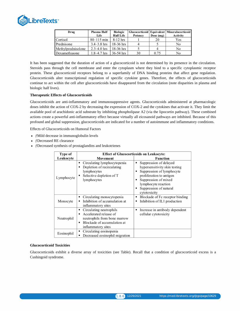
1.8.6 12/29/2021 https://med.libretexts.org/@go/page/10629
It has been suggested that the duration of action of a glucocorticoid is not determined by its presence in the circulation.Steroids pass through the cell membrane and enter the cytoplasm where they bind to a specific cytoplasmic receptorprotein. These glucocorticoid receptors belong to a superfamily of DNA binding proteins that affect gene regulation.Glucocorticoids alter transcriptional regulation of specific cytokine genes. Therefore, the effects of glucocorticoidscontinue to act within the cell after glucocorticoids have disappeared from the circulation (note disparities in plasma andbiologic half lives).
Therapeutic Effects of Glucocorticoids
Glucocorticoids are anti-inflammatory and immunosuppressive agents. Glucocorticoids administered at pharmacologicdoses inhibit the action of COX-2 by decreasing the expression of COX-2 and the cytokines that activate it. They limit theavailable pool of arachidonic acid substrate by inhibiting phospholipase A2 (via the lipocortin pathway). These combinedactions create a powerful anti-inflammatory effect because virtually all eicosanoid pathways are inhibited. Because of thisprofound and global suppression, glucocorticoids are indicated for a number of autoimmune and inflammatory conditions.
Effects of Glucocorticoids on Humoral Factors
ƒMild decrease in immunoglobulin levelsƒDecreased RE clearanceƒDecreased synthesis of prostaglandins and leukotrienes
Glucocorticoid Toxicities
Glucocorticoids exhibit a diverse array of toxicities (see Table). Recall that a condition of glucocorticoid excess is aCushingoid syndrome.

1.8.7 12/29/2021 https://med.libretexts.org/@go/page/10629
Steroid Withdrawal and Glucocorticoid Replacement
Because of suppression of the pituitary-adrenal axis by chronic glucocorticoid therapy, patients that undergo surgicalprocedures or acute medical illness should receive stress dose steroids generally equivalent to 300 mg of hydrocortisoneadministered as a split dose over a 24 hour period. Patients may require very slow and low reductions of steroids back tobaseline to minimize the symptoms of steroid withdrawal, which include joint and muscle pain, nausea, lethargy, weightloss, and fever.
Anti-Cytokine Agents
Rheumatoid arthritis (RA) is a chronic, systemic autoimmune and inflammatory disease that primarily attacks the joints.Autoimmune targeting of normal joint proteins results in inflammation with local release of cytokines, especially TNFα,growth factors, and interleukins, all of which induce COX-2 expression. Levels of TNFα, COX-2, and PGE2are markedlyelevated in the synovial fluid of affected joints. PGE2 binds to synovial cell receptors on and stimulates release of matrixmetalloproteinases (MMPs) which directly damage joint tissue. TNFα also stimulates production of IL-1 and IL-6; thesepro- inflammatory mediators along with COX-2 derived prostaglandins activate the surrounding endothelial tissue torecruit inflammatory cells.
The newest therapeutic agents inhibit the pro-inflammatory effects of the cytokines TNFα and IL-1. By limiting TNFactivity, the generation of pro-inflammatory cytokines IL-1 and IL-6 is diminished. Three strategies have emerged: 1)creating monoclonal antibodies to the TNFα and IL-1 proteins, 2) solubilizing forms of the endogenous receptors of TNFα,and 3) making recombinant versions of endogenous receptor antagonist.
Comparison of Infliximab, Etanercept, and Adalimumab in rheumatoid arthritis:
ƒInfliximab administered IV (by doctor); etanercept and adalimumab given subcutaneously (by patient)ƒInfliximab 3 mg/kg loading dose with similar doses at 2 weeks, 6 weeks, and then every 8 weeks thereafter.Etanercept 25 mg twice weekly or 50 mg weekly. Adalimumab 40 mg every other week.ƒInfliximab must be administered in combination with methotrexate. Etanercept and adalimumab may be monotherapyor may be combined with methotrexate.ƒAll reduce signs, symptoms, and structural damage (joint erosion on radiograph).
Anti-Cytokine Agent Toxicities:
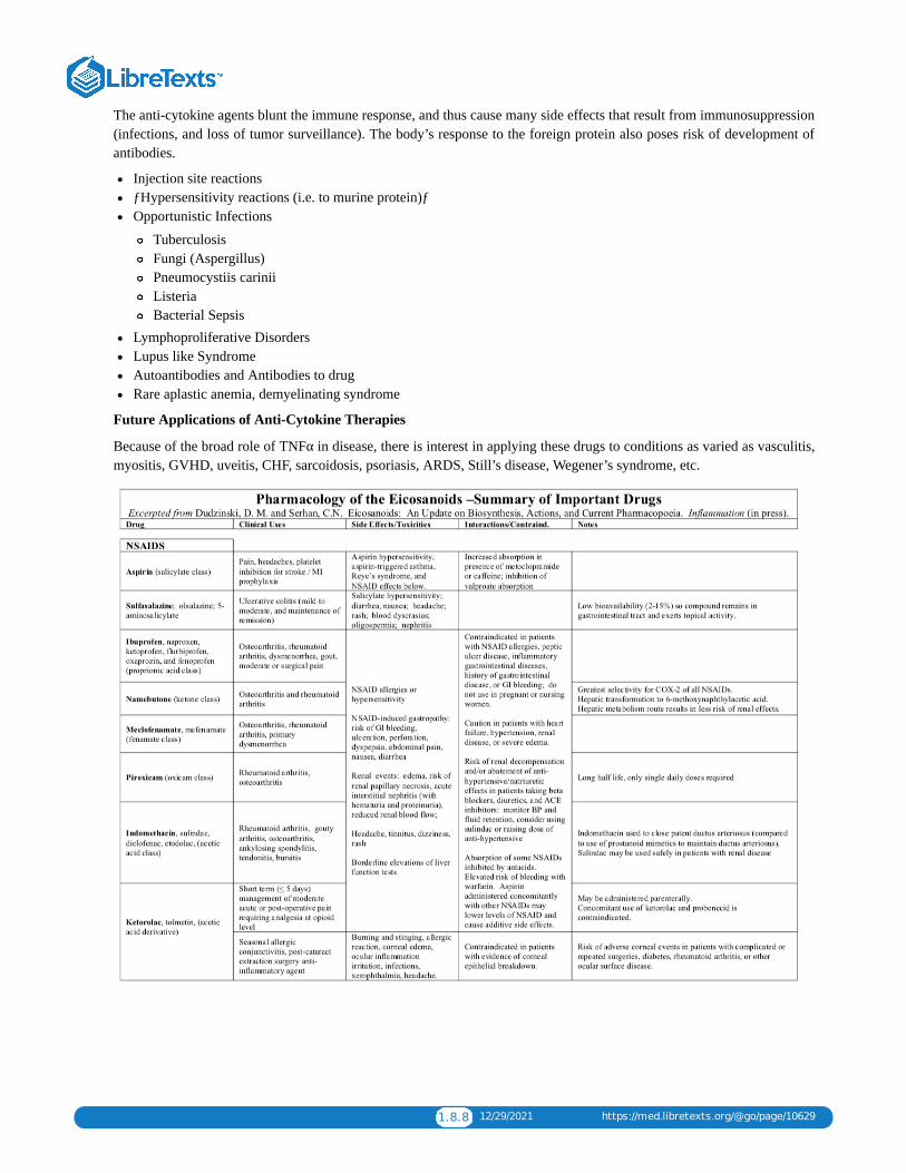
1.8.8 12/29/2021 https://med.libretexts.org/@go/page/10629
The anti-cytokine agents blunt the immune response, and thus cause many side effects that result from immunosuppression(infections, and loss of tumor surveillance). The body’s response to the foreign protein also poses risk of development ofantibodies.
Injection site reactionsƒHypersensitivity reactions (i.e. to murine protein)ƒOpportunistic Infections
TuberculosisFungi (Aspergillus)Pneumocystiis cariniiListeriaBacterial Sepsis
Lymphoproliferative DisordersLupus like SyndromeAutoantibodies and Antibodies to drugRare aplastic anemia, demyelinating syndrome
Future Applications of Anti-Cytokine Therapies
Because of the broad role of TNFα in disease, there is interest in applying these drugs to conditions as varied as vasculitis,myositis, GVHD, uveitis, CHF, sarcoidosis, psoriasis, ARDS, Still’s disease, Wegener’s syndrome, etc.

1.9.1 1/5/2022 https://med.libretexts.org/@go/page/10630
1.9: Vasoactive Drugs I
Hypertension & its Pharmacological Management
What constitutes “hypertension?”
Hypertensive emergencies (malignant hypertension) are defined as severe hypertension coupled with acute end-stageorgan damage.
How common is hypertension?
Hypertension effects approximately 25% of the adult American population.
What are signs or symptoms of hypertension? There are usually no symptoms or signs of hypertension, and thus it is called the “silent killer”. Since humans arecompletely unaware of excessive blood pressure, it is only through measurements that it becomes detected. The exceptionis malignant hypertension, which can cause headache, congestive heart failure, stroke, seizure, papilledema, renal failureand anuria.
What are consequences of long-standing hypertension?
Long-standing hypertension causes accelerated atherosclerosis, which in turns leads to all of the biological fallout of thisdisease. Some consequences include: stroke, coronary artery disease, myocardial infarction, aneurysmal and occlusiveaortic disease. Long-standing hypertension also causes the heart to remodel and undergo a process of hypertrophy (leftventricular hypertrophy or LVH). Hypertrophy can lead to diastolic dysfunction, which can lead to congestive heart failure(CHF) since the heart is too stiff to relax properly. (This will be covered in more detail in the next lecture.) The stiffenedheart requires elevated filling pressures, and this can worsen the dysfunction. Long-standing hypertension can also causethe heart to dilate and lose its ability to pump during systole (systolic congestive heart failure). Lastly, the kidneys areinjured by long-standing hypertension and this is a significant cause of renal failure in the U.S.
What causes hypertension? Over 90% of hypertension in the U.S. is “essential” or idiopathic hypertension, i.e., without an identifiable cause. About10% of hypertension is secondary to some identifiable cause such as steroids, renal vascular disease, renal parenchymaldisease, pregnancy related, pheochromocytoma, Cushing’s syndrome, coarctation of the aorta or primaryhyperaldosteronism to name a few.
The physiological framework for understanding hypertension: Ventriculo-arterial coupling

1.9.2 1/5/2022 https://med.libretexts.org/@go/page/10630
Key: 1 = End diastole, just prior to LV contraction. The pressure at 1 is the left ventricular end diastolic pressure (LVEDP) andthe volume is the left ventricular end diastolic volume (LVEDV) (1 to 2 = isovolemic contraction)
2 = Opening of the aortic valve and beginning of ejection into the aorta (2 to 3 is the volume ejected from the LV into the aorta which is the stroke volume (SV))
3 = End systole. The pressure at 3 is known as the end-systolic pressure (ESP). The aortic valve shuts just after 3. (3 to 4 is isovolumic relaxation)
4 = Beginning of passive diastolic filling. 4 to 1 is diastolic filling along the dotted curve. This dotted curve is the end-diastolic pressure volume relation (EDPVR).
ESPVR = End-systolic pressure volume relation. This also called Emax or Es which stand for maximal elastance orelastance at end-systole, respectively. This characterizes the strength of the LV irrespective of the systolic load it faces.
Ea = Effective arterial elastance. This is characterizes the arterial tree and the load it presents to the LV during systole. Eais primarily determined by arterial resistance but arterial compliance effects it too.
Ea and ESPVR “Couple” to exactly determine the stroke volume. In essence, the volume lost by one chamber is exactlyequal to the volume gained by the other. The elastance of each chamber (heart and vascular tree) determines the pressure.The exact systolic and diastolic pressures that obtain are dependent on arterial properties (Ea), ventricular properties(ESPVR) and the filling state (LVEDV).
There are FOUR possible mechanisms for hypertension
1. The volume ejected from the LV can be too high. This could result from an excessive contraction during systole (a veryhigh ESPVR). This mechanism is described in the medical literature but is not typical. A hyperdynamic circulation isthought to play a role in the hypertension seen in some young, otherwise fit African-American males.
2. The intravascular volume may be too high causing an excess of venous return, leading to an elevated LVEDV. The veryfull heart would then eject a large volume into the arterial tree thus leading to hypertension. The high intravascularvolume could be caused by renal dysfunction with subsequent fluid retention, or it could be due to exogenousadministration. There does seem to be a subset of patients that has an elevated intravascular volume Nevertheless, theexcessive intravascular volume mechanism appears to occur infrequently since many newly diagnosed hypertensivepatients actually have a contracted intravascular volume. The excessive intravascular volume mechanism also impliesthat the cardiac output would be elevated, but it is usually normal.
3. Excess venous return could also occur even with a reduced intravascular volume if thevenous tone were significantlyelevated. This would cause a rise in the LVEDV even with a normal or low actual blood volume. Whether this occursas a regular feature of hypertension is not known.

1.9.3 1/5/2022 https://med.libretexts.org/@go/page/10630
4. The effective arterial elastance (Ea) can be too high. This can occur either because the resistance is too high or becausethe compliance is too low. Many forms of hypertension are associated with an elevated arterial resistance. Furthermore,in older humans, the arterial tree becomes stiffer and less compliant. Thus, for a given stroke volume delivered into thearterial tree, the pressure goes up, especially the systolic pressure.
Rational pharmacotherapy of hypertension is based on the four mechanisms outlined above.
1. Reduce LV systolic performance (reduce the ESPVR): negative inotropes (beta-blockers (metoprolol, atenolol,propranolol) and calcium channel blockers (verapamil,diltiazem)).
2. Reduce blood volume and thus drop LVEDV: diuretics (thiazide – hydrochlorothiazide, loop diuretics – furosemide,bumetanide and potassium sparing diuretics – spironolactone, amiloride, triamterene).
3. Reduce venous tone and thus venous return: Central sympatholytics such as clonidine act to reduce overall sympathetictone.
4. Reduce arterial tone (i.e. resistance) and thus reduce Ea: Effective arterial dilators include angiotensin convertingenzyme inhibitors (ACE inhibitors – lisinopril, captopril), angiotensin receptor blockers (ARB’s – valsartan, losartan),calcium channel blockers (nifedipine, amlodipine), potassium channel openers (minoxidil), nitric oxide donors(nitroprusside), alpha1 blockers (prazosin, terazosin, doxazosin), and mixed alpha and beta-blockers (labetalol)
Major Antihypertensive Drug Classes(These descriptions are intended as a supplement to the more complete discussions in the text.)
Diuretics (thiazide, loop, and potassium-sparing diuretics).
Thiazide diuretics such as hydrochlorothiazide and chlorthalidone are among the most commonly used drugs fortreating hypertension. They inhibit reabsorption of Na and Cl in the distal tubule and lose effectiveness when GFR islow. Their initial effects are said to be mediated by decreasing intravascular volume, however (as mentioned above)most untreated hypertensives have contracted intravascular volume. Diuretics cause peripheral vascular resistance tofall through an unknown mechanism. Unfortunately thiazide diuretics have a number of undesirable metabolic effectssuch as hypercalcemia, hypokalemia, hyponatremia, hyperglycemia, hyperlipidemia, and hyperuricemia. These sideeffects often dictate which drugs to use. When thiazide diuretics are used in low doses, their side effects seem to beminimized.Loop diuretics such as furosemide inhibit the Na/K/Cl cotransporter in the ascending limb of the loop of Henle. Theycause a very brisk diuresis, but their anti-hypertensive effects are actually not that strong. Acute intravenousadministration of furosemide can cause venodilation by an unknown mechanism. Loop diuretics are often part oftreatment for malignant hypertension and hypertension with hypervolemia (e.g., renal insufficiency). The metabolicderangements produced by these drugs (particularly hypokalemia, and hypocalcemia) can be profound. This class is notrecommended as initial monotherapy for hypertension.Potassium-sparing diuretics such as spironolactone, amiloride, and triamterene are not as efficacious as thiazides orloop diuretics in reducing blood pressure, however, they do correct the potassium loss associated with thiazide and loopdiuretics. Amiloride and triamterene inhibit the Na/proton exchanger in the distal and collecting tubules.Spironolactone inhibits the Na/K exchanger affected by aldosterone, and it is particularly effective in the face ofhyperaldosteronism. If potassium-sparing diuretics are given to patients on ACE inhibitors, particular care must betaken since both classes cause elevations in serum potassium.
Sympatholytics (beta-blockers, mixed alpha and beta-blockers, alpha-blockers and central sympatholytics).
Beta adrenergic blockers such as propranolol, metoprolol or atenolol are typical first-line agents for treatinghypertension. They have negative chronotropic and negative inotropic effects. The acute effect of blocking beta-2receptors is an increase in SVR, however chronic administration can decrease peripheral resistance, probably bydecreasing plasma renin and angiotensin II. Unfortunately beta-blockers can elevate triglycerides and reduce HDL. Inaddition, they can produce glucose intolerance, impotence, and depression. In patients prone to bronchospasm (i,e.,asthmatics), non-selective beta-blockers can theoretically worsen the problem, although the risks are somewhatoverplayed. These side effects often dictate drug choices for the hypertensive patient.Alpha-1 adrenergic blockers such as prazosin, terazosin and doxazosin are effective at reducing sympatheticvasoconstriction and thereby reducing vascular resistance. These drugs are also useful for men who have benign

1.9.4 1/5/2022 https://med.libretexts.org/@go/page/10630
prostatic hypertrophy because they can reduce bladder outlet obstruction. Unlike the beta blockers and thiazidediuretics, the alpha blockers have not been shown to decrease mortality. In fact, doxazosin caused an increase incongestive failure in the ALLHAT trial. Thus, the indications for these drugs in hypertension are currently unclear, andthey are not considered first line treatments. Non-selective alpha blockers such as phenoxybenzamine andphentolamine are not used for hypertension because they produce an excessive amount of reflex tachycardia. However,the profound alpha blockade possible with the non-competitive antagonist, phenoxybenzamine, has proven very usefulin the treatment of pheochromocytoma. These patients are usually given alpha- blockade first and then beta-blockade tocontrol the reflex tachycardia.Central sympatholytics such as clonidine stimulate central alpha-2 receptors and thereby reduce sympathetic outflow.These drugs are effective in decreasing heart rate, contractility and vasomotor tone, however, they cause sedation andare usually not first line therapies.Mixed alpha and beta antagonists such as labetalol.and carvedilol block both alpha receptors and beta receptors, sothe reduction in blood pressure is usually not associated with reflex tachycardia. Labetalol is a very effectiveintravenous antihypertensive, but it is less frequently used chronically in its oral form. Carvedilol has had its primaryuse in the treatment of chronic congestive heart failure.
Vasodilators (calcium-channel blockers, direct arterial vasodilators, and sodium nitroprusside).
Calcium channel blockers such as verapamil, diltiazem, nifedipine and amlodipine block L- type calcium channelsand are effective arterial vasodilators. The dihydropyridine agents nifedipine and amlodipine act primarily asvasodilators and have minimal direct effects on the heart. In contrast, verapamil and diltiazem act principally asnegative inotropes and negative chronotropes, and thus decrease heart rate, contractility and cardiac conduction speed.In addition, they reduce vascular resistance. There is controversy over the use of short-acting dihydropyridines inpatients with angina because they can cause reflex sympathetic activation and worsen ischemia. When using verapamilor diltiazem one has to expect a reduction in LV systolic function as well as a reduction in cardiac conduction. Thus, inpatients with congestive heart failure of the systolic type or in those with a significant conduction defect, these drugsshould be avoided. Verapamil and diltiazem are synergistic with beta-blockers and the combination can cause severebradycardia, heart block or pump dysfunction.Direct arterial vasodilators such as minoxidil and hydralazine have relatively limited use. Neither has much effect onvenous tone. The mechanism of action of hydralazine is not known. Minoxidil appears to increase potassiumconductance in vascular smooth muscle, and the resultant hyperpolarization reduces calcium entry. Both drugs cancause reflex tachycardia (particularly minoxidil) and fluid retention. These side effects can be managed with theaddition of a beta-blocker and/or a diuretic. Neither drug is effective for sustained periods. They are usually reservedfor the short-term treatment of refractory hypertension, especially in patients with renal failure. Each of these drugs hasa unique side effect: hydralazine can cause a lupus-like syndrome (cf. Drug Allergy case), and minoxidil can producehair growth (and is sold for the purpose!).Sodium nitroprusside breaks down non-enzymatically to form nitric oxide. It is an extremely potent arteriolar andvenous dilator that is used intravenously for rapid control of hypertensive crises and for blood pressure control duringoperations. Reflex increases in heart rate and contractility usually require treatment with beta blockers.
Renin-angiotensin system (RAS) blockers comprise two broad categories: angiotensin converting enzyme inhibitors(ACE inhibitors) and angiotensin type 1 receptor blockers (ARB’s).
ACE inhibitors like captopril, enalapril, and lisinopril decrease the conversion of angiotension I to angiotensin II(ATII). This reduces peripheral vascular resistance and promotes both natriuresis and hyperkalemia, since a reductionin ATII leads to a reduction in aldosterone. ACE also breaks down bradykinin, so inhibiting this enzyme can increasebradykinin levels and cause more vasodilation. ACE inhibitors have been shown to reduce morbidity (and possiblymortality – see below), and their relatively benign side-effect profile makes them frequent choices for first-line ormonotherapy. Of note, ACE inhibitors are associated with a definite improvement in renal function in patients withdiabetes and it has been shown that renal injury due to long-standing diabetes is reduced. Diabetics who do not have acontraindication for this class of drugs should be taking them for renal protective purposes. ACE inhibitors areassociated with a 5-10% incidence of dry cough, probably caused by the elevated bradykinin levels. For patients whohave reduced renal perfusion pressure (e.g., renal artery stenosis), ACE inhibitors can cause renal dysfunction or renalfailure. (Patients with bilateral renal artery stenosis have high levels of endogeneous angiotensin II which is used to
1.9.5 1/5/2022 https://med.libretexts.org/@go/page/10630
maintain glomerular filtration and ACE inhibitors disrupt that compensatory process.) Finally, ACE inhibitors areassociated with a rare, but potentially fatal, angioedema of the airway. .Angiotensin receptor blockers (ARB’s) like losartan and valsartan cause arteriolar vasodilation by blocking theeffects of angiotensin II at the angiotensin Type I receptor. Since the mechanism is essentially the same as for the ACEinhibitors, the indications and contraindications are the same. The blockade is downstream, so bradykinin is notelevated, and this class of drugs is not associated with a cough.
Treating Hypertension
As a first principle, one should always couple any chemical therapy with lifestyle modifications (maintaining ideal bodyweight, engaging in aerobic physical exercise, eating a healthy diet low in saturated and total fats, limiting sodium intakeand reducing alcohol intake). Each of these lifestyle modifications has been shown to reduce blood pressure modestly.These modifications are inexpensive and pose very little risk. Compliance remains the primary trouble with thesemethodologies. As a second principle, additional risk factors for coronary artery disease and stroke should be aggressivelymanaged in all patients with hypertension. In particular, patients should be counseled on smoking cessation, lipid reductionand diabetic management. When these diseases occur in combination, the probability of end-organ damage goes upsignificantly and careful management of each of the co-morbidities is all the more important.
Does treating hypertension ameliorate the long-standing negative consequences of having hypertension?
There is overwhelming evidence that normalization of the blood pressure (using a variety of therapies) is very effective inreducing end-organ damage such as left ventricular hypertrophy (LVH), myocardial infarction, stroke and renal failure.There are studies addressing each particular end-organ and its responsiveness to reductions in blood pressure.
Have certain drugs been shown to reduce the morbidity and mortality due to hypertension?
Thiazide diuretics and beta-blockers have been shown to reduce the risk of stroke, coronary disease and overall mortalityfrom cardiovascular disease in patients with hypertension. Other drugs used to treat hypertension are being studied at thepresent time and the mortality benefit they offer is being clarified. For example, ACE inhibitors likely reduce the risk ofstroke, coronary disease and major cardiac events and death from cardiovascular causes.
Do physicians do a good job treating hypertension in the United States?
Not usually. On the whole, physicians are adequately treating less than 50% of patients with hypertension in the UnitedStates today. Despite being “easy to treat”, significant numbers of patients do not have their hypertension under adequatecontrol.
What is the best initial therapy for the newly diagnosed hypertensive patient?
Therapeutic interventions usually begin with lifestyle modifications for the first six months to one year. If this does notrectify the situation, then one moves to diuretics – particularly thiazide diuretics. If the diuretic is not fully successful, thenone can add a sympatholytic such as a beta-blocker. Thereafter, vasodilators such as calcium-channel blockers, ACEinhibitors, or ARB’s are instituted. Thiazide diuretics or beta-blockers are considered by many to be first line agents in thetreatment of hypertension because they are inexpensive and have proven efficacy in reducing overall mortality.Unfortunately, the side effects of these drugs are troubling to some patients, and this may decrease compliance. For thisreason, many patients are started early on more expensive drugs like ACE inhibitors. The long-term effects on morbidityand mortality are still being determined, and what is considered “first- line therapy” is likely to be a moving target in thecoming years. The concept of stepped care is very important in the treatment of hypertension. If one therapy fails toachieve the targeted blood pressure, one adds an additional therapy. In general, giving small doses of two or moreantihypertensives from different classes can cause additive or synergistic effects on blood pressure while minimizing sideeffects. In most cases this is preferable to giving a larger dose of a single drug.
Demographic factors
Patients of African descent are more responsive to diuretics and calcium-channel blockers than to beta-blockers or ACEinhibitors. A notable exception is the previously mentioned young African-American who may do well on beta-blockertherapy due to a “hyperdynamic circulation”.
Elderly Patients are said to respond quite favorably to diuretics and calcium-channel blockers. However, due to theirfrequent conduction system disease, many of these patients need to be watched carefully when they are introduced to beta-

1.9.6 1/5/2022 https://med.libretexts.org/@go/page/10630
blockers. Beta-blockers and diuretics reduce mortality in patients with isolated systolic hypertension (very common in theelderly).
Hypertensive crisis (malignant hypertension).
This is an uncommon form of acute severe hypertension that can rapidly progress to stroke, MI, renal failure, orencephalopathy. These patients are admitted to the Intensive Care Unit for invasive hemodynamic monitoring and carefulreduction of their blood pressure with fast- acting potent vasodilators such as sodium nitroprusside.
Disease processes which are affected by anti-hypertensive drugs:
Diabetes – Beta-blockers and thiazide diuretics may make glycemic control difficult. ACE inhibitors can protectthe kidney.Coronary Artery Disease – Beta-blockers offer a mortality benefit (in general). Short-acting calcium channelblockers can worsen ischemia.Congestive Heart Failure (compensated vs. un-compensated) – Beta-blockers offer a mortality benefit as do ACEinhibitors. Beta-blockers should not be used in uncompensated CHF.Hyperlipidemia – Beta-blockers and thiazide diuretics may affect lipid profile unfavorably.COPD/Asthma – Beta-blockers need to be used with caution.Peripheral Vascular Disease (with Symptoms) – Beta-blockers need to be used with discretion.Renal Artery Stenosis (bilateral vs. unilateral) – ACE inhibitor or ARB’s are relatively contraindicated.Cardiac Conduction Defects – Beta-blockers, diltiazem and verapamil can exacerbate conduction defects.Benign Prostatic Hypertrophy – Alpha-1 blockers can provide symptomatic improvement.Depression – Beta-blockers may exacerbate.Raynaud’s Syndrome – Beta-blockers may exacerbate.Renal Failure – ACE inhibitors may cause a reduction in renal performancePregnancy – ACE inhibitors and ARB’s are contraindicated.Aortic Stenosis – Vasodilators need to be introduced with caution.Hyperuricemia (Gout) – Thiazide diuretics may increase uric acid levels.

1.10.1 12/22/2021 https://med.libretexts.org/@go/page/10631
1.10: Vasoactive Drugs II - Heart Failure
Congestive Heart Failure& its Pharmacological Management
The physiological framework for understanding CHF: The Pressure-Volume Loop of the LV
Key:
1 = End diastole, just prior to LV contraction. The pressure at 1 is known as the left ventricular end diastolic pressure(LVEDP) (1 to 2 = isovolemic contraction)
2 = Opening of the aortic valve and beginning of ejection into the aorta (2 to 3 is the volume ejected from the LV into the aorta which is the stroke volume (SV))
3 = End systole. The pressure at 3 is known as the end-systolic pressure (ESP). The aortic valve shuts just after 3. (3 to 4 is isovolumic relaxation)
4 = Beginning of passive diastolic filling. 4 to 1 is diastolic filling along the dotted curve. This dotted curve is the end-diastolic pressure volume relation(EDPVR).
ESPVR = End-systolic pressure volume relation. This also called Emax or Es which stand for maximal elastance orelastance at end-systole, respectively. This characterizes the strength of the LV irrespective of the systolic load itfaces.
Ea = Effective arterial elastance. This is characterizes the arterial tree and the load it presents to the LV duringsystole. Ea is primarily determined by arterial resistance but arterial compliance effects it too.
Ea and ESPVR “Couple” to exactly determine the stroke volume.
What is meant by “congestive heart failure” (CHF)?
Congestive heart failure simply means that the pulmonary blood volume is expanded and, therefore, the pulmonarycirculation is congested with blood. The congestion arises because of elevated left ventricular end-diastolic pressure(LVEDP). An elevated LVEDP is a hallmark of uncompensated congestive heart failure. Common symptoms includeshortness of breath, fatigue, orthopnea and paroxysmal nocturnal dyspnea (PND). Once a patient is treated for CHF theymay become asymptomatic. This is termed compensated CHF and does NOT imply that the underlying disease process hasgone away! It is entirely reasonable and even common for patients who are well medically managed to have a markedlyreduced ejection fraction (EF) yet be capable of most normal activities. This handout pertains primarily to LEFTventricular failure. Although RIGHT ventricular failure occurs, it is less common and not the focus of this handout.

1.10.2 12/22/2021 https://med.libretexts.org/@go/page/10631
How long do people live if they have congestive heart failure?
For all comers, the mortality is 50% in five years. However, if a patient is symptomatic, despite treatment, one-yearmortality can approach 50%. Approximately five million people have congestive heart failure (CHF) in the United Statesat any given time. The great majority of these patients have primarily left ventricular failure.
There are FIVE main physiologic ways to get CHF
1. Decrease the strength of the LV and thereby decrease its ejection – this causes the LVEDP to rise (CHF). This ischaracterized by a reduction in the slope of the ESPVR line.Acute causes include: myocardial ischemia, myocardial infarction, sepsis syndrome, myocardial contusions, excessbeta blockers or excess calcium channel blockers. Chronic causes include: dilated cardiomyopathy due to certain viral illnesses, multiple myocardial infarctions or largeterritory myocardial infarctions, excessive alcohol consumption and certain chemotherapeutic agents such asadriamycin. Prolonged pressure overload (hypertension and aortic stenosis) also causes a weak heart after initiallycausing hypertrophy. With continued overload, the compensation of hypertrophy progresses to a failing myocardium (alow ESPVR)
2. Provide much too much venous return (and hence filling) to the LV– this causes the LVEDP to rise (CHF). Acute causes include: excessive volume administration or large increased in venous tone as with somevasoconstrictors. Exercise causes a large increase in venous return and if the LV cannot eject all that it receives, there isan elevation in the LVEDP (CHF). Chronic causes include profound chronic anemia, longstanding mitral regurgitation or aortic regurgitation. Poorcompliance with diuretic therapy or with high salt intake can cause a rise in the plasma volume which in turn elevatesthe venous return.
3. Alter the passive filling characteristics of the LV such that a normal filling volume is associated with a high pressure –this causes the LVEDP to rise (CHF). This is calleddiastolic CHF. Acute causes include: myocardial ischemia Chronic causes include: prolonged pressure overload (hypertension and aortic stenosis) which cause this problem byinitially causing hypertrophy of the cardiac muscle. The thickened myocardium does not relax well. Certain infiltrativediseases like amyloidosis cause diastolic CHF.
4. Present an enormous load to the LV such that it cannot eject well – this causes the LVEDP to rise (CHF). This ischaracterized by having a severe increase in the arterial load and is shown by a steep increase in the slope of Ea, theeffective arterial elastance. Acute causes include: pheochromocytoma and catechol release, very poorly controlled hypertension. Chronic causes include: pressure overload with aortic stenosis or long term severe hypertension
5. Reduce the function of the heart with other problems: Acute causes include: severe tachycardia or bradycardia, pericardial tamponade, acute ventricular septal defect, acutemitral regurgitation.
Regardless of the cause of CHF, certain management strategies pertain to all patients.
Salt restriction and fluid restriction are regular features of any congestive heart failure management program. Secondly,any underlying cause should be treated aggressively. For example, hypertension is a very frequent precipitator of congestive heart failure and should be managed aggressively.Similarly, coronary artery disease should be managed with the full armamentarium available for that disease. Patientshaving profound anemia and who suffer from high output congestive heart failure should have their anemia and underlyingpathology managed. For patients who are drinking excessive alcohol and have alcohol- induced cardiomyopathy, treatingthe underlying alcoholic behaviors are essential. Additional risk factor management clearly includes smoking cessation,lipid optimization, optimizing diabetes management and so on.
We will now consider how to treat CHF for the first four mechanisms outlined above.
In almost all cases of symptomatic CHF, the LVEDP is too high. This is responsible for the symptom of shortness ofbreath. When the pulmonary blood volume is congested with blood, the lungs become stiff and the work of breathing goesway up. There are multiple acute ways to reduce the LVEDP:

1.10.3 12/22/2021 https://med.libretexts.org/@go/page/10631
Improve LV systolic performance (improve the ESPVR): inotropes (digoxin, milrinone, dobutamine).Digoxin is the hallmark drug for augmenting systolic performance. Although digoxin certainly helps in thesymptomatic management of systolic heart failure, it does not confirm a mortality benefit.Improve arterial loading conditions (reduce Ea): arterial dilators such as angiotensin converting enzymeinhibitors (ACE inhibitors – lisinopril, captopril), and angiotensin receptor blockers (ARB’s – valsartan,losartan). An important feature of ACE inhibitors is their mortality benefit. All patients with CHF who do nothave contraindications should be taking ACE inhibitors. For patients who cannot tolerate ACE inhibitors,angiotensin receptor blockers are very likely to be an adequate substitute and their mortality benefit is beingstudied presently. Other drugs that reduce Ea include hydralazine, minoxidil and nitroprusside. These drugs donot confer a mortality benefit.Improve the diastolic filling characteristics of the LV: All underlying causes such as myocardial ischemiashould be treated.
Reduce the blood volume and thus drop the LVEDP: diuretics (loop diuretics – furosemide, bumetanide andpotassium sparing diuretics – spironolactone). Of the diuretic therapies, only spironolactone has been shown to have amortality benefit in treating patients with CHF.
The Secondary Physiologic Response to LV Failure.
The failing LV precipitates a host of secondary neuroendocrine events which are well described in figure 23-10 fromGolan.
Image removed for copyright reasons. See Fig. 23-10 in Golan.
Management of secondary neuroendocine effects.
It is the correction of neuroendocrine abnormalities (elevated renin, angiotensin II, and aldosterone) that is most likelyresponsible for prolonging the lives of patients with CHF. Remodeling is the process that the left ventricle undergoes whenit is insulted either by chronic unfavorable loading conditions or chronic neuroendocrine “siege.” It involves a change inventricular mass or dimension without a corresponding change in the number of ventricular myocytes. The functional leftventricle first begins to hypertrophy, then begins to dilate, and finally becomes fibrotic. The exact mechanisms that causeremodeling are unclear but the neuroendocrine effects seem to be responsible. It is currently thought that reducing theneuroendocrine effects of CHF is what leads to longer lives for those patients having CHF. This may explain why ACEinhibitors, ARB’s, spironolactone (blocks aldosterone) and beta-blockers (block renin production) all confer a mortalitybenefit.
The use of beta-blockers is, paradoxically, one of the fundamental treatments for systolic congestive heart failure.These drugs offer a definite mortality benefit.
Beta-blockers are well known to depress LV systolic function (i.e. reduce ESPVR) and thus appear counterintuitive inmanaging congestive heart failure. However, beta-blockers have been found to offer an important mortality benefit topatients with congestive heart failureincluding those with severely decreased LV function. It is most likely that beta-blockers interfere with the secondary neuroendocrine events (such as an activated sympathetic nervous system andelevated renin) associated with congestive heart failure. The beta-blockade should be started in all patients who do nothave significant contraindications to these drugs. Presently metoprolol and carvedilol are the two drugs with a provenmortality benefit. Patients with symptomatic congestive heart failure (uncompensated CHF) are poor candidates for thesedrugs. However, once their symptoms are resolved (with diuretics and ACE inhibitors along with other managementstrategies), the introduction of beta-blockers is reasonable. Initial doses are usually low and titrated upwards, as tolerated.
Knowing that Beta-Blockers offer a mortality benefit, do physicians use beta-blockers as the data suggest?
No. For reasons that are not entirely clear, physicians often deny patients beta-blockers.
Severe Decompensated CHF.
In the case of severe, Class IV/Stage D CHF (see appendix), patients are often admitted to the hospital for IV therapy usinginotropes such as Digoxin, Dobutamine or inodilators such asMilrinone or Amrinone These drugs treat symptoms of CHF,but do not offer a mortality benefit. In fact, chronic therapy with some of these drugs can actually increase mortality. Theyare only used as a “bridge” to more definitive therapy or until resolution of the precipitating event can be accomplished. Inpatients with acute severe decompensated CHF, a number of highly invasive therapies exist. Intra-aortic balloon pumping
1.10.4 12/22/2021 https://med.libretexts.org/@go/page/10631
(IABP) offers a bridge to cardiac transplantation. Many patients with dilated cardiomyopathies die due to malignantdysrhythmias. Patients with dilated hearts and severely reduced ejection fraction have a survival benefit with automaticimplantable cardiac defibrillators (AICD’s). Lastly, with severely reduced ejection fractions and cardiac conduction defects, cardiac resynchronizing pacemakers offersymptomatic relief of CHF.
AppendixThe Clinical Characterization of CHF follows two schemes. One is older and based on symptoms (NYHC), the other isnewer and based on disease progression (Stages A, B, C, D).
New York Heart Classifications of symptoms (NYHC I, II, III, IV)
Class I: Patients with no limitation of activities and no symptoms from ordinary activities.Class II: Patients with slight, mild limitation of activity; they are comfortable with rest or with mild exertion.Class III: Patients with marked limitation of activity; they are comfortable only at rest.Class IV: Patients who should be at complete rest; confined to bed or chair; any physical activity brings ondiscomfort and symptoms occur at rest.
The current Staging system deals with progression of the underlying disease.
Stage A: Patients at high risk for developing heart failure are those with:
● Hypertension ● Diabetes mellitus ● Coronary artery disease (including being S/P myocardial infarction) ● History of cardiotoxic drug therapy ● History of alcohol abuse ● History of rheumatic fever ● Family history of cardiomyopathy
Stage B: Pateints with known heart disease but who have never had symptoms of heart failure.Stage C: Patients with known heart disease with current or prior symptoms. Symptoms include:
● Shortness of breath ● Fatigue ● Reduced exercise tolerance
Stage D: Presence of advanced symptoms even with optimized medical care.
AppendixAll types of CHF start with an elevated LVEDP. There are multiple ways to get an elevated LVEDP. In each case below,the dotted line shows the circumstance prior to developing symptoms of CHF. Examples are from Suga and Sagawa.
Type I is from too much venous return from any cause Type II is from too much ‘afterload’ (Ea is too high) Type III is from diastolic dysfunction from any cause (unfavorable EDPVR)Type IV is from a weak LV (ESPVR is toolow) from any cause
1.11.1 1/12/2022 https://med.libretexts.org/@go/page/10632
1.11: Lipid Lowering Drugs - Hyperlipidemia and AtherosclerosisAtherosclerosis: A chronic inflammatory disease characterized by enzymatic destruction of the normal arterial skeleton(largely elastin, collagen and smooth muscle), and replacement by disorganized collagen and elastin, cholesterol, and foamcells.
1. Afflicts all long-lived mammals.2. Major risk factors:
LongevityHypertensionDiabetes – glycosylation of plasma proteins and arterial wall proteinsDyslipoproteinemiaCigarette Smoking
3. Homocysteinemia
Plasma homocysteine levels controlled by 3 genes related to methionine metabolismHigh homocysteine is toxic to the endothelium and eventually atherogenic
4. Lp(a) lipoproteinLevels variable and genetically determinedInhibits tissue plasminogen activator and allows thrombus formation, which may be atherogenicIncreases likelihood of thrombosis and clinical catastrophe when atherosclerosis is present
5. Chronic bacterial infection6. Transmembrane Receptors on Mammalian Cells – Three Broad Classes
1. Receptors mediating transmembrane signaling (e.g. β receptor)Serve to amplify the effect of a tiny concentration of ligand
2. Receptors regulating intracellular substrate concentration (e.g., LDL receptor)Bind tiny fraction of substrateRapid cholesterol turnover involves translocation into the cellReceptors supply cholesterol, when needed, to rapidly growing cellsNormally strongly down-regulated except in liver
3. Scavenger receptors (e.g., asialoglycoprotein receptor)Receptors of normal catabolismRemove certain “worn out” proteins from the plasma or extracellular fluid.Oldest of these, the asialoglycoprotein receptor, was described more than 30 years ago. Removes liver proteinswhich have become desialated over time from the plasma.Recently, more scavenger receptors described which scavenge oxidized albumin, oxidized LDL, and manyothersSR-B1 is the HDL scavenger receptor
7. Apolipoproteins
1. Proteins involved in the solubilization of fat for transport into and out of cells, from one place in the body toanother.
2. Many types, but most important are A1, B, and EAll three involved in cholesterol transportA1 and B in triglyceride as wellApoE has 3 common variants. Plays a critical role in cholesterol absorption, reverse cholesterol transport, and ininhibiting the accumulation in cells of certain hydrophobic proteins.
8. Sterols1. Distinguishing feature between plants and animals is not the presence or absence of chlorophyll, but rather the
sterols they synthesize.Major plant sterol is sitosterol
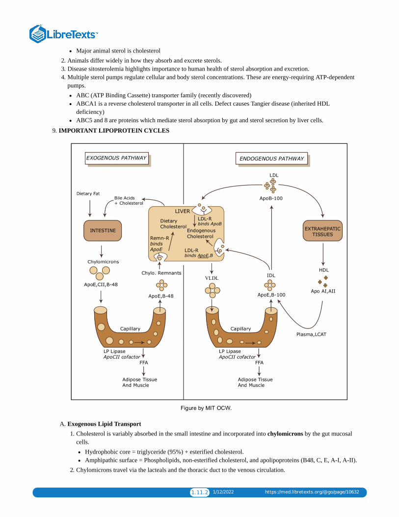
1.11.2 1/12/2022 https://med.libretexts.org/@go/page/10632
Major animal sterol is cholesterol2. Animals differ widely in how they absorb and excrete sterols.3. Disease sitosterolemia highlights importance to human health of sterol absorption and excretion.4. Multiple sterol pumps regulate cellular and body sterol concentrations. These are energy-requiring ATP-dependent
pumps.ABC (ATP Binding Cassette) transporter family (recently discovered)ABCA1 is a reverse cholesterol transporter in all cells. Defect causes Tangier disease (inherited HDLdeficiency)ABC5 and 8 are proteins which mediate sterol absorption by gut and sterol secretion by liver cells.
9. IMPORTANT LIPOPROTEIN CYCLES
A. Exogenous Lipid Transport1. Cholesterol is variably absorbed in the small intestine and incorporated into chylomicrons by the gut mucosal
cells.Hydrophobic core = triglyceride (95%) + esterified cholesterol.Amphipathic surface = Phospholipids, non-esterified cholesterol, and apolipoproteins (B48, C, E, A-I, A-II).
2. Chylomicrons travel via the lacteals and the thoracic duct to the venous circulation.
1.11.3 1/12/2022 https://med.libretexts.org/@go/page/10632
3. In muscle and adipose cells the triglyceride core is progressively hydrolyzed by hormone sensitive lipase(HSL) to form free fatty acids. This leaves a so-called chylomicron remnant. (Apo C obtained from circulatingHDL is required for this step.)
4. Apo A and C are then removed and recycled to HDL, and the chylomicron remnants are taken up byhepatocytes. This process involves LDL receptor-mediated endocytosis and requires Apo E.
5. The liver may now excrete the cholesterol into bile (either unchanged or as bile acids), incorporate it intomembranes, or resecrete it into plasma as lipoprotein cholesterol.
B. Endogenous Lipid Transport: The VLDL-LDL Cycle (Apo B100 System)1. This cycle allows the hepatocyte to export triglycerides and cholesterol to peripheral tissues as VLDL. VLDL
synthesis requires microsomal triglyceride transfer protein (MTP).
Hydrophobic core = triglycerides (55-80%) + esterified cholesterol (5- 15%)Surface = phospholipids (10-20%), Apo B100, C, E
2. The triglycerides in hepatocytes come from two sources:FFA synthesized de novo in liverFFA and glycerol taken up from plasma and re-esterified. These are produced in adipose tissue and muscleby the action of HSL. HSL activity is regulated by adrenergic nerves and circulating catecholamines.
3. The cholesterol in hepatocytes also comes from two sourcesUptake of chylomicron remnantsSynthesized de novo by HMG CoA-reductase
4. Once the VLDL are circulating, the triglycerides may be hydrolyzed by HSL in plasma, and the fatty acids maybe used to provide fuel for muscle cells, or re- esterified and stored in adipocytes. When the VLDL particle hasbeen depleted of triglycerides it becomes a smaller, denser particle called a VLDL remnant orIDL.
5. Like chylomicron remnants, the triglyceride-poor VLDL remnants may reenter the liver. Unlike chylomicrons,the VLDL remnants may be further metabolized to become LDL.
6. The major determinant of LDL concentration in plasma is the number/activity of LDL receptors.Present on nearly all cells and account for 70-80% of LDL catabolism.Most LDL taken up by liver and the rest by peripheral tissues, adrenals and gonads (the latter needcholesterol for steroid synthesis).Apo E is critical ligand for binding of lipid particles to LDL receptor
C. Endogenous Lipid Transport: The HDL Cycle (Apo A-I System)1. This “antiatherogenic” cycle allows cholesterol to be scavenged from chylomicrons, VLDL and peripheral
tissues by HDL particles.Core = triglycerides (5-10%) + esterified cholesterol (15-25%)Surface = phospholipids, + Apo A-I, A-II, C, E
2. Transport of cholesterol from tissue stores to HDL is mediated by ABCA1transporter. It is then esterified bylecithin-cholesterol acyltransferase (LCAT)to make bigger HDL particles. Esterified cholesterol is then disposedof by three primary mechanisms:
Transfer to VLDL, LDL, IDL, and chylomicron remnants by cholesteryl ester transfer protein (CETP) andsubsequent endocytosis by hepatocytes.Direct uptake in liver, adrenals, and gonads by the scavenger HDL receptor called SR-BI.Hydrolysis by hepatic lipase
10. Lipid Lowering Drugs1. HMG-CoA Reductase Inhibitors: Statins
Mechanism of Action: Structural analogs of 3-hydroxy-3-methylglutaric acid (HMG) that competitively inhibitHMG-CoA reductase, the rate-limiting step in cholesterol synthesis.Endogenous Regulation: Hepatocytes maintain critical intracellular sterol pools. The genes for HMG-CoAreductase and the LDL receptor are under the transcriptional control of an SRE (sterol responsive element).When enough sterol is present in the cell, a repressor binds to the SRE inhibiting the transcription of enzymeand receptor and thus the production and recycling of more cholesterol.

1.11.4 1/12/2022 https://med.libretexts.org/@go/page/10632
Physiologic Response to HMG-CoA Reductase Inhibitors: By inhibiting cholesterol production, statinsdeplete sterol pools, “activating” the production of HMG-CoA reductase and the LDL receptor. The increase inLDL receptor levels results in the uptake of more IDL and LDL from the plasma. The net effect is that a newsteady state is established with lower levels of plasma LDL. The most effective statins, such as atorvastatin androsuvastatin can lower LDL by 60-70%.It is thought that apoB-100 synthesis (requisite for VLDL) may also be inhibited resulting in decreased VLDLproduction. This may be one factor that explains the fall in triglycerides from 10% to 30%.Usage: Statins are useful agents in all hyperlipidemias (except for homozygous LDL-R deficiency)Adverse Effects:1. Co-administration with triazole antifungals, and certain other drugs can virtually arrest cholesterol synthesis,
but produces severe toxicity.2. As a group, the statins are quite tolerable with rare serious adverse effects. Some of those effects can include
rhabdomyolysis and liver abnormalities.2. Bile Acid Binding Resins: Cholestyramine, Colestipol,
Mechanism of Action: These are anion exchange resins that are not absorbed by the intestine. They exchangechloride anions for negatively charged bile acids. This results in increased excretion of bile acids.Physiologic Response to Bile Acid Binding Resins: Since fewer bile acids are recycled, hepatocytes increaseconversion of cholesterol to the production of bile acid. Again this depletes the intracellular sterol pool leadingto upregulation of cholesterol synthesis enzymes and LDL receptor. Thus, hepatocyte pools are replenished as aresult of increased production of cholesterol as well as enhanced uptake of LDL from plasma. A new steadystate is reached with 10-25% less plasma LDL.Usage: Resins are useful generally in hyperlipidemia (again except for homozygous LDL-R deficiency).Adverse Effects: Since these agents are not absorbed, they are very safe. Gastrointestinal side effects includebloating, constipation, and abdominal discomfort. They also interfere with the absorption of many other drugs,although this problem can be minimized by appropriate timing of drug administration.
3. Cholesterol absorption inhibitors: Sitostanol-ester margarine, Colesevelam, EzetimibeMechanism of Action: Sitostanol-ester margarine is created by saturating the B-ring of sitosterol to producesitostanol and then esterifying it. Colesevelam is a non-absorbed synthetic soluble fiber. These agents inhibitcholesterol absorption by unknown mechanisms. Ezetimibe is thought to inhibit ABC sterol pumps in gut andliver, reducing the absorption of cholesterol and increasing its secretion into bile. It is absorbed andglucuronidated and undergoes enterohepatic recirculation.Physiologic response: All of these drugs lower LDL by 10-15%. None has much effect on HDL ortriglycerides.Usage: The margarine is available over the counter. The other drugs are prescribed most often as adjunctivetherapy. Ezetimibe is marketed in a combination product with simvastatin. The combination has additive effects,so a large decrease in LDL occurs with a lower dose of statin.Adverse effects: Almost none.
4. Niacin (Nicotinic Acid, Vitamin B3)Mechanism of Action and Physiologic Response: Niacin inhibits HSL in adipose tissue. This decreases thelevels of free fatty acids in the plasma and the amount delivered to hepatocytes. As a result, less VLDL andtriglycerides are synthesized. The reduction in plasma VLDL leads to a 10-15% decrease in LDL. Niacin alsoproduces substantial increases in HDL, probably by decreasing the clearance of its major apolipoprotein, apoAI.Niacin is the only known lipid-lowering agent that has been reported to decrease Lp(a) levels.Usage: This drug can produce a long-term improvement in both cardiovascular and total death rate. Niacin isvery inexpensive and extremely useful for many patients. A multitude of annoying and occasionally dangerousside effects keep it from being a first-line agent for many.Adverse Effects: Cutaneous flushing, headaches, pruritis, dermatitis. Some effects can be decreased bypretreatment with NSAIDs or use of sustained- release preparations. Niacin can cause hyperglycemia (andsometimes overt diabetes), hyperuricemia or gout, gastritis and GI bleeding. Serious liver abnormalities canoccur when the drug is taken in large doses.

1.11.5 1/12/2022 https://med.libretexts.org/@go/page/10632
5. Fibric Acid Derivatives: Gemfibrozil and fenofibrateMechanisms of Action and Physiologic Response: These agents stimulate the nuclear receptor peroxisomeproliferator-activated receptor α, increasing the expression of many proteins involved in lipid metabolism. Theystimulate HSL in muscle and thus, catabolism of triglyceride rich lipoproteins such as VLDL. This can lower thelevel of triglycerides in the plasma by as much as 35%. Fibrates have also been reported to decrease productionof VLDL in hepatocytes by inhibiting fatty acid synthesis. The decrease in VLDL usually leads to somedecrease in LDL.Fibrates can increase increase HDL levels by 15-25%. This is due to both an increase in HDL production and anincrease in reverse cholesterol transport.Usage: These drugs are used for hypertriglyceridemia, especially when HDL is low. They are also used infamilial dysbetalipoproteinemia.Adverse Effects: GI distress, cholelithiasis, myositis, and interaction with warfarin and other albumin bounddrugs.
11. Non-drug Treatment: LDL apheresis
Two systems for selective removal of LDL from plasma by vein to vein apheresis in U.S. marketHighly effective in lowering LDL, even with homozygous LDL receptor deficiency.Produces arrest and regression of both xanthomas and atherosclerosisLimited by cost and inconvenience
12. Investigational Treatments1. ACAT (acyl cholesterol acyl transferase) inhibitors
Mechanism of Action: Inhibits the enzyme that esterifies cholesterol for storage in tissues and preventscholesterol absorption and its storage in arterial foam cells.Adverse effects: Unfortunately, it also prevents storage in the adrenals and gonadsResearch to identify selective ACAT inhibitors – an arterial selective inhibitor may appear soon
2. MTP (microsomal triglyceride transfer protein) inhibitors
Mechanism of Action: Inhibits VLDL production by the liver and lowers cholesterol by preventing its exitfrom the liver in VLDL and LDL.Adverse Effects: Produces fatty liver and threat of cirrhosis.
3. CETP (cholesterol ester transfer protein) inhibitorsMechanism of Action: Decreases reverse cholesterol transport from HDL to VLDL or IDL, thereby increasingHDL while decreasing LDL production.
4. Gene Therapy – still far off
1.12.1 1/5/2022 https://med.libretexts.org/@go/page/10634
1.12: Neuropharmacology I - Drugs for Movement Disorders
Neuropharmacology I Parkinson’s Disease and Movement Disorders
What are movement disorders?
These are a diverse group of neurologic disorders in which the normal functions of the motor system are impaired.Parkinson’s disease is by far the most common disorder of movement, affecting >3% of individuals over the age of 65.Other common movement disorders include:
Tremor - rest, postural or intentionChorea - typified by Huntington’s chorea, an autosomal dominant disorderDystoniaTic disorders - Tourette’s syndrome
Pharmacological Approaches to Treatment of Parkinson’s Disease
Symptomatic treatmentsmost are based on dopamine augmentation
“Neuroprotective” treatments
none presently provenmost current studies are based on “oxidative stress hypothesis”
Parkinson’s disease
Cardinal signs of PD are:rest tremorbradykinesiarigidityimpairment of postural reflexes.
Pathologically, PD is characterized by a loss of dopaminergic neurons from the substantia pars compacta (SNpc) in themidbrain, with the presence of Lewy bodies. This results in a loss of dopaminergic innervation of the striatum (caudateand putamen).The cause of most cases of Parkinson’s disease is unknown. Rare families with genetic mutations causing Parkinson’shave been identified, but most cases are sporadic. Increasing evidence implicates a) the protein alpha-synuclein and b)the role of environmental exposures, including pesticides.Some other, relatively rare disorders may give rise to similar clinical features - examples include striatonigraldegeneration, progressive supranuclear palsy, and multiple cerebral infarcts. In general, these do not respond as well tomedication as idiopathic PD

1.12.2 1/5/2022 https://med.libretexts.org/@go/page/10634
Biochemistry of Dopamine:
Synthesis, storage and releasesynthesized from tyrosine by tyrosine hydroxylasestored in presynaptic vesicles by active transport mechanism (blocked by reserpine)Released by calcium ion-dependent exocytosis
Termination of action and catabolism
reuptake (blocked by cocaine, amphetamine)catabolism - COMT and MAOcatabolic process may lead to the production of toxic free radicals
Dopamine receptorsPharmacological classification - based on effect on intracellular cAMP - D1 stimulates, D2 inhibitsMolecular cloning has revealed that there are 5 DA receptor proteins. These each have 7 transmembrane domains,and are part of the superfamily of G-protein coupled receptorsd1 and d2 are abundant in striatumd5 (D1 type) and d3, d4 (D2 type) are primarily extrastriatal
Dopamine and the etiology of Parkinsonism: (see Reference Section at the end of the handout).Essential feature is the differential effect of DA on the output of striatal neuronsCholinergic interneurons have an important regulatory role

1.12.3 1/5/2022 https://med.libretexts.org/@go/page/10634
The “Oxidative Stress” hypothesis
Proposes that dopamine cell death is caused by the reactive free radicals produced by the catabolism of dopamineSuggests that treatments which reduce catabolism of dopamine should slow the progress of the disease
Treatment of Parkinson’s disease
Levodopa
Most effective agent for the treatment of the symptoms of PDMetabolic precursor of dopamine - acts by augmenting the effectiveness of remaining nigrostriatal neurons.Converted to dopamine by L-aromatic acid decarboxylaseAlmost always administered in combination with carbidopa, an inhibitor of the decarboxylase which does not cross theblood-brain barrierOnset of action is rapid - 30 - 60 min - but affected by gastric pH and emptying. Uptake from GI system and transportinto brain by active mechanism - other aromatic amino acids competeDuration of action is variable, and is greatly affected by extent of disease. Declining duration of effect is the primarylimitation of long-term levodopa therapy.Available in both standard (e.g., Sinemet® 10/100, 25/100, 25/250) and controlled-release formulation (e.g., SinemetCR® 50/200)Adverse effects:
peripheral:nausea and vomitinghypotensionreduced by carbidopa
central:psychosis
Major limitation of long term use is the induction of “motor complications” – wearing off and dyskinesia.
Dopamine agonists
Act directly at dopamine receptors. Four currently available:older drugs: ergot derivatives
bromocriptine - d2,3,4 agonist, partial d1/d5 antagonist

1.12.4 1/5/2022 https://med.libretexts.org/@go/page/10634
pergolide - d1-d5 agonistnewer drugs: non-ergots
pramipexole - selective d2/d3 agonistropinerol - selective d2/d3 agonist
Newer drugs are much better tolerated than older agents, and have expanded the use of this category of medicationsAdverse effects
Most adverse effects related to dopaminergic actions and similar to levodopaPramipexole and Ropinirole produce less nauseaBoth ergot and non-ergot drugs can lower blood pressure and cause peripheral edema.Pergolide recently linked to cardiac valve fibrosis.All of the agonists, as well as levodopa, can cause somnolence.
Dopamine agonists vs. levodopa/carbidopa – which to use?Recent evidence suggests that use of an agonist rather than levodopa leads to a reduced incidence of wearing offand dyskinesiasBut this comes at a price – increased side effects (somnolence, hallucinations, peripheral edema).There is also some recent evidence to suggest that the choice of drug may have an effect on the rate of progressionof the disease – although interpretation of these studies remains controversial.
COMT inhibitors
New class of drugs which act by inhibiting the breakdown of levodopa.When given alone have no effect on PD, but when combined with levodopa increase the duration of action
Tolcapone
First agent releasedFavorable kinetics – relatively long half life, both central and peripheral inhibition of COMTAfter released to market, associated with 3 fatal cases of fulminant hepatic failure – use now limited to patients notresponding to other treatments.
EntacaponeNo effect unless administered together with levodopaLess favorable kinetics- short half life, does not cross blood brain barrierA useful and relatively safe treatment for levodopa-associated wearing off.
Other Agents
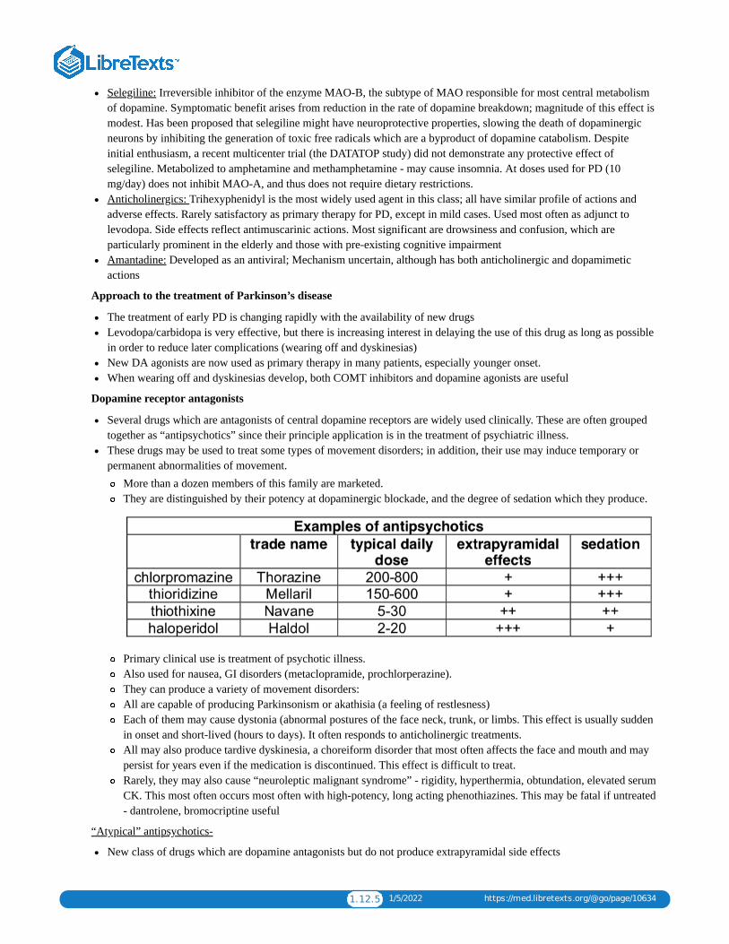
1.12.5 1/5/2022 https://med.libretexts.org/@go/page/10634
Selegiline: Irreversible inhibitor of the enzyme MAO-B, the subtype of MAO responsible for most central metabolismof dopamine. Symptomatic benefit arises from reduction in the rate of dopamine breakdown; magnitude of this effect ismodest. Has been proposed that selegiline might have neuroprotective properties, slowing the death of dopaminergicneurons by inhibiting the generation of toxic free radicals which are a byproduct of dopamine catabolism. Despiteinitial enthusiasm, a recent multicenter trial (the DATATOP study) did not demonstrate any protective effect ofselegiline. Metabolized to amphetamine and methamphetamine - may cause insomnia. At doses used for PD (10mg/day) does not inhibit MAO-A, and thus does not require dietary restrictions.Anticholinergics: Trihexyphenidyl is the most widely used agent in this class; all have similar profile of actions andadverse effects. Rarely satisfactory as primary therapy for PD, except in mild cases. Used most often as adjunct tolevodopa. Side effects reflect antimuscarinic actions. Most significant are drowsiness and confusion, which areparticularly prominent in the elderly and those with pre-existing cognitive impairmentAmantadine: Developed as an antiviral; Mechanism uncertain, although has both anticholinergic and dopamimeticactions
Approach to the treatment of Parkinson’s disease
The treatment of early PD is changing rapidly with the availability of new drugsLevodopa/carbidopa is very effective, but there is increasing interest in delaying the use of this drug as long as possiblein order to reduce later complications (wearing off and dyskinesias)New DA agonists are now used as primary therapy in many patients, especially younger onset.When wearing off and dyskinesias develop, both COMT inhibitors and dopamine agonists are useful
Dopamine receptor antagonists
Several drugs which are antagonists of central dopamine receptors are widely used clinically. These are often groupedtogether as “antipsychotics” since their principle application is in the treatment of psychiatric illness.These drugs may be used to treat some types of movement disorders; in addition, their use may induce temporary orpermanent abnormalities of movement.
More than a dozen members of this family are marketed.They are distinguished by their potency at dopaminergic blockade, and the degree of sedation which they produce.
Primary clinical use is treatment of psychotic illness.Also used for nausea, GI disorders (metaclopramide, prochlorperazine).They can produce a variety of movement disorders:All are capable of producing Parkinsonism or akathisia (a feeling of restlesness)Each of them may cause dystonia (abnormal postures of the face neck, trunk, or limbs. This effect is usually suddenin onset and short-lived (hours to days). It often responds to anticholinergic treatments.All may also produce tardive dyskinesia, a choreiform disorder that most often affects the face and mouth and maypersist for years even if the medication is discontinued. This effect is difficult to treat.Rarely, they may also cause “neuroleptic malignant syndrome” - rigidity, hyperthermia, obtundation, elevated serumCK. This most often occurs most often with high-potency, long acting phenothiazines. This may be fatal if untreated- dantrolene, bromocriptine useful
“Atypical” antipsychotics-
New class of drugs which are dopamine antagonists but do not produce extrapyramidal side effects

1.12.6 1/5/2022 https://med.libretexts.org/@go/page/10634
clozapine - d4 antagonist, effective in treatment of refractory psychosis. Numerous adverse effects, includingneutropenia (which may be fatal) and seizures (1-2%). Requires intensive monitoring.Risperidone, olazepine, quetiapine - newer, less selective, but more favorable adverse effect profile.
Reference section: A model of the basal ganglia. For more information about these models, see Albin RL, Young AB,Penney JB (1989) The functional anatomy of basal ganglia disorders. Trends Neurosci 12:366-375.

1.13.1 1/12/2022 https://med.libretexts.org/@go/page/10635
1.13: Nitric OxideThe major objective of this lecture is to describe the effects of inhaling low levels of nitric oxide (NO) on thehemodynamic and gas exchange function of both the normal and diseased lung. Considerable attention will be paid tosafety and hazards of inhaled NO therapy. During the past few years remarkable progress has been made in understandingthe NO guanylate cyclase signal transduction system. NO has been given considerable clinical investigation in pulmonaryartery hypertension and adult respiratory distress syndrome (ARDS) patients. This lecture concentrates on this area ofclinical research.
Pulmonary hypertension with severe hypoxemia may complicate the care of patients with diseases such as chronicpulmonary hypertension, ARDS, chronic respiratory failure, congenital heart disease, and after cardiopulmonary bypass.
Numerous vasodilator therapies aimed at reducing pulmonary hypertension have been tested in these patients. Systemicvasodilation and hypotension occur with all the currently available intravenous vasodilators tested in dosages sufficient toreduce the pulmonary artery pressure. In addition, intravenous infusions of systemic vasodilators such as nitroprusside orprostacyclin (PGI2) markedly increase the venous admixture (1,2).
NITRIC OXIDE
In 1987, the gaseous molecule NO was identified as an endothelium derived relaxing factor (EDRF) (3,4). NO is an ideallocal transcellular messenger because of its small size, lipophilic nature, and short duration of action (5) and its numerousfunctions in various tissues have been reviewed (6). In vascular endothelial cells, NO is synthesized from the terminalguanidine nitrogen of L-arginine and diffuses rapidly into subjacent vascular smooth muscle (7). There, NO binds to theheme iron complex of soluble guanylate cyclase. The resulting nitrosylheme activates guanylate cyclase, stimulating theproduction of cyclic guanosine 3',5'- monophosphate (cGMP) and subsequently relaxing vascular smooth muscle (7,8).When NO diffuses into the intravascular space, its biologic activity is limited by avid binding to hemoglobin. Interestingly,the nitroso vasodilators we have used for decades, such as nitroglycerin and nitroprusside, act by releasing NO (9).
Endothelium-dependent relaxation in pulmonary arteries occurs in response to a variety of physical and pharmacologicstimuli (10). Endogenous NO can be measured in the exhalation of rabbits, guinea pigs, and humans (11). In normal lungs,however, baseline pulmonary vascular tone is very low and the administration of acetylcholine or the addition ofexogenous NO has little effect on pulmonary vascular resistance (12 - 14). In patients with pulmonary hypertension, on theother hand, acetylcholine infusion or NO inhalation can reduce pulmonary vascular resistance (12,13). It is possible that insome acute and chronic pulmonary hypertensive states, such as ARDS, or chronic pulmonary hypertension, the productionof endogenous NO is impaired (15,16). This might produce further vasoconstriction and foster platelet aggregation (17).Evidence supporting this hypothesis is indirect at this time. Such patients may have an intact response to inhaled NO eventhough their response to intravenous acetylcholine is impaired (18).
NO Inhalation in ARDS
We hypothesized that inhaled NO should diffuse into the pulmonary vasculature of ventilated lung regions and causerelaxation of pulmonary vascular smooth muscle, thereby decreasing pulmonary hypertension in ARDS (19,20). Since theNO is inhaled, the gas should be distributed predominantly to well-ventilated alveoli and not to collapsed or fluid-filledareas of the lung. In the presence of increased vasomotor tone, selective vasodilation of well-ventilated lung regions shouldcause a "steal" or diversion of pulmonary artery blood flow towards well-ventilated alveoli, improving the matching ofventilation to perfusion and improving arterial oxygenation during ARDS. Such an effect would be in marked contrast tothe effects of intravenously administered conventional vasodilators (such as nitroprusside, nitroglycerin, or prostacyclin).These intravenous agents also decrease PA pressure, but by nonselectively dilating the pulmonary vasculature, theyaugment blood flow to nonventilated areas, thereby increasing right-to-left shunting and reducing the PaO . Also unlikeavailable intravenous vasodilators, inhaled NO, because it is avidly bound to hemoglobin and rapidly inactivated, shouldnot produce systemic vasodilation.
Rossaint and coworkers compared the effects of NO inhalation (18 and 36 parts per million (ppm)) to intravenouslyinfused prostacyclin in nine patients with ARDS (21). NO selectively reduced mean pulmonary artery pressure from 37 + 3to 30 + 2 mmHg (mean + SE). Oxygenation improved due to a decreased venous admixture (QVA/Qt). During NObreathing, the PaO /FIO2 ratio increased from 152 + 15 mmHg to 199 + 23 mmHg. While the intravenous infusion of
2
2

1.13.2 1/12/2022 https://med.libretexts.org/@go/page/10635
prostacyclin also reduced pulmonary artery pressure, mean arterial pressure and PaO decreased as QVA/Qt increased.Subsequent reports documented that inhalation of lower concentrations of NO (< 20 ppm) effectively reduced pulmonaryartery pressure and improved PaO2 (22 - 25). Even very small inhaled concentrations (as low as 250 parts per billion NO)may be effective in some patients (26). Right ventricular ejection fraction may increase in some patients responding toinhaled NO, suggesting that the observed decreases of pulmonary artery pressure may be hemodynamically important(24,25).
A marked variation has been reported for the hemodynamic and respiratory effects of NO inhalation, both among patientsand within the same patient at different times in their illness (22,27,28). It is possible that preexisting pulmonary disease aswell as the concomitant administration of other vasoactive drugs may contribute to the observed variability. In general, thebaseline level of pulmonary vascular resistance appears to predict the degree of pulmonary vasoconstriction reversible byNO inhalation. Those with the greatest degree of pulmonary hypertension appear to respond best to NO inhalation (22,28).Dellinger recently reported a dose-response analysis of a randomized trial of NO in 177 ARDS patients (29), a trial whichwas too small to obtain significant outcome data.
Tachyphylaxis has not been observed even when NO inhalation was continued for up to 53 days (21). Pulmonary arterypressure and PaO2quickly return to baseline values, however, after discontinuation of the gas. Occasionally, suddendiscontinuation of inhaled NO can produce problematic pulmonary vasoconstriction and possibly bronchoconstriction(22,30,31). The reason for this is unclear. Possibly, the addition of exogenous NO may decrease NO synthase activity (32)or increase tissue cGMP phosphodiesterase activity.
The vasoconstrictor almitrine besylate has been given intravenously to enhance pulmonary vasoconstriction during NObreathing. This agent has further reduced Qs/Qt in ARDS in combination with NO inhalation (33).
NO Inhalation in Neonatal Respiratory Failure
At birth, there is a sustained decrease of pulmonary vascular resistance and an increase of pulmonary blood flow, in partdue to increasing oxygen tensions. If this does not occur, persistent pulmonary hypertension of the newborn (PPHN) mayresult. Persistent pulmonary hypertension of the newborn is a syndrome characterized by an increased pulmonary vascularresistance, increased right-to-left shunting across the ductus arteriosus and foramen ovale, and severe hypoxemia.Extracorporeal membrane oxygenation (ECMO) is often used to support these infants, because conventional vasodilatortherapy is limited by severe systemic hypotension and may reduce PaO by increasing right-to- left shunting. It has beenhypothesized that endogenous production of NO by the pulmonary vasculature might be decreased in PPHN. If so, theninhaled NO might provide an effective therapy for these severely ill infants (34, 35). Multiple small clinical studies of NOinhalation have been performed in neonates, infants, and children with various types of acute respiratory failure. Ingeneral, pulmonary hypertension is reduced and systemic arterial oxygenation is improved with inhalation of less than 20ppm NO. Nitric oxide inhalation in babies with PPHN and hypoxic respiratory failure has been studied in randomizedmulticenter trials(36,37). As in adults, however, the response is variable. In the neonatal lung, the degree of improvementwith NO appears to depend upon the presence of mature surfactant.
Laboratory studies of the neonatal pulmonary circulation have also documented that inhaled nitric oxide is an effectivepulmonary vasodilator (38). Additionally, important experimental evidence is accumulating that the inhalation of nitricoxide attenuates chronic hypoxic pulmonary vascular remodeling of the pulmonary circulation (39,40). Conceivably,inhaled nitric oxide therapy might be used to limit the chronic pulmonary vascular changes which accompany neonatalacute respiratory failure.
REFERENCES1. Zapol WM, Snider MT, Rie MA, Frikker M, Quinn DA. Pulmonary circulation during adult respiratory distress
syndrome. In: Acute Respiratory Failure. Zapol WM, Falke KJ (eds.) New York: Marcel Dekker, 1985;241-270.2. Radermacher P, Stanek B, Wust HJ, Tanrow J, Falke KJ. Prostacyclin for the treatment of pulmonary hypertension in
the adult respiratory distress syndrome: Effects on pulmonary capillary pressure and ventilation-perfusion distributions.Anesthesiology 1990;72:238-244.
3. Ignarro LJ, Buga GM, Wood KS, Byrns RE. Endothelium-derived relaxing factor produced and released from arteryand vein is NO. Proc Natl Acad Sci USA 1987;84:9265-9269.
2
2

1.13.3 1/12/2022 https://med.libretexts.org/@go/page/10635
4. Palmer RMJ, Ferrige AG, Moncada S. Nitric oxide release accounts for the biological activity of endothelium-derivedrelaxing factor. Nature1987;327:524-526.
5. Ignarro LJ. Signal transduction mechanisms involving nitric oxide.Biochem Pharmacol 1990;41:485-490.6. Moncada S, Higgs A. Mechanisms of disease: The L-arginine-nitric oxide pathway. N Engl J Med 1993;329:2002-
2012.7. Palmer RMJ, Ashton DS, Moncada S. Vascular endothelial cells synthesize nitric oxide from L-arginine. Nature
1988;333:664-666.8. Ignarro LJ. Biological actions and properties of endotheliumderived nitric oxide formed and released from artery and
vein. Circ Res1989;65:1-21.9. Gruetter CA, Gruetter DY, Lyon JE, Kadowitz PJ, Ignarro LJ. Relationship between cyclic guanosine 3':5'-
monophosphate formation and relaxation of coronary artery arterial smooth muscle by glyceryl trinitrate, nitroprusside,nitrite and nitric oxide. J Pharmacol Exp Ther 1981;219:181-186.
10. Dinh Xuan AT, Higenbottam TW, Clelland C, Pepke-Zaba, Wells FC, Wallwork J. Acetylcholine and adenosinedisphosphate causes endothelium-dependent relaxation of isolated human pulmonary arteries. Eur Respir J 1990;3:633-638.
11. Gerlach H, Rossaint R, Pappert D. Autoinhalation of nitric oxide after endogenous synthesis in nasopharynx. Lancet1994;343:518-519.
12. Pepke-Zaba J, Higenbottam TW, Dinh-Xuan AT, Stone D, Wallwork J. Inhaled nitric oxide as a cause of selectivepulmonary vasodilation in pulmonary hypertension. Lancet 1991;338:11731174.
13. Frostell CG, Blomqvist H, Hedenstierna G, Lundberg J, Zapol WM. Inhaled nitric oxide selectively reverses humanhypoxic pulmonary vasoconstriction without causing systemic vasodilation.Anesthesiology 1993;78:427-435.
14. Högman M, Frostell C, Arnberg H, Hedenstierna G. Inhalation of nitric oxide modulates methacholine-inducedbronchoconstriction in the rabbit. Eur Respir J 1993;6:177-180.
15. Cremona G, Dinh Xuan AT, Higenbottam TW. Endothelium-derived relaxing factor and the pulmonary circulation.Lung 1991;169:185- 202.
16. Dinh Xuan AT, Higenbottam TW, Clelland C, Pepke-Zaba J, Cremona G, Wallwork J. Impairment of endothelium-dependent pulmonary artery relaxation in chronic obstructive lung disease. N Engl J Med1991;324:1539-1547.
17. Radomski MW, Palmer RMJ, Moncada S. Endogenous nitric oxide inhibits human platelet adhesion to vascularendothelium. Lancet1987;2:1057-1058.
18. Adatia I, Thompson J, Landzberg M, Wessel DL. Inhaled nitric oxide in chronic obstructive lung disease. Lancet1993;341:307-308.
19. Frostell C, Fratacci M-D, Wain JC, Jones R, Zapol WM. Inhaled nitric oxide: A selective pulmonary vasodilatorreversing hypoxic pulmonary vasoconstriction. Circulation 1991;83:2038-2047.
20. Fratacci M-D Frostell CG, Chen T-Y, Wain JC, Robinson DR, Zapol WM. Inhaled nitric oxide: A selective pulmonaryvasodilator of heparin-protamine vasoconstriction in sheep. Anesthesiology1991;75:990-999.
21. 22 Bigatello LM, Hurford WE, Kacmarek RM, Roberts JD, Jr, Zapol WM. The hemodynamic and respiratory responseof ARDS patients to prolonged nitric oxide inhalation. Am Rev Respir Dis 1993;147:A720.
22. Puybasset L, Rouby JJ, Cluzel P, Mourgeon E, Belin M-F, Arthaud M, Landault C, Viars P. Inhaled nitric oxidereverses the increase in pulmonary vascular resistance induced by permissive hypercapnia in patients with acuterespiratory distress syndrome. Anesthesiology1994;80:1254-1267.
23. Puybasset L, Rouby JJ, Cluzel P, Mourgeon E, Belin M-F, Arthaud M, Landault C, Viars P. Inhaled nitric oxidereverses the increase in pulmonary vascular resistance induced by permissive hypercapnia in patients with acuterespiratory distress syndrome. Anesthesiology1994;80:1254-1267.
24. Gerlach H, Pappert D, Lewandowski K, Rossaint R, Falke KJ. Long- term inhalation with evaluated low doses of nitricoxide for selective improvement of oxygenation in patients with adult respiratory distress syndrome. Intensive CareMedicine 1993;19:443-449.
25. Wysocki M, Vignon P, Roupie E, Humbert M, Adnot S, Lemaire F, Brochard L. Improvement in right ventricularfunction with inhaled nitric oxide in patients with the adult respiratory distress syndrome (ARDS) and permissivehypercapnia. Am Rev Respir Dis1993;147:A350.
26. Zapol WM, Falke KJ, Rossaint R. Inhaled nitric oxide for the adult respiratory distress syndrome. N Engl J Med(author's reply) 1993;329:207.
27. Ricou B, Suter PM (1993) Variable effects of nitric oxide (NO) in ARDS patients. Am Rev Respir Dis 1993;147:A350.
1.13.4 1/12/2022 https://med.libretexts.org/@go/page/10635
28. Rich GF, Murphy GD, Ross CM, Johns RA. Inhaled nitric oxide: A selective pulmonary vasodilation in cardiacsurgical patients.Anesthesiology 1993;78:1028-1035.
29. Dellinger RP, Zimmerman JL, Taylor RW, Straube RC, Hauser DL, Criner GJ, Davis K, Hyers TM, Papadakos P.Effects of inhaled nitric oxide in patients with acute respiratory distress syndrome: Results of a randomized phase IItrial: Crit Care Med 1998;26:15-23.
30. Grover R, Murdoch I, Smithies M, Mitchell I, Bihari D. Nitric oxide during hand ventilation in a patient with acuterespiratory failure.Lancet 1992;340:1038-1039.
31. Dupuy PM, Shore SA, Drazen JM, Zapol WM. Bronchodilator action of inhaled nitric oxide in guinea pigs. J ClinInvest 1992;90:421-428.
32. Rengasamy A, Johns RA. Regulation of nitric oxide synthase by nitric oxide. Mol Pharmacol 1993;44:124-128.33. Wysocki M, Delclaux C, Roupie E, Langeron O, Liu N, Herman B, Lemaire F, Brochard L. Additive effect on gas
exchange of inhaled nitric oxide and intravenous almitrine bismesylate in the adult respiratory distress syndrome.Intensive Care Medicine 1994;20:254- 259.
34. Roberts JD, Polaner DM, Lang P, Zapol WM. Inhaled nitric oxide in persistent pulmonary hypertension of thenewborn. Lancet1992;340:818-819.
35. Kinsella JP, Shaffer E, Neish SR, Abman SH: Low-dose inhalational nitric oxide in persistent pulmonary hypertensionof the newborn.Lancet 1992;340:8819-8820.
36. Roberts JD, et al. Inhaled nitric oxide and persistent pulmonary hypertension of the newborn. N Engl J Med1997;336(9):605-610.
37. The Neonatal Inhaled Nitric Oxide Study Group, Inhaled nitric oxide in full-term and nearly full-term infants withhypoxic respiratory failure.N Engl J Med 1997;336(9)597-604.
38. Roberts JD, Lang P, Bigatello LM, Vlahakes GJ, Zapol WM: Inhaled nitric oxide in congenital heart disease.Circulation 1993;87:447-453.
39. Kouyoumdjian C, Adnot S, Levame M, Eddahibi S, Bousbaa H, Raffestin B: Continuous inhalatioin of nitric oxideprotects against development of pulmonary hypertension in chronically hypoxic rats. J Clin Invest 1994;94:578-584.
40. Roberts JD, Roberts CD, Jones RC, Zapol WM: Continuous nitric oxide inhalation reduces pulmonary arterialstructural changes, right ventricular hypertrophy, and growth retardation in the hypoxic newborn rat. Circ Res1995;76:215-222.

1.14.1 1/12/2022 https://med.libretexts.org/@go/page/10636
1.14: Neuropharmacology II - Anxiolytics and AntidepressantsDepression is a frequent problem, affecting up to 5% of the population. Common presentations include low mood, loss ofenergy, disinterest in activities. May also include weight loss, sleep disturbance, or psychosis. Should be considered inpatients with atypical dementia and chronic pain
Diagnosis of Depression - DSM-IV
Five of the following present during the same 2-week period and represent a change from previous functioning:
depressed moodmarkedly diminished interest or pleasure in all, or almost all, activitiessignificant weight loss when not dieting or weight gaininsomnia or hypersomniapsychomotor agitation or retardationfatigue or loss of energyfeelings of worthlessness or excessive or inappropriate guiltdiminished ability to think or concentrate, or indecisivenessrecurrent thoughts of death, recurrent suicidal ideation or a suicide attempt
The symptoms cause clinically significant distress or impairment in social, occupational, or other important areas offunctioning.The symptoms are not due to the direct physiological effects of a substance (e.g., a drug of abuse, a medication) or ageneral medical condition (e.g., hypothyroidism).The symptoms are not better accounted for by Bereavement
Pathophysiology of depression
At present, mechanism is unknown - may be more than one mechanism.No useful biomarkers or imaging abnormality during lifeStudy of postmortem brain has not revealed any consistent structural or neurochemical abnormalityMajority of the currently available medications were discovered empiricallyMost current theories are based on “amine hypothesis”
Biogenic amines

1.14.2 1/12/2022 https://med.libretexts.org/@go/page/10636
Classes of Antidepressants
Tricyclics and heterocyclicsSelective serotonin reuptake inhibitors (SSRI’s)BupropionNonselective MAO inhibitorsNon-pharmacological therapy
ECT

1.14.3 1/12/2022 https://med.libretexts.org/@go/page/10636
Psychotherapy
Tricyclics and heterocyclics - Clinical pharmacology
Large family of structurally related compoundsMultiple pharmacological actionsTherapeutic effect probably due to ability to block reuptake of serotonin and/or norepinephrineAll may be sedating, although some much more than othersMany of these drugs have anticholinergic (anti-muscarinic) actions - leads to somnolence, dry mouth, urinary retention
Tricylics and heterocyclics - pharmacokinetics and toxicity
All are primarily metabolized by the liver, and undergo first pass metabolismBiochemical half-lives range from 4 to more than 24 hours, but clinical response is much slower - typically severalweeks of therapy is required to observe any clinical improvementOverdose of tricylics (more than 1 gram) is often lethal due to cardiac conduction disturbances. Great care must betaken when these drugs are prescribed for potentially suicidal patients.
Some commonly used tricylics and heterocyclics
Amitriptiline (Elavil®)
Inhibits serotonin & NE reuptakeProminent anticholinergic effectsMetabolite is nortriptyline
Desipramine (Norpramine®)Inhibits NE reuptakeMild anticholinergic effects
Trazodone (Desyrel®)HeterocyclicInhibits serotonin reuptakeMinimal anticholinergic effectsSedating
Selective Serotonin Reuptake Inhibitors (SSRI’s)
Act by inhibition of presynaptic reuptake of serotonin in central synapses.Not as sedating as many of the tricylic compoundsAlso do not have the anticholinergic side effects of the tricyclicsSome are potent inhibitors of P450 enzyme systems, and may lead to drug interactions
Some commonly used SSRI’s
Fluoxetine (Prozac®)Sertaline (Zoloft®)Citalopam (Celexa®)Paroxetine (Paxil®)
All are potent inhibitors of serotonin reuptakeAdverse effects: anxiety, tremorOverdose of SSRI alone is rarely lethalShould not be administered with nonselective MAO inhibitorsSuicide as an adverse effect?
Bupropion
Structurally related to the tricyclics, but seems to have a different therapeutic mechanism, related to altered release ofNENot sedating or anticholinergic, but does sometime induce hallucinations or seizuresAlso effective in treating tobacco addiction

1.14.4 1/12/2022 https://med.libretexts.org/@go/page/10636
MAO Inhibitors
Non-selective, irreversible enzyme inhibitors - long duration of actionTherapeutic effect is due to is enhancement of CNS amine levelsMajor adverse effects are due to excessive accumulation of amines in the circulation
Tyramine: the “cheese effect.”Drug interactions: SSRI’s, sympathomimetics
Safe in carefully controlled circumstances, but “real world” use may lead to serious adverse effects.
Treatment of depression
Many patients will not report symptoms of depression unless asked specificallyPatients who are depressed may be suicidal - it is essential to inquiry about their intentionsThe response of an individual patient to a particular antidepressant cannot be predicted, and treatment often requiressequential trials of several drugsIn severely depressed patients, ECT often produces a rapid improvement and may be the best initial treatment
Sedatives and hypnotics
Used to reduce anxiety, or induce sleepVery commonly prescribedTwo principal chemical classes:
BenzodiazepinesBarbiturates
Both work by enhancing activity of the inhibitory neurotransmitter, GABA
GABA (γ-aminobutyric acid)
Principal inhibitory transmitter of the mammalian brainReceptors:
GABA : ligand gated ion channels, regulate chloride ion, at least 15 different subunit proteinsGABA : G-protein coupled receptors
Effects of benzodiazepines and barbiturates on GABA Receptors
A
B
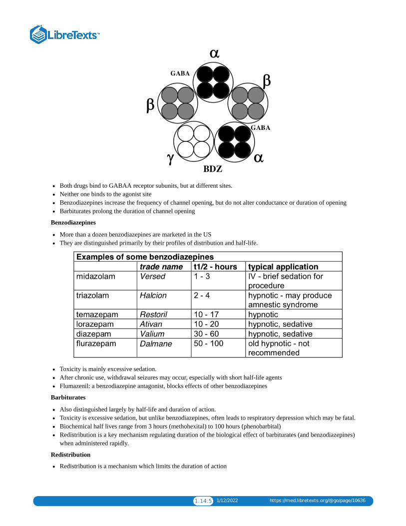
1.14.5 1/12/2022 https://med.libretexts.org/@go/page/10636
Both drugs bind to GABAA receptor subunits, but at different sites.Neither one binds to the agonist siteBenzodiazepines increase the frequency of channel opening, but do not alter conductance or duration of openingBarbiturates prolong the duration of channel opening
Benzodiazepines
More than a dozen benzodiazepines are marketed in the USThey are distinguished primarily by their profiles of distribution and half-life.
Toxicity is mainly excessive sedation.After chronic use, withdrawal seizures may occur, especially with short half-life agentsFlumazenil: a benzodiazepine antagonist, blocks effects of other benzodiazepines
Barbiturates
Also distinguished largely by half-life and duration of action.Toxicity is excessive sedation, but unlike benzodiazepines, often leads to respiratory depression which may be fatal.Biochemical half lives range from 3 hours (methohexital) to 100 hours (phenobarbital)Redistribution is a key mechanism regulating duration of the biological effect of barbiturates (and benzodiazepines)when administered rapidly.
Redistribution
Redistribution is a mechanism which limits the duration of action

1.14.6 1/12/2022 https://med.libretexts.org/@go/page/10636
Effect is greatest when:Agent is administered rapidly (e.g., intravenous)Agent is highly lipophilic
Can lead to very short duration of action (minutes) even though biochemical half life is longer (hours).
Clinical use of sedatives
Anxiolytic useUsually a medium to long acting benzodiazepine, such as diazepam, administered orally.
Hypnotic useUsually a short to medium acting benzodiazepine, such as temazepam, administered orally - but note that allhypnotics lose efficacy if taken daily.
Sedative use (for surgical procedures)A short acting benzodiazepine, such as midazolamA short acting barbiturate, such as thiopentalAdministered intravenously, and action terminated by redistribution.
Tolerance, cross-tolerance, and addiction
Chronic use of sedatives of either class (benzodiazepine or barbiturate) induces tolerance to all members of the class,and cross-tolerance to members of the other class.Both also induce tolerance to ethanol, which acts in part through GABA receptors.Both benzodiazepines and barbiturates may produce dependence and are susceptible to abuse. Potentially lethal actionsof the barbiturates makes them particularly problematic when abused.Rapid withdrawal from either class of sedatives may lead to anxiety, agitation, and seizures
1.15.1 11/24/2021 https://med.libretexts.org/@go/page/10637
1.15: Neuropharmacology III - Anticonvulsants
Neuropharmacology IIIAnticonvulsants
What are seizures?
Seizures are episodes of neurologic dysfunction arising from abnormal synchronous activity of neurons.Alterations of consciousness and abnormal motor activity are the most common manifestationsEpilepsy (recurring seizures without a clear precipitant) is common, affecting about 1% of the populationPharmacological treatment is very successful in the majority of cases, but requires accurate diagnosis and classificationof seizures
Classification of seizures:
Partial seizures (focal onset)Simple partial seizures most common; “Jacksonian march” sometimes observed. May also affect sensory andautonomic systemsComplex partial: impairment of consciousness, with or without motor or other signsEither simple or complex partial seizures may become secondarily generalized, producing a tonic-clonic seizure.
Primary generalized seizures - bilateral onsetTypified by absence (“petit mal”) seizures, which can be recognized by clinical characteristics as well as interictalEEG abnormalities (3Hz spike and wave).
Strategies for the discovery of anticonvulsant drugs:“Traditional” – screening of compounds in animals models of epilepsy“Rational” – based on presumed mechanism of seizure initiation or propagation
Inhibit repetitive activity – e.g., blockade of voltage-dependent Na channelsIncrease inhibitory input – e.g., GABA enhancersReduce excitatory input – e.g., glutamate antagonists
Drugs for treatment of partial seizures or generalized tonic-clonic seizures:
phenytoinOne of the oldest and most widely used anticonvulsants.Mechanism uncertain, but probably related to effect on Na+ channels.May be administered orally or IV.Pharmacokinetics are complex:
oral absorption is good but rate is variable.highly protein-bound - important to note that usual laboratory test measures total, not free phenytoinmetabolism is primarily hepatic. Exhibits saturation kinetics, so that small increment in dose can produce anabrupt rise in equilibrium concentration.half-life averages 22 hours but highly variable; bid dosing usually satisfactoryinduces hepatic metabolism of other anticonvulsants as well as anticoagulantsAcute toxicity of oral form is usually nystagmus, ataxia and diplopia; sedation may also occur.Chronic administration causes hirsuitism, gingival hyperplasia, cerebellar dysfunction, and peripheralneuropathyHypersensitivity with fever, rash which may progress to exfoliation (Stevens-Johnson syndrome) is relativelyinfrequent, but requires discontinuation of the drug in most cases.
Fosphenytoin (Cerebyx) a “prodrug”fosphenytoin is rapidly metabolized to phenytoinfosphenytoin is water soluble; allows IM administration, and eliminates toxicity of propylene glycol vehiclerequired for phenytoin1200 mg phenytoin = $1.50; fosphenytoin = $119.00
+

1.15.2 11/24/2021 https://med.libretexts.org/@go/page/10637
CarbamazepineStructural features similar to phenytoin; mechanism of action likely similar as well.Available in oral form only; rate of absorption variable.Protein binding less than that of phenytoin.Metabolism is primarily hepatic; induces own metabolism, as well as that of other drugs (OCP’s, warfarin, otheranticonvulsants particularly problematic).Half-life is 10-20 hours; tid dosing usually satisfactory, although qid sometimes requiredSeveral active metabolites, including a 10,11 epoxide, contribute to both anticonvulsant activity and toxicityMost common effects of toxicity are ataxia and diplopia, sedation also observed. May also cause hyponatremia.Aplastic anemia may occur and can be fatal. This is rare (6-8/million patients/year) but requires monitoring of CBC.Oxcarbazepine – a derivative, does not form epoxide metabolites and may have lower incidence of adverse effects.
BarbituratesFamily of drugs used for hypnotic, anesthetic and anticonvulsant applicationsMechanism probably related to increased GABA-mediated chloride conductanceTwo members of class commonly used as anticonvulsants:
PhenobarbitalMay be administered PO, IM or IVLong half-life (about 100 hours), hepatic metabolism. Strong inducer of microsomal system.Frequently used in infants; less commonly used in adults because of dose-related sedation.
PrimidoneParent drug has anticonvulsant properties, but is metabolized rapidly by the liver to phenobarbital andPEMA.Toxicity similar to that of phenobarbital
Valproic acid
Carboxylic acid, structurally distinct from other current classes of anticonvulsants.Mechanism uncertain - effective against both partial and primary generalized seizures; drug of choice for myoclonicepilepsyOral or IV administrationHepatic metabolism, with half life 8-12 hours. Induces metabolism of other anticonvulsantsCommon adverse effects are tremor, weight gain, nausea.Most significant risk is hepatotoxicity, which may be fatal. Occurs most often in infants under 2 years when takingmultiple anticonvulsants.
Drugs for primary generalized epilepsy

1.15.3 11/24/2021 https://med.libretexts.org/@go/page/10637
Ethosuccimidedrug of choice for treatment of absence seizure. Also effective in other forms of primary generalized epilepsy,but not usually effective in partial seizures.
Valproic acid
effective in generalized as well as focal epilepsy; particularly useful when several seizure types are present.New anticonvulsants
Because they are new, the clinical indications for these agents are not yet completely defined, and none arecurrently used as the first treatment for epilepsy.Felbamate was approved by the FDA in early 1994 and was the first new drug for epilepsy to be approved in 15years. Although there was great initial enthusiasm for this agent, in less than a year post- marketing surveillancerevealed an unacceptably high rate of drug- related aplastic anemia.Lamotrigine was approved in late 1994. It is thought to act by blockade of sodium channels; useful in partialseizures and possibly also in primary generalized seizures.Gabapentin is approved for use as an “add-on” medication for treatment of partial seizures. Mechanism is uncertain;toxicity is low and does not induce or inhibit metabolism of other anticonvulsants.Topiramate approved in 1997; unknown mechanism, possibly acts on voltage-gated Na+ channels.Tiagabin - an inhibitor of GABA reuptake, approved in 1997 as “add- on” for treatment of partial seizures.Levetiracetam – analog of piracetam, mechanism uncertain, approved as “add-on” for refractory partial seizuresVigabatrin - an inhibitor of GABA transaminase, the degradative enzyme for GABA; approved as an “add-on”agent in refractory epilepsyZonisamide, a sulfonamide derivative approved for partial seizures; acts on Na channels
Benzodiazepines:An important anticonvulsant use of benzodiazepines is in setting of urgent treatment of status epilepticus (seebelow). Two agents are frequently used, diazepam and lorazepam. These are particularly suitable because of rapidaction after intravenous injectionNote that although biological half-life of diazepam is long, duration of action when used IV is short, becauseactivity is terminated by redistributionOral benzodiazepines are not frequently used alone in primary treatment of epilepsy, although sometimes a usefuladjunct in both focal and generalized seizures
Principles for the Management of epilepsy
Attempt to classify, localize and investigate underlying etiologyNot every seizure is an indication for anticonvulsant therapyIn general, monotherapy is preferred to the use of multiple drugsSerum drug levels are a guide to therapy, but you should treat the patient, not the numbersRoughly 80% of patients with epilepsy can achieve good control with one agent; >90% with two or more.In refractory epilepsy, surgical treatment may be appropriate
Pregnancy and the use of anticonvulsant drugs
All of the anticonvulsant drugs have been reported to have teratogenic effectsAlso important to recognize that uncontrolled seizures have an adverse effect on the fetus. Most important period isfirst 12 weeksIn general, the best approach is to keep the number of drugs low (monotherapy if possible) and use the lowest dosewhich provides adequate control.Valproic acid should probably be avoided if at all possible, as the increased incidence of neural tube defects with thisdrug is well documented.Abrupt discontinuation of anticonvulsants during pregnancy is not advisable
Emergency medicine: treatment of status epilepticus
Definition and identificationStatus epilepticus is a state of repeated or continuous seizures.
+

1.15.4 11/24/2021 https://med.libretexts.org/@go/page/10637
Often defined operationally as a single seizure lasting more than 20 minutes, or repeated seizures without recoveryof consciousnessProlonged status epilepticus leads to irreversible brain injury and has a very high rate of mortality. Goal of therapyshould be to achieve control of seizure within 60 minutes or less
ManagementABC’s - Airway, Breathing, CirculationIV access - obtain initial labs (electrolytes, ABG, CBC, tox screen, anticonvulsant levels). History and examinationshould be performed concurrentlyAdminister glucose (50g IV) and thiamin (100mg IV)Initial treatment: - lorazepam, 1-2 mg IV, repeat at 3-5 min intervals to 10 mg totalAdminister a long acting agent - phenytoin or fosphenytoin - 15-20 mg/kg IV. With phenytoin, do not exceed 50mg/min. Not compatible with IV fluids containing glucose. Often causes hypotension, and may provoke arrhythmia- continual monitoring required. Fosphenytoin much safer - but also more expensive !If seizures persist, next agent is phenobarbital. Initial dose is 5 mg/kg IV. May be repeated to 10-15 mg/kg total.These large doses of phenobarbital often produce respiratory depression or arrest, as well as hypotension;intubation, respiratory support, and pressors may be required.Seizures which are refractory to these measures require urgent expert consultation. Barbiturate coma induced byhigh doses of pentobarbital, a short half-life barbiturate, is used in many centers.
References:
Rogawski MA, Loscher W. The neurobiology of antiepileptic drugs. Nat Rev Neurosci. 2004 Jul;5(7):553-64

1.16.1 12/22/2021 https://med.libretexts.org/@go/page/10638
1.16: Antimicrobials I and II
Pharmacologic Principles of Antimicrobial TherapyA. Successful antimicrobial therapy occurs when an effective concentration of drug is delivered to the site of infection for
a sufficient period of time. Minimum effective concentrations are those needed to inhibit growth (bacteriostaticconcentration, MIC) or kill (bacteriocidal concentration,MBC) the pathogen in question.
Bacteriocidal therapy required in the following conditions
a. Bacterial infection in the neutropenic host b. Endocarditis (and other intravascular infections) c. Meningitis and brain abscess d. Staphylococcal (and probably other forms of) osteomylitis e. Prosthetic device infection
B. Drug Absorption1. The determinants of drug absorption are poorly understood and can only be determined by clinical studies.2. Must determine effects of food, gastric pH, and antacids on drug absorption.
a. Food absorption usually decreases, may increaseb. Ketoconazole requires acid pHc. Chelation of tetracyclines and fluoroquinolones by cations in antacids may block absorption.
C. Drug Elimination
Three major routes of elimination:
1. Kidneys– renal elimination may occur either by glomerular filtration or tubular secretion; in general, tubularsecretion (seen with penicillins and many cephalosporins) is more efficient than glomerular filtration, and results inshorter serum half-lives. Probenicid blocks active secretion.
2. Hepatobiliary – if significant hepatobiliary elimination occurs, then little dosage adjustment is needed in renalfailure.
3. Metabolism – generally occurs in the liver, and can lead to drug interactions, because of effects on liver enzymesystems.
D. Distribution of Antimicrobial Agents in Tissues
There are three major determinants of distribution of drugs between the plasma (central compartment) andextravascular space (peripheral compartment)
1. Nature of the Capillary Bed – In most tissues and organs the capillary bed is fenestrated by small pores that permitthe ready diffusion of substances with molecular weights up to 1000 daltons (most antimicrobial agents). A fewlocations in the body, termed specialized sites, have unfenestrated capillaries. As drugs must pass through theendothelial cells of the capillaries to reach extravascular space in these specialized sites, the rate of diffusion islimited by the degree of lipid solubility of the drug.a. The most clinically important specialized sites, are the central nervous system, the retina and the prostate gland.b. Such drugs as the β-lactams, aminoglycosides, mosttetracyclines and vancomycin are weakly lipid soluble and
penetrate specialized site poorly.2. Degrees of Serum Binding – only free drug is available for diffusion and is active. The major binding protein for
most drugs is albumin.3. Active Transport Pumps – The best studied of these pumps act on organic anions and are located in the choroid
plexus of the brain, the retina and the proximal tubule of the kidney. These pump out β-lactam drugs and arecompletely inhibited by probenicid.
E. Site of Infection –
The site of infection determines not only the choice of the agent, but also its dose and the route by which it isadministered.
1. In general, we wish to exceed to MIC.

1.16.2 12/22/2021 https://med.libretexts.org/@go/page/10638
2. Effects of subinhibitory concentrationsa. Alter the bacterial morphology and adherence propertiesb. Decrease opsonic requirementsc. Enhance phagocytosisd. Aid in intracellular killing
PenicillinsA. Structure
Three components: A thiazolidine ring, the β-lactam ring, and a side chain. The side chain determines in large part toantibacterial spectrum and pharmacologic properties of a particular penicillin.
B. Mechanism of Action – surprisingly incompletely understood.1. Penicillin inhibits bacterial growth by interfering with the synthesis of the bacterial cell wall after binding to
penicillin binding proteins (many of these are enzymes are involved in cell wall biosynthesis).2. Although penicillins are bacteriocidal drugs, the mechanisms by which they kill bacteria vary for different species.
For pneumococcus and E. coli, killing is by lysis resulting from deregulation of the autolytic enzyme system (i.e.,peptidoglycan hydrolases). Penicillin may also directly enhance autolytic activity. In the case of streptococcus,penicillin induces hydrolysis of cellular RNA.
3. Post-antibiotic effects are observed with gram-positive, but not gram negative bacteria.C. Spectrum of Activity
see appendix for details generally active against cocci, many bacilli and anaerobes. activity against enterobacteriaceaeand pseudomonas seen with aminopenicillins.
D. Resistance (see XII A for more details)1. The most important mechanism of bacterial resistance to penicillin is enzymatic hydrolysis of the β-lactam bond by
β-lactamases.
a. S. aureus – plasmid encoded and inducible. This plasmid is increasingly found in enterococcus.b. In gram negatives, β-lactamases can be chromosomally or plasmid mediated, constitutive or inducible, and
active against only certain β-lactams or broad spectrum.
The ability of penicillin to inhibit growth of gram negative bacilli is dependent on the rate of influx across theouter membrane being greater than the rate of hydrolysis by β-lactamases. Alteration in the penicillin side chaingoverns gram negative activity, generally by enhancing penetration across the outer membrane rather thanreducing the rate of hydrolysis.
2. Alteration in penicillin binding site.a. Penicillin resistant pneumococcib. Methicillin resistant staphylococci
3. Tolerance – MBC > 16 x MIC. Organisms exhibiting tolerance appear to have realigned or altered autolytic actionwith exposure to penicillin.a. Enterococci are naturally resistantb. Some S. aureus and streptococci.

1.16.3 12/22/2021 https://med.libretexts.org/@go/page/10638
4. Altered permeability of the outer membrane of gram negative bacilli provides another mechanism for resistance tothe penicillins. Mutants with reduced or altered porion channels show 2-16 fold higher MICs to the broad spectrumpenicillins. This mechanism often occurs jointly with altered PBPs or inducible β-lactamases.
E. Adverse Reactions1. Hypersensitivity2. CNS—seizures (primarily penicillin G and in patients with renal failure).3. Gastrointestinal—C. difficile, and nonspecific GI upset—diarrhea, nausea, and vomiting.
CephalosporinsA. Structure
B. Mechanism of Action—essentially the same as penicillin.C. Spectrum of Activity—The cephalosporins are broad spectrum agents. As a rule, gram positive activity diminishes
while gram negative activity increases as one progresses from first to third generation agents. None of thecephalosporins is active against enterococci, Listeria monocytogenes, or methicillin-resistant S. aureus.
see appendix for details.
D. Mechanism of Resistance (see XII A)1. β-lactamase production2. Alterations in target penicillin binding proteins.3. Inability of the drug to reach its binding site: In order to reach its target PBP, a cephalosporin must penetrate an
organism’s cell envelope. This is done relatively easily in the case of gram-positive organisms, as the peptidoglycanstructure that comprises the cell wall routinely allows the passage of cephalosporin-sized particles. Gram-negativeorganisms possess a more formidable barrier, a complex structure composed of polysaccharides, lipids, andproteins. Materials penetrate this outer cell envelope through water-filled channels, or porions, produced by variousouter membrane proteins. Passage by a cephalosporin depends on channel size, charge, and hydrophilic properties.
E. Adverse Reactions1. Hypersensitivity2. Gastrointestinal, including hepatitis.
F. Other Beta-Lactam StrategiesA. β-lactamase Inhibition
1. Sulbactam (ampicillin-sulbactam, Unasyn).2. Clavulanic acid (amoxicillin-clavulanate, Augmentin; ticarcillin- clavulanate, Timentin).3. Tazobactam (pipercillin-tazobactam, Zosyn).
B. Aztreonam—a monocyclic β−lactam relatively resistant to β−lactamases. Spectrum similar to gentamycin.

1.16.4 12/22/2021 https://med.libretexts.org/@go/page/10638
C. Imipenem—broadest spectrum β−lactam. A carbapenem.
1. Unique pharmacologic problem: After imipenem is removed from the circulation by glomerular filtration andsecreted, it is metabolized by a renal peptidase which is located on the brush border of the proximal renaltubules. The metabolites are nephrotoxic.
2. To overcome this problem, a specific peptidase inhibitor, cilastin, was synthesized, which totally blocks themetabolism of imipenem in the kidney, thus blocking toxicity. Cilastin has no antimicrobial activity.
3. Compound drug is imipenem-cilastin combination (Primaxin).4. Particular toxicity is seizures, primarily in renal failure or in the face of ongoing or preceding brain injury.
AminoglycosidesA. Structure—All aminoglycosides consist of central six-memberedaminocyclitol ring linked to two or more aminosugar
residues by glycosidic bonds. The aminoclycitol of streptomycin is streptidine, whereas that of all other availableaminoclycosides is 2-deoxystreptamine.
B. Mechanism of Action—incompletely understood
1. Aminoglycosides bind to ribosomes, with the different aminoglycosides binding to different sites (streptomycin tothe 30S subunit, the others at other sites; streptomycin binding does not compete with binding of the otheraminoglycosides).
2. The consequences of the interaction of aminoglycosides are numerous. The two best-documented consequences arethe inhibition of protein synthesis and an infidelity in correctly reading the genetic code.
3. These, however, don’t explain the bactericidal effect of aminoglycosides. Aminoglycoside transport across the cellmembrane, with accumulation in the cytosol is central to this effect. The transport process is energy-dependent andpH dependent (thus, aminoglycosides don’t work well in situations of low pH and anaerobiosis; i.e., an abscess).
C. Spectrum of Activity1. Aerobic and facultative gram negative bacilli. Especially useful against enteric organisms.

1.16.5 12/22/2021 https://med.libretexts.org/@go/page/10638
2. Partner in synergetic killing with β-lactama. Absolutely required for Enterococci.b. In vitro, (in vivo?) for Staphylococcus aureus, Streptococcus pneumonia, other streptococci.c. Pseudomonas (one of a handful of useful drugs).
D. Mechanisms of Resistance
1. Aminoglycoside-modifying enzymes—diverse array; carried on plasmid and transposons; Ex: acetylation of anamino group, adenylation of a hydroxyl group, phosphorylation of a hydroxyl group. The resulting compound bindspoorly to ribosomes. This is the major form of resistance.
2. Mitochondrial alterations of ribosomes—binding sites modified due to chromosomal interaction; as yet, rare.3. Mutations interfering with aminoglycoside uptake—results in resistance to all aminoglycosides.
E. Adverse Reactions1. Renal toxicity.2. VIII cranial nerve toxicity.
TetracyclineA. Structure
B. Mechanism of Action
Tetracyclines are bacteriostatic drugs and act on the bacterial ribosome. Penetration of the bacterial wall by tetracyclineprobably occurs as a result of both passive diffusion and an active transport system. Once the drug is within thebacterial cell, inhibition of protein synthesis occurs by binding to the 30S ribosomal subunit, so as to block the bindingof the aminoacyl- tRNA to the acceptor site of the mRNA ribosome complex. This prevents the addition of new aminoacids to the growing peptide chain.
C. Spectrum of Activity1. First of the broad spectrum antibacterial agents; now superceded by other agents for conventional bacteria.2. Major use now is in the treatment of
a. Chlamydiab. Mycoplasma speciesc. Rickettsiaed. Spirochetes (including Lyme Disease agent)
3. Also useful as part of combination therapy fora. Plague (with streptomycin).b. Melioidosis (with chloramphenicol).c. Brucellosis (with streptomycin).d. Tularemia (with streptomycin).
D. Mechanisms of Resistance1. Primarily related to plasmid encoded decrease in the influx transport system and/or increasing the ability of the cell
to export the antibiotic.2. Also can be on a transposon (tetm).
E. Adverse Effects

1.16.6 12/22/2021 https://med.libretexts.org/@go/page/10638
1. Skin—photosensitivity.2. Teeth and bones.3. Gastrointestinal—fatty liver; diarrhea, nausea and vomiting.
ErythromycinA. Structure—Erythromycin is one of the 14-membered macrolides consisting of a macrocyclic lactone ring attached to
two sugar moieties.
B. Mechanism of Action1. An inhibitor of protein synthesis in susceptible organisms, erythromycin binds reversibly to a single high-affinity
site on the 50S subunit of the 70S bacterial ribosome. It does not bind to mammalian 80S ribosomes, thusexplaining its lack of toxicity.
2. Generally bacteriostatic; under special conditions bactericidal.C. Spectrum of Activity
1. First line drug against Legionella, Chlamydia, Mycoplasma pneumonia.2. Second line drug against the others.
D. Mechanism of Resistance (see XII B)1. Impermeability of the bacterial cell wall -- in some gram negatives.2. Plasmid mediated methylation of RNA adenosine of ribosome.3. Plasmid mediated inactivation of erythromycin by an esterase.
E. Adverse Reactions1. Gastrointestinal—hepatic plus gut.2. Temporary sensorineural hearing loss with large doses.
F. New Macrolides—Clarithromycin, azithromycin1. Broad spectrum of activity: H. influenza, tuberculosis.2. Better GI tolerance.3. Unusual pharmacokinetics of azithromycin.
VancomycinA. Structure—a bactericidal glycopeptide antibiotic

1.16.7 12/22/2021 https://med.libretexts.org/@go/page/10638
B. Mechanism of Action1. Inhibits cell wall synthesis by binding to D-Ala-D-Ala dipeptide intermediate in cell wall biosynthesis.2. Injures protoplasts by altering the permeability of their cytoplasmic membrane.3. Impairs RNA synthesis.
C. Spectrum of Activity1. Gram positive.2. Particularly useful against methicillin-resistant compounds.3. NB: CNS penetration unreliable.
D. Mechanisms of Resistance (see XII, A, 1c and 2b)E. Adverse Effects
1. VIIIth nerve.2. “Red man syndrome.”3. Nephrotoxicity.
FluoroquinolonesA. Structure
B. Mechanism of Action
Bactericidal effect due to inhibition of DNA topoisomerases (gyrases), which are required to supercoil strands ofbacterial DNA into the bacterial cell.
C. Spectrum of Activity1. Broad gram negative action. Most do not cover anaerobes. see appendix for details.2. Particularly useful because of high concentration in tissue interstitium intracellularly.3. Do not penetrate the CNS.
D. Mechanism of Resistance1. Mutations in the gene encoding DNA gyrase so that there is reduced quinolone binding to its target.2. Mutations that change the outer membrane porions.

1.16.8 12/22/2021 https://med.libretexts.org/@go/page/10638
E. Adverse Effects1. CNS toxicity.2. Gastrointestinal.3. NB: cartilage abnormalities in beagle puppies. Therefore, contraindicated in children & pregnancy.
General Principles of Antimicrobial UseA. Factors to be considered in the initial choice of antibiotics
1. The identity of the infecting organism must be known, or at the very least, it must be possible to make a probabilityassessment of the most likely culprit(s)
2. The likely antimicrobial susceptibility pattern of the invading organisms must be estimated.3. Individual Hospital and ICU Variation
a. Particular issues today: Methicillin-resistant Staphylococcus aureus(“MRSA”); antibiotic resistant gramnegative bacilli; Vancomycin, ampicillin, gentamycin-resistant enterococci (“VRE”)
b. Possible issues tomorrow: penicillin-resistant pneumococci4. The presence or absence of host factors that can modify the choice of antimicrobial agents
a. History of previous adverse reactions - must be specific as to nature of reaction. (e.g., nausea, vomiting, diarrheanot a major contraindication to repeat use of a drug; history of anaphylaxis or Stevens-Johnson syndrome is amajor contraindication)
b. Age of patientNeonates - chloramphenicol normally conjugated to glucuronide by liver; hepatic glucuronyl transferaselevels in neonate very low, toxicity is very common sulfonamides compete with bilirubin for binding sites on serum albumin, can contribute to kernicterusChildren - quinolones cause cartilage damage and arthropathy in young animals, therefore contraindicated inprepubescent children.tetracyclines - bind to developing bone and tooth structures, causing purplish browndiscoloration of teeth, and even enamel hypoplasia.Elderly - isoniazid - above age 50, incidence of hepatotoxicity is 2.3%, under age 30 it is 0.3% increased nephrotoxicity with aminoglycosides and other similar drugs, likely secondary to decreased GFRassociated with aging
c. Pregnancy - As a general rule, the published data are totally inadequate for making recommendations. Thefollowing statements at present appear reasonable1. Penicillins (with the exception of ticarcillin), cephalosporins, and erythromycin are unlikely to be teratogenic
and appear to be safe for use in pregnancy.2. Metronidazole and ticarcillin are teratogenic in rodents and should never be used3. Rifampin and trimethoprim should be avoided on theoretical grounds4. Tetracyclines (in addition to effects on teeth of infant) are associated with fatty necrosis of the liver,
pancreatitis, and probably renal damage in the pregnant woman5. Aminoglycosides cross the placenta, ?effects on VIIIth nerve function of fetus. Ex: streptomycin6. Isoniazid - ? associated with psychomotor retardation, myoclonus, and seizures in infant.
Pharmacokinetics are altered in pregnancy - larger volume of distribution and more rapid clearance fromblood, therefore lower serum levels.Essentially all antimicrobial agents appear in breast milk. Therefore, need to consider potential effects oninfant
d. Genetic or metabolic abnormalities - This is an area that will expand rapidly in the next decade.Slow acetylators of INH (45-64% of Americans) - at risk for polyneuritis. Therefore give everyonepyridoxine.G6PD deficiency - sulfonamides, sulfones, nitrofurantoin, chloramphenicol will precipitate hemolysis
e. Renal and Hepatic function
Dosage adjustment in renal dysfunction or failure is highly variable.As a general rule, the amount of dosage manipulation necessary in renal failure depends upon the extent towhich nonrenal routes of clearance (primarily hepatobiliary) can compensate.

1.16.9 12/22/2021 https://med.libretexts.org/@go/page/10638
Erythromycin, chloramphenicol, lincomycin, and clindamycin should be used with caution in patients withimpaired hepatic function.
5. Site of Infection - the site of infection determines not only the choice of the agent but also its dose and the route bywhich it should be administered.a. ability to achieve effective concentration at sites of interest: e.g., CSFb. local factors that may modify drug efficacy
1. PusAminoglycosides and polymixins bind to (and are inactivated by) pus.Beta-lactamases produced by such organisms as Bacteroides fragilis can cause local inactivation of beta-lactam antibiotics at the site of mixed infection.
c. pH – e.g. aminoglycosides have low activity at low pH.d. presence of foreign body.
B. Rational Use of Antimicrobial Combinations in Infectious Disease process1. For preventing emergence of resistant organisms2. High probability of a polymicrobial infection3. Provision of broad antimicrobial spectrum as initial therapy when patient seriously ill and etiology unclear.4. Combination therapy to permit lower doses and decrease toxicity.5. To achieve antimicrobial synergy
only examples to be clinically proven to be of importance: penicillin + aminoglycoside for serious enterococcalinfection; anti-pseudomonal beta-lactam + tobramycin for Pseudomonasamphotericin + flucytosine forCryptococcus neoformansmany examples of test tube synergy with questionable clinical importance.
6. Disadvantages of antimicrobial combinations:AntagonismCostSide-effects.
C. ChoiceofRouteofAdministration.
In addition to issues related to the intrinsic pharmacokinetic properties of a drug, the major reasons for utilizingparenteral therapy (usually IV) are:
1. Serious illness that requires immediately achieving high blood and tissue concentrations2. Inadequate GI tract function - i.e., the presence of ileus, nausea and vomiting, etc.
D. First Two Commandments of Antimicrobial Therapy1. Buy Time. The first concern is to keep the patient alive until you know the etiology and antimicrobial susceptibility
of the invading pathogen and thereby precisely target treatment. Up until that point, you need to make thisimportant distinction:a. therapeutic emergency -- "front load" antibioticsb. diagnostic dilemma -- "after load" antibiotics.
2. Look for abnormality. The second question is whether the patient has an abnormality that increases the risk from aninadequately treated bacteremia.
Ex: abnormal heart valve, prosthetic joint, prosthetic vascular graft. If yes, "front load" with bactericidal therapy.
Antimicrobial Resistance in the New MillenniumA. Particular problems in Antimicrobial Resistance that are Emerging
“We are about to enter the post-antibiotic era.”
"We are constantly heading towards antimicrobial resistance."

1.16.10 12/22/2021 https://med.libretexts.org/@go/page/10638
The Second Law of Thermodynamics states that the world is constantly heading to Chaos; the application of theSecond Law to infectious disease practice is to state,
1. Antimicrobial Resistance of Staphylococci - Penicillin and other beta-lactams act by binding to enzymes calledpenicillin-binding proteins (PBPs) that, in staphylococci, mediate transpeptidation and carboxypeptidationreactions, important for the cross- linking of the peptidoglycan backbone in the bacterial cell wall. The normalsubstrate of the PBPs is acyl D-alanyl-D-alanine; penicillin acts as its analog. Therefore, penicillin disruptspeptidoglycan synthesis and causes eventual death and lysis of the bacterium. Susceptible isolates of S. aureusproduce four PBPs: PBP1, PBP2, PBP3, and PBP4. PBPs 1, 2, 3 are considered the major targets for beta-lactams.a. Penicillin resistance - Initially, in the early 1940's, staphylococci were universally penicillin sensitive. Penicillin-
resistance, first recognized in 1942; by 1949, -75% of hospital isolates were penicillin resistance; by 1967 >85%of both community-acquired and hospital-acquired strains resistant.1. Mechanism of Resistance: beta-lactamase production by the organism; usually encoded for on a plasmid; can
be part of a transposon that is now integrated into the chromosome; beta-lactamase production is usuallyinducible, with rare strains exhibiting constitutive production of the enzyme.
2. Strategy to Control Problem: the isolation of the penicillin precursor, 6-amino-penicillanic acid in 1959 madethe production of semisynthetic penicillins possible. Modifications of the acyl side chain resulted in steric

1.16.11 12/22/2021 https://med.libretexts.org/@go/page/10638
protection of the beta-lactam ring, which prevented hydrolysis by beta- lactamase. Such drugs as methicillin,oxacillin, nafcillin, cloxacillin, etc., became widely used, and effectively dealt with this problem.
b. Methicillin resistance - Both S. aureus (“methicillin resistant S. aureus, MRSA”) and S. epidermidis strainsresistant to the semisynthetic penicillins emerged, again first in the hospital (particularly ICUs) then spreadinginto nursing homes, and, finally, the community. In addition, many of the MRSA also produced beta-lactamaseand seemed to be a reservoir for resistance determinants for a variety of other antimicrobials, includingquinolones, streptomycin, tetracycline, sulfonamides, chloramphenicol, erythromycin, clindamycin, fusidic acid,gentamicin, and neomycin.1. Mechanism of Resistance: Methicillin-resistant staphylococci, both S. aureus and S. epidermidis, produce a
unique PBP called PBP 2’ or 2a, a 78 kDa protein with low binding affinity for beta-lactam antimicrobialagents. The gene encoding this novel PBP is called mecA and is present on the chromosome of MRSAisolates.
2. Strategy to Control Problem: vancomycin. or teicoplanin
c. Vancomycin Resistance: In 1997, outbreak in a Japanese hospital of partial vancomycin resistance (as well asmethicillin resistance and beta-lactamase production) of an epidemic strain of S. aureus. Mechanism is as yetunknown, but increased production of PBP2 may be involved.
2. Antibiotic Resistance of Enterococci- Enterococci-, like penicillin-sensitive strains of staphylococci, are inhibitedby low concentrations of penicillin. However, with staphylococci, binding to the PBPs also triggers the activation ofautolytic enzymes present in the bacterial cell wall, “a suicide mechanism,” so that the concentration of drugnecessary to kill (bactericidal effect) is essentially identical to the concentration necessary to inhibit growth(bacteriostatic effect). With enterococci, this signal transduction does not occur, and for a bactericidal effect, a cellwall active agent (e.g., penicillin, ampicillin, or vancomycin) needs to be combined with an aminoglycoside (this isof critical importance in the treatment of bacterial endocarditis). In recent years, this inherent problem withenterococci. has been compounded by:a. Penicillin resistance -- some enterococci- produce beta-lactamases, which render them resistant to penicillin and
ampicillin, and some are inherently resistant.b. Vancomycin-resistant enterococci (VRE) - an epidemic throughout the developed world.
1. Mechanism of resistance: Under normal conditions of peptidoglycan synthesis in enterococci, two moleculesof D-alanine are joined by a ligase enzyme to form D-ala-D-ala, which is then added to UDP-N-acetylmuramyl-tripeptide to form the UDP-N-acetylmuramyl-pentapeptide that, when incorporated into thenascent peptidoglycan (transglycosylation), permits the formation of cross-bridges (transpeptidation) thatcontribute to the strength of the peptidoglycan layer. Vancomycin binds with high affinity to the D-ala-D-alatermini of the pentapeptide precursor units, blocking their addition to the growing peptidoglycan chain andpreventing subsequent cross-linking. Resistance is mediated by a variety of genes that favor the productionof D-ala-D- lactate or D-ala-D-serine which is not susceptible to vancomycin effect.
2. Strategy to Control: Linezolid or quinupristin-dalfopristin are last ditch treatments for VRE colonization orinvasive infection. Otherwise, the only strategies available are surgical ablation and quarantine.
B. Mechanisms of Antimicrobial Resistance
1. Inactivation of antimicrobial agentExamples of enzymes that inactivate antimicrobial agents include beta- lactamase, chloramphenicolacetyltransferase, aminoglycoside-modifying enzymes, esterases that inactivate macrolide antibiotics, etc.
2. Permeability Alterationsa. Natural Characteristics. Virtually all gram negative bacilli are intrinsically resistant to penicillin G because of its
inability to easily traverse the outer cell envelope of these organisms. Likewise, lack of permeability also plays arole in relative resistance of enterococci to aminoglycosides, and of gram negatives to macrolides.
b. Acquired CharacteristicsChromosomal mutations that alter porin proteins in gram-negative bacilli can lead to increased resistance tocephalosporins and carbapenems.
1.16.12 12/22/2021 https://med.libretexts.org/@go/page/10638
Mutations that alter the membrane transport system for aminoglycosides can lead to resistance to S. aureusand a variety of gram negatives.Efflux systems can result in resistance to tetracyclines, macrolides,- chloramphenicol, and the quinolones.
3. Alterations in Target Sitesa. Alterations in penicillin-binding proteins (PBPs) leading to methicillin resistance in S. aureus, penicillin
resistance in pneumococci, and relative resistance to penicillin in enterococci.b. Some bacteria are able to produce alternative resistant targets. For example, plasmid-mediated resistance to
trimethoprim-sulfamethoxazole may be due to plasmid-mediated production of a second set of enzymes in thefolic acid synthesis pathway that are resistant to the effects of these agents.
Specific Examples of Resistance to Antimicrobial DrugsA. Resistance to Beta-Lactam Antibiotics
May result from any previously described mechanisms, either alone or in combination:
1. Alterations in penicillin-binding protein (PBPs) decrease affinity for penicillin (or produce increased resistance toinactivation by beta-lactams).
2. Beta-lactam resistance due to permeability barriers:
a. In general, most gram-positive cocci have no permeability barriers to beta-lactams.b. The outer cell envelope of gram-negative bacteria, however, is a natural permeability barrier to beta-lactam
antibiotics, which must penetrate this (usually via porin proteins) in order to reach their target sites.Alterations in porin proteins caused by chromosomal mutation can lead to striking decreases in permeabilityand resistance to a variety of penicillins, cephalosporins, and even carbapenems.
3. Beta-lactam resistance due to elaboration of beta-lactamases, enzymes capable of hydrolyzing the beta-lactam ring
Chromosomally-mediated beta-lactams are inducible and are primarily active against the cephalosporins.
Genes for these enzymes are found in Enterobacter cloacae, Citrobacter freundii, Serratia marcescens,Pseudomonas aeruginosa, and indole- positive Proteus species. Because these enzymes are inducible, in vitrotesting may suggest false susceptibility to cephalosporins under certain test conditions.Moreover, the use of cephalosporins (including “third generation” cephalosporins) has resulted in therapeuticfailure in infections caused by organisms with these genes, primarily associated with the selection of “stablederepressed” mutants. These mutants are usually resistant to all present beta-lactams except amdinocillin, thepenems and carbapenems. The recent description of plasmid-mediated, transferable Group 1 enzymes inKlebsiella pneumonia is a particularly disturbing development!Finally, certain metalloenzymes may inactivate even imipenem and other carbapenems. Although these enzymesare chromosomally located in most instances, and their occurrence has been rare thus far (limited primarily toXanthomonas maltophilia and rare isolates of B. fragilis, Aeromonas hydrophilia, Flavorobacter odoratum,Serratia marcescens, Legionella gormaniae, Bacillus cereus, and Pseudomonas aeruginosa), the recentdescription of plasma-mediated, transferable metalloenzymes conferring resistance to imipenem inPseudomonas aeruginosa in Japan is most worrisome.
B. Resistance to Macrolide Antibiotics
Many macrolide antibiotics have pharmacokinetic or toxicologic advantages overerythromycin, and some haveenhanced spectra of activity as well. Extensive use of these antibiotics, as well as cross-resistance with erythromycin, ishighly likely; hence the importance of understanding the potential for development of resistance. Examples of newmacrolides are azithromycin and clarithromycin.
Under circumstances of heavy utilization, resistance to erythromycin in gram-positive organisms, such as staphylococciand Group A streptococci, has emerged with almost explosive rapidity, and in hospital settings, over 50% of S. aureushave become resistant to erythromycin when it has been utilized exclusively for treating hospital-acquiredstaphylococcal infections. In Japan and Finland, outbreaks of erythromycin- resistant Group A streptococci have beendescribed, and in the late 1970s, over 60% of Group A streptococci in Japan were erythromycin-resistant.
Mechanism of macrolide antibiotic resistance:

1.16.13 12/22/2021 https://med.libretexts.org/@go/page/10638
1. Major mechanism of resistance to macrolides in gram-positive cocci is due to alterations of the ribosomal target,which results in decreased affinity for macrolides and lincosamides.
2. Other mechanisms: Resistance may occur by any of the major mechanisms previously described in this outline.Intrinsic resistance to macrolides in most gram-negative bacilli is almost certainly due to inability of themacrolide to penetrate these organisms.In addition, coagulase-negative staphylococci with the "MS" phenotype have been shown to have an ATP-binding transport protein that causes efflux of macrolides.Novel macrolides capable of binding to methylated ribosomes have been developed recently. Although presentanalogs have only modest antimicrobial activity, this technology may become more important if extensive use ofthese agents turns the possibility of resistance among gram-positive organisms into reality.

1.17.1 11/24/2021 https://med.libretexts.org/@go/page/10639
1.17: Chemotherapy
Principles of Clinical Cancer Chemotherapy and Drug Resistance
Cancer Mortality: 2000 = 553,091; Est.2003 = 556,500 Cancer Chemotherapy -- Effectiveness by Disease
Curative
Acute Lymphocytic Leukemia, Hodgkin’s Disease, Diffuse Histiocytic Lymphoma, Burkitt’s LymphomaTesticular Cancer, ChoriocarcinomaWilms’ Tumor,* Ewing’s Sarcoma,* Embryonal Rhabdomyosarcoma*
Probably Curative
Acute Myelogenous LeukemiaSmall Cell Lung Cancer, Breast Cancer,* Osteogenic Sarcoma*
Major Therapeutic Benefit (Short of Cure)
Head and Neck Cancer, Cervical Cancer, Metastatic Breast Cancer, Ovarian CancerSoft Tissue SarcomaNodular Lymphomas, Chronic LeukemiasInsulinomas
Limited Effectiveness
Lung CancerGI CancerProstate CancerMelanoma
* Adjuvant chemotherapy: Drugs administered after removal of all detectable disease.
First Order Kinetics
A given dose/unit time of chemotherapy will kill a constant percentage of cells, not a constant number. This means thatthe same dose which decreases the tumor burden from 10 to 10 cells will be needed to decrease the burden from 10to 100 cells.
Acute Lymphocytic Leukemia: Induction of Chemotherapy
6 3 3

1.17.2 11/24/2021 https://med.libretexts.org/@go/page/10639
Assuming an initial tumor burden of 106 cells, a treatment which is 99.9% effective will still leave 103 cellsuntouched. Thus, treatment may eliminate clinical symptoms, but the tumor can recur. Treatment should aim for 10-1cells or less remaining to ensure a high percentage of cures.
“Log-kill hypothesis”
Dashed line = no treatment.Top solid line = moderate, infrequent dosing of chemotherapy which prolongs survival but results in recurrentsymptoms and eventual death.Middle line = aggressive treatment. Cell kill exceeds regrowth and treatment is sufficiently long to sterilize tumor(patient is cured).Bottom line = primary tumor is surgically removed or debulked, and adjuvant chemotherapy is used to killremaining occult tumor.
Host Toxicity
Therapeutic effects on tumor (solid line) and toxicity (broken line) are related to drug dose. Both curves are steep, anddisplacem ent of the toxicity curve to the right reflects the therapeutic index (median toxic dose/median effective dose)which is usually low for antitumor agents.

1.17.3 11/24/2021 https://med.libretexts.org/@go/page/10639
Selectivity
Normal bone marrow cell kill and recovery (solid line) are compared to tumor cell kill and recovery (broken line).Each chemotherapy course causes a similar destruction of normal bone marrow and tumor cells, Rate of recovery ofnormal bone marrow, however, is greater than that of tumor cells. Number of normal bone marrow progenitors doeseventually decrease with continued therapy.
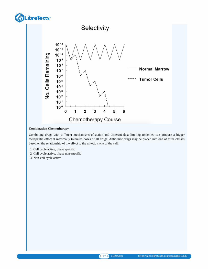
1.17.4 11/24/2021 https://med.libretexts.org/@go/page/10639
Combination Chemotherapy
Combining drugs with different mechanisms of action and different dose-limiting toxicities can produce a biggertherapeutic effect at maximally tolerated doses of all drugs. Antitumor drugs may be placed into one of three classesbased on the relationship of the effect to the mitotic cycle of the cell:
1. Cell cycle active, phase specific2. Cell cycle active, phase non-specific3. Non-cell cycle active
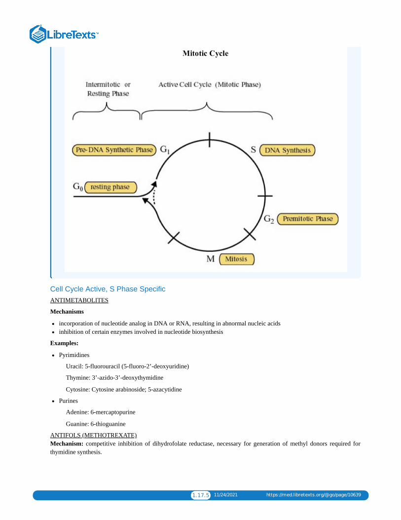
1.17.5 11/24/2021 https://med.libretexts.org/@go/page/10639
Cell Cycle Active, S Phase SpecificANTIMETABOLITES
Mechanisms
incorporation of nucleotide analog in DNA or RNA, resulting in abnormal nucleic acidsinhibition of certain enzymes involved in nucleotide biosynthesis
Examples:
Pyrimidines
Uracil: 5-fluorouracil (5-fluoro-2’-deoxyuridine)
Thymine: 3’-azido-3’-deoxythymidine
Cytosine: Cytosine arabinoside; 5-azacytidine
Purines
Adenine: 6-mercaptopurine
Guanine: 6-thioguanine
ANTIFOLS (METHOTREXATE) Mechanism: competitive inhibition of dihydrofolate reductase, necessary for generation of methyl donors required forthymidine synthesis.

1.17.6 11/24/2021 https://med.libretexts.org/@go/page/10639
Toxicity (general)
interfere with replication of all rapidly proliferating cellsbone marrow --- myelosuppressionGI mucosa ---- diarrhea, stomatitis
Toxicity (specific)
6-mercaptopurine --- cholestatic jaundiceMethotrexate
renal – high doses may block tubules and cause acute renal failure hepatic – usually seen with chronic daily administration of low doses (i.e., psoriasis treatment) CNS – encephalopathy in patients given prior irradiation
Cell Cycle Active, G2/M Phase Specific
BLEOMYCIN
Mechanisms
induces single-strand and double-strand DNA breaksselectivity for G2 phase, but cells in G1 can also be killed
Toxicity
subacute or chronic pneumonitislittle myelosuppression
PLANT ALKALOIDS: VINCRISTINE (VCR), VINBLASTINE (VBL), TAXOL, EPIDOPHYLLOTOXINS (VP-16,VM-26), CAMPTOTHECINS
Mechanisms
Tubulin Binders VCR, VBL inhibit polymerization of tubulin Taxol blocks depolymerization; stabilizes microtubule

1.17.7 11/24/2021 https://med.libretexts.org/@go/page/10639
VP-16, VM-26 target topoisomerase II; non-intercalatingCamptothecins (topotecan, irinotecan) inhibit topoisomerase I
Toxicity
neurotoxicityparesthesiasconstipationdecreased deep tendon reflexesmyelosuppressionSIADH (VCR, VBL)
Cell Cycle Active, Phase Non-SpecificALKYLATING AGENTS
Mechanisms
base alkylation resulting in DNA cross-linkingsingle strand breaksdouble strand breaks and strand misreading
Examples:
Nitrogen MustardCyclophosphamideNitrosoureasCis-platinumBusulfan
Toxicities (general)
myelosuppressionstomatitisnausea/vomitingalopeciaimpaired ovulation and spermatogenesismutagenesis and carcinogenesis
Toxicities (specific)
Cyclosphosphamide: hemorrhagic cystitis, bladder fibrosis, cardiotoxicity (reversible), SIADHBusulfan: interstitial pulmonary fibrosisNitrosoureas: cumulative myelosuppressionPlatinum: acute tubular necrosis, ototoxicity
ANTHRACYCLINES
Mechanisms
intercalate between strands of DNA double helixformation of drug free radicalsinhibition of topoisomerase II
Examples:
daunorubicindoxorubicin (Adriamycin)
Toxicities
myelosuppressionstomatitis

1.17.8 11/24/2021 https://med.libretexts.org/@go/page/10639
cardiotoxicity (irreversible, dose-related)
Non Cell Cycle ActiveCORTICOSTEROIDS
Mechanisms
unclear –induce apoptosis of lymphoblasts and effective in lymphoid malignancieswork via nuclear receptors
Examples
prednisonedexamethasone
Toxicity
typical steroid toxicity –relatively modest in this context
L-ASPARAGINASE (E. COLI, ERWINIA)
Mechanisms
l-asparaginase converts asparagine to aspartate and NH3. Normal cells can reverse this process to form asparagine.Drug has activity in acute lymphocytic leukemia. Lymphoblasts lack asparagine synthetase and die without preformedasparagine in plasma.
Toxicity
Hypersensitivity (urticaria, anaphylaxis)PancreatitisHepatotoxicity
Classification by Important Toxicity1. Renal - Acute Tubular Necrosis
PlatinumStreptozotocinMethotrexate
2. Hepatic6-Mercaptopurine (cholestatic jaundice)L-asparaginase (abnormal liver function tests)Anthracyclines (dependence on biliary excretion)
3. Bladder (hemorrhagic cystitis)Cyclophosphamide
4. Neurotoxicity (paresthesias)Plant alkaloids
5. Pulmonary (interstitial fibrosis)BleomycinBusulfanNitrosoureas (high doses)
6. Cardiac
Anthracyclines (chronic cardiomyopathy)Cyclophosphamide (acute arrhythmias)
7. CarcinogenesisAlkylating agentsProcarbazine

1.17.9 11/24/2021 https://med.libretexts.org/@go/page/10639
8. SIADHVincristineCyclophosphamide
Requirements for Combination Chemotherapy1. Several different drugs, each independently active against a given disease2. Each drug should have
A different mechanism of actionA different dose-limiting toxicity
3. As a result:
Each drug is given at full doseThe rate of cell kill increasesThe chance of emergence of a drug-resistant clone decreases
Examples:
Combination Chemotherapy – ALL
Combination Chemotherapy: Other Curative Regimens
MOPP – Hodgkin’s Disease
M-BACOP – Diffuse Lymphoma
VBP – Testis

1.17.10 11/24/2021 https://med.libretexts.org/@go/page/10639
Adriamycin = doxorubicin Oncovin = vincristine
Tumor Host ResistanceA. Changes in the tumor
1. Location of tumor cellsPharmacologic sanctuaryBlood supply to the anatomical regionExtravascular distance for drug diffusionDrug metabolism by extracellular enzymes and normal cells that surround the tumor
2. Effect of tumor sizeFirst order kineticsPenetration of drugGradients of oxygen, nutrients
3. Growth Characteristics
Mean volume doubling times for certain human tumors
There is an inverse correlation between volume doubling time and response to chemotherapy (slower growth =worse response). Each bar in the graph below represents the average doubling time of a different human metastatictumor:
® ®

1.17.11 11/24/2021 https://med.libretexts.org/@go/page/10639
Gompertzian growth
Relationship between tumor size (A), growth fraction (B), and growth rate (C)
Although growth fraction is maximal at time of initiation of growth, the growth rate is maximal when the tumor isabout 37% of its limiting size.
B. Changes in the Host1. Altered absorption, distribution or excretion of a drug so that less reaches the tumor.2. Increased synthesis of enzymes from non-malignant cells which inactivate the drug.3. Increased sensitivity of normal tissues to the effect of a drug.
Cellular ResistanceA. Natural Drug Resistance – lack of sensitivity of a tumor cell to drugs prior to therapy.B. Acquired Drug Resistance – genotypic and phenotypic changes during therapy that render a tumor cell insensitive to
the lethal effects of a drug.

1.17.12 11/24/2021 https://med.libretexts.org/@go/page/10639
1. Goldie-Coldman Hypothesis: the probability of selecting a single cell with resistance to a specific drug is related toboth population size and mutation frequency. Drug exposure provides the pressure for selection of a resistant cellpopulation.
2. The frequency of drug resistant mutants is increased by antitumor agents which are also mutagens. Heidelberger etal. demonstrated that treatment with chemical mutagens and single-step selection results in the emergence of tumorcells resistant to fluoropyrimidines. The one-step selection following exposure to a mutagen may be relevant to theclinical use of combinations such as CMF.
C. Mechanisms of Acquired Drug ResistanceDecreased Expression of a gene product
Increased expression of a gene product
Expression of an altered gene product

1.17.13 11/24/2021 https://med.libretexts.org/@go/page/10639
D. Acquired Methotrexate Resistance
Acquired MTX resistance has been attributed to a variety of mechanisms. For example, the following have beenidentified in MTX-resistant sublines of a human squamous cell carcinoma (SCC15) established in culture byprogressive dose escalation:
1. Altered transport2. Defective polyglutamylation
Polyglutamate derivatives (MTX-PGs) with 2 to 5 γ-linked glutamyl moieties (MTX-Glu2 to MTX-Glu5) areselectively retained by cells.MTX-PGs have a higher affinity for DHFR, cause prolonged inhibition of DNA synthesis and increasecytotoxicity.
3. Increased production of DHFR (gene amplification)Abnormal homogeneous staining regions (HSRs): sites identified in MTX resistant cells which representamplified DHFR genes on chromosome 2 (mouse) and 5 (human). HSRs are associated with stable resistance.Double minute chromosomes (DMs): small chromosomes of varying size without centromeres, usuallyoccurring in pairs. These chromosomes do not segregate and therefore are lost during the process of celldivision. Gene amplification on the double minute chromosome is thus unstable in the absence of selectingagents.
4. Altered DHFRDHFRs in some resistant cells have a low affinity for MTX.An altered DHFR gene has a mutation in the codon for amino acid 22. This mutation (arginine for leucine)decreases both binding of MTX and function of the enzyme.
E. Multidrug or Pleotropic Resistance
1. Tumor cells exposed to a single drug develop cross-resistance to structurally unrelated compounds with differentmechanisms of action. The affected drugs include a wide spectrum: anthracyclines, vinca alkaloids, actinomycin,podophyllotoxins.
2. Resistant cells have an impaired ability to accumulate and retain drug. Drug efflux is probably more efficient.3. An over-expressed plasma membrane glycoprotein, designated the P- or permeability glycoprotein, with a MW of
170,000 daltons, is consistently found in multidrug- resistant human and animal cell lines, and in transplantabletumors.
Drug resistance is related to the amount of P-glycoproteinCells which regain drug sensitivity no longer express the membrane alteration.Transfer of DNA from drug-resistant cells confers multidrug resistance and plasma membrane glycoproteinexpression.Transfer of multidrug resistance with a cDNA coding for the P-glycoprotein demonstrates that overexpression ofthis single gene is sufficient to confer the resistance phenotype.
4. The P-glycoprotein or MDR1 gene is a member of a small family of genes. Not all members of this family conferthe multidrug-resistant phenotype, thus suggesting that there may be functionally distinct classes of P-glycoproteinisoforms.
5. Double minute chromosomes (DMs) and homogeneous staining regions (HSRs) are found in multidrug resistantlines. The degree of amplified DNA fragments correlates with the degree of drug resistance. The HSRs representamplified P-glycoprotein genes and are reversible.
6. The P-glycoprotein is comprised of 1280 amino acids with 12 hydrophobic segments that act as transmembranedomains. There are two similar domains with cytoplasmic sites that bind ATP. Both domains appear to be required

1.17.14 11/24/2021 https://med.libretexts.org/@go/page/10639
for function. P-glycoprotein is primarily expressed on plasma membranes while smaller amounts have beendetected in the ER and Golgi membranes.
7. Vinblastine photoaffinity analogs bind to the P-glycoprotein, and binding is competitively antagonized by unlabeledvinblastine and anthracycline. The vinblastine protein complex is precipitated by a monclonal antibody to the P-glycoprotein. Moreover, vinblastine is transported by an ATP-dependent process through membranes that contain P-glycoprotein. These findings have established that the P-glycoprotein is a transporter molecule.
The P-glycoprotein is also homologous to bacterial transport proteins which are involved in ATP-dependenttransport of specific molecules, particularly hemolysin, through the bacterial inner cell membrane.
8. High levels of MDR1 gene expression have been found in liver, colon, small intestine, kidney, adrenal cortex andadrenal medulla. The P-glycoprotein is localized in a highly polar fashion on the bile canalicular surface ofhepatocytes, the lumenal surface of the proximal tubule cells of the kidney, and on the lumenal mucosal surface ofintestinal columnar cells. MDR1 RNA levels are usually high in those cancers derived from normal tissues whichthemselves have high MDR1 expression.
9. Intrinsic multidrug resistance appears to be related to persistent expression of a gene involved in normal cellularfunction. Similar biochemical changes occur in multidrug resistant human breast cancer cells and in rat hyperplasticliver nodules that develop resistance to a wide variety of hepatotoxins (Solt Farber model). Mechanisms of de novoresistance to therapy in tumors associated with increased carcinogen exposure (colon, lung cancer) may therefore besimilar to those associated with acquired resistance to anti-neoplastic agents.
10. Drugs such as verapamil, diltiazem and quinidine have been found to overcome multidrug resistance in cell cultureand in some animal experiments. For example, verapamil has been used to reverse adriamycin resistance in humanovarian cancer cells. Verapamil competes with vinblastine for binding, suggesting that drug binding to the P-glycoprotein is a necessary step in the process of drug resistance.
Other agents that reverse P-glycoprotein-mediated multidrug resistance include steroids and steroid antagonists(progesterone and tamoxifen) and reserpine. The most potent chemosensitizers are hydrophobic molecules with abasic nitrogen atom and two planar aromatic rings (termed the “pharmacophore”).
11. Verapamil has been used clinically in an attempt to reverse multidrug resistance. A Phase I-II study failed todemonstrate a potentiation of doxorubicin therapy with verapamil in eight drug-resistant ovarian cancer patients.However, a more recent study has shown a therapeutic benefit for three of eight patients with multiple myeloma andnon-Hodgkin’s lymphoma by adding a continuous infusion of verapamil to the VAD regimen. The three respondingpatients had P-glycoprotein positive tumors. Dose-limiting toxicity of verapamil was hypotension and cardiacarrhythmias. Myelosuppression was not increased in this trial. In this regard, in vitrostudies have demonstrated thatdoxorubicin cytotoxicity is not increased by verapamil in normal human marrow cells. Other clinical studies areunderway using amiodarone and quinidine as inhibitors of P-glycoprotein mediated resistance.
F. Acquired Resistance to Alkylating Agents
Prolonged exposure of human cells to alkylating agents has resulted in a maximum of 10- 15 fold resistance. Thisfinding for alkylating agents is distinct from patterns of resistance seen with other chemotherapeutic agents. Alkylatingagents therefore more closely resemble X-irradiation where significant resistance has not been demonstrated withrepetitive treatment.
ReferenceBast Fr. RC, Kufe DW, Pollack RE, Weichselbaum RR, Holland JF, Frei III E.: Cancer Medicine, 5th Edition. BCDecker, Inc., 2000

1.18.1 12/1/2021 https://med.libretexts.org/@go/page/10640
1.18: Opioid PharmacologyOpium – a mixture of alkaloids from Papaver somniferum. An opiate is a naturally occurring alkaloid, i.e., morphine orcodeine, and an opioid is any natural or synthetic compound, which has morphine-like properties. Hundreds of opioidalkaloids and peptides have been synthesized, but all clinically available opioid analgesics are alkaloids.
Structure-Activity RelationshipsMost opioid analgesics are related to morphine (Figure 16.1). Distinctive features of morphine include 5 rings, 3- and 6-hydroxyl groups (phenolic and alcoholic), piperidine ring with an N-methyl group, and a quaternary carbon at position 13.Morphine is optically active, and only the levorotatory isomer is an analgesic.
Figure 16.1
Simple modifications of morphine make active analgesics, e.g., Codeine is morphine O-methylated at position 3 andgeroin is morphine O-acetylated at positions 3 and 6. Replacing the N-methyl with something larger (allyl, cyclopropyl,cyclobutyl) usually produces a compound with opioid antagonist properties. N-allyl substitution of morphine andoxymorphone produces the antagonists nalorphine and naloxone, respectively. Morphine may be modified extensively, butstill have agonist activity. Meperidine (Demerol) is a synthetic opioid with only fragments of the morphine structure(Figure 16.1).
Opioid Classification
1. Based on intrinsic activityAgonists (morphine, fentanyl)Pure antagonists (naloxone, naltrexone)Mixed agonist-antagonists (nalbuphine, butorphanol)
2. Based on interaction with μ, κ, or δ opioid receptor subtypes
All three receptors have been cloned, and knockout mice created.Each receptor thought to have 2-3 (or more) subtypes, but no distinct gene products have been identified. All belongto the superfamily of G-protein coupled receptors.Most opioid analgesics are relatively selective μ opioid agonists. The various μ effects are discussed below.A few analgesics (pentazocine, nalbuphine, butorphanol) are κ agonists, although they are not highly selective.Experimental selective κ drugs produce analgesia, but also unique effects like diuresis and dysphoria.The selective δ agonists are mainly peptides. Receptor may function permissively with μ receptor (allostericinteraction?).
Endogenous Opioid Peptides
1. Enkephalins include several compounds derived from a large proenkephalin molecule (also called proenkephalin A).
Most important compounds are pentapeptides, methionine- and leucine- enkephalin. Relatively selective δ ligands.Widely distributed in CNSAct like morphine to modulate neurotransmitter release (see p. 3)Found with catecholamines in sympathetic terminals and adrenal.
2. Endorphins (chiefly β-endorphin) are derived from the large precursor molecule pro opiomelanocortin (POMC).POMC also the precursor for ACTH and MSH, which are found together withβ-endorphin.

1.18.2 12/1/2021 https://med.libretexts.org/@go/page/10640
β-endorphin is a 31 amino acid peptide which has analgesic activity in man and animals. It binds preferentially to μreceptors.Localized primarily in pituitary and hypothalamus.
3. Dynorphins are derived from a prodynorphin molecule (also called proenkephalin B).Dynorphin A is a 17 amino-acid peptide which is a potent and highly selective agonist at κ receptors.Similar distribution to the enkephalins.
4. Opioid peptides are located in places which allow them to function as neurotransmitters or neuromodulators.5. Probably modulate pain transmission in the cord and alter acetylcholine release in the myenteric plexus.6. Postulated to play fundamental roles in areas as diverse as hormonal secretion, thermoregulation, and cardiovascular
control.
Opioid Agonists -Pharmacodynamics1. General Mechanisms
Opioids inhibit adenylyl cyclase via interaction with G /G .
Hyperpolarize postsynaptic neurons by increasing outward K+ currents
i 0

1.18.3 12/1/2021 https://med.libretexts.org/@go/page/10640
Act presynaptically to block Ca++ uptake and consequently inhibit neurotransmitter release. Opioids have beenshown to inhibit the release of many neurotransmitters, including substance P, acetylcholine, norepinephrine,glutamate, and serotonin.Opioids produce highly specific depressant and stimulant effects by acting at discrete CNS sites. For example,morphine stimulates the vagal nuclei in the medulla while depressing respiratory centers only a few millimetersaway.The mechanism for neuronal stimulation is often the depression of an inhibitory interneuron .
2. General Clinical Properties
Acute and Chronic Effects of Opioids
All of the clinically-used μ opioid agonists produce these effects.The few qualitative differences between drugs (e.g. histamine release) usually do not involve specific opioidreceptor mechanisms.Opioids differ greatly in physicochemical properties as well as speed of onset and duration of action, so clinicalselection is frequently based on pharmacokinetic considerations.
3. CNS Effectsa. Analgesia and Mood
Mechanisms:
Clinical characteristics:
Processing of pain information is inhibited by a direct spinal effect at the dorsal horn. Probably involvespresynaptic inhibition of the release of tachykinins like substance P.Rostrad transmission of pain signals decreased by activation of descending inhibitory pathways in the brainstem.Emotional response to pain altered by opioid actions on the limbic cortex.Opioids may act at receptors located peripherally on sensory neurons. Possibly important in painful conditionsaccompanied by tissue inflammation.Selective relief of pain at doses which do not produce hypnosis or impair sensation.Typically, patients report that pain is still present, but the intensity is decreased and it no longer bothers them asmuch.Mood elevation, sometimes frank euphoria can occur. Sense of well-being and cloudy detachment thought to bean important reason for opioid abuse.Some types of pain more responsive to opioids than others. More effect in prolonged, burning pain than sharppain of an incision. Neuropathic pain (e.g. pain of nerve root compression) can be very resistant.Relative potencies (see text) usually determined in postoperative pain. Similar data for other pain statesgenerally not available. Actual dose administered will vary greatly from patient to patient.
b. Sedation-Hypnosis
Drowsiness, feelings of heaviness, and difficulty concentrating are common.Sleep may occur with relief of pain, although these drugs are not hypnotics.

1.18.4 12/1/2021 https://med.libretexts.org/@go/page/10640
Most likely to occur in elderly or debilitated patients and in those taking other CNS depressants (EtOH,benzodiazepines).
c. CNS Toxicity
Dysphoria and agitation occur infrequently (incidence higher with meperidine and codeine).Seizures can be produced by meperidine—major metabolite, normeperidine, is a convulsant.Opioids generally avoided in head injury or when elevated intracranial pressure (ICP) is suspected.1. ↓ ventilation can ↑ PaCO2 and raise ICP further.2. Pupil effects may mask changing neurologic signs.
d. Respiratory Depression
Mechanisms:
Clinical Characteristics:
Direct effects on respiratory centers in the medulla.Dose-related depression of ventilatory response to hypercarbia and hypoxia. This shifts CO2 response curve tothe right (see figure).May involve a distinct subset of μ2 receptors.With usual analgesic doses, arterial O2 saturation often decreases.Drive to breathe may be abnormal despite an apparently normal respiratory rate and state of consciousness.Effects are dose related. First CO2 and hypoxic response are depressed, then respiratory rate slows. Very largedoses may cause irregular or periodic breathing and eventually apnea.Trouble most likely to occur with pre-existing pathology (such as hypothyroidism, pulmonary or CNS disease)or previous drug administration (alcohol, general anesthetics, benzodiazepines).Sleep depresses the response to CO2 and potentiates the opioid effect.Respiratory depression is the major toxicity of opioids and nearly always the cause of death from overdose.Equianalgesic doses of all opioids produce equivalent amounts of respiratory depression. There is no convincingevidence than any analgesic is more or less dangerous than morphine in this regard.
Both analgesia and respiratory depression are reduced by administration of an opioid antagonist or by thedevelopment of tolerance. This has two important clinical implications:1. Tolerant individuals who require large amounts of opioid for relief of pain are not at proportionately
increased risk for respiratory depression

1.18.5 12/1/2021 https://med.libretexts.org/@go/page/10640
2. Respiratory depression is difficult to reverse without reversing some analgesia (see "Naloxone").e. Cough Suppression
Depression of cough centers in the medulla (and possibly, the periphery).Different molecular mechanism than analgesia or respiratory depression— cough suppressed by dextro-isomersof opioids (e.g. dextromethorphan), compounds which have no analgesic activity.
f. Pupillary ConstrictionStimulation of Edinger-Westphal (parasympathetic) nucleus of the oculomotor nerve to produce miosis.Pinpoint pupil is a pathognomonic sign of opioid overdose.Antagonized by naloxone, atropine or ganglionic blockers.
g. Nausea and VomitingComplex effects on vomiting centers in the medulla.
Direct stimulation of the chemoreceptor trigger zone (CTZ) in the area postrema on the floor of the fourthventricle. This activates the vomiting center properEmetic effects markedly potentiated by stimulation of the vestibular apparatus, so ambulatory patients are muchmore likely to vomit than those lying quietly.In animals (and man?), very high doses can depress the vomiting center
h. Muscle RigidityLarge i.v. doses can cause generalized stiffness of skeletal muscle. Thought due to μ-mediated increase instriatal dopamine synthesis and inhibition of striatal GABA release.Most common with fentanyl and congeners.May play a role in some overdose fatalities.
4. Cardiovascular Effects
Decrease in central sympathetic tone causes vasodilation and orthostatic hypotension.Effects on both capacitance and resistance vessels.Bradycardia by stimulating central vagal nucleiLittle or no myocardial depression.
5. Histamine ReleaseMorphine, codeine, meperidine cause non-immunologic displacement of histamine from tissue mast cells.Occasionally redness, hives, itching near injection site. Rarely, hypotension, generalized flushing.Not an allergy—true allergic responses to opioids are very rare.Facial itching and warmth are common after opioids—probably a dysesthesia which has nothing to do withhistamine.

1.18.6 12/1/2021 https://med.libretexts.org/@go/page/10640
6. Smooth Muscle Effectsa. Intestine and Stomach
Spasm of smooth muscle all along the GI tract. Both small and large bowel become hypertonic, but rhythmicpropulsive activity is diminished. Delay in intestinal transit time and spasm of the anal sphincter causeconstipation.Delayed gastric emptying. Important because it may slow absorption of oral medications.Mechanism involves both CNS effects and peripheral actions on opioid receptors in the enteric plexus. Smoothmuscle effects of morphine > meperidine > agonist-antagonist opioids.Chronic administration of opioids frequently necessitates the administration of laxatives and stool softeners totreat constipation. Recent evidence that poorly-absorbed quaternary opioid antagonists are also effective inreversing this local effect.Constipating effect is used therapeutically for treatment of diarrhea. Diphenoxylate (in Lomotil) and loperamide(Imodium) are poorly-absorbed opioids that do not produce central effects.
b. Biliary SystemContraction of smooth muscle along the biliary tree and spasm of the sphincter of Oddi.Can precipitate biliary colic on rare occasions.Effect antagonized by naloxone and partially reversed by glucagon, nitroglycerin, or atropine.
c. Urinary TractIncrease contractions of the ureter and tone of the urinary sphincter, but decrease force of detrusor musclecontraction. Decreased attention to full bladder. Can cause urinary retention.Probably both central and peripheral mechanisms involved.
7. Effects on Pregnancy and the Neonate
All cross the placenta.No teratogenic effects, but chronic use may cause physical dependence in utero. Neonatal withdrawal after deliverycan be life-threatening.Opioids given during labor can cause respiratory depression in baby.
8. ToleranceReduction in effect with repeated dosing (or higher dose to produce same effect). First indication usually decreasedduration of analgesia, then decreased intensity. Can be profound.Cross-tolerance to other opioids.Mechanism not known precisely. Involves adaptive response of adenylyl cyclase and/or G protein coupling. Not apharmacokinetic effect.Develops most rapidly to depressant effects like analgesia, respiratory depression, euphoria, but much less toleranceto stimulatory effects like constipation or miosis. This has some important clinical consequences:1. Heroin addicts or methadone maintenance patients may have little euphoria from high doses but continue to
experience constipation and miosis.2. Terminal cancer patients and others requiring high doses for analgesia are also tolerant to respiratory depression
(cf. p. 6), but they frequently require treatment for constipation.9. Physical Dependence
Adaptation which produces stereotyped withdrawal syndrome (abstinence) when drug is stopped. Symptoms stopwhen small dose of opioid is given.Giving antagonist (naloxone) to physically dependent person causes rapid onset of more severe precipitatedabstinence.Withdrawal symptoms include runny nose, vomiting, diarrhea, gooseflesh, mydriasis, shaking chills, drug seekingbehavior.Physical dependence not the same as psychological dependence or addiction. Mild physical dependence may becommon.Addiction produced by appropriate medical treatment is a very unusual event. Irrational fear of addicting patientscited as a frequent cause for inadequate pain treatment.

1.18.7 12/1/2021 https://med.libretexts.org/@go/page/10640
10. Use of Methadone in Opioid Physical Dependence:Opioid Detoxification—patient switched from short-acting opioid to methadone (T1⁄2 = 35 hr) and tapered slowly.Withdrawal symptoms protracted, though mild. Adjuvants like clonidine and sedatives may be helpful.Maintenance—chronic methadone to maintain a state of tolerance and physical dependence. Several putativebenefits:1. ↓ withdrawal symptoms, so drug seeking (& illegal activity) decreased.2. Tolerance develops to opioid euphoria, so injection of illegal heroin is not reinforcing. (Behavior may or may
not decrease.)3. Methadone given orally, so risk of needles reduced.4. Obtaining methadone requires regular contact with caregivers and access to counseling and other treatment.
Opioid Agonists – Pharmacokinetics
Onset and duration most often the basis for selection of an opioid. Huge variation in physicochemical properties andtherefore absorption and distribution throughout the body.
Physicochemical Properties of Some Opioid Agonists
A. Pharmacokinetics of Morphine1. Rapid absorption, wide distribution, and rapid clearance from plasma.2. Clearance mainly by hepatic biotransformation (70% first pass).
Primarily 3-glucuronide (inactive)6-glucuronide. A highly active metabolite, but role in clinical effects is uncertain. May account for opioiddepression reported in renal failure. May also be important with chronic dosing.N-demethylation to normorphine
3. Polar metabolites cleared by kidney.4. Relatively hydrophilic drug, so CNS penetration and exit are slow. This accounts for slow onset and long duration.
Effects lag behind changes in plasma concentrations.B. Pharmacokinetics of Meperidine

1.18.8 12/1/2021 https://med.libretexts.org/@go/page/10640
1. Rapid absorption, wide distribution, and rapid clearance from plasma.2. Clearance mainly by hepatic biotransformation (48-56% first pass).
N-demethylation to normeperidine, oxidation to meperidinic acid or normeperidinic acid.Normeperidine is a CNS stimulant and can produce convulsions in man. Metabolite has T1⁄2 of 8-12 hr sosignificant amounts may accumulate. Toxicity most likely with high doses in renal failure.
C. Pharmacokinetics of Fentanyl
1. Rapid absorption, wide distribution, moderately rapid hepatic clearance2. More than 60% first-pass metabolism to inactive metabolites.3. Extremely lipophilic. Rapidly crosses BBB and other membrane barriers so effects parallel changes in plasma
concentrations.4. Fat solubility means that drug may be administered by multiple routes: useful analgesic effects by transdermal
patch, intranasal spray, and buccal mucosa (fentanyl “lollipop”).
Opioid Agonists – Individualization of Dosage1. Analgesic requirements are enormously variable. Usual adult morphine dose (10 mg) only 70% effective in acute pain.2. Range of effective concentrations (the “therapeutic window”) is narrow for each patient but varies widely between
patients. Implication: “cookbook” analgesia likely to be inadequate or excessive much of the time.3. Lower starting doses for elderly, hypovolemic, debilitated, hypothyroid or those given other CNS depressants.4. Do not be afraid to give adequate treatment to patients who have become highly tolerant.5. Watch for accumulation of parent drug and/or metabolites in hepatic or renal failure.
Opioid Antagonists1. Naloxone
Pure, competitive antagonist at μ, κ, and δ receptors (highest affinity at μ)Given alone, almost no effect. Some behavioral effects in animals.Rapidly reverses opioid overdose, but effect short due to redistribution. Patient may become renarcotized.
2. Naltrexone
Used orally in high doses to treat detoxified heroin addicts (blocks euphoria from injected heroin).Effects primarily from active metabolite, 6-β-naltrexol.
Opioid Agonist-Antagonists1. Developed in search for less abusable potent analgesics.2. All have analgesic (agonist) properties as well as ability to antagonize morphine effects.3. Two basic mechanisms:
Partial agonists at μ receptor. Buprenorphine has high affinity, but limited efficacy at μ receptor. Given alone, it hasmorphine-like effects. Competes effectively with agonists like morphine and may reduce effect.Agonists/Partial agonists at κ receptor. Nalorphine, pentazocine, nalbuphine, butorphanol act as κ agonists (probablyκ3) to produce analgesia. Also act as competitive antagonists at μ receptors (high affinity but no efficacy at thisreceptor).
1. Clinical properties:
Potent analgesics effective in moderate to severe pain.Relatively limited toxicity (respiratory dep., smooth muscle)Decreased abuse potential, but also decreased patient acceptance (mood elevation may be clinically important!).Occasional dysphoria or hallucination with κ agonistsAntagonist properties mean they can precipitate withdrawal in patients already receiving chronic treatment with opioidagonists.
1. Neither agonist vs. antagonist potency nor μ/κ selectivity seem to predict clinical utility or patient acceptance.

Index
AAnticholinesterase
1.5: Case Study - Anticholinesterase Anticonvulsants
1.15: Neuropharmacology III - Anticonvulsants Antidepressants
1.14: Neuropharmacology II - Anxiolytics andAntidepressants Antidysrhythmics
1.7: Local Anesthetics Antiinflammatory Drugs
1.8: Antiinflammatory Drugs Anxiolytics
1.14: Neuropharmacology II - Anxiolytics andAntidepressants atherosclerosis
1.11: Lipid Lowering Drugs - Hyperlipidemiaand Atherosclerosis Autonomic Pharmacology
1.6: Autonomic Pharmacology
BBacteriocidal therapy
1.16: Antimicrobials I and II
CChemotherapy
1.17: Chemotherapy congestive heart failure
1.10: Vasoactive Drugs II - Heart Failure
Ddepression
1.14: Neuropharmacology II - Anxiolytics andAntidepressants
EED50
1.2: Introduction to Pharmacology
Hhypertension
1.9: Vasoactive Drugs I
IImmunosuppression
1.1: Immunosuppression for Solid OrganTransplantation
LLipid Lowering Drugs
1.11: Lipid Lowering Drugs - Hyperlipidemiaand Atherosclerosis Local Anesthetics
1.7: Local Anesthetics
Nnitric oxide
1.13: Nitric Oxide
Oopiates
1.18: Opioid Pharmacology opioids
1.18: Opioid Pharmacology
PPharmacokinetics
1.3: Pharmacokinetics I pharmacology
1.2: Introduction to Pharmacology
VVasoactive Drugs
1.9: Vasoactive Drugs I

Glossary
Sample Word 1 | Sample Definition 1

Glossary
Sample Word 1 | Sample Definition 1


