ORIGINAL ARTICLE Phenolic composition and medicinal usage of Psidium guajava Linn.: Antifungal activity or inhibition of virulence? Maria F.B. Morais-Braga a, * , Joara N.P. Carneiro a , Antonio J.T. Machado a , De´bora L. Sales a , Antonia T.L. dos Santos a , Aline A. Boligon c , Margareth L. Athayde c,1 , Irwin R.A. Menezes b , Djair S.L. Souza d , Jose´ G.M. Costa b , Henrique D.M. Coutinho b a Department of Biological Sciences, Regional University of Cariri, Crato, Ceara ´, Brazil b Department of Biological Chemistry, Regional University of Cariri, Crato, Ceara ´, Brazil c Department of Industrial Pharmacy, Federal University of Santa Maria, Santa Maria, Rio Grande do sul, Brazil d ESAM, Federal University of the Semi Arid, Mossoro ´, Rio Grande do Norte, Brazil Received 20 May 2015; revised 15 September 2015; accepted 22 September 2015 Available online 30 September 2015 KEYWORDS Fungistatic effect; Inhibition of dimorphism; Tea; Tincture Abstract Psidium guajava is a Myrtaceae plant whose medicinal properties are recognized in sev- eral locations. The use of teas and tinctures prepared from their leaves has been used to combat infections caused by fungi of the genus Candida. In this study, aqueous extracts of leaves and hydroethanolic were tested to verify the antifungal potential and its chemical composition has been investigated. The microbiological assays were performed by broth microdilution to determine the minimum inhibitory concentration (MIC) and from these the minimum fungicidal concentration was performed (MFC) by subculturing on solid media. A cell viability curve was obtained for demonstration of inhibition of fungal growth of strains of Candida albicans and Candida tropicalis. Tests to check morphological changes by the action of the extracts were performed in microcultive cameras depleted environment at concentrations of MIC/2, MIC and MIC 2. Extracts analyzed by high performance liquid chromatography demonstrated flavonoids and phenolic acids. The extracts showed fungistatic effect and no fungicide with MIC >8192 lg/mL, MFC above 8192 lg/mL. The IC 50 was calculated ranging from 1803.02 to 5623.41 lg/mL. It has been found * Corresponding author. Tel.: +55 88 3102 1212. E-mail address: fl[email protected](M.F.B. Morais-Braga). 1 In memorian. Peer review under responsibility of King Saud University. Production and hosting by Elsevier Saudi Journal of Biological Sciences (2017) 24, 302–313 King Saud University Saudi Journal of Biological Sciences www.ksu.edu.sa www.sciencedirect.com http://dx.doi.org/10.1016/j.sjbs.2015.09.028 1319-562X Ó 2015 The Authors. Production and hosting by Elsevier B.V. on behalf of King Saud University. This is an open access article under the CC BY-NC-ND license (http://creativecommons.org/licenses/by-nc-nd/4.0/).
Transcript
Saudi Journal of Biological Sciences (2017) 24, 302–313
King Saud University
Saudi Journal of Biological Sciences
www.ksu.edu.sawww.sciencedirect.com
ORIGINAL ARTICLE
Phenolic composition and medicinal usage
of Psidium guajava Linn.: Antifungal activity
or inhibition of virulence?
* Corresponding author. Tel.: +55 88 3102 1212.
E-mail address: [email protected] (M.F.B. Morais-Braga).1 In memorian.
Peer review under responsibility of King Saud University.
Production and hosting by Elsevier
http://dx.doi.org/10.1016/j.sjbs.2015.09.0281319-562X � 2015 The Authors. Production and hosting by Elsevier B.V. on behalf of King Saud University.This is an open access article under the CC BY-NC-ND license (http://creativecommons.org/licenses/by-nc-nd/4.0/).
Maria F.B. Morais-Bragaa,*, Joara N.P. Carneiro
a, Antonio J.T. Machado
a,
Debora L. Sales a, Antonia T.L. dos Santos a, Aline A. Boligon c,
aDepartment of Biological Sciences, Regional University of Cariri, Crato, Ceara, BrazilbDepartment of Biological Chemistry, Regional University of Cariri, Crato, Ceara, BrazilcDepartment of Industrial Pharmacy, Federal University of Santa Maria, Santa Maria, Rio Grande do sul, BrazildESAM, Federal University of the Semi Arid, Mossoro, Rio Grande do Norte, Brazil
Received 20 May 2015; revised 15 September 2015; accepted 22 September 2015
Available online 30 September 2015
KEYWORDS
Fungistatic effect;
Inhibition of dimorphism;
Tea;
Tincture
Abstract Psidium guajava is a Myrtaceae plant whose medicinal properties are recognized in sev-
eral locations. The use of teas and tinctures prepared from their leaves has been used to combat
infections caused by fungi of the genus Candida. In this study, aqueous extracts of leaves and
hydroethanolic were tested to verify the antifungal potential and its chemical composition has been
investigated. The microbiological assays were performed by broth microdilution to determine the
minimum inhibitory concentration (MIC) and from these the minimum fungicidal concentration
was performed (MFC) by subculturing on solid media. A cell viability curve was obtained for
demonstration of inhibition of fungal growth of strains of Candida albicans and Candida tropicalis.
Tests to check morphological changes by the action of the extracts were performed in microcultive
cameras depleted environment at concentrations of MIC/2, MIC and MIC � 2. Extracts analyzed
by high performance liquid chromatography demonstrated flavonoids and phenolic acids. The
extracts showed fungistatic effect and no fungicide with MIC >8192 lg/mL, MFC above
8192 lg/mL. The IC50 was calculated ranging from 1803.02 to 5623.41 lg/mL. It has been found
Phenolic composition and medicinal usage of Psidium guajava Linn 303
that the extracts affect the morphological transition capability, preventing the formation of pseudo-
hyphae and hyphae. Teas and tinctures, therefore, have the potential antifungal, by direct contact,
causing inhibition of fungal multiplication and its virulence factor, the cell dimorphism, preventing
tissue invasion. Further studies are needed to elucidate the biochemical pathways and genes assets
involved in these processes.
� 2015 The Authors. Production and hosting by Elsevier B.V. on behalf of King Saud University. This is
an open access article under theCCBY-NC-ND license (http://creativecommons.org/licenses/by-nc-nd/4.0/).
1. Introduction
Microorganisms of the genus Candida can be found naturallycomposing the microbiota of the human organism inhabiting
your gastrointestinal tract and mucous membranes (Lu et al.,2014; Shao et al., 2007). Changes in dynamic of the host organ-ism too favor the proliferation of these fungi and the distur-
bance caused in homeostasis can lead to a range ofinfections that range in their level and location and can onlybe superficial, in skin and mucosal (oral, vaginal candidiasis)
or systemic, compromising the life of an individual (Mayeret al., 2013; Sardi et al., 2013).
Usually the infections caused by Candida spp. in its magni-
tude are assigned to the species Candida albicans, however, ill-ness caused by Candida non-albicans (CNAM) had increasedincidence over the years and yeasts of Candida glabrata, Can-dida tropicalis, Candida krusei and Candida parapsilosis have
been increasingly identified as human pathogens (Sardi et al.,2013; Silva et al., 2012).
Mechanisms of resistance to commercial drugs developed
by these microorganisms have been constantly investigatedand reported and the continuous evolution for resistance isextremely worrying considering the limited number of antifun-
gal classes currently available (Maubon et al., 2014; Xie et al.,2014). The search for different therapeutic alternatives is aconstant and the use of natural products of plant origin oftenserves as a reference to the search for active compounds and, in
this sense, a ethno directed approach has directed pharmaceu-tical research (Albuquerque and Hanazaki, 2006), in this case,in order to antifungal discovery potential.
Member of the Myrtaceae, Psidium guajava Linn. species(guava), plant native to tropical America (Okamoto et al.,2009), has its widespread medicinal use among the world’s
populations. Being a plant of tropical and subtropical regions,can be found on plantations, in backyards of homes, or natu-rally in other areas, and could even be considered invasive spe-
cies (Richardson and Rejmanek, 2011).The medicinal attributes from all parts of the species are
spread over several generations and therefore, make up manylists of ethnobotanical studies, showing great versatility and
value in use, being mentioned for the treatment of varioustypes of diseases (Dakappa-Shruthi et al., 2013; Gutierrezet al., 2008).
This therapeutic use recorded in different locations includesa significant number of body systems such as disorders of thesensory system: vertigo (Dakappa-Shruthi et al., 2013;
Gutierrez et al., 2008); disorders of the respiratory system:laryngitis, sore throat, colds, coughs, tuberculosis, lung prob-lems, bronchitis, catarrh, rhinitis (Dakappa-Shruthi et al.,
2013; Gutierrez et al., 2008; Ogbole and Ajaiyeoba, 2010;Waruruai et al., 2011); disorders of the genito-urinary system:menstrual disorders, vaginal discharge, childbirth, nephritis,
premenstrual syndrome, gonorrhea, non-specified venereal dis-eases, leucorrhea (Dakappa-Shruthi et al., 2013; Gutierrezet al., 2008; Van Vuuren and Naidoo, 2010); disorders of the
nervous system: anorexia, epilepsy, cerebral ailments, chorea,convulsions, nervousness (Dakappa-Shruthi et al., 2013;Gomez-Estrada et al., 2011; Gutierrez et al., 2008); disorders
of the digestive system: diarrhea, dysentery, stomachache,digestive problems, gastric insufficiency, ulcers, dyspepsia, gas-troenteritis, gastritis, bowel disorders, colic, toothache, consti-pation (Dakappa-Shruthi et al., 2013; Gomez-Estrada et al.,
2011; Gutierrez et al., 2008); disorders of the circulatory sys-tem: piles, swelling, hypertension, edema (Dakappa-Shruthiet al., 2013; Gutierrez et al., 2008); the musculoskeletal system
and connective tissue diseases: gout, spasm, rheumatic pain(Dakappa-Shruthi et al., 2013; Gutierrez et al., 2008); notdefined conditions or pain not defined: aches (Dakappa-
Shruthi et al., 2013); skin diseases and tissue subcutaneous:inflamed mucous membranes, mouth – swelling, skin problems,ulcers, itch, scabies, skin sores, wounds, dermatosis, sores, boil,gingivitis (Dakappa-Shruthi et al., 2013; Gutierrez et al., 2008);
diseases of the endocrine glands, nutrition andmetabolism: dia-betes (Gutierrez et al., 2008); infectious and parasitic diseases:cholera, worms, bacterial infections, herpes, mycoses, thrush,
pox, measles (Dakappa-Shruthi et al., 2013; Gutierrez et al.,2008; Waruruai et al., 2011); neoplasms: cancer (Alonso-Castro et al., 2011), and disease of the blood and blood-
forming organs: hemorrhages, blood cleansing (Dakappa-Shruthi et al., 2013; Gutierrez et al., 2008).
The P. guajava is popularly used in the treatment of infec-
tious diseases, particularly against those caused by fungi, it iscommon practice registered in different countries such as Bra-zil, Cuba and South Africa where it is used to treat thrush, leu-corrhoea, and vaginitis, pathologies associated with infections
caused by Candida spp. (Borba and Macedo, 2006; Fenneret al., 2006; Oliveira et al., 2010; Ramırez et al., 2007; VanVuuren and Naidoo, 2010).
Considering the pharmacological potential of the species P.guajava described in ethnobotanical reports, especially withregard to its therapeutic use in treatments against diseases
caused by fungi, this study aims to scientifically validate theantifungal properties of tea and tincture prepared with leavesof guava and evaluate the effect of natural products in viru-
lence strains of C. albicans and C. tropicalis, particularly itsmorphological transition process.
2. Material and methods
2.1. Collection area
The collection was realized in the rainy season at the county ofMilagres, Ceara, Northeastern region of Brail (07� 17.1190 S
and 038� 51.7790 W, 388 m of altitude; 07� 17.1200 S and038� 51.7780 W, 389 m of altitude; 07� 17.1220 S and 038�51.7760 W, 392 m of altitude; 07� 17.1190 S and 038� 51.7790
W, 388 m of altitude) at ‘‘Sıtio” Malhada. The climate issemi-arid, with temperatures ranging between 24� and 26 �C(IPECE, 2013).
2.2. Plant material
The study was conducted using young, healthy leaves of a
Psidium species locally known as red guava, which were col-lected and transported to the Laboratory of Microbiologyand Molecular Biology at the Regional University of Cariri
– URCA. Twigs with flowers of the species were also collectedand vouchers were produced and deposited in the HerbariumDardano de Andrade Lima at the university under No.10935, where the species was identified as Psidium guajava
Linn. The collection period included January, February,March and April, known as the ‘‘wintry block of the CaririCeara region.” Collections were made between 8:30 and
10:30 am, and the plant material was taken to the laboratorywhere it was subjected to qualitative screening and cleaningbefore being weighed and stored under refrigeration. Alto-
gether, there was 2650 g of leaves in perfect condition, and thisquantity was divided for preparation of three types of extracts:Aqueous Extract of P. guajava Infusion (AEPGI), AqueousExtract of P. guajava Decoction (AEPGD) and: Hydroethano-
lic Extract of P. guajava (HEPG).
2.3. Preparation of extracts
2.3.1. Aqueous extracts
Two types of aqueous extracts were prepared, each using
399.9 g of leaves mixed with 6 L of water (based on a propor-tion of 10 g/150 mL, equivalent to one cup of tea – 150 cc).The decoction was made by mixing roughly cut leaves in cold
water and then boiling for 15 min. Afterward, the tea wasallowed to cool, filtered and then stored under refrigeration.As for the infusion, the water was boiled without leaves,which were placed in the water after turning off the heat.
The pot was covered with a lid and allowed to stand untilthe tea cooled down (Matos, 2002), and the preparationwas then filtered and stored under refrigeration and both
infusion and decoction were frozen (�60 �C) and lyophilizedto dryness. The powdered extracts were stored under refriger-ation for testing, using 14.46 g (yield 3.62%) and 15 g (yield
3.75%) extract powder from the decoction and infusion,respectively.
2.3.2. Hydroethanolic extract
The hydroethanolic extract (70%) was prepared by triturationwith cold extraction, using a total of 1846.5 g leaves in a pro-portion of 5 g/mL of hydroethanolic solution (Matos, 2002).
The leaves were cut to increase contact surface with the sol-vent, and the mixture was left at room temperature protectedfrom air and light, for a period of 96 h for maximum extrac-tion efficiency. The mixture was then filtered and placed in a
rotary evaporator (Q-344B – Quimis – Brazil) at 40 rpm and60 �C to concentrate the extract. Finally, the crude extractwas frozen, lyophilized (50.8 g – yield 2.75%) and then stored
under refrigeration.
2.4. Chemical analysis
2.4.1. Chemical, apparatus and general procedures
All chemicals were of analytical grade. Methanol, acetic acid,
gallic acid, caffeic acid and chlorogenic acid were purchasedfrom Merck (Darmstadt, Germany). Quercetin, quercitrin, iso-quercitrin, rutin, kaempferol, luteolin, catechin and epicate-chin were acquired from Sigma Chemical Co. (St. Louis,
MO, USA). High performance liquid chromatography(HPLC–DAD) was performed with a Shimadzu ProminenceAuto Sampler (SIL-20A) HPLC system (Shimadzu, Kyoto,
Japan), equipped with Shimadzu LC-20AT reciprocatingpumps connected to a DGU 20A5 degasser with a CBM20A integrator, SPD-M20A diode array detector and LC solu-
tion 1.22 SP1 software.
2.4.2. High performance liquid chromatography with diode arraydetection (HPLC–DAD)
Reverse phase chromatographic analyseswere carried out undergradient conditions using C18 column (4.6 mm � 250 mm)packed with 5 lm diameter particles; the mobile phase was
water containing 2% acetic acid (A) and methanol (B), andthe composition gradient was: 5% (B) for 2 min; 25% (B) until10 min; 40, 50, 60, 70 and 80% (B) every 10 min; following themethod described by Silva et al. (2014) with slightmodifications.
P. guajava extracts (hydroethanolic – EHEPG, infusion – EAIPGand decoction – EADPG) and mobile phase were filteredthrough 0.45 lmmembrane filter (Millipore) and then degassed
by ultrasonic bath prior to use, the extracts of P. guajava wereanalyzed at a concentration of 20 mg/mL. The flow rate was0.6 mL/min and the injection volume was 50 lL. The sample
andmobile phase were filtered through 0.45 lmmembrane filter(Millipore) and then degassed by ultrasonic bath prior to use.Stock solutions of standard references were prepared in
water: methanol (1:1; v/v) at a concentration range of0.025–0.300 mg/mL catechin, epicatechin, quercetin, quercitrin,isoquercitrin, kaempferol, luteolin and rutin, and 0.035–0.300 mg/mL for gallic, caffeic and chlorogenic acids. Quantifi-
cation was carried out by integration of the peaks using theexternal standard method, at 254 nm for gallic acid, 281 nmfor catechin and epicatechin, 327 nm for chlorogenic and caffeic
acids, and 366 for quercetin, quercitrin, isoquercitrin, luteolin,kaempferol and rutin. The chromatography peaks were con-firmed by comparing its retention time with those of reference
(r = 0.9990). All chromatography operations were carried outat ambient temperature and in triplicate.
2.4.3. Limit of detection (LOD) and limit of quantification(LOQ)
LOD and LOQ were calculated based on the standard devia-tion of the responses and the slope using three independent
Phenolic composition and medicinal usage of Psidium guajava Linn 305
analytical curves, as defined by Kamdem et al. (2013). LODand LOQ were calculated as 3.3 and 10 r/S, respectively,where r is the standard deviation of the response and S is
the slope of the calibration curve.
2.5. Antifungal assay
2.5.1. Strains and culture media used
Standard types of strains were obtained from the Culture Col-
lection of Oswaldo Cruz of the Brazilian Institute of QualityControl in Health (INCQS) and clinical isolates of the yeastsC. albicans and C. tropicalis were provided by Dr. Edeltrudes
Oliveira Lima (Mycology Laboratory of Paraıba FederalUniversity), namely CA INCQS 40006, CA LM 62, CA LM77, CA LM 109, CA LM 111, CA LM 122, CT INCQS40042, CT LM 18, CT LM 20 and CT LM 23. These strains
were inoculated into Sabouraud Dextrose Agar (SDA,KASVI) and incubated for 24 h at 37 �C. Afterward, small ali-quots of yeast were transferred to test tubes each containing
3 mL of sterile saline (0.9%). Using the McFarland scale, theconcentration of the inoculum was standardized by comparingits turbidity with the 0.5 standard, giving a standard yeast sus-
pension of 1 � 105 cells/mL (NCCLS, 2002). The inocula thusprepared were used to determine the minimum inhibitory con-centration (MIC) in Sabouraud Dextrose Broth (SDB, HIME-DIA), double concentrated. Another culture medium was used
for analysis of yeast micromorphology. The potato dextroseagar (PDA, DIFCO) was prepared by diluting it more thanthat recommended by the manufacturer to make it a depleted
medium capable of stimulating yeast to produce hyphae. Agarwas added to this diluted medium to obtain a solid medium.
2.5.2. Drugs, reagents and preparation of solutions
Dimethyl sulfoxide (DMSO, Merck, Darmstadt, Germany)was used for dilution of the extracts, and the antifungal
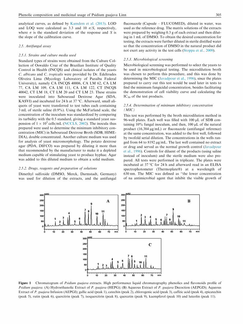
Figure 1 Chromatogram of Psidium guajava extracts. High perform
Psidium guajava. (A) Hydroethanolic Extract of P. guajava (HEPG); (
Extract of P. guajava Infusion (AEPGI); gallic acid (peak 1), catechin (p
fluconazole (Capsule – FLUCOMED), diluted in water, wasused as the reference drug. The matrix solutions of the extractswere prepared by weighing 0.3 g of each extract and then dilut-
ing in 1 mL of DMSO. To obtain the desired concentration fortesting, the extracts were further diluted in sterile distilled waterso that the concentration of DMSO in the natural product did
not exert any activity in the test cells (Stoppa et al., 2009).
2.5.3. Microbiological screening
Microbiological screening was performed to select the yeasts to
be used in microbiological testing. The microdilution brothwas chosen to perform this procedure, and this was done bydetermining the MIC (Javadpour et al., 1996), since the plates
prepared to carry out this test would be used later in tests tofind the minimum fungicidal concentration, besides facilitatingthe demonstration of cell viability curve and calculating the
IC50 of the test products.
2.5.4. Determination of minimum inhibitory concentration(MIC)
This test was performed by the broth microdilution method in96-well plates. Each well was filled with 100 lL of SDB con-taining 10% fungal inoculum, and then, 100 lL of the natural
product (16,384 lg/mL) or fluconazole (antifungal reference)at the same concentration, was added to the first well, followedby twofold serial dilution. The concentrations in the wells ran-ged from 64 to 8192 lg/mL. The last well contained no extract
or drug and served as the normal growth control (Javadpouret al., 1996). Controls for diluent of the products (using salineinstead of inoculum) and the sterile medium were also pre-
pared. All tests were performed in triplicate. The plates wereincubated at 37 �C for 24 h and afterward read in an ELISAspectrophotometer (Thermoplate�) at a wavelength of
630 nm. The MIC was defined as ‘‘the lower concentrationof na antimicrobial agent that inhibit the visible growth of
ance liquid chromatography phenolics and flavonoids profile of
B) Aqueous Extract of P. guajava Decoction (AEPGD); Aqueous
AEPGI: Aqueous Extract of P. guajava Infusion; AEPGD: Aqu-
eous Extract of P. guajava Decoction; HEPG: Hydroethanolic
Extract of P. guajava: INCQS: Instituto Nacional de Controle de
Qualidade em Saude; LM: Laboratorio de Micologia.
306 M.F.B. Morais-Braga et al.
na microorganism in dilution assays” (CLSI, 2002). The resultsobtained in the ELISA readout were used to construct the cell
viability curve and the IC50 of the extracts of P. guajava.
2.5.5. Determination of minimum fungicidal concentration
(CFM)
For this test, a small sterile rod was placed in each well of theMIC test plate (except for sterility control). After mixing themedium in each well, the rod was taken to a large petri dish
containing SDA, streaking its surface and transferring thesolution (medium + inoculum + natural product) for subcul-ture of yeast and checking cell viability. After 24 h incubation,
the plates were inspected for any formation of colonies of Can-dida (Ernst et al., 1999, with modifications). The concentrationat which there was no growth of fungal colonies was consid-ered the MFC of the natural product.
2.5.6. Effect of natural products on fungal morphology
To determine if the natural product caused any change in fun-
gal morphology, by inhibiting the development of hyphae,sterile micromorphological chamber slides were prepared for
observation of yeasts. Three milliliters of PDA mediumdepleted by dilution were added to chambers, containing the
natural product concentrations MIC/2, MIC and MIC � 2.Aliquots of the inoculi were taken from the petri dishes tomake two parallel steaks on the solid medium, which were then
covered with a sterile coverslip. The chambers were placed inthe incubator for 24 h (37 �C) and inspected under a lightmicroscope using a 40� objective. A camera was attached to
the microscope to capture images. A control for yeast growth(hyphae stimulated by depleting medium) was performed, aswell as a control with the conventional antifungal fluconazolefor comparative purposes and a control with DMSO at 100%
and 0.5% (the concentration in the natural products used inthe tests). The assays were performed according to Sidrinand Rocha (2010) and Mendes (2011), with some
modifications.
2.6. Statistical analysis
The results of the tests were done in triplicate. Data obtainedfor each sample and concentration were checked for their nor-mal distribution and then analyzed by one-way ANOVA by
post hoc Tukey test. EC50 values were obtained by nonlinearregression for the purpose of interpolating values from stan-dard curves (using the software Graphpad Prism, v. 5.0) ofthe % growth values plotted against concentration and EC50
values are expressed as lg/mL.
3. Results and discussion
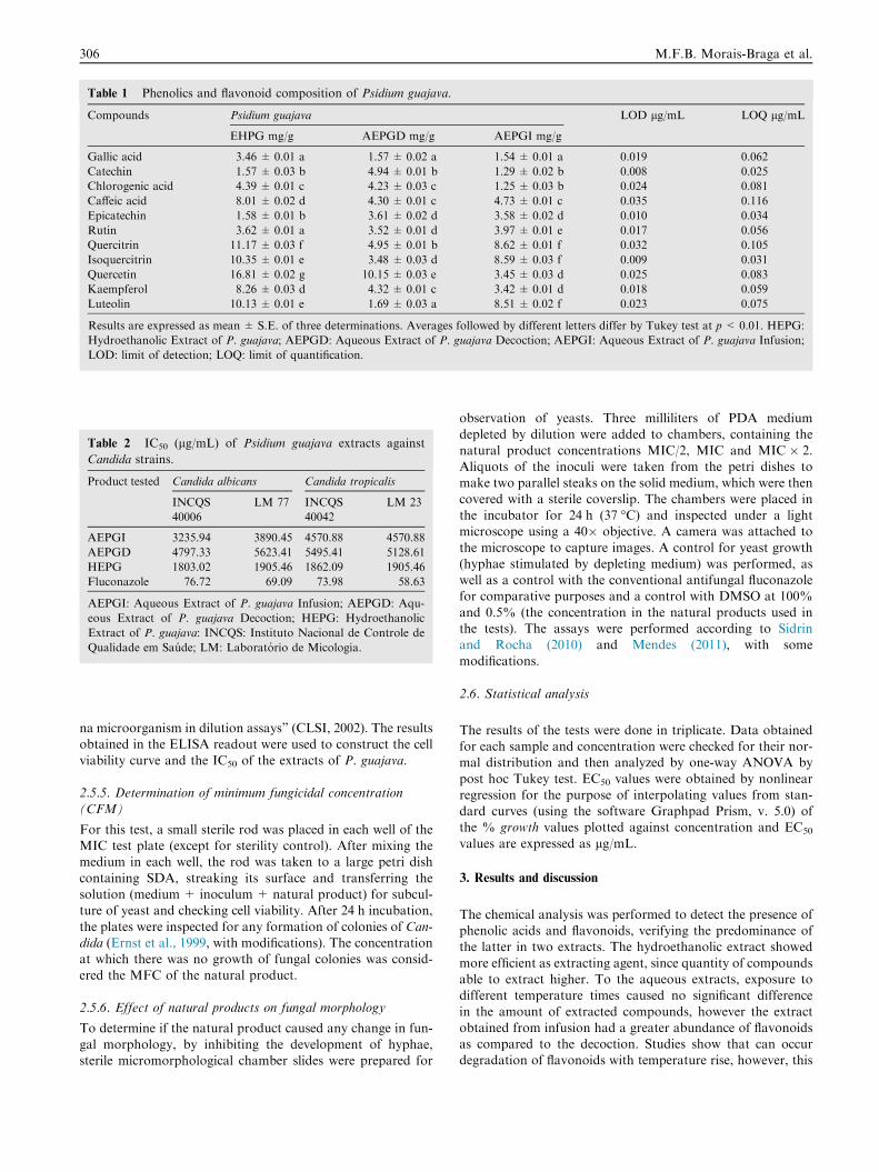
The chemical analysis was performed to detect the presence ofphenolic acids and flavonoids, verifying the predominance ofthe latter in two extracts. The hydroethanolic extract showed
more efficient as extracting agent, since quantity of compoundsable to extract higher. To the aqueous extracts, exposure todifferent temperature times caused no significant difference
in the amount of extracted compounds, however the extractobtained from infusion had a greater abundance of flavonoidsas compared to the decoction. Studies show that can occurdegradation of flavonoids with temperature rise, however, this
Phenolic composition and medicinal usage of Psidium guajava Linn 307
process also depends on the chemical structure and the interac-tion between them (Baby et al., 2007; Mello et al., 2010). Inthis sense, the decoction longer exposure to elevated tempera-
ture may have been the cause of the reduction of the level offlavonoids. The major compound differed only in the analysisof each extract into the aqueous infusion made, which in this
case is the quercitrin, while the other the quercitrin appearsto be the most expressive content. The chromatogram extractsof the species are shown in Fig. 1, and the results representing
their chromatographic profiles in front the parameters used aredetailed in Table 1.
The chemical composition of P. guajava has been widelyinvestigated and studies have reported that plant extracts are
constituted alkaloids, triterpenoids, tannins, saponins, glyco-sides, flavonoids, and phenolic compounds and other com-pounds (Dakappa-Shruthi et al., 2013; Tambe et al., 2014).
A chemical analysis of tea from the leaves of guava (Changet al., 2013) highlighted a polyphenol profile in which the maincomponents were found: quercetin, myricetins, catechin, gallic
and ellagic acids and their derivatives, but the researchers callattention to differences in chemical composition, stating thatseveral factors such as time of collection, form of collection
and processing, temperature, among others, may influencethe outcome of the chemical prospecting.
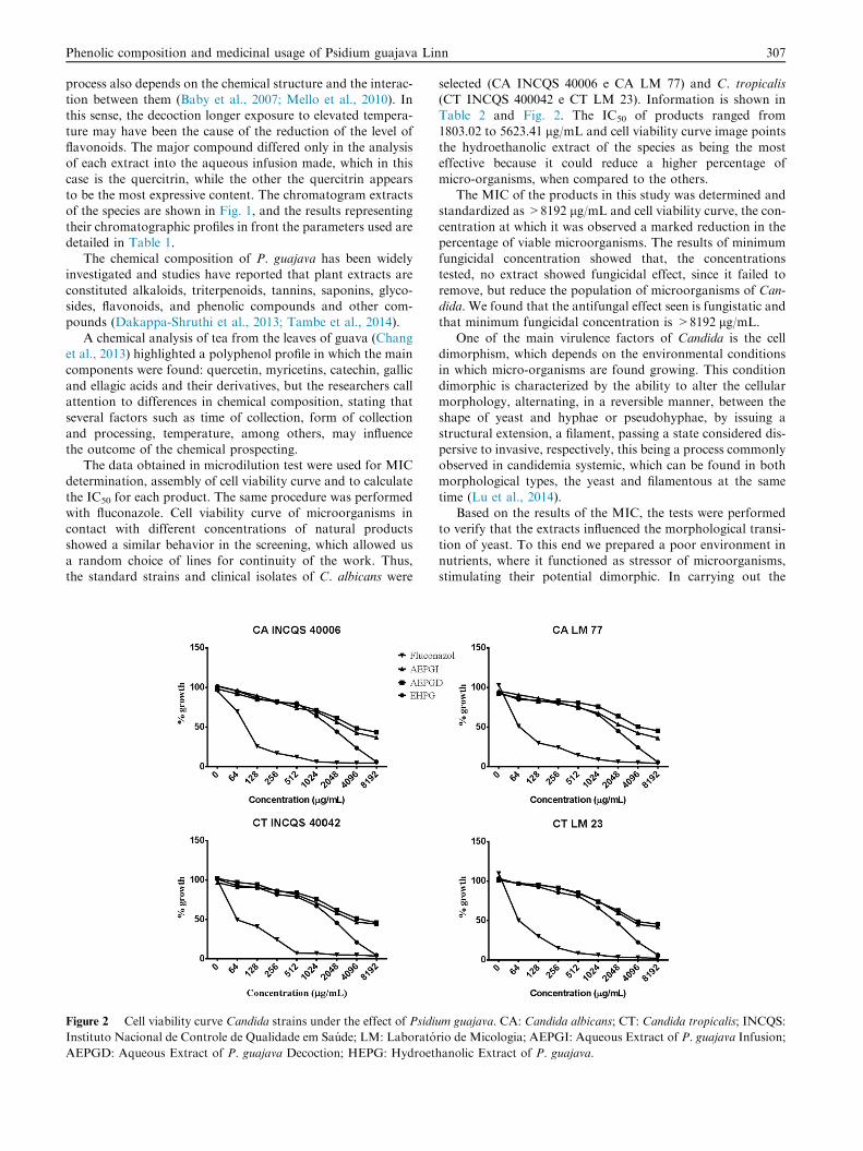
The data obtained in microdilution test were used for MICdetermination, assembly of cell viability curve and to calculate
the IC50 for each product. The same procedure was performedwith fluconazole. Cell viability curve of microorganisms incontact with different concentrations of natural products
showed a similar behavior in the screening, which allowed usa random choice of lines for continuity of the work. Thus,the standard strains and clinical isolates of C. albicans were
Figure 2 Cell viability curve Candida strains under the effect of Psidiu
Instituto Nacional de Controle de Qualidade em Saude; LM: Laborato
AEPGD: Aqueous Extract of P. guajava Decoction; HEPG: Hydroeth
selected (CA INCQS 40006 e CA LM 77) and C. tropicalis(CT INCQS 400042 e CT LM 23). Information is shown inTable 2 and Fig. 2. The IC50 of products ranged from
1803.02 to 5623.41 lg/mL and cell viability curve image pointsthe hydroethanolic extract of the species as being the mosteffective because it could reduce a higher percentage of
micro-organisms, when compared to the others.The MIC of the products in this study was determined and
standardized as >8192 lg/mL and cell viability curve, the con-
centration at which it was observed a marked reduction in thepercentage of viable microorganisms. The results of minimumfungicidal concentration showed that, the concentrationstested, no extract showed fungicidal effect, since it failed to
remove, but reduce the population of microorganisms of Can-dida. We found that the antifungal effect seen is fungistatic andthat minimum fungicidal concentration is >8192 lg/mL.
One of the main virulence factors of Candida is the celldimorphism, which depends on the environmental conditionsin which micro-organisms are found growing. This condition
dimorphic is characterized by the ability to alter the cellularmorphology, alternating, in a reversible manner, between theshape of yeast and hyphae or pseudohyphae, by issuing a
structural extension, a filament, passing a state considered dis-persive to invasive, respectively, this being a process commonlyobserved in candidemia systemic, which can be found in bothmorphological types, the yeast and filamentous at the same
time (Lu et al., 2014).Based on the results of the MIC, the tests were performed
to verify that the extracts influenced the morphological transi-
tion of yeast. To this end we prepared a poor environment innutrients, where it functioned as stressor of microorganisms,stimulating their potential dimorphic. In carrying out the
m guajava. CA: Candida albicans; CT: Candida tropicalis; INCQS:
rio de Micologia; AEPGI: Aqueous Extract of P. guajava Infusion;
anolic Extract of P. guajava.
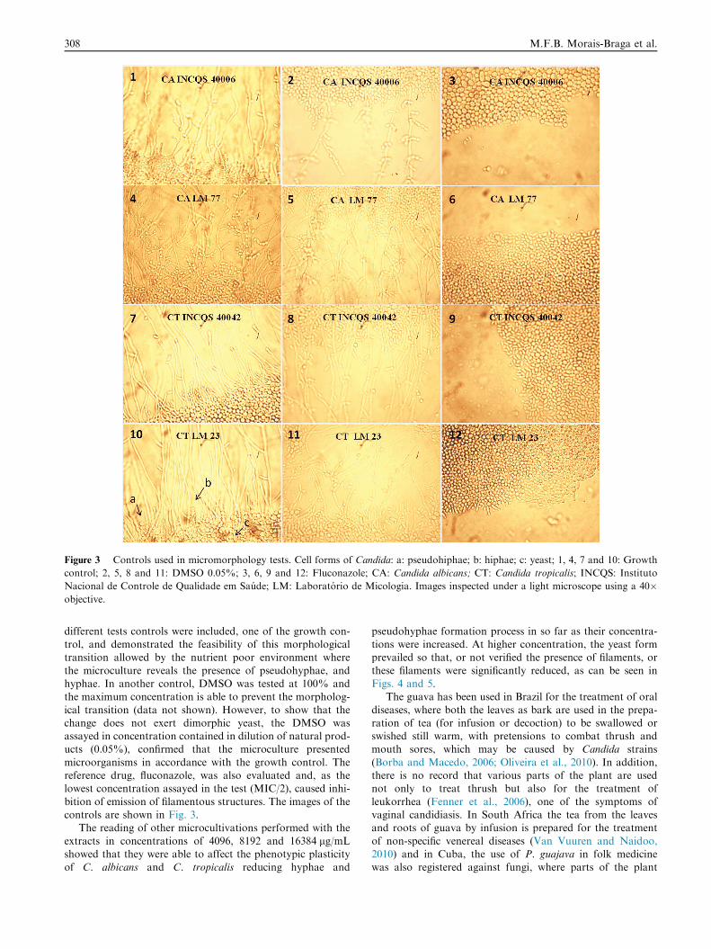
Figure 3 Controls used in micromorphology tests. Cell forms of Candida: a: pseudohiphae; b: hiphae; c: yeast; 1, 4, 7 and 10: Growth
control; 2, 5, 8 and 11: DMSO 0.05%; 3, 6, 9 and 12: Fluconazole; CA: Candida albicans; CT: Candida tropicalis; INCQS: Instituto
Nacional de Controle de Qualidade em Saude; LM: Laboratorio de Micologia. Images inspected under a light microscope using a 40�objective.
308 M.F.B. Morais-Braga et al.
different tests controls were included, one of the growth con-trol, and demonstrated the feasibility of this morphologicaltransition allowed by the nutrient poor environment where
the microculture reveals the presence of pseudohyphae, andhyphae. In another control, DMSO was tested at 100% andthe maximum concentration is able to prevent the morpholog-
ical transition (data not shown). However, to show that thechange does not exert dimorphic yeast, the DMSO wasassayed in concentration contained in dilution of natural prod-ucts (0.05%), confirmed that the microculture presented
microorganisms in accordance with the growth control. Thereference drug, fluconazole, was also evaluated and, as thelowest concentration assayed in the test (MIC/2), caused inhi-
bition of emission of filamentous structures. The images of thecontrols are shown in Fig. 3.
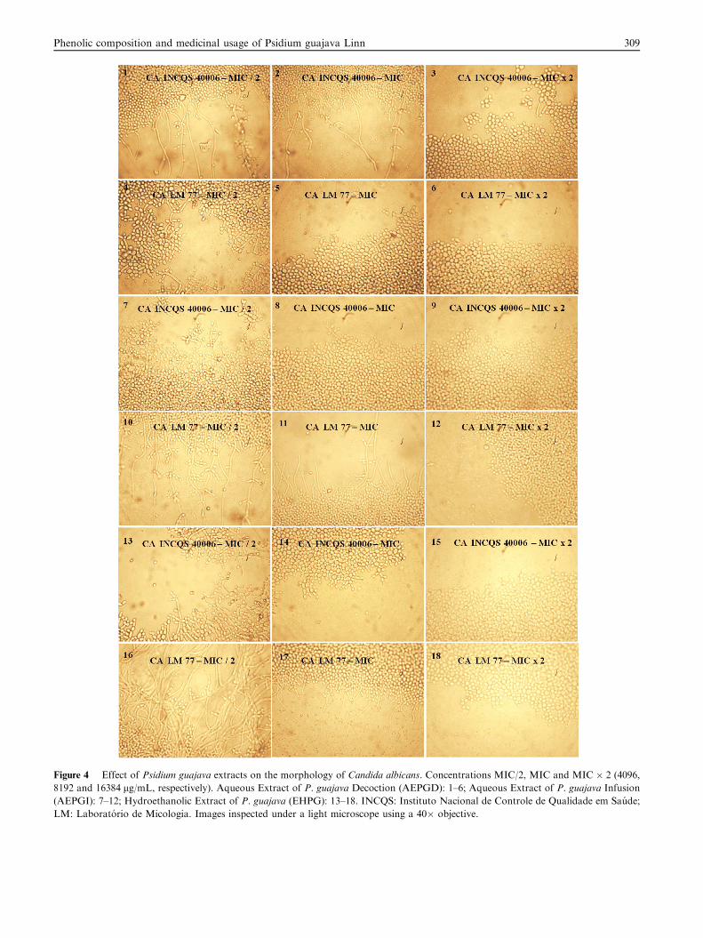
The reading of other microcultivations performed with the
extracts in concentrations of 4096, 8192 and 16384 lg/mLshowed that they were able to affect the phenotypic plasticityof C. albicans and C. tropicalis reducing hyphae and
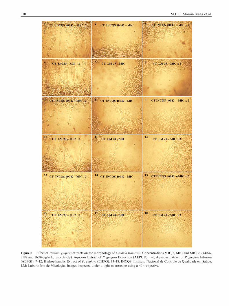
pseudohyphae formation process in so far as their concentra-tions were increased. At higher concentration, the yeast formprevailed so that, or not verified the presence of filaments, or
these filaments were significantly reduced, as can be seen inFigs. 4 and 5.
The guava has been used in Brazil for the treatment of oral
diseases, where both the leaves as bark are used in the prepa-ration of tea (for infusion or decoction) to be swallowed orswished still warm, with pretensions to combat thrush andmouth sores, which may be caused by Candida strains
(Borba and Macedo, 2006; Oliveira et al., 2010). In addition,there is no record that various parts of the plant are usednot only to treat thrush but also for the treatment of
leukorrhea (Fenner et al., 2006), one of the symptoms ofvaginal candidiasis. In South Africa the tea from the leavesand roots of guava by infusion is prepared for the treatment
of non-specific venereal diseases (Van Vuuren and Naidoo,2010) and in Cuba, the use of P. guajava in folk medicinewas also registered against fungi, where parts of the plant
Figure 4 Effect of Psidium guajava extracts on the morphology of Candida albicans. Concentrations MIC/2, MIC and MIC � 2 (4096,
8192 and 16384 lg/mL, respectively). Aqueous Extract of P. guajava Decoction (AEPGD): 1–6; Aqueous Extract of P. guajava Infusion
(AEPGI): 7–12; Hydroethanolic Extract of P. guajava (EHPG): 13–18. INCQS: Instituto Nacional de Controle de Qualidade em Saude;
LM: Laboratorio de Micologia. Images inspected under a light microscope using a 40� objective.
Phenolic composition and medicinal usage of Psidium guajava Linn 309
Figure 5 Effect of Psidium guajava extracts on the morphology of Candida tropicalis. Concentrations MIC/2, MIC and MIC � 2 (4096,
8192 and 16384 lg/mL, respectively). Aqueous Extract of P. guajava Decoction (AEPGD): 1–6; Aqueous Extract of P. guajava Infusion
(AEPGI): 7–12; Hydroethanolic Extract of P. guajava (EHPG): 13–18. INCQS: Instituto Nacional de Controle de Qualidade em Saude;
LM: Laboratorio de Micologia. Images inspected under a light microscope using a 40� objective.
310 M.F.B. Morais-Braga et al.
Phenolic composition and medicinal usage of Psidium guajava Linn 311
are used in the preparation of dye, powder and elixir (Ramırezet al., 2007). Based on these ethnobotanical reports, we canassume that the main form of therapeutic use P. guajava
against fungi is the topical use, as the natural product is placeddirectly on the skin or mucosa, used in mouthwash and gargle,in sitz baths and even in tea administration, which when taken
favors the contact of the natural product with the intestinallumen, where the infection causing microorganisms can beaccommodated.
If we consider the context of this research, a cup of tea(made by decoction and infusion) with 150 mL of water and10 g of fresh leaves, will be contained in this volume, just over4 times the concentration considered as MIC, was able to
reduce the percentage of viable microorganisms by direct con-tact. The same situation is extended to hydroethanolic extract.In 150 mL of tincture is contained 25 times the minimum inhi-
bitory concentration. If relate to the preparation of virulencepotential inhibitor in 150 mL of aqueous extracts of plantshave about 2 times the concentration at which the filamentous
structures of Candida have been reduced. In this same volumehydroethanolic extract is contained 12 times the inhibitoryconcentration dimorphism. Thus a direct contact of tea or
tincture prepared in the above relation not only reduces thepercentage of viable microorganisms, but also disturbs the pro-cess of morphological yeast transition that remains in placeafter the addition of home-made preparations, neutralizing
one of its virulence factors the ability to invade substrates.Due to the ethnomedicinal use of P. guajava observed both
in the traditional medicine as complementary and alternative
medicine, the plant is now part of the list of medicinal plantsof the World Health Organization (WHO). Based on funda-mental criteria such as common use in at least two regions
of WHO and satisfactory amount of scientific data, this orga-nization has promoted the development of monographs inwhich relevant information about this and other species of
medicinal relevance, was made available to the public access(WHO, 2009). P. guajava also reported in national lists ofmedicinal plants in some countries and is covered in publicpolicy programs focused on primary health care, as occurs,
for example, in Brazil (BRASIL, 2009; RENISUS, 2009).The belief system of some people, low economic power of
users, the medications available at minimal cost, the lack of
access to another type of therapeutic resource in conflict areas(especially in poor countries), the fact that they are naturalproducts and considered by some to be more effective than
allopathic medicines and cause side effects or milder sideeffects compared to commercial drugs are some of the reasonsgiven to justify the significant use of medicinal plants (Adnanet al., 2014; Khan et al., 2014; OMS, 2002), including the spe-
cies under study.Regarding the popular therapy with P. guajava, several fac-
tors can influence the final result of a treatment as a contact
time of natural product with the infection microorganisms,duration of treatment, methods of use, among others.
Although we are talking about parts of a plant that has its
fruit habitually used in nutrition for human populations fromdifferent locations, the use of tea fresh leaves for infusion hashad its cytotoxic potential investigated. The aqueous extract
intragastric administration in rats of both sexes (doses of 0.2,2.0 and 20.0 g/day) for prolonged period (six months) resultedin signs of hepatotoxicity and renal problems as hydronephro-sis in males and pyelonephritis, and nephrocalcinosis in
females. The LD50 of the extract was more than 20.0 g/kg(Attawish et al., 1995).
Almeida et al. (2006) evaluated in vitro the cytotoxicity of
tea made by infusing peritoneal macrophages of mice. Theinfusion of the leaves was prepared and tested both immedi-ately as a few hours after preparation. Soon after preparation,
the infusion was added to the culture environment whichexhibited 10% mortality rate, increasing to 31.82% after stor-age at 4 �C for a period of 48 h. After this period, the index
rose to 76.18% revealing that the infusion, therefore, presentsan immunotoxic effect. In this sense, taking tea in the same dayit is prepared can prevent damage being caused to the cells ofthe immune system, which, according to the authors, may be
due to flavonoids oxidation and subsequent release of theirderivatives capable to generate radicals free, which wouldcause toxicity.
People affected with candidiasis usually present with someimpairment of their immune responses and in this sense, wouldbe at serious risk, and may further compromise the body’s
defense case, for lack of such risks, adopt an inadequate alter-native therapy. These studies therefore point to a cautious useof tea as much as the duration of treatment, preparation and
storage even as the administration of excessive amounts.The antifungal effect of the species P. guajava reported here
may be due to the presence of phenolic compounds in theextracts, since they are able to promote both inhibition of
growth of Candida lineages (Alves et al., 2014; Barros et al.,2013; Candiracci et al., 2011; Tempesti et al., 2012; Vashisthet al., 2013; Candiracci et al., 2012), as well as their filamentous
structures resulting from the transition process (Candiracciet al., 2012; Canonico et al., 2014). The percentage of phenoliccompounds of the hydroethanolic extract was more pro-
nounced compared to aqueous extracts as well as their poten-tial inhibitor, as can be seen in cell viability curve. However,further investigations are needed to elucidate the mechanisms
by which act the extracts and which in fact, are the phytochem-icals contained therein, responsible for the observed effect.
P. guajava, in subsequent studies, had its antifungal poten-tial investigated obtaining results favorable for different
methodologies (Assunccedil et al., 2013; Jebashree et al.,2011; Mailoa et al., 2014; Suwanmanee et al., 2014), but thiswas the first report which was investigated and verified its
influence on a virulence factor of Candida.
4. Conclusion
The use of teas, pastes, plasters and sitz baths prepared fromleaves of P. guajava (red guava) for different populationshad, in this study, their potential bioactive scientifically justi-
fied through tests with leaf extracts by direct contact, since,besides provoking a decrease in the population of micro-organisms of the genus Candida, affected an important fungalvirulence factors, morphological transition, and consequently
their invasive potential of tissues. The observed antifungaleffect is fungistatic and not fungicidal, since he did not killthe fungi. However, it is important to remember that the exist-
ing cultural complex systems in these populations allow differ-ent forms of therapeutic preparations with amounts ofingredients that may be different from that used in our tests,
it is known that there is no standardization when it comes tothe use of medicinal plants. Further studies are needed to
312 M.F.B. Morais-Braga et al.
understand the genetic and biochemical processes involved inboth dynamic fungistatic as in inhibiting emissions of cellextensions of C. albicans and C. tropicalis in its virulence.
Declaration of interest
The authors declare that there is no conflict of interests regard-
ing the publication of this paper.
Acknowledgments
The authors are grateful to the Brazilian Research agencies:CNPq, CAPES and FUNCAP by the financial and grant
support.
References
Adnan, M., Ullah, I., Tariq, A., Murad, W., Azizullah, A., Khan, A.,
Ali, N., 2014. Ethnomedicine use in the war affected region of
northwest Pakistan. J. Ethnobiol. Ethnomed. 10, 16.
Albuquerque, U.P., Hanazaki, N., 2006. As pesquisas etnodirigidas na
descoberta de novos farmacos de interesse medico e farmaceutico:
fragilidades e pespectivas. Rev. Bras. Farmacogn. 16 (sSupl.).