61 UDC 666.9.014 : 549.766.21 APTEFF, 34, 1–148 (2003) BIBLID: 1450–7188 (2003) 34, pp. 61–70 Original scientific paper PHOSPHOGYPSUM SURFACE CHARACTERISATION USING SCANNING ELECTRON MICROSCOPY Miloš B. Rajković and Dragan V. Tošković This paper presents the results of application of Scanning Electron Microscopy (SEM) to examinations of the samples of natural gypsum and phosphogypsum. Phosphogypsum has a well developed crystalline structure, and appear in two polymorphous forms, of rombic and hexagonal shape crystals. Natural gypsum has a poorly crystalline structure. The differences in crystalline structure influence the chemical behavior of these row materials. KEYWORDS: scanning electron microscopy (SEM), natural gypsum, phosphogypsum INTRODUCTION Phosphogypsum is a by–product of the phosphate fertilizer which is produced in large quantities world–wide during the production of phosphoric acid. The wet phosphoric acid process is the most common process used to produce phosphoric acid. It can be summa- rized by the following equation (1): Ca 10 (PO 4 ) 6 F 2 + 10H 2 SO 4 + 20H 2 O → 6H 3 PO 4 + 10CaSO 4 ×2H 2 O + 2HF phosphogypsum Phosphogypsum consist mainly of gypsum (CaSO 4 ×2H 2 O), but may also contain minor quantities of environmentally sensitive constituents such as heavy metals and na- turally–occurring radionuclides (2). The presence of these minor constituents has raised some concerns regarding the stockpiling of phosphogypsum and its utilization in the other industries. Dr. Miloš B.Rajković, Professor, Institute of Food Technology and Biochemistry, Faculty of Agriculture, 11080 Belgrade–Zemun, Nemanjina 6, Serbia and Montenegro; Dr. Dragan V.Tošković, Professor, Faculty of Technology, 76000 Zvornik, Karakaj b.b., Republic of Srpska
PHOSPHOGYPSUM SURFACE CHARACTERISATION USING SCANNING ELECTRON MICROSCOPY
Miloš B. Rajković and Dragan V. Tošković
This paper presents the results of application of Scanning Electron Microscopy (SEM) to examinations of the samples of natural gypsum and phosphogypsum. Phosphogypsum has a well developed crystalline structure, and appear in two polymorphous forms, of rombic and hexagonal shape crystals. Natural gypsum has a poorly crystalline structure. The differences in crystalline structure influence the chemical behavior of these row materials.
KEYWORDS: scanning electron microscopy (SEM), natural gypsum, phosphogypsum
INTRODUCTION
Phosphogypsum is a by–product of the phosphate fertilizer which is produced in large quantities world–wide during the production of phosphoric acid. The wet phosphoric acid process is the most common process used to produce phosphoric acid. It can be summa-rized by the following equation (1):
Phosphogypsum consist mainly of gypsum (CaSO4×2H2O), but may also contain minor quantities of environmentally sensitive constituents such as heavy metals and na-turally–occurring radionuclides (2). The presence of these minor constituents has raised some concerns regarding the stockpiling of phosphogypsum and its utilization in the other industries.
Dr. Miloš B.Rajković, Professor, Institute of Food Technology and Biochemistry, Faculty of Agriculture, 11080 Belgrade–Zemun, Nemanjina 6, Serbia and Montenegro; Dr. Dragan V.Tošković, Professor, Faculty of Technology, 76000 Zvornik, Karakaj b.b., Republic of Srpska
62
The determination of elementary structure of matter, whether it is known or if it is necessary to verify the composition of a new or a well–known material to be used in further work, presents a frequent demand in the contemporary analytical practice. Some-times these demands are combined with the need to determine the morphology or the structure of the surface roughness of a sample, its homogeneity or the presence of defects in the structure. The electron microscopic and spectroscopic techniques, whose rapid deve-lopment to the commercial systems has been enabled first of all by the achievements in the electronic and computer technologies since the seventies to date, have become power-ful tools to date for attaining the above requirements (3,4,5).
This paper presents the results of application of the Scanning Electron Microscopy (SEM) to examinations of the samples of natural gypsum and phosphogypsum, for the purpose of characterizing the composition and defining the morphological composition (6).
EXPERIMENTAL
All investigations were performed with phosphogypsum obtained from Chemical In-dustry IHP Prahovo by a technological procedure in which raw phosphates are treated with H2SO4. Phosphogypsum purification was carried out by the following procedure (7,8): solution of H2SO4 in water was made, with concentration of 28 wt.%, which was heated up to 90°C. Phosphogypsum was added to the solution and an emulsion was made with continuous stirring. After phosphogypsum, BaSO4 was added also at continuous stirring, until the temperature reached 85°C. Then the mixture was cooled. After cooling, it was filtered through a special Buchner funnel. Grinding of the purified phosphogypsum was carried out in a laboratory mixer to the particle size mainly between 100 and 200 µm, with only 12% particles bigger than 200 µm.
Phosphogypsum samples were analysed using scanning electron microscopy SEM, JSM–84OA, JEOL, Tokyo, Japan, with the possibility of englargment by 180.000 times and the resolution of 10 nm.
The adsorbed, specially sprayed and secondary electrons make it possible to obtain the picture by the scanning electron microscopy. For that reason, the sample does not have to be thin, which simplifies its application, and the energy of electrons of the primary beam does not have to be high, because electrons do not have to pass through the sample. Besides, the additional electromagnetic field (of the scanning coil) enables a controlled and programmed shifting of the electron beam. In such way, particularly small areas of a sample may be, line by line, examined – scanned (5).
X–ray diffraction analysis was carried out with a powder diffractometer SIEMENS D–500 with Ni–filtered CuKα radiation. Identification of crystalline phases in recorded samples has been carried out by matching position and intensity of diffraction profiles with the JC PDS data.
Thermogravimetric analyses of phosphogypsum were performed on a Dermatograph STANTON, England, with a heating rate up to 7°C/min in air stream in mixture with Al2O3. Samples were analysed in ceramic crucibles at temperature up to 500°C.
63
RESULTS AND DISCUSSION
The results of the analysis of chemical and mineralogical composition of natural gypsum, phosphogypsum taken from filter from the process of phosphoric acid production by ”wet procedure” and previously processed and purified phosphogypsum by the new procedure (9) are presented in Table 1.
Table 1. Chemical and mineralogical composition of natural gypsum, phosphogypsum taken from plant’s pipe and purified phosphogypsum
From Table 1. it can be seen that phosphogypsum is in the form of dihydrate, immedia-
tely after separation from filters (74.16 wt.%), while the natural gypsum is essentially hemihydrate (81.34 wt.% CaSO4×1/2H2O), and all the remaining parameters are far closer to natural gypsum than to phosphogypsum.
In both unpurified and purified phosphogypsum, P2O5 contents are 0.84 and 0.09 wt.%, respectively, the content in natural gypsum being lower. A positive characteristics of purified phosphogypsum is the presence of CaO (as CaCO3, like that in natural gypsum 5.09%). The advantageous feature of phosphogypsum is the proportion of CaO and SO3, which indicates the presence of other salts with sulphur – MgSO4, Al2(SO4)3, FeSO4.
To establish morphological composition and structure, their homogeneity and the presence of faults in their structure of samples of natural gypsum and various phospho-gypsum, previously prepared (7,10), and mixtures of natural gypsum–phosphogypsum were subjected to SEM analysis.
The obtained micrographs are presented in Figs. 1 – 5.
64
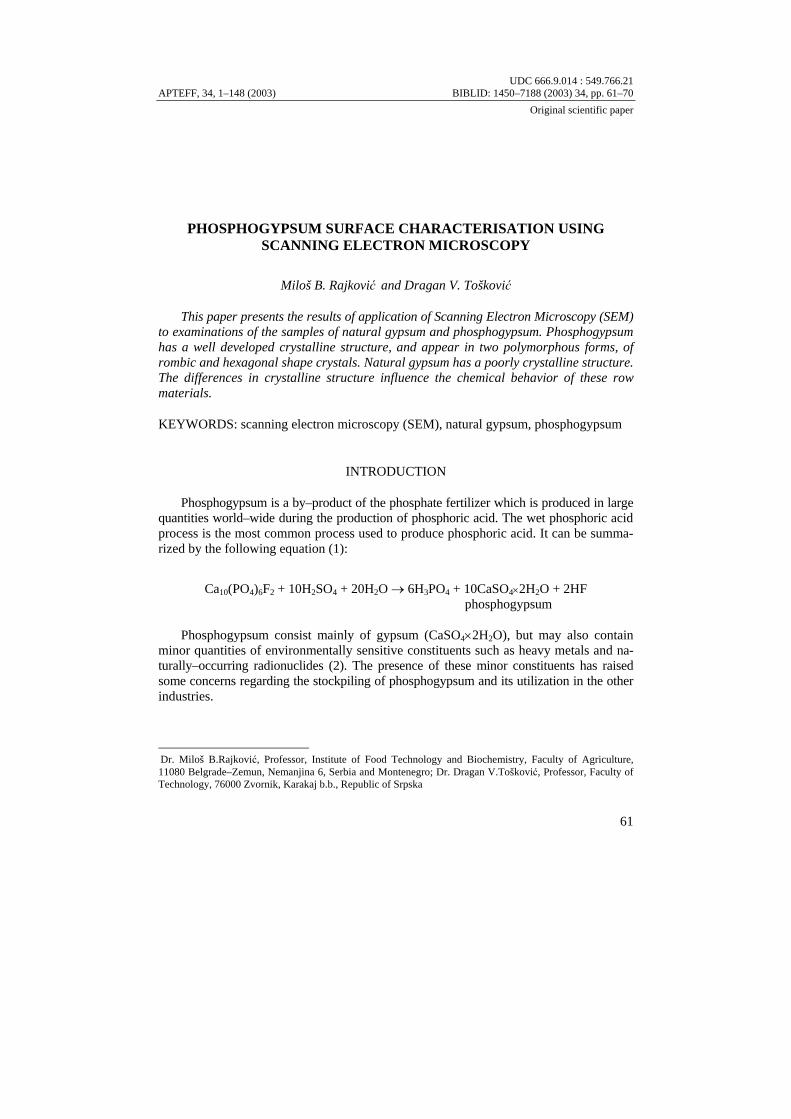
Fig. 1. SEM micrograph of crystalline structure of natural gypsum (×500)
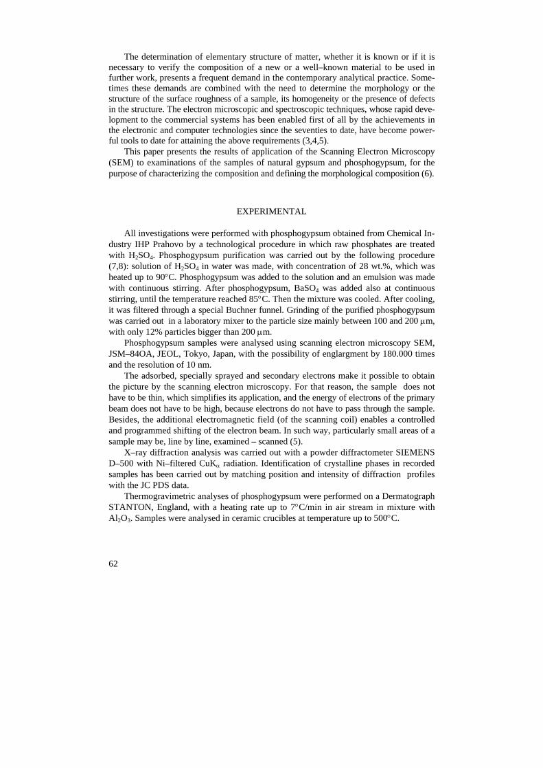
Fig. 2. SEM micrograph of crystalline structure of phosphogypsum
taken from plant’s pipe (×1,000)
65
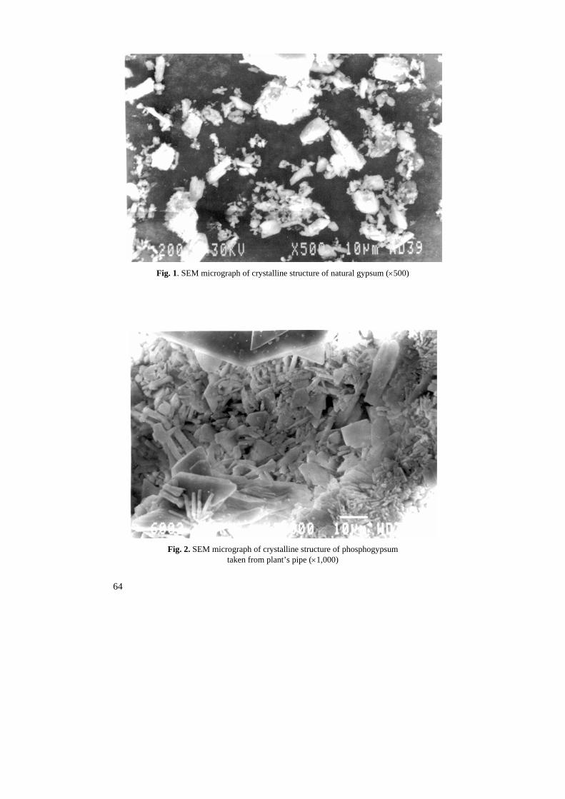
Fig. 3. SEM micrograph of crystalline structure of mixture of 75 wt. % natural gypsum
and 25 wt. % phosphogypsum (×500)
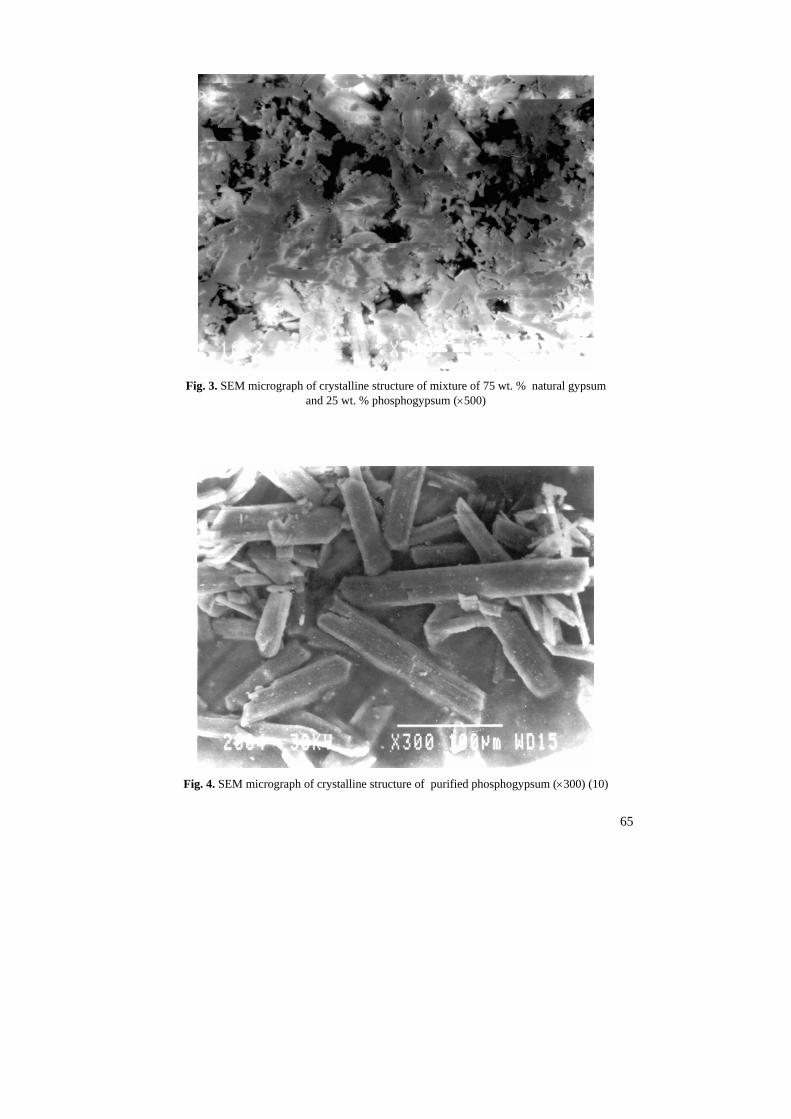
Fig. 4. SEM micrograph of crystalline structure of purified phosphogypsum (×300) (10)
66
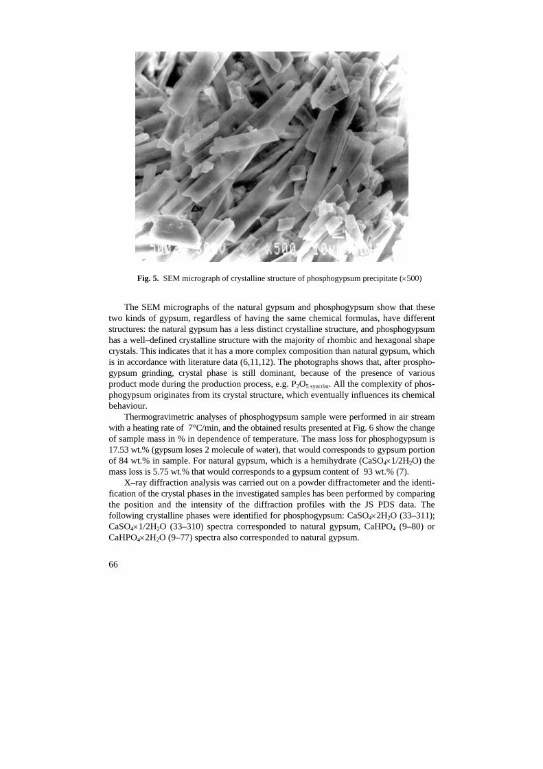
Fig. 5. SEM micrograph of crystalline structure of phosphogypsum precipitate (×500)
The SEM micrographs of the natural gypsum and phosphogypsum show that these two kinds of gypsum, regardless of having the same chemical formulas, have different structures: the natural gypsum has a less distinct crystalline structure, and phosphogypsum has a well–defined crystalline structure with the majority of rhombic and hexagonal shape crystals. This indicates that it has a more complex composition than natural gypsum, which is in accordance with literature data (6,11,12). The photographs shows that, after prospho-gypsum grinding, crystal phase is still dominant, because of the presence of various product mode during the production process, e.g. P2O5 syncrist. All the complexity of phos-phogypsum originates from its crystal structure, which eventually influences its chemical behaviour.
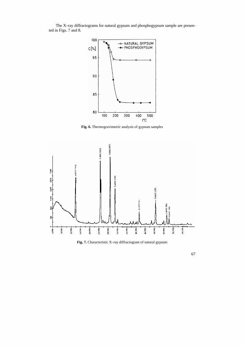
Thermogravimetric analyses of phosphogypsum sample were performed in air stream with a heating rate of 7°C/min, and the obtained results presented at Fig. 6 show the change of sample mass in % in dependence of temperature. The mass loss for phosphogypsum is 17.53 wt.% (gypsum loses 2 molecule of water), that would corresponds to gypsum portion of 84 wt.% in sample. For natural gypsum, which is a hemihydrate (CaSO4×1/2H2O) the mass loss is 5.75 wt.% that would corresponds to a gypsum content of 93 wt.% (7).
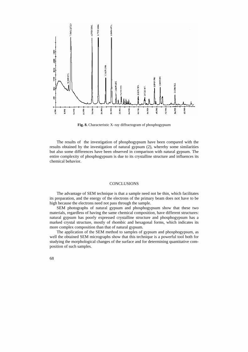
X–ray diffraction analysis was carried out on a powder diffractometer and the identi-fication of the crystal phases in the investigated samples has been performed by comparing the position and the intensity of the diffraction profiles with the JS PDS data. The following crystalline phases were identified for phosphogypsum: CaSO4×2H2O (33–311); CaSO4×1/2H2O (33–310) spectra corresponded to natural gypsum, CaHPO4 (9–80) or CaHPO4×2H2O (9–77) spectra also corresponded to natural gypsum.
67
The X–ray diffractograms for natural gypsum and phosphogypsum sample are presen-ted in Figs. 7 and 8.
Fig. 6. Thermogravimetric analysis of gypsum samples
Fig. 7. Characteristic X–ray diffractogram of natural gypsum
68
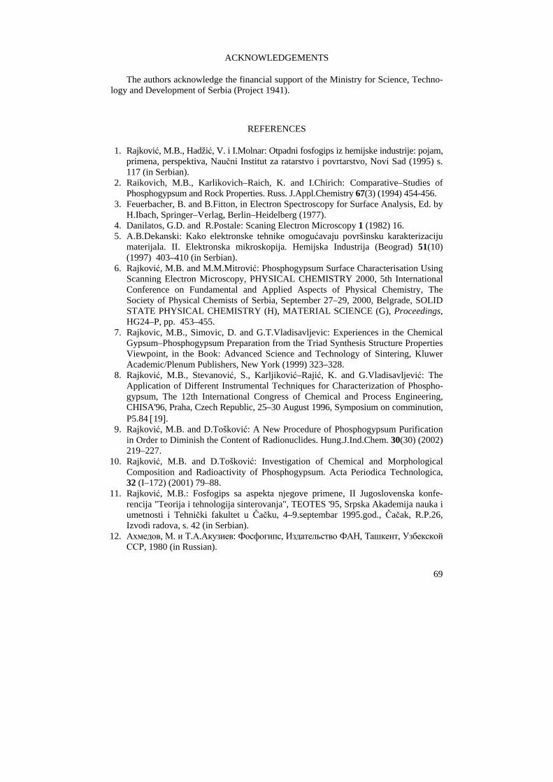
Fig. 8. Characteristic X–ray diffractogram of phosphogypsum
The results of the investigation of phosphogypsum have been compared with the results obtained by the investigation of natural gypsum (2), whereby some similarities but also some differences have been observed in comparison with natural gypsum. The entire complexity of phosphogypsum is due to its crystalline structure and influences its chemical behavior.
CONCLUSIONS
The advantage of SEM technique is that a sample need not be thin, which facilitates its preparation, and the energy of the electrons of the primary beam does not have to be high because the electrons need not pass through the sample.
SEM photographs of natural gypsum and phosphogypsum show that these two materials, regardless of having the same chemical composition, have different structures: natural gypsum has poorly expressed crystalline structure and phosphogypsum has a marked crystal structure, mostly of rhombic and hexagonal forms, which indicates its more complex composition than that of natural gypsum.
The application of the SEM method to samples of gypsum and phosphogypsum, as well the obtained SEM micrographs show that this technique is a powerful tool both for studying the morphological changes of the surface and for determining quantitative com-position of such samples.
69
ACKNOWLEDGEMENTS
The authors acknowledge the financial support of the Ministry for Science, Techno-logy and Development of Serbia (Project 1941).
REFERENCES
1. Rajković, M.B., Hadžić, V. i I.Molnar: Otpadni fosfogips iz hemijske industrije: pojam, primena, perspektiva, Naučni Institut za ratarstvo i povrtarstvo, Novi Sad (1995) s. 117 (in Serbian).
2. Raikovich, M.B., Karlikovich–Raich, K. and I.Chirich: Comparative–Studies of Phosphogypsum and Rock Properties. Russ. J.Appl.Chemistry 67(3) (1994) 454-456.
3. Feuerbacher, B. and B.Fitton, in Electron Spectroscopy for Surface Analysis, Ed. by H.Ibach, Springer–Verlag, Berlin–Heidelberg (1977).
4. Danilatos, G.D. and R.Postale: Scaning Electron Microscopy 1 (1982) 16. 5. A.B.Dekanski: Kako elektronske tehnike omogućavaju površinsku karakterizaciju
materijala. II. Elektronska mikroskopija. Hemijska Industrija (Beograd) 51(10) (1997) 403–410 (in Serbian).
6. Rajković, M.B. and M.M.Mitrović: Phosphogypsum Surface Characterisation Using Scanning Electron Microscopy, PHYSICAL CHEMISTRY 2000, 5th International Conference on Fundamental and Applied Aspects of Physical Chemistry, The Society of Physical Chemists of Serbia, September 27–29, 2000, Belgrade, SOLID STATE PHYSICAL CHEMISTRY (H), MATERIAL SCIENCE (G), Proceedings, HG24–P, pp. 453–455.
7. Rajkovic, M.B., Simovic, D. and G.T.Vladisavljevic: Experiences in the Chemical Gypsum–Phosphogypsum Preparation from the Triad Synthesis Structure Properties Viewpoint, in the Book: Advanced Science and Technology of Sintering, Kluwer Academic/Plenum Publishers, New York (1999) 323–328.
8. Rajković, M.B., Stevanović, S., Karljiković–Rajić, K. and G.Vladisavljević: The Application of Different Instrumental Techniques for Characterization of Phospho-gypsum, The 12th International Congress of Chemical and Process Engineering, CHISA'96, Praha, Czech Republic, 25–30 August 1996, Symposium on comminution, P5.84 [19].
9. Rajković, M.B. and D.Tošković: A New Procedure of Phosphogypsum Purification in Order to Diminish the Content of Radionuclides. Hung.J.Ind.Chem. 30(30) (2002) 219–227.
10. Rajković, M.B. and D.Tošković: Investigation of Chemical and Morphological Composition and Radioactivity of Phosphogypsum. Acta Periodica Technologica, 32 (I–172) (2001) 79–88.
11. Rajković, M.B.: Fosfogips sa aspekta njegove primene, II Jugoslovenska konfe-rencija "Teorija i tehnologija sinterovanja", TEOTES '95, Srpska Akademija nauka i umetnosti i Tehnički fakultet u Čačku, 4–9.septembar 1995.god., Čačak, R.P.26, Izvodi radova, s. 42 (in Serbian).
12. Ахмедов, М. и Т.А.Акузиев: Фосфогипс, Издательство ФАН, Ташкент, Узбекской ССР, 1980 (in Russian).
Елементарни састав материје, било да је непознат или да је потребно провери-ти састав новог или познатог материјала који ће се користити у даљем раду, пред-ставља чест захтев у савременој аналитичкој пракси. Некада се овим захтевима при-дружује и потреба да се утврди морфологија или структура површинске храпавости узорка, његова хомогеност или присуство дефеката у структури. Електронске микро-скопске и спектроскопске технике, чији је нагли развој, све до комерцијалних система, од седамдесетих година до сада, омогућен пре свега достигнућима у електронским и компјутерским технологијама, постале су снажно оруђе за остваривање помену-тих захтева.
У раду су приказани резултати примене скенирајуће електронске микроскопије (SEM) на испитивања површине узорака природног гипса и фосфогипса, ради карак-теризације састава и дефинисања морфолошког састава.
SEM снимци природног гипса и отпадног фосфогипса указали су да ова два гипса, иако имају исте хемијске (молекулске) формуле – CaSO4×2H2O, имају разли-читу структуру: природни гипс има слабије изражену кристалну структуру а отпад-ни фосфогипс има изразиту кристалну структуру, претежно ромбичног и хексаго-налног облика, што указује на његов сложенији састав од природног, а што је у складу са литературним подацима.