Photonic nanoarchitectures occurring in butterfly scales as selective gas/vapor sensors L. P. Biró *a , K. Kertész a , Z. Vértesy a , Zs. Bálint b a Research Institute for Technical Physics and Materials Science, H-1525, Budapest, POB 49, Hungary b Hungarian Natural History Museum, H-1088, Budapest, Baross utca 13, Hungary ABSTRACT Photonic band gap material type nanoarchitectures occurring in the wing scales of butterflies possessing structural color were investigated as selective gas/vapor sensors. From 20 examined butterfly species all showed selective sensing when various volatile organic compounds were introduced as additives in ambient air. Four butterflies species: Chrysiridia ripheus (Geometridae), Pseudolycena marsyas, Cyanophrys remus (both Lycaenidae) and Morpho aega (Nymphalidae) were selected to demonstrate the possibilities of selective sensing offered by these natural nanoarchitectures. Each butterfly species gives characteristic response both for species, i.e., for its typical nanoarchitecture, and for the seven test vapors used. Fast response time, reproducible and concentration dependent signals are demonstrated. Keywords: butterfly wing scales, nanoarchitecture, photonic crystal, photonic band gap (PBG), selective gas/vapor sensor, volatile organic compounds (VOCs), optical reading, bioinspiration. 1. INTRODUCTION Beyond their charming beauty butterflies may reveal a number of “design principles” which were carefully distilled to perfection during many millennia of natural evolution. This is particularly true for those colors which have role in sexual communication or cryptic behavior. Individuals with the “wrong color” have lesser chances to find a mating partner, or are predated early, before they could have the chance to reproduce. In the present paper we are interested in those natural designs which concern photonic nanoarchitectures occurring in the wings of butterflies. These nanoarchitectures are named photonic crystals (PhC), or photonic band gap (PBG) materials by physicists and materials scientists. The concept of these composite materials built of two materials with distinct optical properties (refractive index) was introduced 20 years ago by Yablonovitch [1] and John [2]. The wings of butterflies are covered by scales, which are the cuticular product of a single cell. Typical scale dimensions are in the 100 x 50 x 1 µm 3 range, the building material is mainly chitin. Generally speaking, the scales have the shape of a flattened sack with an unstructured side facing the wing membrane, while the side looking away from the wing membrane usually has a clearly visible system of longitudinal ridges spaced at a distance of the order of one µm from each other. The ridges are frequently connected by so called cross-ribs, oriented normally to the direction of the ridges. The interior volume of the scale may be filled with a complex nanoarchitecture, which has chitin as main constituent. The nanoarchitectures occurring in butterfly scales were classified from a biological point of view by Ghiradella [3]. Recently a renewed interest is focused on these nanoarchitectures from the point of view of physics and materials science and of biology, too [4 – 8]. Moreover, similar photonic nanoarchitectures were reported in various organisms, like beetles [9], birds [10] marine organism [11], etc., for recent reviews on this topic see [12, 13]. As PBG materials in butterfly scales are constituted mainly from chitin and air, when the chemical composition of air changes the optical properties of the PhC type material are shifted. This offers a handy way of using the natural photonic nanoarchitectures as gas/vapor sensors. Potyrailo et al., used sophisticated data handling methods to prove that the wings of Morpho sulkowskyi butterfly can act as selective chemical sensors able to distinguish between water and ethanol vapors [14]. In the present work we show that many butterflies possessing structural color, i.e., BPG material type nanoarchitectures in their scales, are able to act as selective chemical sensors. Moreover, the butterfly wing sensors * [email protected]; phone: +36-1-3922681; fax: +36-1-3922226; http://www.nanotechnology.hu/ The Nature of Light: Light in Nature II, edited by Katherine Creath, Proc. of SPIE Vol. 7057, 705706, (2008) · 0277-786X/08/$18 · doi: 10.1117/12.794910 Proc. of SPIE Vol. 7057 705706-1
Transcript
Photonic nanoarchitectures occurring in butterfly scales as selective gas/vapor sensors
L. P. Biró*a, K. Kertésza, Z. Vértesya, Zs. Bálintb
a Research Institute for Technical Physics and Materials Science, H-1525, Budapest, POB 49, Hungary
b Hungarian Natural History Museum, H-1088, Budapest, Baross utca 13, Hungary
ABSTRACT Photonic band gap material type nanoarchitectures occurring in the wing scales of butterflies possessing structural color were investigated as selective gas/vapor sensors. From 20 examined butterfly species all showed selective sensing when various volatile organic compounds were introduced as additives in ambient air. Four butterflies species: Chrysiridia ripheus (Geometridae), Pseudolycena marsyas, Cyanophrys remus (both Lycaenidae) and Morpho aega (Nymphalidae) were selected to demonstrate the possibilities of selective sensing offered by these natural nanoarchitectures. Each butterfly species gives characteristic response both for species, i.e., for its typical nanoarchitecture, and for the seven test vapors used. Fast response time, reproducible and concentration dependent signals are demonstrated. Keywords: butterfly wing scales, nanoarchitecture, photonic crystal, photonic band gap (PBG), selective gas/vapor
1. INTRODUCTION Beyond their charming beauty butterflies may reveal a number of “design principles” which were carefully distilled to perfection during many millennia of natural evolution. This is particularly true for those colors which have role in sexual communication or cryptic behavior. Individuals with the “wrong color” have lesser chances to find a mating partner, or are predated early, before they could have the chance to reproduce. In the present paper we are interested in those natural designs which concern photonic nanoarchitectures occurring in the wings of butterflies. These nanoarchitectures are named photonic crystals (PhC), or photonic band gap (PBG) materials by physicists and materials scientists. The concept of these composite materials built of two materials with distinct optical properties (refractive index) was introduced 20 years ago by Yablonovitch [1] and John [2]. The wings of butterflies are covered by scales, which are the cuticular product of a single cell. Typical scale dimensions are in the 100 x 50 x 1 µm3 range, the building material is mainly chitin. Generally speaking, the scales have the shape of a flattened sack with an unstructured side facing the wing membrane, while the side looking away from the wing membrane usually has a clearly visible system of longitudinal ridges spaced at a distance of the order of one µm from each other. The ridges are frequently connected by so called cross-ribs, oriented normally to the direction of the ridges. The interior volume of the scale may be filled with a complex nanoarchitecture, which has chitin as main constituent. The nanoarchitectures occurring in butterfly scales were classified from a biological point of view by Ghiradella [3]. Recently a renewed interest is focused on these nanoarchitectures from the point of view of physics and materials science and of biology, too [4 – 8]. Moreover, similar photonic nanoarchitectures were reported in various organisms, like beetles [9], birds [10] marine organism [11], etc., for recent reviews on this topic see [12, 13]. As PBG materials in butterfly scales are constituted mainly from chitin and air, when the chemical composition of air changes the optical properties of the PhC type material are shifted. This offers a handy way of using the natural photonic nanoarchitectures as gas/vapor sensors. Potyrailo et al., used sophisticated data handling methods to prove that the wings of Morpho sulkowskyi butterfly can act as selective chemical sensors able to distinguish between water and ethanol vapors [14]. In the present work we show that many butterflies possessing structural color, i.e., BPG material type nanoarchitectures in their scales, are able to act as selective chemical sensors. Moreover, the butterfly wing sensors
The Nature of Light: Light in Nature II, edited by Katherine Creath, Proc. of SPIE Vol. 7057, 705706, (2008) · 0277-786X/08/$18 · doi: 10.1117/12.794910
originating from different species of butterflies exhibit specific and distinct signals for the same vapor present in air, so that they are suitable for being used in sensor arrays which can “fingerprint” a certain gas, or vapor in a similar way like chemically functionalized carbon nanotube based sensors [15]. Selective chemical sensors that could be read optically have several advantages over those that operate electronically: no extensive wiring is needed, no danger of explosion, optical sensors could operate in airtight enclosures without the need of feedthrough, etc.. The more so, that recent concerns regarding the indoor air pollution [16] are increasing with the spreading of airtight buildings to reduce energy consumption and with the increase of the time fraction people in developed countries spend in closed environment to values close to 90% [16]. The major concern is generated by several volatile organic compounds (VOCs) which may have various sources in the artificial environment [17].
Fig. 1. Electron micrographs showing the nanoarchitectures occurring in the dorsal cover scales of the Pseudolycena marsyas (a, c) and of the Chrysiridia ripheus (b, d). SEM images (a, b), TEM images (c, d).
Although we have investigated the selective sensing capability of more than 20 different butterflies and moths with structural (PBG) color, in the present paper we will concentrate on only four species, one day flying geometrid moth and two lycaenid and one nymphalid butterfly species: Chrysiridia ripheus (Geometridae), Pseudolycena marsyas, Cyanophrys remus (both Lycaenidae) and Morpho aega (Nymphalidae). These four species convincingly illustrate that very different natural PhC type nanoarchitectures can be used for selective gas/vapor sensing and that different nanoarchitectures may give different response to the same gas/vapor. We believe that the natural nanoarchitectures occurring in butterfly scales can be used as low cost, selective optical gas/vapor sensors. The low cost is provided by the very simple way in which these nanoarchitectures can be produced by rearing butterflies like it is done with many spectacular butterflies which have structural colors. One may wonder to what extent the colors are “reproducible”? As already pointed out, those colors which have role in sexual signaling or cryptic behavior very likely are highly optimized by evolution and stable. In an earlier work we investigated the blue dorsal color and scale nanostructure in the monophyletic Polyommatus eros species group and in two closely related species Polyommatus ariana and Polyommatus icarus used as out-group species [18]. The three species from the P. eros group: P. eros, P. eroides and P. erotides share a hypothetical a common ancestor which inhabited Europe and Asia before the last glacial period. Although, presently the three species live in geographically isolated regions (two in Europe, one in Asia), they still show similar spectral characteristics, which are very clearly different from the two other blue lycaenids, used as out-group comparison species [18].
Proc. of SPIE Vol. 7057 705706-2
I '
:
j I
-D )
C
D
—
Co
(p
—
Ref
lect
ance
(%
) -
- U
i 0
Ui
0 U
i 0
Ui
0 U
i
0 0
Ui 0
0 0 Ui 0 0 0 0 —I 0 0
2. METHODOLOGY The investigated male butterfly and month samples were obtained from the Lepidoptera Collection of the Hungarian Natural History Museum. Flat pieces of wings were cut and fixed to frames to provide safety in handling during spectral characterization. Reflectivity measurements were carried out with an Avaspec 2048/2 fiber-optic spectrometer, in a 45o backscattering geometry, as described in detail earlier [5], a diffuse white standard (Avaspec) was used as comparison sample. This measurement geometry was chosen because of several reasons: i) to be able during sensing experiments to decouple the illuminating light reflected from the transparent quartz window from the light reflected by the wing piece; ii) this decoupling is made possible by the nonspecular reflectance of butterfly wings possessing structural color; iii) as certain butterfly species like the blue morphos have a very intense but narrow angle normal incidence reflectance we preferred a measurement geometry which diminishes the intensity differences between the various butterfly wings we used. In order to reveal the characteristic structural elements of the investigated nanoarchitectures, the butterfly wings were examined by optical microscopy, scanning electron microscopy (SEM) and transmission electron microscopy (TEM). To avoid charging during SEM examination the wing pieces were coated by a thin layer of gold. The TEM specimens of 70 nm thickness were cut in a plane transversal to the plane of the scales by ultramicrotome, from blocks of special resin in which wing pieces were previously embedded in as received state, without any chemical staining.
Fig. 2. (Color on-line) Reflectance maxima of the four investigated wing pieces at 45o in backscattering
geometry, recorded to white diffuse standard.
All the sensing experiments were carried out using streaming ambient air, the air was pumped through a small metallic cell provided with a quartz window using a membrane pump. The framed wing pieces were placed under the quartz window. The sample was illuminated at normal incidence by a 150 W halogen lamp, the light reflected by the butterfly wing under an angle of 45o was collected by the pick-up fiber of the fiber optic spectrophotometer (see inset of Fig. 3b). The spectrophotometer was operated in scope mode, i.e., the intensity of the reflected light was measured without the use of any standard. The test vapors were introduced in the air flow by passing the ingoing air through a bubbler containing the liquid to be vaporized. When measuring the concentration dependence, the liquid in the bubbler was diluted in several steps. In some experiments the CIE L*a*b* [19] parameters provided by the software of the spectrophotometer were used, to quantify the color changes upon introducing various vapors in the streaming air. The a* , characterizing the shift on the green to red axis will be used in the present paper.
3. EXPERIMENTAL RESULTS AND DISCUSSION The electron micrographs are shown for two of the four investigated species in Fig. 1. The characteristic nanoarchitectures of the Morpho-type scales are well know [4, 14, 20], our SEM and TEM data in agreement with earlier works are not presented here. We reported earlier the scale nanoarchitectures of Cyanophrys remus, both for the dorsal and for the ventral sides of the wing [5], here we used only ventral side wing samples. Under the aspect of their nanoarchitectures the four butterfly scales used can be classified as detailed below.
Fig. 3. (Color on-line) Reflected light intensity in scope mode from the wing of Pseudolycaena marsyas. (a) Reflected light intensity in streaming ambient air and in the mixture of streaming air and ethanol; (b)
difference of the spectra shown in (a). The inset in the right upper corner shows schematically the measurement arrangement.
Fig. 4. (Color on-line) Difference in reflected light intensity recorded under identical experimental
conditions for the same two butterfly wings for: (a) air to air end ethanol; (b) air to air and benzene. One may note that the response is specific both for butterflies and vapors.
Proc. of SPIE Vol. 7057 705706-4
Pseudolycena marsyas (dorsal side): has a so called “pepper-pot” type structure occurring frequently in Lycaenid butterflies, which can be characterized by various degrees of order from perfect single crystalline [5], to fully amorphous type structure [21, 22], like in the case of P. marsyas. It is worth to point out that in the case of P. marsyas, as shown in Fig. 1c., the pigmentation within the body of the scale shows a clear delimitation: the layer closer to the wing membrane is strongly pigmented, while the layer above has little pigment if any. Chrisyrida ripheus (earlier Urania ripheus) (dorsal side): has multilayer type scales as reported earlier [23]. As this moth, because of its various structural colors ranging from blue-green to purplish yellow, also known as the Madagascan sunset moth, offers the possibility to chose various colors, we decided to use its orange-red wing region, to cover as much of the visible spectrum as possible, see Fig. 2. (Color on-line) Reflectance maxima of the four investigated wing pieces at 45o in backscattering geometry, recorded to white diffuse standard. At a first glance on the SEM image in Fig. 1b, one may find it surprising that such an apparently closed structure reacts at all to modification in the ambient atmosphere, the TEM image in Fig. 1d shows that this structure also has a significant air fraction, so it’s band gap is shifted with the changes in the composition of the atmosphere. The regions where the gas exchange occurs between the interior of the scale and the surrounding atmosphere are located on the sides of the ridges.
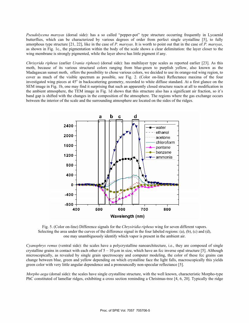
Fig. 5. (Color on-line) Difference signals for the Chrysiridia ripheus wing for seven different vapors.
Selecting the area under the curves of the difference signal in the four labeled regions: (a), (b), (c) and (d), one may unambiguously identify which vapor is present in the ambient air.
Cyanophrys remus (ventral side): the scales have a polycrystalline nanoarchitecture, i.e., they are composed of single crystalline grains in contact with each other of 5 – 10 µm in size, which have an fcc inverse opal structure [5]. Although microscopically, as revealed by single grain spectroscopy and computer modeling, the color of these fcc grains can change between blue, green and yellow depending on which crystalline face the light falls, macroscopically this yields green color with very little angular dependence and a pronouncedly non-specular reflectance [5]. Morpho aega (dorsal side): the scales have single crystalline structure, with the well known, characteristic Morpho-type PhC constituted of lamellar ridges, exhibiting a cross section reminding a Christmas-tree [4, 6, 20]. Typically the ridge
Proc. of SPIE Vol. 7057 705706-5
is composed of a stack of 6 - 7 lamellas of 70 nm thickness, the average maximal length of the lamellas as measured from the center of the ridge being 110 nm, the average distance between neighboring ridges is 635 nm.
Fig. 6. (Color on-line) Relative variation of the a* as provide by the spectrophotometer software under
cyclic alternation of ambient air only to ambient air and water vapor, for Morpho aega wing. The difference signal shifts to the negative half plane when water vapors are present. One may note that after 5
minute a stationary state is reached.
Fig. 7. (Color on-line) Relative variation of the a* as provide by the spectrophotometer software under
cyclic alternation of ambient air only to ambient air and increasing concentrations of ammonia ( B/A = 2; C/A = 5; D/A= 10; E/A = 20; F/A = 40) for the M. aega wing.
In a typical gas/vapor sensing experiment the cell is illuminated under normal incidence with white light and the reflected light intensity is measured under an emergent angle of 45o, as shown in the inset of Fig. 3b. The difference of
Proc. of SPIE Vol. 7057 705706-6
light intensities recorded in only air and in air with ethanol Fig. 3b, although much smaller in amplitude than the maximum in Fig. 3a, produces a clearly distinguishable maximum. In Fig. 4 it is illustrated that the measured difference signal is specific (in spectral position and intensity variation) both for the butterfly and for the vapors introduced in the ambient air. In Fig. 5 the difference signals for seven different test vapors (five volatile organic compounds) are show for the wing of C. ripheus. As can be clearly seen from the curves, all signals are in the well measurable range and only the distinction between acetone and chloroform may pose some difficulties. Our measurements show that if needed, in principle, it would be possible to realize a selective sensor using the wings of only one butterfly species, but using several different species can significantly improve the selectivity. There are some other aspects to be examined if sensor applications are proposed, like: response time, reproducibility and concentration sensitivity. In these experiments we used the CIE a* parameter, i.e., the shift on the green-red axis. Using the a* directly yields the shift in color, making possible to skip the subtraction of two consecutive spectra (air and air + vapors). In Fig. 6, the response time and the reproducibility are illustrated in the case of M. aega wing and water vapors. The same wing was used to test the concentration dependence using increasing NH3 concentrations in the solution in the bubbler vessel, the results are shown in Fig. 7. Both Fig. 6 and Fig. 7 show that the measurable signal builds up in matter of seconds and vanishes in similarly short times without any need of regeneration by heating as it occurs in the case of most solid state type sensor, ceramic [24] or semiconductor [25] sensors operated at elevated temperatures. However, in the case of ammonia a more significant persistent modification is observed as compare to water vapors. This may indicate that ammonia produces the modification of optical properties not only by modifying the optical properties of one of the materials constituting the PBG nanoarchitectures, but may interact more deeply with the chitin. Further studies are needed to get more insight in this phenomenon. Even so, a clear concentration dependence of the signal can be seen in Fig. 7, and for a certain concentration range satisfactory reproducibility is obtained.
4. CONCLUSIONS The wings of the four investigated butterflies showed specific response to various test vapors mixed with ambient air both under the aspect of butterfly species and the aspect of the chemical nature of the vapors. This clearly demonstrates that butterfly wings are suitable sensor elements for selective and “intelligent” sensor arrays, which will be able to fingerprint certain gas/vapor species and to recognize these species by comparing the actually measured data with files stored in the memory of a computer. Measurements on the wings of Morpho aega demonstrate that the sensors can have a fast and reproducible response and concentration sensitivity was achieved, too. Further work is needed to elucidate the cause of more pronounced persistent modifications in the case of ammonia vapors. Photonic nanoarchitectures occurring in the scales of butterflies possessing structural colors may constitute a cheap and convenient material for the production of selective and sensitive sensors, which can be read optically without the need of electrical wiring. On the other hand, if suitable ways are found to reproduce these structures artificially, they may constitute a bioinspired “library” of potential sensor nanoarchitectures.
ACKNOWLEDGEMENTS The butterfly specimens used in the present study were donated by the Hungarian Natural History Museum. The work was supported by EU6 NEST/PATHFINDER/ BioPhot-01915. The work in Hungary was partly supported by OTKA-NKTH No. K67793.
REFERENCES
[1] Yablonovitch E., “Inhibited Spontaneous Emission in Solid-State Physics and Electronics,” Phys. Rev. Lett. 58, 2059 (1987). [2] John S., “Strong localization of photons in certain disordered dielectric superlattices,” Phys. Rev. Lett. 58, 2486 (1987). [3] Ghiradella H., “Hairs, Bristles, and Scales,” in: Microscopic Anatomy of Invertebrates Vol. 11A, Insecta, M. Locke ed., Wiley-Liss, 1998. pp. 257 -287.
Proc. of SPIE Vol. 7057 705706-7
[4] Vukusic P., Sambles J. R., Lawrence C. R. and Wootton R. J., “Quantised interference and diffraction in single Morpho butterfy scales,” Proc. R. Soc. Lond. B 266, 1403 (1999). [5] Kertész K., Bálint Zs., Vértesy Z., Márk G. I., Lousse V, Vigneron J. P., Rassart M., and Biró L. P., “Gleaming and dull surface textures from photonic-crystal-type nanostructures in the butterfly Cyanophrys remus,” Phys. Rev E. 74, 021922 (2006). [6] Berthier S., Charron E., and Boulenguez J., “Morphological structure and optical properties of the wings of Morphidae,” Insect Science 13, 145 (2006). [7] Prum R. O., Quinn T. and Torres R. H., “Anatomically diverse butterfly scales all produce structural colours by coherent scattering,” J. Exper. Biol. 209, 748 (2006). [8] Stavenga D. G., Giraldo M. A., and Hoenders B. J., “Reflectance and transmittance of light scattering scales stacked on the wings of pierid butterflies,” Opt. Express 14, 4880 (2006). [9] Vigneron J. P., Pasteels J. M., Windsor D. M., Vértesy Z., Rassart M., Seldrum Th., Dumont J., Deparis O., Lousse V., Biró L. P., Ertz D., Welch W., “Switchable reflector in the Panamanian tortoise beetle Charidotella egregia (Chrysomelidae: Cassidinae),” Phys. Rev. E 76, 031907 (2007). [10] Vigneron J. P., Colomer J-F., Rassart M, L. Ingram A. L., and Lousse V., “Structural origin of the colored reflections from the black-billed magpie feathers,” Phys. Rev. E 73, 021914 (2006). [11] Welch V., Vigneron J. P., Lousse V., Parker A. R., “Optical properties of the iridescent organ of the comb-jellyfish Beroë cucumis (Ctenophora),” Phys. Rev. E 73, 041916 (2006). [12] Vukusic P. & Sambles J. R., “Photonic structures in biology,” Nature 424, 852 (2003). [13] Parker A. R. & Townley H. E., “Biomimetics of photonic nanostructures,” Nature Nanotechnology 2, 347 (2007). [14] Potyrailo R. A., Ghiradella H., Vertiatchikh A., Dovidenko K., Cournoyer J. R., Olson E., “Morpho butterfly wing scales demonstrate highly selective vapour response,” Nature Photonics 1, 123 (2007). [15] Horváth Z. E., Kertész K., Koós A. A., Horváth E., Vértesy Z., Molnár G., Ádám M., Dücső Cs., Gyulai J., Nemes-Incze P., Darabont Al., Kónya Z., Kiricsi I., Biró L. P., “Mats of Functionalized Carbon Nanotubes for Gas/Vapor Sensing,” Nanopages, 1, 209 (2006). [16] Indoor Air Pollution: “An Introduction for Health Professionals, American Lung Association,” the American Medical Association, the U.S. Consumer Product Safety Commission, and the U.S. Environmental Protection Agency, http://www.epa.gov/iaq/pubs/hpguide.html (as of 2008May 28). [17] Reiser R., Meile A., Hofe C., Knutti R., “Proceedings of Indoor Air 2002” (on CD ROM) (http://www.indair.org/index.htm) pp. 1004, (2002). [18] Bálint Zs., Horváth Z. E., Kertész K., Véretesy Z., Biró L. P., “Observations on scale structures and spectroscopic properties of Polyommatus lycaenid butterflies (Lepidoptera: Lycaenidae),” Annales Historico-Naturales Musei Nationalis Hungarici 99, 115 (2007). [19] Wikipedia, “CIELAB,”, http://en.wikipedia.org/wiki/Lab_color_space (as of 2008May 29). [20] Kinoshita S., Yoshioka K., Fujii Y., Okamoto N., “Photophysics of Structural Color in the Morpho Butterflies,” Forma 17, 103 (2002). [21] Biró L. P., Bálint Zs., Kertész K., Vértesy Z., Márk G. I., Tapasztó L., Lousse V. and Vigneron J. P., “Quasiordered photonic band gap materials of biologic origin: butterfly scales,” Proc. SPIE Vol. 6593, 659318-1 (2007). [22] Kertész K., Molnár G., Vértesy Z., Koós A. A. , Horváth Z. E., Márk G. I., Tapasztó L., Bálint Zs., Tamáska I., Deparis O., Vigneron J. P., L.P. Biró, “Photonic band gap materials in butterfly scales: A possible source of “blueprints”,” Mat. Sci. Eng. B 149, 259 (2008). [23] Yoshioka S. and Kinoshita K, “Polarization-sensitive color mixing in the wing of the Madagascan sunset moth,” Opt. Express 15, 2691 (2007). [24] Xiao D., Yu P., Du R., Zhu J., Peng S., Li P., Zhuang Y., “Investigation on New Types of Semiconducting Ceramic Gas Sensors,” J. Mater. Synt. & Proc. 6, 428, (1998). [25] Fleischer M., Meixner H., “Selectivity in high-temperature operated semiconductor gas-sensors,” Sensors and Actuators B 52, 179 (1998).


![Novel Design for Photonic Crystal Ring Resonators Based ...jopn.miau.ac.ir/article_3046_01eb01affabdaa909e9328069782f311.pdf · employing photonic crystals [4]. In recent years, photonic](https://static.documents.pub/doc/80x56/5e7ed386707cf3599e6c8522/novel-design-for-photonic-crystal-ring-resonators-based-jopnmiauacirarticle304601eb01affabd.jpg)

