Page 1
PONTIFICIA UNIVERSIDAD CATOLICA DE CHILE
Facultad de Ciencias Biológicas
Programa de Doctorado en Ciencias Biológicas
Mención Genética Molecular y Microbiología
EVALUATING THE IMPACT OF ASYMPTOMATIC HERPES SIMPLEX VIRUS
TYPE 1 INFECTION ON MULTIPLE SCLEROSIS DISEASE
IN A MOUSE MODEL
Tesis entregada a la Pontificia Universidad Católica de Chile en cumplimiento parcial de
los requisitos para optar al Grado de Doctor en Ciencias con mención en Genética
Molecular y Microbiología
Por
LUISA FERNANDA DUARTE PEÑALOZA
Director de tesis: Dr. Pablo A. González Muñoz
Agosto 2020
Page 2
1
AGRADECIMIENTOS
Quiero agradecer en primer lugar a Dios y a mi angelito David en el cielo, de quién aprendí acá
en la tierra que siempre hay que celebrar la vida y que se puede llegar al infinito y mas allá!.
A traves de estas líneas quiero expresar también mi más sincera gratitud a todas las personas e
instituciones que con su soporte humano, científico o económico hicieron posible el desarrollo
de esta tesis. Agradezco a la Vicerrectoria de Investigación por su becas de ayudante e instuctor
durante todo el programa de doctorado y la beca otorgada para realizar mi pasantía, a la Facultad
de Ciencias Biológicas por su apoyo administrativo y económico, a CONICYT por sus becas de
asistencia a eventos científicos y al instituto Milennio de Inmunología e Inmunoterapia por su
apoyo científico y económico. Muy especialmente a mi tutor, por su acertada orientación en
todo mi proceso de formación como estudiante de doctorado. Por su soporte, confianza y
discusión crítica de experimentos y ciencia, que me permitieron un buen avance en el trabajo
realizado y crecimiento como profesional científico. A los investigadores que colaboraron en
mi proyecto, al Dr. Alexis Kalergis, la Dra. Susan Bueno y Dra. Claudia Riedel por acogerme
en sus laboratorios y brindarme soporte científico. A los miembros de mi comisión: Dra. Carola
Otth, Dr. Marcelo Lopez y Dr. Rodrigo De La Iglesia por su guia y consejos. Quiero agradecer
a Maria José Altamirano por su apoyo en el manejo de los animales de experimentación, a Omar
Vallejos por el apoyo con las histologías junto a Catalina y Romina. A la Dra. Cecilia Opazo,
Máximo Diaz y Bárbara Gutierrez en la UNAB, por enseñarme a trabajar con el modelo de EAE
y por su continua ayuda cada vez que la necesitaba cuando iba a visitarlos. Al compatriotra
Jorge Tabares por su ayuda en los procesamientos y las citometrías, y a mis compañeros de
PGLab por sus aportes en discusiones de resultados y su compañía a diario.
Agradezco a #GMM2016, mis compañeros de batalla, por todos los buenos momentos
compartidos. Por escucharme, apoyarme, reirse de mí o conmigo y siempre estar presente
durante este recorrido. Desde el inicio cuando nos juntabamos a estudiar por horas , hasta el
final donde cualquier motivo era una excusa para nuestra junta mensual incluso durante la
cuarentena, creánme esas juntas fueron fundamentales para el desarrollo de esta tesis. Gracias
Alejandro, Miguel, Aldo, Bárbara, Verito, Pablo y Kevin, los quiero mucho. A mis amig@s
colombian@s siempre pendientes a distancia. Y como no agradecer a mi amiga Lili, quien me
motivó para presentarme al programa y quien ha estado siempre pendiente de mi avance, estoy
feliz de haber compartido este tiempo contigo, dándonos apoyo mutuo no solo a nivel científico
sino también personal.
Finalmente, agradezco a mi familia, tanto colombiana como chilena, a mis padres por sus
enseñanzas a lo largo de la vida y por todo el esfuerzo realizado por nosotros. A mis hermanos,
de quienes he aprendido a ser constante, agradecida, luchadora y fuerte. A mis sobrinos, que son
mis hijos prestados, a Valeria mi diseñadora estrella. A mis cuñados y suegros, gracias por su
apoyo incondicional con mis bebes. De manera muy especial a mis hijos Santiago y Simón, que
son mi regalo más grande del cielo y mi principal motivación, y a mi esposo Alfredo quien ha
estado siempre compartiendo mis alegrías y angustias, siendo un gran ejemplo a seguir como
profesor y científico, por su paciencia y constante apoyo. Por permanecer a mi lado hasta llegar
al final de este recorrido, esta tesis va dedicada a ellos.
Page 3
2
INDEX
FIGURE INDEX ....................................................................................................................... 4
TABLE INDEX ......................................................................................................................... 5
ABSTRACT ............................................................................................................................... 6
RESUMEN ................................................................................................................................ 8
1. THEORETICAL BACKGROUND .................................................................................. 10
1.1 Epidemiology and life cycle of Herpes simplex virus type-1 (HSV-1) .......................... 10
1.2 HSV-1 at the central nervous system .............................................................................. 15
1.3 HSV-1 and neurodegeneration ........................................................................................ 17
1.4 ICP34.5 is a neurovirulent factor of HSV-1.................................................................... 21
1.5 Multiple sclerosis disease ................................................................................................ 26
1.6 Animal models to study the relationship between virus and multiple sclerosis disease. 28
1.7 HSV-1 and multiple sclerosis disease ............................................................................. 32
2. HYPOTHESIS AND AIMS ............................................................................................... 35
3. ASYMTOMATIC HERPES SIMPLEX VIRUS TYPE 1 INFECTION CAUSES AN
EARLIER ONSET AND MORE SEVERE EXPERIMENTAL AUTOIMMUNE
ENCEPHALOMYELITIS ..................................................................................................... 37
3.1 Abstract ........................................................................................................................... 38
3.2 Introduction ..................................................................................................................... 39
3.3 Material and methods ...................................................................................................... 42
3.3.1 Mice and Viruses ...................................................................................................... 42
3.3.2 Infections and EAE Induction ................................................................................... 42
3.3.3 Blood-brain barrier integrity assay .......................................................................... 43
3.3.4 Histological analysis and immunohistochemistry .................................................... 44
3.3.5 Western blot analysis ................................................................................................ 45
3.3.6 Mononuclear cell isolation, staining and flow cytometry ........................................ 46
3.3.7 Quantitative PCR (qPCR) and reverse transcription quantitative PCR (RT-qPCR) 47
3.3.8 ELISAs Assays .......................................................................................................... 47
3.3.9 Statistical Analyses ................................................................................................... 48
3.4 Results ............................................................................................................................. 49
3.4.1 Asymptomatic HSV-1 infection alters the permeability of the blood-brain barrier . 49
Page 4
3
3.4.2 Asymptomatic HSV-1 infection accelerates the onset and increases the severity of
EAE .................................................................................................................................... 50
3.4.3 Asymptomatic HSV-1 infection increases EAE-associated inflammation ................ 57
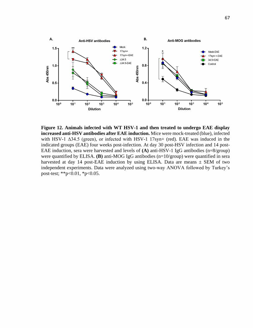
3.4.4 Asymptomatic mice infected with WT HSV-1 display increased amounts of anti-
HSV-1 antibodies after EAE induction .............................................................................. 63
3.5 Discussion ....................................................................................................................... 68
3.6 Acknowledgements ......................................................................................................... 72
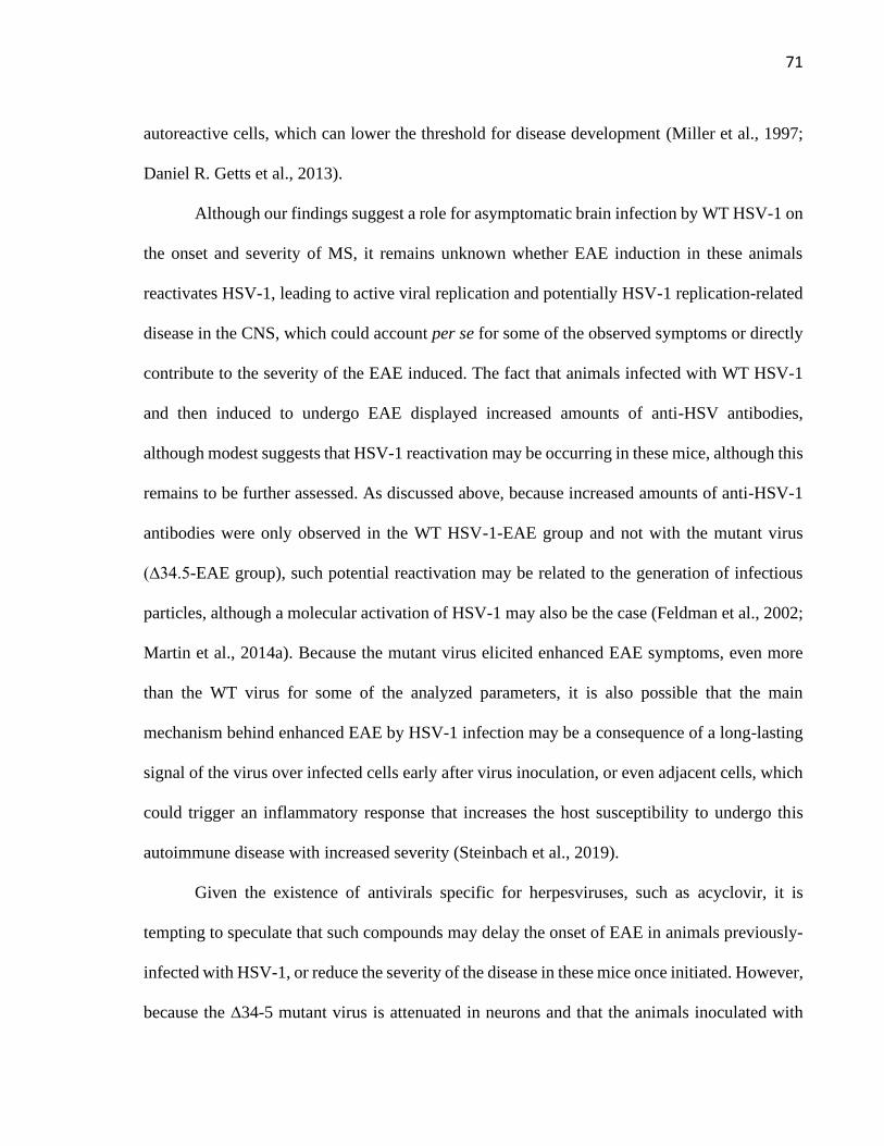
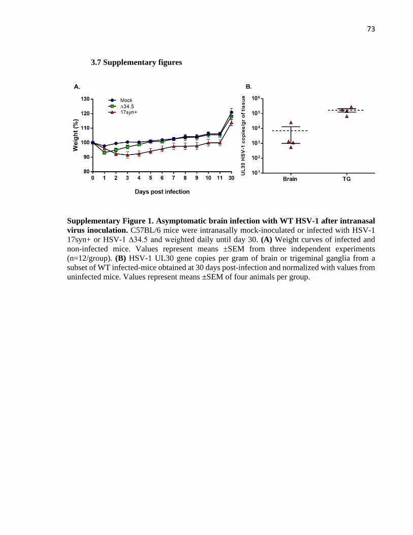
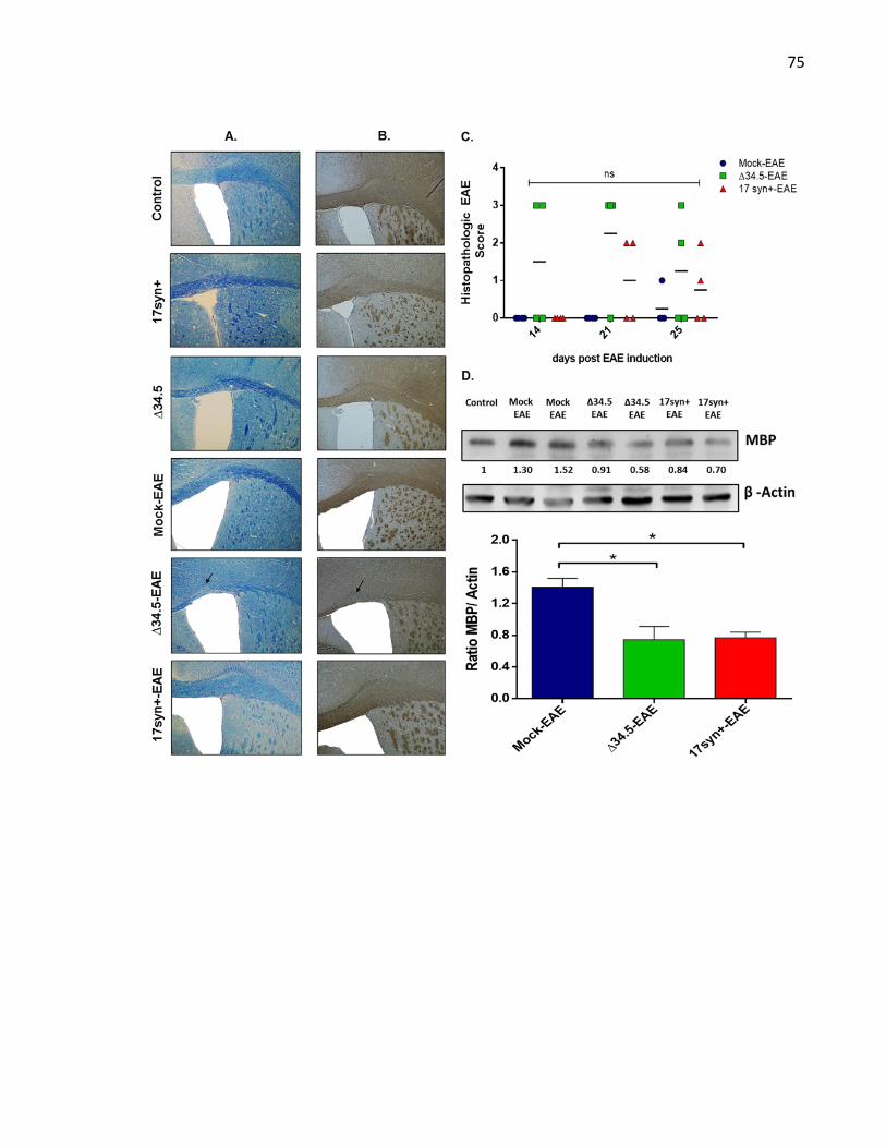
3.7 Supplementary figures .................................................................................................... 73
4. DISCUSSION ...................................................................................................................... 80
5. CONCLUDING REMARKS ............................................................................................. 92
6. APPENDIX .......................................................................................................................... 94
6.1 Contribution in scientific publications during this thesis and PhD training. .................. 94
6.2 Scientific meetings attended during this thesis and awards. ........................................... 99
REFERENCES ...................................................................................................................... 100
Page 5
4
FIGURE INDEX
Figure 1. Life cycle of HSV-1 .................................................................................................. 13
Figure 2. Central nervous system infection with HSV-1 .......................................................... 16
Figure 3. Acute and chronic neuroinflammation by HSV-1 brain infection ............................ 19
Figure 4. Functions of the neurovirulence factor ICP34.5 ....................................................... 23
Figure 5. Inflammatory process after EAE induction ............................................................... 31
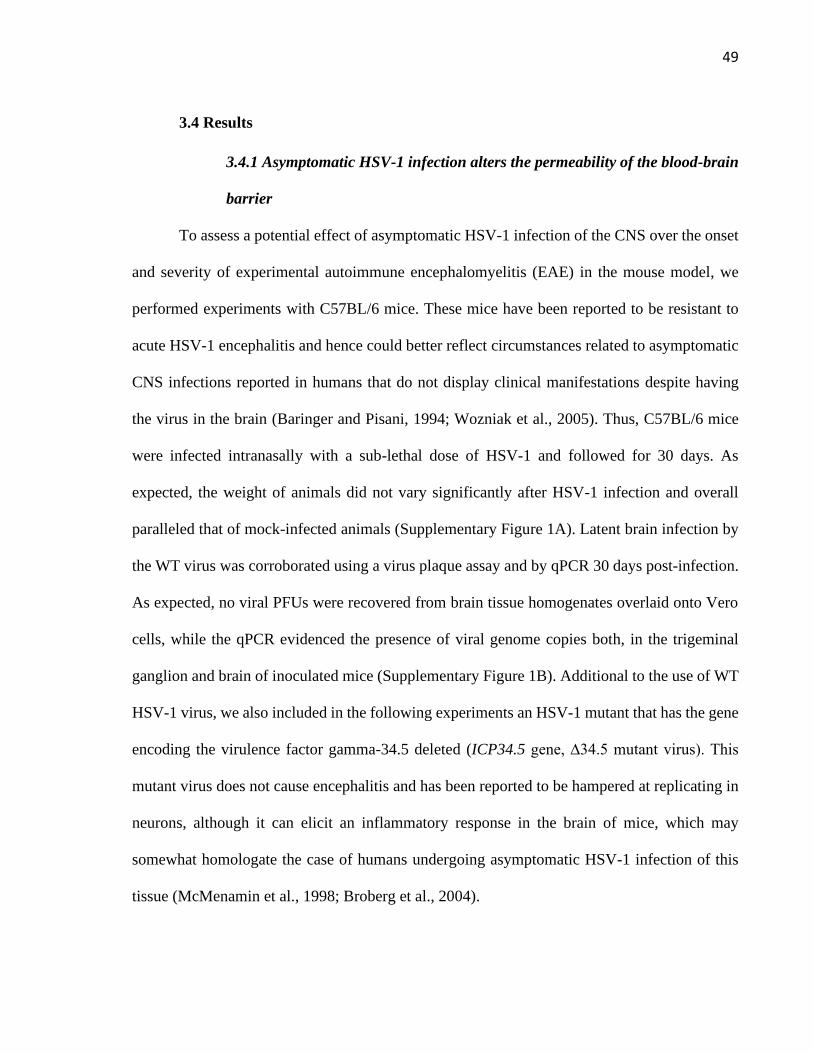
Figure 6. Asymptomatic HSV-1 infection increases BBB permeability in vivo ...................... 51
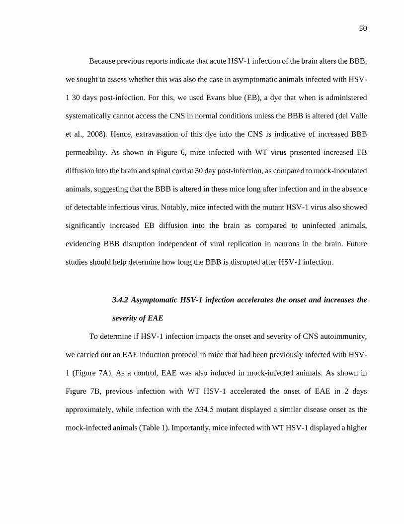
Figure 7. Asymptomatic HSV-1 infection accelerates the onset and increases the severity of
EAE ........................................................................................................................................... 52
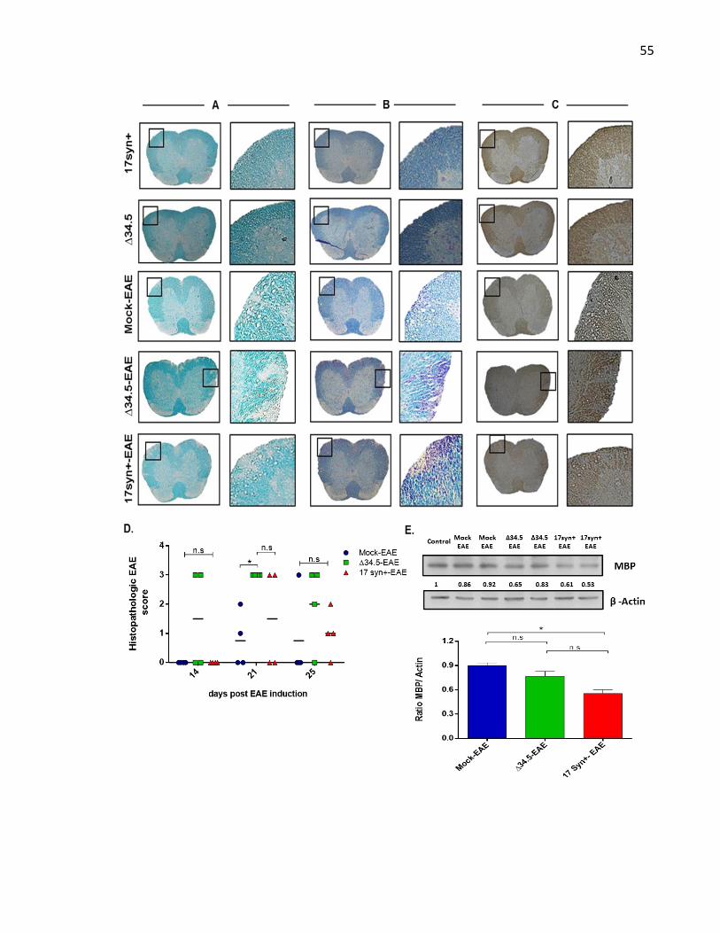
Figure 8. Asymptomatic HSV-1 infection increases spinal cord demyelination after EAE
induction. .................................................................................................................................. 56
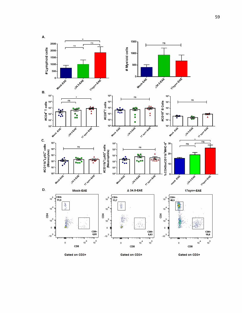
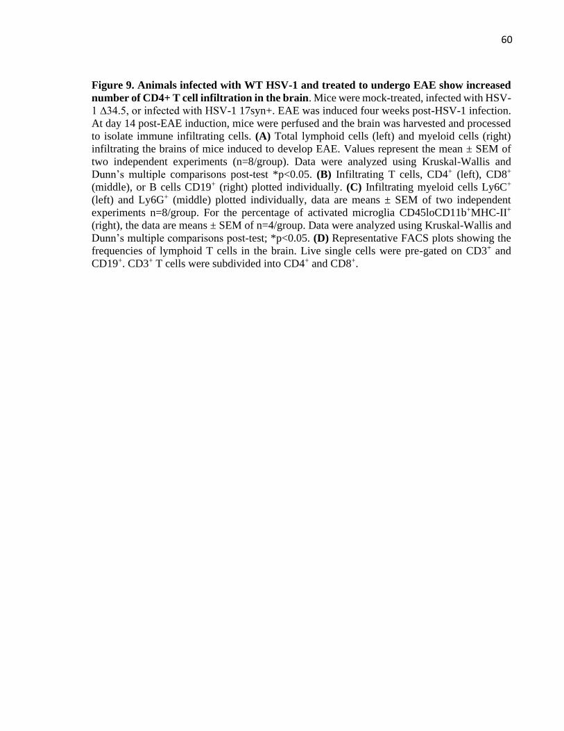
Figure 9. Animals infected with WT HSV-1 and treated to undergo EAE show increased number
of CD4+ T cell infiltration in the brain ..................................................................................... 60
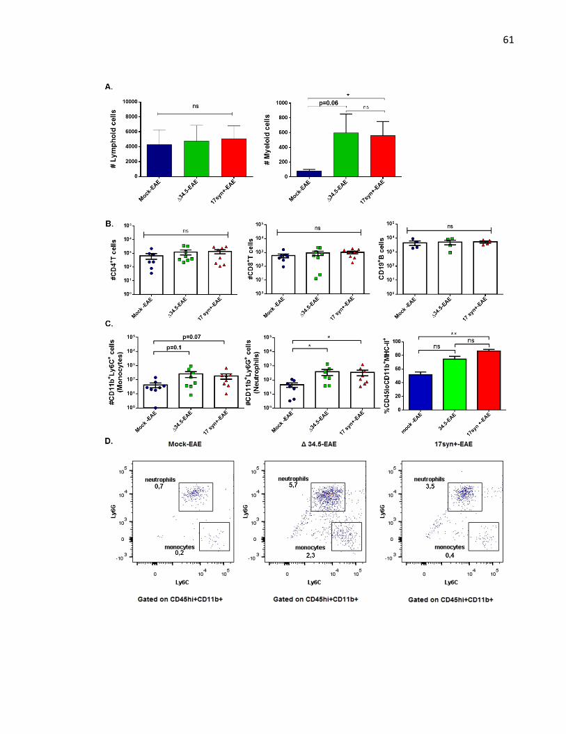
Figure 10. Animals infected with HSV-1 and treated to undergo EAE display increased number
of neutrophils infiltrating the spinal cord ................................................................................. 62
Figure 11. Asymptomatic HSV-1 infection increases the expression of pro-inflammatory
cytokines in the CNS of mice with EAE. ................................................................................. 65
Figure 12. Animals infected with WT HSV-1 and then treated to undergo EAE display increased
anti-HSV antibodies after EAE induction ................................................................................ 67
Page 6
5
TABLE INDEX
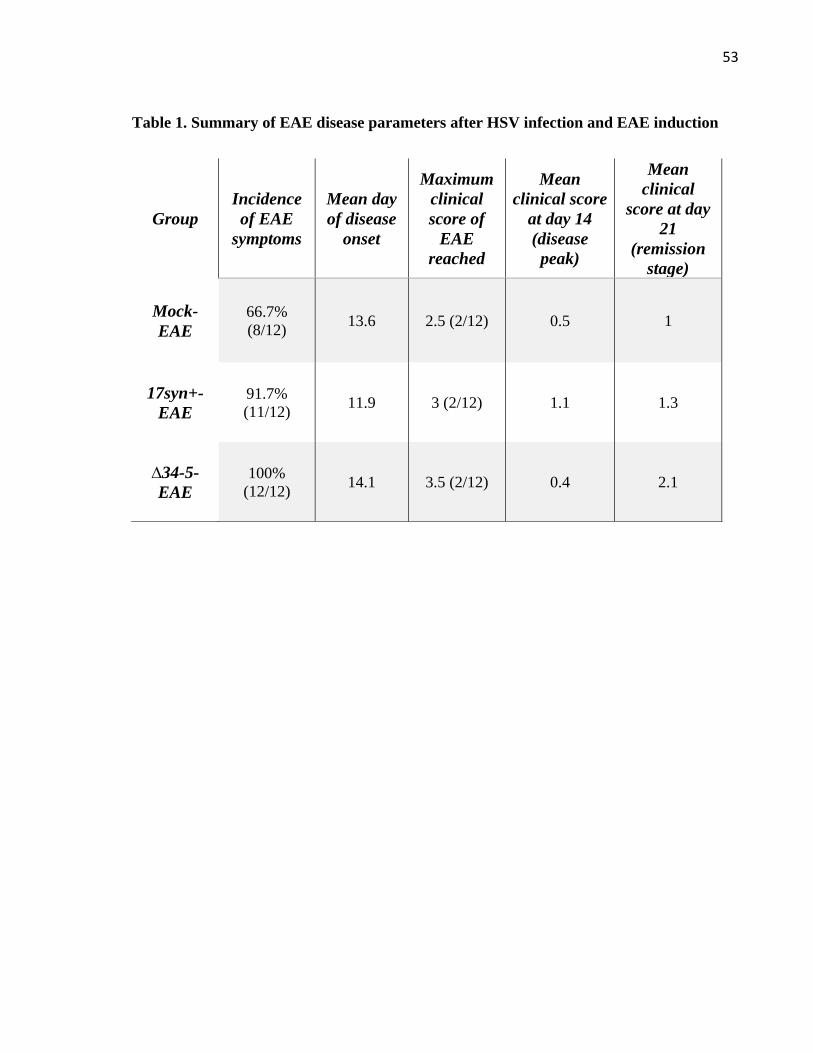
Table 1. Summary of EAE disease parameters after HSV infection and EAE induction ........ 53
Page 7
6
ABSTRACT
Multiple sclerosis is a demyelinating autoimmune disease of the central nervous system
(CNS) that severely impairs the individual’s motor and sensory functions. At present, its cause
or causes are unknown, and the available treatments only decrease the frequency of
inflammatory relapses but do not prevent chronic damage and neurologic decline. There is
evidence that suggests that viruses may play roles in multiple sclerosis onset and pathogenesis
by acting as environmental triggers. Importantly, viruses belonging to the Herpesviridae family,
which are acquired at early stages of life, and cause lifelong infections have been defined as
major candidates for triggering or exacerbating this disease. Currently, only a few studies have
assessed a potential role for herpes simplex viruses in multiple sclerosis. Noteworthy, herpes
simplex virus type 1 (HSV-1) DNA has been found in cerebrospinal fluid and peripheral blood
of patients with multiple sclerosis relapses, as well as more frequently in post-mortem brain
samples of individuals with multiple sclerosis than healthy controls. Notably, HSV-1 infections
are mainly asymptomatic, and this virus may reach the brain throughout life without evident
clinical symptoms. Moreover, accumulating data suggests that persistent HSV-1 infection in the
brain could produce prolonged neuroinflammation due to continuous subclinical reactivations
leading to neurodegenerative disorders in susceptible individuals. The goal of this thesis was to
determine whether asymptomatic HSV-1 infection favors the onset of multiple sclerosis and its
severity. We studied this question by using animals that recapitulate several aspects related to
multiple sclerosis disease and HSV-1 infection in humans. First, we infected mice with a sub-
lethal dose of HSV-1, waited for their recovery from acute infection at least 30 days, and then
we induced experimental autoimmune encephalomyelitis (EAE), the main animal model used
Page 8
7
for studying multiple sclerosis disease. The onset and severity of multiple sclerosis symptoms
in the EAE mouse model was compared with non-infected animals. We determined the
populations of immune cells infiltrating the CNS of mice after HSV-1 infection and EAE
induction, as well as cytokines produced in this tissue once autoimmunity was initiated. We also
assessed the permeability of the blood-brain barrier (BBB) 30 days post-HSV-1 infection. Our
results show that a previous infection with HSV-1 accelerates the onset of EAE and enhances
disease severity. Moreover, the animals previously infected with HSV-1, and induced to develop
EAE undergo increased CNS inflammation as compared to uninfected animals, which was
characterized by prolongated microglia cell activation, an elevated infiltration of CD4+ T cells
in the brain and increased infiltration of neutrophils in the spinal cord, as well as significant
levels of IL-6 and IL-1β mRNA expression in these tissues. Notably, we also found that after
asymptomatic HSV-1 infection, the BBB remains disrupted for up to 30 days when virions are
not detectable. We expect that this study will help to better define the possible contribution of
HSV-1 infection in multiple sclerosis disease and warrant future studies and trials with antiviral
interventions as a potential treatment for this disease to slow its progression.
Page 9
8
RESUMEN
La esclerosis múltiple es una enfermedad autoinmune desmielinizante del sistema
nervioso central (SNC) que perjudica severamente las funciones sensoriales y motoras del
individuo. Hoy en día, la causa o causas de esta enfermedad son desconocidas y el tratamiento
disponible solo disminuye la frecuencia de las recaídas inflamatorias, pero no previene del daño
crónico y el declive neurológico. Existe evidencia que sugiere que los virus pueden tener un
papel importante en el inicio y la patogénesis de la esclerosis múltiple por actuar como
gatillantes ambientales. Notablemente, virus que pertenecen a la familia Herpesviridae, los
cuales son adquiridos en etapas tempranas de la vida y causan infecciones latentes de por vida,
han sido definidos como principales candidatos para iniciar o exacerbar esta enfermedad.
Actualmente, pocos estudios han evaluado el potencial papel de los virus del herpes simple en
esclerosis múltiple. Cabe resaltar, que el virus del herpes simple tipo 1 (VHS-1) se ha detectado
en líquido cefalorraquídeo y en sangre periférica de pacientes con esclerosis múltiple durante
recaídas inflamatorias, así como también en mayor frecuencia en muestras de cerebro post
muerte de individuos con esclerosis múltiple que en individuos sanos. Además, las infecciones
producidas por VHS-1 son principalmente asintomáticas y este virus podría alcanzar el cerebro
a lo largo de la vida sin síntomas clínicos evidentes. Además, datos acumulados sugieren que la
infección persistente con VHS-1 en el cerebro produce prolongada neuroinflamación debido a
continuas reactivaciones subclínicas que conduce a desordenes neurodegenerativos en personas
susceptibles. El objetivo de esta tesis fue determinar si la infección asintomática con VHS-1
favorece el inicio de la esclerosis múltiple y su severidad. Nosotros abordamos esta pregunta
usando animales que recapitulan varios aspectos relacionados con la enfermedad de la esclerosis
Page 10
9
múltiple y la infección con VHS-1 en humanos. Primero, infectamos ratones con una dosis no
letal de VHS-1, esperamos a la recuperación de la infección aguda, al menos 30 días, y luego
inducimos la enfermedad de encefalomielitis autoinmune experimental (EAE), la cual es el
principal modelo animal usado para estudiar la enfermedad de esclerosis múltiple. El inicio y
severidad de síntomas de esclerosis múltiple en el modelo murino de EAE fue comparado con
animales no infectados. Determinamos las poblaciones de células inmunes infiltrando SNC de
ratones después de la infección con VHS-1 e inducción de EAE, así como también las citoquinas
producidas en este tejido luego del inicio de la autoinmunidad. También evaluamos la
permeabilidad de la barrera hemato-encefálica 30 días post infección con VHS-1. Nuestros
resultados muestran que una infección previa con VHS-1 acelera el inicio de EAE y aumenta la
severidad de la enfermedad en el modelo murino. Además, animales previamente infectados con
VHS-1 e inducidos a desarrollar EAE padecen una mayor inflamación de SNC que los animales
no infectados, lo cual se caracterizó por prolongada activación de microglía, una elevada
infiltración de células T CD4+ en el cerebro y neutrófilos en la médula espinal, y niveles de
expresión significativos de mRNA de las citoquinas IL-6 e IL-1β en estos tejidos. Notablemente,
también encontramos que después de la infección asintomática con VHS-1, la barrera hemato-
encefálica permanece alterada hasta por 30 días cuando no son detectados viriones. Esperamos
que este estudio ayude a definir mejor la posible contribución de la infección por VHS-1 en la
enfermedad de la esclerosis múltiple y a garantizar futuros estudios y ensayos con
intervenciones antivirales como un potencial tratamiento de esta enfermedad para retardar su
progresión.
Page 11
10
1. THEORETICAL BACKGROUND
1.1 Epidemiology and life cycle of Herpes simplex virus type-1 (HSV-1)
HSV-1 is an enveloped double-stranded DNA virus belonging to the Herpesviridae
family, that has a genome of approximately 152 Kbp with more than 80 different open reading
frames (ORFs) (Boehmer and Nimonkar, 2003). Importantly, all herpesviruses cause lifelong
latent infections in their hosts with sporadic reactivations (Perng and Jones, 2010). HSV-1 is a
neurotropic pathogen with a wide spectrum of clinical symptoms ranging from harmless
manifestations, such as oral and facial lesions to severe infection of the eyes and the central
nervous system (CNS) (Suazo et al., 2015). This virus is the most common cause of sporadic
encephalitis in adults, as well as infectious blindness due to herpetic keratitis (Lairson et al.,
2003; Whitley, 2015). HSV-1 is usually acquired during childhood, and worldwide
approximately 65% of people have antibodies against this virus. However, only 20–40% of
infected individuals develop symptoms (Dobson et al., 2003), but they are reservoirs that
contribute to viral transmission towards new hosts through asymptomatic shedding (Miller and
Danaher, 2008; Ramchandani et al., 2016).
HSV-1 can alternate between a lytic infection phase that produces virions, or a latent
state characterized by transcriptional repression of all viral lytic genes (Whitley and Roizman,
2001). HSV-1 enters epithelial cells at the initial site of infection by fusing its envelope with the
cell membrane, through a process that is mediated and assisted by several viral glycoproteins.
The fusion of membranes leads to the release of the viral capsid surrounded by tegument
proteins into the cell cytoplasm, then travels associated to microtubules, to the cell nucleus. The
viral DNA is delivered into the nucleus and transcribed in a cascade-dependent manner, with
Page 12
11
three major waves of transcription: first, the expression of immediate early genes (IE or alpha
genes), followed by the expression of early genes (E or beta genes) and lastly, late genes (L or
gamma genes). Furthermore, the latter are sometimes sub-divided into late-early and late genes
(or gamma-1 and gamma-2 genes, respectively) (Honess and Roizman, 1974; Ibáñez et al.,
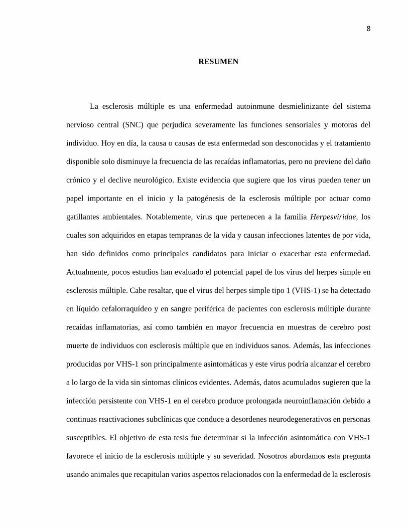
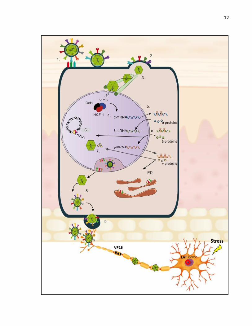
2018) (Figure 1). For IE mRNAs, a viral transactivator called VP16 plays an important role in
promoting their transcription by binding to cellular factors namely the octamer-binding protein
1 (Oct1) and the host cell factor-1 (HCF-1) (Herrera and Triezenberg, 2004). Some IE viral
genes play key roles in the evasion of the host cellular antiviral response. As IE proteins are
expressed, some of them will act as transcription factors for E viral genes, and then is promoted
the synthesis of E proteins that play roles in viral processes, such as DNA replication (Suazo et
al., 2015). Finally, late gene expression occurs thanks to the transactivation properties of viral
IE genes (Honess and Roizman, 1975). These later genes encode, among others, for structural
components of the virion, such as capsid, tegument, and viral surface proteins (Herrera and
Triezenberg, 2004). Once viral DNA is replicated, it is packaged into new capsids that are
released into the cytoplasm, where they are complemented with viral tegument and
glycoproteins. Finally, new infectious viral particles are released to the extracellular and the
virus gains access to the termini of sensory neurons that innervate the skin reaching the cell
body of these cells by retrograde transport through neuronal axons (Antinone and Smith, 2010).
Here, the virus can spread through a lytic cycle or enter latency (Figure 1). During facial
infections that affect the mouth, face or eyes, viral progeny from HSV-1 replication in the
epithelium will reach the cell bodies of sensory and autonomic nerve terminals of neurons in the
trigeminal ganglia (TG). Virus within neurons can enter in a latency phase in which viral DNA
remains as an episome in the nucleus with reduced-to-none virus protein expression
Page 14
13
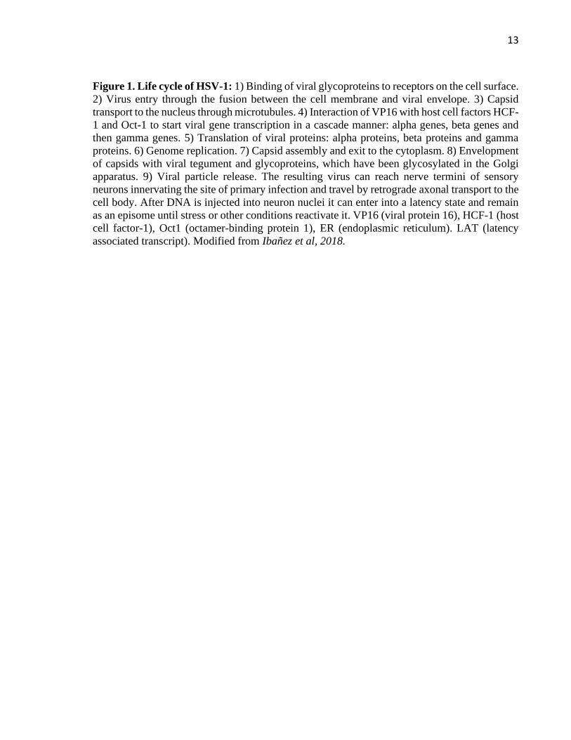
Figure 1. Life cycle of HSV-1: 1) Binding of viral glycoproteins to receptors on the cell surface.
2) Virus entry through the fusion between the cell membrane and viral envelope. 3) Capsid
transport to the nucleus through microtubules. 4) Interaction of VP16 with host cell factors HCF-
1 and Oct-1 to start viral gene transcription in a cascade manner: alpha genes, beta genes and
then gamma genes. 5) Translation of viral proteins: alpha proteins, beta proteins and gamma
proteins. 6) Genome replication. 7) Capsid assembly and exit to the cytoplasm. 8) Envelopment
of capsids with viral tegument and glycoproteins, which have been glycosylated in the Golgi
apparatus. 9) Viral particle release. The resulting virus can reach nerve termini of sensory
neurons innervating the site of primary infection and travel by retrograde axonal transport to the
cell body. After DNA is injected into neuron nuclei it can enter into a latency state and remain
as an episome until stress or other conditions reactivate it. VP16 (viral protein 16), HCF-1 (host
cell factor-1), Oct1 (octamer-binding protein 1), ER (endoplasmic reticulum). LAT (latency
associated transcript). Modified from Ibañez et al, 2018.
Page 15
14
(Nicoll et al., 2012). It has been hypothesized that VP16 may be lost during axonal transport
and latency state is favored due to the lack of a viral transactivator (Kim et al., 2012).
Remarkably, latency is mainly characterized by the transcription of only one viral transcript
from the viral genome, which is non-coding and is termed the latency-associated transcript
(LAT) (Nicoll et al., 2016). Importantly, in latently-infected cells LAT is processed into
miRNAs that silence the expression of viral genes that are required for productive virus
replication (Umbach et al., 2008). In addition, LAT promoter in neurons has been associated
with epigenetic markers of active transcription during the latent state (i.e. particular acetylation
patterns at histone H3) (Kubat et al., 2004). In contrast, the promoters of lytic viral genes were
found to display methylations associated to heterochromatin (Cliffe et al., 2009; Cliffe and
Wilson, 2017). Nevertheless, sporadic expression of lytic viral genes in neurons during latency
in the form of mRNA has been reported by several groups (Feldman et al., 2002; Margolis et
al., 2007; Ma et al., 2014), which was followed in some cases by protein synthesis without
production of new viral particles suggesting that HSV-1 persistence is a dynamic process that
includes not only a latent state with sporadic productive reactivations, but also spontaneous
molecular reactivations without productive progeny production (Du et al., 2011; Kim et al.,
2012; Martin et al., 2014a). Ultimately, under stress conditions HSV‐1 can reactivate from
neurons releasing new viral particles that can cause recurrent lesions close to the initial site of
infection, spread asymptomatically to new hosts, or enter into the CNS by anterograde transport
(Halford et al., 1996).
Page 16
15
1.2 HSV-1 at the central nervous system
HSV‐1 can invade the brain and replicate in neuronal cells causing herpes simplex
encephalitis (HSE) (Gnann and Whitley, 2017) or creating a reservoir for virus production with
asymptomatic reactivations. About 30% of HSE cases are related to primary infection (more
commonly in children and adolescents), while 70% of cases are attributed to viral reactivation
from previous infection (mainly adults). Figure 2 shows the different strategies used by HSV-1
to infect the brain. One of them is associated with a primary infection via olfactory tracts
(Burgos et al., 2006; Jennische et al., 2015). In fact, studies using animal models have shown
the spread of HSV-1 from the nasal cavity to the CNS after infection of the olfactory epithelium,
which is connected with the olfactory bulb and consequently the limbic system, resulting in
focal encephalitis in the brain (Figure 2A) (Twomey et al., 1979; Dinn, 1980). Regarding
neonatal HSV-1 infections, the olfactory route is frequently considered responsible and widely
described as the result of close contact between the newborn olfactory tissue and HSV-1 virions
present in the birth canal of the mother at the time of birth. However, a study in mice suggests
that vertical transmission is predominantly hematogenous (Burgos et al., 2006). This study
showed that placenta had high number of viral genomes, indicating that HSV-1 could reach the
brain of fetuses by this route through the maternal bloodstream (Burgos et al., 2006). Another
route by which HSV-1 may gain access to the brain, is peripheral viral reactivation with
subsequent anterograde axonal transport, associated with latent virus in TG acquired in a
previous orolabial or eye infection (Figure 2B) (Whitley et al., 1982). Finally, latent HSV-1 in
the brain may be a source of productive reactivations that seed infection to other sites within
this tissue, or cause HSE in some susceptible individuals (Figure 2C). In the past, sensory
ganglia was understood to be the only place of HSV-1 latency, but autopsy studies have
Page 17
16
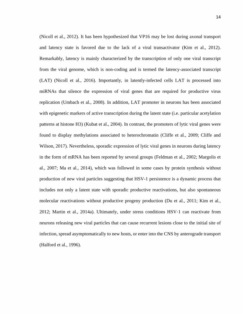
Figure 2. Central nervous system infection with HSV-1. A) HSV-1 CNS infection through
the olfactory route: HSV-1 can infect the termini of olfactory neurons enervating the nasal
epithelium and access the CNS by retrograde axonal transport through neurons until reaching
the olfactory bulb in the brain. B) HSV-1 can also infect the CNS because of HSV-1 peripheral
reactivation. HSV-1 can reactivate from neurons in the trigeminal ganglia (TG) and reach either
the skin or CNS through anterograde axonal transport. C) HSV-1 can also reach different regions
of the CNS because of HSV-1 reactivation within the brain. Reactivation of latent virus within
the CNS has been reported to reach the cerebellum, olfactory bulb, frontal cortex, or
hippocampus. Modified from Duarte et al, 2019.
Page 18
17
demonstrated the presence of HSV-1 DNA in brain tissue in individuals with no known
neurologic disease, suggesting the possibility that HSV-1 could establish latency in the CNS
(Baringer and Pisani, 1994). Moreover, some studies have reported viral reactivation in ex vivo
brainstem tissue explants following latent infection with HSV-1 in mice (Chen et al., 2006).
Also, infectious virus was recovered in the brainstem of latently infected mice, which were
induced to viral reactivation by hyperthermia and latent viral genomes were detected in the
cerebellum, olfactory bulbs, frontal cortex, and hippocampus (Yao et al., 2014). That study
indicates that this virus can reach the brain and remain there in a latent state, from where it can
reactivate after stress conditions leading to a symptomatic or an asymptomatic spread.
1.3 HSV-1 and neurodegeneration
There is accumulating evidence suggesting that HSV-1 infection of the brain both, in
symptomatic and asymptomatic individuals could lead to neuronal damage and eventually, to
neurodegenerative disorders, such as multiple sclerosis or Alzheimer´s disease (extensively
reviewed in Duarte et al.,2019). Indeed, neurological sequelae, such as epilepsy, amnesia or
cognitive and behavioral alterations are common after HSE despite treatment with antivirals that
limit virus replication (Misra et al., 2008; Riancho et al., 2013). Noteworthy, immune-related
mechanisms have been defined as main players that induce chronic neurologic damage
(Marques et al., 2008). In addition, subclinical reactivations from brain neurons may eventually
occur and produce local and regional effects in this tissue which may ultimately lead to
neurodegenerative manifestations (Perng and Jones, 2010; Martin et al., 2014b).
Page 19
18
Importantly, studies using mouse models support the above-mentioned notions and have
allowed to deepen our knowledge on the chronic alterations elicited by HSV-1 infection over
the CNS both, in mice that are more susceptible of undergoing severe viral encephalitis
(Marques et al., 2008; Martin et al., 2014a), as well as in C57BL/6 mice that are resistant to
HSV-1 encephalitis under certain experimental conditions, given by their rapid and effective
innate alpha/beta interferon (IFN-α/β) response that reduces viral pathogenesis and increases
their survival, leading to asymptomatic brain infection (Halford et al., 2004; Kastrukoff et al.,
2012; Zimmermann et al., 2017).
A study using BALB/c mice showed that early during HSE, the immune response in the
brain is dominated by the influx of macrophages and neutrophils, which play a critical role in
viral clearance (Figure 3A) (Marques et al., 2008; Terry et al., 2012). Moreover, macrophages
secrete TNF-α and microglial cells express high levels of IL-1β, which affect the blood-brain
barrier (BBB) by upregulating endothelial cell adhesion molecules (Fields et al., 2006). Non-
productive HSV-1 infection of microglia can also lead to the expression of others cytokines and
pro-inflammatory molecules, such as TNF-α, IL-6, IL-8, CCL5 and chemokine CXCL10
(Lokensgard et al., 2001). After 14 days post infection T lymphocytes begin to be a predominant
leucocyte cell type infiltrating the brain, which is composed mainly by CD8+ T cells that persist
in this tissue up to 30 days post-infection without detectable viral replication (Figure 3B)
(Marques et al., 2008; Terry et al., 2012). Importantly, infiltrating CD8+ T cells express IFN-γ
which is known to synergize with TNF-α to increase NO-induced neurodegeneration and
demyelination in the brains of mice (Blais and Rivest, 2004). Moreover, prolonged microglial
activation has also been reported in the brains of mice latently-infected with HSV, as indicated
by high MHC class-II expression levels up to 30 days post-infection
Page 20
19
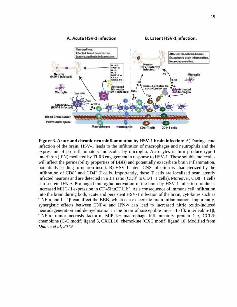
Figure 3. Acute and chronic neuroinflammation by HSV-1 brain infection: A) During acute
infection of the brain, HSV-1 leads to the infiltration of macrophages and neutrophils and the
expression of pro-inflammatory molecules by microglia. Astrocytes in turn produce type-I
interferon (IFN) mediated by TLR3 engagement in response to HSV-1. These soluble molecules
will affect the permeability properties of BBB) and potentially exacerbate brain inflammation,
potentially leading to neuron insult. B) HSV-1 latent CNS infection is characterized by the
infiltration of CD8+ and CD4+ T cells. Importantly, these T cells are localized near latently
infected neurons and are detected in a 3:1 ratio (CD8+ to CD4+ T cells). Moreover, CD8+ T cells
can secrete IFN-γ. Prolonged microglial activation in the brain by HSV-1 infection produces
increased MHC-II expression in CD45intCD11b+. As a consequence of immune cell infiltration
into the brain during both, acute and persistent HSV-1 infection of the brain, cytokines such as
TNF-α and IL-1β can affect the BBB, which can exacerbate brain inflammation. Importantly,
synergistic effects between TNF-α and IFN-γ can lead to increased nitric oxide-induced
neurodegeneration and demyelination in the brain of susceptible mice. IL-1β: interleukin-1β,
TNF-α: tumor necrosis factor-α, MIP-1α: macrophage inflammatory protein 1-α, CCL5:
chemokine (C-C motif) ligand 5, CXCL10: chemokine (CXC motif) ligand 10. Modified from
Duarte et al, 2019.
Page 21
20
(Figure 3B) (Marques et al., 2008) . In addition, asymptomatic reactivation in BALB/c mice
was demonstrated by the detection of viral ICP4 protein in the TG and cerebral cortex of mice
60 days post-infection, and was accompanied by the up-regulation of markers of
neuroinflammation, such as toll-like receptor (TLR) 4, interferon α/β, and phosphorylated
interferon regulatory factor 3 (p-IRF3) (Martin et al., 2014a).
On the other hand, another study using C57BL/6 mice that survived an acute phase of
ocular infection accompanied with virus dissemination to the CNS, showed that LAT was
mainly concentrated within the lateral ventricles and the hippocampus (ependymal zone), as
well as the brainstem 30- and 60-days post-infection (Menendez et al., 2016). Surprisingly, the
ependymal region in the brain evidenced HSV-1 lytic gene transcripts being expressed at these
time-points post-infection, in contrast to the brainstem and TG, in which the expression of lytic
genes was decreased (Menendez et al., 2016). Interestingly, this study proposes the hypothesis
that a specific tropism of HSV-1 to the ependymal zone may be linked to chronic inflammatory
responses in the brain and that this zone may have particular conditions that provide an
environment that enhances viral persistence, potentially leading to neurodegeneration (Webb et
al., 1989; Conrady et al., 2013). A more recent study showed that the ependymal zone harbors
neural progenitor cells that are vulnerable to acute HSV-1 infection and viral lytic-associated
proteins were detected in these cells during latency (Chucair-Elliot et al., 2014). Importantly,
viral persistence in the ependymal zone of the brain was related to T cells expressing exhaustion
markers (LAG-3, TIM-3, PD1, CD160 and KLRG-1), which were unable to control HSV-1
infection ex vivo and secreted less IFN-γ and granzymes in comparison to T cells isolated from
TG (Wherry and Kurachi, 2015; Menendez et al., 2016).
Page 22
21
At the molecular level, the matrix metalloproteinases 2 and 9 (MMP-2 and MMP-9) have
been shown to be elevated in the brains in both, acute and latent HSV-1 infections. These MMPs
could degrade the extracellular matrix and cell surface proteins of the BBB and modulate its
permeability, which could lead to persistent cell infiltration increasing neuroinflammation
(Martínez-Torres et al., 2004; Weiser et al., 2007).
Finally, it has been reported that HSV-1 negatively modulates apoptosis-related
pathways in neurons favoring its persistence in the brain (Du et al., 2012; Carpenter et al., 2015),
and can disrupt autophagy-related processes leading to protein accumulation and cellular
toxicity in this tissue (Lussignol et al., 2013; O’Connell and Liang, 2016). Moreover, HSV-1
infection can produce mitochondrial dysfunction, which increases the production of reactive
oxygen species (Wnek et al., 2016). Therefore, HSV-1 could significantly contribute to the
pathogenesis of neurons, by interfering with these processes in the brain (Lussignol et al., 2013;
Carpenter et al., 2015; Wnek et al., 2016). On the other hand, because the immune system of an
individual tends to decay upon aging, opportunities arise for HSV-1 to reactivate in the organism
and spread to tissues such as the brain contributing to neurodegenerative disorders in humans
(Dobson et al., 2003; Otth et al., 2009; Martin et al., 2011; Buscarinu et al., 2017).
1.4 ICP34.5 is a neurovirulent factor of HSV-1
To productively replicate in the host nervous system, HSV-1 encodes several viral
proteins that counteract the host antiviral response (Suazo et al., 2015). The gamma-34.5 gene
encodes a neurovirulence factor named infected cell protein 34.5 (ICP34.5 or gamma-34.5),
which is present in two copies in the viral genome and is located in the inverted repeats of the
Page 23
22
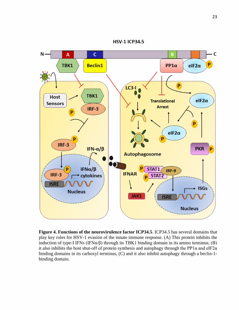
regions flanking the unique long (UL) sequence (Wilcox and Longnecker, 2016). This viral
protein has several binding-domains that target specific host proteins that are involved in several
effector pathways, such as type-I interferon (IFN-I) induction, host shutoff of protein synthesis,
and the inhibition of autophagy (Figure 4) (Wilcox and Longnecker, 2016).
Host cells respond to HSV-1 infection through the recognition of pathogen-associated
molecular patterns (PAMPs) that trigger IFN-I production, which in turn induces the expression
of an array of antiviral genes (Rasmussen et al., 2009). Recognition of PAMPs by host sensors,
such as toll-like receptors (TLRs), retinoid acid-inducible gene-I (RIG-I), melanoma
differentiation associated gene 5 (MDA5) or DNA-dependent activator of IFN-regulatory factor
(DAI), leads to downstream signaling events that ultimately activate TANK-binding kinase 1
(TBK1), which is responsible of phosphorylating and activating IRF3, the primary transcription
factor regulating the induction of type-I IFNs (Fitzgerald et al., 2003). Importantly, ICP34.5
abolishes the induction of IFN-I production through direct binding to TBK1 through its amino
terminus (Ma et al., 2012). This hijacking of TBK1 prevents IRF3 phosphorylation and its
consequently nuclear translocation for the transcriptional activation of IFN-I genes (Figure 4A)
(Verpooten et al., 2009). Nevertheless, if type-I IFNs are produced, they are detected by the
IFN-I receptor (IFNAR), which activates the JAK-STAT signaling pathway and initiates the
transcription of interferon stimulated genes (ISGs), which enhance their antiviral state (Ivashkiv
and Donlin, 2014). One of such ISGs is the double-stranded RNA–dependent protein kinase R
(PKR), which inhibits protein synthesis by phosphorylating the translation initiation factor
eIF2a (Mohr, 2004). Importantly, the carboxyl terminus of ICP34.5 binds to the host
phosphatase PP1α, which in turn binds to eIF2α and leads to eIF2α dephosphorylation and the
Page 24
23
Figure 4. Functions of the neurovirulence factor ICP34.5. ICP34.5 has several domains that
play key roles for HSV-1 evasion of the innate immune response. (A) This protein inhibits the
induction of type-I IFNs (IFNα/β) through its TBK1 binding domain in its amino terminus, (B)
it also inhibits the host shut-off of protein synthesis and autophagy through the PP1α and eIF2α
binding domains in its carboxyl terminus, (C) and it also inhibit autophagy through a beclin-1-
binding domain.
Page 25
24
reversing of protein synthesis shutoff in the cell (Figure 4B) (Wilcox et al., 2015a). In addition,
eIF2α phosphorylation promotes the induction of autophagy (Acevo-Rodríguez et al., 2020).
Autophagy acts as a defense mechanism against different infectious agents, promoting
lysosomal degradation of microorganisms, as well as playing key roles in immune signaling. It
also plays roles in antigen processing for pathogen-derived peptide presentation in MHC
molecules and for the delivery of viral nucleic acids to endosomal TLRs (Lussignol and
Esclatine, 2017). Importantly, autophagy has been reported to be key for controlling HSV-1
infection in neurons (O’Connell and Liang, 2016). This finding is in sharp contrast with
epithelial cells, where an IFN-I response is sufficient alone to control HSV-1 infection and in
which case autophagy is not required (Yordy et al., 2012). However, although autophagy
protects the adult brain from viral encephalitis, contrasting results have been reported in
newborn mice, where autophagy seems to be detrimental for the host and was described to
promote neuronal apoptosis. Interestingly, these findings suggest an age-dependent role for
autophagy during HSV-1 brain infection (Wilcox et al., 2015b). Notably, ICP34.5 inhibits
autophagy indirectly through eIF2α dephosphorylation by PP1α, as well as directly through its
interaction with the autophagy-inducing protein beclin-1 and interfering with the formation of
autophagosomes and antigen presentation in dendritic cells (DCs) (Figure 4C) (Orvedahl et al.,
2007; Gobeil and Leib, 2012; Wilcox et al., 2015a).
Because of the aforementioned functions of this protein, previous investigations have
studied HSV-1 mutant viruses that have the ICP34.5 gene deleted. Interestingly, these viruses
can replicate at the mucosae and epithelial tissues, although yielding lower titers and lasting for
fewer days as compared to the wild type virus (Whitley et al., 1993). These results indicate
ICP34.5 positively modulates the replication ability of HSV-1 early during infection when the
Page 26
25
virus challenges the innate immune response. Moreover, these mutants did not cause lethal
encephalitis due to its impaired ability to evade the antiviral response, reporting a reduced ability
to replicate in the nervous system, and also to establish latency and reactivate as determined ex
vivo (Orvedahl et al., 2007).
Nevertheless, some studies have shown that despite the apparent attenuated phenotype
of ICP34.5-deleted viruses, these mutants can cause the destruction of ependymal cells, as well
as neurons that are exposed to high amounts of the virus, which lead to inflammation in the
brain (Kesari et al., 1998; Markovitz and Roizman, 2000). A study evaluating the effect of the
∆34.5 mutant HSV-1 in the brain of different strains of rats and mice reported robust immune
responses consisting of macrophages and T cells in the brain in all the animal strains tested, yet
significant weight loss was only seen in some animals, which was accompanied by signs of
clinical disease (McMenamin et al., 1998). These results suggest that the dose of the virus used,
as well as the host immune system can impact the overcoming of the infection and limit or not
the severity of the infection and related disease. This is an important observation, as these mutant
viruses have been exploited for the delivery of disease-limiting cytokines in cancer and tumor
therapies, yet the immune responses elicited against these HSV-1 vectors have not been fully
elucidated (Broberg and Hukkanen, 2005). More recently, another study evaluated the
replication efficiency of numerous ∆34.5 HSV-1 mutants in nervous system tissues after
intranasal, corneal or intralabial infection routes, as well as the effects of the viruses over the
immune response after intranasal infection (Broberg et al., 2004). Importantly, this study
reported that the intranasal inoculation of HSV-1 mutants is an effective route for viral spread
into the CNS, with poor replication of the virus in this tissue, but viral DNA persistence even
21-days post-infection. Regarding the immune response, the infection with HSV-1 mutants
Page 27
26
alone, or encoding IL-4 or IL-10 transgenes induced Th2-type cytokine responses (Broberg et
al., 2004). However, viruses encoding the IL-10 transgene or without any transgenes produced
Th1-type cytokines, namely IFN-γ and IL-23 in the brain. Additionally, the transgene-free
mutant virus elicited a higher number of lymphoid T cells and CD11c+ antigen presenting cells
in the spleen as compared to WT HSV-1 (Broberg et al., 2004). Taken together, these results
suggest an additional immunomodulatory role for ICP34.5 and calls for further studies of the
immune responses produced by these mutants viruses that are being used as vectors in gene
therapy (Broberg and Hukkanen, 2005; Hukkanen and Nygårdas, 2013). It is important to
guarantee desired immune responses that aid as therapies, while avoiding possible adverse
effects.
1.5 Multiple sclerosis disease
Multiple Sclerosis (MS) is a neurodegenerative disorder affecting the CNS, where the
protective myelin sheath that covers the nerve cells in the brain, spinal cord and optic nerves are
damaged, inflamed and hardened by attacking of myelin-specific autoreactive T cells or B cells,
and myeloid cells that infiltrate the CNS mediating an inflammatory response that results in
demyelination and axon degradation, that disrupts the ability of neurons to transmit the nerve
impulse, resulting in a widespread of signs and symptoms depending of the site of lesion,
including physical, sensorial, cognitive and sometimes psychiatric problems (Compston and
Coles, 2008; Thomas, 2012; Dobson and Giovannoni, 2019). MS is the most common cause of
non-traumatic neurological dysfunction affecting principally young adults between the age of
20 and 50 with an average age of onset of 29, which generate a great socio-economic burden
because the disease may hinder ability to maintain studies and work (Msif, 2013). It is estimated
Page 28
27
that approximately 2.3 million people suffer from this disease worldwide, with highest
prevalence in countries in North America and Europe (140 and 108 cases per 100,000
individuals, respectively) and lowest in African and Asian countries (2.1 and 2.2 cases per
100.000 individuals, respectively) (Msif, 2013). Chile is considered a low to medium risk
country for MS, because in the Magallanes region there is a prevalence of 13 to 14 cases per
100,000 individuals (Melcon et al., 2013), with all geographical regions in Chile showing a
cumulative prevalence rate of 5.69 per 100,000 individuals and an annual incidence rate of 0.90
(Díaz et al., 2012).
MS exhibits a heterogeneous progression and symptomatology. The first evident sign of
its appearance is called clinically isolated syndrome (CIS), an event with observed
demyelination involving the optic nerve, brain or spinal cord (Miller et al., 2005; Filippi et al.,
2018). 85% of newly diagnosed patients present a relapsing-remitting form (RRMS) of MS,
which is display a worsening of neurological function called relapse or exacerbation. Disease is
followed by periods of remission in which the neurological functionality is partially recovered
within weeks to months. It has been estimated that up to 80% of these individuals will develop
secondary progressive MS (SPMS), one to two decades post-diagnosis. In SPMS, the
inflammation of CNS is reduced, however progressive neurological decline and CNS atrophy
are observed. Finally, approximately 10% of patients with MS are diagnosed with primary
progressive disease (PPMS), which shows a progressive decline from the onset and an absence
of relapses (Dendrou et al., 2005; Filippi et al., 2018; Dobson and Giovannoni, 2019).
The pathology of the disease is characterized by focal demyelinated plaques caused by
activated self-reactive cells that recognize myelin antigens and migrate to the CNS after
disruption of the BBB. These infiltrating cells may also lead to reactive gliosis, loss of
Page 29
28
oligodendrocytes, and axonal damage (Dendrou et al., 2005; Haider et al., 2016). The
mechanisms underlying the BBB breakdown are not entirely determined but seem to be
mediated by the direct effects of proinflammatory cytokines, such as IL-1β and IL-6, or
chemokines released by resident CNS cells (microglia, astrocytes and endothelial cells) or
lymphoid and myeloid infiltrating cells (Argaw et al., 2006; Aubé et al., 2014; Wang et al.,
2014).
There is no cure for this disease because its cause is unknown. Currently, two models
have been proposed to explain the development of MS. Whereas in the first model, autoreactive
T cells are activated by a peripheral stimulus and then migrate to the CNS by crossing the BBB,
in the second model the demyelination is caused by an inflammatory response mounted against
an infection in the CNS, and the activation and infiltration of self-reactive T-cell occur as a
secondary phenomenon (Dendrou et al., 2005). However, what triggers the loss of peripheral
immunologic tolerance leading to the activation of these autoreactive immune cells in an
individual and what determines their infiltration into the CNS remains at present somewhat
unknown. It is thought that MS develops as an interplay between multiple factors, such as
genetic predisposition, the host immune system and environmental factors (Beecham et al.,
2013). Importantly, viral infections have been defined as environmental triggers that could play
an important role in disease development and progress.
1.6 Animal models to study the relationship between virus and multiple sclerosis
disease.
Animal models of demyelinating diseases have allowed advances in the understanding
mechanisms involving virus in autoimmunity. As support to the intrinsic theory, some viral
Page 30
29
infection in the CNS can produce demyelinating disease by epitope spreading or bystander
activation. As an example, Theilers´s murine encephalomyelitis virus (TMEV) causes a
persistent infection in CNS without complete viral clearance and reactivity to myelin antigens
emerges after the onset of viral-induced clinical symptoms, which is due to epitope spreading
after initial virus-specific Th1-mediated demyelination (Karpus et al., 1995; Miller et al., 1997).
In contrast, during CNS infection by neurotropic mouse hepatitis virus (MHV), infectious virus
is not detected in the brain tissue, and MHV persistence is characterized by presence of viral
RNA and proteins, which have been associated with T cell retention. Likely, chronic
inflammation releases myelin antigen leading to the bystander activation of myelin-specific T
cells (Bergmann et al., 2006).
Because of the difficulty in identifying direct causal effectors over MS initiation in
humans, animal models that mimic MS or share disease traits with this disease are highly
valuable for this purpose. Experimental autoimmune encephalomyelitis (EAE) is a disease in
animals that shares numerous molecular and cellular signatures with MS and can be actively
induced using different CNS antigens and peptides, as well as through passive adoptive transfer
of activated CD4+ T cells that recognize such self-antigens (Baxter, 2007). One such model is
based on peripheral immunization of mice with oligodendrocyte glycoprotein-derived peptide
(MOG35-55) and the disruption of the BBB with pertussis toxin (Kastrukoff et al., 1987).
Approximately 12 days after treatment, mice develop ascending paralysis due to spinal cord
inflammation, which leads to demyelination, neuron dysfunction and death in its severe form
when using high doses of MOG peptide and pertussis toxin (Constantinescu et al., 2011;
Robinson et al., 2014). Immune cell infiltrations in the brain are atypical in this mouse model
of MS and if present, are restricted to the meninges. Infiltrating CD4+ T cells are re-activated in
Page 31
30
the CNS by antigen-presenting cells (APCs), with the resulting inflammatory response leading
to monocyte recruitment into the CNS. Currently, Th1 and Th17 are considered the main CD4+
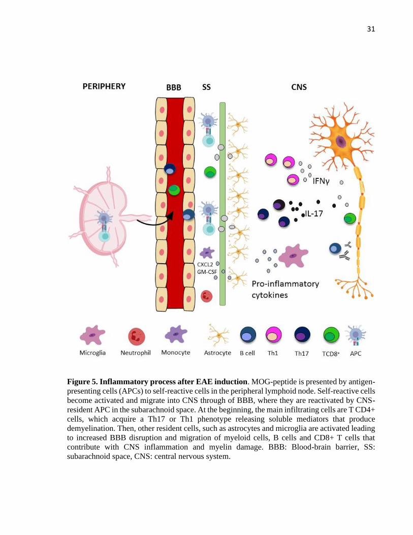
T cell sub-sets implicated in this disease (Figure 5) (Constantinescu et al., 2011).
Interestingly, some herpesviruses such as Epstein-Barr virus (EBV) and human
herpesvirus 6 (HHV-6) have received particular attention for their ability to remain latent in
lymphoid cells and potentially to modulate the onset and relapse of MS in humans. Animals
models have contributed to study the molecular and cellular events that could interfere with the
disease course (Casiraghi et al., 2012; Reynaud and Horvat, 2013; Casiraghi et al., 2015;
Leibovitch et al., 2018). In fact, a study investigated the role of the murine gamma-herpesvirus
γHV-68 (a homologue of EBV in humans), on the pathogenesis of relapsing-remitting EAE in
SJL mice. Importantly, this study found that infection with live γHV-68, but not UV-inactivated
virus exacerbated EAE disease (Peacock et al., 2003). Additionally, a follow-up study found
that latent-infection with γHV-68 virus, prior to EAE induction was capable of increasing the
pathogenesis of active EAE, which was associated with increased CD4+ and CD8+ T cell
responses in the brain and spinal cord, yet was independent of viral reactivation (Casiraghi et
al., 2012). On the other hand, human herpesvirus-6 (HHV-6) has also been investigated as an
environmental trigger of EAE. As rodents are not susceptible to HHV-6 infection, a recent study
used non-human primates to examine the impact of HHV-6 infection on EAE disease. Although
the viral infections were asymptomatic, MS-like disease in these animals was significantly
accelerated in all virally-inoculated animals with detection of viral antigens in the brain, which
showed a marked colocalization with CD3+ cells, suggesting that this virus may participate in
MS in humans (Leibovitch et al., 2018). However, the mechanism underlying this potential
relation and its impact in MS patients requires further studies.
Page 32
31
Figure 5. Inflammatory process after EAE induction. MOG-peptide is presented by antigen-
presenting cells (APCs) to self-reactive cells in the peripheral lymphoid node. Self-reactive cells
become activated and migrate into CNS through of BBB, where they are reactivated by CNS-
resident APC in the subarachnoid space. At the beginning, the main infiltrating cells are T CD4+
cells, which acquire a Th17 or Th1 phenotype releasing soluble mediators that produce
demyelination. Then, other resident cells, such as astrocytes and microglia are activated leading
to increased BBB disruption and migration of myeloid cells, B cells and CD8+ T cells that
contribute with CNS inflammation and myelin damage. BBB: Blood-brain barrier, SS:
subarachnoid space, CNS: central nervous system.
Page 33
32
1.7 HSV-1 and multiple sclerosis disease
At present, an association between HSV-1 and MS disease may be considered based on
the finding of virus genetic material in tissue samples or in body fluids of patients with MS. In
1964, HSV-1 was isolated for the first time in the brain of a postmortem patient with MS
(Gudnadottir et al., 1964). Then, HSV-1 was isolated from the cerebrospinal fluid in alive patient
during the first episode of MS (Bergstrom et al., 1989). More recently, a case-control study
evaluated the prevalence of HSV-1 in peripheral blood mononuclear cells (PBMCs) of patients
with RRMS, and HSV-DNA was founded in 45.1% of patients with MS, in comparison with
3.4% of healthy subjects (Najafi et al., 2016). Another study also detected DNA and mRNA of
HSV-1 in the peripheral blood of patients with MS during clinical acute attack, and it probably
play a role in the triggering of MS relapses (Ferrante et al., 2000). Finally, HSV-DNA has been
reported more frequently in postmortem MS brain tissues than control subjects, and HSV-DNA
was found more in active plaques than inactive plaques in these tissues (Sanders et al., 1996).
On the other hand, HSV-1 seropositivity has been associated with increased risk of MS
in those individuals that do not have the DRB1*15 allele, or decreased risk in those that have it
(Waubant, 2011). Importantly, these observations somewhat support the idea that this virus may
play a role in MS in individuals with particular genotypes (Kastrukoff et al., 2012). Moreover,
another study showed that depletion of macrophages causes CNS demyelination in mice
ocularly infected with HSV-1 (Mott et al., 2011; Zandian et al., 2011). Likewise, a recombinant
HSV-1 expressing IL-2 produced autoreactive T cells and CNS demyelination, supporting the
hypothesis that within an environment that promotes T cell activation, HSV-1 may be enough
for initiating processes that end with the destruction of the myelin in the CNS (Osorio et al.,
Page 34
33
2005; Mott et al., 2013). A subsequent study determined that the mechanism that led to CNS
demyelination in these HSV-1-infected mice was the suppression of IL-12p70 formation by IL-
2 or after macrophage depletion (Lee et al., 2017). Moreover, a recent study showed that the
HSV-1 host-pathogen interactome is highly concentrated in susceptibility genes associated with
neurological disorders, such as MS with enrichment values at 4-fold (Carter, 2017).
Additionally, microorganisms may also contribute to the pathogenesis of MS by inducing the
activation and clonal expansion of self-reactive lymphocytes by mimicry molecular
(Wucherpfennig and Strominger, 1995). For instance, the Hy.1B11 T cell receptor (TCR)
originated from a patient with MS showed to be cross-reactive with a peptide derived from
HSV-1 (UL15154-166) (Sethi et al., 2013).
Taken together, although some studies support a role for HSV-1 infection in MS
(Ferrante et al., 2000; Najafi et al., 2016), this has been poorly studied in animal models which
could help define whether HSV-1 infection plays a direct role in MS. In 1977, a study performed
in rats showed that repeated inoculations of HSV-1 elicit clinical and histological evidence of
recently exacerbated EAE. However, the authors did not determine the mechanism behind this
observation (Hochberg et al., 1977). Moreover, the approach available in that time of EAE
disease in rats was characterized by inflammation and edema leading to paralysis without
demyelination, which differs from what happens in MS (Robinson et al. 2014). In contrast,
MOG-induced EAE is characterized by CNS demyelination and can follow a relapsing–
remitting or chronic disease course as MS, depending on the induction conditions (Berard et al.,
2010). Importantly, this model has been widely used to develop and evaluate therapies to treat
MS (Robinson et al., 2014). For this reason, for this thesis we proposed to assess the impact of
asymptomatic HSV-1 infection over MOG-induced EAE in C57BL/6 mice to determine the
Page 35
34
possible roles of HSV-1 infection on multiple sclerosis disease. First, we infected mice with a
neurovirulent strain of HSV-1 that reaches the brain after intranasal inoculation. Notably,
C57BL/6 mice can be resistant to acute encephalitis after CNS infection by HSV-1, which we
consider can recapitulate several aspects of asymptomatic HSV-1 infection in humans, which
undergo infection without clinical manifestations, despite having this virus in the brain
(Kastrukoff et al., 2012). Moreover, we also evaluated the effects of an attenuated viral strain
of HSV-1, which does not cause encephalitis and has an impaired ability to establish latency
and reactivate from the nervous system. This study could help better understand the relationship
between HSV-1 infection and multiple sclerosis disease, as well as help identify new factors
contributing to the progression of this disease.
Page 36
35
2. HYPOTHESIS AND AIMS
According to the previous evidence described above it is possible that HSV-1 may modulate
the severity and susceptibility to MS because:
1. HSV-1 infects an important percentage of the population.
2. HSV-1 is acquired early in life and causes lifelong persistent infection.
3. HSV-1 infects neurons and can remain in a latent state from which it may reactivate
periodically causing symptomatic or asymptomatic shedding.
4. HSV-1 can reach the brain throughout life without inducing clinical symptoms.
5. Recurrent subclinical reactivations during a persistent brain infection may produce
neuroinflammation and chronic neuron damage.
6. Acute and latent brain infection elevates the MMP-2 and MMP-9 expression, which
could affect the BBB integrity.
To assess a possible relationship between HSV-1 and MS, we proposed to evaluate the following
hypothesis and aims:
Hypothesis:
“Asymptomatic HSV-1 infection enhances MOG-induced EAE disease severity in the mouse
model by increasing the permeability of the blood-brain barrier”.
Page 37
36
Main goal:
To assess the impact of asymptomatic HSV-1 infection on the onset and severity of multiple
sclerosis in a mouse model.
Specific Aims:
1. To evaluate the clinical and histopathologic score after EAE induction in HSV-1-
infected and non-infected animals.
2. To determine the immune cells infiltrating the CNS after EAE induction in HSV-1-
infected and non-infected animals.
3. To determine the cytokine environment in the CNS after EAE induction in HSV-1-
infected and non-infected animals.
4. To quantify MOG or HSV-1 specific antibodies levels in the sera of HSV-1-infected and
non-infected animals after EAE induction.
5. To investigate whether asymptomatic HSV-1 infection increases BBB permeability.
Page 38
37
3. ASYMTOMATIC HERPES SIMPLEX VIRUS TYPE 1 INFECTION CAUSES AN
EARLIER ONSET AND MORE SEVERE EXPERIMENTAL AUTOIMMUNE
ENCEPHALOMYELITIS
Luisa F. Duarte1, 2, María J. Altamirano-Lagos1,2, Jorge H. Tabares-Guevara1,2, Ma.
Cecilia Opazo1,3, Máximo Díaz1,3, Romina Navarrete1,2, Catalina Muza1,2, Omar P.
Vallejos1,2, Claudia A. Riedel1,3, Susan M. Bueno1,2, Alexis M. Kalergis1,2,4 and Pablo A.
González1,2,*.
1Millennium Institute on Immunology and Immunotherapy, 2Departamento de Genética
Molecular y Microbiología, Facultad de Ciencias Biológicas, Pontificia Universidad Católica
de Chile, Santiago, Chile. 3Departamento de Ciencias Biológicas, Facultad de Ciencias de la
Vida, Universidad Andrés Bello, Santiago, Chile 4Departamento de Endocrinología, Facultad
de Medicina, Escuela de Medicina, Pontificia Universidad Católica de Chile, Santiago, Chile.
*Corresponding author: Dr. Pablo A. González, Millennium Institute on Immunology and
Immunotherapy, Departamento de Genética Molecular y Microbiología, Facultad de Ciencias
Biológicas, Pontificia Universidad Católica de Chile, Santiago, Chile. Avenida Libertador
Bernardo O’Higgins 340, Santiago, Chile. Email: [email protected]
Keywords: HSV-1, viral infection, multiple sclerosis, experimental autoimmune
encephalomyelitis
Page 39
38
3.1 Abstract
Herpes simplex virus type 1 (HSV-1) infection is highly prevalent in the human
population, yet its presence is generally unnoticed as the virus can establish asymptomatic
infection and remains latent in the host with periodic reactivations. Importantly, the virus may
undergo subclinical reactivations and shed onto other tissues or individuals. Noteworthy, HSV-
1 infects neurons and may eventually reach and expand within the central nervous system (CNS)
with no apparent disease. Multiple sclerosis (MS) is an increasingly prevalent progressive
autoimmune and debilitating chronic disease that involves the recognition of CNS antigens by
the immune system. Although significant progress has been made in the last decades on the
biology of MS and the identification of novel therapies to treat its symptoms, the triggers of this
disease remain unknown. However, recent studies have suggested that viral latent infections
may contribute to disease onset. Interestingly, a potential association between HSV-1 infection
and MS have been reported, yet a direct relationship between both has not been conclusively
demonstrated. Experimental autoimmune encephalomyelitis (EAE) recapitulates several aspects
of MS in humans and is widely used to study this disease. Here, we evaluated the effect of
asymptomatic brain infection by HSV-1 on the onset and severity of EAE in C57BL/6 mice, as
well as by an HSV-1-mutant that is attenuated in neurovirulence and does not cause encephalitis.
Importantly, we observed a more severe EAE in mice previously infected with either, with the
wild-type (WT) or the mutant HSV-1, as compared to uninfected control mice. These findings
support the notion that a previous exposure to HSV-1 can accelerate and enhance EAE, which
suggests a potential contribution of HSV-1 to the onset and severity of MS.
Page 40
39
3.2 Introduction
Multiple sclerosis (MS) is an autoimmune inflammatory disorder of the central nervous
system (CNS) that affects both, the brain and spinal cord in which multifocal autoreactive
lymphocytic infiltrations lead to damage of the myelin and the axons of neurons (Karandikar et
al., 2004; Dendrou et al., 2005). Defining what triggers the loss of immunologic tolerance to
CNS antigens and the onset of autoreactivity with infiltration into the associated tissues remains
elusive (Compston and Coles, 2008; Steelman, 2015). Likely, MS develops as an interplay
between genetic predisposition, the immune system and environmental factors, such as viral
infections (Beecham et al., 2013).
Herpes simplex virus type 1 (HSV-1) infection is highly prevalent in the human
population with nearly two thirds of the world population infected with this virus (Suazo et al.,
2015). HSV-1 is neurotropic and causes a wide spectrum of clinical manifestations, ranging
from mild symptoms such as oral and facial lesions (e.g. herpes labialis, herpetic
gingivostomatitis), to more severe more diseases affecting the eyes and CNS (e.g. herpetic
keratitis, retinitis, encephalitis and meningitis) (Arduino and Porter, 2008; Rechenchoski et al.,
2017). Importantly, HSV-1 can access the CNS with no apparent pathology (asymptomatic)
establishing a persistent latent infection (Looker et al., 2015). Accumulating evidence indicates
that healthy individuals frequently have HSV-1 in the brain, which could eventually favor the
development, or enhance the severity of neurodegenerative disorders by altering normal
neuronal cell function (Duarte et al., 2019). Subclinical HSV-1 reactivations within CNS
neurons may also contribute to local and regional dissemination of the virus, as well as long-
term detrimental effects in this tissue(Marques et al., 2008; Duarte et al., 2019). Importantly,
HSV-1 infection of the CNS is characterized by persistent lymphocytic cell infiltrations and
Page 41
40
elevated levels of cytokine transcripts (e.g. IFN-γ, TNF-α), as well as increased amounts of
chemokine mRNAs (e.g. CXCL10, CCL5), suggesting that latent HSV-1 infection can be
accompanied by a chronic inflammatory process in this tissue (Theil et al., 2003). Moreover,
increased levels of matrix metalloproteinases 2 and 9 (MMP-2 and MMP-9) have been detected
in HSV-1 latently-infected CNS, which could contribute to the degradation of the surrounding
extracellular matrix and cell surface proteins leading to a partial breakdown of the blood-brain
barrier (BBB), which plays an important role in MS (Martínez-Torres et al., 2004; Weiser et al.,
2007). This inflammatory response could be in response to low-level expression of viral genes
during HSV-1 latency of the CNS (Feldman et al., 2002), which could facilitate an inflammatory
environment that modulates the onset and severity of neurological disorders (Steiner and
Benninger, 2013).
Importantly, viruses belonging to the Herpesviridae family have been suggested as
potential triggers and positive modulators of MS (Wuest et al., 2014). For instance, human
herpesvirus 6 (HHV-6) was recently shown to increase the severity of MS-like symptoms in
non-human primates treated to undergo experimental autoimmune encephalomyelitis (EAE)
(Leibovitch et al., 2018). In another study, latent-infection with the homologous of Epstein-Barr
virus in mice (γHV-68 virus), prior to EAE induction was shown to enhance the pathogenesis
of active EAE, which was associated with increased CD4+ and CD8+ T cell responses in the
brain and spinal cord, yet was independent of viral reactivation (Casiraghi et al., 2012, 2015).
On the other hand, a study performed in rats showed that repeated inoculations of HSV-1 elicited
clinical and histological evidence of exacerbated EAE, but the possible mechanisms behind this
observation were not determined (Hochberg et al., 1977). Additionally, HSV-1 genetic material
has been found more frequently in the cerebrospinal fluid and blood of MS patients than control
Page 42
41
subjects, suggesting an association between this virus and MS (Sanders et al., 1996; Ferrante et
al., 2000; Najafi et al., 2016). However, a direct relationship between both, as well as the
mechanisms underlaying a role of HSV-1 over MS, or vice versa has not been elucidated. Here,
we assessed whether a sub-lethal infection of the CNS with HSV-1 that produces an
asymptomatic infection in the mouse, modulates the severity of MS-like symptoms upon the
induction of EAE, which is widely used as a surrogate model for multiple sclerosis. Importantly,
we used C57BL/6 mice, which are resistant to HSV-1 acute brain infection and to HSV-1-
induced demyelinating lesions throughout the brain (Kastrukoff et al., 2012), to facilitate the
assessment of asymptomatic brain infection by HSV-1 over EAE disease. We also performed
experiments with an HSV-1 mutant virus that has the gamma-34.5 gene (ICP34.5) deleted. This
mutant has been reported to replicate in peripheral tissues, but is attenuated in neurons and does
not cause encephalitis (Whitley et al., 1993).
Noteworthy, we found that HSV-1 infection with the wild-type (WT) virus accelerated
the onset of EAE. Furthermore, previous infection with both, the WT and the attenuated mutant
virus elicited a more severe EAE disease in mice, which was accompanied by increased CNS
inflammation, as well as histological alterations in these tissues. Additionally, infected animals
induced to undergo EAE showed an increase in activated microglia in the brain and spinal cord,
more infiltrating CD4+T cells in the brain and higher amounts of neutrophils in the spinal cord.
We also found significantly higher levels of IL-6 and IL-1β mRNA in these tissues.
Interestingly, we found that infection with either viruses elicited prolonged alterations to the
BBB, which may account for some of the effects described above. Taken together, our results
suggest a direct relationship between asymptomatic HSV-1 infection after intranasal viral
Page 43
42
inoculation and an increased susceptibility to undergo a more severe form of EAE. The
implications of these findings are discussed.
3.3 Material and methods
3.3.1 Mice and Viruses
Five-week-old C57BL/6 female mice were obtained from The Jackson Laboratories (Bar
Harbor) and maintained with environment enrichment, sterile food and water ad libitum at the
central animal facility at the Pontificia Universidad Católica de Chile. Virus stocks were
prepared and titters were determined in Vero cells (ATCC® CCL-81) and kept at -80°C until
use. WT 17syn+ HSV-1 and the R3616 HSV-1 mutant used in this study were kindly provided
by Dr. Carola Otth (Universidad Austral de Chile, Chile). R3616 lacks the gamma-34.5 gene
(∆ICP34.5) and was generated and generously donated by Dr. Bernard Roizman (University of
Chicago, USA) (Chou et al., 1990). All procedures in this study were approved by the Scientific
Ethical Committee for Animal and Environmental Care of the Pontificia Universidad Católica
de Chile and the Biosafety Committee of the same institution (Protocol #170705018) and were
performed according to the National Institutes of Health Guide for Care and Use of Animals
(National Research Council (US), 2011).
3.3.2 Infections and EAE Induction
Five-week-old C57BL/6 female mice were infected intranasally with a sub-lethal dose
of 106 plaque forming units (PFU) of 17 syn+ or ∆34.5 HSV-1, as previously described (Broberg
et al., 2004; Zimmermann et al., 2017). Mock (vehicle)-inoculated mice were used as controls.
During the first two weeks post-infection, mice were clinically scored daily based on
Page 44
43
physiological parameters, appearance, posture, and neurological signs of herpes simplex
encephalitis (i.e. seizures, paralysis). EAE was induced 30-35 days post-infection after
asymptomatic HSV-1 infection. Briefly, mice were anesthetized with a mixture of ketamine and
xylazine, and injected subcutaneously with 50 μg of myelin oligodendrocyte glycoprotein-
(MOG)-derived peptide (MOG35-55, sequence MEVGWYRSPFSRVVHLYRNGK; Pan Web,
Stanford University) emulsified in complete Freund’s adjuvant (Thermo Scientific)
supplemented with heat-inactivated Mycobacterium tuberculosis H37 RA (DIFCO). Mice also
received two intraperitoneal injections of 350 ng of pertussis toxin (List biological laboratories,
Inc) at the time of induction and 48 hours later. Mice were scored daily based on an EAE scale
as follows: 0, no changes in motor function; 0.5, tip of tail is limp; 1, limp tail; 2, limp tail and
weakness of hind legs; 2.5, limp tail, and one hind limb paralyzed; 3, limp tail, and complete
paralysis of hind limbs; 3.5, hind limbs and one fore limb paralyzed; 4, hind limbs and forelimbs
completely paralyzed; 5, moribund.
3.3.3 Blood-brain barrier integrity assay
The integrity of blood-brain barrier (BBB) of HSV-1-infected mice was evaluated using
an Evans blue (EB, Sigma-Aldrich) dye exclusion test, as previously reported (del Valle et al.,
2008). 30 days post-infection, mice were transcardially perfused with 50 mL of phosphate-
buffered saline (PBS, pH 7.4), followed by 50 ml of the EB 2% in PBS under lethal
ketamine/xylazine dose. Brains and spinal cords were dissected, fixed in 4% of p-formaldehyde
(PFA) and cryopreserved in PBS with 30% sucrose for 24 h. Later, organs were embedded in
cryostat-embedding compound (OCT, Sakura), cut into 20 μm thick sections on a cryostat at
−22°C and mounted on Superfrost slides (Thomas Scientific). Slides were examined under a
Page 45
44
confocal laser microscope (Leica TCS LSI), and EB extravasation was visualized as red
fluorescence using a 543-nm laser. Additionally, the amount of EB entering the CNS was
quantified by spectrophotometry at 620 nm after tissue homogenization in 50% of
trichloroacetic acid in PBS and normalized according to the weight of the tissue (EB ng/mg
tissue) (Wang and Lai, 2014).
3.3.4 Histological analysis and immunohistochemistry
Mice infected with HSV-1 and induced to develop EAE were transcardially perfused
with 50 mL of PBS to remove intravascular leukocytes. Lumbar regions in the spinal cords and
corpus callosum in the brain were dissected and carefully processed for histological analyses.
Briefly, tissues were fixed for 24 h in 4% PFA, dehydrated with ethanol and embedded in
paraffin. 6-μm thick sections were obtained using a microtome, and slices were stained with
Luxol Fast Blue solution (LFB) (0.1%, 2 h at 60 °C) and counterstained with Cresyl violet
(0.1%, 6 min) to evaluate demyelination and cell infiltrates, respectively. Four to five sections
per mice were analyzed using an Axio Vert.A1 microscope (Zeiss) with a 10X and a 20X
objective, and histopathologic score was determined as follows: 0, no detected inflammation or
demyelination; 1, one inflammation focus with slight demyelination; 2, two inflammation foci
with moderate demyelination; 3, three or more inflammation foci with severe or complete
demyelination, as previously described (Paintlia et al., 2009). Additionally,
immunohistochemistry against the myelin basic protein (MBP) was carried out using the
Mouse-on-Mouse HRP-Polymer Bundle kit (Biocare Medical). The procedure was carried out
following the manufacturer's instructions. Briefly, sections were deparaffinized with xylene and
rehydrated with decreasing concentrations of alcohol. Endogenous peroxidase was quenched
Page 46
45
with 3% H2O2 in PBS for 20 min, followed by several washes in PBS. Antigen retrieval was
performed using the reagent Rodent Decloaker 1X (Biocare medical) at 95°C for 40 min in a
steamer. Then, slides were incubated for 30 min at room temperature (RT) in Rodent Block M
for 30 min (Biocare medical), followed by 60 min of incubation at 37°C with a dilution 1:1000
of primary anti-MBP antibody (SMI-99P, Biolegend) in 1% bovine serum albumin (BSA,
Winkler) in PBS and 0.1% Triton X-100. After washes with PBS pH 7.4, Mouse-on-Mouse
HRP-Polymer was added for 30 min. Finally, immunostaining was performed using 0.05%
diaminobenzidine and 0.015% H2O2, and counterstained with hematoxylin for 5 min. Slides
without primary antibody were used as controls.
3.3.5 Western blot analysis
Western blot analyses were performed to evaluate the expression of MBP in lumbar
regions in the spinal cord and corpus callosum in the brain of mice infected with HSV-1 and
induced to develop EAE. Samples were homogenized, placed in lysis buffer (150 mM NaCl, 1
mM EDTA, 10 mM Tris-HCl, 1 mM phenylmethanesulfonyl fluoride, 0.5% NP40, 0.5%
Sodium Deoxicholate, and 0.1% SDS), and total protein was determined using the Pierce BCA
Protein Assay Kit (Thermo Scientific) following the manufacturer's instructions. Proteins were
resolved using 12% sodium dodecyl sulfate (SDS) polyacrylamide gel electrophoresis and
transferred to nitrocellulose membranes (Bio-Rad). After blocking with 5% BSA, membranes
were incubated overnight at 4°C with a 1:300 dilution of mouse anti-MBP (SMI-99P,
Biolegend) or a 1:1000 dilution of anti-β-actin (2F1-1, Biolegend) for 2 h at RT. A horseradish
peroxidase (HRP)-conjugated anti-mouse antibody was used as secondary antibody
(GenScript), and proteins were visualized by chemiluminescence using a ChemiDoc®MP
Page 47
46
Imaging System (Bio-Rad). Band intensity was calculated using ImageJ (U.S. National
Institutes of Health).
3.3.6 Mononuclear cell isolation, staining and flow cytometry
Single cells suspensions were generated from the spinal cord and brain of HSV-1-
infected EAE-induced mice perfused with PBS, as previously reported (Manglani et al., 2018).
Infected and uninfected mice without EAE were used as controls. Tissues were incubated with
1 mg/ml collagenase IV (Thermo Scientific) and 50 µg/ml DNAse I (Roche) in RPMI (Thermo
Scientific) at 37°C for 30 min. Mononuclear cells (MNCs) were isolated using 30/70% Percoll
gradients (GE healthcare). For staining, MNCs were treated with CD16/32 Fc-block (BD
Biosciences) to inhibit nonspecific antibody binding and incubated with anti-mouse immune
cell surface markers for 45 min at 4°C. The following antibodies were used: anti-CD3 (Clone
17A2), anti-CD4 (clone 6K1.5), anti-CD8 (clone 53-6.7), anti-CD19 (clone 1D3), anti-CD45
(clone 30-F11), anti-CD11b (clone M1/70), anti-Ly6C (clone HK 1.4), and anti-Ly6G (clone
RB6-8C5) and anti-MHC-II (clone AF6-120.1) (BioLegend). Dead cells were detected using
the fixable Zombie Violet kit (BioLegend) and excluded from the analyses. Cells were
enumerated by adding CountBright™ absolute counting beads (Thermo Scientific) to each
sample before acquisition using a FACSCanto II flow cytometer (BD Biosciences) and data was
analyzed using FlowJo software (Tree Star, Inc). This work was supported by the Cytometry
Core UC (FCC UC).
Page 48
47
3.3.7 Quantitative PCR (qPCR) and reverse transcription quantitative PCR
(RT-qPCR)
Total DNA from brain and trigeminal ganglia tissues was isolated using phenol-
chloroform (Winkler) for quantifying the number of viral genomes. 200 ng of DNA was used
for qPCR analyses with the following primers and probe for the viral polymerase UL30 gene:
Fwd-GGCCAGGCGCTTGTTGGTGTA, Rev-ATCACCGACCCGGAGAGGGA and Probe-
CCGCCGAACTGAGCAGACACCCGC (Integrated DNA Technologies) and an Applied
Biosystems StepOnePlus thermocycler, as previously described (Retamal-Díaz et al., 2017).
Total RNA was isolated from tissues for cytokine expression analysis using TRIzol reagent
(Thermo Scientific) according to the manufacturer’s instructions. cDNA synthesis from total
RNAs was performed using SuperScript™ II Reverse Transcriptase (Thermo Scientific) and
random primers. RT-qPCR reactions were carried out using PowerUp™ SYBR™ Green Master
Mix (Thermo Scientific) and primers for the detection of IL-1β, IFN-γ, TNF-α, IL-10, IL-17
and IL-6 (Zaheer et al., 2007) using a Mx3000P™ QPCR System (Stratagene) with the
following cycling conditions: one cycle of 50°C for 2 min and 95°C for 2 min, followed by 40
cycles of 95°C for 15 s, 57° for 15 s and 72°C for 1 min. The abundance of each target mRNA
was determined by relative expression to the β-actin housekeeping gene and the 2^-delta delta
cycle threshold (2-ΔΔCT) method (Rao et al., 2013).
3.3.8 ELISAs Assays
Antibodies against HSV-1 were detected by ELISA using sera obtained before and after
EAE induction. MaxiSorp ELISA plates (Nunc/Thermo Scientific) were coated with 20 µg/mL
of protein extracts from uninfected-Vero cells or 10 µg/mL of protein extracts from infected-
Page 49
48
Vero cells and incubated at 4°C overnight in a humidity chamber. Plates were blocked with
PBS-BSA 1% and then incubated with serial dilutions of the sera. To reduce non-specific
antibody binding to the infected protein extracts, the sera were pre-adsorbed over plates with
uninfected-Vero protein extracts for 2 h at RT and then transferred to plates with infected-Vero
protein extracts and incubated at 4°C overnight in a humidity chamber. After three washes with
PBS-Tween 20 0.05%, the wells were incubated with an HRP-conjugated anti-mouse-IgG
antibody diluted 1:2000 (Thermo Scientific) for 1 h at RT, washed 3 times with PBS-Tween 20
0.05%, developed with 1-Step™ Ultra TMB-ELISA Substrate Solution (Thermo Scientific) for
10 minutes, and read on a Multiskan ELISA plate reader at 450 nm after adding H2SO4 2N to
stop the enzymatic reaction. Anti-MOG antibodies were also detected in the sera from
uninfected-EAE and infected-EAE mice carrying out the steps mentioned above and using 10
µg/mL of MOG peptide to coat the ELISA plates.
3.3.9 Statistical Analyses
Statistical significance between experimental groups was assessed by one-way analysis
of variance (ANOVA) with Dunn’s multiple comparisons post-test for parametric data, Kruskal-
Wallis with Dunn’s multiple comparisons post-test for non-parametric data (three or more
groups) or two-way ANOVA with Tukey’s multiple comparison post-test (two independent
variables) using GraphPad Prism software (GraphPad Software, La Jolla California USA).
Page 50
49
3.4 Results
3.4.1 Asymptomatic HSV-1 infection alters the permeability of the blood-brain
barrier
To assess a potential effect of asymptomatic HSV-1 infection of the CNS over the onset
and severity of experimental autoimmune encephalomyelitis (EAE) in the mouse model, we
performed experiments with C57BL/6 mice. These mice have been reported to be resistant to
acute HSV-1 encephalitis and hence could better reflect circumstances related to asymptomatic
CNS infections reported in humans that do not display clinical manifestations despite having
the virus in the brain (Baringer and Pisani, 1994; Wozniak et al., 2005). Thus, C57BL/6 mice
were infected intranasally with a sub-lethal dose of HSV-1 and followed for 30 days. As
expected, the weight of animals did not vary significantly after HSV-1 infection and overall
paralleled that of mock-infected animals (Supplementary Figure 1A). Latent brain infection by
the WT virus was corroborated using a virus plaque assay and by qPCR 30 days post-infection.
As expected, no viral PFUs were recovered from brain tissue homogenates overlaid onto Vero
cells, while the qPCR evidenced the presence of viral genome copies both, in the trigeminal
ganglion and brain of inoculated mice (Supplementary Figure 1B). Additional to the use of WT
HSV-1 virus, we also included in the following experiments an HSV-1 mutant that has the gene
encoding the virulence factor gamma-34.5 deleted (ICP34.5 gene, ∆34.5 mutant virus). This
mutant virus does not cause encephalitis and has been reported to be hampered at replicating in
neurons, although it can elicit an inflammatory response in the brain of mice, which may
somewhat homologate the case of humans undergoing asymptomatic HSV-1 infection of this
tissue (McMenamin et al., 1998; Broberg et al., 2004).
Page 51
50
Because previous reports indicate that acute HSV-1 infection of the brain alters the BBB,
we sought to assess whether this was also the case in asymptomatic animals infected with HSV-
1 30 days post-infection. For this, we used Evans blue (EB), a dye that when is administered
systematically cannot access the CNS in normal conditions unless the BBB is altered (del Valle
et al., 2008). Hence, extravasation of this dye into the CNS is indicative of increased BBB
permeability. As shown in Figure 6, mice infected with WT virus presented increased EB
diffusion into the brain and spinal cord at 30 day post-infection, as compared to mock-inoculated
animals, suggesting that the BBB is altered in these mice long after infection and in the absence
of detectable infectious virus. Notably, mice infected with the mutant HSV-1 virus also showed
significantly increased EB diffusion into the brain as compared to uninfected animals,
evidencing BBB disruption independent of viral replication in neurons in the brain. Future
studies should help determine how long the BBB is disrupted after HSV-1 infection.
3.4.2 Asymptomatic HSV-1 infection accelerates the onset and increases the
severity of EAE
To determine if HSV-1 infection impacts the onset and severity of CNS autoimmunity,
we carried out an EAE induction protocol in mice that had been previously infected with HSV-
1 (Figure 7A). As a control, EAE was also induced in mock-infected animals. As shown in
Figure 7B, previous infection with WT HSV-1 accelerated the onset of EAE in 2 days
approximately, while infection with the ∆34.5 mutant displayed a similar disease onset as the
mock-infected animals (Table 1). Importantly, mice infected with WT HSV-1 displayed a higher
Page 52
51
Figure 6. Asymptomatic HSV-1 infection increases BBB permeability in vivo. 30 days post-
infection mice were transcardially perfused with Evans Blue dye (2 % w/v). (A) Evans blue
visualization by confocal microscopy in brain (left panels) and spinal cord sections (right panels)
in uninfected mice or animals inoculated with ∆34.5 HSV-1, or 17syn+ HSV-1. Representative
images of two independent experiments are shown. The original magnification of the
photomicrographs is 10x. The brain image is a composite of 10 serial images. (B) Quantification
of Evans blue incorporated into the brain (upper panel) and spinal cord (lower panel) by
spectrophotometry at 620 nm. Values represent means ±SEM of two independent experiments
(n=7/group). Data were analyzed using Kruskal-Wallis and Dunn’s multiple comparisons post-
test; **p<0.01; *p<0.05.
Page 53
52
Figure 7. Asymptomatic HSV-1 infection accelerates the onset and increases the severity
of EAE. (A) Schematic representation of the experimental design carried out in this study. (B)
EAE was scored for each mouse after EAE induction, which was carried out 30-35 days post-
HSV-1 infection. Mice were followed until day 21 post-EAE induction. The graph shows the
means of disease scores ± SEM for mice mock-treated (blue circles), infected with ∆34.5 HSV-
1 (green squares), or infected with 17syn+ HSV-1 (red triangles) in three independent
experiments (n=12/group). Data were analyzed using two-way ANOVA followed by Turkey’s
post-test; **** p<0.0001, *** p<0.001, * p<0.05.
Page 54
53
Table 1. Summary of EAE disease parameters after HSV infection and EAE induction
Group
Incidence
of EAE
symptoms
Mean day
of disease
onset
Maximum
clinical
score of
EAE
reached
Mean
clinical score
at day 14
(disease
peak)
Mean
clinical
score at day
21
(remission
stage)
Mock-
EAE
66.7%
(8/12) 13.6 2.5 (2/12) 0.5
1
17syn+-
EAE
91.7%
(11/12) 11.9 3 (2/12) 1.1 1.3
∆34-5-
EAE
100%
(12/12) 14.1 3.5 (2/12) 0.4 2.1
Page 55
54
incidence and scores of EAE symptoms than non-infected animals with EAE (Table 1, and
Figure 7B). On the other hand, mice infected with the ∆34.5 mutant virus had a higher incidence
and increased EAE clinical scores than WT HSV-1-inoculated animals (Table 1, and Figure
7B). In addition, a subset of animals was monitored for an extended period of time (25 days
post-EAE induction) to evaluate the remission stage. Unlike the mock-EAE treated animals,
which showed mild EAE symptoms, the animals infected either, with the WT or mutant HSV-
1 showed a chronic progressive course of EAE symptoms up to permanent paralysis, which
would normally be observed in C57BL/6 mice after severe MOG35-55-induced EAE
(Supplementary Figure 2) (Berard et al., 2010).
To characterize the impact of asymptomatic HSV-1 infection on the integrity of CNS
tissues after EAE induction, we performed histological and molecular analyses of brain and
spinal cord samples. Histological analyses with Luxol Fast Blue (LFB), which stains the myelin
was contrasted with Cresyl violet to evidence cellular infiltration. Additionally, we performed
myelin basic protein (MBP) expression analysis by immunohistochemistry and western blot for
this protein. As shown in Figures 8A-C, histology analysis of spinal cord tissues revealed
morphological alterations after staining with LFB and performing MBP immunohistochemistry,
that were more evident for the experimental group infected with the ∆34.5 mutant virus induced
to undergo EAE. In these animals, this tissue displayed significant cellular infiltration and loss
of myelin, consistent with more severe EAE than the other groups at day 21 post-EAE induction
(Figure 8D). Importantly, histological samples of mice infected with WT HSV-1 and treated to
undergo EAE did not display significant differences respect to mock-infected group, possibly
because these animals experienced fewer maximum disease score than the ∆34.5-inoculated
group. Surprisingly, the expression of the MBP protein in western blot assays was lower in mice
Page 57
56
Figure 8. Asymptomatic HSV-1 infection increases spinal cord demyelination after EAE
induction. (A) Representative images of lumbar sections of spinal cords stained with Luxol
Fast Blue showing tissue demyelination. (B) Representative images of Luxol Fast Blue staining
contrasted with Cresyl violet showing cellular infiltration. Myelin staining is observed in blue
in the white matter and cell nuclei are colored purple. (C) Representative images of
immunohistochemistry performed against the MBP protein. Representative images of three
independent experiments are shown. Image magnifications are 10x (left) and 20x(right) and
correspond to day 21 post-EAE induction. (D) Quantitative histopathological analyses of spinal
cord lumbar sections. Values represent the mean ± SEM of three independent experiments. Data
were analyzed with two-way ANOVA followed by Turkey’s post-test; *p<0.05 (n=12, 4/group
per day evaluated). (E) Representative western blot images for MBP (upper panel) and actin
(lower panel) in the spinal cord at day 14 post-EAE induction. The graph shows densitometric
analyses for MBP bands that were normalized to actin. Data represent the mean ± SEM.
Comparisons between ratios were performed using one-way ANOVA with Dunnett’s multiple
comparison post-test; *p<0.05.
Page 58
57
previously infected with the WT virus, as compared to the ∆34.5 mutant virus-infected group,
which is somewhat unexpected, as the latter displayed increased histological pathology as
compared to the animals infected with the WT virus (Figure 8E). These differences may be due
to more regional damage in this tissue in the ∆34.5-EAE group, as compared to the WT-EAE
group.
On the other hand, as shown in the Supplementary Figure 3, brain tissues showed some
regions of evident demyelination only in HSV-1-infected animals induced to develop EAE. This
was not the case for HSV-1-infected mice without EAE induction which were used as controls.
Similarly, mock-inoculated animals treated to undergo EAE did not show significant
histological alteration, which was expected as the protocol used for inducing EAE in our
experimental setting was mild, consistent with mild disease score values and no significant
demyelination in the brain in the absence of previous viral infection (Supplementary Figure 3).
Regarding the western blot assays in the brain, animals infected with HSV-1 either, with the
WT or mutant virus and treated to develop EAE, showed a decrease in the expression of MBP.
Taken together, these results indicate that asymptomatic infection with HSV-1 either,
with a WT virus or mutant virus that cannot replicate in neurons significantly affects the
outcome of EAE, suggesting a direct relationship between both, the virus and this autoimmune
disease.
3.4.3 Asymptomatic HSV-1 infection increases EAE-associated inflammation
To determine if previous asymptomatic infection with HSV-1 favors the infiltration of
immune cells into the CNS after EAE is induced, we performed flow cytometry analysis of the
brain and spinal cord at day 14 post-EAE induction and assessed the presence of CD4+ T cells
Page 59
58
(CD3+/CD4+ cells), CD8+ T cells (CD3+/CD8+ cells), or B cells (CD19+ cells) (Supplementary
Figure 4), as well as myeloid cells, namely monocytes (CD45hi+CD11b+Ly6C+ cells),
neutrophils (CD45hi+CD11b+Ly6G+ cells), and activated microglia (CD45lo+CD11b+MHC-II+)
(Supplementary Figure 5). As shown in Figure 9A, the brains of mice infected with WT HSV-
1 and induced to undergo EAE displayed significantly more cellular infiltration of lymphoid
cells than other groups. In contrast, those previously infected with the ∆34.5 mutant virus had
more infiltration of myeloid cells in this tissue, although the differences were not statistically
significant. Because HSV-1 latent brain infection has been reported to be accompanied by
persistent T cell infiltration (Marques et al., 2008), we sought to determine if this would be the
case in our HSV-EAE model. As a control, mice infected with WT or the mutant virus alone,
without EAE induction were evaluated at equivalent time-points as mice infected and then
treated to undergo EAE (6 weeks post-infection). As shown in Supplementary Figure 6A and
6B, animals infected with HSV-1 alone did not display increased amounts of T cells in the brain
or spinal cord as compared to healthy mice. Surprisingly, mice infected with WT HSV-1 and
treated to undergo EAE displayed a significantly higher number of CD4+ T cells in the brain as
compared to the mock-EAE group (Figures 9B, and 9D). Regarding the myeloid cells analyzed
in the brain, significant differences were observed for activated microglia expressing the MHC-
II surface marker, which was higher in the WT HSV-1-EAE group than in the other groups
(Figure 9C). On the other hand, no significant differences were observed between the different
groups in terms of the number of infiltrating lymphoid cells in the spinal cord (Figures 10A and
10B). However, HSV-1-infected mice induced to experience EAE had a greater number of
infiltrating myeloid cells than mock-EAE group (Figure 10A), which were mainly neutrophils
as shown in Figures 10C and 10D. Moreover, the amount of activated microglia in the spinal
Page 61
60
Figure 9. Animals infected with WT HSV-1 and treated to undergo EAE show increased
number of CD4+ T cell infiltration in the brain. Mice were mock-treated, infected with HSV-
1 ∆34.5, or infected with HSV-1 17syn+. EAE was induced four weeks post-HSV-1 infection.
At day 14 post-EAE induction, mice were perfused and the brain was harvested and processed
to isolate immune infiltrating cells. (A) Total lymphoid cells (left) and myeloid cells (right)
infiltrating the brains of mice induced to develop EAE. Values represent the mean ± SEM of
two independent experiments (n=8/group). Data were analyzed using Kruskal-Wallis and
Dunn’s multiple comparisons post-test *p<0.05. (B) Infiltrating T cells, CD4+ (left), CD8+
(middle), or B cells CD19+ (right) plotted individually. (C) Infiltrating myeloid cells Ly6C+
(left) and Ly6G+ (middle) plotted individually, data are means ± SEM of two independent
experiments n=8/group. For the percentage of activated microglia CD45loCD11b+MHC-II+
(right), the data are means ± SEM of n=4/group. Data were analyzed using Kruskal-Wallis and
Dunn’s multiple comparisons post-test; *p<0.05. (D) Representative FACS plots showing the
frequencies of lymphoid T cells in the brain. Live single cells were pre-gated on CD3+ and
CD19+. CD3+ T cells were subdivided into CD4+ and CD8+.
Page 63
62
Figure 10. Animals infected with HSV-1 and treated to undergo EAE display increased
number of neutrophils infiltrating the spinal cord. Mice were mock-treated, infected with
HSV-1 ∆34.5, or infected with HSV-1 17syn+. EAE was induced four weeks post-infection.
At day 14 post-EAE induction, mice were perfused and the spinal cords were harvested and
processed to isolate immune cells infiltrating this tissue. (A) Total lymphoid cells (left) and
myeloid cells (right) infiltrating the spinal cords of mice induced to develop EAE. Values
represent the mean ± SEM of two independent experiments (n=8/group). Data were analyzed
using Kruskal-Wallis and Dunn’s multiple comparisons post-test *p<0.05. (B) Infiltrating T
cells, CD4+ (left), CD8+ (middle), or B cells CD19+ (right) plotted individually. (C) Infiltrating
myeloid cells Ly6C+ (left) and Ly6G+ (middle) plotted individually. Data are means ± SEM of
two independent experiments n=8/group. For the percentage of activated microglia
CD45loCD11b+MHC-II+ (right), the data are means ± SEM of n=4/group. Data were analyzed
using Kruskal-Wallis and Dunn’s multiple comparisons post-test *p<0.05. (D) Representative
FACS plots showing the frequencies of infiltrating myeloid cells in the spinal cords. Live single
cells were pre-gated on CD45+ and CD11b+. CD45hi+/CD11b+ infiltrating myeloid cells were
subdivided into neutrophils (Ly6G+) and monocytes (Ly6C+).
Page 64
63
cord of WT-infected mice was significantly higher than in uninfected mice in this tissue (Fig
10C).
Next, to evaluate whether asymptomatic infection with HSV-1 modulates the cytokine
environment in the CNS upon EAE induction, we performed RT-qPCR for a set of ytokines that
either, promote an inflammatory state in this tissue (i.e. IL-1β, IL-6, IL-17, TNF-α and IFN-γ)
or an anti-inflammatory environment (i.e. IL-10). As shown in Figure 11A, the brain of mice
infected with WT HSV-1 or the ∆34.5 mutant virus and treated to undergo EAE showed
increased expression of all the cytokines evaluated, as compared to mock-infected animals.
Notably, more IL-1β mRNA was expressed in the brain of infected animals with EAE than
equivalent tissue obtained from mice induced to develop EAE without a previous HSV-1
infection (Figures 11A). Moreover, IL-6 mRNA levels were also significatively increased in the
brain of mice infected with ∆34.5 mutant virus (Figure 11A). Cytokines mRNAs in the spinal
cord displaying important variations, as compared to mock-infected animals were IL-6 and IL-
10 in the WT HSV-1-EAE group, as shown in Figure 11B. IL-17 and IFN-γ also showed some
differences among the evaluated groups, and although these changes were not-significant these
cytokines also showed a tendency to be increased in the brain and spinal cord of mice infected
with either virus and treated to undergo EAE (Figures 11A and 11B).
3.4.4 Asymptomatic mice infected with WT HSV-1 display increased amounts
of anti-HSV-1 antibodies after EAE induction
Given the results obtained above, it is possible that asymptomatic infection with HSV-1
predisposes the animals to undergo increased EAE severity, but it is also possible that the
Page 66
65
Figure 11. Asymptomatic HSV-1 infection increases the expression of pro-inflammatory
cytokines in the CNS of mice with EAE. Mice were mock-treated, infected with HSV-1 Δ34.5,
or infected with HSV-1 17syn+. Four weeks post-infection EAE was induced. 14 days post-
EAE induction, tissue homogenates were evaluated by RT-qPCR to assess cytokine expression
at the mRNA level using the 2-ΔΔCT method with actin as a reference gene. (A) Relative
expression levels of proinflammatory cytokines (IL-6, IL-1β, TNF-α, IFN-γ and IL-17), and the
anti-inflammatory cytokine IL-10 in the brain of HSV-1 17syn+-infected mice (red triangles),
HSV-1 ∆34.5-infected mice (green squares), and mock-treated mice (blue circles) plotted
individually. B) Relative expression levels of cytokines in the spinal cord of HSV-1 17syn+-
infected mice (red triangles), HSV-1 ∆34.5-infected mice (green squares), and mock-treated
mice (blue circles) plotted individually. Values represent means ± SEM of two independent
experiments (n=8/group). Data were analyzed using Kruskal-Wallis and Dunn’s multiple
comparisons post-test; **p<0.01, *p<0.05.
Page 67
66
induction of EAE in previously-infected animals may promote virus reactivation in the CNS or
periphery and facilitate enhanced neurodegenerative disease. To preliminarily assess this latter
scenario, we assessed the concentrations of circulating antibodies against HSV-1 in the serum
of infected animals before- and 14 days after EAE induction. Interestingly, we found that those
animals that were previously infected with WT HSV-1 and then treated to undergo EAE
displayed a modest, yet significantly increase in the quantity of anti-HSV-1 antibodies in the
serum (Figure 12A). Although these differences are not substantial, this result suggests possible
viral reactivation, either productive (new infectious particles) or at the molecular level
(expression of HSV-1 antigens without the release of new infectious particles), which requires
further attention. However, because infections with the ∆34.5 mutant virus previous to EAE
induction did not increase the quantity of HSV-1-specific antibodies after EAE induction,
suggest the increased amount of anti-HSV-1 antibodies in the WT HSV-1-EAE group may be
due to viral reactivation (Figure 12A).
Additionally, we assessed the quantity of MOG-specific antibodies in the sera of animals
infected or not with HSV-1 and then treated to undergo EAE. As shown in Figure 11B, although
mice infected with WT HSV-1 displayed significantly higher amounts of anti-MOG antibodies
after EAE induction as compared to control healthy mice, no significant differences were
observed between the animals of the WT HSV-1 EAE, ∆34.5 HSV-1-EAE mice or mock-EAE
group (Figure 12B).
Page 68
67
Figure 12. Animals infected with WT HSV-1 and then treated to undergo EAE display
increased anti-HSV antibodies after EAE induction. Mice were mock-treated (blue), infected
with HSV-1 ∆34.5 (green), or infected with HSV-1 17syn+ (red). EAE was induced in the
indicated groups (EAE) four weeks post-infection. At day 30 post-HSV infection and 14 post-
EAE induction, sera were harvested and levels of (A) anti-HSV-1 IgG antibodies (n=8/group)
were quantified by ELISA. (B) anti-MOG IgG antibodies (n=10/group) were quantified in sera
harvested at day 14 post-EAE induction by using ELISA. Data are means ± SEM of two
independent experiments. Data were analyzed using two-way ANOVA followed by Turkey’s
post-test; **p<0.01, *p<0.05.
Page 69
68
3.5 Discussion
Infections with human herpesviruses has been suggested as potential triggers or
enhancers of MS in recent reports (Casiraghi et al., 2015; Leibovitch et al., 2018), yet studies
that assess or support a role for HSV-1 infection are relatively scarce and a direct relationship
between this virus and this disease has not been reported before (Ferrante et al., 2000; Ferrò et
al., 2012; Rizzo et al., 2016; Buscarinu et al., 2017). Although the fact that HSV-1 infects the
CNS makes this virus a suspect candidate in MS, the fact that HSV-1 infection is highly
prevalent in the human population, unlike MS somewhat argues against this idea. However,
asymptomatic HSV-1 infection in the CNS may be insufficient for developing MS per se and
the initiation of the disease likely requires other contributing elements, such as genetic and
environmental factors (Briggs et al., 2010; Kakalacheva et al., 2011; Waubant, 2011). However,
the prevalence of CNS infection with HSV-1 in otherwise healthy individuals is somewhat
unknown, as this is not a routine analysis to be performed after death. Despite the fact that CNS
infection with HSV-1 in healthy individuals is undetermined, it is possible to foresee that the
chances of having HSV-1 infection of the CNS will likely increase with aging, as progressive
senescence of the immune system may allow HSV-1 to reactivate from peripheral tissues, such
as the trigeminal ganglia and spread within the brain (Jamieson et al., 1991; Wozniak et al.,
2005; Itzhaki and Lathe, 2018). Furthermore, repeated HSV-1 reactivations throughout the life
of an individual may provide opportunities for increased number of neurons to be infected with
this virus as a person gets older. Additionally, neuronal senescence may also facilitate
neurodegenerative disorders by HSV-1 and eventually facilitate MS initiation and progression
(Menendez et al., 2016; Duarte et al., 2019).
Page 70
69
Here, we observed that a previous infection with HSV-1 after intranasal virus inoculation
can predispose the host to an earlier onset and more severe EAE disease. Our results showed a
significant increased demyelination of spinal cords in animals previously infected with HSV-1,
which was more evident for those infected with ∆34.5 mutant virus. Surprisingly, these results
suggest that viral replication in the brain may not be necessary for experiencing increased EAE
severity after infection with HSV-1.
Although we did not observe significant histological alterations in the brain tissues
obtained from mice that displayed an earlier onset in EAE, or increased EAE severity after a
previous infection with HSV-1, several molecular markers associated with inflammation and
cellular infiltration in the CNS of these animals could be detected by other means. As reported
above, we found that IL-6 mRNA was elevated in both, in the brain and spinal cord of infected
animals. Importantly, this cytokine has been reported to be a key player in the development of
autoimmune diseases by differentiating autoreactive proinflammatory CD4+ T cell responses
towards a Th-17 phenotype, as well as by inhibiting the induction of regulatory T cells (Tregs)
(Maimone et al., 1997; Kimura and Kishimoto, 2010). Studies performed in humans with RRMS
show that IL-6 supports T cell effector function resistance to regulation by Tregs, which may
contribute to disease severity (Schneider et al., 2013). This could explain why although there
were increased levels of IL-10 mRNA in the spinal cord of the WT HSV-1 group, these animals
suffered a more severe disease than the mock-EAE group. It is possible that the anti-
inflammatory effect of IL-10 may be disrupted by the high levels of IL-6 in this tissue, thus
favoring a Th-17 phenotype. However, this remains to be evaluated. Moreover, the elevated
levels of IL-1β mRNA observed in the brain may also promote BBB permeability, possibly
through previously reported mechanisms over astrocytes (Wang et al., 2014; Lin and Edelson,
Page 71
70
2017). An interesting finding was the fact that the BBB of asymptomatic HSV-1-infected mice
remained permeable to the Evans blue dye 30 days after infection. Although alterations in the
BBB during HSV-1 infection had been reported before, this phenomenon was only observed in
in vitro BBB models, or during acute CNS infection with this virus (HSV-1 encephalitis) (Liu
et al., 2019b, 2019a; He et al., 2020). Our results show that the disruption of the BBB occurs
independent of encephalitis and persists in the absence of infectious virus in the CNS. Moreover,
these results suggest that intranasal virus inoculation is enough to disrupt the BBB for a long
period. However, it remains to be determined how long these alterations last and whether they
are key for the observations reported herein.
On the other hand, while CD4+ T cells have been shown to play a key role over EAE
onset and severity (Constantinescu et al., 2011), and that we observed that these cells were
increased in the brain of WT-HSV-1-EAE mice, relevant roles for other immune cells, such as
neutrophils are emerging as a relevant immune component contributing to CNS damage (Aubé
et al., 2014; Rumble et al., 2015; Woodberry et al., 2018). Importantly, we found that these cells
were increased in the CNS of the experimental groups infected with HSV-1, as compared to
mock-infected mice. It would be important to characterize the phenotype of these cells to
determine if they are contributing to the enhanced disease severity observed, which would
support the notion of a detrimental role for neutrophils in EAE, and eventually MS pathogenesis.
Additionally, it will be interesting to assess the contribution and role of virus-specific CD4+ and
CD8+ T cells in these experiments, as these cells may be contributing to CNS inflammation by
promoting immune cell access to the CNS, cytokine secretion in these tissues or direct neuron
damage (Steinbach et al., 2019). Previous reports suggest that viral infections can increase the
susceptibility to autoimmune diseases by eliciting bystander inflammation and the activation of
Page 72
71
autoreactive cells, which can lower the threshold for disease development (Miller et al., 1997;
Daniel R. Getts et al., 2013).
Although our findings suggest a role for asymptomatic brain infection by WT HSV-1 on
the onset and severity of MS, it remains unknown whether EAE induction in these animals
reactivates HSV-1, leading to active viral replication and potentially HSV-1 replication-related
disease in the CNS, which could account per se for some of the observed symptoms or directly
contribute to the severity of the EAE induced. The fact that animals infected with WT HSV-1
and then induced to undergo EAE displayed increased amounts of anti-HSV antibodies,
although modest suggests that HSV-1 reactivation may be occurring in these mice, although this
remains to be further assessed. As discussed above, because increased amounts of anti-HSV-1
antibodies were only observed in the WT HSV-1-EAE group and not with the mutant virus
(∆34.5-EAE group), such potential reactivation may be related to the generation of infectious
particles, although a molecular activation of HSV-1 may also be the case (Feldman et al., 2002;
Martin et al., 2014a). Because the mutant virus elicited enhanced EAE symptoms, even more
than the WT virus for some of the analyzed parameters, it is also possible that the main
mechanism behind enhanced EAE by HSV-1 infection may be a consequence of a long-lasting
signal of the virus over infected cells early after virus inoculation, or even adjacent cells, which
could trigger an inflammatory response that increases the host susceptibility to undergo this
autoimmune disease with increased severity (Steinbach et al., 2019).
Given the existence of antivirals specific for herpesviruses, such as acyclovir, it is
tempting to speculate that such compounds may delay the onset of EAE in animals previously-
infected with HSV-1, or reduce the severity of the disease in these mice once initiated. However,
because the ∆34-5 mutant virus is attenuated in neurons and that the animals inoculated with
Page 73
72
this virus displayed more severe EAE, the use of such drugs may not necessarily have
therapeutic effects. Nevertheless, it will be interesting to perform the experiments carried out in
this study in the presence of drugs such as acyclovir after virus infection to determine the
contribution of HSV-1 replication in the different stages of EAE.
3.6 Acknowledgements
We are grateful to Dr. Luis Larrondo for sharing equipment for visualizing western blots.
This work was supported by the Millennium Institute on Immunology and Immunotherapy
(P09/016-F) from the Millennium Science Initiative of the Agencia Nacional de Investigación
y Desarrollo (ANID, Chile); and FONDECYT (ANID, Chile) grant #1190864.
Page 74
73
3.7 Supplementary figures
Supplementary Figure 1. Asymptomatic brain infection with WT HSV-1 after intranasal
virus inoculation. C57BL/6 mice were intranasally mock-inoculated or infected with HSV-1
17syn+ or HSV-1 ∆34.5 and weighted daily until day 30. (A) Weight curves of infected and
non-infected mice. Values represent means ±SEM from three independent experiments
(n=12/group). (B) HSV-1 UL30 gene copies per gram of brain or trigeminal ganglia from a
subset of WT infected-mice obtained at 30 days post-infection and normalized with values from
uninfected mice. Values represent means ±SEM of four animals per group.
Page 75
74
Supplementary Figure 2. Asymptomatic HSV-1-infected mice show a chronic course of
EAE disease. EAE disease was scored for each mouse after EAE induction, which occurred 30
days post-HSV-1 infection. Mice were followed until day 25 post-EAE induction. The graph
shows the mean ± SEM of EAE disease scores for mice mock-treated (blue circles), infected
with HSV-1 ∆34.5 (green squares), or HSV-1 17syn+ (red triangles) (n=4/group). Data were
analyzed using two-way ANOVA followed by Turkey’s post-Test; ** p<0.01, * p<0.05.
D a y s p o s t E A E in d u c tio n
EA
E c
lin
ica
l S
co
re
5 7 9 1 1 1 3 1 5 1 7 1 9 2 1 2 3 2 5
0
1
2
3M ock-E A E
1 7 s y n + -E A E
3 4 .5 -E A E
* *****
*
Page 77
76
Supplementary Figure 3. Asymptomatic HSV-1 infection contributes to brain
demyelination after EAE induction. (A) Representative images of brain sections stained with
Luxol Fast Blue showing corpus callosum demyelination. (B) Representative images of
immunohistochemistry against the MBP protein in brain samples. Images are representative of
three independent experiments. Image magnification is 10x and correspond at day 14 post-EAE
induction. Arrows show demyelination sectors with reduced myelin in the corpus callosum. (C)
Quantitative histopathological analyses of brain tissue samples. Values represent means ± SEM
of three independent experiments. Data were analyzed using two-way ANOVA followed by
Turkey’s post-test; (n=12, 4/group per day evaluated). (D) Representative western blot images
for MBP (upper panel) and actin (lower panel) in brain tissue at day 14 post-EAE induction.
The graph shows densitometric analyses for MBP bands that were normalized to actin. Data
represent the mean ± SEM. Comparisons between ratios were performed using one-way
ANOVA with Dunnett’s multiple comparison post-test; *p<0.05.
Page 78
77
Supplementary Figure 4. Flow cytometry gating strategy to phenotype lymphoid cells
isolated from CNS tissues. Infiltrating cells were selected on the forward versus side scatter
(FSC vs SSC) gating. Then, exclusion of doublets was performed by plotting the height against
the area for forward scatter, and the live single cells were pre-gated on CD3+ and CD19+. Finally,
CD3+ T cells were subdivided into CD4+ and CD8+.
Page 79
78
Supplementary Figure 5. Flow cytometry gating strategy to phenotype myeloid cells
isolated from CNS tissues. Infiltrating cells were selected on the forward versus side scatter
(FSC vs SSC) gating. Then, exclusion of doublets was performed by plotting the height against
the area for forward scatter, and the live single were pre-gated on CD45+ and CD11b+.
CD45hi+/CD11b+ infiltrating myeloid cells were subdivided into neutrophils (Ly6G+) and
monocytes (Ly6C+). On the other hand, CD45lo+/CD11b+ (microglia) was evaluated for the
activation marker MHC-II.
Page 80
79
Supplementary Figure 6. Asymptomatic HSV-1 infection per se does not increase T cell
infiltration in the CNS of C57BL/6 mice. Mice were mock-treated (black circles), infected
with HSV-1 ∆34.5 (green squares), or infected with HSV-1 17syn+ (red triangles). Six weeks
post-infection, mice were perfused, and tissues were harvested and processed to isolate immune
infiltrating cells in the tissue. (A) Infiltrating T cells, CD4+ (left) and CD8+ (right) in the brain.
(B) Infiltrating T cells, CD4+ (left) and CD8+ (right) in the spinal cord. Data are means ± SEM
of two independent experiments (n=3-6/group). Data were analyzed using Kruskal-Wallis and
Dunn’s multiple comparisons post-test. No significant differences were observed between the
analyzed groups.
Page 81
80
4. DISCUSSION
Despite significant advances in the identification of immune system components that
participate in MS disease, there is still a poor understanding on the initial events that lead to the
onset and progression of this disease. Autoreactive T cells that have escaped negative selection
in the thymus are frequently detected in the blood of healthy individuals but only rarely induce
autoimmune disease, because they are controlled by different regulatory mechanisms and
usually do not have access to the CNS (Raddassi et al., 2012; Cao et al., 2015). Therefore, it is
thought that environmental factors in genetically susceptible individuals could play important
roles in MS development (Beecham et al., 2013). Noteworthy, viral infections have been
identified as potential environmental triggers that could lead to disease onset and/or
exacerbation (Kakalacheva et al., 2011; Steelman, 2015).Thus, studying their effects in MS may
help identifying determinants that contribute to the onset and progression of the disease, as well
as help in the development novel strategies to prevent or treat MS.
Our current results show for the first time that previous infection with HSV-1 alters the BBB
increasing its permeability to small compounds, such as the dye Evans blue, for at least 30 days
post infection in the absence of infectious virus, and independent of viral encephalitis. This
finding is highly relevant in the context of MS, as for developing this disease autoreactive cells
need to enter the CNS. In the animal model, pertussis toxin is used to permeabilize the BBB,
followed by MOG peptide immunization and interference with the BBB is key for initiating an
autoimmune response to MOG. The severity of EAE is also somewhat proportional to the
amounts of pertussis toxin used (Iruretagoyena, 2004; Berard et al., 2010; Albornoz et al., 2013).
Importantly, EAE induced in our experiments represent mild- to moderate- scenarios of disease
Page 82
81
as compared to other models, specifically with the aim of assessing in a more physiological
context the potential relationship between HSV-1 infection and EAE. Moreover, the dose of
HSV-1 used herein was sub-lethal and the mouse strain we used is considered resistant to HSV-
1-induced encephalitis under the experimental conditions applied, which was evidenced by the
fact that the virus-inoculated mice rapidly recovered from infection and did not succumb to
death (Kastrukoff et al., 2012; Martin et al., 2014a). Furthermore, the amount of MOG peptide
(50 µg) and pertussis toxin (350 ng) used in our study for inducing EAE are comparatively low
side by side to other studies, in such a way to induce a mild form of EAE, which is evidenced
by the fact that not all animals manifest disease (i.e. 67% in the mock-EAE group) and the
maximum scores (mean of maximum score 2) are overall below those generally reported for
severe EAE, where total paralysis is observed with clinical scores of 4 or death (Iruretagoyena,
2004). Several studies have reported that the breakdown of the BBB is an early event in EAE
development, which causes cell infiltration into the CNS with subsequent myelin damage
(Bennett et al., 2010). Therefore, the induction of EAE in infected animals would occur in the
context of an previously altered BBB enabling facilitated and faster migration of immune cells
into the CNS, which could shorten the inductive phase of EAE and explain why WT HSV-1-
infected animals present symptoms before the uninfected animals, as well as higher scores.
Importantly, our findings suggest that because of HSV-1 interrupts the permeability of the BBB
for long periods, EAE may be induced in mice previously infected with HSV-1 simply by
immunizing with the MOG peptide. However, not all animals showed BBB alterations after
HSV-1 infection and it is unknown what is the impact of the timing between HSV-1 infection
and the induction of EAE disease over the latter. HSV-1 modulation of the BBB could be
temporal, and once enough time has passed since infection, the BBB may recover and require
Page 83
82
its disruption again with pertussis toxin to induce EAE, but further studies are needed to evaluate
that. Moreover, although at a lower extent than the WT HSV-1, the ∆34.5 mutant virus also
showed significant BBB permeability in the brain. The attenuated phenotype of the ∆34.5
mutant virus could explain its decreased ability to affect the BBB. As reported by several
studies, the gamma-34.5 protein inhibits IFN-I responses, autophagy, and host-mediated shut-
off of protein synthesis in order to evade the host immune response, and the targeting of these
host pathways and processes by HSV-1 is also required for its dissemination and disease, and
contributes to HSV-1-related pathogenesis in the brain (Orvedahl et al., 2007; Wilcox and
Longnecker, 2016). Given that ∆34.5-infected mice displayed a significant increase of
demyelination in the spinal cord without BBB alterations in this tissue, particularly at 30 days
post-infection, the hypothesis of this thesis on the role of the BBB in the increased onset or
severity of EAE can only be partially validated, as infection with the mutant HSV-1 disrupted
the BBB in the brain and not the spinal cord. Given this result, it is possible that disruption of
the BBB at the brain is sufficient for EAE-related damage in the spinal cord. Alternatively, other
additional mechanisms, different from increased BBB permeability may also play a role in the
EAE exacerbation by HSV-1 infection.
EAE is associated with increased immune cell infiltration into the CNS, likely due to the
recruitment of CNS antigen-specific T cells that recognize autoantigens and secrete soluble
factors that recruit more immune cells into this tissue (Zamvil, 1990). Although the type and
nature of immune cells infiltrating the CNS during EAE have been well documented, it is
unknown whether the same amounts and type of immune cells are recruited to these tissues
when previous infection with HSV-1 exists. In this study, we determined the amounts and types
of immune cells infiltrating the CNS of animals infected with HSV-1 and in which EAE has
Page 84
83
been induced and compared them with animals in which EAE has been induced without previous
HSV-1 infection. Notably, we found some differences that could partially explain the increased
severity of the disease. On the one hand, inflammation accompanied by T cell infiltrations were
observed in the brain, which is rarely found in mild EAE, where ascending paralysis is mainly
due to spinal cord inflammation and demyelination. CD4+ T cells were the predominant T cell
type invading the brain of WT HSV-1 infected mice. However, the specific antigenicity and
phenotype of these cells remain to be identified. It could be possible that activated CD4+ T cells
that are not specific for CNS epitopes (i.e. HSV-specific T cells) are also able to enter the brain
parenchyma and participate in sustaining a pro-inflammatory environment that recruits
additional immune cells. On the other hand, myeloid cells seem play a key role in spinal cord
demyelinating during EAE after HSV-1 infection. Importantly, some studies have confirmed
the pathogenic role of neutrophils in MS in humans and animal models, which is related with
BBB breakdown and augmented Th17 immune responses (Aubé et al., 2014). Indeed,
neutrophils have been found in the cerebrospinal fluid in MS patients during relapse both in
adults and children (Chabas et al., 2010; Kostic et al., 2014). Moreover, post-mortem CNS
tissues revealed neutrophil infiltration associated with regions of BBB leakage in a MS patient
(Aubé et al., 2014), and the neutrophil-to-lymphocyte ratio in peripheral blood has been
proposed to be a marker of MS disease activity (Bisgaard et al., 2017). Notably, in some cases
neutrophils can have an immune suppressive functions depending on the inflammatory
environment (Ioannou et al., 2012; Ma and Xia, 2018), for which in our case it would be
important to characterize the phenotype of these cells to determine their contribution during
disease development and progression, including the production of key mediators of effector
functions, such as ROS, neutrophil elastase, myeloperoxidase, peptidylarginine deiminase 4
Page 85
84
(PAD4), neutrophil extracellular traps (NETs), and the anti-inflammatory cytokine IL10,
arginase-1 (Arg-1) and inducible nitric oxide synthase (iNOS) as suppressors factors. Studies in
Alzheimer’s disease discuss the possibility that BBB breakdown is mediated through NETs
(Zenaro et al., 2015). Similarly, we could increase our panel of soluble mediators including the
quantification of the levels of important chemokines involved in neutrophil recruitment to the
CNS, such as CXCL2 and granulocyte-macrophage colony-stimulating factor (GM-CSF).
Notably, we observed that HSV-1-infected animals tend to have increased levels of IL-17
mRNA, which is also a cytokine that favors neutrophil migration (Simmons et al., 2014;
McGinley et al., 2020).
Moreover, the obtained profile of cytokines in our study provides an overall picture of what
inflammatory events are occurring in the CNS of the infected animals. We found significatively
higher levels of IL-6 and IL-1β mRNA in the brain and spinal cord of previously infected mice
as compared to uninfected animals induced to undergo EAE disease. However, because several
regulatory processes occur after mRNA expression, such as post-transcriptional modifications,
translational regulation, and protein degradation control, the results obtained in our experiments
should be corroborated at the protein level. Regarding the role of IL-6 in MS, this cytokine has
been reported exacerbates clinical manifestations and spinal cord pathology in EAE, mainly by
promoting the differentiation of CD4+ T cells toward a Th17 phenotype, which initiate and
perpetuate neuroinflammation and demyelination in this model (Samoilova et al., 1998).
Importantly, IL-6 can be produced by several cells in the CNS and it could be important to know
which cells would be producing this cytokine in high amounts in the context of HSV-1 infection,
as well as after EAE induction in that infected-animals. A study in mice with IL-6 deficiency in
astrocytes (Ast-IL-6 KO) induced to develop EAE showed that lack of astrocytic IL-6 produces
Page 86
85
a delay in the onset of clinical symptoms with fewer inflammatory infiltrates and decreased
demyelination (Erta et al., 2016). These attenuated symptoms of EAE are likely observed in our
mock-infected mice and suggest that IL-6 could be released by chronically activated astrocytes
and elicit EAE enhancement in previously infected animals. However, further studies are needed
to evaluate this hypothesis. On the other hand, IL-1β is also found augmented in the blood and
cerebrospinal fluid of MS patients, and post-mortem CNS tissues from ill people with MS
(Hauser et al., 1990; Dujmovic et al., 2009). In addition, clinical EAE is significantly attenuated
in IL-1 receptor-deficient and IL-1β-deficient mice (Schiffenbauer et al., 2000; Li et al., 2011).
Moreover, similar to IL-6 several immune cell types serve as critical producers of IL-1β during
EAE, with this cytokine inducing responses in hematopoietic and CNS resident cells (Di Paolo
and Shayakhmetov, 2016). A recent study using an IL-1β reporter mouse identified neutrophils
and monocyte-derived macrophages as the main cells subsets expressing IL-1β in the spinal
cord after EAE (Lévesque et al., 2016). Furthermore, some studies have shown that Th17 cells
polarized in vitro express higher levels of the IL-1β receptor than Th1 or Th2 cells, and that
IL1β enhances GM-CSF production by Th17 cells, which as mentioned above is important for
neutrophil recruitment and the pathogenicity of EAE (Chung et al., 2009; Guo et al., 2009).
Regarding CNS resident cells, there is evidence supporting BBB breakdown in EAE by IL-1β
over astrocytes or directly over endothelial cells. This cytokine can lead to the production in
astrocytes of hypoxia inducible factor-1 (HIF-1) and vascular endothelial growth factor-A
(VEGF-A), which are potent inducers of BBB permeability and angiogenesis (Argaw et al.,
2006). More specific activities of IL-1β over astrocytes were also reported by others and include
the stimulation of chemokine production (CCL2, CCL20, CXCL2), which might recruit and
activate leukocytes (Wang et al., 2014; Rothhammer and Quintana, 2015). Although microglia
Page 87
86
have been defined as a key producer of IL-1β in CNS, a recent study showed that during acute
EAE infiltrating macrophages are activated and are the main producers of this cytokine, whereas
microglia remained suppressed (Vainchtein et al., 2014). In contrast, we found that microglia
displayed a significant increase of the activation marker MHC-II in mice infected previously
with WT HSV-1. Therefore, microglia could be playing an important role enhancing the BBB
breakdown, which was more pronounced in these animals than those infected with the mutant
virus that did not show a significant increase of activated microglia. Our results open the
possibility for assessing, later on, the contribution of particular cell types over the release of
specific cytokines and modulate the observed phenotypes by cell depletion or cytokine
neutralization with antibodies, or alternatively using knock-out mice.
To further evaluate the dependence of viral replication in the CNS, or HSV-1 reactivation
over EAE initiation and severity, we tested a mutant HSV-1 virus that lacks a gene associated
to neurovirulence (ICP34.5 gene), which is attenuated for replication in neurons and does not
cause acute encephalitis, yet elicits an inflammatory response in the brain (Broberg et al., 2004).
Moreover, this virus has also shown be attenuated in the establishment of latency, as well as in
its capacity to reactivate (Whitley et al., 1993). Importantly, we observed that ∆34.5-infected
animals showed a worse EAE score than non-infected, and WT HSV-1-infected animals, which
could be relevant for figuring out mechanisms behind the modulation of MS disease by HSV-1
infection. Although some studies in the past have characterized the replication, establishment of
latency and reactivation of HSV-1 ∆34.5 mutants in some mice models, differences have been
found between the reported results and data regarding the course of infection of C57BL/6 mice
with this virus are lacking (Whitley et al., 1993; Broberg et al., 2004). Some time ago, Whitley
et al. reported that the ∆34.5-mutant virus assessed herein had lost the capacity to spread from
Page 88
87
the nasal mucosae to the CNS and replicate in this latter tissue, as well as displayed a reduced
ability to establish latency and reactivate ex vivo. Indeed, after intranasal infection of Swiss
Webster and BALB/c mice with 105 to 106 PFU of the mutant virus, no infectious viral particles
were detected in the brain or TG at any of the evaluated time-points (1, 3,5 and 7 d.p.i.) and
latent viral genome was only detected in the TG of a single animal out of five at 28 d.p.i; also
the amount of virus detected in this tissue was much lower than that recovered with the WT
virus (Whitley et al., 1993). Later, another study using BALB/c mice reported that intranasal
infection was an effective way to spread the ∆34.5-mutant virus in the CNS. While the virus did
not grow in cultures derived from brain samples, the viral DNA was detected in brain
preparations up to 21 d.p.i. Viral reactivation from the trigeminal ganglia in the explant cultures
was not detected (Broberg et al., 2004). The differences observed between the different studies
evaluating viral spread in the nervous system seem to be associated with the amount of virus
used during infection, as the last study compared intranasal infections with either, 106 or 107
PFUs of the mutant virus and only found a significant increase in viral spread to both, the
trigeminal ganglia and brain when using 107 PFU (Broberg et al., 2004). Given that our
experiments were performed using 106 PFUs of the ∆34.5 mutant virus, it is possible that viral
spread to the nervous system was limited and that the virus was not able to establish a latent
infection. This notion is further supported by the fact that a previous study reported that HSV-1
enters, replicates, spreads and establishes latent infections similarly in C57BL/6 and BALB/c
mice (Halford et al., 2004), and that the resistance to HSV-1 encephalitis in the C57BL/6 mouse
strain is conditional and depends on the amount of the inoculum, the viral strain used, and viral
resistance to the host IFN response (Lopez, 1975; Zawatzky et al., 1981; Halford et al., 2004).
Because the mutant virus lacks ICP34.5, which is important for inhibiting the IFN-I pathway,
Page 89
88
the infection by this mutant may be rapidly contained by the a strong innate IFN α/β response
elicited in C57BL/6 mice that impairs viral progression into the nervous system (Zawatzky et
al., 1982; Halford et al., 2004).
Although it is currently unknown to us if the mutant virus was completely cleared in the
infected mice, or if the virus reached the nervous system establishing a latent infection, based
on previous studies discussed above we suggest that the enhanced EAE severity observed in our
experiments after asymptomatic HSV-1 infection could be due to an inflammatory signature
imprinted in infected tissues early after infection, rather than an effect of latent virus in the
nervous system or viral reactivation from this tissue. Interestingly, our results may reinforce
data reported in previous studies, in which mice showed increased susceptibility to severe EAE
after a cleared viral infection (Chen et al., 2017; Steinbach et al., 2019). A study reported that a
transient brain viral infection induces the formation of tissue-resident memory T cells (TRM)
clusters with a persisting chemotactic signal with CCL5, which increased autoimmune lesions
in the brain after EAE induction by a virus-independent mechanism (Steinbach et al., 2019).
Importantly, similar TRM have been reported in various mucosal and epithelial tissues after
peripheral infections, which could also predispose the host to a long-term permissive
inflammatory environment that may modulate autoimmune diseases (Steinbach et al., 2018).
Despite the well-characterized protective function of TRM acting as sentinels to trigger an
antigen-specific response against reinfections (Gebhardt et al., 2009; Iijima and Iwasaki, 2014;
Mueller and Mackay, 2016), evidence of a possible harmful role of these cells in autoimmune
diseases is emerging. Recently, it has been proposed that these cells could contribute to the
recruitment and reactivation of self-reactive cells through bystander mechanisms (Park and
Kupper, 2015; Steinbach et al., 2018, 2019). In addition, a long-term exacerbation of EAE in
Page 90
89
mice was observed after a resolved influenza infection. In this study, the researchers attributed
the increase in EAE severity to an inflammatory environment in the lung and mediastinal lymph
nodes 50 days post-influenza virus inoculation, which likely modulated the course of EAE
leading to a higher amount of Th1 T cells infiltrating the CNS in the animals (Chen et al., 2017).
However, on the other hand our results differ from those reported with another herpesvirus,
in which case the modulation of the course of EAE disease was suggested to depend on the
latent virus in B cells (Casiraghi et al., 2012, 2015). Mice latently infected with the herpesvirus
γHV-68, a murine homolog of EBV, showed an earlier onset, and a worse clinical EAE outcome
that was accompanied by enhanced T cell infiltrations inside the CNS with a potent Th1
response (Casiraghi et al., 2012). Here, EAE was induced during the acute phase of infection
with the WT virus or in animals infected with mutant γHV-68 virus that is deficient in latency
in order to evaluate the role of latency in the observed overcome (Casiraghi et al., 2015). This
study showed a delay in the onset of EAE when the disease was induced during acute infection,
and that the disease scores were similar to those reported in the uninfected mice. In line with
this observation, mice infected with the virus deficient in latency also displayed a less severe
disease course and lower amounts of T cells infiltrating the CNS. No viral DNA was detected
in the splenocytes of mice infected with this virus, indicating that the virus was cleared before
latency was established (Casiraghi et al., 2015). Interestingly, enhanced EAE disease was
associated with STAT-1 and CD40 upregulation in uninfected dendritic cells, which was
abolished in mice infected with the virus deficient in latency.
Noteworthy, we cannot rule out that asymptomatic brain infection with WT HSV-1 could
be modulating the outcome of EAE disease by other virus-dependent mechanisms. In this
regard, because the induction of EAE, and EAE per se is associated with CNS inflammation
Page 91
90
and alterations of the BBB which allows the infiltration of immune cells into this tissue that
secrete pro-inflammatory cytokines (i.e. MOG-specific T cells) (Bennett et al., 2010), it is
possible that latent WT HSV-1 in the CNS may be reactivated during EAE. Furthermore,
increased BBB permeability during EAE likely favors the infiltration of bystander T cells into
the CNS (Liu et al., 2019b), which could favor the infiltration of T cells into this tissue that
recognize HSV-1 antigens and hence further increase CNS inflammation. Moreover, although
our findings indicate that asymptomatic CNS infection with WT HSV-1 before EAE induction
increases the onset and severity of EAE, it remains unknown to us whether the observed effects
over EAE in WT HSV-1-infected animals may also be due to replicating virus, after viral
reactivation, or molecular viral reactivation with the expression of some viral proteins (Feldman
et al., 2002; Nicoll et al., 2012).
Importantly, it is unknown whether HSV-1 infection in humans could either, initiate or
aggravate the progression of MS or be a consequence of MS disease. Numerous studies have
reported reduced percentages of CD8+ T cells in peripheral blood of MS patients, which could
be associated with impaired responses against viral infections in these persons (Thompson et
al., 1986; Pender et al., 2012). Additionally, a recent study showed that EBV-specific CD8+ T
cells in individuals suffering MS displayed limited cytokine production, evidencing an
exhaustion-like phenotype (Pender et al., 2017). Others have found that CD8+ CD57+ T cells
have increased expression of the inhibiting surface molecule programmed death-1 (PD-1) in
patients with MS, as compared to healthy individuals, and was associated with a negative
regulation of cytotoxic responses against EBV (Cencioni et al., 2017). Thus, it is possible that a
defective control of HSV-1 infection by T cells in MS or the EAE model, together with T cell
exhaustion may lead to HSV-1 reactivation. Further studies should be performed regarding the
Page 92
91
specific immune cells infiltrating the CNS under the conditions described in our study to draw
a more comprehensive picture of the events occurring after EAE induction in mice previously
infected with HSV-1 and elucidate possible interrelationships between EAE and HSV-1 latent
infection.
Taken together, we report that a previous asymptomatic HSV-1 infection enhances EAE
disease, even in the absence of latent or reactivated virus, and that the mechanism seems be
mediated by an inflammatory environment permissive for autoimmunity, which remains to be
further investigated in future studies. Although similar inflammatory environments could be
generated by other stimuli, our study could help to elucidate the participation of HSV-1 over
MS, revealing some of the pathways involved in this interrelationship, which could aid find new
pharmacological targets to treat or prevent the progression of this disease.
Page 93
92
5. CONCLUDING REMARKS
Based on the results obtained during the development of this thesis, we conclude the following:
- Asymptomatic infection with WT HSV-1 accelerates the clinical symptoms of EAE and
enhances EAE severity. Infection with the ∆34.5 mutant virus increases the clinical
course of EAE.
- Asymptomatic infection with WT HSV-1 produces an increased infiltration of T CD4+
cells into the brain after EAE induction. Moreover, a previous infection with either, WT
HSV-1 or the ∆34.5 mutant virus, lead to a higher infiltration of neutrophils into the
spinal cord after EAE induction.
- A previous infection with HSV-1 either, WT or the ∆34.5 mutant virus elicits a higher
expression of pro-inflammatory cytokines after EAE induction both, in the brain and
spinal cord.
- Asymptomatic infection with WT HSV-1, after intranasal inoculation elicits prolonged
BBB disruption in both, the brain and spinal cord at least up to 30 days post-infection.
BBB disruption also occurs after infection with the ∆34.5 mutant virus, but to a lesser
extent and only in the brain.
These results indicate that under certain conditions that predispose the development of EAE,
which is a murine model for multiple sclerosis disease in humans, HSV-1 infection could play
a role on the onset and severity of the disease. Finally, although the breakdown of the BBB may
explain the increased number of T CD4+ cells infiltrating the brain of the WT HSV-1-infected
Page 94
93
animals together with a faster onset of EAE symptoms, as well as exacerbated demyelination in
this tissue in animals infected with both viruses (WT and mutant), the alteration of the BBB
does not seem to be a determining factor regarding disease severity at spinal cord level, as a
worst course of EAE was observed in the animals infected with the mutant virus. This latter
virus did not significantly increase the permeability of the BBB in the spinal cord, as compared
to the uninfected group.
Overall, given the findings described above we partially validate the hypothesis
“Asymptomatic HSV-1 infection enhances MOG-induced EAE disease severity in the mouse
model by increasing the permeability of the blood-brain barrier”, because other mechanisms
besides BBB disruption could be responsible for the increased disease observed in HSV-1-
infected animals. Furthermore, since the mutant virus used herein is defective in the
establishment of latency and reactivation from the nervous system, we suggest that HSV-1
enhances the severity of EAE by an indirect immune-mediated mechanism, likely mediated by
an inflammatory signature in the infected tissues that is imprinted early after HSV-1 infection
of the host.
Page 95
94
6. APPENDIX
6.1 Contribution in scientific publications during this thesis and PhD training.
Castillo, E.*, Duarte, L. F.*, Arriagada, J., Corrales, N., Álvarez, D. M., Farías, M. A., et al.
(2020). Anti-herpetic activity of Macrocystis pyrifera and Durvillaea Antarctica algae extracts
against HSV-1 and HSV-2. Frontiers in Microbiology. *Equal contribution. Accepted.
Alvarez, D. M., Duarte, L. F., Corrales, N., Smith, P. C., & González, P. A. (2020).
Cetylpyridinium chloride blocks herpes simplex virus replication in gingival fibroblasts.
Antiviral research, 179, 104818.
Álvarez, D. M., Castillo, E., Duarte, L. F., Arriagada, J., Corrales, N., Farías, M. A., et al.
(2020). Current Antivirals and Novel Botanical Molecules Interfering With Herpes Simplex
Virus Infection. Front. Microbiol. 11, 1–19.
Duarte, L. F., Farías, M. A., Álvarez, D. M., Bueno, S. M., Riedel, C. A., & González, P. A.
(2019). Herpes Simplex Virus Type 1 Infection of the Central Nervous System: Insights Into
Proposed Interrelationships With Neurodegenerative Disorders. Frontiers in cellular
neuroscience, 13, 46.
Ibáñez, F. J., Farías, M. A., Gonzalez-Troncoso, M. P., Corrales, N., Duarte, L. F., Retamal-
Díaz, A., et al. (2018). Experimental dissection of the lytic replication cycles of herpes simplex
viruses in vitro. Front. Microbiol. 9, 2406.
Page 100
99
6.2 Scientific meetings attended during this thesis and awards.
- Awarded with the “2nd place of three-minute thesis 3MT® competition 2019 Award”
from Pontificia Universidad Católica de Chile.
- SOMICH Congress 2019 and XLI SOMICH anual metting. 5th to 8th of November 2019.
Puerto Varas, Chile (Poster presentation). Awarded with the "Metting Attendance Award
2019" from the Sociedad de Microbiología de Chile.
- 3rd Americas School of Neuroimmunolgy Course. 23rd to 26th of September 2019.
Montreal, Canada.(E-poster presentation). Awarded with the "Grant award ASNI 2019"
from the International society of Neuroimmunology.
- 4th Innovative Approaches for Identification of Antiviral Agents Summer School.
Cagliari, Italy. 24th to 28th of September 2018 (Oral presentation). Awarded with the
“Best oral communication award” from the European Society for Virology.
- Awarded with the “Metting Attendance Grant 2018-2019” from Comisión Nacional de
Investigación Científica y Tecnológica. Chile.
Page 101
100
REFERENCES
Acevo-Rodríguez, P. S., Maldonado, G., Castro-Obregón, S., and Hernández, G. (2020).
Autophagy Regulation by the Translation Machinery and Its Implications in Cancer. Front.
Oncol. 10, 1–17. doi:10.3389/fonc.2020.00322.
Albornoz, E. A., Carreño, L. J., Cortes, C. M., Gonzalez, P. A., Cisternas, P. A., Cautivo, K.
M., et al. (2013). Gestational Hypothyroidism Increases the Severity of Experimental
Autoimmune Encephalomyelitis in Adult Offspring. Thyroid 23, 1627–1637.
doi:10.1089/thy.2012.0401.
Antinone, S. E., and Smith, G. A. (2010). Retrograde Axon Transport of Herpes Simplex Virus
and Pseudorabies Virus : a Live-Cell Comparative Analysis. J. Virol. 84, 1504–1512.
doi:10.1128/JVI.02029-09.
Arduino, P. G., and Porter, S. R. (2008). Herpes Simplex Virus Type 1 infection: Overview on
relevant clinico-pathological features. J. Oral Pathol. Med. 37, 107–121.
doi:10.1111/j.1600-0714.2007.00586.x.
Argaw, A. T., Zhang, Y., Snyder, B. J., Zhao, M.-L., Kopp, N., Lee, S. C., et al. (2006). IL-1β
Regulates Blood-Brain Barrier Permeability via Reactivation of the Hypoxia-Angiogenesis
Program. J. Immunol. 177, 5574–5584. doi:10.4049/jimmunol.177.8.5574.
Aubé, B., Lévesque, S. A., Paré, A., Chamma, É., Kébir, H., Gorina, R., et al. (2014).
Neutrophils Mediate Blood–Spinal Cord Barrier Disruption in Demyelinating
Neuroinflammatory Diseases. J. Immunol. 193, 2438–2454.
doi:10.4049/jimmunol.1400401.
Baringer, J. R., and Pisani, P. (1994). Herpes Simplex Virus Genomes in Human Nervous
System Tissue Analyzed by Polymerase Chain Reaction. Ann. Neurol. 36, 823–829.
doi:10.1002/ana.410360605.
Baxter, A. G. (2007). The origin and application of experimental autoimmune
encephalomyelitis. Nat. Rev. Immunol. 7, 904–912. doi:10.1038/nri2190.
Beecham, A. H., Patsopoulos, N. A., Xifara, D. K., Davis, M. F., Kemppinen, A., Cotsapas, C.,
et al. (2013). Analysis of immune-related loci identifies 48 new susceptibility variants for
multiple sclerosis. Nat. Genet. 45, 1353–1360. doi:10.1038/ng.2770.
Bennett, J., Basivireddy, J., Kollar, A., Biron, K. E., Reickmann, P., Jefferies, W. A., et al.
(2010). Blood-brain barrier disruption and enhanced vascular permeability in the multiple
sclerosis model EAE. J. Neuroimmunol. 229, 180–191.
doi:10.1016/j.jneuroim.2010.08.011.
Berard, J. L., Wolak, K., Fournier, S., and David, S. (2010). Characterization of Relapsing –
Remitting and Chronic Forms of Experimental Autoimmune Encephalomyelitis in C57BL
/ 6 Mice. Glia 58, 434–445. doi:10.1002/glia.20935.
Bergmann, C. C., Lane, T. E., and Stohlman, S. A. (2006). Coronavirus infection of the central
nervous system: host–virus stand-off. Nat. Rev. Microbiol. 4, 121–132.
doi:10.1038/nrmicro1343.
Bergstrom, T., Andersen, O., and Vahlne, A. (1989). Isolation of herpes simplex virus type 1
during first attack of multiple sclerosis. Ann. Neurol. 26, 283–285.
doi:10.1002/ana.410260218.
Bisgaard, A. K., Pihl-Jensen, G., and Frederiksen, J. L. (2017). The neutrophil-to-lymphocyte
ratio as disease actvity marker in multiple sclerosis and optic neuritis. Mult. Scler. Relat.
Disord. 18, 213–217. doi:10.1016/j.msard.2017.10.009.
Page 102
101
Blais, V., and Rivest, S. (2004). Effects of TNF-alpha and IFN-gamma on nitric oxide-induced
neurotoxicity in the mouse brain. J. Immunol. 172, 7043–52.
doi:10.4049/jimmunol.172.11.7043.
Boehmer, P. E., and Nimonkar, A. V (2003). Herpes Virus Replication. IUBMB Life 55, 13–22.
doi:10.1080/1521654031000070645.
Briggs, F. B. S., Goldstein, B. A., McCauley, J. L., Zuvich, R. L., De Jager, P. L., Rioux, J. D.,
et al. (2010). Variation within DNA repair pathway genes and risk of multiple sclerosis.
Am. J. Epidemiol. 172, 217–224. doi:10.1093/aje/kwq086.
Broberg, E., and Hukkanen, V. (2005). Immune Response to Herpes Simplex Virus and γ1 34.5
Deleted HSV Vectors. Curr. Gene Ther. 5, 523–530. doi:10.2174/156652305774329267.
Broberg, E. K., Peltoniemi, J., Nygardas, M., Vahlberg, T., Roytta, M., and Hukkanen, V.
(2004). Spread and Replication of and Immune Response to 134.5-Negative Herpes
Simplex Virus Type 1 Vectors in BALB/c Mice. J. Virol. 78, 13139–13152.
doi:10.1128/jvi.78.23.13139-13152.2004.
Burgos, J. S., Alfaro, J. M., Sastre, I., and Valdivieso, F. (2006). Hematogenous Vertical
Transmission of Herpes Simplex Virus Type 1 in Mice. J. Virol. 80, 2823–2831.
doi:10.1128/JVI.80.6.2823.
Buscarinu, M. C., Fornasiero, A., Romano, S., Ferraldeschi, M., Renié, R., Trasimeni, G., et al.
(2017). Coincident onset of multiple sclerosis and Herpes simplex virus 1 encephalitis : a
case report. 10–13. doi:10.1186/s40893-017-0023-5.
Cao, Y., Goods, B. A., Raddassi, K., Nepom, G. T., Kwok, W. W., Love, J. C., et al. (2015).
Distinct Inflammatory Profiles of Myelin-Reactive T cells from Patients with Multiple
Sclerosis HHS Public Access. Sci Transl Med 7, 287–74.
doi:10.1126/scitranslmed.aaa8038.
Carpenter, D., Hsiang, C., Jiang, X., Osorio, N., Jones, C., and Wechsler, S. L. (2015). The
herpes simplex virus type 1 (HSV-1) latency-associated transccript (LAT) protects cells
against cold-chock-induced apoptosis by maintaining phosphorilation of protein kinase B
(AKT). J. neurovirology 21, 568–575. doi:10.1007/s13365-015-0361-z.
Carter, C. J. (2017). Susceptibility genes are enriched in those of the herpes simplex virus 1 /
host interactome in psychiatric and neurological disorders. FEMS Pathog. Dis. 69, 240–
261. doi:10.1111/2049-632X.12077.
Casiraghi, C., Citlali Márquez, A., Shanina, I., and Horwitz, M. S. (2015). Latent virus infection
upregulates CD40 expression facilitating enhanced autoimmunity in a model of multiple
sclerosis. Sci. Rep. 5, 13995. doi:10.1038/srep13995.
Casiraghi, C., Shanina, I., Cho, S., Freeman, M. L., Blackman, M. A., and Horwitz, M. S.
(2012). Gammaherpesvirus latency accentuates EAE pathogenesis: Relevance to Epstein-
Barr virus and multiple sclerosis. PLoS Pathog. 8, 1–16.
doi:10.1371/journal.ppat.1002715.
Cencioni, M. T., Magliozzi, R., Nicholas, R., Ali, R., Malik, O., Reynolds, R., et al. (2017).
Programmed death 1 is highly expressed on CD8+ CD57+ T cells in patients with stable
multiple sclerosis and inhibits their cytotoxic response to Epstein–Barr virus. Immunology
152, 660–676. doi:10.1111/imm.12808.
Chabas, D., Ness, J., Belman, A., Yeh, E. A., Kuntz, N., Gorman, M. P., et al. (2010). Younger
children with MS have a distinct CSF inflammatory profile at disease onset. Neurology 74,
399–405. doi:10.1212/WNL.0b013e3181ce5db0.
Chen, Q., Liu, Y., Lu, A., Ni, K., Xiang, Z., Wen, K., et al. (2017). Influenza virus infection
Page 103
102
exacerbates experimental autoimmune encephalomyelitis disease by promoting type I T
cells infiltration into central nervous system. J. Autoimmun. 77, 1–10.
doi:10.1016/j.jaut.2016.10.006.
Chen, S.-H., Yao, H.-W., Huang, W.-Y., Hsu, K.-S., Lei, H.-Y., Shiau, A.-L., et al. (2006).
Efficient Reactivation of Latent Herpes Simplex Virus from Mouse Central Nervous
System Tissues. J. Virol. 80, 12387–12392. doi:10.1128/JVI.01232-06.
Chou, J., Kern, E. R., Whitley, R. J., and Roizman, B. (1990). Mapping of Herpes Simplex
Virus-I Neuovirulence to y34.5, a Gene Nonessential for Growth in Culture. Science (80-.
). 250, 1262–1266.
Chucair-Elliot, A. J., Conrady, C. D., Zheng, M., Kroll, C., Lane, T. E., and Carr, D. J. J. (2014).
Microglia-induced IL-6 protects against neuronal loss following HSV-1 infection of neural
progenitor cells. doi:10.1002/glia.22689.Microglia-induced.
Chung, Y., Chang, S. H., Martinez, G. J., Yang, X. O., Nurieva, R., Kang, H. S., et al. (2009).
Critical Regulation of Early Th17 Cell Differentiation by Interleukin-1 Signaling.
Immunity 30, 576–587. doi:10.1016/j.immuni.2009.02.007.
Cliffe, A. R., Garber, D. A., and Knipe, D. M. (2009). Transcription of the Herpes Simplex
Virus Latency-Associated Transcript Promotes the Formation of Facultative
Heterochromatin on Lytic Promoters . J. virilogy 83, 8182–8190.
doi:10.1128/JVI.00712-09.
Cliffe, A. R., and Wilson, A. C. (2017). Restarting Lytic Gene Transcription at the Onset of
Herpes Simplex Virus Reactivation. J. Virol. 91, 1–6. doi:10.1128/JVI.01419-16.
Compston, A., and Coles, A. (2008). Multiple sclerosis. Lancet 372, 1502–1517.
doi:10.1016/S0140-6736(08)61620-7.
Conrady, C. D., Zheng, M., Rooijen, N. Van, Drevets, D. A., Royer, D., Alleman, A., et al.
(2013). Microglia and a Functional Type I IFN Pathway Are Required To Counter HSV-1
− Driven Brain Lateral Ventricle Enlargement and Encephalitis. J. Immunol. 190, 2807–
2817. doi:10.4049/jimmunol.1203265.
Constantinescu, C. S., Farooqi, N., O’Brien, K., and Gran, B. (2011). Experimental autoimmune
encephalomyelitis (EAE) as a model for multiple sclerosis (MS). Br. J. Pharmacol. 164,
1079–1106. doi:10.1111/j.1476-5381.2011.01302.x.
Daniel R. Getts, Emily M. L. Chastain, Terry, R. L., and Miller, S. D. (2013). Virus infection,
antiviral immunity, and autoimmunity. Immnol Rev. 255, 197–209.
doi:10.1111/imr.12091.Virus.
del Valle, J., Camins, A., Pallàs, M., Vilaplana, J., and Pelegrí, C. (2008). A new method for
determining blood-brain barrier integrity based on intracardiac perfusion of an Evans Blue-
Hoechst cocktail. J. Neurosci. Methods 174, 42–49. doi:10.1016/j.jneumeth.2008.06.025.
Dendrou, C., Fugger, L., and Friese, M. . (2005). Immunopathology of multiple sclerosis. Nat.
Rev. Immunol. 15, 545–558. doi:10.1038/nri3871.
Di Paolo, N. C., and Shayakhmetov, D. M. (2016). Interleukin 1α and the inflammatory process.
Nat Immunol 17, 906-913file:///G:/Mi unidad/EAE nueva/Artículos nu.
doi:10.1038/ni.3503.
Díaz, V., Barahona, J., Antinao, J., Quezada, R., Delgado, I., Silva, C., et al. (2012). Incidence
of multiple sclerosis in Chile. A hospital registry study. Acta Neurol. Scand. 125, 71–75.
doi:10.1111/j.1600-0404.2011.01571.x.
Dinn, J. J. (1980). Transolfactory spread of virus in herpes simplex encephalitis * Acute
haemolysis and renal failure after nomifensine overdosage. Br. Med. J. 281, 1392.
Page 104
103
Dobson, C. B., Wozniak, M. A., and Itzhaki, R. F. (2003). Do infectious agents play a role in
dementia? Trends Microbiol. 11, 312–317. doi:10.1016/S0966-842X(03)00146-X.
Dobson, R., and Giovannoni, G. (2019). Multiple sclerosis – a review. Eur. J. Neurol. 26, 27–
40. doi:10.1111/ene.13819.
Du, T., Zhou, G., and Roizman, B. (2011). HSV-1 gene expression from reactivated ganglia is
disordered and concurrent with suppression of latency-associated transcript and miRNAs.
Proc. Natl. Acad. Sci. 108, 18820–18824. doi:10.1073/pnas.1117203108.
Du, T., Zhou, G., and Roizman, B. (2012). Induction of apoptosis accelerates reactivation of
latent HSV-1 in ganglionic organ cultures and replication in cell cultures. Proc. Natl. Acad.
Sci. U. S. A. 109, 14616–14621. doi:10.1073/pnas.1212661109.
Duarte, L. F., Farías, M. A., Álvarez, D. M., Bueno, S. M., Riedel, C. A., and González, P. A.
(2019). Herpes simplex virus type 1 infection of the central nervous system: Insights into
proposed interrelationships with neurodegenerative disorders. Front. Cell. Neurosci. 13,
1–23. doi:10.3389/fncel.2019.00046.
Dujmovic, I., Mangano, K., Pekmezovic, T., Quattrocchi, C., Mesaros, S., Stojsavljevic, N., et
al. (2009). The analysis of IL-1 beta and its naturally occurring inhibitors in multiple
sclerosis: The elevation of IL-1 receptor antagonist and IL-1 receptor type II after steroid
therapy. J. Neuroimmunol. 207, 101–106. doi:10.1016/j.jneuroim.2008.11.004.
Erta, M., Giralt, M., Jiménez, S., Molinero, A., Comes, G., and Hidalgo, J. (2016). Astrocytic
IL-6 influences the clinical symptoms of EAE in mice. Brain Sci. 6.
doi:10.3390/brainsci6020015.
Feldman, L. T., Ellison, A. R., Voytek, C. C., Yang, L., Krause, P., and Margolis, T. P. (2002).
Spontaneous molecular reactivation of herpes simplex virus type 1 latency in mice. Proc.
Natl. Acad. Sci. U. S. A. 99, 978–983. doi:10.1073/pnas.022301899.
Ferrante, P., Mancuso, R., Pagani, E., Guerini, F. R., Calvo, M. G., Saresella, M., et al. (2000).
Molecular evidences for a role of HSV-1 in multiple sclerosis clinical acute attack. J.
Neurovirol. 6, S109-114. Available at: http://www.ncbi.nlm.nih.gov/pubmed/10871797.
Ferrò, M. T., Franciotta, D., Prelle, A., Bestetti, A., and Cinque, P. (2012). Active intrathecal
herpes simplex virus type 1 (HSV-1) and human herpesvirus-6 (HHV-6) infection at onset
of multiple sclerosis. J. Neurovirol. 18, 437–440. doi:10.1007/s13365-012-0110-5.
Fields, M., Zheng, M., Zhang, M., and Atherton, S. S. (2006). Tumor necrosis factor alpha and
macrophages in the brain of herpes simplex virus type 1-infected BALB/c mice. J.
Neurovirol. 12, 443–455. doi:10.1080/13550280601039030.
Filippi, M., Bar-Or, A., Piehl, F., Preziosa, P., Solari, A., Vukusic, S., et al. (2018). Multiple
sclerosis. Nat. Rev. Dis. Prim. 4, 1–27. doi:10.1038/s41572-018-0041-4.
Fitzgerald, K. A., McWhirter, S. M., Faia, K. L., Rowe, D. C., Latz, E., Golenbock, D. T., et al.
(2003). IKKE and TBKI are essential components of the IRF3 signalling pathway. Nat.
Immunol. 4, 491–496. doi:10.1038/ni921.
Gebhardt, T., Wakim, L. M., Eidsmo, L., Reading, P. C., Heath, W. R., and Carbone, F. R.
(2009). Memory T cells in nonlymphoid tissue that provide enhanced local immunity
during infection with herpes simplex virus. Nat. Immunol. 10, 524–530.
doi:10.1038/ni.1718.
Gnann, J. W., and Whitley, R. J. (2017). Herpes Simplex Encephalitis: an Update. Curr. Infect.
Dis. Rep. 19, 1–12. doi:10.1007/s11908-017-0568-7.
Gobeil, P. a M., and Leib, D. a (2012). Herpes Simplex Virus γ 34 . 5 Interferes with
Autophagosome. MBio 3, e00267-12. doi:10.1128/mBio.00267-12.Editor.
Page 105
104
Gudnadottir, M., Helgadottir, H., Bjarnason, O., and Jonsdottir, K. (1964). Virus isolated from
the brain of a patient with multiple sclerosis. Exp. Neurol. 9, 85–95. doi:10.1016/0014-
4886(64)90008-1.
Guo, L., Wei, G., Zhu, J., Liao, W., Leonard, W. J., Zhao, K., et al. (2009). IL-1 family members
and STAT activators induce cytokine production by Th2, Th17, and Th1 cells. Proc. Natl.
Acad. Sci. U. S. A. 106, 13463–13468. doi:10.1073/pnas.0906988106.
Haider, L., Zrzavy, T., Hametner, S., H??ftberger, R., Bagnato, F., Grabner, G., et al. (2016).
The topograpy of demyelination and neurodegeneration in the multiple sclerosis brain.
Brain 139, 807–815. doi:10.1093/brain/awv398.
Halford, W. P., Balliet, J. W., and Gebhardt, B. M. (2004). Re-Evaluating Natural Resistance to
Herpes Simplex Virus Type 1. J. Virol. 78, 10086–10095. doi:10.1128/jvi.78.18.10086-
10095.2004.
Halford, W. P., Gebhardt, B. M., and Carr, D. J. (1996). Mechanisms of herpes simplex virus
type 1 reactivation. J. Virol. 70, 5051–60. Available at:
http://www.ncbi.nlm.nih.gov/pubmed/8764012%5Cnhttp://www.pubmedcentral.nih.gov/
articlerender.fcgi?artid=PMC190459.
Hauser, S. L., Doolittle, T. H., Lincoln, R., Brown, R. H., and Dinarello, C. A. (1990). Cytokine
accumulations in CSF of multiple sclerosis patients. Neurology 40, 1735 LP – 1735.
doi:10.1212/WNL.40.11.1735.
He, Q., Liu, H., Huang, C., Wang, R., Luo, M., and Lu, W. (2020). Herpes Simplex Virus 1-
Induced Blood-Brain Barrier Damage Involves Apoptosis Associated With GM130-
Mediated Golgi Stress. Front. Mol. Neurosci. 13, 1–16. doi:10.3389/fnmol.2020.00002.
Herrera, F. J., and Triezenberg, S. J. (2004). VP16-Dependent Association of Chromatin-
Modifying Coactivators and Underrepresentation of Histones at Immediate-Early Gene
Promoters during Herpes Simplex Virus Infection. 78, 9689–9696.
doi:10.1128/JVI.78.18.9689.
Hochberg, F. H., Lehrich, J. R., and Arnason, B. G. W. (1977). Herpes simplex infection and
experimental allergic encephalomyelitis. Neurology 27, 584–587.
Honess, R. W., and Roizman, B. (1974). Regulation of Herpesvirus Macromolecular Synthesis
I. Cascade Regulation of the Synthesis of Three Groups of Viral Proteins 1. J. Virol. 14,
8–19. doi:10.1128/jvi.14.1.8-19.1974.
Honess, R. W., and Roizman, B. (1975). Regulation of Herpesvirus Macromolecular Synthesis:
Sequential Transition of Polypeptide Synthesis Requires Functional Viral Polypeptides*
(herpes simplex 1/amino acid analogues/polyacrylamide gel electrophoresis). Proc. Natl.
Acad. Sci. 72, 1276–1280. Available at:
https://www.ncbi.nlm.nih.gov/pmc/articles/PMC432515/pdf/pnas00047-0062.pdf.
Hukkanen, V., and Nygårdas, M. (2013). The virtues and vices of harnessing HSV vectors for
CNS autoimmunity modulation. Immunotherapy 5, 911–913. doi:10.2217/imt.13.100.
Ibáñez, F. J., Farías, M. A., Gonzalez-Troncoso, M. P., Corrales, N., Duarte, L. F., Retamal-
Díaz, A., et al. (2018). Experimental dissection of the lytic replication cycles of herpes
simplex viruses in vitro. Front. Microbiol. 9. doi:10.3389/fmicb.2018.02406.
Iijima, N., and Iwasaki, A. (2014). A local macrophage chemokine network sustains protective
tissue-resident memory CD4 T cells. Science (80-. ). 346, 93–98.
doi:10.1126/science.1257530.
Ioannou, M., Alissafi, T., Lazaridis, I., Deraos, G., Matsoukas, J., Gravanis, A., et al. (2012).
Crucial Role of Granulocytic Myeloid-Derived Suppressor Cells in the Regulation of
Page 106
105
Central Nervous System Autoimmune Disease. J. Immunol. 188, 1136–1146.
doi:10.4049/jimmunol.1101816.
Iruretagoyena, M. I. (2004). Andrographolide Interferes with T Cell Activation and Reduces
Experimental Autoimmune Encephalomyelitis in the Mouse. J. Pharmacol. Exp. Ther.
312, 366–372. doi:10.1124/jpet.104.072512.
Itzhaki, R. F., and Lathe, R. (2018). Herpes Viruses and Senile Dementia: First Population
Evidence for a Causal Link. J. Alzheimer’s Dis. 64. doi:10.3233/JAD-180266.
Ivashkiv, L. B., and Donlin, L. T. (2014). Regulation of type i interferon responses. Nat. Rev.
Immunol. 14, 36–49. doi:10.1038/nri3581.
Jamieson, G. A., Maitland, N. J., Craske, J., Wilcock, G. K., and Itzhaki, R. F. (1991). Detection
of herpes simplex virus type 1 DNA sequences in normal and Alzheimer’s disease brain
using polymerase chain reaction. Biochem. Soc. Trans. 19, 122S. doi:10.1042/bst019122s.
Jennische, E., Eriksson, C. E., Lange, S., Trybala, E., and Bergström, T. (2015). The anterior
commissure is a pathway for contralateral spread of herpes simplex virus type 1 after
olfactory tract infection. J. Neurovirol. 21, 129–147. doi:10.1007/s13365-014-0312-0.
Kakalacheva, K., Münz, C., and Lünemann, J. D. (2011). Viral triggers of multiple sclerosis.
Biochim. Biophys. Acta 1812, 132–140. doi:10.1016/j.bbadis.2010.06.012.
Karandikar, N. J., Crawford, M. P., Yan, S. X., Ortega, S. B., Mehta, R. S., Hewitt, R. E., et al.
(2004). High prevalence of autoreactive, neuroantigen-specific CD8+ T cells in multiple
sclerosis revealed by novel flow cytometric assay. Blood 103, 4222–4231.
doi:10.1182/blood-2003-11-4025.
Karpus, W. J., Pope, J. G., Peterson, J. D., Dal Canto, M. C., and Miller, S. D. (1995). Inhibition
of Theiler’s virus-mediated demyelination by peripheral immune tolerance induction. J
Immunol 155, 947–957. Available at: http://www.ncbi.nlm.nih.gov/pubmed/7608570.
Kastrukoff, L. F., Lau, A. S., and Kim, S. U. (1987). Multifocal CNS demyelination following
peripheral inoculation with herpes simplex virus type 1. Ann. Neurol. 22, 52–59.
doi:10.1002/ana.410220113.
Kastrukoff, L. F., Lau, A. S., and Thomas, E. E. (2012). The effect of mouse strain on herpes
simplex virus type 1 (HSV-1) infection of the central nervous system (CNS). Herpesviridae
3, 4. doi:10.1186/2042-4280-3-4.
Kesari, S., Lasner, T. M., Balsara, K. R., Randazzo, B. P., Lee, V. M. Y., Trojanowski, J. Q., et
al. (1998). A neuroattenuated ICP34.5-deficient herpes simplex virus type 1 replicates in
ependymal cells of the murine central nervous system. J. Gen. Virol. 79, 525–536.
doi:10.1099/0022-1317-79-3-525.
Kim, J. Y., Mandarino, A., Chao, M. V, Mohr, I., and Wilson, A. C. (2012). Transient Reversal
of Episome Silencing Precedes VP16- Dependent Transcription during Reactivation of
Latent HSV-1 in Neurons. PLoS Pathog. 8. doi:10.1371/journal.ppat.1002540.
Kimura, A., and Kishimoto, T. (2010). IL-6: Regulator of Treg/Th17 balance. Eur. J. Immunol.
40, 1830–1835. doi:10.1002/eji.201040391.
Kostic, M., Dzopalic T, Zivanovic S, Zivkovic N, Cvetanovic, A., Stojanovic, I., et al. (2014).
IL‐17 and Glutamate Excitotoxicity in the Pathogenesis of Multiple Sclerosis_kostic.pdf.
Scand. J. Immunol. 79, 181–186. doi:doi: 10.1111/sji.12147.
Kubat, N. J., Tran, R. K., McAnany, P., and Bloom, D. C. (2004). Specific Histone Tail
Modification and not DNA Methylation Is a Determinant of Herpes simplex virus type 1
Latent Gene Expression. J. Virol. 78, 1139–1149. doi:10.1128/JVI.78.3.1139.
Lairson, D. R., Begley, C. E., Reynolds, T. F., and Wilhelmus, K. R. (2003). Prevention of
Page 107
106
herpes simplex virus eye disease: a cost-effectiveness analysis. Arch. Ophthalmol. 121,
108–112. Available at: http://www.ncbi.nlm.nih.gov/pubmed/12523894.
Lee, D. H., Zandian, M., Kuo, J., Mott, K. R., Chen, S., Arditi, M., et al. (2017). Suppression of
IL-12p70 formation by IL-2 or following macrophage depletion causes T-cell
autoreactivity leading to CNS demyelination in HSV-1-infected mice. PLoS Pathog. 13,
1–24. doi:10.1371/journal.ppat.1006401.
Leibovitch, E. C., Caruso, B., Ha, S. K., Schindler, M. K., Lee, N. J., Luciano, N. J., et al.
(2018). Herpesvirus trigger accelerates neuroinflammation in a nonhuman primate model
of multiple sclerosis. Proc. Natl. Acad. Sci. 115, 11292–11297.
doi:10.1073/pnas.1811974115.
Lévesque, S. A., Paré, A., Mailhot, B., Bellver-Landete, V., Kébir, H., Lécuyer, M. A., et al.
(2016). Myeloid cell transmigration across the CNS vasculature triggers IL-1ß-driven
neuroinflammation during autoimmune encephalomyelitis in mice. J. Exp. Med. 213, 929–
949. doi:10.1084/jem.20151437.
Li, Q., Powell, N., Zhang, H., Belevych, N., Ching, S., Chen, Q., et al. (2011). Endothelial IL-
1R1 is a critical mediator of EAE pathogenesis. Brain. Behav. Immun. 25, 160–167.
doi:10.1016/j.bbi.2010.09.009.
Lin, C.-C., and Edelson, B. T. (2017). New Insights into the Role of IL-1β in Experimental
Autoimmune Encephalomyelitis and Multiple Sclerosis. J. Immunol. 198, 4553–4560.
doi:10.4049/jimmunol.1700263.
Liu, H., Huang, C. X., He, Q., Li, D., Luo, M. H., Zhao, F., et al. (2019a). Proteomics analysis
of HSV-1-induced alterations in mouse brain microvascular endothelial cells. J.
Neurovirol. 25, 525–539. doi:10.1007/s13365-019-00752-z.
Liu, H., Qiu, K., He, Q., Lei, Q., and Lu, W. (2019b). Mechanisms of Blood-Brain Barrier
Disruption in Herpes Simplex Encephalitis. J. Neuroimmune Pharmacol. 14, 157–172.
doi:10.1007/s11481-018-9821-6.
Lokensgard, J. R., Hu, S., Sheng, W., VanOijen, M., Cox, D., Cheeran, M. C., et al. (2001).
Robust expression of TNFa, IL-1b, RANTES , and IP-10 by human microglial cells during
nonproductive infection with herpes simplex virus. J. Neurovirol. 7, 208–219.
doi:10.1080/13550280152403254.
Looker, K. J., Magaret, A. S., May, M. T., Turner, K. M. E., Vickerman, P., Gottlieb, S. L., et
al. (2015). Global and Regional Estimates of Prevalent and Incident Herpes Simplex Virus
Type 1 Infections in 2012. PLoS One 10, 1–17. doi:10.1371/journal.pone.0140765.
Lopez, C. (1975). Genetics of natural resistance to herpesvirus infections in mice. Nature 258,
152–153. doi:10.1038/258152a0.
Lussignol, M., and Esclatine, A. (2017). Herpesvirus and autophagy: “All right, everybody be
cool, this is a robbery!” Viruses 9, 1–21. doi:10.3390/v9120372.
Lussignol, M., Queval, C., Bernet-Camard, M.-F., Cotte-Laffitte, J., Beau, I., Codogno, P., et
al. (2013). The Herpes Simplex Virus 1 Us11 Protein Inhibits Autophagy through Its
Interaction with the Protein Kinase PKR. J. Virol. 87, 859–871. doi:10.1128/JVI.01158-
12.
Ma, H., and Xia, C. Q. (2018). Phenotypic and functional diversities of myeloid-derived
suppressor cells in autoimmune diseases. Mediators Inflamm. 2018, 15–17.
doi:10.1155/2018/4316584.
Ma, J. Z., Russell, T. A., Spelman, T., Carbone, F. R., and Tscharke, D. C. (2014). Lytic Gene
Expression Is Frequent in HSV-1 Latent Infection and Correlates with the Engagement of
Page 108
107
a Cell- Intrinsic Transcriptional Response. PLoS Pathog. 10, 1–14.
doi:10.1371/journal.ppat.1004237.
Ma, Y., Jin, H., Valyi-Nagy, T., Cao, Y., Yan, Z., and He, B. (2012). Inhibition of TANK
Binding Kinase 1 by Herpes Simplex Virus 1 Facilitates Productive Infection. J. Virol. 86,
2188–2196. doi:10.1128/jvi.05376-11.
Maimone, D., Guazzi, G. C., and Annunziata, P. (1997). IL-6 detection in multiple sclerosis
brain. J. Neurol. Sci. 146, 59–65. doi:10.1016/S0022-510X(96)00283-3.
Manglani, M., Gossa, S., and McGavern, D. B. (2018). Leukocyte Isolation from Brain, Spinal
Cord, and Meninges for Flow Cytometric Analysis. Curr. Protoc. Immunol. 121, e44.
doi:10.1002/cpim.44.
Margolis, T. P., Elfman, F. L., Leib, D., Pakpour, N., Apakupakul, K., Imai, Y., et al. (2007).
Spontaneous Reactivation of Herpes Simplex Virus Type 1 in Latently Infected Murine
Sensory Ganglia. J. Virol. 81, 11069–11074. doi:10.1128/JVI.00243-07.
Markovitz, N. S., and Roizman, B. (2000). Replication-competent herpes simplex viral vectors
for cancer therapy. Adv. Virus Res. 55, 409–424. doi:10.1016/s0065-3527(00)55010-6.
Marques, C. P., Cheeran, M. C.-J., Palmquist, J. M., Hu, S., Urban, S. L., and Lokensgard, J. R.
(2008). Prolonged microglial cell activation and lymphocyte infiltration following
experimental herpes encephalitis. J. Immunol. 181, 6417–6426.
doi:10.4049/jimmunol.181.9.6417.
Martin, C., Aguila, B., Araya, P., Vio, K., Valdivia, S., Zambrano, A., et al. (2014a).
Inflammatory and neurodegeneration markers during asymptomatic HSV-1 reactivation. J.
Alzheimer’s Dis. 39, 849–859. doi:10.3233/JAD-131706.
Martin, C., Leyton, L., Arancibia, Y., Cuevas, A., Zambrano, Á., Concha, M. I., et al. (2014b).
Modulation of the AMPK/Sirt1 axis during neuronal infection by herpes simplex virus type
1. J. Alzheimer’s Dis. 42, 301–312. doi:10.3233/JAD-140237.
Martin, C., Solis, L., Concha, M. I., and Otth, C. (2011). Herpes simplex virus tipo 1 como
factor de riesgo asociado con la enfermedad de Alzheimer. Rev. Med. Chil. 139, 779–786.
doi:10.4067/S0034-98872011000600013.
Martínez-Torres, F. J., Wagner, S., Hass, J., Kehm, R., Sellner, J., Hacke, W., et al. (2004).
Increased presence of matrix metalloproteinases 2 and 9 in short- and long-term
experimental herpes simplex virus encephalitis. Neurosci. Lett. 368, 274–278.
doi:10.1016/j.neulet.2004.06.064.
McGinley, A. M., Sutton, C. E., Edwards, S. C., Leane, C. M., DeCourcey, J., Teijeiro, A., et
al. (2020). Interleukin-17A Serves a Priming Role in Autoimmunity by Recruiting IL-1β-
Producing Myeloid Cells that Promote Pathogenic T Cells. Immunity 52, 342-356.e6.
doi:10.1016/j.immuni.2020.01.002.
McMenamin, M. M., Byrnes, A. P., Charlton, H. M., Coffin, R. S., Latchman, D. S., and Wood,
M. J. A. (1998). A γ34.5 mutant of herpes simplex 1 causes severe inflammation in the
brain. Neuroscience 83, 1225–1237. doi:10.1016/S0306-4522(97)00513-7.
Melcon, M., Melcon, C., Bartoloni, L., Cristiano, E., Duran, J., Grzesiuk, A., et al. (2013).
Towards establishing MS prevalence in Latin America and the Caribbean. Mult. Scler. J.
19, 145–152. doi:10.1177/1352458512441985.
Menendez, C. M., Jinkins, J. K., and Carr, D. J. J. (2016). Resident T Cells Are Unable To
Control Herpes Simplex Virus-1 Activity in the Brain Ependymal Region during Latency.
J. Immunol. 197, 1262–1275. doi:10.4049/jimmunol.1600207.
Miller, C. S., and Danaher, R. J. (2008). Asymptomatic shedding of herpes simplex virus ( HSV
Page 109
108
) in the oral cavity. Oral Surgery, Oral Med. Oral Pathol. Oral Radiol. Endod. 105, 43–
50. doi:10.1016/j.tripleo.2007.06.011.
Miller, D., Barkhof, F., Montalban, X., Thompson, A., and Filippi, M. (2005). Clinically
isolated syndromes suggestive of multiple sclerosis , part I : natural history , pathogenesis
, diagnosis , and prognosis.
Miller, S. D., Vanderlugt, C. L., Begolka, W. S., Pao, W., Yauch, R. L., Neville, K. L., et al.
(1997). Persistent infection with Theiler’s virus leads to CNS autoimmunity via epitope
spreading. Nat. Med. 3, 1133–1136. doi:10.1038/nm1097-1133.
Misra, U. K., Tan, C. T., and Kalita, J. (2008). Viral encephalitis and epilepsy. Epilepsia 49,
13–18. doi:10.1111/j.1528-1167.2008.01751.x.
Mohr, I. (2004). Neutralizing innate host defenses to control viral translation in HSV-1 infected
cells. Int. Rev. Immunol. 23, 199–220. doi:10.1080/08830180490265600.
Mott, K. R., Gate, D., Zandian, M., Allen, S. J., Rajasagi, N. K., van Rooijen, N., et al. (2011).
Macrophage IL-12p70 signaling prevents HSV-1-induced CNS autoimmunity triggered by
autoaggressive CD4+ tregs. Investig. Ophthalmol. Vis. Sci. 52, 2321–2333.
doi:10.1167/iovs.10-6536.
Mott, K. R., Zandian, M., Allen, S. J., and Ghiasi, H. (2013). Role of Interleukin-2 and Herpes
Simplex Virus 1 in Central Nervous System Demyelination in Mice. J. Virol. 87, 12102–
12109. doi:10.1128/JVI.02241-13.
Msif (2013). Atlas of MS 2013: Mapping Multiple Sclerosis Around the World. Mult. Scler. Int.
Fed., 1–28.
Mueller, S. N., and Mackay, L. K. (2016). Tissue-resident memory T cells: Local specialists in
immune defence. Nat. Rev. Immunol. 16, 79–89. doi:10.1038/nri.2015.3.
Najafi, S., Ghane, M., Poortahmasebi, V., Jazayeri, S., and Yousefzadeh-Chabok, S. (2016).
Prevalence of herpes simplex virus in patients with relapsing-remitting multiple sclerosis:
A case-control study in the North of Iran. Arch Clin Infect Dis 9, 1–6.
doi:10.5812/archcid.36576.
National Research Council (US) Committee for the Update of the Guide for the Care and Use
of Laboratory Animals (2011). Guide for the Care and Use of Laboratory Animals. Natl.
Acad. Press 8th editio. doi:10.17226/12910.
Nicoll, M. P., Hann, W., Shivkumar, M., Harman, L. E. R., Connor, V., Coleman, H. M., et al.
(2016). The HSV-1 Latency-Associated Transcript Functions to Repress Latent Phase
Lytic Gene Expression and Suppress Virus Reactivation from Latently Infected Neurons.
PLoS Pathog. 12, 1–24. doi:10.1371/journal.ppat.1005539.
Nicoll, M. P., Proenca, J. T., and Efstathiou, S. (2012). The molecular basis of herpes simplex
virus latency. FEMS Microbiol. Rev. 36, 684–705. doi:10.1111/j.1574-6976.2011.00320.x.
O’Connell, D., and Liang, C. (2016). Autophagy interaction with herpes simplex virus type-1
infection. Autophagy 12, 451–459. doi:10.1080/15548627.2016.1139262.
Orvedahl, A., Alexander, D., Tallóczy, Z., Sun, Q., Wei, Y., Zhang, W., et al. (2007). HSV-1
ICP34.5 Confers Neurovirulence by Targeting the Beclin 1 Autophagy Protein. Cell Host
Microbe 1, 23–35. doi:10.1016/j.chom.2006.12.001.
Osorio, Y., La Point, S. F., Nusinowitz, S., Hofman, F. M., and Ghiasi, H. (2005). CD8+-
dependent CNS demyelination following ocular infection of mice with a recombinant
HSV-1 expressing murine IL-2. Exp. Neurol. 193, 1–18.
doi:10.1016/j.expneurol.2004.12.004.
Otth, C., Zambrano, Á., and Concha, M. I. (2009). The Possible Link Between Herpes Simplex
Page 110
109
Virus Type 1 Infection and Neurodegeneration. Curr. Hypotheses Res. Milestones
Alzheimer’s Dis. 2, 181–188. doi:10.1007/978-0-387-87995-6_15.
Paintlia, A. S., Paintlia, M. K., Singh, I., Skoff, R. B., and Singh, A. K. (2009). Combination
therapy of lovastatin and rolipram provides neuroprotection and promotes neurorepair in
inflammatory demyelination model of multiple sclerosis. Glia 57, 182–193.
doi:10.1002/glia.20745.
Park, C. O., and Kupper, T. S. (2015). The emerging role of resident memory T cells in
protective immunity and inflammatory disease. Nat. Med. 21, 688–697.
doi:10.1038/nm.3883.
Peacock, J. W., Elsawa, S. F., Petty, C. C., Hickey, W. F., and Bost, K. L. (2003). Exacerbation
of experimental autoimmune encephalomyelitis in rodents infected with murine
gammaherpesvirus-68. Eur. J. Immunol. 33, 1849–1858. doi:10.1002/eji.200323148.
Pender, M. P., Csurhes, P. A., Burrows, J. M., and Burrows, S. R. (2017). Defective T-cell
control of Epstein-Barr virus infection in multiple sclerosis. Clin. Transl. Immunol. 6,
e126-17. doi:10.1038/cti.2016.87.
Pender, M. P., Csurhes, P. A., Pfluger, C. M., and Burrows, S. R. (2012). CD8 T cell deficiency
impairs control of Epstein-Barr virus and worsens with age in multiple sclerosis. J Neurol
Neurosug Psychiatry 83, 353–354. doi:10.1136/jnnp.2011.241349.
Perng, G., and Jones, C. (2010). Towards an Understanding of the Herpes Simplex Virus Type
1 Latency-Reactivation Cycle. 2010. doi:10.1155/2010/262415.
Raddassi, K., Kent, S. C., Yang, J., Bourcier, K., Bradshaw, E. M., Seyfert-Margolis, V., et al.
(2012). Increased frequencies of MOG/MHC Class II binding CD4 cells in patients with
multiple sclerosis. J Immunol 187, 1039–1046. doi:10.4049/jimmunol.1001543.Increased.
Ramchandani, M., Kong, M., Tronstein, E., Selke, S., Mikhaylova, A., Magaret, A., et al.
(2016). Herpes simplex virus type 1 shedding in tears, and nasal and oral mucosa of healthy
adults. Sex trans dis 43, 756–760. doi:10.1097/OLQ.0000000000000522.
Rao, X., Huang, X., Zhou, Z., and Lin, X. (2013). An improvement of the 2ˆ(-delta delta CT)
method for quantitative real-time polymerase chain reaction data analysis. Biostat.
Bioinforma. Biomath. 3, 71–85. Available at:
http://www.ncbi.nlm.nih.gov/pubmed/25558171%0Ahttp://www.pubmedcentral.nih.gov/
articlerender.fcgi?artid=PMC4280562.
Rasmussen, S. B., Jensen, S. B., Nielsen, C., Quartin, E., Kato, H., Chen, Z. J., et al. (2009).
Herpes simplex virus infection is sensed by both Toll-like receptors and retinoic acid-
inducible genelike receptors, which synergize to induce type I interferon production. J.
Gen. Virol. 90, 74–78. doi:10.1099/vir.0.005389-0.
Rechenchoski, D. Z., Faccin-Galhardi, L. C., Linhares, R. E. C., and Nozawa, C. (2017).
Herpesvirus: an underestimated virus. Folia Microbiol. (Praha). 62, 151–156.
doi:10.1007/s12223-016-0482-7.
Retamal-Díaz, A., Weiss, K. A., Tognarelli, E. I., Freire, M., Bueno, S. M., Herold, B. C., et al.
(2017). US6 gene deletion in Herpes simplex virus type 2 enhances dendritic cell function
and T cell activation. Front. Immunol. 8. doi:10.3389/fimmu.2017.01523.
Reynaud, J. M., and Horvat, B. (2013). Human Herpesvirus 6 and Neuroinflammation. ISRN
Virol. 2013, 1–11. doi:10.5402/2013/834890.
Riancho, J., Alvarado-Delgado, M., Sedano, M. J., Polo, J. M., and Berciano, J. (2013). Herpes
simplex encephalitis : clinical presentation , neurological sequelae and new prognostic
factors. Ten years of experience. Neurol. Sci. 34, 9–11. doi:10.1007/s10072-013-1475-9.
Page 111
110
Rizzo, R., Bortolotti, D., Fainardi, E., Gentili, V., Bolzani, S., Baldi, E., et al. (2016). KIR2DL2
inhibitory pathway enhances Th17 cytokine secretion by NK cells in response to
herpesvirus infection in multiple sclerosis patients. J. Neuroimmunol. 294, 1–5.
doi:10.1016/j.jneuroim.2016.03.007.
Robinson, A. P., Harp, C. T., Noronha, A., and Miller, S. D. (2014). The experimental
autoimmune encephalomyeltis (EAE) model of MS: utility for understanding disease
pathophysiology and treatment. Handb. Clincial Neurol. 122, 173–189. doi:10.1016/B978-
0-444-52001-2.00008-X.The.
Rothhammer, V., and Quintana, F. J. (2015). Control of autoimmune CNS inflammation by
astrocytes. Semin. Immunopathol. 37, 625–638. doi:10.1007/s00281-015-0515-3.
Rumble, J. M., Huber, A. K., Krishnamoorthy, G., Srinivasan, A., Giles, D. A., Zhang, X., et al.
(2015). Neutrophil-related factors as biomarkers in EAE and MS. J. Exp. Med. 212, 23–
35. doi:10.1084/jem.20141015.
Samoilova, E. B., Horton, J. L., Hilliard, B., Liu, T.-S. T., and Chen, Y. (1998). IL-6 deficient
mice are resistant to experimental autoimmune encephalomyelitis: Roles of IL-6 in the
activation and diferentiation of autoreactive T cells. J. Immunol. 161, 6480–6486.
Sanders, V., Waddell, A., Felisan, S., Li, X., Conrad, A., and Tourtellotte, W. (1996). Herpes
Simplex Virus in Postmortem Multiple Sclerosis Brain Tissue. Arch neurol 53, 125–133.
doi:10.1001/archneur.1996.00550020029012.
Schiffenbauer, J., Streit, W. J., Butfiloski, E., Labow, M., Edwards, C., and Moldawer, L. L.
(2000). The induction of EAE is only partially dependent on TNF receptor signaling but
requires the IL-1 type I receptor. Clin. Immunol. 95, 117–123.
doi:10.1006/clim.2000.4851.
Schneider, A., Long, S. A., Cerosaletti, K., Ni, C. T., Samuels, P., Kita, M., et al. (2013). In
active relapsing-remitting multiple sclerosis, effector T cell resistance to adaptive Tregs
involves IL-6-mediated signaling. Sci. Transl. Med. 5. doi:10.1126/scitranslmed.3004970.
Sethi, D. K., Gordo, S., Schubert, D. A., and Wucherpfennig, K. W. (2013). Crossreactivity of
a human autoimmune TCR is dominated by a single TCR loop. Nat. Commun. 4, 1–10.
doi:10.1038/ncomms3623.
Simmons, S. B., Liggitt, D., and Goverman, J. M. (2014). Cytokine-Regulated Neutrophil
Recruitment Is Required for Brain but Not Spinal Cord Inflammation during Experimental
Autoimmune Encephalomyelitis. J. Immunol. 193, 555–563.
doi:10.4049/jimmunol.1400807.
Steelman, A. J. (2015). Infection as an environmental trigger of multiple sclerosis disease
exacerbation. Front. Immunol. 6. doi:10.3389/fimmu.2015.00520.
Steinbach, K., Vincenti, I., Egervari, K., Kreutzfeldt, M., van der Meer, F., Page, N., et al.
(2019). Brain-resident memory T cells generated early in life predispose to autoimmune
disease in mice. Sci. Transl. Med. 11, eaav5519. doi:10.1126/scitranslmed.aav5519.
Steinbach, K., Vincenti, I., and Merkler, D. (2018). Resident-Memory T Cells in tissue-
restricted immune responses: For better or worse? Front. Immunol. 9, 1–15.
doi:10.3389/fimmu.2018.02827.
Steiner, I., and Benninger, F. (2013). Update on Herpes Virus Infections of the Nervous System.
Curr. Neurol. Neurosci. 13, 414. doi:10.1007/s11910-013-0414-8.
Suazo, P. A., Ibañez, F. J., Retamal-Díaz, A. R., Paz-Fiblas, M. V., Bueno, S. M., Kalergis, A.
M., et al. (2015). Evasion of early antiviral responses by herpes simplex viruses. Mediators
Inflamm., 1–16. doi:10.1155/2015/593757.
Page 112
111
Terry, R. L., Getts, D. R., Deffrasnes, C., Vreden, C. Van, Campbell, I. L., and King, N. J. C.
(2012). Inflammatory monocytes and the pathogenesis of viral encephalitis. J.
Neuroinflammation 9, 1–10. doi:10.1186/1742-2094-9-270.
Theil, D., Derfuss, T., Paripovic, I., Herberger, S., Meinl, E., Schueler, O., et al. (2003). Latent
herpesvirus infection in human trigeminal Ganglia Causes Chronic Immune Response. Am.
J. Pathol. 163, 2179–2184. doi:10.1016/S0002-9440(10)63575-4.
Thomas, F. P. (2012). Multiple Sclerosis. Pathy’s Princ. Pract. Geriatr. Med. Fifth Ed. 1, 823–
833. doi:10.1002/9781119952930.ch70.
Thompson, A. J., Brazil, J., Whelan, C. A., Martin, E., Hutchinson, M., and Feighery, C. (1986).
Peripheral blood T lymphocyte changes in multiple sclerosis: a marker of disease
progression rather than of relapse? J Neurol Neurosug Psychiatry 49, 905–912.
Twomey, J. A., Barker, C. M., Robinson, G., and Howell, D. A. (1979). Olfactory mucosa in
herpes simplex encephalitis. J. Neurol. Neurosurg. psychiatry 42, 983–987.
Umbach, J. L., Kramer, M. F., Jurak, I., Karnowski, H. W., Donald, M., and Cullen, B. R.
(2008). MicroRNAs expressed by herpes simplex virus 1 during latent infection regulate
virual mRNAs. Nature 454, 780–783. doi:10.1038/nature07103.
Vainchtein, I. D., Vinet, J., Brouwer, N., Brendecke, S., Biagini, G., Biber, K., et al. (2014). In
acute experimental autoimmune encephalomyelitis, infiltrating macrophages are immune
activated, whereas microglia remain immune suppressed. Glia 62, 1724–1735.
doi:10.1002/glia.22711.
Verpooten, D., Yijie, M., Hou, S., Yan, Z., and He, B. (2009). Control of taNK-binding kinase
1-mediated signaling by the γ34.5 protein of herpes simplex virus 1. J. Biol. Chem. 284,
1097–1105. doi:10.1074/jbc.M805905200.
Wang, H. L., and Lai, T. W. (2014). Optimization of Evans blue quantitation in limited rat tissue
samples. Sci. Rep. 4, 1–7. doi:10.1038/srep06588.
Wang, Y., Jin, S., Sonobe, Y., Cheng, Y., Horiuchi, H., Parajuli, B., et al. (2014). Interleukin-
1β induces blood-brain barrier disruption by downregulating sonic hedgehog in astrocytes.
PLoS One 9, 1–8. doi:10.1371/journal.pone.0110024.
Waubant, E. et al (2011). Common viruses associated with lower pediatric multiple sclerosis
risk. Neurology 76, 1989–1995. doi:10.1212/WNL.0b013e31821e552a.
Webb, S. J., Eglin, R. P., Reading, M., and Esiri, M. M. (1989). Experimental murine herpes
simplex encephalitis: immunohistochemical detection of virus antigens. Neuropathol.
Appl. Neurobiol. 15, 165–174. doi:10.1111/j.1365-2990.1989.tb01218.x.
Weiser, S., Miu, J., Ball, H. J., and Hunt, N. H. (2007). Interferon-γ synergises with tumour
necrosis factor and lymphotoxin-α to enhance the mRNA and protein expression of
adhesion molecules in mouse brain endothelial cells. Cytokine 37, 84–91.
doi:10.1016/j.cyto.2007.02.021.
Wherry, E. J., and Kurachi, M. (2015). Molecular and cellular insights into T cell exhaustion.
Nat. Rev. Immunol. 15, 486–499. doi:10.1038/nri3862.
Whitley, R. J. (2015). Herpes Simplex Virus Infections of the Central Nervous System.
Continuum (N. Y). 21, 1704–1713.
Whitley, R. J., Kern, E. R., Chatterjee, S., Chou, J., and Roizman, B. (1993). Replication,
establishment of latency, and induced reactivation of herpes simplex virus γ1 34.5 deletion
mutants in rodent models. J. Clin. Invest. 91, 2837–2843. doi:10.1172/JCI116527.
Whitley, R. J., and Roizman, B. (2001). Herpes simplex virus infections. Lancet 357, 1513–
1518. doi:10.1016/S0140-6736(00)04638-9.
Page 113
112
Whitley, R., Lakeman, A., Nahmias, A., and Roizman, B. (1982). DNA restriction-enzime
analysis of herpes simplex virus isolates obtained from patients with encephalitis. N. Engl.
J. Med. 307, 1060–1062. doi:10.1056/NEJM198210213071706.
Wilcox, D. R., and Longnecker, R. (2016). The Herpes Simplex Virus Neurovirulence Factor
γ34.5: Revealing Virus – Host Interactions. PLoS Pathog. 12, 1–7.
doi:10.1371/journal.ppat.1005449.
Wilcox, D. R., Muller, W. J., and Longnecker, R. (2015a). HSV targeting of the host
phosphatase PP1α is required for disseminated disease in the neonate and contributes to
pathogenesis in the brain. Proc. Natl. Acad. Sci. U. S. A. 112, E6937–E6944.
doi:10.1073/pnas.1513045112.
Wilcox, D. R., Wadhwani, N. R., Longnecker, R., and Muller, W. J. (2015b). Differential
Reliance on Autophagy for Protection from HSV Encephalitis between Newborns and
Adults. PLoS Pathog. 11, 1–13. doi:10.1371/journal.ppat.1004580.
Wnek, M., Ressel, L., Ricci, E., Rodriguez-Martinez, C., Villalvazo, C., Ismail, Z., et al. (2016).
Herpes simplex encephalitis is linked with selective mitochondrial damage ; a post-mortem
and in vitro study. Acta Neuropathol 132, 433–451. doi:10.1007/s00401-016-1597-2.
Woodberry, T., Bouffler, S., Wilson, A., Buckland, R., and Brüstle, A. (2018). The Emerging
Role of Neutrophil Granulocytes in Multiple Sclerosis. J. Clin. Med. 7, 511.
doi:10.3390/jcm7120511.
Wozniak, M. A., Shipley, S. J., Combrinck, M., Wilcock, G. K., and Itzhaki, R. F. (2005).
Productive Herpes Simplex Virus in Brain of Elderly Normal Subjects and Alzheimer’s
Disease Patients. J. Med. Virol. 75, 300–306. doi:10.1002/jmv.20271.
Wucherpfennig, K. W., and Strominger, J. L. (1995). Molecular Mimicry in T Cell-Mediated
Autoimmunity : Viral Peptides Activate Human T Cell Clones Specific for Myelin Basic
Protein. Cell 80, 695–705.
Wuest, S. C., Mexhitaj, I., Chai, N. R., Romm, E., Scheffel, J., Xu, B., et al. (2014). A complex
role of herpes viruses in the disease process of multiple sclerosis. PLoS One 9, 1–14.
doi:10.1371/journal.pone.0105434.
Yao, H.-W., Ling, P., Tung, Y.-Y., Hsu, S.-M., and Chen, S.-H. (2014). In Vivo Reactivation
of Latent Herpes Simplex Virus 1 in Mice Can Occur in the Brain before Occurring in the
Trigeminal Ganglion. J. Virol. 88, 11264–11270. doi:10.1128/JVI.01616-14.
Yordy, B., Iijima, N., Huttner, A., Leib, D., and Iwasaki, A. (2012). A Neuron-Specific Role for
Autophagy in Antiviral Defense against Herpes Simplex Virus. Cell Host Microbe 12,
334–345. doi:10.1016/j.chom.2012.07.013.
Zaheer, A., Sahu, S. K., Wu, Y., Zaheer, A., Haas, J., Lee, K., et al. (2007). Diminished cytokine
and chemokine expression in the central nervous system of GMF-deficient mice with
experimental autoimmune encephalomyelitis. Brain Res. 1144, 239–247.
doi:10.1016/j.brainres.2007.01.075.
Zamvil, S. (1990). The T Lymphocyte In Experimental Allergic Encephalomyelitis. Annu. Rev.
Immunol. 8, 579–621. doi:10.1146/annurev.immunol.8.1.579.
Zandian, M., Mott, K. R., Allen, S. J., Chen, S., Arditi, M., and Ghiasi, H. (2011). IL-2
suppression of IL-12p70 by a recombinant HSV-1 expressing IL-2 induces T-cell auto-
reactivity and CNS demyelination. PLoS One 6. doi:10.1371/journal.pone.0016820.
Zawatzky, R., Gresser, I., DeMaeyer, E., and Kirchner, H. (1982). The role of interferon in the
resistance of C57BL/6 mice to various doses of herpes simplex virus type 1. J. Infect. Dis.
146, 405–410. doi:10.1093/infdis/146.3.405.
Page 114
113
Zawatzky, R., Hilfenhaus, J., Marcucci, F., and Kirchner, H. (1981). Experimental infection of
inbred mice with herpes simplex virus type 1. I. Investigation of humoral and cellular
immunity and of interferon induction. J. Gen. Virol. 53, 31–38. doi:10.1099/0022-1317-
53-1-31.
Zenaro, E., Pietronigro, E., Bianca, V. Della, Piacentino, G., Marongiu, L., Budui, S., et al.
(2015). Neutrophils promote Alzheimer’s disease-like pathology and cognitive decline via
LFA-1 integrin. Nat. Med. 21, 880–886. doi:10.1038/nm.3913.
Zimmermann, J., Hafezi, W., Dockhorn, A., Lorentzen, E. U., Krauthausen, M., Getts, D. R., et
al. (2017). Enhanced viral clearance and reduced leukocyte infiltration in experimental
herpes encephalitis after intranasal infection of CXCR3-deficient mice. J. Neurovirol., 1–
10. doi:10.1007/s13365-016-0508-6.