AUS DER ABTEILUNG MOLECULAR CELL BIOLOGY LABORATORY
DEPARTMENT OF NEUROLOGY
DER FAKULTÄT FÜR MEDIZIN DER UNIVERSITÄT REGENSBURG
POST-TRANSLATIONAL MODIFICATION
AND REGULATION OF HUMAN SPIR PROTEIN
Inaugural – Dissertation zur Erlangung des Doktorgrades
der Biomedizinischen Wissenschaften
der Fakultät für Medizin
der Universität Regensburg
vorgelegt von
SREEJA LAKSHMI
2011
Dekan: Prof. Dr. Dr. Torsten E. Reichert 1. Berichterstatter: Prof. Dr. Eugen Kerkhoff
2. Berichterstatter: Prof. Dr. Jens Schlossmann
Tag der mündlichen Prüfung: 13/12/2011
Erklärung
Hiermit versichere ich, dass ich die vorliegende Arbeit selbständig angefertigt und keine anderen als die hier angegebenen Quellen und Hilfsmittel verwendet habe. ……………………………………… Sreeja Lakshmi
Abstract
i
Abstract Spir proteins are the primodial members of the emerging group of actin nucleation
factors, which initiate actin polymerization by binding monomeric actin to one or multiple
Wiskott-Aldrich Syndrome protein (WASp) homology-2 domains. Spir proteins are implicated
in diverse cellular processes including actin dynamics, vesicle trafficking as well as
Drosophila and mammalian oogenesis. Despite the biological roles of Spir was interpreted to
an extent in the fields of protein and membrane interactions, the exact mechanisms by which
the protein is regulated is still exists to be unknown. A previous study by Otto et al., (2000)
disclosed Drosophila p150-Spir as a direct link between JNK (c-Jun N-terminal kinase) and
actin organization, by being a downstream target of JNK function. The p150-Spir was
phosphorylated by a constitutively active form of JNK, JNK-MKK7, both in vivo and in vitro.
This finding came up with a new proposal for the regulatory mechanism for the Spir proteins,
through the phosphorylation by Mitogen-activated protein kinases, eventhough a
comprehensive phosphorylation profile was not convincingly uncovered. Phosphorylation of proteins being one of the most relevant and ubiquitous post-
translational modification, it carries interest as well as importance to gain more insights into
the influence phosphorylation on the biological activities of Spir. In analogy with the previous
finding, the present study is directed to elucidate the phosphorylation profile of mammalian
Spir proteins, which has not been addressed yet. Precise identification of phospho-residues
was carried out by combining biochemical and contemporary mass spectrometry analysis.
Mammals exhibit two Spir proteins, Spir-1 and Spir-2. Using nano LC-MS/MS (nano-
Liquid Chromatography Tandem Mass spectrometry), the present study could localize the
phosphorylated aminoacids in peptide sequences with three phospho-moieties reliably.
Following the identification and characterization of phosphorylation sites, description of the
biological events following the phosohorylation was depicted. Formin proteins are well known
to be the prominent interaction partners of the Spir. Recently, it was identified that both
mammalian Spir proteins interact with both mammalian Fmn subgroup proteins, formin-1 and
formin-2 and the interaction is mediated by the KIND domain of Spir and and the Formin Spir
Interaction (FSI) sequence at the very C-terminus of the Fmn proteins. (Pechlivanis et al.,
2009). Concomitantly, the autoregulatory interaction mediated by the N-terminal KIND
domain and the C-terminal FYVE domain was also characterized by the phopshorylation.
The study will generate a unique knowledge regarding the influence of post-translational
modification on the regulatory events of Spir proteins by analysing inter and intra molecular
interactions with the accompaniment of protein interaction studies.
Zusammenfassung
ii
Zusammenfassung Spir Proteine sind primodiale Elemente einer neuen Gruppe von Aktin-Nukleations-Faktoren,
die Aktin Polymerisation initiieren, indem sie Aktin Monomere an ein oder mehrere Wiskott-
Aldrich-Syndrom Protein Homologie-2 Domänen (WASp) binden. Spir Proteine werden mit
diversen zellulären Prozessen in Verbindung gebracht, einschließlich Aktin-Dynamik,
Vesikel-Transport und Oogenese in Drosophila und Mammalia. Obwohl die biologische Rolle
von Spir bezüglich Protein und Membran-Interaktionen zu einem gewissen Ausmaß
untersucht wurde, sind die genauen Mechanismen der Protein Regulation immer noch
unbekannt. Eine vorangegangene Studie von Otto et al., (2000) deckte Drosophila p-150
Spir als direkten Link zwischen JNK (c-Jun- N-terminale Kinase) und Aktin Organisation auf
und identifizierte p-150 Spir als downstream target von JNK. Das p-150 Spir wurde sowohl in
vivo als auch in vitro von einer konstitutiv aktiven Form von JNK, JNK-MKK7 phosphoryliert.
Durch die Phosphorylierung Mitogen- aktivierter Proteinkinasen erkannte man einen neuen
Ansatz für Mechanismen zur Regulation von Spir Proteinen, obgleich ein umfassendes
Phosphorylierungsprofil nicht beschrieben werden konnte. Da es sich bei der
Phosphorylierung von Proteinen um eine der wichtigsten und universellsten Formen der
posttranslationalen Modifikation handelt, ist es sowohl von Interesse als auch von Bedeutung
mehr Einblicke zu bekommen, wie Phosphorylierung von Spir die biologische Aktivität des
Proteins beeinflusst. Analog zu den vorherigen Erkenntnissen hat die vorliegende Studie die
Aufklärung des Phosphorylierungsprofils der Spir Proteine in der Klasse der zum ziel
Mammalia. Phosphorylierungsstellen wurden mit Hilfe biochemischer und modernster
massenspektrometrischer Methoden präzise identifiziert. Die Klasse der Mammalia weist
zwei Spir Proteine auf, Spir- 1 und Spir-2. Die vorliegende Untersuchung konnte durch den
Einsatz von LC-MS/MS (nano-Liquid Chromatography Tandem Mass spectrometry) die
phosphorylierten Aminosäuren in der Peptid Sequenz mit drei Phosphorresten zuverlässig
identifizieren. Nach Identifizierung und Charakterisierung der Phosphorylierungsstellen
wurde eine Beschreibung der biologischen Ereignisse, welche auf einer Phosphorylierung
beruhen, dargestellt. Formin Proteine sind bedeutende Interaktionspartner von Spir. Kürzlich
wurde herausgefunden, dass beide Säuger-Spir- Proteine mit beiden Säuger-Fmn-
Untergruppen Proteinen, formin-1 und formin-2, interagieren und dass die Interaktion über
die KIND Domäne von Spir und der Formin Spir Interaction (FSI) Sequenz des C- Terminus
der Fmn Proteine erfolgt. (Pechlivanis et al., 2009) Begleitend wurde die autoregulierte
Interaktion, die durch die N-terminale KIND Domäne und die C-terminale FYVE Domäne
vermittelt wird, durch die Phosphorylierung charakterisiert. Die Studie wird wichtige
Erkenntnisse über den Einfluss posttranslationaler Modifikationen von Spir Proteinen
generieren, indem inter-und intramolekulare Interaktionen zusammen mit Protein
Interaktionsstudien durchgeführt werden...................................................................................
Table of contents
iii
Table of Contents Page Abstract .................................................................................................. i
Zusammenfassung ............................................................................... ii
1. Introduction ...................................................................................1 1.1. Biology of Actin .................................................................................................1
1.2. Actin : Structure and Dynamics ....................................................................... 2
1.2.1. Actin structure .................................................................................................. 2
1.2.2. Actin dynamics ................................................................................................. 3
1.3. Actin nucleation machinary .............................................................................. 5
1.3.1. The Arp2/3 complex ......................................................................................... 5
1.3.2. Formins ............................................................................................................ 7
1.3.2.1. Formin-2 .............................................................................................. 10
1.3.3. WH2 domain-containing nucleators ................................................................11
1.4. Synergy amongst two distinct nucleators, Spir and formin ............................ 16
1.5. Spir-regulation ................................................................................................17
1.6. An overview on Post-translational modifications and Mass spectrometry … 18
1.6.1. Post-translational modifications …………………………………………………. 18
1.6.2. Mass spectrometry ………………………………………………………………...19
2. Materials ..................................................................................... 23
2.1. Chemicals ...................................................................................................... 23
2.2. Analytical Kits ................................................................................................ 24
2.3. Bacterial Strains ............................................................................................. 25
2.4. Eukaryotic cell line ......................................................................................... 25
2.5. Expression vectors ........................................................................................ 25
2.6. Enzymes ....................................................................................................... 25
Table of contents
iv
2.7. Molecular weight standards ........................................................................... 26
2.8. Antibodies ...................................................................................................... 26
2.8.1. Primary antibodies ........................................................................................ 26
2.8.2. Secondary antibodies ................................................................................... 26
2.9. Cell culture media and suppliments ............................................................... 26
2.10. Plastic ware and other materials .................................................................... 27
2.11. Equipments .................................................................................................... 27
2.12. Media, Buffers and Solutions ......................................................................... 28
3. Methods ...................................................................................... 33
3.1. Molecular Biology .......................................................................................... 33
3.1.1. DNA amplification by polymerase chain reaction .......................................... 33
3.1.2. Agarose Gel Electrophoresis ......................................................................... 33
3.1.3. DNA digestion ................................................................................................ 33
3.1.4. Transformation ............................................................................................... 34
3.1.5. Plasmid DNA preparation .............................................................................. 34
3.1.6. Sequencing .................................................................................................... 34
3.1.7. Site-directed mutagenesis.............................................................................. 34
3.2. Cell Biology .................................................................................................... 35
3.2.1. Cell culture ..................................................................................................... 35
3.2.1.1. Poly-L-Lysine coating of culture plates .................................................... 35
3.2.1.2. Transfection ............................................................................................. 35
3.3. Protein Biochemical methods ........................................................................ 36
3.3.1. Electrophoretic seperation of proteins ........................................................... 36
3.3.2. Coomassie staining ....................................................................................... 36
3.3.3. Ponceau S staining ........................................................................................ 37
3.3.4. Western blotting and immunodetection ......................................................... 37
3.3.5. Blot stripping .................................................................................................. 37
3.3.6. Recombinant protein expression in prokaryotes ............................................38
Table of contents
v
3.3.6.1. Expression and purification of GST-tagged recombinant proteins ……… 38
3.3.7. Methods for the detection of protein-protein interaction ……………………… 39
3.3.7.1. GST fusion protein pull-down technique .................................................. 39
3.3.7.2. Immunoprecipitation ................................................................................. 40
3.3.8. Phosphatase assay ....................................................................................... 41
3.4. Fluorescence anisotropy ............................................................................... 41
3.5. Mass Spectrometry .........................................................................................42
4. Results ........................................................................................ 43
4.1. Phosphorylation of human Spir-2 .................................................................. 43
4.1.1. Identification of phosphorylation sites in Spir-2 ............................................. 43
4.1.1.1. Expression and purification of GST-Fmn-2-eFSI ..................................... 43
4.1.1.2. Interaction between Spir-2 and Fmn-2 ..................................................... 44
4.1.1.3. Spir-2 is phosphorylated by JNK-MKK7 ................................................... 45
4.1.1.4. Identification of phosphorylated residues in Spir-2 ...................................47
4.1.1.5. LC-MS/MS analysis of Spir-2 protein ....................................................... 48
4.1.1.6. Phosphatase treatment abrogated the phosphorylation of Spir-2 ............ 55
4.1.1.7. Effect of kinase-inactive mutant on Spir-2 phosphorylation ..................... 56
4.2. Investigation of the role of Ser136 in the biological activities of Spir
proteins ........................................................................................................... 57
4.2.1. Effect of JNK-MKK7 on wild type and mutant forms of Spir-2 protein ........... 58
4.2.2. Mutational analysis of Ser136 on Spir/Formin interaction ............................. 60
4.2.3. Effect of phosphorylation on autoregulatory interaction of Spir proteins.........63
5. Discussion .................................................................................. 65
5.1. Analysis of phosphorylation of human Spir-2 ................................................ 65
5.1.1. Phosphorylation of Spir-2 by JNK-MKK7........................................................ 65
5.1.2. Dephosphorylation of Spir-2 by alkaline phosphatase.................................... 67
Table of contents
vi
5.1.3. Determination of novel phosphorylation sites in Spir-2 protein by Mass
spectrometry .................................................................................................. 68
5.2. Functional relevance of phosphorylated Ser136 in Spir-KIND domain........... 70
5.2.1. Influence of phosphorylation on Spir/formin cooperation................................ 71
5.2.2. Phosphorylation on autoregulatory backfolding of Spir proteins..................... 72
6. Conclusion and perspectives ................................................... 73
APPENDIX ………………………………………………………………….. 74
Appendix -I- Abbreviations and acronyms ....................................... 74 Appendix -II- Human Spir-2 sequence .............................................. 78
Appendix -III- Construct overview ..................................................... 80
Acknowledgements ............................................................................ 81
Bibliography ........................................................................................ 83
1. Introduction
1
1. Introduction
The cellular cytoskeleton is an intricate network of filamentous proteins. The
cytoskeleton confers upon the cells to resist deformation, to change shape during movement
and act as a scaffolding for the attachment of many organells. Actin filaments, microtubules
and intermediate filaments are equally involved in making up the cytoskeleton as well as
undergo continual reorganization as a necessary requirement for cellular and morphological
dynamics and motility.
1.1. Biology of Actin Actin is a universal protein expressed in all organisms of the present day world. Since
it´s discovery in 1942 (Bruno.F.Straub), actin has seen a long and fruitful period of
investigations. It is manily located in the cytoplasm of the cells but is also present in nucleus
(Dos Remedios et al., 2003). Until a short time ago, actin was thought to have only evolved
in eukaryotic cells. Recently, prokaryotic proteins, like Murein formation cluster E B (MreB)
and Partitioning M (ParM), with structural similarities to eukaryotic actin assemble into
filaments have been discovered indicating their representment as ancestral actin
cytoskeleton (Campellon and Welch, 2010).
Actin filaments are major components of at least 15 distinct structures in metazoan
cells. They get assembled in response to different stimuli into a multifarious higher-order
cellular structures ranging from lamellipodia to microvilli, each exicuting diverse cellular
functions. The most cited assemblies of actin include muscle sarcomeres, synapses,
membrane protrusions like-lamellipodia/lamella, ruffles, phagocytic cups, podosomes and
invadopodia, filopodia and microvilli, stress fibers and the contractile ring (Higgs and
Chabbra, 2007; Welch and Mullins, 2002; Small, Rottner and Kaverina, 1999). Thus the actin
cytoskeleton participates in many important cellular processes like muscle contraction, cell
motility, cell division and cytokinesis, vesicle and organelle movement, cell signalling and the
establishment and maintenance of cell junctons and cell shape.
To perform all these functions, the constitution of actin cytoskeleton must be tightly
regulated, which comprise the organization of actin monomers into actin polymers and
further to filamentous network. A large repertoire of interacting partners that are still
increasing in number, orchestrate spatial and temporal control of these phenomena which
attribute versatile functions to actin.
1. Introduction
2
1.2. Actin : Structure and Dynamics
1.2.1. Actin structure
Actin is one of the most abundant and highly conserved protein in eukaryotic cells
that fall into three broad classes: α, β and γ-isoforms.
Actin, the chief component of the actin cytoskeleton, exist in two forms in the cell:
monomers (G-actin or globular actin; Figure 1A) and filaments (F-actin or filamentous actin;
Figure 1B).
Monomeric actin (G-actin) is composed of a single polypeptide chain of about 375
aminoacids resulting in an approximately 43 KDa protein. The first atomic-resolution 3D
structure of G-actin in complex with DNase I was proposed by Kabsch et al., in 1990. G-actin
has two main domains each of which is subdivided into 4 subdomains (here they are
represented as I -IV). N- and C- termini are both present in subdomain I. The main domains
are seperated by a deep cleft, which occupies a tightly bound adenosine-derived nucleotide
with a divalent cation (Mg2+ or Ca2+) (Kabsch et al., 1990). The bound nucleotide contacts
residues from all four subdomains and functions as the coordinating center of the molecule
(Page et al., 1998). The most favourable state for actin filament assembly is Mg-ATP-G-actin
in the physiological state since Mg-ATP-actin nucleates about three orders of magnitude
faster than Ca-ATP-actin (Estes et al., 1992). The actin molecule can undergo
conformational changes according to the nucleotide and cation state or depending on the
actin binding proteins (Schüler et al., 2001).
Filamentous actin (F-actin) is formed by the reversible endwise polymerization of
monomeric G-actin. The first high-resolution structural model of the actin filament at a
resolution of 8 A° was proposed by Holmes et al., in 1990. The filaments are of
approximately 7-10 nm in diameter. F-actin appears morphologically as either a single-start
left-handed helix or two right-handed helices that slowly rotate each other (Chabbra. D and
Dos Remedios, 2010). Actin filaments are polar structures with two ends that are
differentiated by their structural and biochemical characterestics (Welch and Mullins, 2002).
This polarity is key to the mechanism of actin assembly in cells. The fast growing end is
called the barbed or (+) end and the slow growing end is the pointed or (-) end. The
nomenclature of the filament is based on the arrowhead-like appearance of the F-actin when
decorated with myosin subfragment I (S-I). These ends correspond to subdomains I and III
and subdomains II and IV, respectively (Dos Remedios et al., 2003).
1. Introduction
3
Figure 1. Ribbon representations of molecular structures of naturally occuring forms of actin.
A) Crystal structure of monomeric G-actin (PDB code:1J6Z) with nucleotide binding cleft (ATP in G-
actin and ADP in F-actin) (red) and divalent cation (gold). Locations of the subdomains marked and
numbered from I-IV (Kabsch et al., 1990). Exposed subdomains I and III, and II and IV represent
barbed and pointed ends respectively. B) Holmes model of F-actin (Holmes et al., 1990) with
individual actin monomers in five different colours.
1.2.2. Actin dynamics
Actin polymerization is a classic example of self assembly and the cells regulate all
aspects of actin assembly through a fascinating repertoire of proteins (Pollard, 2007).
Polymerization involves three main features: 1) a slow initial association of actin monomers
to a dimer that is more likely to rapid dissociation to monomers than association, (kinetics
place the dissociation equilibrium constant for an actin dimer as high as 4-5 M (Welch and
Mullins, 2002)); 2) The formation of a stable trimer that represents the nucleus of
polymerization, a state where actin assembly is more likely than disassembly; and 3) the
elongation phase during which actin monomers are rapidly assembled (Dos Remedios et al.,
2003) (Figure 2).
The relative instablity of polymerization intermediates and the actin monomer binding
proteins (ABPs), that buffer the large pool of actin monomers in the cells, supress the
spontaneous actin polymerization. This makes nucleation as the rate limiting step in filament
formation. Actin nucleation is the assembly of a stable F-actin like “seed“ from ATP actin
(Winkler and Schafer, 2007). A minimum of three actin molecules need to be assembled to
1. Introduction
4
form a polymerization-competent nucleus (Qualmann and Kessels, 2009). The factors that
help to overcome the kinetic barrier to filament nucleation and elongation are called actin
nucleation factors, which control the time and location of polymerization and influence the
structures of actin networks they generate (Pollard, 2007; Chesarone and Goode, 2009;
Dominguez and Holmes, 2010).
Figure 2. The stages in the polymerization of monomeric G-actin to form filamentous actin. G-
actin carrying ATP will form a stable nucleus in the process, nucleation, which enable the binding of
succeeding actin monomers and undergo elongation to give rise to filamentous, F-actin. Addition of G-
actin take place at the barbed end. As the filament ages, ATP get hydrolysed to ADP and
depolymerization of ADP-G-actin take place at the pointed end of the filament. The released ADP is
exchanged for ATP to take another G-actin inorder to start a new cycle of polymerization. (Adapted
from Molecular Cell Biology, 6th Edition).
An actin filament is structurally assymmetric which is reflected in a difference in rate
of addition of G-actin at two ends- elongation at the barbed end is 10-20 times faster than
that at the pointed end. Subunit addition at the barbed end is determined by the Critical
concentration (Cc) of G-actin. Under physiological conditions in the cell, Cc of Mg-ATP actin
is lower at the barbed end (0.1µM) and than at the pointed end (0.7µM) (Weber A. 1999).
Thus polymerization take place by the preferential addition of ATP-loaded actin monomers
primarily at the barbed end since the critical concentration of the actin monomers, is lower at
the barbed end than at the pointed end. Hydrolysis of ATP by polymerized actin and
dissociation of the γ-phosphate appear to be an internal timer that indicates the age of a
filament and triggers processes that disassemble actin filaments in cells (Pollard, 2003). As
the filament ages, ATP is hydrolyzed to ADP and inorganic phosphate (Pi) is released. ATP
hydrolysis is irriversible and fast with a half time of about 2s and phosphate dissociation is
much slower with a half time of 350s, so ADP-Pi-actin is relatively long-lived intermediate in
freshly assembled filaments. The different rates of monomer association and dissociation at
the two ends eventually developes a “steady state“ where the pointed end depolymerization
replenishes the G-actin for the barbed end polymerization enabling the filament to perform
“treadmilling“. The process will result in the net incorporation of ATP-actin at the (+) end
1. Introduction
5
which equalizes the loss of ADP-actin at the (-) end. The released ADP-actin gets recharged
with ATP for another round of polymerization (Qualmann and Kessels, 2009; Dos Remedios
et al., 2003; Baum and kunda, 2005; and Pollard, 2003).
In vivo, all steps in actin dynamics are controlled and modulated by actin-binding
proteins. Efficient exchange of ADP for ATP is ensured by G-actin binding protein, profilin.
Thymosin β4 control the pool of non-filamentous actin in cells by binding to actin monomers.
Capping proteins, at the pointed end, serves protection from depolymerization and at the
barbed end, stop actin polymerization reactions, therefore prevent excessive actin filament
polymerization.
1.3. Actin nucleation machinary Current knowledge on the complexity of nucleation mechanisms has tremendously
increased with a vast array of nucleators and nucleation mechanisms. They can be divided
into three principal groups, the Arp2/3 complex and it´s large family of nucleation promoting
factors, formins and WH2 domain - containing nucleators. Apart from the short description of
Arp2/3 complex and formins, the rest of this thesis book gives a close-grained illustration of
the novel class of actin nucleation factor, Spir, a WH2 domain - containing nucleator, that
may allow a better understanding of it´s functions and localization.
1.3.1. The Arp2/3 complex
Arp2/3 complex, the founding member of actin nucletaion factors, was first isolated
from Acanthamoeba castellanii (Machesky et al., 1994). The 220 KDa complex is composed
of seven stably associated polypeptides that are highly conserved in virtually all eukaryotic
organisms (Goley and Welch, 2006). The polypeptides comprise two name giving actin-
related proteins, Arp2 and Arp3, held together by five additional Arp2/3 complex components
(ARPC), ARPC1 (p41-Arc), ARPC2 (p34-Arc), ARPC3 (p21-Arc), ARPC4 (p20-Arc) and
ARPC5 (p16-Arc). Metazoans, fungi, amoeba and plants express all of these subunits
(Pollard, 2007). The crystal structure of bovine Arp2/3 complex at 2.0 A° was solved by X-ray
crystallography giving insights to it´s organization and functions (Robinson et al., 2001).
Among the known nucleators, Arp2/3 complex is unique in it´s ability to nucleate
filaments as well as to organize them into branched networks. Arp2/3 complex is referred to
as the primary nucleator of actin filaments in most crawling cells owing to their localization in
lamellipodia and pseudopodia in the leading edge of the motile cells. The complex is also
functionally important for actin polymerization during phagocytosis (May et al., 2000).
The actin-related proteins Arp2 and Arp3 are postulated to mimic an actin dimer and
serves as a nucleation site. Upon binding to an existing filament, Arp2 and Arp3 likely
1. Introduction
6
reorient into a dimer that acts as the first two subunits of the new filament. The ARPC2–
ARPC4 heterodimer provides the main surface for interaction of the complex with the mother
filament and anchors Arp3, the first subunit of the daughter filament (Campellone and Welch,
2010). Arp2/3 complex has only a very moderate actin nucleation capacity on it´s own, but
can be activated by binding to nucleation-promoting factor (NPF) proteins (Goley and Welch,
2006). In presence of nucleation promoting factors, Arp2/3 complex catalyzes the actin
polymerization by binding to the sides of an existing (mother) filament and initiates the
assembly of a new (daughter) filament in a Y-branch configuration with a 70° branch angle
(Campellon and Welch, 2010) (Figure 3). This will generate highly branched actin filaments
that form entangled ´dendritic` networks and is central to it´s functions in vivo. Although actin
filaments are flexible, branch junctions appear to be very rigid (Blanchoin et al.,2000). Potent
nucleation also requires phosphorylation of Thr or Tyr residues in Arp2 (LeClaire III et al.,
2008).
Figure 3. Schematic representation of Arp2/3 complex and the actin filament elicited by the
complex. A) Cartoonic representation of Arp2/3 complex- a seven subunit protein complex containing
Arp2, Arp3 and five additional subunits, ARPC1-5. B) When activated by nucleation promoting factors
(NPF) the Arp2/3 complex initiates the formation of new actin filaments from the sides of existing
mother filaments in a Y-branch configuration with a regular branch angle of 70° (Goley and Welch,
2006).
NPFs, are classified into Class1 and Class II based on the mechanism by which they
activate Arp2/3 complex and their effect on the Y- branching reaction (Goley and Welch,
2006). Class 1 NPFs- bacterial protein ActA, WASP (Wiskott-Aldrich syndrome protein),
WASP family verproline homologue (WAVE; also known as SCAR), WASP and SCAR
homologue (WASH), WASP homologue associated with actin, membranes and microtubules
(WHAMM) and junction-mediating regulatory protein (JMY). All proteins of WASP family
possess a common c-terminal WCA domain with a W (WH2) domain binding to G-actin,
amphipathic connector (C) region and an acidic (A) peptide that collectively bind Arp2/3
1. Introduction
7
complex and thus activate the complex to polymerize branched actin filaments (Campellon
and Welch, 2010). Class II include S.cerevisiae actin binding protein-1 (Abp1) (Goode et al.,
2001), Pan1 (Duncan et al., 2001) as well as metazoan cortactin (Weaver et al., 2001),
possessing an F-actin binding region rather than the G-actin binding W domain in Class 1
NPFs. NPFs are typically found associated with membranes, and they specify the front of a
cell, ensuring that the nucleation of new filaments in a dendritic actin-filament network occurs
only from filaments growing towards the membrane (Fletcher and Mullins, 2010). The
activities of NPFs, in turn, are tightly regulated by the Rho-family GTPases, Cdc42 and Rac
(Higgs and Pollard, 2000). Improper functioning of the ARP2/3 complex and its regulators
can lead to disease like WAS, X-linked genetic disorder, caused by mutation in WASP
encoding gene (Goley and Welch, 2006). The complex disfunction might also be associated
with cancer metastasis (Wang et al.,2005) and might represent targets for the therapeutic
invention.
1.3.2. Formins
Formins are a group of recently emerged key regulators of actin polymerization,
which promote not only nucleation but also processive barbed-end elongation (Goode and
Eck, 2007). Formins catalyze the formation of unbranched (linear) actin filaments involved in
actin-based processes including polarized cables in budding yeast, contractile rings and
interphase cables in fission yeast and stress fibers and filopodia in animal cells (Paul and
Pollard, 2009 ; Dahlgaard et al., 2007).
Formins are named for the mouse limb deformity (ld) gene, the first formin gene
identified (Zigmond, 2004). Even though much of knowledge regarding the structure and
biochemical activity of formins were originated from the studies in yeast homologue Bni1
(Schönichen and Geyer, 2010), nucleation activity has since been observed for vertebrates.
Phylogenetic analysis have classified the fifteen distinct mammalian formin genes into 7
subfamilies– Diaphanous formins (Dia 1, -2 and -3), Dishevelled-associated activator of
morphogenesis (DAAM-1 and -2), formin-related proteins identified in leucocytes (FRL-1, -2
and -3; also called FMNL-1, -2 and -3), `Inverted` formins (INF-1,-2), Formin homology
domain proteins (FHOD-1,-2), Delphilin and formin sub family of Formins (Fmn-1 and Fmn-2)
(Higgs and Peterson, 2005; Goode and Eck, 2007).
Formins are large multidomain proteins of > 1000 aminoacid residues that are
composed of various combinations of different functional domains. Formins are defined by a
unique and highly conserved C-terminal formin homology 2 (FH2) domain usually flanked by
formin homology 1 (FH1) domain, N-terminal to FH2 domain (Figure 4) (Faix and Grosse,
2006). The N-terminal part is very divergent within the formin protein family.
1. Introduction
8
FH2 domain typically ~ 400–500 amino acids long, is necessary and sufficient for
actin nucleation. The domain mediates actin binding, actin nucleation and processive
capping. The FH2 domains form homodimers, which assume a ring-like structure that
encircles the elongating actin filament at its fast-growing end, promoting its elongation and
protecting it from other capping proteins. Thus FH2 domain appears to be a processive cap
that walks with the barbed end of the filament as it elongates. FH2 domains are active as
dimers (Moseley et al, 2004) and mutations that disrupt the dimerization abolish actin
polymerization activity (Xu et al, 2004).
Figure 4. Domain array of mammalian formin proteins, Fmn-1, Fmn-2 and mDia1. Mammalian
formin-1 (mm formin-1), mammalian formin-2 (mm formin-2) proteins correspond to Swiss-Prot entry
numbers Q05860 and Q9JL04 are shown. Proline rich FH1 domain is succeeded by actin binding FH2
domain. FH1 domain of Fmn-2 has extensively repeated proline motifs. At the very C- terminus a
conserved sequence motif, FSI is depicted, which is unique for Fmn subfamily of formins. Several
members of the formin superfamily contain an autoregulatory peptide (DAD) in their C-termini at the
position where the FSI sequence is located. In Diaphanous family formins (the structure of mDia1
protein is shown) the intramolecular interaction between the C-terminal DAD domain and a N-terminal
DID results in an autoinhibited conformation of the formin proteins. Abbreviations: FH1, Formin
homology 1 ; FH2, Formin homology 2; FSI, Formin Spir Interaction sequence; GBD, GTPase binding
domain; DID, Diaphanous inhibitory domain; DD, Dimerization domain; DAD, Diphanous
autoregulatory domain.
The FH1 domain is a proline-rich region that binds to profilin–actin complexes and
enhances the delivery of new actin monomers onto the growing filaments (Paul and Pollard,
2008). Two principal roles of the FH1 domain is appeared to be, 1) the FH1 domain is
required for formins to efficiently use profilin-bound actin to build filaments; 2) FH1-profilin
interactions increase the rate of filament elongation. In short, FH2 domain is sufficient in
catalyzing the filament nucleation where as FH1 domain in stimulating filament elongation.
Mammalian Fmn subgroup of formins possess a FSI (Formin Spir Interaction)
sequence at the very C-terminus of the protein which mediate the interaction with the KIND
domain of mammalian Spir proteins (Figure 4) (Pechlivanis et al., 2009).
1. Introduction
9
Formins govern two distinct phases of actin assembly: nucleation and elongation
(Figure 5). The mechanism of actin assembly primarily involves the high affinity binding of
their dimeric donut-shaped FH2 domains to the barbed ends of actin filaments. Since FH2
domain lacks detectable affinity for actin monomers, it has been hyothesised that they
catalyze polymerization by binding and stabilizing spontaneously formed actin dimers or
trimers and thus defeating the kinetic hurdle (Pring et al., 2003). These interactions depend
on the ability of FH2 domains to dimerize. Each functional half of the FH2 dimer is called a
`hemi-dimer`or bridge and contains two F-actin-binding sites (Ottomo et al., 2005). During
the filament elongation FH2 dimer exists in alternating morphologies at the barbed end –
`open` and `closed` structural states. In closed conformation, both FH2 monomers bind the
two terminal actin subunits at the filament end, blocking further actin incorporation. In open
conformation, actin incorporation is enabled and the barbed end can assoiate with a new
actin subunit (Campellone and Welch, 2010). Once a filament is nucleated, the dimeric FH2
domain moves processively with the growing barbed end, shielding it from capping proteins
terminating the elongation, while permitting the rapid addition of new subunits.
Figure 5. Actin nucleation by formins. Formins are large multidomain proteins that initiate the
polymerization of unbranched actin filaments. The formin homology-2 (FH2) domain initiates filament
assembly and remains associated with the fast-growing barbed end, enabling further addition of actin
subunits while protecting the end from capping proteins. Proline rich formin homology-1 (FH1) domain,
recruits profilin–G-actin complexes to the barbed ends of the growing filament (Pieta K. Mattila &
Pekka Lappalainen, 2008).
Following nucleation, the adjecant Pro-rich FH1 domain comes in action for the
subsequent elongation of the filament. Profilin is a ubiquitous actin monomer binding protein
with separate binding sites for monomeric actin and polyproline tracts. Therefore, the
interaction between profilin and the FH1 domain are crucial for the recruitment of new ATP-
1. Introduction
10
G-actin residues from profilin-actin complexes to the FH2 domain for incorporation into the
growing filaments at their barbed ends (Chesarone, DuPage and Goode, 2010; Faix and
Grosse, 2006). FH1-FH2 dependent acceleration of barbed end elongation is as much as 5-
fold over the rate of elongation at the free barbed ends (Kovar et al., 2006).
Regulation of formins appears significantly different for every formin family
(Schönichen and Geyer, 2010). Many formins `rest` in an autoinhibited state in the cytosol
and must be activated by specific ligands. Rho family GTPases are known to play an
important role in activating both mammalian and yeast systems (Heasman and Ridely, 2008).
The best understood mechanism of regulation is the autoinhibitory interaction which
was well studied in Diaphanous-related formins (DRFs), a conserved subfamily of formins
and known to be the direct effectors of Rho-family GTPases as well. The DRFs include the
Dia, DAAM and FRL formins in mammals and Bni1, Bnr1 and SepA in yeast (Goode and
Eck, 2007). Formin autoinhibition is mediated by interactions between the N- and C-terminal
functional halves. The actin assembly apparatus in the C-termini encompass FH1-FH2
module and the Diaphanous auto-regulatory domain (DAD). The N-termini is the regulatory
region which include GTPase binding domain (GBD), which binds to Rho family GTPases,
Diaphanous inhibitory domain (DID), that participate in autoinhibition, the dimerization
domain (DD) and a coiled-coil region (Li and Higgs, 2003; Ottomo et al., 2005). The binding
of DAD to the GBD-DID is obstructing the actin polymerizing activity of FH2 domain (Wallar
et al., 2006). This inhibitory interaction is disrupted by the binding of RhoA to the GBD-DID
resulting in the activation of FH2 (Lammers et al., 2005). For other metazoan formins, auto-
inhibitory sequences are less clear. Formins of FHOD group possess DAD regions but not
DID and that of FMN, INF and Delphilin groups contain neither DID or DAD (Higgs H N,
2005). The differences in the diversity of domains attributed to regulatory mechanisms
underscores the importance of their structural analysis.
1.3.2.1. Formin-2
The Fmn-2 genes in mouse and humans share conservation of sequence and
genomic location, and are expressed throughout the brain and spinal cord (Leader B and
Leder P, 2000). Human and mouse formin-2 share 90% identity over the C-terminus and
79% identity over most of the N-terminus. Also, human formin-2 showed 74.7% total-amino-
acid identity with mouse Fmn2, and 31.9% total-amino-acid identity with human formin-1
(Katoh and Katoh, 2004). Formin-2 is also similar to Drosophila Cappuccino with 37%
identity in the C-terminus. The high similarity among formin-1, formin-2 and Cappuccino bring
them all together in the Fmn group of formins. Formin-2 contains both the FH1 and the FH2
domains. The FH1 domain of formin-2 possess the most extensively repeated proline motif
yet described in the family (Leader and Leder, 2000). Recently, a new conserved sequence
1. Introduction
11
motif namely FSI (Formin Spir Interaction sequence) have been identified in the very C-
terminus of formin-2 protein adjecant to FH2 which is unique among the members of Fmn
subgroup of formins from flies, fish, birds and mammals. The FSI region of both mammalian
formin (formin-1/2) proteins is mediating the interaction with KIND domain of both
mammalian Spir (Spir-1/2) (Figure 4). (Pechlivanis et al., 2009).
Formin-2 is preferentially expressed in developing Central Nervous System (CNS)
during embryogenesis. The expression begins in the developing spinal cord and brain
structures and continues in neonatal and adult brain structures including the olfactory bulb,
cortex, thalamus, hypothalamus, hippocampus and cerebellum. The similar expression
pattern of mouse and human formin-2 in the developing and mature CNS suggests a similar
role for the human and mouse genes. It also shares similar expression pattern with a distinct
actin nucleation factor, Spir-1 which has predominant expression in brain. Both genes were
found to be expressed in the developing nervous system. The expression of both the genes
was again identical in the adult brain, with the highest expression in CA1, CA2 and CA3
fields in the hippocampus, garanular layer of dentate gyrus and in the Purkinje cells in
cerebellum. The strong overlapping expression patterns of two distinct classes of actin
nucleators, Spir-1 and Formin-2, suggests a strong functional correlation between the two
(Schumacher et al, 2004). Even though formin-2 is predominantly expressed in the nervous
system, the corresponding deficient mice have no abnormalities in the nervous system. Fmn-
2 deficient mice exhibit hypofertility due to a failure in correct metaphase spindle positioning
during meiosis I. In 2002, Leader B et al. showed that Fmn2 is expressed in the developing
mammalian oocyte and required for DNA-spindle positioning during meiosis I. Later, Schuh
and Ellenberg (2008) showed in live mouse oocytes that spindle relocation, which is
essential for fertility, requires a continuosly reorganizing cytoplasmic actin network nucleated
by Fmn-2.
1.3.3. WH2 domain-containing nucleators
Following the buzz of Arp2/3 complex and formin family into the world of actin
polymerization, a new genere of nucleating proteins were launched that contain one or
multiple actin binding WH2 motifs, as their signature. They include Spir (4 WH2) (Quinlan et
al., 2005), Cordon-Bleu (Cobl) (3 WH2) (Ahuja et al., 2007), Leiomodin (Lmod) (1 WH2)
(Chereau et al., 2008) as well as bacterial nucleators– VopF/VopL (3 WH2) (Liverman et al.,
2007) and TARP (1 WH2) (Jewett et al., 2006). Even though the WH2 domain is shared by
NPFs of ARP2/3 complex, WASP, and the amino-terminal portion of Thymosin β4 (Paunola
et al., 2002), neither can trigger the actin nucleation on its own and this feature put Spir
proteins in the world of it´s own as a versatile actin nucleation factor.
1. Introduction
12
Spir Proteins
Spir proteins are the primodial member of the emerging group of actin nucleation
factors with tandem cluster of WH2 domains. In 2005, Quinlan et al. revealed a fillip to give a
new lease on actin nucleation machinary catalyzed by Spir proteins which were about to
complete the trio of factors, along with Arp2/3 complex and Formins.
Spir gene was first identified in Drosophila together with Capuccino, Drosophila
formin, in a Drosophila screen to elucidate their requirement for proper development of
oocytes and embryos. Mutations in either gene resulted in the failure of the egg, and later the
developing embryo, in establishing the polarity (Manseau and Schüpbach, 1989). Since then
Spir proteins have been exclusively identified in metazoans.
Vertebrate genome encode two spir genes, Spir-1 and spir-2 (Schumacher et al.,
2004) and the corresponding proteins Spir-1 and Spir-2 have a high similarity. Spir-1
expression was identified in developing nervous system as well as in adult brain. In the adult
brain, high expression was found in the Purkinje cells of cerebellum, neuronal cells of
hippocampus and dentate gyrus. In addition to the nervous system, Spir-1 was also detected
in fetal liver and adult spleen (Schumacher et al., 2004). Spir-2 gene possess broader
expression pattern when compared to Spir-1 gene, which was detected throughout the
digestive tract, brain, testis and kidney with no significant expression in spleen, lung and liver
(Pleiser et al., 2010). Until now, the class of Spir proteins comprises – Drosophila Spir
(dSpir), vertebrate Spir-1 and Spir-2, the sea squirt Ciona Savignyi PEM-5 (posterior end
mark-5) (Wellington et al., 1999) and pEg6 of the African clawed frog Xenopus (Le Goff et
al., 2006).
Spir has a unique mechanism in actin nucleation and polymerization, sharing only
limited functional hallmarks with the Arp2/3 complex and formins. Similar to formins but
unlike Arp2/3 complex Spir induce unbranched, linear actin filaments (Quinlan et al., 2005).
Spir resembles Arp2/3 complex in that it remains bound to the pointed end of newly formed
actin filaments whereas formins processively move along with the barbed ends of the actin
strutures. But, a recent study put direct evidence in front showing that Spir binds to the
barbed end of the actin filament, under conditions where Spir accelerates actin
polymerization (Takuto et al., 2011). Otherwise Spir has no sequence homology to the formin
family of proteins or to the subunits of Arp2/3 complex (Quinlan et al., 2005).
Spir proteins form a well-conserved family in animals. They are single polypeptides
with multiple domains starting from the N-terminus to C-terminus: KIND, a cluster of four
WH2 domains, Spir box and FYVE domain (Figure 6).
1. Introduction
13
Figure 6. Domain architecture of Spir proteins: All known Spir proteins share a common structural
array with an N-terminal Kinase non-catalytic C-lobe domain (KIND) followed by a cluster of four
WASP homology-2 (WH2) domains in the center. The C-terminus comprises Spir Box and modified
FYVE-Zn-finger domain.
KIND (Kinase non-catalytic C-lobe domain) was first identified by Ciccarelli et al.,
as a conserved region in the N-terminal half of Spir proteins and named the region based on
its sequence similarity to the C-lobe of the protein kinase fold (Ciccarelli et al., 2003). The
KIND domain is found only in metazoa. The region significantly matched various catalytic
domains of protein kinases but the best similarity was found to p21-activated kinase (PAK), a
serine threonine phosphotransferase (Ciccarelli et al., 2003). Multiple sequence analysis of
different protein kinases defined a characterestic eukaryotic protein kinase catalytic domain,
`a kinase fold`, with about ∼ 200-350 amino acids, with 11 major conserved motifs (Hanks
and Quinn, 1991). The fold harbours two structurally independent subdomains connected by
a short linker region: a short N lobe formed by a β sheet and an α helix that contributes to the
binding of ATP, and a larger and mainly helical C-lobe that contains the catalytic residues
and the activation loop. Spir protein has the entire C-lobe of the kinase domain without the
essential catalytic residues required for the kinase activity, which names the novel region as
Kinase non-catalytic C-lobe domain. Also Spir proteins are devoid of the N-lobe as well as
the linker region of the kinase domain (Ciccarelli et al., 2003). But the KIND domain evolved
from a functional kinase turned into an interaction module since the substrates of the protein
kinases interacts with the α helical regions in the C-lobe. This fact was initially characterized
by the discovery of the specific interaction between the Spir-KIND domain and Fmn
subgroup of Formin superfamily, prominent interaction partners of Spir (Quinlan et al., 2007).
The first atomic structure of KIND domain was revealed recently (Zeth K et al., 2011). The
crystal structure of KIND domain was determined at 2.05 A° and showed an almost complete
α-helical fold with a small three-stranded β-sheet (Zeth K. et al., 2011).
The central region of Spir proteins encodes a cluster of four WH2 repeats- the name
derived from the WASP (Wiskott-Aldrich syndrome protein) homology domain 2. They
comprise 17-27 amino acid long actin binding motifs. The actin nucleation activity of Spir
proteins resides in the WH2 domains (Figure 7). These WH2 domains, designated A, B, C,
and D (from N- to C-terminal) connected together through intervening sequences, linker 1
through to linker 3 (L-1, L-2, L-3) (Quinlan et al., 2005). The sequence allignments revealed
1. Introduction
14
that the four WH2 domains, especially WH2-B and WH2-D (Wellington et al., 1999), and the
linker regions are conserved in Spir proteins belonging to different species (Kerkhoff, 2006).
The C-terminal half of the WH2 cluster with WH2-C, L-3 and WH2-D, are crucial for
nucleating actin assembly (Quinlan et al., 2005). A stabilized actin dimer is formed by the
close coordinated activity of WH2-C and WH2-D through L-3, in that each taking an actin
monomer. Afterwards, WH2-B and WH2-A add the third and fourth monomer to the initial
dimer. Mutational studies indicate that among the four WH2 domains of Spir, the C and D
domains have the strongest nucleation potential and removal of the C-D fragment and the
replacement of L-3 nearly abolish the nucleation activity (Quinlan et al., 2005).
Figure 7. Mechanism of actin nucleation by Spir proteins: A) The tandem WH2 domains are
designated as A, B, C and D, and the coresponding linker regions as L-1, L-2 and L-3. First two actin
monomers get bound to WH2-D and WH2-C to initiate the formation of an actin dimer and are
stabilized by L-3. Subsequently G-actin is added to WH2-B and WH2-A to form trimers and tetramers.
B) Four actin monomers are bound to the cluster of four WH2 domains. C) Further polymerization
occurs at the barbed end whereas Spir proteins remain bound to the ponted end of the actin filament,
(Kerkhoff, 2006).
Spir localization at the intracellular membrane structures is attributed to the integrity
of the C-terminal half of the protein encompassing- modified FYVE zinc finger domain and
Spir box (SB) located N-terminal to the FYVE domain.
Spir box (SB) is a highly conserved region among Spir family proteins sharing
sequence homology to α-helical domain of rabphilin-3A, also located adjacent to its FYVE
finger-related zinc finger motif (Ostermeier and Brunger, 1999). The rabphilin-3A α helix N-
terminal to its FYVE-related zinc-finger motif mediates the interaction with GTP loaded
1. Introduction
15
Rab3A and Rab3A GTPase. The homology of the Spir-box and the rabphilin-3A α helix
suggested a role for the Spir-box in mediating the association of the Spir actin organizers
with Rab GTPase (Kerkhoff et al., 2001). Spir proteins colocalize with Rab11 GTPase, at the
trans-Golgi network, post-Golgi vesicles and the recycling endosomes. Still, a direct
interaction of Spir with Rab GTPase has not been shown yet.
The modified FYVE zinc finger domain (mFYVE) is named after the four cysteine-
rich proteins: Fab 1 (yeast orthologue of PIKFYVE), YOTB, Vac 1 (vesicle transport protein),
and EEA1 (Early Endosomal Antigen 1), in which it has been found. FYVE domains are
membrane binding modules. The FYVE finger has eight potential zinc coordinating cysteines.
The structure forms a `turret loop` which helps in penetrating the membrane (Hurley, 2006).
A hallmark of FYVE domains is the cluster of basic amino acids between cysteines 2 and 3
of the consensus sequence, mediating the interaction with phosphatidylinositol 3-phosphate
(Stenmark, 2005). The Spir zinc finger motif is a modified version of the FYVE domains
(mFYVE), lacking the basic cluster between cysteines 2 and 3 and having a loop insertion
between cysteines 6 and 7. The Spir actin organizers are targeted to intracellular
mambranes by their modified FYVE zinc finger domain and is involved in vesicle transport
processes (Kerkhoff, 2001 and Morel et al., 2009).
WH2- domain containing actin nucleators other than Spir include:
Cobl, has three WH2 domains for actin binding and promote nonbundled,
unbranched actin filaments. It seems to be a vertebrate-specific nucleator (Qualmann and
Kesels, 2009) with restricted expression in brain (Ahuja et al., 2007). Even if the cellular
functions of Cobl are just beginning to be explored, it has been found out that in neurones
Cobl is crucial for neuritogenesis and dendritic branching (Ahuja et al., 2007).
Lmod is a muscle specific actin nucleation factor (Chereau et al., 2008), with
isoforms, Lmod-1 expressed in smooth muscles, Lmod-2, latest actin nucleator to be
characterized, expressed in cardiac muscles and the uncharacterized Lmod-3, fetal
leiomodin (Conley et al., 2001). The domain organization of Lmods has resemblence to
tropomodulins (Tmods), proteins that cap actin filament pointed ends (Chereau et al., 2008).
Unlike Cobl, Lmod has only one WH2 motif in the C-terminus and elicites unbranched actin
filaments. The Lmod is localized to the middle of muscle sarcomeres and the knock down of
the protein severely affects the sarcomere assembly in cultured muscle cells.
Recently two remarkable bacterial actin nucleators have been found out- VopF in
Vibrio cholerae and VopL in Vibrio parahaemolyticus, each with three WH2 domains for
accelerating the actin polymerization and both nucleate the actin filaments that grow from the
barbed end (Tam et al., 2007). TARP (translocated actin recruiting phosphoprotein) from
Chlamydia trachomatis also contains one WH2 domain (Jewett et al., 2006). These
1. Introduction
16
observations suggest that pathogens adopted Spir-like actin nucleation mechanism to
manipulate host cytoskeleton.
1.4. Synergy amongst two distinct nucleators, Spir and formin Formins are the known prominent interaction partners of Spir (Quinlan et al., 2007;
Pechlivanis et al., 2009). Cappuccino (Capu; in Drosophila) and formin-1 and formin-2 (Fmn-
1 and fmn-2 in mammals) belong to the formin (Fmn) subgroup of formins. Like formins,
mammals have two homologous Spir proteins, Spir-1 and Spir-2. Progressive studies to
elucidate the interaction between Spir and formin was revealed by the finding that the actin
nucleation factors, Spire and Capu, are required to construct as well as to maintain the
polarity in developing Drosophila oocytes using both actin and microtubule systems
(Theurkauf et al., 1992). Both proteins cooperate in the generation of a dynamic actin mesh
in the oocyte that prevents premature ooplasmic streaming and loss of either have an
identical phenotype in early Drosophila oogenesis (Dahlgaard et al., 2007), inducing
premature cytoplasmic streaming, loss of oocyte polarity, and female sterility (Theurkauf et
al., 1992; Emmons et al., 1995; Wellington et al., 1999). Analogous to Drosophila
counterparts, Spire and Cappu, mammalian formin-2 and spir-1 genes are coexpressed in
the developing and adult nervous system, and the corresponding proteins interact each other
(Schumacher et al., 2004; Quinlan et al., 2007; Pechlivanis et al., 2009). Recently it was
found that a dynamic actin mesh, as during Drosophila oogenesis, is also required for mouse
oogenesis. The correct localization of the meiotic spindle during mouse oogenesis and the
resulting asymmetric division depends on an actin mesh that is built up by formin-2. Myosin-2
generates the pulling forces required for spindle movement (Schuh and Ellenberg, 2008).
Quinlan et al. (2007) observed the interaction between Spir and Capu both in vivo
and in vitro which showed that the Spir-KIND/Capu-FH2 has more affinity in interaction at a
stoichiometry of 2:2 (two KIND monomers to one FH2 dimer) when compared to that in
between Spir-WH2/Capu-FH2. Subsequently the interaction inhibits actin nucleation by Capu
but enhances that by Spir (Quinlan et al., 2007). The interaction was also studied with
mammalian isoforms, Spir-1 and formin-2. The KIND-FH2 interaction is evolutionarily
conserved. Further anatomization of this interaction by Pechlivanis et al., (2009) revealed
high affinity Spir binding site at the very C-terminus of formin-2, designated as Formin Spir
Interaction (FSI) sequence adjacent to its core FH2 domain. The FSI sequence was found to
be highly conserved within the Fmn subfamily of formin proteins and absent in other formin
subfamilies. Both mamalian Spir proteins Spir-1/2 interact with both mammalian formin
proteins Fmn-1/2 and FSI interacts with the KIND domains of both Spir-1 and Spir-2 with a
similar affinity (Pechlivanis et al., 2009).
1. Introduction
17
1.5. Spir- regulation The regulation of Spir proteins can be proposed on the basis of protein interaction
and protein phosphorylation even though a better understanding is necessary. An approach
towards the regulation of Spir was proposed on the basis of the phosphorylation of
Drosophila p150-Spir proteins by the mitogen-activated protein kinase (MAPK), JNK (c-Jun
N-erminal kinase) (Otto et al., 2000). MAPKs are specific serine/threonine kinases which
respond to various stimuli and control a variety of cellular activities including gene
expression, mitosis, cell differentiation and cell survival/apoptosis. Signal transduction occurs
by a series of three kinases that form a phosphorylation relay. The activated MAPKKK, by
the phopshorylation of Ser and Thr residues, phosphorylates MAPK-kinase (MAPKK), which
then becomes active and phosphorylates the MAP kinase (MAPK). MAPKK is a “dual
specific“ enzyme, phosphorylates Thr-X-Tyr motif located in the kinase activation loop (Davis
R J, 2000). Studies of a large number of MAPKs demonstrate that this mechanism of
activation is conserved in many organisms, including plants, yeast, nematodes, insects, and
mammals (Cathy Tournier et al., 2001). This phosphorylation enables the MAPK to
translocate to nucleus and phosphorylate their target protein(s) (Cavigelli et al., 1995). Three
best characterised subfamilies in vertebrates are named, extracellular-signal regulated
kinase (ERK), c-Jun N-terminal kinase (JNK) and p38. Two different MAP kinases, MKK4
and MKK7, are implicated in the activation of JNK group of kinases (Wang et al., 2006).
Although both MKK4 and MKK7 are dual-specificity protein kinases that can phosphorylate
JNK on Tyr and Thr, in vitro experiments indicate that these sites are phosphorylated
selectively by MKK4 and MKK7, respectively (Cathy Tournier et al., 2001). And of these two,
MKK7 selectively activates only JNK (Jacobs et al., 1999 and Cathy Tournier et al., 2001).
C-terminal region of p150-Spir with a smallest fragment comprising 53 amino acids at
the DEJL motif (docking site for Erk and JNK containing LXL motif), which is not highly
conserved within the Spir-family proteins, mediates the interaction between Drosophila p150-
Spir and JNK (Otto et al., 2000). Colocalization studies in NIH 3T3 cells transiently
transfected with both p150-Spir and JNK, revealed that JNK translocated to and colocalizes
with p150-Spir at discrete spots around the nucleus. Moreover, C-terminal sequences of
p150-Spir were shown to get phosphorylated both in vitro and in vivo by JNK-MKK7, a
constitutively active JNK. Since Erk and JNK MAP kinases are recruited to substrate proteins
via docking sites, enabling the kinases to phosphorylate serine or threonine residues
adjacent to prolines (S/TP motifs) (Jacobs D et al., 1999) and as p150-Spir contains a JNK
docking site (the DEJL motif) with several potential S/TP phosphorylation motifs,
electrophoretic mobility shift exhibited by p150-Spir supports the fact that p150-Spir is a
phosphorylation target of JNK. Colocalization of p150-Spir with F-actin and induction of
clustering of F-actin around the nucleus following the co-expression with p150-Spir, observed
1. Introduction
18
in mouse fibroblasts led to the conclusion that besides being a downstream target of JNK
function, p150-Spir also provides a direct link between JNK and actin organization (Otto et
al., 2000). In analogy to the p150-Spir, this part of the study points on the post-translational
modification (phosphorylation) of mammalian Spir proteins.
1.6. An overview on Post-translational modifications and Mass spectrometry 1.6.1. Post-translational modifications (PTMs)
The term post-translational modification refers to the addition or removal of a
functional group from an aminoacid, resulting in reversible modification of the protein activity.
PTMs of a protein can determine its activity state, localization, turnover, and interactions with
other proteins (Mann and Jensen, 2003). About 300 types of post-translational modifications
of proteins are known to occur physiologically (Jensen, 2004). However, only some of them
have been reported to play crucial roles in protein function in general.
Of all known post-translational protein modifications, protein phosphorylation has
turned out to be one of the most biologically relevant and ubiquitous PTM, since the isolation
of phosphoserine (first described as serine phosphoric acid) in 1932 by Phoebus A.Levene
and Fritz A.Lipmann (Pradela and Albar, 2007). It is estimated that one-third of eukaryotic
proteins are phosphorylated as a result of carefully regulated protein kinase and protein
phosphatase activities, which differ in their kinetic properties, substrate specificities and
cellular or tissue distribution. Phosphorylation and dephosphorylation catalyzed by these
counteracting enzymes can modify the function of a protein. Thus protein phosphorylation
has been shown to be the key regulator in cell division, signal transduction etc. The
simplicity, flexibility and reversibility of phosphorylation, coupled with the ready availability of
ATP as a phosphoryl donor, explain its selection as the most general regulatory device
adopted by eukaryotic cells (Cohen P, 2002). Among the aminoacids that can be
phosphorylated, O-phosphates are the most common class and are mostly attached to
serine (Ser), threonine (Thr) and tyrosine (Tyr) residues. The occurence of phosphorylation
on Ser and Thr residues is more frequent than on Tyr residues, with the ratio of
pSer/pThr/pTyr in the order of 1800:200:1 (Reinders and Sickmann, 2005; Witze et al.,
2007). Protein phosphorylation events are detected by increase in aminoacid residue mass
of +80Da, which report the addition of HPO3 (Witze et al, 2007).
Despite its relevance, the analysis of protein phosphorylation has been revealed as
one of the most challenging tasks due to its highly dynamic nature and low stoichiometry. A
comprehensive study of protein phosphorylation should include the identification of
phosphoproteins and sites of phosphorylation, identification of proteins involved in the
process (kinases and phosphatases) and description of the biological events following the
phosphorylation (Pradela and Albar, 2008).
1. Introduction
19
Identification of sites of post-translational modification is crucial for elucidating the
biological roles of any given protein. Low stoichiometry of many PTMs makes
characterization of the sites of modification challenging. Several approaches can be
employed for the identification of PTMs of proteins like Mass spectrometry, 2D gel analysis,
sequencing by Edman´s degradation and computational prediction (Johnson and Eyers,
2010; Hjerrild and Gammeltoft, 2006). Due to it´s sensitivity and specificity, Mass
spectrometry (MS) has become a powerful analytical strategy in proteomics and a method of
choice for unbiased analysis of protein phosphorylation.
1.6.2. Mass spectrometry
A mass spectrometer produces ions from the substance under investigation,
seperates them according to their mass-to-charge ratio (m/z), and records the relative
abundance of each ionic species present. MS measurements are carried out in gas phase.
A Mass spectrometer can be divided into three fundemental parts,
• Ionisation source
• (m/z) analyzer and
• Detector
Individual components of a mass spectrometer are shown in Figure 8. MS analysis of
the sample can be performed in two ways: bottom-up and top-down. In the bottom-up
approach, proteins are enzymatically digested into peptides prior to MS analysis, whereas in
a top-down method an intact protein is analyzed. Of these bottom-up approach is the most
popular one for the protein identification by peptide sequencing. In this approach, digestion of
protein is carried out by proteolytic enzymes (usually Trypsin) to obtain peptide mixtures
which are further fractionated by reversed-phase liquid chromatography (RP-LC) followed by
mass spectrometry. The obtained peptides are fragmented preferentially via collision induced
dissociation and the corresponding tandem mass spectra are collected. The peptide
sequence information from the resulting fragment ions are then searched in a database using
appropriate algorithms.
The mass spectrometer used for the study is Tandem Mass Spectrometer (Tandem
MS or MS/MS) namely QTOF-mass spectrometer (QStar XL, Applied Biosystems GmbH,
Darmstadt, Germany) directly coupled to nano-HPLC system (Agilent Technologies GmbH,
Boeblingen, Germany). A tandem MS is used to determine structural features of a compound
(here protein) comprising the aminoacid sequence, site of attachment and the type of PTM.
In this case, detailed structural features of the peptides can be obtained from the analysis of
the masses of the resulting fragmented ions inside the mass spectrometer.
1. Introduction
20
Figure 8 . Components of a Mass spectrometer.
Outline of the working of Mass spectrometer.
1. Sample introduction
The method of sample introduction to the ionisation source often depends on the ionisation
method being used, as well as the type and complexity of the sample.
The sample can be inserted directly into the ionisation source, or can undergo some
type of chromatography en route to the ionisation source. This latter method of sample
introduction usually involves the mass spectrometer being coupled directly to a high pressure
liquid chromatography (HPLC), gas chromatography (GC) or capillary electrophoresis (CE)
separation column, and hence the sample is separated into a series of components which
then enter the mass spectrometer sequentially for individual analysis. The current study uses
reversed phase liquid chromatography (RP-LC). The outlet of a RP column can be directly
coupled to the inlet of the MS to analyze complex peptide mixtures, a method known as LC–
MS. In a typical LC–MS experiment, the peptides are eluted from a RP column according to
their hydrophobicity, are ionized via ESI and, then, transferred with high efficiency into the
MS for analysis.
2. Ionization
For analyzing biomolecules by MS, the ability to transfer large and polar analytes of
interest into gas-phase ions are taken in credit. The development of soft ionization methods
like, MALDI and ESI, have enabled the transfer of polar, nonvolatile, and thermally unstable
proteins and peptides into the gas phase without extensive degradation. The study employs
1. Introduction
21
ESI, a technique used to produce gas-phase ions from the analyte in solution. During ESI,
the analyte is dissolved in volatile solvent and is passed through a high voltage needle at
atmospheric pressure over which a voltage of 3-5 kV is applied. As a consequence of this
strong electric field, the sample emerging from the tip of the needle is dispersed into aerosol
of tiny charged droplets assisted by nebulising gas. Solvent from the droplet is evaporated
and the droplet gets smaller. The process of repeated evaporation ultimately release multiply
charged individual gas phase analyte ions and make their way to the analyzer of the MS.
3. Mass analyzers
Mass analyzers are an integral part of each instrument because they can store ions
and separate the ions formed in the ionisation source of the mass spectrometer according to
their m/z ratios. Among the numerous mass analyzers, the better known of which include:
• Time-of-flight (TOF) mass analyzers
• Quadrupole (Q) mass analyzers
• Ion trap (Quadrupole ion trap, QIT; linear ion trap, LIT or LTQ) mass analyzers
• Fourier-transform ion cyclotron resonance (FTICR) mass analyzers
• Orbitrap mass analyzers
Mass analyzers mainly differ in how they determine the m/z ratios of the peptides.
The tandem MS employed in this study uses Quadrapole-Time-of-flight geometry
(QTOF). Since the analysers are of two different types, the respective MS is a hybrid one.
The two analyzers are seperated by a collision cell into which an inert gas is admitted to
collide with the selected sample ions and bring about their fragmentation.
3.1. Fragmentation methods (Peptide sequencing by tandem mass spectrometry)
Within an MS, individual proteins or peptides are separated and fragmented for
sequencing and for the localization of post-translational modification site(s). The fragment
mass data can then be used to search the database for peptide identification.
Collision-induced dissociation (CID), also called collision-activated dissociation
(CAD), is the most common peptide fragmentation method. There are three different types of
bonds that can fragment along the amino acid backbone: the NH-CH, CH-CO, and CO-NH
bonds. Each bond breakage gives rise to two species, one neutral and the other one
charged, and only the charged species is monitored by the mass spectrometer. The charge
can stay on either of the two fragments depending on the chemistry and relative proton
affinity of the two species. Figure 9 shows a peptide with four aminoacids having six possible
fragment ions for each amino acid residue and are labelled with the a, b, and c ions having
the charge retained on the N-terminal fragment, and the x, y, and z ions having the charge
retained on the C-terminal fragment. The most common cleavage sites are at the CO-NH
bonds which give rise to the b and/or the y ions. The mass difference between two adjacent
1. Introduction
22
b ions, or y ions, is indicative of a particular amino acid residue.
Figure 9. Fragmentation of a peptide containing four aminoacids. The cleavage can happen at
different positions. If the charge remains on the N terminal fragment, the ion is named as either a, b or
c. If the charge remains on the C terminal fragment, the ion type is either x, y, or z. The cleavage of
the peptide bond is very common for a CID process. In the MS/MSspectrum, most ions are detected
as y and b ions.
4. Detection and recording of sample ions.
The detector monitors the ion current, amplifies it and the signal is then transmitted
to the data system where it is recorded in the form of mass spectra. The m/z values of the
ions are plotted against their intensities to show the number of components in the sample,
the molecular mass of each component, and the relative abundance of the various
components in the sample. The type of detector is supplied to suit the type of analyser; the
more common ones are the photomultiplier, the electron multiplier and the micro-channel
plate detectors.
5. Computational algorithms for peptide identification
The profiling of proteins in a sample can be achieved by search programmes that
match the MS/MS spectra of peptides recorded during sample analysis with the theoretical
spectra. A series of computational methods for the assignment of peptide sequences based
on automated interpretation of the MS/MS spectra and protein sequence database searching
have been developed. The search programmes calculate a similarity score that evaluates the
degree of match between the experimental MS/MS spectrum and a theoretical spectrum
(e.g., SEQUEST, Thermo Corp., http://www.thermo.com/) or uses a statistical approach to
evaluate the probability of observing the MS/MS fragment ions (e.g., Mascot, Matrix Science,
http://www.matrixscience.com/).
2. Materials
23
2. Materials 2.1. Chemicals
Agarose UltraPure™ Invitrogen, Molecular Probes 15510-027
Agar select Sigma-Aldrich A5054
Acrylamide(30%)/ Bisacrylamide(0,8%)(37.5:1) Carl-Roth 3029.1
Ammonium persulfate (APS) Sigma-Aldrich A3678
Ampicillin; Sodium crystalline Sigma-Aldrich A9518
Ammonium bicarbonate AppliChem A3689.0500
Acetonitrile VWR 83640.320
ß-Mercaptoethanol Carl-Roth 4227.3
Bovine serum albumin, Cohn V fraction Sigma-Aldrich A4503
Boric acid Sigma-Aldrich B0252
Bromophenol blue Carl-Roth A512.1
Calcium chloride Carl-Roth A119.1
Chloramphenicol Sigma-Aldrich C0378
Complete, Mini, EDTA-free; Protease inhibitor Roche 11836170001
Coomassie Blue G Carl-Roth 9598.1
1,4-Dithioerythritol (DTE) Sigma-Aldrich 109K1022
EDTA Sigma-Aldrich E5134
Ethanol >99.8%, p.a. Carl-Roth 9065.2
Ethanol denatured (+1% Mek) Carl-Roth K928.1
Ethidiumbromide Carl-Roth HP46.1
Formic acid MERCK KGaA 1.00264.1000
Glutathione Sigma-Aldrich G4251
Glutathione Sepharose beads GE/Healthcare 17-0756-01
Glycerine Carl-Roth 3783.1
Glycine Sigma-Aldrich G7126
2. Materials
24
Hydrochloric acid Carl-Roth 6331.1
IPTG Sigma-Aldrich I9003
Isopropanol AppliChem A0900
LB Sigma-Aldrich L3022
Lipofectamine Invitrogen, Molecular Probes 18324012
Magnesium chloride, hexahydrate
Carl-Roth 2189.2
Milk powder, blotting grade Carl-Roth T145.2
Methanol, 99.9% Carl-Roth 4627.2
Nonidet P-40 Biochemica; distributed by A. hartenstein Laborbedarf
A1694,0500
PBS buffer (10x Dulbecco´s) powder
Applichem A0965
Ponceau S Sigma-Aldrich P3504
Protein-G-Agarose beads Roche 11243233001
Sodium chloride Sigma-Aldrich S3014
Sodium dodecyl sulfate Sigma-Aldrich L4390
Sodium hydroxide pellets Merck 106462
TEMED Sigma-Aldrich T9281
Triton X 100 Carl-Roth 3051.3
Tween 20 AppliChem A1389
Tris AppliChem A1086, 1000
Trypsin (Trypsin Gold; MS grade)
Promega V5280
Trifluoroacetic acid Merck KGaA 1.08178.0050
2.2. Analytical Kits
Kit Supplier Order Number NucleoTrap DNA Gel
extraction Kit Macherey-Nagel 740609.50
DNA Plasmid Maxi Kit Qiagen 12163
DNA Plasmid Mini Kit Qiagen 12123
ECL Western Blotting Detection
GE/Healthcare RPN2109
2. Materials
25
2.3. Bacterial Strains
Strain Supplier Description Order number
NEB10 NEB
Genotype:araD139∆(ara,leu)7697 fhuA lacX74 galK16 galE15 mcrA f80d(lacZ∆M15) recA1 relA1 endA1 nupG rpsL rph spoT1∆(mrr hsdRMS mcrBC)
C3019H
E. coli Rosetta Novagen /Merck
Increased protein expression rate by providing tRNAs rarely used in E.coli. Genotype: F- ompT hsdSB(rB- mB-) gal dcm pRARE (CamR)
70953
2.4. Eukaryotic cell line
Cell line ATTC number Annotation HEK 293 CRL-1573 Human embryonic kidney
293 cells 2.5. Expression vectors
Vector Supplier Annotation
pGEX-4T-1 GE/Healthcare
Bacterial expression vector. Genes cloned into the MCS will be expressed as fusions to the C-terminus of GST
pProEX HTb Invitrogen, Molecular Probes
Bacterial expression vector. Genes cloned into the MCS will be expressed as fusion proteins with an N-terminally 6xHis tag.
pcDNA3 Invitrogen, Molecular Probes Mammalian expression vector
pEGFP-C1
Clontech
Mammalian expression vector. Genes cloned into the MCS will be expressed as fusions to the C-terminus of eGFP
2.6. Enzymes
Enzyme Supplier Order number Pfu -Polymerase Promega M774B
Restriction enzymes NEB - T4 DNA ligase (supplied with
10XReaction Buffer) NEB M0202
Calf Intestinal phosphatase (CIP) NEB 0531005
2. Materials
26
2.7. Molecular weight standards
Standards Supplier Order number DNA ladder 1 Kb PeqLab 25-2031
DNA ladder 100 bp PeqLab 25-2010
Precision Plus Protein dual colour standard Bio-Rad 1610374
2.8. Antibodies
2.8.1. Primary Antibodies
Antigen Antibody Species Supplier Order number
Working dilution
Anti-myc Myc 9E10 mouse monoclonal Santa Cruz sc-40 1:500
Anti-EGFP Living-colors
EGFP A.v PeptideAntibody
Rabbit polyclonal Clontech 632376 1:100
Anti-Spir-2 Anti-Spir-2 antibody
Rabbit polyclonal Lab source - 1:1000
2.8.2. Secondary Antibodies
Antigen Antibody Species Supplier Order number
Working dilution
Anti-mouse anti-mouse IgG
horseradishperoxidase linked
Sheep polyclonal
Amersham Pharmacia
biotech NA 9310 1:5000
Anti-rabbit anti-rabbit IgG
horseradishperoxidase linked
sheep polyclonal
Amersham Pharmacia
biotech NA 9340 1:5000
2.9. Cell culture media and suppliments
Material Supplier Order number DMEM Invitrogen, Molecular Probes 41965039
L-Glutamine 200 mM Invitrogen, Molecular Probes 25030024 Poly-L-Lysine; 0,01%
solution Sigma-Aldrich P 4707
Trypsin 0,05% EDTA Invitrogen, Molecular Probes 25300054 PBS Invitrogen, Molecular Probes 14190094
Fetal calf serum Fetalclone III Thermo Scientific; Hyclone SH30109.03
2. Materials
27
2.10. Plastic ware and other materials
Item Supplier Order number Blotting papers Grade 3 MM
Chr Whatman 3030.917
Cell culture flask (T75) Sarstedt 83.1813.002 Cell culture plates 6-Well Greiner Bio-One 657160 Cell culture glass bottom
dish Willco wells GWSt-5040
Conical reaction tubes (15 ml) Greiner Bio-One 188271
Parafilm® � `M` � Laboratory Film Pechiney Plastic Packaging PM-996
Pasteur pipettes distributed by A. Hartenstein Laborbedarf PP07
PCR-tubes (0.2 ml) Axygen, distributed by Abimed PCR-0208-CP-C
Pipette tips (0.5-10 µl) Axygen, distributed by Abimed T-300
Pipette tips (100-1000 µl) Axygen, distributed by Abimed T-1000-B
Pipette tips (2-20 µl) and (20-200 µl) Sarstedt 70.760.002
Petri dishes (94 x 16 mm) distributed by A. Hartenstein Laborbedarf
PP90
Reaction tubes (1.5 ml) distributed by A. Hartenstein Laborbedarf RK1G
Reaction tubes (2.0 ml) distributed by A. Hartenstein Laborbedarf RK2G
Syringe (1 ml) with needle 0.45 x 10 mm Becton-Dickinson 300015
Syringe (10 ml) Braun 4606108V Syringe (50 ml) Dispomed 22050
Syringe filter (0.2 µm) Sartorius Stedim Biotech 17597 X-ray film FujiFilm 47410 08379
2.11. Equipments
Apparatus Supplier Autoclave tape (180°C) distributed by A. Hartenstein Laborbedarf Autoclave tape (120°C) distributed by A. Hartenstein Laborbedarf
Neubauer counting chamber Brand Centrifuges Eppendorf Computer Apple Incubator Heraeus, Thermo electron corporation
Thermocycler Eppendorf Gel Documentation system Image Quant 4000, GE Healthcare
Photometer GeneQuant 1300, GE Healthcare
Nano-HPLC system Agilent 1100, (Agilent Technologies GmbH, Boeblingen, Germany)
C18 columns (for nano seperations) Nieuwkoop, The Netherlands
Q-TOF-Mass spectrometer QstarXL, Applied BiosystemsGmbH, Darmstadt, Germany
2. Materials
28
2.12. Media, Buffers and Solutions Cell culture medium for HEK 293
• 10% FCS
• 100 U/ml Penicillin
• 100 µg/ml Streptomycin
• 0.2 mM L-glutamate in DMEM
LB Medium for Bacterial propagation 20g LB broth (with 10g/L Trypton, 5g/L NaCl and 5 g/L Yeast extract) in 1L of dH2O
Autoclave for 15 minutes at 121°C.
Buffers and solutions for Protein SDS-PAGE and Western blotting Seperating Gel
6.5 % 7.5% 10% 11% 12%
Dist.Water 6.7 mL 5.7mL 4.8mL 4.5mL 4.1mL
3M Tris-HCl, pH-9.0 1.3mL 1.3mL 1.3mL 1.3mL 1.3mL
Acrylamide 30 2.3mL 2.6mL 3.5mL 3.9mL 4.2mL
20% SDS 50µL 50µL 50µL 50µL 50µL
TEMED 10µL 10µL 10µL 10µL 10µL
10% APS 50µL 50µL 50µL 50µL 50µL
Stacking Gel
Dist.water 2.6 mL
1M Tris-HCl,pH-6.8 420µL
Acrylamide 30 550µL
20%SDS 17µL
TEMED 5µL
10% APS 33µL
2. Materials
29
10x Running Buffer
250 mM Tris (base)
190 mM Glycine
0.1% (w/v) SDS
H2O dest
Blotting (Transfer) Buffer (1.5L)
25 mM Tris (base)
192 mM Glycine
20% (v/v) Methanol
Ponceau S
0,2 % (w/v) Ponceau S
3 % (w/v) Trichloroacetic acid
1X PBS
9.5 g 10x PBS powder in 1L H2O dest
PBS-Tween
0.05% (v/v) Tween 20 in 1x PBS
Blocking solution (5% Dried milk solution)
5 g Dried milk powder in 100 ml 1x PBST
Stripping buffer
62,5 mM Tris
2 % (w/v) SDS
pH 6,7
1x SDS sample buffer (Protein sample buffer)
60 mM Tris-HCl pH 6,8
10 % (v/v) Glycerin
3 % (w/v) SDS
5 % (v/v) ß-Mercaptoethanol
0,005 % (w/v) Bromophenolblue
2. Materials
30
5x SDS sample buffer (Protein sample buffer)
300 mM Tris-HCl pH 6,8
50 % (v/v) Glycerin
15 % (w/v) SDS
25 % (v/v) ß-Mercaptoethanol
0,025 % (w/v) Bromophenolblue
Immunoprecipitation buffer
25 mM Tris, pH 7.4 150 mM NaCl 1 mM EDTA 0.1% (v/v) NP 40 10% (v/v) Glycerin
Purification of GST-tagged proteins Binding buffer
2.5 mM Tris pH 7.4
1X PBS
Elution buffer
20 mM Tris pH 7.8
100 mM NaCl
20 mM Glutathione
5 mM DTE
GST Pull down assay Lysis buffer
25 mM Tris pH 7.4
150 mM NaCl
1 mM EDTA
0.1% (v/v) NP40
10% (v/v) glycerol
1 mM PMSF
Roche-Protease inhibitor cocktail
2. Materials
31
Washing buffer
20 mM Tris pH 7.6
50 mM NaCl
1 mM EDTA
5% (v/v) glycerol
1 mM DTE
0.1% Triton-X-100
Roche-Protease inhibitor cocktail
Lysis buffer, without EDTA (for GST-Spir-1-KIND/ Myc-Spir-1-CT pull down)
25 mM Tris pH 7.4
150 mM NaCl
5 mM MgCl2
0.1% (v/v) NP-40
10% (v/v) glycerol
1 mM PMSF
Roche-Protease inhibitor cocktail
Washing buffer without EDTA (for GST-Spir-1-KIND/ Myc-Spir-1-CT pull down)
20 mM Tris pH 7.6
50 mM NaCl
5 mM MgCl2
5% (v/v) glycerol
2 mM DTE
0.1% Triton-X-100
Roche-Protease inhibitor cocktail
6x DNA loading buffer
9 mM Tris-HCl pH 7,4
0,45 mM EDTA
46 % (v/v) Glycerin
0,2 % (w/v) SDS
0,05 % (w/v) Bromophenolblue
2. Materials
32
TBE buffer
0.89 M Tris –Base, pH 8.3
25 mM EDTA
0.89 M Boric acid
CIP buffer
50 mM Tris-HCl pH 7,9
10 mM MgCl2
100 mM NaCl
1 mM DTT
Coomassie (G-250) solution
0.1% Coomassie Brilliant Blue G-250
2% Phosphoric acid
5% Aluminium sulphate
20% Methanol
3.Methods
33
3. Methods 3.1. Molecular Biology 3.1.1. DNA amplification by polymerase chain reaction (PCR) PCR is a powerful technique used for the amplification of a specific DNA sequence of
interest using a DNA polymerase enzyme, like Pfu polymerase and two sequence specific
oligonucleotide primers that bind to the sense and antisense strands of the DNA template.
The PCR is commonly carried out in an automated Thermal cycler (Eppendorf) which put the
reaction through a series of 20-40 cycles of denaturation of DNA template, annealing of
primers to the DNA template, elongation of the primer catalyzed by the polymerase and the
final elongation, with three different temperatures. The denaturation temperature is in the
range of 94-96°C. The annealing temperature is about 3-5 degrees below the Tm (melting
temperature) of the primers used. The time requred for the elongation is dependent on the
length of the desired PCR product. For Pfu polymerase 1 minute elongation was sufficient for
1kb of plasmid length.
3.1.2. Agarose Gel Electrophoresis Agarose gel electrophoresis is a technique used to identify and separate DNA
fragments based on their size. Agarose gel with a concentration of 0.8% is prepared by
dissolving Agarose (w/v) in 0.5x TBE buffer and boiled in microwave until the agarose is
dissolved. Afterwards the solution is cooled to 50°C, and supplimented with 0.5 µg/ml of the
fluorescent DNA-intercalating ethidium bromide. This was poured into the gel trays and the
combs were inserted. After the soldification of the gel, the trays were put into the
electrophoresis chamber and the combs were removed. DNA samples were mixed with 6x
DNA loading buffer, loaded onto the gel and electrophoresis was conducted at 120 volts for
35 minutes until the blue dye reaches the front of the gel. 1Kb DNA ladder was used to
determine the size of the DNA fragments which were visualised using a UV transilluminator.
The fragments were excised and recovered from the gel using NucleoTrap Gel Extract Kit
(Macherey & Nagel) following the manufacturers´ instructions.
3.1.3. DNA digestion DNA fragment with specific restriction sites was incubated with restiction
endonucleases (NEB) following the manufacturers´ recommendations. The exact digestion
was verified by agarose gel electrophoresis.
3.Methods
34
3.1.4. Transformation
Uptake of plasmid DNA into a bacterium is called transformation and the bacterial
cells that are capable of transformation are called competent cells. Competent cells are
prepared by treating them with Rubedium chloride (RbCl), which promotes the binding of the
plasmid DNA to the cell surface which further pass into the cell.
Competent E.coli (NEB 10) cells, kept as glycerol stocks, were initially thawed on ice.
100 µl of the cell suspension was mixed with the DNA sample, gently mixed and kept on ice
for 45 minutes. The cells were heat shocked for 55 seconds at 42°C and chilled on ice for 2
minutes. 900µl LB was added and incubated at 37°C for 1 hr with shaking. The cells were
collected by centrifugation at 6000 rpm for 10 minutes at room temperature. Supernatent was
discarded and the pellet was resuspended in 150µl LB. 50 and 100µl were plated out on LB
agar plates containing appropriate antibiotic selection marker and incubated overnight at
37°C.
3.1.5. Plasmid DNA preparation A single bacterial colony from the overnight transformants was added to LB medium
supplimented with appropriate antibiotic were incubated at 37°C for overnight. The cells were
pelleted and the DNA was isolated using Plasmid Mini Kit and Plasmid Maxi Kit (Qiagen).
The purified DNA was digested with the desired restriction enzymes. The digested sample
was loaded on 0.8% agarose gel and conducted electrphoresis for verification.
3.1.6. Sequencing DNA plasmids were sequenced by Eurofins MWG/ Operon (Ebersburg).
3.1.7. Site-directed Mutagenesis
Serine136 in pcDNA3-Myc-hs-Spir-2 (Spir-2 full length) and S150 in pGEX-4T1-
NTEV-hs-Spir-1-KIND were mutated to Ala and Glu using QickChange Site-directed
Mutagenesis Kit. This method is performed using PfuTurbo® DNA polymerase. The basic
procedure utilizes a supercoiled double stranded DNA (dsDNA) vector with an insert of
interest and two synthetic oligonucleotide primers containing desired mutation. The primers,
each complimentary to opposite strands of the vector, are extended during temperature
cycling by PfuTurbo® DNA polymerase. Incorporation of oligonucleotide primers generates
mutated plasmid containing staggered nicks. PCR was carried out in 50µl containing, 50ng
of DNA to be mutated, 125ng each of 5´ and 3´ primers, 5µl Pfu 10x buffer, 1µl of 10mM
dNTPs and 1.5µl of PfuTurbo® DNA polymerase in autoclaved water. Thermal cycler carry
out the reaction through the following settings:
3.Methods
35
1. 95°C for 30sec for the first denaturation of the double stranded DNA template
2. 95°C for 30sec for the denaturation of the DNA template
3. 67°C for 1 min for annealing of the primer to the DNA template
4. 68°C for 1 min/kb of plasmid length elongation.
The following primers (primer sequence 5´to 3´) were used for Myc-Spir-2- S136A:
gag agc gag gag cgc gaa ctc gcc cct cag ctg gag cgg ctc atc ; Myc-Spir-2- S136E: gag agc
gag gag cgc gaa ctc gaa cct cag ctg gag cgg ctc atc ; Myc-Spir-1-KIND-S150E : aag gag aat
gaa gaa agg gaa tta gag cct ccc cta gag cag ctt atc.
The non-mutated methylated DNA template was selectively degraded by DpnI
endonuclease by adding 1 µL DpnI (10 U/µL) per reaction mixture and incubated at 37°C for
1 hour. The non-methylated amplified mutation-harboring plasmid was then transformed into
NEB10. Plasmid DNA was isolated from the transformed colonies by Plasmid Mini and Maxi
kits and mutations were verified by sequencing.
3.2. Cell biology 3.2.1. Cell culture
HEK 293 cells were grown in Dulbecco´s modified Eagle´s medium supplimented with
10% fetal calf serum, 2mM L-glutamate, 100U/ml Penicillin and 100µg/ml Streptomycin in a
humidified atmosphere of 10% CO2 and at a temperature of 37°C.
3.2.1.1. Poly-L-Lysine coating of culture plates Poly-L-lysine is a synthetic aminoacid widely used as a coating to enhance cell to
improve cell attachment even in reduced and serum free conditions. Culture plates were
incubated with 10% Poly-L-Lysine solution in H20 dest. for 2-3 minutes at room temperature.
Afterwards, the solution was removed and wells were allowed to get dried. Before culturing
the cells in the plates, washing 2 times with 1x PBS is performed.
3.2.1.2. Transfection Transient transfections were carried out by using Lipofectamine according to
manufacturers´recommendations. Cells were seeded approximately 24 hours before
transfection in DMEM full medium (10% FCS, Pen/Strep, L-Glu). At the day of transfection
the cells reached a confluency of 80-90%. DNA and transfection reagent were suspended in
DMEM without FCS, Pen/Strep and L-Glu. Appropriate amount of Lipofectamine solution was
incubated with corresponding concentrations of DNA, for 20 minutes before being added to
the cells. After 5-6 hr, the medium was changed to DMEM containing FCS, Pen/Strep and L-
Glu. Cells were allowed to express proteins for 24-36 hours prior to harvesting.
3.Methods
36
3.3. Protein biochemical methods 3.3.1. Electrophoretic seperation of proteins
Polyacrylamide gel electrophoresis (PAGE) is applied to separate protein mixtures
based on their migration in solution in response to an electric field. Proteins are amphoteric
compounds. Therefore their net charge is determined by the pH of the solution. Throughout
the course of this study, protein samples were separated under denaturing conditions using
anionic detergent, SDS (Sodium dodecyl sulphate), which denature the proteins without
breaking the peptide bonds. SDS disrupts the secondary and tertiary structure of the proteins
and confers a negative charge to the polypeptides. Proteins charged negatively by the
binding of SDS separate within a matrix of polyacrylamide gel in an electric field according to
their molecular weights.
Gels were casted using casting chambers. The casting chamber was filled with
seperating gel solution and overlaid by isopropanol. Formation of the polyacrylamide matrix
was done by polymerization reaction of the monomers acrylamide and bisacrylamide induced
by adding N, N, N’, N’- tetraethylmethylendiamine (TEMED) and the polymerization starter,
ammonium persulfate (APS). After polymerization, isopropanol was removed and the
separating gel was overlaid by the stacking gel solution and the combs were inserted.
Different concentrations of acrylamide were used for separating gels. Gels were placed in gel
chambers and 1x SDS electrophoresis buffer (Running buffer) was added in the buffer
chamber. Prior to loading, protein samples were boiled with 1x, 2x or 5x SDS-loading buffer
and boiled for 5 minutes at 90°C. Electrophoresis was performed at 45 mA at room
temperature until the bromphenol blue dye reached the bottom of the gel. Protein sizes were
analyzed using the pre-stained Protein Ladder (Bio-Rad) as molecular weight marker.
3.3.2. Coomassie staining To visualise the proteins with Coomassie staining, Coomassie Brilliant Blue G-250 or
R-250 is employed. The former differ from the latter in having two additional methyl groups.
This staining method is fast but much less sensitive (about 100 ng per protein band) than
silver staining. After electrophoresis, the gels were dipped in the stain for 15 min with shaking
and excess stain is removed by gentle agitation with dH2O until the clear band of desired
protein is viewed. Coomassie Brilliant Blue G-250 is highly compatible with mass
spectrometry and is therefore employed for staining the gels for the purpose of identification
of phosphorylation sites by mass spectrometry. Coomassie Brilliant Blue G-250 is able to
form colloids in acidic media containing ammonium sulfate (Neuhoff et al., 1988) i.e., there is
a very low concentration of free dye resulting in minimal background staining. The colloids
act as a reservoir of dye molecules, so that enough dye is present to occupy all binding sites
3.Methods
37
of the proteins, provided that staining is prolonged to the steady state. The electrophoresed
gel was stained with Coomassie Brilliant blue G250 for overnight and washed with distilled
water for 4-5 times with gentle shaking.
3.3.3. Ponceau S staining To control the efficient transfer of the proteins to the nitrocellulose membrane, the
membrane was incubated in Ponceau solution for 1 minute and gently washed with PBST
buffer. Afterwards, the membrane was destained in PBST buffer for several times until the
red colour was completely removed.
3.3.4. Western blotting and Immunodetection Proteins were transferred efficiently from a polyacrylamide gel to a nitrocellulose membrane
by electroblotting (western blot) using a Sigma blotting tank. The transfer was carried out for
1.5 hours at 150 mA at room temperature in blotting buffer. Protein transfer was verified by
staining the nitrocellulose membrane with Ponceau S solution for 1 minute at room
temperature followed by rinsing the membrane with distilled water 2-3 times to reveal the
protein distribution on the nitrocellulose membrane. Thereafter, the membrane was
destained with PBS-Tween (PBST) until staining faded away completely. For
immunodetection, the nitrocellulose membrane was blocked over night in blocking buffer (5%
Milk solution) at 4°C. After blocking, the membrane was incubated in blocking buffer
containing the corresponding primary antibody for 1.5 hour at room temperature.
Subsequently, the membrane was washed 3 times with PBST for 10 min at room
temperature with shaking. Further the membrane was incubated in PBST: blocking buffer 3:1
(v/v) containing the secondary HRP-linked antibody for 35 minutes at room temperature,
followed by a second washing as described above. Detection was performed with an ECL
reagent kit (Enhanced Chemiluminescence, GE Healthcare) and experimental datas were
digitalized with an ImageQuant LAS 4000 CCD camera imaging system and a PeqLab gel
documentation system which was further processed with Adobe Photoshop.
3.3.5. Blot stripping Membrane stripping is the removal of primary (1°) and secondary (2°) antibodies from
a western blot membrane to allow the incubation of new 1° and 2° antibodies for the assay of
new protein by western blot. Western blot stripping buffer removes both the antibodies
without removing or damaging the immobilized antigen. This allows blots to be stripped and
reprobed. Blot stripping buffer is prewarmed at 50°C. The nitrocellulose membrane to be
stripped, is incubated with stripping buffer for 15 minutes at 50°C. This is repeated for 2 more
times. After the third incubation, the membrane is washed with 0.05% PBST, 4 times for 10
3.Methods
38
minutes and blocked in blocking solution overnight before being reprobed by corresponding antibodies.
3.3.6. Recombinant protein expression in prokaryotes
The expression of the fusion protein may be affected by a variety of factors such as
the (a) E. coli strain, (b) cell growth conditions (e.g. temperature, aeration, cell density, IPTG
concentration, etc.), (c) toxicity of the target protein, (d) codon usage and (e) structure and
stability of mRNA. For the prokaryotic expression of proteins, the E.coli strain Rosetta
(Novagen) has been used. Rosetta strains are BL21 derivatives designed to enhance the
expression of proteins that contain codons rarely used in E.coli. These strains supply tRNAs
for AGG/AGA (Arginine), AUA (Isoleucine), CUA (Leucine), CCC (Proline) and GGA
(Glycine), codons on a compatible chloramphenicol-resistant plasmid, pRARE. By supplying
rare codons, the Rosetta strains provide for ″universal″ translation, where translation would
otherwise be limited by the codon usage of E.coli. Protein expression is under the control of
the lacZ promotor and is induced by the lactose analog isopropyl-ß-D-thio-galactoside
(IPTG). IPTG binds to the repressor and inactivates it, but is not a substrate for β-
galactosidase. As it is not metabolized by E. coli, its concentration remains constant.
Therefore the expression rate of the recombinant protein is also constant.
3.3.6.1. Expression and purification of GST-tagged recombinant proteins
Glutathione-S-transferase (GST) from Schistosoma japonicum is used as tag for
proteins for expression and purification applications. GST is a 26 KDa protein which binds to
glutathione with high affinity and specificity. Recombinant proteins fused to GST can then be
selectively purified based on its high affinity for immobilized glutathione (glutathione
sepharose). Sepharose is a bead form of agarose. The binding occurs under
nondenaturating conditions since GST loses its ability to bind glutathione sepharose when
denatured. The cDNA of interest cloned into pGEX-4T1-NTEV was used.
Procedure : Expression and purification of GST-fusion proteins for GST-pull down
assay (GST-Fmn2-eFSI, GST-Spir-1-KIND and GST-Spir-1-KIND-S150E)
Escherichia coli Rosetta pLysS bacteria were transformed with plamid encoding GST-
tagged Spir and formin constructs. Initially, the recombinant bacteria were grown in LB
medium with 10% Ampicillin (1:1000) and 34 mg/liter Chloromphenicol (1:1000) at 37° C with
shaking (160 rpm). The next day 200 ml of LB medium with 10% Ampicillin (1:1000) and 34
mg/liter Chloromphenicol (1:1000) was inoculated with primary culture (10:1) and incubated
at 37°C with shaking at 160 rpm until A 600 ~ 0.5 – 0.6 is reached. When the desired OD is
reached, the temperature was lowered to 21°C and protein expression was
3.Methods
39
induced with 120µM IPTG and allowed to proceed at 21°C for overnight. Cells were
harvested at 4000 x g for 20 minutes at 4° C and the pellet were resuspended in buffer A
(2.5 mM DTE in 1x PBS, pH 7.4). Bacteria were lysed by ultrasonication and centrifuged at
20000 x g for 30 minutes at 4°C. The high speed supernatant were incubated with 300 µL
GSH- Sepharose 4B resin (GE Healthcare) for 2h-overnight at 4°C on a rotating wheel. The
beads were washed 5 times with buffer A and proteins were eluted with 600 µL of elution
buffer (20 mM Tris, pH 7.8, 100 mM NaCl, 20 mM glutathione and 5 mM DTE). Proteins were
concentrated by ultrafiltration using Amicon Ultra-4 ultracentrifugation devices with molecular
mass cut offs of 10000 Da (Millipore). Protein concentration was measured by Bradford
method using bovine serum albumin as standard. Fractions were collected, stored at – 20°C.
Aliquots of the fractions were analyzed by SDS-PAGE followed by Coomassie staining.
3.3.7. Methods for the detection of protein-protein interactions
Since protein-protein interactions are intrinsic to every cellular processes and carries
extreme relevance, there are a multitude of methods to detect them. Each approaches has
its own advantages and disadvantages regarding the sensitivity and specificity. The study
make use of two methods: Pull down assays and Immunoprecipitation.
3.3.7.1. GST fusion protein pull-down technique.
As a relatively easy and straightforward method to confirm the known protein-protein
interactions and to map the interaction sites, it is worthwhile to employ pull-down assays.
Glutathione-S-transferase (GST) fusion proteins have had a range of applications since their
introduction as tools for synthesis of recombinant proteins in bacteria. GST pull-down
experiments are used to identify interactions between a probe protein and unknown targets
and to confirm suspected interactions between a probe protein and a known protein. The
probe protein is a GST fusion protein whose coding sequence is cloned into an IPTG –
inducible expression vector. This fusion protein is expressed in bacteria and purified by
affinity chromatography on GSH-Sepharose beads. The target proteins are usually lysates of
cells. The cell lysate and the GST fusion protein are incubated together with GSH-Sepharose
beads. Complexes recovered from the beads are resolved by SDS-PAGE and analyzed by
western blotting or staining.
For GST Pulldown experiments with the GST-Fmn2-eFSI / Myc-hs-Spir-2 and GST-
Spir-KIND domains / EGFP-tagged Spir-1-FYVE. 50µg of purified GST/GST fusion protein
bound to 13µl of GSH-Sepharose 4B resin (GE Healthcare) were used. Cell lysates were
prepared by lysing ~36 x 105 HEK 293 cells in 900 µl of Lysis buffer (25 mM Tris pH 7.4, 150
mM NaCl, 1 mM EDTA, 0.1%(v/v) NP40, 10%(v/v) Glycerol, 1 mM PMSF ; which was also
3.Methods
40
supplemented with one tablet of Protease inhibitor mixture (Roche Applied Science) per 7ml
of buffer) at 4°C. Care should be taken to avoid EDTA from lysis and washing buffer when
dealing with HEK 293 transfected with Spir-CT. The lysate was centrifuged at 20,000×g for
20 minutes at 4°C. 50µg of GSH-Sepharose 4B- bound GST fusion proteins were incubated
with the high speed supernatant of the cell lysate for 2 hours at 4°C. Beads were gently
washed 4 times with Washing buffer (20 mM Tris pH 7.6, 50 mM NaCl, 1 mM EDTA, 5% (v/v)
Glycerol, 1mM DTE, 0.1% (v/v) Triton X- 100; supplemented with 1 tablet of Protease
inhibitor mixture per 7 ml of buffer) and boiled in 2x SDS protein sample buffer. The samples
were subjected to SDS-PAGE followed by Western blot analysis. c-Myc-9E10 mouse
monoclonal antibody (1:500 ; Santa Cruz Biotechnology, Inc.) and anti-Spir-2 antibody
(1:1000 ; Lab stock), rabbit A.V- living colors antibody (1:100; Takara/Clontech) were used
as primary antibodies, which were detected with horseradish peroxidiase- conjugated anti-
mouse and anti-rabbit secondary antibodies (1:5000 ; GE Healthcare) respectively, and
visualized with the enhanced chemiluminescence kit (GE Healthcare). Experimental datas
were digitalized with an ImageQuant LAS 4000 CCD camera imaging system and a PeqLab
gel documentation system which was further processed with Adobe Photoshop.
3.3.7.2. Immunoprecipitation (IP)
IP is a technique of precipitating a protein antigen out of solution using an antibody
that specifically binds to that particular protein. The present study makes use of the indirect
method of IP in which antibody that is specific for the particular protein are directly added to
the mixture of proteins. The Protein G agarose beads are then added to the mixture of
antibody and the protein, so that antibody bound to the targets will stick to the beads. One of
the major technical hurdles with IP is the difficulty in generating an antibody that specifically
target a single known protein which can be overcome by using tags onto either N- or C-
terminal end of the protein of interest. Target proteins used in the study is Myc-tagged and to
immunoprecipitate them anti-Myc 9E10 antibody is used. HEK293 cells (3 x 10⁵ per well)
were seeded in a 6-well plate one day before transfection. Cells were transfected with Myc-
Spir-2 alone and together with Myc-JNK-MKK7. 24 hours post-transfection the cells were
lysed with 333µl immunoprecipitation buffer (25 mM Tris, pH- 7.4, 150 mM NaCl, 1
mMEDTA, 0.1% (v/v) NP40 and 10% glycerol). The cell lysate was centrifuged for 5 minutes
at 40,000 rpm at 4°C. Anti-myc-9E10 antibody was added to the cleared supernatent to a
final concentration of 4 µg/ml and incubated for 1 h on ice. 30µl of Protein- G-Agarose
(Roche) beads was added, and the sample was rotated for 2 hours at 4°C. The beads were
washed three times with IP buffer and the reaction was stopped by boiling the beads in 1x
SDS protein sample buffer. The samples were resolved by SDS-PAGE and detected by
Western blotting.
3.Methods
41
3.3.8. Phosphatase assay
HEK 293 cells were transfected with Myc-Spir-2 in presence and absence of JNK-
MKK7 using Lipofectamine following manufacturers´ ́instructions. After 24 hours post-
tansfection, the cells were harvested in immunoprecipitation (IP) buffer and were further
undergone immunoprecipitation with anti-Myc-antibody (4µg/ml) for 1hour in ice. The
complex is incubated with 30 µl Protein-G-Agarose beads (Roche) for 2 hours at 4°C on a
rotating wheel. The beads were washed 3 times with IP buffer and after the last washing the
beads were resuspended in CIP buffer (50 mM Tris- HCl, pH-7.9, 10 mM MgCl2, 100 mM
NaCl and 1 mM DTT). 10 units of Calf Intestinal Phosphatase (CIP; 10,000U/ml) was added
to 25µl beads suspension and incubated at 37°C for 1 hour. Reaction was stopped by the
addition of 40µl of 1X SDS sample buffer and heated for 5 mins at 90°C. The samples were
subjected to 7.5% SDS-PAGE followed by western blotting using anti-Myc-antibody.
3.4. Fluorescence anisotropy Anisotropy is a measure of the correlation between polarization aof fluoresence
excitation and emission. The anisotropy is maximal when a fluorophore is absolutely fixed in
space. Fluorescence anisotropy can be used for measuring the binding interaction between
two molecules, to determine the binding constant (or the inverse, the dissociation constant)
for the interaction. Protein interactions can be detected when one of the interacting partners
is fused to a fluorophore. Upon binding of the partner molecule a larger, more stable complex
is formed which will tumble more slowly (thus, increasing the polarization of the emitted light
and reducing the "scrambling" effect). This technique works best if a small molecule is fused
to a fluorophore and binds to a larger partner (this maximizes the difference in signal
between bound and unbound states). If the fluorophore is attached to the larger protein in a
binding pair, the difference in polarization between bound and unbound states will be smaller
(because the unbound protein will already be fairly stable and tumble slowly to begin with)
and the measurement will be less accurate. Fluorescence anisotropy measurements were
performed in a Horriba Jobin Yvon Fluoromax-4 spectrophotometer in anisotropy buffer (10
mM Hepes, pH 7.0,100 mM NaCl) at 20 °C. Proteins for anisotropy measurements, were
labelled on cysteines with the thiol reactive maleimidocaproyl-linked BodipyFL™-fluorophore
(Probes). Proteins were buffer exchanged into labelling buffer (10mM Hepes, pH 7.0, 100mM
NaCl, 0.25 mM Tris (2-carboxy-ethyl) phosphine) using Nap 10 columns (GE Healthcare). A
2-fold molar excess of the dye was coupled with the protein (1-5g/L) for 16-18 h at 4°C.
Excess non-reacted dye was quenched with 20mM dithioerythritol and subsequently
removed using Nap 10 columns. The BodipyFL™ fluorophore was excited at 495 nm, and
emission was collected at 510 nm, with an integration time of 2s for the Bodipy-labelled
3.Methods
42
formin peptides (100 nM) and 4s for the BodipyFL™-labeled Spir-1-KIND domain (100 nM).
The slit width of the emission and excitation monochromators was set to 2 nm for the Bodipy-
FLTM-labeled formin peptides and 3 nm for the labeled Spir-1-KIND domain, respectively.
Each data point of the binding curve which is the mean of at least eight collected polarization
signals. Data analysis and processing was done with Sigma Plot 9.0 (Systat Software).
3.5. Mass Spectrometry measurements
Gel bands in the Coomassie staining (Coomassie Brilliant Blue G250) corresponding
to the phosphorylated and non-phosphorylated fragments of Spir-2 protein, were excised and
washed according to Shevchenko et al. (1996). Briefly, gel pieces were washed 3 times
alternatively with 50µl of 50 mM ammonium bicarbonate and 25 mM ammonium bicarbonate
in 50% acetonitrile. Subsequently, the gel slices were dried in vaccum centrifuge. 5µl of
Trypsin solution (12.5 ng/µl in 50 mM ammonium bicarbonate) were added to each gel piece
and incubated at 37°C overnight for in-gel digestion. The obtained peptides were eluted with
20µl of 5% formic acid and subjected to nano-liquid chromatography-MS/MS analysis.
Therefore, an Agilent 1100 nano- HPLC system (Agilent Technologies GmbH, Boeblingen,
Germany) was used. The samples were pre- concentrated on a 100 µm inner diameter, 2cm
C-18 column (Nanoseperations, Nieuwkoop, The Netherlands) using 0.05% trifluroaceticacid
with a flow rate of 8ml/minute. The peptides were then separated on a 75µm inner diameter,
15cm, ZorbaxSB300-C18-column (flow rate 300µl/minute; Agilent Technologies GmbH,
Boeblingen, Germany) using a 2h binary gradient from 5-50% solvent B (Solvent A: 0.2%
formic acid; Solvent B: 0.2% formic acid, 84% acetonitrile). The nano-HPLC was directly
coupled to QTOF-mass spectrometer (QStar XL, Applied Biosystems GmbH, Darmstadt,
Germany) acquiring repeatedly one full-MS and two tandem-MS spectra of the most
intensive ions in the respective full MS scan. The tandem-MS spectra were searched against
the SwissProt database using Mascot algorithm version 2.2 (Matrix Science Ltd., London,
UK) using the following adjustments: trypsin as protease, one missed cleavage site,
oxidation of methionine, phosphorylation of serine, threonine and tyrosine, pyroglutamic acid
for N-terminal Gln as variable modifications, 0.2 Da tolerance for MS and MS/MS signals,
and only doubly and triply charged ions.
4. Results
43
4. Results 4.1. Phosphorylation of human Spir-2 4.1.1. Identification of phosphorylation sites in human Spir-2
Drosophila p150-Spir was found to be a phosphorylation target of JNK activity and it
was also observed that JNK-MKK7, a constitutively active form of JNK, induces an
electrophoretic mobility shift of p150-Spir as a result of protein phosphorylation (Otto et al,
2000). Still, the sites of phosphorylation were not known yet. In analogy to these facts, this
study carries the interest to elucidate the phosphorylation profile of mammalian Spir proteins
by employing a contemporary mass spectrometric (MS) approach. All the phosphorylation
experiments are carried out with Myc tagged human-Spir-2 protein (Myc-hs-Spir-2) together
with the constitutively active JNK-MKK7 kinase. As a prerequisite to get the Spir-2 protein for
the MS analysis, Spir-2 was expressed in HEK 293 cells and subsequently pulled down
using purified GST-tagged Fmn-2-eFSI in a conventional GST-pull down assay, which was
performed based on the fact that mammalian Spir proteins (Spir-1/2) interact with
mammalian formin proteins (Fmn-1/2) and the interaction is mediated by N-terminal Spir
KIND domain and formin-2 formin Spir interaction sequence (FSI) with flanking sequences N-
terminal to it (eFSI) (Pechlivanis et al., 2009). Later on, both the phosphorylated as well as
non-phosphorylated fragments of Spir protein were identified in Coomassie staining as well
as in Western blotting. Initially GST-Fmn-2-eFSI was expressed and purified to perform the
pull-down experiment.
4.1.1.1. Expression and purification of GST-Fmn-2-eFSI As a preliminary step for the GST pull-down assay, GST-Fmn-2-eFSI fusion protein
was purified. E.coli Rosetta strain was transformed with a GST-Fmn-2-eFSI expression
vector and expression was induced with 0.1mM IPTG at 21°C (→ 3.3.6.1). Cells were
harvested, sonicated and the clear supernatent was incubated with Glutathione (GSH) –
Sepharose 4B beads. The beads and associated proteins were undergone repeated spin
down and resuspension cycles and afterwards the protein was eluted from the beads and
concentrated via an Amicon Ultrafiltrate device. The concentration was determined by a
Bradford assay and the purified GST-Fmn-2-eFSI protein corresponding to the size of 33KDa
was identified in coomassie staining (Figure 4.1.1.1) which was further on used for the
GSTpull-down assay with Spir-2 protein.
4. Results
44
Figure 4.1.1.1 Expression and purification of GST-Fmn-2-eFSI. GST-Fmn-2-eFSI was expressed
in Rosetta and the expression was induced using 0.1mM IPTG at 21°C. The cells were harvested,
sonicated and centrifuged. Supernatent and pellet fractions are from the high speed centrifugation of
the sonicated cell lysates. The clear supernatent was coupled to GSH-Sepharose beads. The beads
were washed and the protein was eluted and concentrated via Amicon Ultra4 centrifugation devices.
SDS-PAGE was conducted on a 10% Polyacrylamide gel and the protein was visualized using
Coomassie staining. The band of GST-Fmn-2-eFSI protein is indicated.
Abbreviations: M, Marker; KDa, kilo Dalton; to, non-induced control; t1, induced control.
4.1.1.2. Interaction between Spir-2 and Fmn-2 To purify transiently expressed Spir-2 from HEK 293 cell lysates, a GST pull-down
assay was performed. HEK 293 cells were transiently transfected with plasmid encoding
Myc-hs-Spir-2 (pcDNA3-Myc-hs-Spir-2 (full length)). After 24 hours transfection, total cell
lysates were made in 900µl of chilled pull-down buffer. The lysate was centrifuged at 20,000x
g for 20 minutes at 4°C. 50 µg each of purified GST-Fmn-2-eFSI and GST (as control)
proteins were allowed to get bound to 13µl of GSH-Sepharose 4B beads (→ 3.3.7.1) for 1
hour. Beads were gently washed with the pull down buffer and incubated with the high speed
supernatant of the cell lysate for 2 hours at 4°C. Beads were again washed for 4 times at 4
°C at 500 x g and boiled in 2x SDS protein sample buffer at 95°C for 5 minutes. The pulled
fractions were subjected to SDS-PAGE followed by western blotting using anti-Myc antibody
as primary antibody and a horseradish peroxidase-conjugated anti-mouse secondary
antibody. The bands were visualized with the Enhanced Chemiluminescence Kit
(Amersham). The blot reveals the interaction between Spir-2 and Fmn-2 (4.1.1.2 B).
4. Results
45
Figure 4.1.1.2. Interaction between Spir-2 and Fmn-2. A. Domain organizations of hs-Spir-2
(GenBank accession No: CAD19439.1) and mm-Fmn-2 (Swiss-Prot entry No: Q9JL04.2)
Abbreviations: KIND, Kinase non-catalytic C-lobe domain; WH2, WASP homology-2 domain; FYVE,
modified FYVE Zn finger; FH1, Formin homology 1 domain; FH2, Formin homology 2 domain; FSI,
Formin-Spir Interaction sequence; eFSI, FSI with flanking sequences from FH2 (aa1523-aa1578); aa,
aminoacid B. GST protein alone or GST-protein fused to Fmn-2-eFSI (GST-Fmn-2-eFSI) have been
coupled to Glutathione-Sepharose beads and incubated with HEK 293 cell lysates expressing Spir-2.
The proteins expressed in the cell lysates were pulled down in a GST pull-down experiment with
purified GST-Fmn-2-eFSI and GST bound to GSH-Sepharose 4B beads, were detected by
immunoblotting using anti-Myc antibody. HEK NT is HEK 293 cells non-transfected with Spir-2 and is
used as control.
4.1.1.3. Spir-2 is phosphorylated by JNK-MKK7 The next step was to address the question, whether mammalian Spir is
phosphorylated. To cross over this question, the same pull-down assay as above was
employed but with the involvement of JNK-MKK7, so as to know the effect of JNK-kinase on
phosphorylation. Briefly, HEK 293 cells were transiently transfected with Spir-2 alone and
together with JNK-MKK7 in order to compare the electrophoretic mobility, if there is any. 24
4. Results
46
hours post-transfection the cell lysates were collected, sonicated, centrifuged and the clear
supernatent was incubated with purified GST and GST-Fmn-2-eFSI. Afterwards pull-down
assay was conducted as above. The pulled fractions were resolved in SDS-PAGE followed
by western blotting with Anti-Spir-2 antibody and horseradish peroxidase conjugated anti-
rabbit antibody as primary and secondary antibodies respectively and visualized using
Enhanced Chemiluminescence Kit.
JNK-MKK7 induced an electrophoretic mobility shift of Spir-2 indicating
phosphorylation of Spir-2 (Figure 4.1.1.3 B). Two prominent bands were observed
corresponding to the molecular weight of non-phosphorylated and phosphorylated fragments
of Spir-2 protein which strongly suggests the phosphorylation of Spir-2 protein in response to
JNK-MKK7. There was no interaction between Spir-2 and GST protein alone.
Figure 4.1.1.3. Phosphorylation of Spir-2 by JNK-MKK7. A. Structure of the JNK–MKK7 fusion
protein. Amino acids 2–426 of rat JNK3 (GenBank accession No. ABD24063) were fused via an (EG)5
linker peptide (L) to amino acids 2–346 of mouse MKK7 (GenBank accession No: AF026216). In
addition, a Myc epitope tag (aminoacids 410–419 of c-Myc; M) was fused to the amino terminus of the
protein (modified from Otto et al., 2000). B. GST protein alone or GST-protein fused to Fmn-2-eFSI
(GST-Fmn-2-eFSI) have been coupled to Glutathione-Sepharose beads and incubated with HEK 293
cell lysates expressing Spir-2 alone and along with JNK-MKK7. The proteins expressed in the cell
lysates were pulled down in the GST pull-down experiment with purified GST-Fmn-2-eFSI and GST
bound to GSH-Sepharose 4B beads, were detected by immunoblotting using a rabbit polyclonal anti-
Spir-2 antibody.
4. Results
47
4.1.1.4 Identification of phosphorylated residues in Spir-2
To confirm the phopshorylation as well as to identify the phopshorylation sites, a
mass spectrometrical approach was employed.
Gel electrophoresis of protein samples, trypsin digestion of target proteins and
analysis of the resulting peptide fragments by Mass Spectrometry (MS) comprise a powerful
method for protein identification and characterization. A fluorescent, coomassie or silver stain
is necessary to visualize proteins that have been separated in 1-D or 2-D gels. Processing
such samples for mass spectrometry necessitates first excising the protein spot of interest,
removing the stain, digesting and eluting the protein in the gel piece using an in-gel tryptic
digestion which is the most commonly used procedure. Accordingly, the same pulled fractions which showed the phosphorylation status of
Spir-2, were again subjected to SDS-PAGE followed by staining with Coomassie Brilliant
Blue (CBB) G-250 (Figure 4.1.1.4) to visualize the protein fragments, followed by MS
analysis to confirm the phosphorylation site assignment of the protein.
Figure 4.1.1.4 Coomassie staining
of GST-Fmn-2-eFSI/Spir-2 (-/+JNK-
MKK7) pull down.
SDS-PAGE gel of the pulled fractions
comprising both phosphorylated and
non- phosphorylated fragments of Spir-
2, alone and along with JNK-MKK7,
from the GST-pull down assay, stained
with Coomassie Brilliant Blue G-250.
Pull-down of Spir-2, with and without
JNK-MKK7 from purified GST protein is
also shown as a control.
Likewise the Westernblot from the pull down experiment, coomassie staining of the
SDS-PAGE gel with the same pulled fractions came up with two sharp bands corresponding
to the coexpression of Spir-2 protein along with JNK-MKK7 pulled from purified GST-Fmn-2-
eFSI protein. On the otherhand, only a single sharp band is clearly visible which points to the
pulled sample containing Spir-2 alone. These facts empower the study to move further with
the MS analysis.
4. Results
48
4.1.1.5. LC-MS/MS analysis of Spir-2 protein MS has become a powerful technology for proteomics and is evolved as a method of
choice for unbiased analysis of protein phosphorylation. To identify the phosphorylation sites,
the study make use of nano-LC-MS/MS (Liquid chromatography-Tandem mass
spectrometry) instrumentation which provide high sensitivity and good reproducibility.
Prior to the entry into MS, the gel bands corresponding to both the potentially
phosphorylated and non-phosphorylated fragments of Spir-2 were excised carefully, washed
and performed in-gel tryptic digestion (→ 3.5). The resulting peptide mixture is eluted with
formic acid and further fractionated by nano-scale high performance liquid chromatography
(HPLC) systems (Agilent 1100) that are directly linked to the inlet of the mass spectrometer
which allows on-line detection and analysis of peptides. The liquid effluent containing the
peptides eluted from the chromatography column is then electrostatically dispersed to
multiply−charged analyte ions by Electrospray ionisation (ESI). Following ionization, the
analyte ions are seperated by mass analyzer and finally detected.
The mass analyzer, QTOF (Quadrapole Time of Flight) enables the ions formed in the
ionisation source of the mass spectrometer to get resolved according to their mass-to-charge
(m/z) ratios. The detector monitors the ionic current, amplifies it and the signal is then
transmitted to the data system where it is recorded in the form of mass spectra. For protein
identification, acquired mass spectra are typically compared to a database that contains all
possible protein sequences. The tandem-MS spectra were searched against the Swiss-Prot
data base (human nr) using the Mascot algorithm version 2.2.
MS/MS spectra of the phosphopeptides obtained in the analysis identified three
individual phosphorylated Serine residues, S136, S456 and S636 (Figure 4.1.1.5 B/C/D and
Table 1). Assignment of phosphorylation sites was verified by manual inspection of the
tandem mass spectra. The results established the phosphorylation of S136, S456, and S636
which were identified for the first time in Spir-2 protein.
To identify post-translational modifications, it is important to obtain good sequence
coverage. The peptide sequence data correspond to an accumulated aminoacid sequence
coverage of 67%, resulted after the tryptic digestion (cut at C-terminal of Lys and Arg
residues) (Figure 4.1.1.5 A).
4. Results
49
Figure 4.1.1.5 A. Illustration of Spir-2 sequence coverage by nano-LC-MS/MS analysis. MS/MS
spectra files were searched against MASCOT search engine. Aminoacid sequence coverage (red
colour) obtained by nano-LC-MS/MS analysis of peptide mixtures after the digestion with trypsin (cut
at K-X or R-X; X is any aminoacid). The aminoacid sequence coverage was determined to 67%.
Verified phosphopeptide regions in the tryptic digests are underlined.
The peptide, 134EL[pS]PQLER141, is found to be mono phopshorylated and the mass
difference between y6 (711.325) and y5 (642.299) represents phopshoserine indicating S136
is phopshorylated. The prominent neutral loss (-97 Da) is a common phenomenon for the
peptides those are phopshorylated on Ser or Thr. ´y´ ions containing phopshorylated Ser
featured loss of 98 Da due to the elimination of phosphoric acid.
The above fact is applicable to the remainig two peptides as well. The peptide 454SF[pS]EHDLAQLR464, is monophosphorylated carrying a phosphorylated Ser residue at
the site 456. The difference between b3 (304.100) and b2 (235.083) is an indication of Ser
phosphorylation since the result is indicating the loss of 18Da (H2O loss). The difference
between y4 (470.00) and y3 (400.209) in the spectra 624FGHIPVYTLGFE[pS]PQR639 also
points to a loss of 18 Da from the molecular weight showing Ser phopshorylation.
4. Results
51
Figure 4.1.1.5 D
Figure 4.1.1.5 B/C/D Peptide Spectra of three phopshorylated peptides in Spir-2. Automatic
nanoflow LC-MS/MS analysis of Spir-2 identified three phopshorylated peptides and three
phopshorylation sites in each. MS/MS peptide sequence data established the presence of phosphate
groups on S136, S456 and S636. N and C-terminal peptide fragment ions (b-ion (in green) and y-ion
(in blue) series respectively) are indicated. ´y´ ions containing phosphorylated Ser, featured loss of 98
Da due to the loss of phosphoric acid are shown.
Conjointly, nano-LC-MS/MS analysis allowed the identification of three phospho-
peptides with one in N-terminal KIND domain and the others in C-terminus of Spir-2 protein,
locating one Ser phosphorylation sites in each peptides (Table 1).
4. Results
52
Table 1. Phosphopeptides identified and sequences by mass spectrometry Peptide sequence Phosphorylation sites
1. 134- EL[pS]PQLER- 141 S136
2. 454- SF[pS]EHDLAQLR- 464 S456
3. 624- FGHIPVYTLGFE[pS]PQR-639 S636
Phosphopeptides identified in phosphorylated Spir-2 protein by nano-LC-MS/MS analysis of
tryptic digests.
Among the three Ser phosphorylation residues, S136 is found to be localized in the
KIND domain of Spir-2, which is a putative protein interaction module. Interestingly, this site
is conserved among the Spir family of proteins (Figure 4.1.1.5 E), raising the possibility that
this site serve a functional guise.
Figure 4.1.1.5 E Sequence alignment of a part of KIND domain of Spir family proteins mapping
the S136 phosphorylation site. Sequence alignment of a portion of N-terminal KIND domain of Spir
family members mapping the singly phosphorylated peptide ELpSPQLER in Spir-2 protein pointing
S136 phosphorylation site which is conserved among Spir family proteins. In mammals S136 is
immediately succeeded by Proline. The sequences were aligned by a ClustalW mutiple sequence
alignment and manually realigned when necessary. Abbreviations : KIND, Kinase non-catalytic C-lobe
domain; JNK, c-Jun-N terminal kinase; Ser, Serine and Thr, Threonine
JNK MAP kinases are recruited to substrate proteins via docking sites, enabling the
kinases to phosphorylate serine or threonine residues adjacent to prolines (S/TP motifs)
(Jacobs D et al., 1999). Since S136 residue of Spir-2 is immediately followed by a proline in
all mammalian Spir proteins, leads to the conclusion that S136 could be definitively assigned
as a positive target motif for JNK in mammalian Spir proteins, whereas the respective site is
not the highest confidence assignment in Drosophila and Ciona Spir (PEM-5), as the
corresponding serine is not followed by a proline. Thus the residue following S136 of Spir-2
4. Results
53
confirm JNK consensus sequence. The fact that, phosphorylation on S136 is specifically
induced by JNK-MKK7 was confirmed using Extracted ion chromatogram, taking the peptide
containing S136. Extracted ion chromatograms of the peptide containing S136, in presence
and absence of JNK-MKK7 were integrated. The abundance of phosphorylation of S136 was
found to be huge in presence of JNK-MKK7 when compared to that in the absence of the
same kinase (Figure 4.1.1.5 F).
The above features reveals that the Ser136 found in the Spir KIND domain, which is
typically involved in protein-protein interactions, and phosphorylation in this domain may
regulate the ability of Spir proteins to bind to it´s interacting protein partners.
Figure 4.1.1.5 F Extracted ion chromatogram for S136 containing peptide. The extracted ion
chromatogram showing the abundance of phopshorylation on S136 in the N-terminal KIND domain of
Spir-2, in presence and absence of JNK. The chromatogram shows that the S136 exhibit a high rate of
phosphorylation when Spir-2 is expressed together with JNK-MKK7.
The second identified phosphorylation site in Spir-2 protein, S456, is located in the C-
terminal half of the protein, specifically in the linker region between the WH2 domain and the
Spir-box. This site is found as not conserved among the Spir family proteins and is specific
for mammalian Spir-2 proteins. However, theoretically, this site can not be assigned as
4. Results
54
definitively assigned motif for JNK since it did not satisfy the consensus sequence residue
(ST/P) following the S456 site (Figure 4.1.1.5 G, upper panel). Eventhough this site is
outside of any described domains of the proteins, it may not imply a lack of functional
significance.
The third Ser phosphorylation site is found to be Ser636, which resides in the FYVE
Zn-finger domain. It is also not a conserved site and is specific for mammalian Spir-2 protein
(Figure 4.1.1.5 G, lower panel). But in Spir-2 this site is immediately followed by Proline,
which is not the case in mouse homologue. FYVE domains are membrane binding modules.
The Spir proteins are specifically recruited to endosomal membranes by a FYVE zinc finger
membrane localization domain which makes point to investigate the role of this
phosphorylation site in the membrane targeting and intracellular membrane transport.
Figure 4.1.1.5 G Sequence alignment of Spir protein mapping the S456 and S636
phosphorylation sites. Sequence alignment of Spir proteins focusing the linker region between the
WH2 domain and Spir Box, containing the monophosphorylated peptide SF[pS]EHDLAQLR with Ser
456 residue (Upper panel) and the Spir-FYVE domain with the S636 in monophosphorylated peptide
FGHIPVYTLGFE[pS]PQR highlighted in red. Both the residues are found to be specific only for
mammalian Spir-2 proteins. The green asterisk represent the corresponding positions of
phosphorylated Ser residues. The sequences were aligned by a ClustalW mutiple sequence alignment
and manually realigned when necessary. Abbreviations : SB, Spir-box; FYVE, FYVE Zn-finger
domain; S, Serine
LC-MS/MS analysis revealed the presence of S136 only in the JNK-MKK7 induced
condition where as the other two phosphorylated residues, S456 and S636 were present
both in the presence and absence of the kinase (Table 2).
4. Results
55
Table 2. Specificity of JNK-MKK7 on phosphorylation residues Kinase (JNK-MKK7) Phosphorylation sites
S136 S456 S636
1. − JNK-MKK7 − + +
2. + JNK-MKK7 + + +
4.1.1.6. Phosphatase treatment abrogated the phosphorylation of Spir-2 As shown by the aforementioned datas, Spir is phosphorylated and to an extent it
clarifies that the phosphorylation is induced by JNK-MKK7. In order to further characterize
the up-shifted band of Spir-2 in the SDS-PAGE, the study employs phosphatase treatment,
which address the question, whether the electrophoretic mobility shift is by JNK-MKK7
induced phosphorylation.
As an opening wedge, HEK-293 cells were transfected with Spir-2 alone and together
with JNK-MKK7 as described previously. The cell lysates harvested in IP buffer were
undergone immunoprecipitation with anti-Myc 9E10 antibody, and incubated with Calf
Intestinal Alkaline phosphatase (CIAP/CIP, 10U), that catalyzes protein dephosphorylation,
for 1 hour at 37°C (→ 3.3.8).
The resulting samples with and without the treatment with CIP were subjected to
Western blotting analysis with the same anti-Myc-antibody. Figure 4.1.1.6 shows an obvious
up-shift of Spir-2 band induced by JNK-MKK7 (Lane 3 and Lane 4) whereas CIP could
effectively diminish the JNK-induced band shift of Spir-2 (Lane 2). Control experiments were
performed with the total HEK-293 cell lysates expressing Spir-2, in the presence and
absence of JNK-MKK7 (Lane 5 and Lane 8) for the clear distinction of intact and
phosphorylated fragments of Spir proteins.
4. Results
56
Figure 4.1.1.6 Phosphatase treatment of Spir-2 protein. HEK 293 cells transfected with plasmids
encoding Spir-2 and JNK-MKK7 alone or together, were subjected to immunoprecipitation using c-Myc
9E10 antibody. A fraction of the immunoprecipitated sample were treated with calf intestinal alkaline
phosphatase (CIAP, 10U) for 1hour at 37°C. Expression of Spir-2 protein in both phosphorylated and
non phosphorylated form were determined by Western blotting using anti-Myc antibody. The
electrophoretic mobility shift of Spir-2 induced by JNK-MKK7 is abolished by CIP treatment (Lane 2).
Lane 5 and Lane 8, HEK 293 total cell lysates expressing Spir-2, with and without JNK-MKK7
respectively; Lane 6 and Lane 7, immunoprecipitate and clear supernatent from c-Myc
immunoprecipitated Spir-2 protein; Lane 3 and Lane 4, immunoprecipitate and clear supernatent from
c-Myc immunoprecipitated Spir-2 protein together with JNK-MKK7 . Lane 1, JNK-MKK7 control.
4.1.1.7. Effect of kinase-inactive mutant on Spir-2 phosphorylation
Inorder to gain insights into the exact phosphorylation characterized by JNK-MKK7,
the study utilises a kinase-inactive mutant, JNK-MKK7 KD. It is also known as kinase dead
form of JNK-MKK7 in which the critical lysine residues in the ATP binding sites of JNK (K55A
and K56A) and MKK7 (K76E) had been replaced by nonphosphorylatable amino acids (Otto
et al., 2000.
To analyse the specificity of JNK-MKK7 in the phosphorylation of Spir-2, HEK 293 cells were
transfected with Spir-2 alone and together with JNK-MKK7 and/or JNK-MKK7 KD. The total
proteins in the cell lysates were seperated by SDS-PAGE, blotted onto a nitrocellulose
membrane and detected by immunoblotting using anti-Myc antibody. Figure 4.1.1.7 shows
that JNK-MKK7-KD did not induce an electrophoretic mobility shift of Spir-2 as JNK-MKK7
does, which straighten out the finding that phopshorylation is induced by JNK-MKK7.
4. Results
57
Figure 4.1.1.7. Characterization of in vitro Spir phosphorylation by JNK-MKK7. HEK 293 cells
were transfected by with DNA vectors directing the expression of either Spir-2 alone and in
combination with JNK-MKK7 or a kinase inactive mutant of JNK-MKK7 (JNK-MKK7 KD). 24 hours
post-transfection, cells were lysed and the expression of total proteins were determined by Western
blotting using anti-Myc antibody. The phosphorylation of Spir-2 is induced by JNK-MKK7 (Lane 2)
where as JNK-MKK7 KD did not phosphorylate the protein ( Lane 1). Lane 3 and Lane 4, represents
the total HEK 293 cell lysates of JNK-MKK7 and Spir-2 as controls. The upshifted band of Spir-2
representing the phosphorylated fragment is depicted with asterisk (red).
Together, these results demonstrated that the electrophoretic mobility shift of Spir-2
detected in the SDS-PAGE is a result of phosphorylation and the phosphorylation is induced
by JNK-MKK7.
4.2. Investigation of the role of Ser136 in the biological activities of Spir proteins
Determining protein phosphorylation sites is often the primodial step in the elucidation
of a regulation mechanism. Knowledge about the protein phosphorylation sites provides
description of the biological events following the phosphorylation events. A prerequisite for
this approach is to mutate potential phosphorylation sites and look for the functional analysis.
Upon scruitinising the interaction of two prominent actin nucleation factors, Spir and
formin, a previous study revealed a high affinity Spir binding site at the very C-terminus of
mammalian formins (FSI sequence) adjecant to it´s core FH2 domain which interact with the
N-terminal KIND domain of Spir. The FSI sequence was found to be highly conserved only
with in the Fmn subfamily of formins (Pechlivanis et al., 2009). Recently revealed crystal
4. Results
58
structure of Spir-KIND/Fmn-2-FSI complex denotes that positively charged residues of Fmn-
FSI peptide is mediating the interaction with acidic residues on the KIND domain (Zeth., et al,
2011).
Based on this identification I speculated that the physiological relevance of
phosphorylation site identified in the Spir-KIND domain, Ser136, may have some effect on
the interaction with formin proteins. The interesting fact on the other side was that Ser136
was also conserved in the Spir family of proteins and it fulfills the consensus sequence of
JNK-MAP kinase as it is immediately succeeded by a proline residue. In order to analyse this
hypothesis, mutation of Ser136 to both alanine (Ala; A) and glutamic acid (Glu; E) were
performed. The S136A mutation was used as a non-phosphorylated non-serine control and
the S136E mutation mimics the phosphorylated state of S136 residue. This may make out
remarks in the Spir/formin cooperation with a dominant negative and a constitutively active
forms with Ala and Glu mutants respectively, of the Spir proteins in parallel.
Before addressing the question of functional relevance of S136 mainly in the
interaction with formins, experiments were conducted to understand whether JNK-MKK7 is
inducing phosphorylation of mutants as it does with the wild type and thus provide a
clarification towards the fact supporting S136 as a JNK-phosphorylation site.
4.2.1. Effect of JNK-MKK7 on the wild type and mutant forms of Spir-2 protein
Site-directed mutagenesis was done using Stratagene Quickchange Site Directed
Mutagenesis Kit and the plasmids created were then sequenced through Eurofins MWG
Operon for confirming the corresponding mutations (Table 3).
Table 3. Primers used for Site directed mutagenesis of S136 in Spir-2-KIND and S150
in Spir-1-KIND domain.
Mutation name Primer sequence (5´to 3´)
S136A gagagcgaggagcgcgaactcgcccctcagctggagcggctcatc
S136E gagagcgaggagcgcgaactcgaacctcagctggagcggctcatc
S150E aaggagaatgaagaaagggaattagagcctcccctagagcagcttatc
Introduced sites of mutagenesis are in bold with underline. Mutations were done in Spir-2
and Spir-1 taking the pcDNA3-Myc-hs-Spir-2 (full length) and pGEX-4T1-NTEV-Spir-1-KIND
domain as templates.
4. Results
59
Eukaryotic expression vector pcDNA3 containing Spir-2 wild type (Spir-2,WT), Spir-2,
Ala mutant (Spir-2, S136A) and Spir-2, Glu mutant (Spir-2, S136E) were transfected to HEK
293 cell line in the presence and absence of JNK-MKK7 inorder to compare the response of
wild type and the mutant forms of Spir upon the coexpression with JNK-MKK7. Total cell
lysates were made at 24 hours post-transfection, were subjected to SDS-PAGE followed by
Western blot analysis using anti-Spir-2 antibody (4.2.1 A) and the blot was stripped for anti-
Myc-antobody (4.2.1 B) as well.
Figure 4.2.1 Effect of JNK-MKK7 on Spir-2 WT/S136A/S136E. HEK 293 cells were transiently
transfected with Spir-2/WT, Spir-2/S136A and Spir-2/S136E, both in the presence and absence of
JNK-MKK7. Proteins were allowed to express after 24 hours of transfection and total cell lysates were
made 100µl 1X Laemmli buffer and allowed to boil for 5 mints at 90°C. The samples were subjected to
SDS-PAGE followed by western blotting with a rabbit polyclonal anti-Spir-2 antibody (A). The blot was
then stripped and reprobed against mouse monoclonal ani-Myc-antibody (B). The blot shows that
Spir-2 wt is phopshorylated by JNK-MKK7 (Lane 3) but the Ala and Glu mutants not (Lane 2 and Lane
1). Asterisk indicate the phospho-Spir-2. Total lysates of HEK 293 cells expressing Spir-2, wt, Spir-2,
S136A and Spir-2, S136E are used as controls (Lane 6, Lane 5 and Lane 4).
The finding implies that that the mutation of the conserved phosphorylated Ser136
residue in the KIND domain of Spir markedly abandoned the phosphorylation induced by
JNK-MKK7 which can be easily detected by a comparison from the phosphorylation status of
Spir-WT (Figure 4.2.1). Lane 3 displays the Spir protein in both non-phosphorylated (lower
4. Results
60
band) and phosphorylated fragments (upper band). The disappearance of phosphorylated
fragment is clearly visible in the case of glutamate (Lane 1) and alanine (Lane 2) mutants
when co-expressed with JNK-MKK7. Lane 6, lane 5 and Lane 4 represents the total HEK
293 cell lysates expressing Spir-2/WT, Spir-2/S136A and Spir-2/S136E mutants, which are
used as controls.. The blot of Anti-Spir-2 antibody is stripped and reprobed against anti-Myc-
antibody.
Concluding, the present finding support the previous results that Spir is
phosphorylated by JNK and Ser136 is a positive target motif for JNK.
4.2.2. Mutational analysis of Serine 136 on Spir/formin interaction 4.2.2.1. GST Pull-down assay to detect the interaction between Spir-2,wt/S136A/S136E
and Fmn-2
Further step to investigate the possible role of the phosphorylated S136 residue of
Spir-2 in the interaction with Fmn-2 were performed with a preliminary GST pull-down assay.
HEK 293 cells were transiently transfected with Spir-2 WT/S136A/S136E constructs all along
with JNK-MKK7. After 24 hours of transfection, cell lysates were extracted in pull-down buffer
and subjected to pull-down assay by incubating with purified GST-Fmn-2-eFSI protein
coupled to GSH-Sepharose 4B beads. A fraction of the same cell lysates pulled with purified
GST protein coupled to Sepharose beads in the same manner, were used a control. The
pulled fractions were subjected to SDS-PAGE followed by western blot with anti-Myc-
antibody (Figure 4.2.2.1).
As figured in 4.2.2.1, the interaction of the Spir-KIND-domain and Fmn-2-eFSI is not
significantly affected by the phosphorylation. In a more precise way, the result points out that
both the mutant variants of Spir-2, S136A and S136E, interact with the Fmn proteins as the
wild type does (Lane 3, Lane 6 and Lane 9). Also, it seems to be intersting that
phosphorylated and non-phosphorylated Spir-2 were pulled out by GST-Fmn-2-eFSI to equal
amounts in cotransfection approaches with JNK-MKK7, which is highlighted in red circles
(Lane 1 and Lane 3). This can be led to the hypothesis that both the phosphorylated and
non-phosphorylated forms of Spir-2 interact with Fmn-2-eFSI with equal affinity, which
indeed require further investigations to confirm. Nevertheless, one could conclude from the
observation that phosphorylation does not have a signifant impact on the interaction between
the mammalian Spir and mammalian Fmn subgroup of formins, since the Ala and Glu
mutants of Spir-2 interact with Fmn-2, in the same manner the Spir-2 wt does.
4. Results
61
Figure 4.2.2.1 Interaction of Spir (WT,S136A and S136E) and formin proteins. Purified GST
protein alone and GST-Fmn-2-eFSI have been coupled to Glutathione-Sepharose beads and
incubated with HEK293 cell lysates expressing wild type Spir-2/JNK-MKK7, Spir-2, S136A/JNK-MKK7
and Spir-2,S136E/JNK-MKK7. Myc-tagged proteins expressd in the cell lysates were pulled down
using GST-Fmn-2-eFSI were detected by immunoblotting with mouse monoclonal anti-Myc antibody.
Spir protein fragmented equally into phosphorylated and non-phosphorylated forms is shown in the
red circles (Lane 1 and Lane 3). JNK-MKK7 is visible at a molecular weight of ∼96 KDa. Lane 2, Lane
5 and Lane 8 shows the pull down of all the three constructs of Spir-2 with GST, as control. Lane 3,
Lane 6 and Lane 9 are the Spir-2,S136A and Spir-2, S136E pulled form GST-Fmn-2-eFSI.
Eventhough it can not be excluded from the pull down assay that there is no apparent
difference in the interaction between Spir-2, wild type as well as mutant forms, with
mammalian formin-2 protein, it is worthwhile to employ a biophysical approach to measure
the strength of interaction between the proteins. Fluorescence anisotropy measurements
provide a very sensitive tool to detect and quantify protein interactions (Dr. Markos
Pechlivanis is acknowledged for the Anisotropy measurements ).
4.2.2.2. Fluorescence polarization measurement to quantify the Spir/formin interaction .
To monitor the strength of interaction between C-terminal extension of Fmn-2 (Fmn-2-
eFSI) and Spir-KIND domain, flurescent BodipyFL-labeled Fmn-2-eFSI and Spir-1-KIND-WT
and Spir-1-KIND-S150E were used (Figure 4.2.2.2 A and B) (The phosphorylated S150 in
human Spir-1 corresponds to the S136 in human Spir-2; the difference exists in the number
4. Results
62
of aminoacids in both proteins as, Spir-1 protein is with more aminoacid residues (756 amino
acids) than Spir-2 (728 amino acids). Also, the phosphorylated Ser residue in the KIND
domain, is highly conserved in both mammalian Spir proteins). From the fluorescence
anisotropy measurements, the strength of interaction between Spir-1-KIND-WT and Fmn-2-
eFSI as well as Spir-1-KIND-S150E and Fmn-2-eFSI was measured. The values of strength
measured for the interaction of both the wild type as well as mutant variant of Spir-1 shows
that, the strength of the Spir-1-KIND WT/Fmn-2-eFSI interaction (Kd~48nM) is almost similar
to Spir-1-KIND-S150E/Fmn-2-eFSI (Kd~63nM). Thus the anisotropy data along with the pull-
down assay validate the fact that both the wild type as well as the mutant forms of Spir
interacts with Fmn-2 protein and also the mutants of Spir restore the similar strength of
interaction with the the Fmn-2 protein as the wild type does.
Figure 4.2.2.2. Fluorescence anisotropy/polarization measurements probing the interactions of
Spir-1-KIND-wt and Spir-1-KIND-S150E with Fmn-2-eFSI. Binding of Spir-1-KIND-S150E (A) and
Spir-1-KIND-wt (B) to BodipyFl-labelled Fmn-2-eFSI (100nM) is shown. The affinities of both the
constructs of Spir-1-KIND, wt as well as mutant, towards the Fmn-2-eFSI reflects no visible difference,
as clearly depicted by the dissociation constants. Fmn-2-eFSI protein is marked by the red boxes
Abbreviations: wt, wild type; ΔP, change in polarization; BodypyFl-labelled
4. Results
63
4.2.3. Effect of phosphorylation on autoregulatory interaction of Spir proteins As phosphorylation has no significant impact on the trans-regulatory interaction
between mammalian Spir and mammalian Fmn proteins, further step to investigate whether
the identified Ser phosphorylated residue in the KIND domain has been influencing the
autoregulatory bakfolding property of the Spir proteins, a GST pull-down assay was
employed. GST fusion proteins, GST-Spir-1-KIND-wt and GST-Spir-1-KIND-S150E, were
tested for their ability to pull down EGFP-Spir-1-FYVE domain. The purified GST fusion
proteins were incubated with HEK 293 cell extracts transiently transfected with EGFP-Spir-1-
FYVE, since the interaction is mediated by the FKI (FYVE - KIND interaction sequence)
residing in the FYVE domain. EGFP-Spir-1-FYVE was detected by immunoblotting using
anti-EGFP antibody (α-living colors GFP) and horseradish peroxidase conjugated anti-rabbit
antibody as primary and secondary antibodies respectively, and detected with Enhanced
Chemiluminescence Kit. The blot shows that the band corresponding to the Spir-1-FYVE
pulled from the glutamate mutant of Spir-1-KIND is stronger when compared to that pulled
from the Spir-1-KIND wild type which conveys that glutamate mutant variant of Spir-1-KIND
is binding more strongly to the Spir-1-FYVE domain when compared to the wild type (Figure
4.2.3 A and B). In a statistical approach, Spir-1-KIND, S150E mutant possess a 1.4 fold
stronger interaction than the Spir-1-KIND, wild type towards the Spir-1-FYVE domain (4.2.3
B). But yet it has to be confirmed by a biophysical approach like fluoresence polarization
measurement to quantify the strength of interaction between the corresponding N- and C-
terminal domains of Spir.
4. Results
64
Figure 4.2.3. Interaction of Spir-KIND WT/S150E and Spir-1-FYVE domain. A. Purified GST
protein alone and GST-Spir-1-KIND-wt/S150E have been coupled to Glutathione-Sepharose beads
and incubated with HEK 293 cell lysates expressing EGFP-Spir-1-FYVE. The cell lysates were pulled
down using GST fusion proteins were detected by immunoblotting with anti-EGFP antibody (α-living
colors GFP) . Ponceau S staining of the bacterially expressed and purified GST, GST-Spir-1-KIND
(wt and S150E) and immunoblots of pulled EGFP-Spir-1-FYVE proteins from HEK-293 lysates are
shown. B. Comparison of the strength of interaction between wt and S150E mutant of Spir-1-KIND
and the Spir-1-FYVE domain. The intensity if the signal is corrected by the loading control. Mean
density values are estimated using the Image J programme. Data represents mean ± SD. +P = 0.045
(Pair sampled T-test). This figure is a representative of two independent experiments. SD, Standard
deviation; P, probability.
5. Discussion
65
5. Discussion
Spir proteins are the founding members of the emerging group of actin nucleation
factors with one or multiple WH2 domains as their signature. Since their discovery
(Wellington et al., 1999; Otto et al., 2000) Spir proteins have seen fruitful investigations which
unveiled their prominent roles including their interaction with formin proteins (Rosales-Nieves
A.E, et al., 2006; Quinlan et al., 2007; Pechlivanis M, et al., 2009), followed by the effect of
this co-operativity in Drosophila and mammalian oogenesis (Dahlgaard K. et al., 2007;
Pfender, S. et al., 2011). Apart from these interactions, Spir proteins provide an important
link to understand the role of actin dynamics in regulating the intracellular membrane
transport through their mambrane localization FYVE domain which makes them to
specifically target towards the endosomal membrane (Kerkhoff et al., 2001; Morel et al.,
2009).
The first member identified, among the Spir family of proteins, to be phosphorylated
was Drosophila p150-Spir and it was shown that the phopshorylation is induced by JNK-
MKK7, a constitutively active form of JNK (Otto et al., 2000). Moreover, it´s role as a direct
link between JNK and actin organization, unveiled a new proposal for regulatory mechanisms
among Spir family proteins through signal cascades (Otto et al., 2000). Gaining interest from
this finding the current work shed light on the phosphorylation status of mammalian Spir
proteins which have not been described yet.
The present study concentrate in elucidating the influence of post-translational
modification on the regulatory events of Spir proteins, which is conducted in two sessions:
the identification and characterization of phosphorylation sites in the mammalian Spir
proteins, and later on, role of identified phosphorylation sites in the biological activities of the
protein.
5.1. Analysis of phosphorylation of human Spir-2
5.1.1. Phosphorylation of Spir-2 by JNK-MKK7 On the basis of the findings observed in p150-Spir protein, phopshorylation studies
were switched to mammalian homologues. A study regarding the phosphorylation of a
particular protein carries us through two preliminary stages which is of utter relevance. The
first attempt was to find out whether the mammalian Spir proteins are phosphorylated or not,
and secondly, if there is a phosphorylation, are there any kinases involved in catalyzing the
specific phosphorylation, if yes, the type of the kinase like Ser-Thr kinases or Tyr kinases.
5. Discussion
66
Further step will be the identification of phosphorylated residues using the most versatile tool
to identify the post-translational modifications of the protein, called Mass spectrometry.
In this study, a phosphorylation profile of human Spir-2 is described for the first time.
To accomplish this, a pull-down assay with GST-fusion protein, GST-Fmn-2-eFSI was
conducted. Pull-down experiments with GST-fusion proteins attached to Glutathione beads
are a screening technique for the identification of protein-protein interaction. The
phosphorylation studies were conducted in Myc-tagged human Spir-2 protein, with 728
amino acids. Since the previous study detected phosphorylation of p150-Spir induced by
JNK-MKK7, we checked the effect of JNK kinase on mammalian Spir. Therefore, Spir-2 in
presence and absence of JNK-MKK7 was expressed in HEK 293 cells and subsequently cell
lysates were subjected to pull-down assay using purified GST-Fmn-2-eFSI. The first
interesting observation was the finding that Spir-2 is phosphorylated by JNK-MKK7 (Figure.
4.1.1.3 B.). The pulled fragment was clearly visible in two prominent bands, corresponding to
the molecular weight of phosphorylated and non-phopshorylated residues of Spir-2, in both
Western blot analysis as well as in Coomassie staining (Figure. 4.1.1.4). The observation
controlled by Spir-2 protein pulled without JNK-MKK7 give us a hint that it may be a target of
the kinase, which is further verified using the kinase dead mutant.
The induction of phosphorylation of Drosophila Spir by JNK-MKK7 was already
described. JNK-MKK7 is a fusion protein formed by fusing JNK 3 (rat) to it´s upstream
activator MKK7 (mouse) via a linker region. A previous finding has shown an in vitro
phosphorylation of aminoterminal c-Jun sequences by JNK-MKK7 which resulted in the
electrophoretic mobility shift of c-Jun (Otto et al., 2000). MKK7 activates JNK by
phosphorylating a TPY motif in the central region of JNK.
In contrast to JNK-MKK7, it´s kinase inactive mutant, JNK-MKK7 KD, which is
otherwise known as kinase dead mutant, failed to phosphorylate the Spir-2 protein. In JNK-
MKK7 KD, critical lysine residues in the ATP-binding sites of JNK (K55A and K56A) and
MKK7 (K76E) were replaced by non phosphorylatable amino acids (Otto et al.,2000).
Supporting the phosphatase data, the migratory and immunoblot behaviour of Spir-2 was
compared when coexpressed with JNK-MKK7 as well as JNK-MKK7 KD. The results showed
that, kinase dead mutant did not induce the electrophoretic mobility shift of Spir-2 (Figure
4.1.1.7). The data indeed proved the role of JNK-MKK7 on Spir phosphorylation. Yet, solely
taking this data under consideration the prediction of specificity of JNK-MKK7 on Spir-2
phosphorylation is impossible. One can not exclude the possibility of the existance of another
kinase in the cascade through which JNK can indirectly phopshorylate the protein. If at all,
this speculation open a new gateway for future studies which reveals the localization pattern
as well as the actin organization. Upon stimulation, JNK kinases translocate from the
cytoplasm to the nucleus where they phosphorylate a variety of transcription factors. In
5. Discussion
67
analyzing the subcellular localization of both the intact and dead mutant of JNK-MKK7, it was
observed that, JNK-MKK7 was found predominantly in the nucleus where as its inactive
mutant form was totally excluded from the nucleus (Rennefahrt et al., 2002). Based on these
findings, it will be interesting to look for the localization pattern of JNK-MKK7 as well as it´s
mutant form when co-expressed with Spir-2 protein. The analysis can also be performed the
other way around to look for the localization of Spir together with MAP kinases. Additionally,
the transient expression of JNK -MKK7 tremendously reduced or led to the complete loss of
actin stress fibers, whereas the inactive form, JNK-MKK7 KD, had no such effect (Rennefahrt
et al., 2002). Spir proteins are prominant actin nucleation factors which elicits unbranched
actin filaments. The above finding thus carry relevance in finding out the changes, if there is
any, in the actin nucleation ability of Spir proteins together with the active and inactive form
of the JNK kinase. These expectations are to be revealed by further studies.
MAP kinases are specifically Ser/Thr kinases and JNK MAP kinases are recruited to
substrate proteins via docking sites, enabling the kinase to phosphorylate the Ser or Thr
residues adjecant to prolins (S/TP motifs) (Jacobs, D et al, 1999). Phosphorylation of Spir-2
has to be further characterized by Mass spectrometric analysis which provide the sequence
of the phosphorylated peptides as well as the precise site of phosphorylation. But before
getting into the detailed sequence analysis of the protein we attempted a phosphatase assay
with Calf Intestinal Alkaline Phosphatase (CIP).
5.1.2. Dephosphorylation of Spir-2 by alkaline phosphatase Inorder to verify that the upshifted band is exactly as a result of phosphorylation, we
conducted a phosphatase assay using Calf Intestinal Alkaline Phosphatase (CIP). CIP is a
phosphomonoesterase purified from calf Intestinal mucosa. CIP effectively dephosphorylate
proteins containing phosphoserine and phosphothreonine, which together account for > 97 %
of protein bound phosphate in eukaryotic cells (Coligan et al., 1997). Transiently transfected
HEK 293 cell lines with Spir-2 together with JNK-MKK7 were lysed and immunoprecipitated
with anti-Myc-antibody and a portion of the immunecomplex was treated with CIP. In Figure.
4.1.1.6, we found that JNK-MKK7 resulted in the appearance of phosphorylated form of Spir-
2 which got erased by the CIP treatment. This observation confirmed that the upshifted band
observed is resulted from phosphorylation. The GST pull-down assay together with the
phosphatase treatment brings the fact that hs-Spir-2 protein as well is phosphorylated by
JNK-MKK7 like the Drosophila homologue.
5. Discussion
68
5.1.3. Determination of novel phosphorylation sites in Spir-2 protein by Mass
spectrometry
Incorporation of one or more phosphate groups on specific amino acid side chains
within a protein, with serine, threonine, tyrosine, and histidine being the most commonly
studied, often induces significant protein conformational change and consequently profound
effects on protein activity and protein–protein interactions (Cohen, P., 2002). Since
phosphorylation is an important regulatory mechanism, we generated a phosphorylation
mapping with human Spir-2 protein. Phosphorylation sites are identified using contemperory
mass spectrometrical approach employing nano-HPLC coupled Tandem mass spectrometry.
Mass spectrometry is ideally suited to the direct identification of protein phosphorylation
sites. Phosphopeptides present in the mixtures can be sequenced at the femtomole level
without the need for extensive purification. A great advantage of mass spectrometry is that
they do not require prior labelling of the target protein with 32P. Regardless of the method
used to map phosphorylation sites, it is imperative that the native phosphorylation state of
the target protein be preserved during isolation.
The Coomassie staining in the Figure 4.1.1.4 showed the bands corresponding to
intact and phosphorylated fragments of Spir-2 protein. To read any protein sample derived by
SDS-PAGE, in mass spectrometer, special preparatory protocols are in need. The process of
elution of proteins from acrylamide gels are more or less inefficient, so the most
sraightforward approach to prepare gel-fractionated proteins for MS analysis is the direct
digestion of protein in the gel (Shevchenko et al., 2007). The fragments corresponding to
non-phosphorylated and phosphorylated Spir-2 proteins are excised from the gel, digested
by trypsin to get smaller peptides which were subjected to nano-HPLC-MS/MS analysis.
MS analysis allowed us to identify a total of three phosphopeptides, with three
phosphorylated serine residues in each, namely S136, S456 and S636 (Figure 4.1.1.5
B/C/D). Table.1 shows each confirmed phosphorylation assignment by sequence position
using QTOF-MS instrument. Out of the three identified phosphorylation sites, S136 takes the
importance when compared to the two other sites because of three main facts:
• S136 is located in the N-terminal KIND domain of Spir-2 (Figure 5.1), whose
role as potential protein interaction module has already unveiled in recent
studies.
• S136 is conserved among the Spir family proteins and
• S136 can be considered as a positive target motif for JNK, since it satisfy the
consensus sequence of JNK as it is immediately followed by Proline In
mammals and it has been proved by MS analysis that the site is promisingly
regulated by JNK-MKK7 (Figure 4.1.1.5 F; Table 2 ).
5. Discussion
69
All these particulars of S136 raise the possibility that this site serve a functional guise.
The second and third phopshorylation residues, S456 and S636, are found to be
situated in the C-terminus of the protein (Figure 5.1). More precisely, S456 in the linker
region between the last WH2 domain and Spir-box where as S636 in the membrane binding
FYVE domain (4.1.1.5 G). Eventhough S456 is outside of known domains, can not be
considered as an irrelevant site. Both are found as conserved only in mammalian Spir-2
protein. From the amino acid sequence allignment it is clearly visible that S456 can not be
considered as a direct target of JNK. In contrast, S636 is succeeded by Proline, in human
Spir-2 only, and could be a JNK substrate, however it´s phopshorylation was not found to be
upregulated by JNK-MKK7 in mass spectrometry. Still, the significance of this finding is
unclear unless put in front for further investigtions.
Figure 5.1. Schematic representation of phosphorylation sites in Spir-2 protein. The structure of
human Spir-2 with the three newly identified phosphorylation sites, S136, S456 and S636 in defenite
regions are shown.
A recent study highlighting Spir/formin synergy, presented a crystal structure of Spir-
KIND domain alone and in complex with Fmn-2-FSI peptide with resolution at 2.05A° and 1.8
A° respectively (Zeth et al., 2011). This finding described the molecular basis of the two
prominant actin nucleation factors, Spir and formin, that the large interface with conserved
and positively charged residues of the Fmn-2-FSI peptide electrostatically interact with the
acidic groove on the surface of KIND domain (Zeth K et al., 2011). In collaboration study with
Dr. Kornelius Zeth, MPI, Tubingen, we could map the phosphorylated S136 (S136 in Spir-2
correspond to S150 in Spir-1) in the crystal structure of Spir-KIND domain alone and in
complex with Fmn-2-FSI peptide.
The interaction between mammalian Spir and mammalian Fmn subfamily of formins
was already anatomized, which revealed a new formin Spir interaction (FSI) sequence in the
very C-terminus of Fmn proteins, which interact with the KIND domain of Spir proteins.
(Pechlivanis et al, 2009). Since Spir/formin interactions studies are heading in the current
days, S136 has been selected for further investigation to examine it´s functional relevance.
5. Discussion
70
Figure 5.2. Crystal structure of Spir-1-KIND domain alone and in complex with Fmn-2-FSI
mapping the S150 phosphorylation site in the KIND domain: The structure of KIND domain
shows a dominance of α-helices with very few β-sheets. The S150 residue seems to be accessible in
the KIND domain alone, but turn towards the inner portion of the structure when complexed with FSI
peptide which again not accessible to the Fmn-2-FSI. The position of S150 in the structure of Spir-1-
KIND alone and in complex with Fmn-2-FSI peptide is shown in red circles, which is highlighted in the
boxes. The phopshorylated residue S136 in Spir-2 protein correspond to the S150 residue in Spir-1 protein. The difference in number
results from the length of the two proteins as hs-Spir-1 is with 756 aminoacids and hs-Spir-2 with 728 aminoacids.
5.2. Functional relevance of phosphorylated Ser136 in Spir-KIND domain To establish that phosphorylation of a given protein plays a crucial role in a process
under study is the evaluation of the effect of non-phosphorylatable mutations in the candidate
substrate. Therefore inorder to observe the possible functional role of the phosphorylated
S136 site in Spir proteins, phosphodeficient and phosphomimic mutants of S136 were
generated, such as S136A and S136E. The effect of JNK-MKK7 on Spir-2 wildtype (wt) and
mutant variants were undertook. We found that JNK-MKK7 induces the phosphorylation of
only the wt type protein and none of the mutants (Figure 4.2.1). This observation make us to
hypothesise that Spir activity might be regulated by JNK kinases which phosphorylate S136.
The peptide 134EL[pS]PQLER141 contains mitogen-activated protein kiinase (MAPK)
phosphorylation site and a SP motif (Proline-directed Ser) which is conserved among Spir
family proteins.
5. Discussion
71
5.2.1. Influence of phosphorylation on Spir/formin cooperation S136 was found in the KIND domain of Spir, which is typically involved in the protein-
protein interactions, which made us to suspect that the phosphorylation site in this domain
may regulate the ability of Spir to bind to its interaction partners. The prominent interaction
partner being formins, we examined how the phosphorylation on Spir protein influence the
transregulatory interaction with the formins. GST pull-down assay to screen the interaction
followed by fluorescent polarization measurements for quantifying the strength of interaction
were undertaken. Spir-2 wt, Spir-2, S136A and Spir-2, S136E together with JNK-MKK7 were
pulled down using GST-Fmn-2-eFSI (Figure 4.2.2.1). The data pointed out two remarkable
observations, first one being, both the phosphorylated as well as the non-phosphorylated
fragments of Spir protein was pulled by the formin protein, which indirectly conveys that both
the fragments are interacting with formin proteins. But it has to be still confirmed and clarified
that whether both the fragments are interacting with the formins with equal strength or not.
The second observation was that, mutation on S136 does not affect the interaction with Fmn-
2 protein, which clears that phosphorylation in the KIND domain has no significant impact on
the interaction between mammalian Spir and mammalian formin proteins.
In accordance with this, we quantified the strength of interaction between the Spir-1-
KIND domain and the Fmn-2-eFSI peptide, using both the wild type as well as the
phosphomimicing mutant, S150E. From the dissociation constants obtained, we could
conclude the above finding that the wt as well as the mutant form of Spir-KIND interact with
the Fmn-2-eFSI with similar strength.
The interaction between mammalian Spir and mammalian formins were previously
validated already by the pull-down assay and colocalization experiments. The colocalization
between Fmn-2-eFSI and a membrane targeted Myc-Spir-1-KIND-CAAX were observed in
Hela cells. Cytoplasmically expressed Fmn-2-eFSI relocated to the plasma membrane along
with the Spir-1-KIND (Pechlivanis et al., 2009). Obvious visualization studies are in need to
investigate the difference in localization pattern of Fmn-2-eFSI when co-expressed with Spir-
1-KIND wt and Spir-1-KIND-S150E.
Like the Fmn subfamily of formins have the FSI domain at the very C-terminus, some
of the members of formin superfamily contain an autoregulatory peptide in their C-termini
called Diaphanous autoregulatory domain (DAD) (Giggs H N, 2005). DAD interacts with the
Diaphanous inhibitory domain (DID) located in the N-termini of the same proteins. This
intramolecular DID/DAD interaction results in an autoinhibited conformation of the formin
proteins. This inhibited confirmation must be released by the Rho small G- protein for the
acivation of formin proteins (Li and Giggs, 2005; Otomo et al., 2005). Homologous DAD/DID
sequences could not be found in the Fmn subfamily members (Pechlivanis et al., 2009). This
conveys that the Fmn subfamily of formins may not be potentially regulated by an
5. Discussion
72
autoinhibitory backfolding interactions but can be through a transregulatory interaction with
Spir-KIND domain utilising their FSI module. More studies have to be added to understand
the effect of kinase mediated phopshorylation on the Spir/formin complex.
5.2.2. Phosphorylation on autoregulatory backfolding of Spir Apart from the intermolecular interaction with Fmn subgroup of formins, we also
performed experiments to look for the effect of phosphorylation on the autoregulatory
interaction of Spir protein which is mediated by the N-terminal KIND and C-terminal FYVE
domain. The specific sequence mediating this binding is FYVE/KIND interaction sequence
(FKI) located in the FYVE domain (Tittel, Dietrich, Pechlivanis, Samol, Pleiser, Schwille and
Kerkhoff., manuscript in preparation). We conducted the experiment in which EGFP tagged
Spir-1-FYVE was pulled from the purified GST-tagged Spir-1-KIND wt and th Spir-1-KIND
S150E. The data figured in 4.2.3, shows that Spir-1-KIND S150E possess more intensive
binding with the Spir-FYVE domain rather than the wt. Even though, the finding must be
validated through a quantitative assay like flurescence anisotropy, the data shows that
phosphomimicing mutant of Spir interact more strongly to the membrane binding FYVE
domain. It raises a possibility that autoinhibition of Spir proteins through backfolding is
regulated by phosphorylation. This finding must be validated by a biophysical approach
which enable the quantification of the strength of FYVE/KIND interaction
FYVE domains are important zinc finger domains which recruite a subset of proteins
to the endosomal membrane by binding to phosphatidylinositol-3-phosphate (PI(3)P)
(Stenmark, 2005). Spir consists of a modified FYVE domain, lacking the basic cluster
between cysteines 2 and 3, mediating the PI(3)P binding and having a loop insertion
between cysteines 6 and 7. Fyve domain form a ´turret loop´ to penetrate the membrane.
Membrane binding property of the Spir proteins depends on the integrity of Spir-box and
FYVE domain (Kerkhoff et al., 2001). If the above finding clarifies the intensive binding if the
mutant when compared to the wild type form of Spir, which is to be studied in detail, then we
could hypothesise that the phopshorylation alters the membrane localization of Spir. More
precisely, when the KIND domain interact with the FYVE domain more strongly, it will result
in pulling the FYVE from the intracellular membranes making the domain more accessible for
binding with the KIND domain. It remains to be determined in future studies.
6. Conclusion and perspectives
73
6. Conclusion and perspectives
The identification of sites of post-translational modification is crucial for fully
deciphering the biological roles of any given protein. The study has demonstrated the power
of combining biochemical and mass spectrometrical analysis in precise identification of
phosphoresidues and provides an essential foundation in elucidating the biological events
following phosphorylation.
The current study generated a phosphorylation profile of mammalian Spir protein
using nano-LC-MS/MS analysis, which revealed three new serine phosphorylation residues,
in human Spir-2 protein. One among the three phosphorylated serine residue, S136, residing
in the N-terminal KIND domain, a protein interaction module, was conserved in both
mammalian Spir proteins, Spir-1 and Spir-2, Drosophila p150-Spir and Spir Ciona savignyi
(Pem-5). Apart from the fact that the sequence following S136 satisfy the JNK consensus
sequence (ST/P motif), mass spectrometrical approaches could ascertain that this site could
be assigned as a positive target motif for JNK,. The other two sites, S456 and S636, residing
in the C-terminus of the Spir-2 protein was not found to be conserved among the Spir family
of proteins. Since KIND domain is a potential protein interaction module, inorder to gain
insights into the functional relevance of conserved S136 in the N-terminal KIND domain,
mutational analysis was performed. Emerging data indicated that phosphorylation has no
significant impact on the interaction between Spir and formin proteins. Moreover, the mutants
restore the similar strength of interaction as the wild type does. Further verification in
understanding the influence of phosphorylation on the autoregulatory interaction between N-
terminal N-terminal KIND and C-terminal FYVE domains of Spir proteins revealed that the
glutamate mutant possess a strong binding affinity towards Spir-FYVE in contrast to the wt,
which has to be verified by a biophysical approach like fluorescence anisotropy
measurements.
Further research should focus on the studies incorporating localization patterns of
Spir proteins under, both phosphorylated and non-phosphorylated conditions.
Immunostaining followed by Fluorescence microscopy will shed light to get an idea whether
phosphorylation inhibit or augment the membrane targeting property of the protein. It also
carries interest to look for the alterations in the localization of Spir protein under serum
starvation, if at all, examining the capability of Spir proteins to induce actin filaments under
serum deprived conditions will open efficacious avenues for future studies.
Appendix -I- Abbreviations and acronyms
74
APPENDIX Appendix -I- Abbreviations and acronyms APS Ammonium persulfate
Arp2/3 actin-related protein 2 and 3
ADP adenosin diphosphate
ATP adenosin triphosphate
bp base pairs
BSA Bovine serum albumin
Cc Critical concentration
Capu Cappuccino
cDNA complementary DNA
Co-IP co-immunoprecipitation
Cobl Cordon-bleu
Cdc42 Cell division cycle 42
CE capillary electrophoresis
CID Collision-induced dissociation
DAAM dishevelled-associated activator of morphogenesis
DAD diaphanous autoregulatory domain
Dia diaphanous
DID diaphanous inhibitory domain
DMEM Dulbecco's modified Eagle's medium
DMSO Dimethyl Sulfoxide
DNA Deoxyribonucleic acid
dSpir Drosophila Spir
eGFP enhanced green fluorescent protein
E. coli Escherichia coli
ECL enhanced chemiluminescent light detection
EDTA Ethylenediaminetetraacetic acid
EEA1 early endosome antigen 1
ERK-1 extracellular-signal-regulated kinase 1
ESI Electrospray ionisation
FYVE Fab1p, YOTB, Vac1p, EEA1
FH1 formin homology 1
Appendix -I- Abbreviations and acronyms
75
FH2 formin homology 2
FHOD formin homology domain-containing protein
FRL formin-related gene in leukocytes
FSI formin-Spir Interaction site
FKI FYVE-KIND Interaction site
Fmn formin
F-actin filamentous actin
FCS fetal calf serum
G proteins guanine nucleotide-binding proteins
G-actin globular actin
GDF GDI-displacement factor
GDI GDP-dissociation inhibitor
GDP Guanosine diphosphate
Glu Glutamic acid
GST glutathione S-transferase
GTP Guanosine-5'-triphosphate
GC Gas chromatography
HEK 293 human embryonic kidney 293 cells
His Histidine
HRP horseradish peroxidase
HPLC High-performance liquid chromatography
IgG Immunoglobulin G
IP Immunoprecipitation
IPTG Isopropyl beta-D-1-thiogalactopyranoside
JMY junction-mediating regulatory protein
JNK c-Jun N-terminal kinase
kb kilo bases
kDa kilo Dalton
kV Kilovolt
KIND kinase non-catalytic C-lobe domain
L-3 linker region 3
LB Luria Bertani
Lmod-2 Leiomodin-2
mA milliampere
mAbp1 mammalian actin-binding protein 1
MCS multiple cloning site
Appendix -I- Abbreviations and acronyms
76
ml milliliter
mM millimolar
µM micromolar
M molar
MWCO molecular-weight cutoff
MreB Murein formation cluster E B
MAPKKK Mitogen activated protein kinase –kinase-kinase
MAPKK Mitogen activated protein kinase –kinase
MAPK Mitogen-activated protein kinase
m/z mass to charge ratio
MS Mass spectrometer/Mass spectrometry
MS/MS Tandem mass spectrometry
MALDI Matrix-assisted laser desorption/ionisation
NPF nucleation promoting factor
OD Optical density
Par M Partitioning M
PA phosphatidic acid
PAGE Polyacrylamide gel electrophoresis
PBS phosphate buffered saline
PCR polymerase chain reaction
Pen Penicillin
PEM-5 Posterior end mark-5
PTM Post-translational modification
pSer phosphorylated Ser
pThr phosphorylated Thr
pTyr phosphorylated Tyr
Pfu Pyrococcus furiosus
Q-TOF Quadrapole-Time-of-flight
rpm rounds per minute
RPLC Reversed phase liquid chromatography
SCAR suppressor of cAMP receptor
SDS Sodium dodecyl sulfate
Strep Streptomycin
SB Spir-Box
Ser serine
s seconds
Appendix -I- Abbreviations and acronyms
77
TARP translocated actin recruiting phosphoprotein
TEMED Tetramethylethylenediamine
TGN trans-Golgi network
Thr Threonine
Tyr Tyrosine
tRNA transfer RNA
VSV-G Vesicular stomatitis virus glycoprotein
WASP Wiskott-Aldrich syndrome protein
WH2 WASP homology domain 2
WAVE WASp family verprolin homologous protein
WASH WASP and SCAR homologue
WB Western Blot
WHAMM Wiskott Aldrich syndrome protein homologue associated with
actin, golgi membranes and microtubules
WHIF1 WH2 domain-containing formin 1
wt wild type
ºC degree Celsius
α Alpha
β Beta
γ Gamma
Appendix -II- Human Spir-2 sequence
78
Appendix -II- Human Spir-2 sequence (NCBI Accession: CAD19439.1) atg gac gcg ggt cgg cgc gcg gga ggc gat gac ggc ccc gcc atg gcc cgg gcg ggc agc < 60 M D A G R R A G G D D G P A M A R A G S < 20 tgc ggc ggc gcc gcg gca ggc gca ggg cgg ccg gag ccc tgg gag ctg tcc ctg gag gag < 120 C G G A A A G A G R P E P W E L S L E E < 40 gtg ctg aag gcc tac gag cag ccg ctc aac gag gag cag gcg ttg gcc gtg tgc ttc cag < 180 V L K A Y E Q P L N E E Q A L A V C F Q < 60 ggc tgc cgc ggg ctg cgg ggc tcg ccg ggc cgg cgc ctg cgg gat acc ggg gac ctc ctg < 240 G C R G L R G S P G R R L R D T G D L L < 80 ctg cgc ggg gac ggc tcg gtc ggg gcg cgg gag ccc gag gcc gcg gaa cct gca acc atg < 300 L R G D G S V G A R E P E A A E P A T M < 100 gtc gtg cca cta gcc agc tcg gaa gcc cag acc gtg cag tcc ctc ggc ttc gcc atc tac < 360 V V P L A S S E A Q T V Q S L G F A I Y < 120 cgc gcg ctg gac tgg ggg ctg gac gag agc gag gag cgc gaa ctc agc cct cag ctg gag < 420 R A L D W G L D E S E E R E L S P Q L E < 140 cgg ctc atc gac ctc atg gcc aac aac gac agc gag gac agc ggc tgc ggt gcc gcc gat < 480 R L I D L M A N N D S E D S G C G A A D < 160 gag ggc tac ggg ggt ccc gag gag gag gag gag gcc gag ggc gta ccc cgc agc gtg cgc < 540 E G Y G G P E E E E E A E G V P R S V R < 180 acc ttt gcc cag gcc atg cgg ctg tgc gcg gcg cgg ctg acc gac ccc cgg ggc gca cag < 600 T F A Q A M R L C A A R L T D P R G A Q < 200 gcg cat tac cag gcc gtg tgc cgc gcg ctc ttc gtg gag acg ctg gag ctg cgg gcc ttc < 660 A H Y Q A V C R A L F V E T L E L R A F < 220 ctg gcc agg gtc cgg gag gcc aag gag atg ctg cag aag ctt cgg gag gac gag ccg cat < 720 L A R V R E A K E M L Q K L R E D E P H < 240 ctg gag acg cct cgg gca gag ctg gac agc ctg ggt cac aca gac tgg gcc cga ctg tgg < 780 L E T P R A E L D S L G H T D W A R L W < 260 gtt cag ctc atg cgg gag ctc cgc cgc gga gtg aag ctg aag aag gtg caa gag cag gag < 840 V Q L M R E L R R G V K L K K V Q E Q E < 280 ttc aac ccc ctc ccc acc gag ttc cag ctc acg ccc ttc gag atg ctg atg cag gac atc < 900 F N P L P T E F Q L T P F E M L M Q D I < 300 cgg gcc cgg aac tac aag ctg cgc aag gtc atg gtg gat ggg gac atc ccg ccc cgg gtg < 960 R A R N Y K L R K V M V D G D I P P R V < 320 aag aag gac gct cac gag ctc atc ctg gac ttt atc cgc tca cgg cct cca ctg aag cag < 1020 K K D A H E L I L D F I R S R P P L K Q < 340 gtc tct gag agg cgg ctg cgc ccg ttg cca cca aag caa agg tcc ctg cat gag aag atc < 1080 V S E R R L R P L P P K Q R S L H E K I < 360 ctg gag gag atc aag cag gag cgg agg ctg cgc ccg gtg cgg ggc gag ggc tgg gct gcc < 1140 L E E I K Q E R R L R P V R G E G W A A < 380 cgc ggg ttt ggc tct ctg ccc tgc atc ctc aac gcc tgc tcc gga gat gcc aag tcc acc < 1200 R G F G S L P C I L N A C S G D A K S T < 400 tcc tgc atc aac ctg tca gtc aca gat gct ggg ggc agc gcc cag cgc ccg cgg ccc cgc < 1260 S C I N L S V T D A G G S A Q R P R P R < 420 gtg ctg ctc aag gcg cct acc ttg gct gaa atg gaa gag atg aat aca tct gag gaa gaa < 1320 V L L K A P T L A E M E E M N T S E E E < 440 gag tct ccg tgt ggg gag gtg acg ctg aaa cgg gac cgc tcc ttc tca gag cat gac ctg < 1380 E S P C G E V T L K R D R S F S E H D L < 460
Appendix -II- Human Spir-2 sequence
79
gcc cag ctc cga agt gag gtg gcc tct ggc ctg cag tcg gcc acc cac ccc cca gga ggg < 1440 A Q L R S E V A S G L Q S A T H P P G G < 480 acg gag cca cca cgg ccc cga gct ggc agt gcg cat gtg tgg agg ccc ggc tcc cga gac < 1500 T E P P R P R A G S A H V W R P G S R D < 500 cag ggt acc tgt ccc gcg agt gtc tct gac ccc agc cac ccc cta ctc agc aac cgg ggc < 1560 Q G T C P A S V S D P S H P L L S N R G < 520 tcc tcg ggg gac aga ccc gag gcc tcc atg acc ccc gat gcc aaa cac ctg tgg ctg gag < 1620 S S G D R P E A S M T P D A K H L W L E < 540 ttc agc cac ccc gtg gag agc ctg gcg ctg act gtg gaa gag gtg atg gac gtg cgc cgt < 1680 F S H P V E S L A L T V E E V M D V R R < 560 gtg ctg gtg aag gcc gag atg gaa aag ttt ttg cag aac aag gag ctc ttc agc agt ctg < 1740 V L V K A E M E K F L Q N K E L F S S L < 580 aag aag ggg aag att tgc tgc tgc tgc cgg gcc aag ttc ccg ctg ttc tcg tgg ccg ccc < 1800 K K G K I C C C C R A K F P L F S W P P < 600 agc tgt ctc ttc tgc aag aga gcc gtc tgc act tcc tgt agc ata aag atg aag atg cct < 1860 S C L F C K R A V C T S C S I K M K M P < 620 tct aag aaa ttt gga cac atc cct gtc tac aca ctg ggc ttt gag agt cct cag agg gta < 1920 S K K F G H I P V Y T L G F E S P Q R V < 640 tca gct gcc aaa acc gcg cca atc cag aga aga gac atc ttt cag tct ctg caa ggg cca < 1980 S A A K T A P I Q R R D I F Q S L Q G P < 660 cag tgg cag agc gtg gag gag gcg ttc ccc cac atc tac tcc cac ggc tgt gtc ctg aag < 2040 Q W Q S V E E A F P H I Y S H G C V L K < 680 gat gtc tgc agt gag tgc acc agc ttt gtg gca gac gtg gtg cgt tcc agc cgc aag agc < 2100 D V C S E C T S F V A D V V R S S R K S < 700 gtg gac gtc ctc aac act acg cca cga cgc agt cgc cag acc caa tcc ctc tac atc cct < 2160 V D V L N T T P R R S R Q T Q S L Y I P < 720 aac acc agg act ctt gac ttc aag tga < 2187 N T R T L D F K * < 728
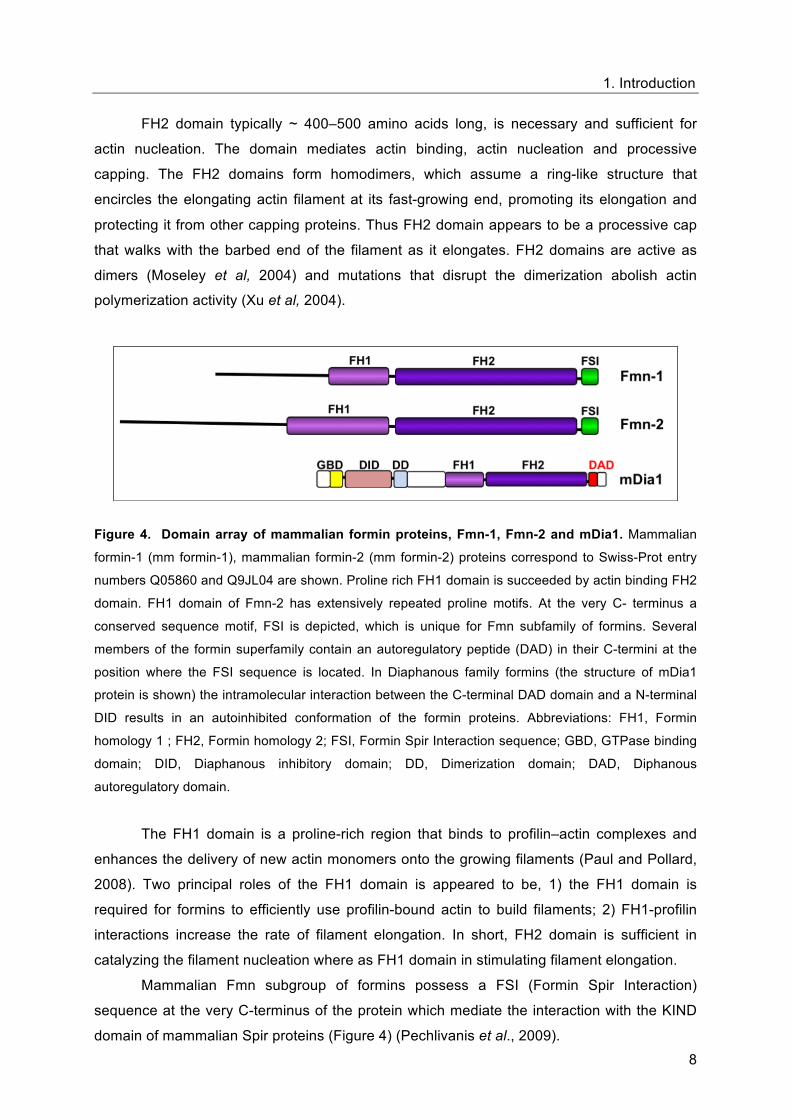
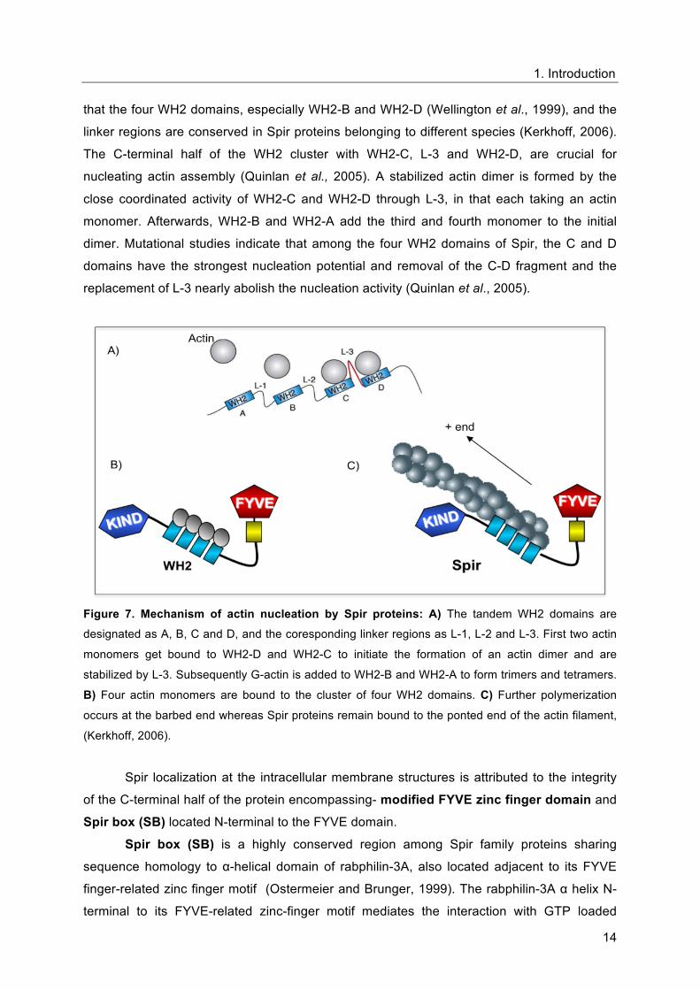
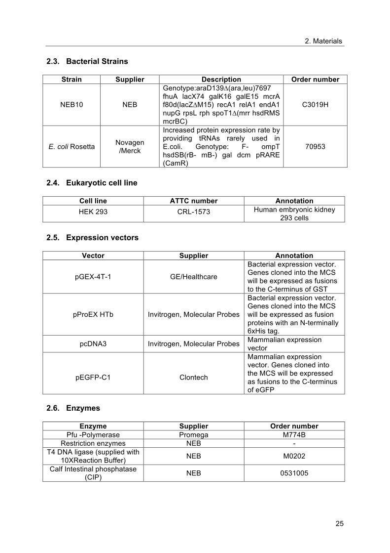
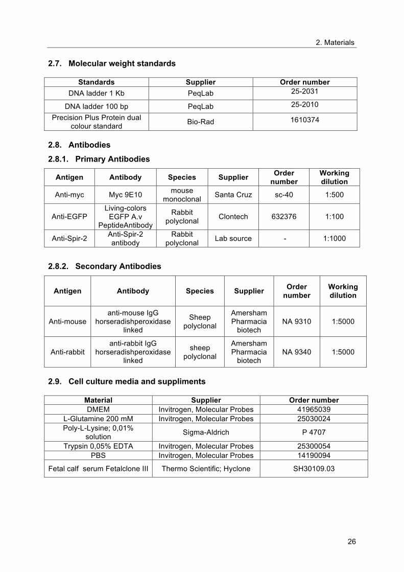
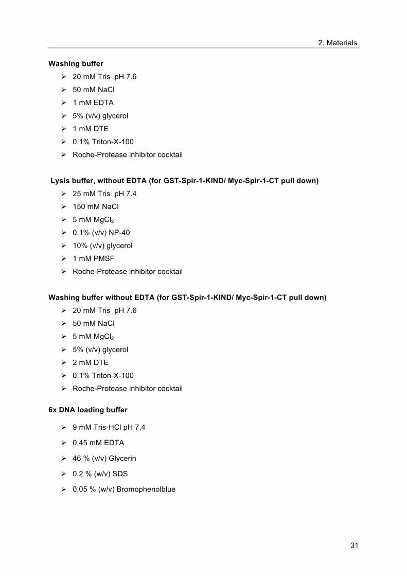
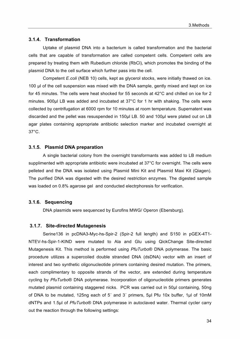
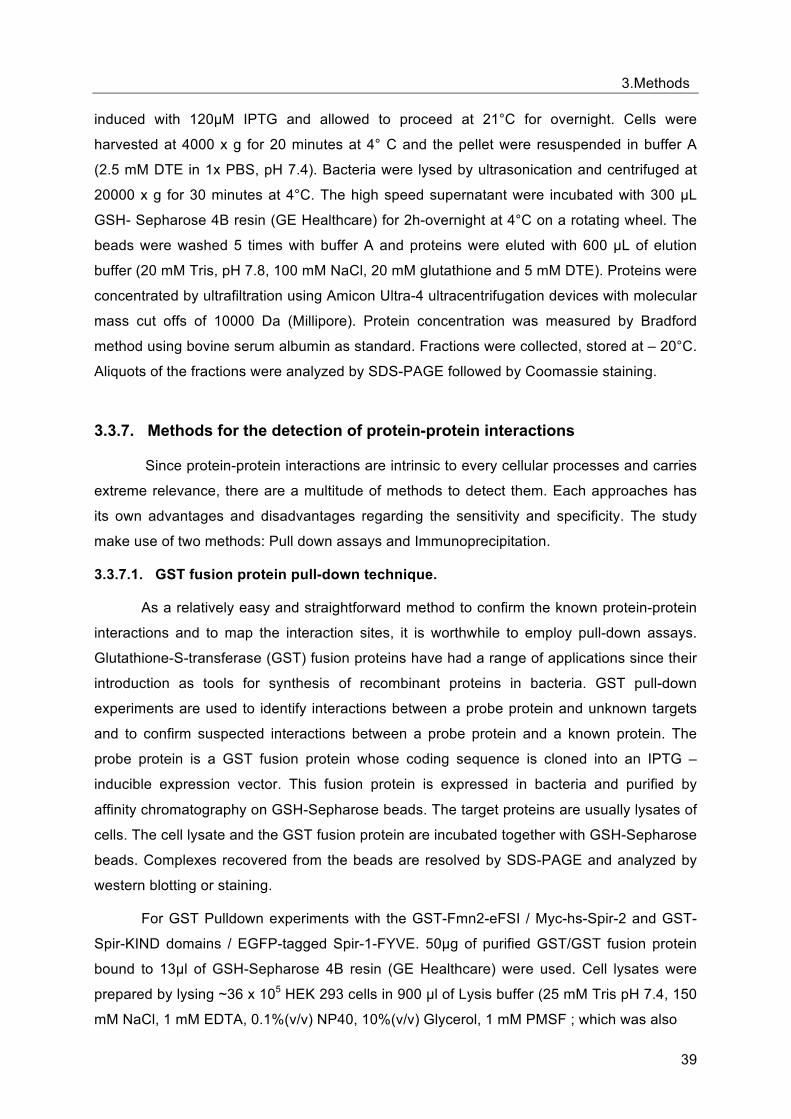
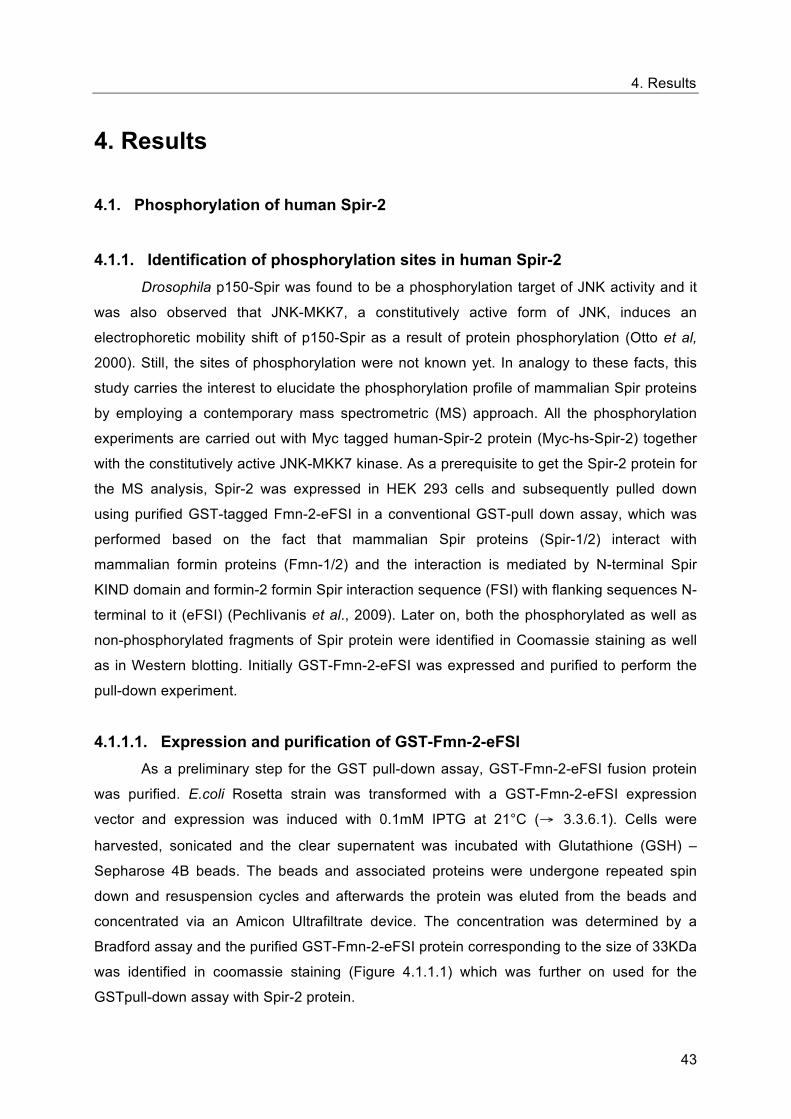
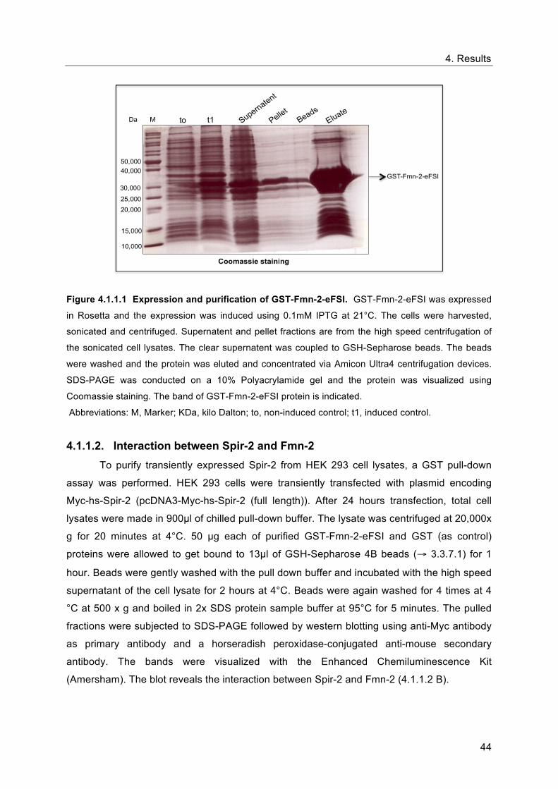
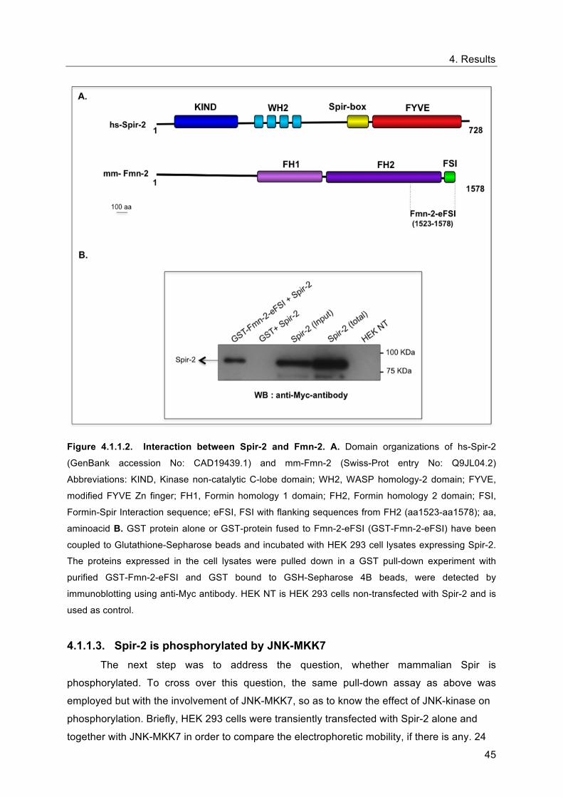
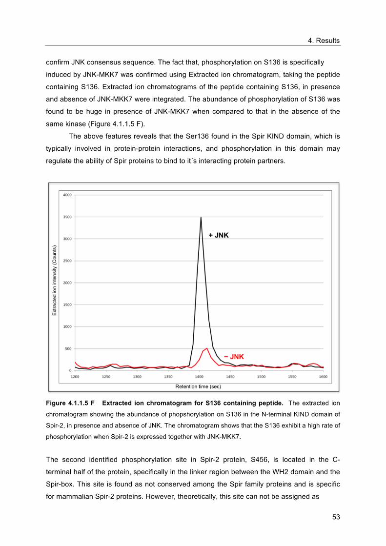
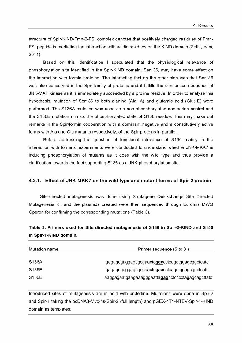
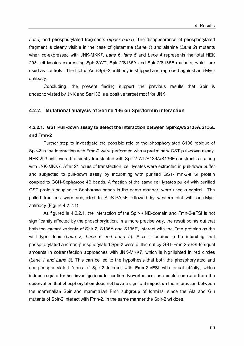
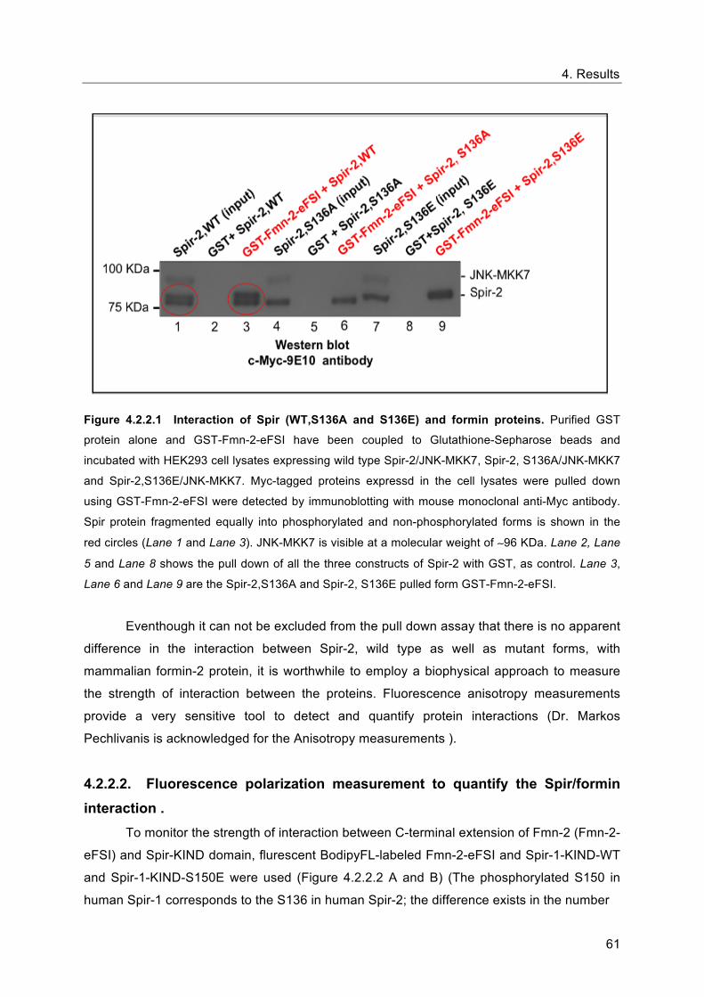
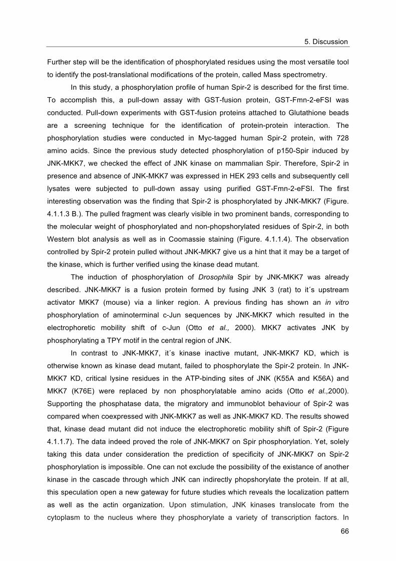
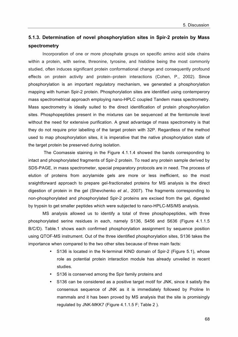
Appendix-III-Construct overview
80
Appendix -III- Construct overview
Construct Description Restriction sites Purpose
pcDNA3-Myc-
hs-Spir-2
Myc-Spir-2 f.l (full length)
BamHI / HindIII
GST pull-down,
Mass spectrometry,
Immunoprecipitation.
pGEX- 4T1- NTEV-
mFmn-2-eFSI GST-Fmn-2-eFSI EcoRI / SalI
GST pull-down;
Anisotropy
pcDNA3-Myc-JNK-
MKK7 Myc-JNK-MKK7 BamHI / XbaI
GST pull-down,
Immunoprecipitation
pcDNA3-Myc-
hs-Spir-2-S136A Myc-Spir-2-S136A BamHI / HindIII
Mutation analysis;
GST pull-down
pcDNA3-Myc-
hs-Spir-2-S136E Myc-Spir-2-S136E BamHI / HindIII
Mutation analysis;
GST pull-down
pGEX- 4T1- NTEV-
hs-Spir-1-KIND GST-Spir-1-KIND BamHI / XhoI GST pull-down
pGEX- 4T1- NTEV-
hs-Spir-1-KIND-
S150E
GST-Spir-1-KIND-
S150E BamHI / XhoI GST pull-down
pEGFP-C1-hs-Spir-1-
FYVE EGFP-Spir-1-FYVE XhoI / BamHI GST pull-down
pProExHtb-hs-Spir-1-
KIND His-Spir-1-KIND BamHI / XhoI Anisotropy
pProExHtb-hs-Spir-1-
KIND-S150E
His-Spir-1-KIND-
S150E BamHI / XhoI Anisotropy
Acknowledgements
81
Acknowledgements With immense pleasure, I am taking this opportunity to express my heartfelt thanks to
all, outside and inside the work circle, for their invaluable contributions to make this voyage
possible and enjoyable. Without these supporters, especially, the select few I am about to
mention, I may not have reached to where I am today.
I must begin by thanking my supervisor, Prof. Dr. Eugen Kerkhoff, for his sincere assistance
and guidance throughout the tenure of my thesis, for making time even when there was little
to spare. Above all, for giving an opportunity to carry out my Ph.D in his lab.
I am perpetually greatful to Prof. Dr. Daniela Männel, Institute of Immunology, for her
encouragements, constructive criticisms and all time expert and wise advices. Boundless
thanks for bringing sunshines and courage to withstand in my tough situations. A` Thank you`
means a lot to me....
I owe a special debt of gratitude to Prof. Dr. Jens Schlossmann, Department of
Pharmacology and Toxicology, for generously accepting my mentorship, for being a part of
my thesis jury and for valuable suggestions and precious support rendered to me.
My hearfelt respect and gratitude goes to Prof. Dr. Oliver Reiser, Institute for Organic
Chemistry, for helping me in the enrollment of my Ph.D thesis.
I am indebted to Prof. Dr. Peter Oefner and Prof. Dr. Anja Bosserhoff, for their kindness and
constant readiness to help me.
I extend my sincere gratitude to Bayerische Forschungsstiftung for their scintillating
scientific and financial support.
I deeply thank Dr. Markos Pechlivanis, for the motivations and timely advices whenever I was
in need of.
I am greatful to Dr. Joerg Reinders, Proteomics group, for introducing the world of Mass
spectrometry. Thank you for your excellent support for carrying out the mass spectometrical
analysis.
Dr. Kornelius Zeth is warmly acknowledged for his collaboration in crystalographic studies.
Acknowledgements
82
Thanks to my labmates, Annette Samol, Sandra Pleiser, Sabine Weiß, Dr. Agnes Pawelec and
Dr. Susanne Dietrich for their support. I remember, my former colleague, Martin Beusch, for
making the ambience fresh and funny with his presence.
Many thanks to Nevels-Paulus Lab, for moulding me to get started with my Ph.D life. Without
daubt I will look back with fond memories and nostalgia on my time in your lab. Thank you
Christina for being a wonderful teacher and friend.
I am thankful to Tanja Janoschek, for helping me to get cleared with the administration stuffs.
Thank you Denise, for being with me whenever there was computer emergencies.
Petra Leukel is thanked for her all time helps with Image J programme..
Sylvia Moeckel, thank you for being with me while I work with Excel.
My friends....
Ines Tschertner, words fail to express my thanks to you. You added much colours to my life in
Germany. Simply, You are Wonderful!
Tina, for being there for me, making my life smooth and funny.
Stefan Benecke and family for their gentle support during tough circumstances.
Anja Thomas, for being a nice and supportive friend.
My family....
My wonderful parents, Mr.Gopalakrishnan and Mrs.Valsala Gopalakrishnan, and my
brother, Manikandan, for their unconditional love and support which always makes me to
step forward- specially, my mother for being my ardent hand and for her contagious
enthusiasm for academia.
The most profoud thanks I owe to my husband, Preetham, it would have been impossible
without his support.
My daughter, Prarthana, for her lovely kicks instigating me to stick to the timetables, to
complete this thesis work on schedule, and above all, for making my life more beautiful.
........ and to the Almighty, under his watchful eyes, I gained strength and perseverence to
tackle challenges head on.
Sreeja Lakshmi, Regensburg, 2011
Bibliography
83
Bibliography
Alberto, P. and Juan, P. A. (2008). Advances in the analysis of protein phosphorylation. J.
Proteome Res. 7, 1809-1818.
Azoury, J., Lee, K. W., Georget,V., Rassinier, P., Leader, B. and Verlhac, M. H. (2008)
Spindle positioning in mouse oocytes relies on a dynamic meshwork of actin filaments.
Curr. Biol.18, 1514-1519.
Ahuja, R., Pinyol, R., Reichenbach, N., Custer, L., Klingensmith, J., Kessels, M. M. and
Qualmann, B. (2007). Cordon-Bleu Is an Actin Nucleation Factor and Controls Neuronal
Morphology. Cell. 131, 337–350.
Blanchoin, L. and Pollard, T. D. (2002). Hydrolysis of ATP by polymerized actin depends on
the bound divalent cation but not proline. Biochemistry. 41, 597-602.
Baum, B. and Kunda, P. (2005). Actin nucleation: spire-actin nucleator in a class of its own.
Curr. Biol. 15, R305-R308.
Blanchoin, L. and Robinson, R.C., (2000). Phosphorylation of Acanthamoeba actophorin
(ADF/cofilin) blocks interaction with actin without a change in atomic structure. J. Mol. Biol.
295, 203-211.
Chalkley, R. (2010). Instrumentation for LC-MS/MS in proteomics. Methods. Mol. Biol. 658,
47-60.
Chesarone, M. A., DuPage, A. G and Goode, B. L. (2010). Unleashing formins to remodel
the actin and microtubule cytoskeletons. Nat. Mol. Cell. Biol.11, 63-74.
Carlier, M. F. (1998). Control of actin dynamics. Curr. Opin. Cell. Biol.10, 45-51.
Chesarone, M. A. and Goode, B. L. (2009). Actin nucleation and elongation factors:
mechanisms and interplay. Curr. Opin. Cell. Biol 21, 28-37.
Campellone, K. G. and Welch, M. D. (2010). A nucleator arms race: cellular control of actin
assembly. Nat. Rev. Mol. Cell. Biol. 11, 237-251.
Bibliography
84
Conley, C. A., Fritz-Six, K. L., Almenar-Queralt, A. and Fowler, V. M. (2001). Leiomodins:
larger members of the tropomodulin (Tmod) gene family. Genomics 73, 127-139.
Chereau, D., Boczkowska, M., Maruszewska, A.S., Fujiwara, I., Hayes, B. H., Rebowski, G.,
Lappalainen, P., Pollard, T. D. and Dominguez, R. (2008). Leiomodin is an actin � filament
nucleator in muscle cells. Science, 320, 239-243.
Chereau, D., Ker, F., Grace, P., Grabarek, Z., Langsetmo, K. and Dominguez, R. (2005).
Actin-bound structures of Wiskott-Aldrich syndrome protein (WASP)- homology domain 2
and the implications for � lament assembly. Proc Natl Acad Sci U S A, 102, 16644-16649.
Chhabra, E. S. and Higgs, H. N. (2007). The many faces of actin: matching assembly factors
with cellular structures. Nature. Cell. Biol. 9, 1110-1121.
Carroll, A. E., Gerrelli, D., Gasca, S., Berg, E., Beier, D. R., Copp, A. J. and Klingensmith, J.
(2003). Cordon-bleu is a conserved gene involved in neural tube formation. Dev. Biol. 262,
16-31.
Ciccarelli, F. D., Bork, P. and Kerkhoff, E. (2003). The KIND module: a putative signalling
domain evolved from the C lobe of protein kinase fold. Trends. Biochem. Sci. 28, 349-352.
Cohen, P. (2002). The origins of protein phosphorylation. Nat. Cell. Biol.4, E127–E130.
Dos Remedios, C. G., Chhabra, D., Kekic, M., Dedova, I. V., Tsubakihara, M., Berry, D.A.
and Nosworthy, N. J. (2003). Actin binding proteins: regulation of cytoskeletal microfilaments.
Physiol. Rev. 83, 433-473.
Domon, B. and Aebersold, R. (2006). Mass spectrometry and protein analysis. Science. 312,
212-217.
Dominguez, R. and Holmes, K. C. (2011). Actin structure and function. Annu. Rev.
Biophys.40, 169-186.
Dahlgaard, K., Raposo, A. A., Niccoli, T. and St Johnston, D. (2007). Capu and Spire
assemble a cytoplasmic actin mesh that maintains microtubule organization in the Drosophila
oocyte. Dev. Cell. 13, 539 –553.
Bibliography
85
Davis, R. J. (2000) Signal transduction by the JNK group of MAP kinases. Cell. 103, 239-
252.
Dumont, J., Million, K., Sunderland, K., Rassinier, P., Lim, H., Leader, B. and Verlhac, M. H.
(2007). Formin-2 is required for spindle migration and for the late steps of cytokinesis in
mouse oocytes. Dev Biol. 301, 254-265.
Duncan, M.C., Cope, M.J., Goode, B. L., Wendland, B. and Drubin, D. G. (2001). Yeast
Eps15-like endocytic protein, Pan1p, activates the Arp 2/3 complex. Nat. Cell. Biol. 3, 687-
690.
Estes, J. E., L. A. Selden, and L. C. Gershman. (1987). Tight binding of divalent cations to
monomeric actin. Binding kinetics support a simplified model. J. Biol. Chem. 262, 4952-
4957.
Emmons, S., Phan, H., Calley, J., Chen, W., James, B. and Manseau, L. (1995).
Cappuccino, a Drosophila maternal effect gene required for polarity of the egg and embryo,
is related to the vertebrate limb deformity locus. Genes Dev. 9, 2482-2494.
Firat-Karalar, E. N. and Welch, M. D. (2011). New mechanisms and functions of actin
nucleation. Curr. Opin. Cell. Biol. 23, 4-13.
Fletcher, D. A. and Mullins, R. D. (2010). Cell mechanics and the cytoskeleton. Nature. 463,
485-92.
Faix, J. and Grosse, R. (2006). Staying in shape with formins. Dev. Cell 10, 693–706.
Foltz, I. N., Gerl, R. E., Wieler, J.S., Luckach, M., Salmon, R.A. and Schrader, J. W. (1998).
Human mitogen-activated protein kinase kinase 7 (MKK7) is a highly conserved c-Jun N-
terminal kinase/stress-activated protein kinase (JNK/SAPK) activated by environmental
stresses and physiological stimuli. J Biol Chem. 273, 9344-9351.
Goley, E. D., Rammohan, A., Znameroski, E. A., Firat-Karalar, E.N., Sept, D. and Welch, M.
D. (2010). An actin-filament-binding interface on the Arp2/3 complex is critical for nucleation
and branch stability. Proc. Natl. Acad. Sci. U S A.107, 8159-8164.
Bibliography
86
Goley, E. D. and Welch, M. D. (2006). The ARP2/3 complex: an actin nucleator comes of
age. Nat. Rev..Mol..Cell..Biol. 7, 713-726.
Goode, B. L., Rodal, A. A., Barnes, G. and Drubin, D. G. (2001). Activation of the Arp2/3
complex by the actin filament binding protein Abp1p. J. Cell Biol. 153, 627–634.
Goode, B. L. and Eck, M. J. (2007). Mechanism and function of formins in the control of actin
assembly. Annu.Rev.Biochem.76, 593-627.
Higgs, H.N. and Peterson, K.J. (2005). Phylogenetic analysis of forming homology 2 domain.
Mol.Biol. Cell 16, 1-13.
Higgs, H.N. and Pollard, T.D. (2000). Activation by Cdc42 and PIP2 of Wiskott-Aldrich
Syndrome protein (WASp) stimulates actin nucleation by Arp2/3 complex. J. Cell Biol. 150,
1311–1320.
Holmes, K. C., Popp, D., Gebhard, W. and Kabsch, W. (1990). Atomic model of the actin
filament. Nature, 347, 44-49.
Hjerrild, M. and Gammeltoft, S. (2006). Phosphoproteomics toolbox: computational biology,
protein chemistry and mass spectrometry. FEBS Lett. 580, 4764-4770.
Higgs, H.N. (2005). Formin proteins: a domain-based approach. Trends. Biochem. Sci. 30,
342–353.
Ito, T., Narita, A., Hirayama, T., Taki, M., Iyoshi, S., Yamamoto, Y., Maéda, Y. and Oda, T.
(2011). Human Spire interacts with the barbed end of the actin filament. J. Mol. Biol. 408,
18–25.
Jewett, T. J., Fischer, E.R., Mead, D.J. and Hackstadt, T. (2006). Chlamydial TARP is a
bacterial nucleator of actin. Proc. Natl. Acad. Sci. U S A.103, 15599-15604.
Johnson, H. and Eyers, C. E. (2010). Analysis of post-translational modifications by LC-
MS/MS. Methods. Mol. Biol. 658, 93-108.
Bibliography
87
Jacobs, D., Glossip, D., Xing, H., Muslin, A. J. and Kornfeld, K. (1999). Multiple docking sites
on substrate proteins form a modular system that mediates recognition by ERK MAP kinase.
Genes Dev. 13, 163-175.
Kabsch,W., Mannherz, H. G., Suck, D., Pai, E. F. and Holmes, K.C.(1990.) Atomic structure
of the actin:DNase I complex. Nature, 347, 37-44.
Kerkhoff, E. (2006). Cellular functions of the Spir actin-nucleation factors. Trends. Cell. Biol.
16, 477- 483.
Kerkhoff, E., Simpson, J.C., Leberfinger, C.B., Otto, I.M., Doerks, T., Bork, P., Rapp, U.R.,
Raabe, T. and Pepperkok, R. (2001). The Spir actin organizers are involved in vesicle
transport processes. Curr. Biol. 11, 1963-1968.
Kerkhoff, E. (2006). Cellular functions of the Spir actin-nucleation factors. Trends Cell Biol
16, 477- 483.
Katoh, M. and Katoh, M. (2004). Identification and characterization of human FHDC1, mouse
Fhdc1 and zebrafish fhdc1 genes in silico. Int. J. Mol. Med. 6, 929–934.
Kerkhoff E. (2010) Actin dynamics at intracellular membranes: The Spir/formin nucleator
complex. Eur.J. Cell. Biol. 90, 922-925.
Kovar, D.R. (2006). Molecular details of formin-mediated actin assembly. Curr. Opin. Cell
Biol. 18, 11–17.
Kerkhoff, E., Simpson, J. C., Leberfinger, C. B., Otto, I. M., Doerks, T., Bork, P., Rapp, U. R.,
Raabe, T. and Pepperkok, R. (2001). The Spir actin organizers are involved in vesicle
transport processes. Curr. Biol. 11, 1963-1968.
Le Goff, C., Laurent, V., Le Bon, K., Tanguy, G., Couturier, A., Le Goff, X. and Le Guellec, R.
(2006). pEg6, a spire family member, is a maternal gene encoding a vegetally localized
mRNA in Xenopus embryos. Biol Cell. 98, 697-708.
Li, F. and Higgs, H. N. (2003). The mouse Formin mDia1 is a potent actin nucleation factor
regulated by autoinhibition. Curr. Biol, 13, 1335-1340.
Bibliography
88
Li, H., Guo, F., Rubinstein, B. and Li, R. (2008). Actin-driven chromosomal motility leads to
symmetry breaking in mammalian meiotic oocytes. Nat. Cell. Biol,10, 1301-1308.
Leader, B. and Leder, P. (2000). Formin-2, a novel formin homology protein of the
cappuccino subfamily, is highly expressed in the developing and adult central nervous
system. Mech Dev, 93, 221-31.
Lammers, M., Rose, R., Scrima, A. and Wittinghofer, A. (2005). The regulation of mDia1 by
autoinhibition and its release by Rho GTP. EMBO J. 24, 4176–4187.
Leader, B., Lim, H., Carabatsos, M. J., Harrington, A., Ecsedy, J., Pellman, D., Maas, R. and
Leder, P. (2002.) Formin-2, polyploidy, hypofertility and positioning of the meiotic spindle in
mouse oocytes. Nat. Cell. Biol, 4, 921-928.
LeClaire, L. L 3rd., Baumgartner, M., Iwasa, J. H., Mullins, R. D. and Barber, D. L. (2008).
Phosphorylation of the Arp2/3 complex is necessary to nucleate actin filaments. J. Cell.
Biol.182, 647-54.
Liverman, A. D., Cheng, H. C., Trosky, J. E., Leung, D. W., Yarbrough, M. L., Burdette, D. L.,
Rosen, M. K. and Orth, K. (2007). Arp2/3- independent assembly of actin by Vibrio type III
effector VopL. Proc Natl Acad Sci USA. 104, 17117–17122.
Mooney, L. M and Whitmarsh, A. J. (2004). Docking interactions in the c-Jun N-terminal
kinase pathway. J. Biol. Chem. 279, 11843-11852.
Mann, M. and Jensen, O. N. (2003). Proteomic analysis of post-translational modifications.
Nat. Biotechnol. 21, 255-61.
Moseley, J.B., I. Sagot., A. L. Manning., Y. Xu., M.J. Eck., D. Pellman, and B.L.Goode .
(2004). A conserved mechanism for Bni1- and mDia1-induced actin assembly and dual
regulation of Bni1 by Bud6 and profi lin. Mol. Biol. Cell . 15, 896 – 907.
Manseau, L. J. and Schüpbach,. T. (1989). Capuccino and spire: two unique maternal-effect
loci required for both the anterioposterior and dorsoventral patterns of the Drosophila
embryo. Genes Dev. 3, 1437-1452.
Molecular Biology of the Cell. Fifth Edition.
Bibliography
89
Morel, E., Parton, R. G., Gruenberg, J. (2009). Annexin A2-dependent polymerization of actin
mediates endosome biogenesis. Dev. Cell 16, 445-457.
Otto, I.M., Raabe, T., Rennefahrt, U.E., Bork, P., Rapp, U.R. and Kerkhoff, E. (2000). The
p150- Spir protein provides a link between c-Jun N-terminal kinase function and actin
reorganization. Curr. Biol. 10, 345-348.
Ostermeier, C. and Brünger, A.T. (1999) Structural basis of Rab effector specificity: crystal
structure of the small G protein Rab3A complexed with the effector domain of rabphilin- 3A.
Cell, 96, 363–374.
Oda, T. and Maeda, Y. (2010). Multiple Conformations of F-actin. Structure, 18, 761-767.
Oda, T., Iwasa, M., Aihara, T., Maeda, Y. and Narita, A. (2009). The nature of the globular- to
fibrous- actin transition. Nature. 457, 441-5.
Olsen, J. V., Ong, S. E. and Mann, M. (2004). Trypsin cleaves exclusively C-terminal to
Arginine and lysine residues. Mol. Cell. Proteomics.3, 608-614.
Otomo, T., D.R. Tomchick, C. Otomo, S.C. Panchal, M. Machius, and M.K.Rosen. (2005).
Structural basis of actin filament nucleation and processive capping by a formin homology 2
domain. Nature. 433, 488–494.
Otterbein, L. R., Grace, P. � and Dominguez, R. (2001). The crystal structure of uncomplexed
actin in the ADP state. Science. 293, 708-711.
Paul, A. S. and Pollard, T.D. (2009). Review of the mechanism of processive actin filament
elongation by formins. Cell. Motil. Cytoskeleton. 66, 606-617.
Paradela, A. and Albar, J. P. (2008). Advances in the analysis of protein phosphorylations.
J. Proteome Res. 7, 1809-1818.
Pring, M., Evangelista, M., Boone, C. and Zigmond, S. (2003). Mechanism of formin-induced
nucleation of actin filaments. Biochemistry. 42, 486-496.
Bibliography
90
Pollard, T D., Blanchoin, L., and Mullins, R. D. (2000). Molecular mechanisms controlling
actin fiament dynamics in nonmuscle cells. Annu. Rev. Biophys. Biomol. Struct. 29, 545-576.
Pollard, T. D. and Borisy, G.G. (2003). Cellular motility driven by assembly and disassembly
of actin filaments. Cell. 112, 453-465.
Pleiser, S., Rock, R., Wellmann, J., Gessler, M. and Kerkhoff, E. (2010). Expression patterns
of the mouse Spir-2 actin nucleator. Gene. Expr. Patterns. 10, 345–350.
Pechlivanis, M., Samol, A., and Kerkhoff, E. (2009). Identification of a short Spir interaction
sequence at the C-terminal end of formin subgroup proteins. J. Biol. Chem 284, 25324-
25333.
Pollard, T.D. (2007). Regulation of actin filament assembly by Arp2/3 complex and formins.
Annu Rev Biophys Biomol Struct 36, 451-477.
Perrin, B. J. and Ervasti, J. M. (2010). The actin gene family: function follows isoform.
Cytoskeleton. 67, 630-634.
Page, R., Lindberg, U. and Schutt, C. E. (1998). Domain motions in actin. J. Mol. Biol. 280,
463-474.
Pfender, S., Kunzetsov, V., Pleiser, S., Kerkhoff, E. and Schuh, M. (2011). Spir-type actin
nucleators co-operate with Formin-2 to drive asymmetric oocyte division. Curr. Biol. 21, 955-
960.
Paunola, E., Mattila, P. K. and Lappalainen, P. (2002). WH2 domain: a small, versatile
adapter for actin monomers. FEBS Lett, 513, 92-97.
Quinlan, M.E., Heuser, J.E., Kerkhoff, E. and Mullins, R.D. (2005). Drosophila Spire is an
actin nucleation factor. Nature. 433, 382-388.
Quinlan. M.E., Hilgert, S., Bedrossian, A., Mullins, R.D., and Kerkhoff, E. (2007). Regulatory
interactions between two actin nucleators, Spire and Cappuccino. J. Cell. Biol 179, 117-128.
Bibliography
91
Quinlan, M.E., and Kerkhoff, E. (2008). Actin nucleation: bacteria get in-Spired. Nat. Cell.
Biol 10, 13-15.
Qualmann, B. and Kessels, M. M. (2009). New players in actin polymerization- WH2-
domain-containing actin nucleators. Rev. Trends. Cell. Biol.19, 276-285.
Qualmann, B. and Kessels, M. M. (2008). Actin nucleation: Putting the breaks on Arp2/3
complex. Curr.Biol.18, R420-422.
Reinders, J. and Sickmann, A. (2005). State-of-the-art in phosphoproteomics. Proteomics. 5,
4052-4061.
Renault, L., Bugyi, B. and Carlier MF. (2008). Spire and Cordon-bleu: multifunctional
regulators of actin dynamics. Trends. Cell. Biol. 18, 494-504.
Robinson, R. C., Turbedsky, K., Kaiser, D. A., Marchand, J. B., Higgs, H. N., Choe, S. and
Pollard, T. D.(2001). Crystal structure of Arp2/3 complex. Science. 294, 1679-1684.
Rennefahrt, U. E., Illert, B., Kerkhoff, E., Troppmair, J. and Rapp, U. R. (2002). Constitutive
JNK activation in NIH 3T3 fibroblasts induces a partially transformed phenotype. J. Biol.
Chem. 277, 29510-29518.
Rosales-Nieves, A. E., Johndrow, J. E., Keller, L. C., Magie, C. R., Pinto-Santini, D. M. and
Parkhurst, S. M. (2006). Coordination of microtubule and microfilament dynamics by
Drosophila Rho1, Spire and Cappuccino. Nat. Cell. Biol. 8, 367-376.
Shevchenko, A., Wilm, M., Vorm, O. and Mann, M.(1996). Mass spectrometric sequencing of
proteins silver-stained polyacrylamide gels. Anal. Chem. 68, 850-858.
Sept, D. and McCammon, J A. (2001). Thermodynamics and kinetics of actin � filament
nucleation. Biophys J, 81, 667-674.
Schumacher, N., Borawski, J. M., Leberfinger, C. B., Gessler, M. and Kerkhoff. E. (2004) � .
Overlapping expression pattern of the actin organizers Spir-1 and formin-2 in the developing
mouse nervous system and the adult brain. Gene. Expr Patterns.4, 249-255.
Bibliography
92
Stenmark, H., Aasland, R., Toh, B. H. and D'Arrigo, A. (1996). Endosomal localization of the
autoantigen EEA1 is mediated by a zinc-binding FYVE finger. J. Biol. Chem, 27, 24048-
24054.
Stenmark, H. (2005). The FYVE finger: A Phosphoinositide Binding Domain. Chapter 19,
Zinc-finger proteins: From Atomic Contact to cellular Function.
Steen, H. and Mann, M. (2004). The ABC's (and XYZ's) of peptide sequencing. Nat Rev Mol
Cell Biol. 5, 699-711.
Schuh, M. and Ellenberg, J. (2008). A new model for asymmetric spindle positioning in
mouse oocytes. Curr Biol.18, 1986-1992.
Shevchenko, A., Tomas, H., Havlis, J., Olsen, J. V. and Mann M. (2006). In-gel digestion for
mass spectrometric characterization of proteins and proteomes. Nat Protoc.1, 2856-2860.
Small, J. V., Rottner, K. and Kaverina, I. (1999). Functional design in the actin cytoskeleton.
Curr. Opin. Cell. Biol.11, 54-60.
Schmidt, A. and Hall, M.N. (1998). Signaling to the actin cytoskeleton. Annu. Rev. Cell. Dev.
Biol.14, 305-338.
Schonichen, A. and Geyer, M. (2010). Fifteen formins for an actin filament: a molecular view
on the regulation of human formins. Biochem Biophys Acta. 1803, 152-163.
Schüler, H. (2001). ATPase activity and conformational changes in the regulation of actin.
Biochem. Biophys.Acta.1549, 137–147.
Theurkauf, W. E. (1994). Premature microtubule-dependent cytoplasmic streaming in
cappuccino and spire mutant oocytes. Science. 265, 2093-2096.
Tournier, C., Dong, C., Turner, T.K., Jones, S. N., Flavell, R. A. and Davis, R. J. (2001).
MKK7 is an essential component of the JNK signal transduction activated by proinflammatory
cytokines. Genes Dev. 15, 1419-1426.
Bibliography
93
Tam, V.C., Serruto, D., Dziejman, M., Brieher, W. and Mekalanos, J.J. (2007). A type III
secretion system in Vibrio cholerae translocates a formin/spire hybrid-like actin nucleator to
promote intestinal colonization. Cell. Host. Microbe 1, 95–107.
Vertommen, D., Rider, M., Ni. Y., Waelkens, E., Merlevede, W., Vandenheede, J. R.and Van
Lint, J. (2000). Regulation of protein kinase D by multisite phosphorylation. Identification of
phosphorylation sites by mass spectrometry and characterization by site-directed
mutagenesis. J. Biol. Chem. 275, 19567-19576.
Wellington. A., Emmons. S., James, B., Calley. J., Grover, M., Tolias, P. and Manseau, L.
(1999). Spire contains actin binding domains and is related to ascidian posterior end mark-5.
Development. 126, 5267-5274.
Wilm. M., Shevchenko, A., Houthaeve, T., Breit, S., Schweigerer, L., Fotsis, T. and Mann, M.
(1996). Femtomole sequencing of proteins from polyacrylamide gels by nano-electrospray
mass spectrometry. Nature. 369, 466-469.
Wang, X., Destrument, A. and Tournier, C. (2007). Physiological roles of MKK4 and MKK7:
insights from animal models. Biochem. Biophys. Acta. 1773, 1349-1357.
Weston, C. R. and Davis, R. J. (2007). The JNK signal transduction pathway. Curr Opin Cell
Biol. 19, 142-149.
Wallar, B.J., Stropich, B.N., Schoenherr, J.A., Holman, H.A., Kitchen, S.M., and Alberts, A.S.
(2006). The basic region of the diaphanous autoregulatory domain (DAD) is required for
autoregulatory interactions with the diaphanous-related formin inhibitory domain. J.
Biol.Chem. 281, 4300–4307.
Weber, A. (1999). Actin binding proteins that change extent and rate of actin monomer-
polymer distribution by different mechanisms. Mol. Cell. Biochem. 190, 67–74.
Winckler, B. and D.A. Schafer. (2007). Cordon-bleu: a new taste in actin nucleation. Cell.
131, 236–238.
Welch, M. D. and Mullins, R. D. (2002). Cellular control of actin nucleation. Annu. Rev. Cell.
Dev. Biol, 18, 247- 288.
Bibliography
94
Weaver, A. M., Karginov, A. V., Kinley, A. W., Weed, S. A., Li, Y., Parsons,J. T. and Cooper,
J. A. (2001). Cortactin promotes and stabilizes Arp2/3-induced actin filament network
formation. Curr. Biol. 11, 370-374.
Wang, R.W., Yang, C.Y., Zhao, G.F. and Yang, J.X. (2005) Fragmentation effects on
diversity of wasp community and its impact on fig/fig wasp interaction in Ficus racemosa L.
Journal of Integrative Plant Biology. 47, 20–26.
Wallar, B. J. and Alberts, A.S. (2003). The formins: active scaffolds that remodel the
cytoskeleton. Trends. Cell. Biol. 13, 435-446.
Witze, E. S., Old, W. M., Resing, K. A. and Ahn, N. G. (2007). Mapping protein post-
translational modifications with mass spectrometry. Nature methods. 4, 798-806.
Xu, Y., Moseley, J.B., Sagot, I., Poy, F., Pellman, D., Goode, B.L. and Eck, M.J. (2004)
Crystal structures of a Formin Homology-2 domain reveal a tethered dimer architecture. Cell
116, 711–723.
Yao, Z., Diener, K., Wang, X. S., Zukowski, M., Matsumoto, G., Zhou, G., Mo, R., Sasaki, T.,
Nishina, H., Hui, C. C., Tan, T. H., Woodgett, J. P. and Penninger, J. M. (1997). Activation of
stress-activated protein kinases/c-Jun N-terminal protein kinases (SAPKs/JNKs) by a novel
mitogen-activated protein kinase. J. Biol. Chem. 272, 32378-32383.
Yi, K., Guo, C., Chen, D., Zhao, B., Yang, B. and Ren, H. (2005). Cloning and functional
characterization of a formin-like protein (AtFH8) from Arabidopsis. Plant. Physiol. 138, 1071-
1082. Zeth, K., Pechlivanis, M., Samol, A., Pleiser, S., Vonrhein, C. and Kerkhoff E. (2011).
Molecular Basis of Actin Nucleation factor Cooperativity: CRYSTAL STRUCTURE OF THE
SPIR-1 KINASE NON-CATALYTIC C-LOBE DOMAIN (KIND).FORMIN-2 FORMIN SPIR
INTERACTION MOTIF (FSI) COMPLEX. J Biol Chem.286, 30732-30739.
Zigmond, S. H. (2004). Formin-induced nucleation of actin filaments. Curr Opin Cell Biol. 16,
99-105.