UCSF Benioff Children’s Hospital Outreach Services, Mission Bay
Disclosures
We have no financial relationships with any commercial interests
No relevant financial relationships exist
Objectives
Discuss abnormal conditions that increase a pregnant woman’s risk for rapid decline
Review the physiological changes of pregnancy that mask the severity of maternal decompensation
Describe the importance of multidisciplinary care teams aimed to provide comprehensive care
Cite at least 3 patient safety bundles aimed to prevent maternal morbidity and death
Objectives
Review normal physiologic changes of pregnancy that impact maternal decompensation
Highlight abnormal conditions that contribute to the severity of obstetric emergencies.
Cite four Maternal Safety Bundles aimed to reduce maternal morbidity and death
Describe how direct care nurses can improve patient safety with their organization
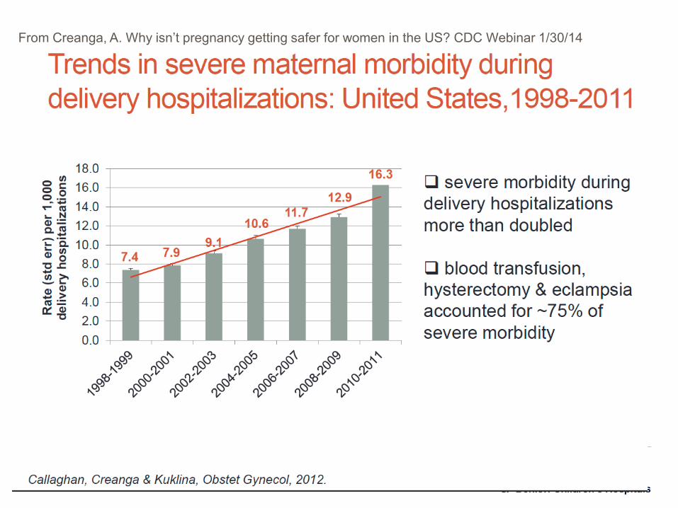
From Creanga, A. Why isn’t pregnancy getting safer for women in the US? CDC Webinar 1/30/14
Current Commentary
The Maternal Early Warning CriteriaA Proposal From the National Partnership for Maternal Safety
Mhyre, J., D’ Oria, R., Hameed, A., et al
Current Commentary
The National Partnership for Maternal SafetyMary E. D’Alton, MD, Elliott K. Main, MD, M. Kathryn Menard, MD, and Barbara S. Levy, MD
The American College ofObstetricians and Gynecologists
WOMEN’S HEALTH CARE PHYSICIANS
Obstetrics & Gynecology
VOL. 123, NO. 5, MAY 2014
Obstetrics & GynecologyVOL. 124, NO. 4, Oct 2014
Maternal Warning Systems
The Joint Commission (2010) requires hospitals to have written criteria to observe change or deterioration in a patient’s condition and how to recruit staff to manage patient care
Signs and symptoms of impending severe maternal illness or collapse went unrecognized in many cases (CEMACH, 2011) due to the relative rarity of such events and normal changes in physiology associated with pregnancy and childbirth compounds the problem
• Recommendation: Develop and adopting systems to alert the team of maternal deterioration to assist in early recognition, intervention and timely referral of treatment of women (CEMACH, 2011)
The National Partnership for Maternal Safety is a multi-stakeholder consensus effort and is comprised of representatives from organizations in women’s health care and other provider, state, federal, and regulatory bodies which supports early warning criteria to promote patient safety http://www.safehealthcareforeverywoman.org/maternal-safety.html
Vital sign assessment is critical during active bleeding. Blood pressure, pulse and respirations have been the standard in assessing vital signs.
Often variations in vital signs are ignored or dismissed as “normal” due to the physiological changes in pregnancy (CEMACH, 2011)
Lack of standardized documentation can result in delays in recording of abnormal results which can effect timeliness of clinical decision making (Yeung, Lapinsky, Granton, Doran, & Cafazzo, 2012)
Maternal Early Warning Systems
Abnormal physiologic signs and symptoms precede critical illness
Early intervention will avoid severe M&M occurrence
Effective policy of escalation of care
Maternal Early Warning Criteria
California Partnership for Maternal Safety
The Maternal Early Warning Criteria: A Proposal From the National Partnership for Maternal Safety.Mhyre, Jill; DOria, Robyn; MA, RNC; Hameed, Afshan; Lappen, Justin; Holley, Sharon; CNM, DPN; Hunter, Stephen; MD, PhD; Jones, Robin; King, Jeffrey; DAlton, Mary
BUNDLE SCIENCE
National Partnership Strategy to Enhance Maternal Safety
A "bundle" is a group of interventionsrelated to a disease process that, when
executed together, result in betteroutcomes than when implemented
individually.
CA-PAMR: Chance to Alter Outcome Grouped Cause of Death; 2002-2004 (N=145)
Grouped Cause of Death Chance to Alter Outcome
Strong /
Good (%)
Some
(%)
None
(%)
Total
N (%)
Obstetric hemorrhage 69 25 6 16 (11)
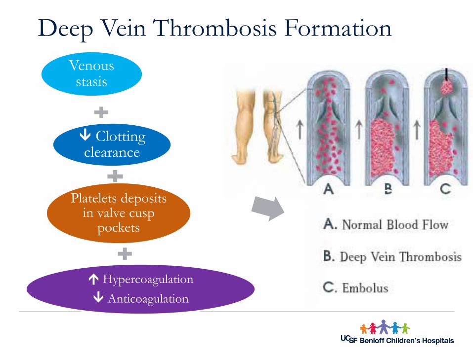
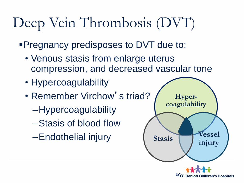
Deep vein thrombosis/
pulmonary embolism53 40 7 15 (10)
Sepsis/infection 50 40 10 10 (7)
Preeclampsia/eclampsia 50 50 0 25 (17)
Cardiomyopathy and other
cardiovascular causes25 61 14 28 (19)
Cerebral vascular accident 22 0 78 9 (6)
Amniotic fluid embolism 0 87 13 15 (10)
All other causes of death 46 46 8 26 (18)
Total (%) 40 48 12 145 7
Normal physiologic changes
Cardiovascular
Hematologic
Pulmonary
Cardiovascular
Cardiac Output
Cardiac Changes
Stroke Volume 30-50%
Heart Rate 20% (~10-20 beats)
Anatomic Changes Uterus
Vascular Resistance
SVR PVR
Normal Cardiac Adaptation during Pregnancy
% C
ha
ng
e
pregnant Weeks of gestation postpartum
Hematologic
Cardiac Output
Total blood volume
Plasma Volume
RBC Volume
% C
ha
ng
e
Weeks of gestationpregnant postpartum
Blood Volume Changes
Total Volume
35% (~ 2,000ml)
Plasma Volume
50% (~ 1,600ml)
RBC Mass
17% (~ 350mL)
Normal Hematologic Events Associated with Pregnancy
Hematologic continued:Clotting Factors During Pregnancy
Parameter ChangeFibrin Increases 40% at term
Plasma fibrinogen Increases 50% (300 – 600) mg/dl
Coagulation factors I, VII, VIII, X, XII Increases markedly
Von Willebrand factor antigen Increases markedly
Coagulation factor XI Decreases 60% - 70%
Coagulation factor XIII Decreases slightly
Coagulation factors II, V Increases slightly or unchanged
Protein S (anticoagulant) activity Decreased
Clotting and bleeding time Unchanged
Prothrombin time Increases slightly or unchanged
Partial plasma thromboplastin time Increases slightly or unchanged
Tissue hypoperfusion metabolic acidosis inflammatory mediators tissue and vascular injury multiple organ failure
Adult Respiratory Distress Syndrome
Hemorrhagic
Shock
Damage to endothelial cells in pulmonary vasculature
Fluid leaks from vascular space into alveoli
Respiratory failure
Case Presentation: Hemorrhage
38 y.o. @40+2 admitted for elective IOL: • Hgb 12.5, Hct 39.2, Plt 195K, Blood Type A+, Antibody screen: neg
Dinoprostone placed, misoprostol X2, AROM, oxytocin started
Epidural is placed
Rapidly progress to 10 cm , MD Notified
15 minute 2nd stage male infant
1 minute later: Placenta delivered spontaneously
Manual exploration of uterus “cleared of clots “
Fundal checks (6) RN charted “moderate”
23
1 hour 22 minutes later patient to MBU – Pulse: 82 BP: 126/70Patient passes “large clot” and “gush” when transferred to MBU bed IM methylergonovine30 minutes later carboprost given5 minutes later misoprostol given30 minutes later 2nd carboprost given10 minutes later VS: Pulse 106, BP: 116/72Foley catheter placed – urine concentrated, amount not documentedShift change
Case Presentation: Hemorrhage
24
Case Presentation: Hemorrhage
RN weighs chux 462 gm
RN reports to MD patient has “bled out” and is short of breath
Patient feels light headed
MD orders type and cross 2 units PRBC’s, another fluid bolus and wants to go to Main OR for D&C
3 more chux “saturated with blood” no clots
Coagulation Lab values obtained and sent to lab
2nd IV is started
1 hour and 20 minutes later, 1st unit of blood is transfused
8 liters of crystalloid up to this point
25
Case Presentation: Hemorrhage
Laboratory values:
• Hct nadir 13, Plts 22K (dysfunctional/abnormal aggregation)
• Fibrinogen 137, D Dimer >35,000, ABG pH: 7.14
Multiple doses of pressor support
• Norepinephrine drip to maintain BP/MAP
• To OR for unplanned emergency Hysterectomy
Patient to ICU intubated on IV pressor support
Blood products received: • 6 units PRBC• 4 units, FFP• 2 units Single Donor Platelets• 2 units of Cryoprecipitate
26
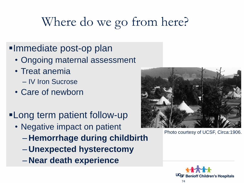
Unplanned Hysterectomy: Postoperative Course
Transfer from ICU
Weak but stable
Loss of choice
Hbg Hct
• Iron—IV (sucrose)
• Rh-Erythropoeitin
• Heparin
Discharge home with support
How Errors Occur
Defenses Harm
Safeguards
Stop the line
Standard work
Flexible staffing
Self-checks
Culture
PoliciesResources
TrainingCommunication
Failures
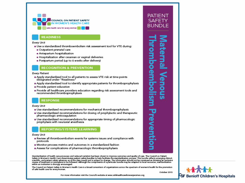
The Maternal Safety Bundle for Obstetric Hemorrhage
Proactive approach
Includes 13 elements
Establishes resources
Manage OB Hemorrhage
29
Hemorrhage
ACOG defines OB hemorrhage as: cumulative blood loss ≥1000 mL accompanied by s/sx of hypovolemia within 24 hrs after birth (including intrapartum blood loss) regardless of mode of birth.
Even with proper management can occur in
• ~ 4% of vaginal births and ~ 6% of cesarean birth
• As a result: 1/20 women will experience PPH
Early or Primary (< 24 hr after birth)
• Highest risk in the first hour after delivery because large venous areas are exposed after placental separation
Late or Secondary (>24 hr to 6 weeks after)
• Caused by infection, placental site subinvolution, retained placental fragments, or coagulopathies (DIC)
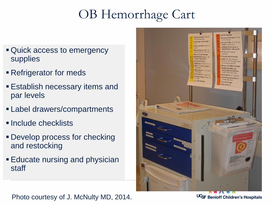
Photos courtesy of Holli M. Mason MD, 2017CPMS Blood Bank Webinar Slide Set
38
Blood Component Therapy
PRODUCT VOLUME
(ML)
CONTENTS EFFECT
(PER UNIT)
Packed Red
Blood Cells
240 RBC, WBC,
plasma
hematocrit 3% &
Hgb 1 g/dl
Platelets 50 Platelets,
RBC,WBC,
plasma
platelet count 5,000-
10,000 mm3 per unit
Fresh Frozen
Plasma
250 Fibrinogen,
antithrombin III,
factors V* & VIII*
fibrinogen by
10mg/dl
Cryoprecipitate 40 Fibrinogen,
factors VIII & XIII
and Von
Willebrand
fibrinogen by
10mg/dl
Laboratory Diagnosis of DIC
All routine screening tests of coagulation yield grossly abnormal results
Laboratory Test Value
Platelets
Fibrinogen
Fibrin Split Products
PT & aPTT
D Dimer
Decreased
Less than 200
Increased
Initially increased
Increased
40
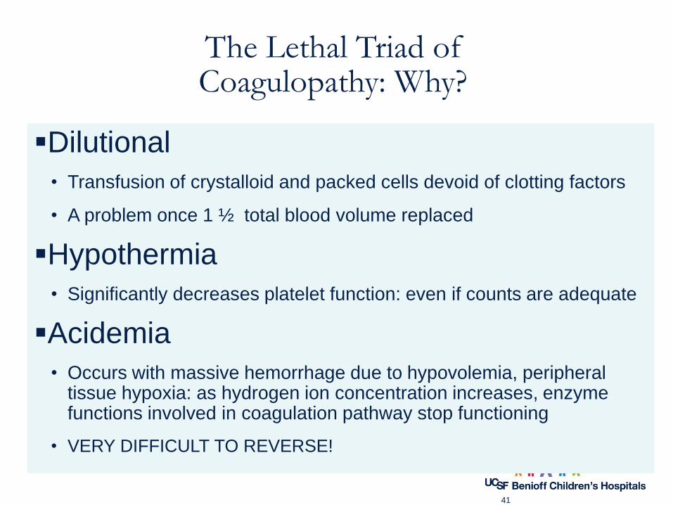
The Lethal Triad ofCoagulopathy: Why?
Dilutional
• Transfusion of crystalloid and packed cells devoid of clotting factors
• A problem once 1 ½ total blood volume replaced
Hypothermia
• Significantly decreases platelet function: even if counts are adequate
Acidemia
• Occurs with massive hemorrhage due to hypovolemia, peripheral tissue hypoxia: as hydrogen ion concentration increases, enzyme functions involved in coagulation pathway stop functioning
• VERY DIFFICULT TO REVERSE!
41
What is DIC?
Underlying disorder
Activates coagulation cascade
• Blood clot formation
• Coagulation factors become depleted
• Results in uncontrolled bleeding
‒Death
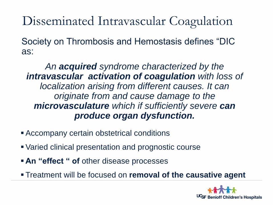
Disseminated Intravascular Coagulation
Accompany certain obstetrical conditions
Varied clinical presentation and prognostic course
An “effect “ of other disease processes
Treatment will be focused on removal of the causative agent
Society on Thrombosis and Hemostasis defines “DIC as:
An acquired syndrome characterized by the intravascular activation of coagulation with loss of
localization arising from different causes. It can originate from and cause damage to the
microvasculature which if sufficiently severe can produce organ dysfunction.
Etiology of DICOB/Gyn
Complications
Infection
Cancer
OB Complications
Placental Tissue
After Birth
Coagulation is initiated to prevent hemorrhage at placentation
Platelet plugs and fibrin clots for to provide hemostasis
• Fibrinogen and platelet counts decrease
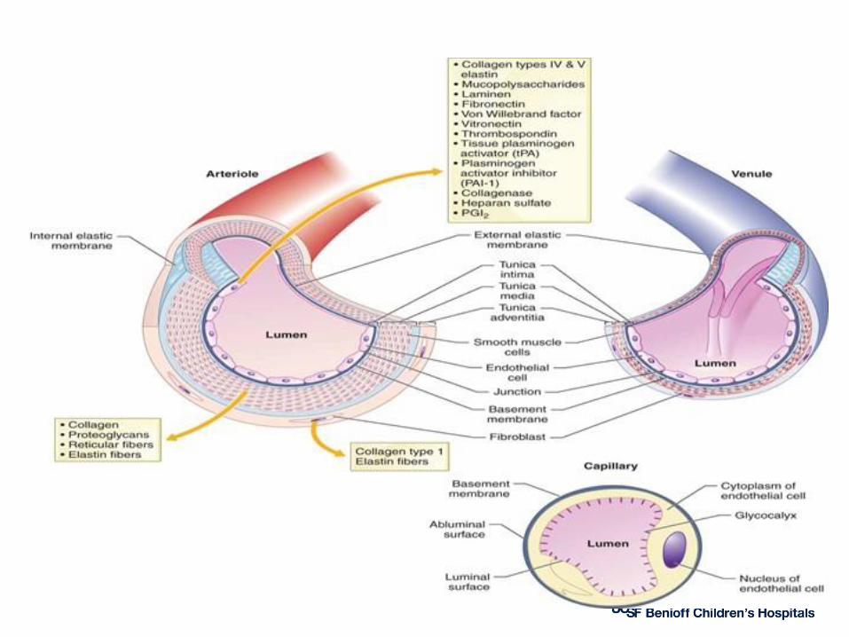
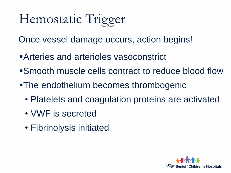
Physiology Review: Hemostasis
Failure or deficiencies in any of the components can lead to varying degrees of uncontrolled hemorrhaging or clotting
Primary components:
• Vascular endothelium
• Circulating platelets
• Circulating proteins
Vascular System: Blood Vessels
Endothelium
• Controls vessel permeability
• Controls blood flow rate
‒vasoconstriction
• Produces and releases substances that inhibit or stimulate platelets, coagulation, and fibrinolysis
Daily Function
Endothelium
Endothelium
Single layer of endothelial cells, lining vessels
Coated by glycocalyx (protein and mucopolysaccarides)
Protects basement membrane
Negatively charged, repels circulating proteins and platelets
Secretes substances to keep the blood vessel in a nonreactive environment
Anatomy
Vascular System
Subendothelium
• Smooth muscle and connective tissue with collagen fibers
Continue for 5 days postpartum• Clinical improvement
Begin oral anticoagulant therapy - Warfarin
Heparin “ High Alert”
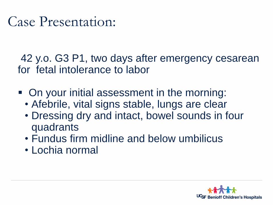
CVD Case Presentation
25 year old obese (BMI 38) African-American G2P2 presents 10 days after an uncomplicated vaginal delivery with fatigue and persistent cough since delivery.
BP 110/80, HR 110, RR 28, afebrile, with O2 sat 94% on room air.
She gets diagnosed with respiratory infection and is prescribed an antibiotic. Fatigue is attributed to lack of sleep.
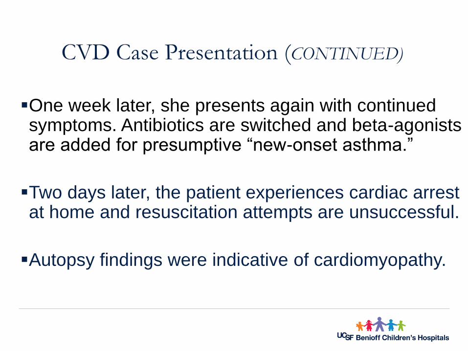
CVD Case Presentation (CONTINUED)
One week later, she presents again with continued symptoms. Antibiotics are switched and beta-agonists are added for presumptive “new-onset asthma.”
Two days later, the patient experiences cardiac arrest at home and resuscitation attempts are unsuccessful.
Autopsy findings were indicative of cardiomyopathy.
Disease in Pregnancy and Postpartum Taskforce. Visit: www.CMQCC.org for details
Rationale for ToolkitCardiovascular Disease is
the leading cause of maternal mortality in CA and U.S.
under-recognized in pregnant or postpartum women
higher among African-American women
25% of deaths attributed to cardiovascular disease may have been
prevented if the woman’s heart disease had been diagnosed earlier.
Pregnancy is a period of frequent interaction with health care
providers and offers an opportunity to detect and treat heart disease,
improve pregnancy outcomes, and affect future cardiovascular health.
Hameed A, Lawton E, McCain CL, et al. Pregnancy-Related Cardiovascular Deaths in California: Beyond Peripartum Cardiomyopathy. American Journal of Obstetrics and
Encourage obstetric and other healthcare providers to retain a high index of suspicion for CVD, particularly among women with risk factors who present with symptoms in late pregnancy or early postpartum period
To serve as resource for generalists who provide maternity care to women, with special emphasis on
Prenatal visits
Postpartum encounters
Emergency room visits
Given that CVD is the leading cause of maternal mortality & morbidity in
California, the Toolkit aims to:
Hameed, AB, Morton, CH and A Moore. Improving Health Care Response to Cardiovascular Disease in Pregnancy and Postpartum Developed under contract
#11-10006 with the California Department of Public Health, Maternal, Child and Adolescent Health Division. Published by the California Department of Public
Disease in Pregnancy and Postpartum Taskforce. Visit: www.CMQCC.org for details
Rationale for ToolkitCardiovascular Disease is
the leading cause of maternal mortality in CA and U.S.
under-recognized in pregnant or postpartum women
higher among African-American women
25% of deaths attributed to cardiovascular disease may have been
prevented if the woman’s heart disease had been diagnosed earlier.
Pregnancy is a period of frequent interaction with health care
providers and offers an opportunity to detect and treat heart disease,
improve pregnancy outcomes, and affect future cardiovascular health.
Hameed A, Lawton E, McCain CL, et al. Pregnancy-Related Cardiovascular Deaths in California: Beyond Peripartum Cardiomyopathy. American Journal of Obstetrics and
Disease in Pregnancy and Postpartum Taskforce. Visit: www.CMQCC.org for details
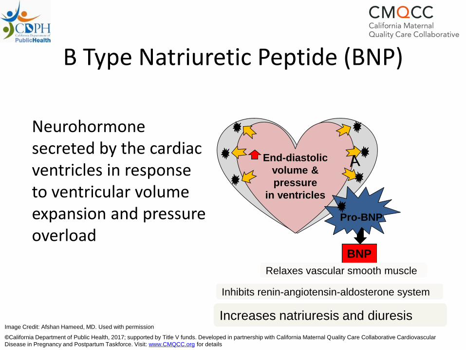
Clinical Uses of BNP in Pregnancy
Diagnosis of heart failure
In pregnant women with dilated CMP, higher BNP predicts adverse cardiovascular outcomes
Asymptomatic left ventricular function
Useful to evaluate shortness of breath
Predictor of cardiovascular outcome
In pregnant women with congenital heart disease, higher BNP levels are associated with poor outcomes
• Blatt A, Svirski R, Morawsky G, et al. Short and long-term outcome of pregnant women with preexisting dilated cardiomypathy: An NTproBNP and echocardiography-
guided study. The Israel Medical Association journal : IMAJ. Oct 2010;12(10):613-616.
• Tanous D, Siu SC, Mason J, et al. B-type natriuretic peptide in pregnant women with heart disease. J Am Coll Cardiol. Oct 5 2010;56(15):1247-1253.
• Kansal M, Hibbard JU, Briller J. Diastolic function in pregnant patients with cardiac symptoms. Hypertens Pregnancy. 2012;31(3):367-374.
Disease in Pregnancy and Postpartum Taskforce. Visit: www.CMQCC.org for details
Key Clinical Pearls
First presentation of cardiovascular disease may be
during pregnancy or early postpartum.
The highest risk period for CVD worsening is between 24-
28 weeks or postpartum.
CVD symptoms or vital sign abnormalities should not be
ignored in pregnant/postpartum women.
New onset or persistent asthma may be a sign of heart
failure.
Bilateral infiltrates on chest x-ray may be due to heart
failure rather than pneumonia.Hameed AB, Morton CH, and A Moore. Improving Health Care Response to Cardiovascular Disease in Pregnancy and Postpartum Developed under contract #11-
10006 with the California Department of Public Health, Maternal, Child and Adolescent Health Division. Published by the California Department of Public Health, 2017.
Disease in Pregnancy and Postpartum Taskforce. Visit: www.CMQCC.org for details
Key Clinical Pearls (continued)
Pregnancy or postpartum women with significant risk factors should be counseled regarding future CVD risk.
Women with known CVD should receive pre- & inter-conception counseling by an experienced perinatologist and cardiologist.
Contraception choices should be tailored to the individual.
Provider and patient education is essential.
High index of suspicion, early diagnosis, appropriate referrals and follow up are the key elements to a successful outcome.
Hameed AB, Morton CH, and A Moore. Improving Health Care Response to Cardiovascular Disease in Pregnancy and Postpartum Developed under contract #11-
10006 with the California Department of Public Health, Maternal, Child an Adolescent Health Division. Published by the California Department of Public Health, 2017.
Disease in Pregnancy and Postpartum Taskforce. Visit: www.CMQCC.org for details
Postpartum Presentations to the ED, PCP or OB Provider
Key Points (2)
New onset asthma is rare in adults.
Bilateral crackles on lung examination are most likely associated with Congestive Heart Failure (CHF).
Improvement of dyspnea with bronchodilators does not confirm the diagnosis of asthma, as CHF may also improve with bronchodilators. Likewise, a lack of response to bronchodilators should prompt the entertainment of a diagnosis other than asthma.
Disease in Pregnancy and Postpartum Taskforce. Visit: www.CMQCC.org for details
Racial Disparities in CVDClinical Implications
Listen to women. Take patient complaints seriously, and maintain a high index of suspicion for CVD especially in ALL African-American women.
Any co-morbidity should further heighten the clinical index of suspicion.
African-American women with chronic or gestational hypertension, high BMI (>35) who present with symptoms suggestive of CVD or vital signs indicated in the CVD Assessment Algorithm should be evaluated carefully and thoroughly for potential CVD.
Hameed AB, Morton CH, and A Moore. Improving Health Care Response to Cardiovascular Disease in Pregnancy and Postpartum Developed under contract #11-
10006 with the California Department of Public Health, Maternal, Child an Adolescent Health Division. Published by the California Department of Public Health, 2017.
Disease in Pregnancy and Postpartum Taskforce. Visit: www.CMQCC.org for details
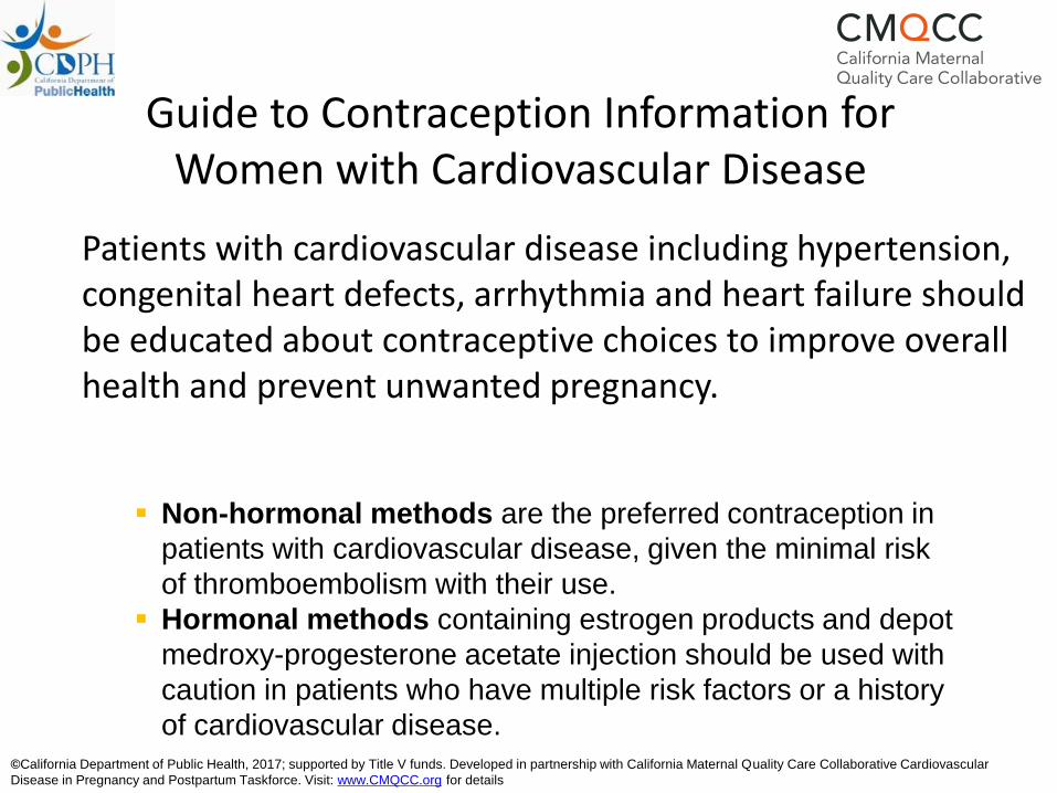
Guide to Contraception Information for Women with Cardiovascular Disease
Patients with cardiovascular disease including hypertension, congenital heart defects, arrhythmia and heart failure should be educated about contraceptive choices to improve overall health and prevent unwanted pregnancy.
Non-hormonal methods are the preferred contraception in
patients with cardiovascular disease, given the minimal risk
of thromboembolism with their use.
Hormonal methods containing estrogen products and depot
medroxy-progesterone acetate injection should be used with
caution in patients who have multiple risk factors or a history