Posttranscriptional Autoregulation of Escherichia coli ThreonyltRNA Synthetase Expression In Vivo
J. SCOTT BUTLER,* MATHIAS SPRINGER, JACQUES DONDON, AND MARIANNE GRUNBERG-MANAGOInstitut de Biologie Physico-Chimique, 75005 Paris, France
Received 3 April 1985/Accepted 18 October 1985
Five mutations in thrS, the gene for threonyl-tRNA synthetase, have been characterized, and the sites of themutations have been localized to different regions of the thrS gene by recombination with M13 phage carryingportions of the thrS gene. Quantitative immunoblotting shows that some of these mutations cause theoverproduction of structurally altered threonyl-tRNA synthetase in vivo. The amounts of in vivo thrS mRNAas measured by quantitative hybridization are, however, the same as wild-type levels for each mutant. Theseresults demonstrate that the expression of threonyl-tRNA synthetase is autoregulated at the posttranscriptionallevel in vivo.
Aminoacyl-tRNA synthetases are a particularly importantand interesting class of enzymes with respect to both enzy-mology and control ofgene expression. Since these enzymesaminoacylate tRNAs, they probably play a role in processessensitive to tRNA aminoacylation (i.e., the stringent re-sponse and the attenuation of transcription). Little is known,however, about how the concentrations of aminoacyl-tRNAsynthetases themselves are controlled. Neidhardt and hisco-workers showed that aminoacyl-tRNA synthetase levelsincrease with increases in cell growth rate (18), and it isknown that the levels of some aminoacyl-tRNA synthetasesundergo transient, or in some cases permanent, derepressionwhen starved for their cognate amino acids (16). Morerecently, alanyl-tRNA synthetase was shown in vitro tocontrol its own expression at the level of transcriptioninitiation (24). Phenylalanyl-tRNA synthetase expression isalso controlled at the transcriptional level, but in this case byan attenuation mechanism mediated by the level of chargedtRNAPhe (4, 27).The gene (thrS) for threonyl-tRNA synthetase (ThrRS) is
located at 38 min on the Escherichia coli chromosome (5).The stop codon of thrS is followed three nucleotides later bythe initiation codon of the gene for translation initiationfactor IF3 (infC) (12). A multicopy plasmid which carriesthrS as well as pheS and pheT (the genes for the small andlarge subunit of phenylalanyl-tRNA synthetase, respec-tively) overproduces phenylalanyl-tRNA synthetase andThrRS in a ratio of 10 to 1 in vivo (21), yet the amounts ofmRNA for each synthetase are the same (22). In an in vitrotranscription translation system the addition of ThrRS spe-cifically inhibits ThrRS synthesis, but has no effect on thesynthesis of thrS mRNA (11). These results suggest that theexpression of thrS may be regulated posttranscriptionally.This hypothesis is supported by experiments which demon-strate that the expression of protein fusions, but not operonfusions, between thrS and lacZ is derepressed in two thrSmutants which overproduce defective ThrRS (25a).The use of gene fusions is, however, only an indirect way
of monitoring gene expression. Thus we decided to measuredirectly the quantities of ThrRS and thrS mRNA in vivo infive thrS mutants. The results of the measurements showthat the overproduction of ThrRS in some of these mutantsis not a result of derepression of transcription of thrS. Thus,
* Corresponding author.
it is likely that the expression of thrS is translationallyautoregulated in vivo.
MATERIALS AND METHODS
Bacterial strains and general methods. The E. coli strainsused in this work are listed in Table 1. The structure of pB21is described by Plumbridge and Springer (21). General genetictechniques are described elsewhere (27). Restriction enzymeswere either from Boehringer Mannheim or New EnglandBiolabs and were used according to the recommendations ofthe manufacturer. [5,6-3H]uridine ([3H]U) (43 Ci/mmol) waspurchased from Amersham.
Isolation of thrS thermosensitive mutants. P1 vir, grown onstrain YMC, was mutagenized with hydroxylamine as de-scribed by Murgola and Yanofsky (15) until the survival (inPFU) was about 10-4 and the transduction capacity of thelysate (as measured by the transduction of pps- to pps+)was reduced to about 1%. After mutagenesis the phage weredialyzed against P1 buffer (14) and stored at 4°C. Transduc-tion with the mutagenized phage was performed (14) usingIBPC4901 (A) (a pps- strain not able to grow on pyruvate orlactate) and minimal A plates (14) supplemented with 0.2%sodium pyruvate, 50 ,ug of arginine per ml, 50 ,ug of histidineper ml, and 200 ,ug of proline per ml (S plates). Transduc-tions were performed at 30°C, and transductants were testedfor growth on S plates at 30 and 42°C. Strains with impairedgrowth at high temperature were purified twice and screenedfor complementation with phage A BR (23), which carriesthrS as well as infC, rplT, and pheST.
Genetic mapping of thrS mutations. We wanted to map thrSmutations by recombination with the male-specific phageM13 carrying parts of the thrS gene. Thus it was necessary tointroduce an F factor into each mutant. Overnight cultureswere crossed with strain TT628 (2) as described by Miller(14), except that the mating was allowed to occur for 3 h at30°C. Tetracycline-resistant recombinants were selected onglucose minimal plates supplemented with arginine, histi-dine, and proline, each at 50 ,ug/ml, tetracycline at 20 ,ug/ml,and, for strain IBPC4771, threonine at 50 ,ug/ml.Each F+ derivative was infected with a different M13
phage at a multiplicity of infection of 100. After incubation at30°C for 10 min, the mixtures were diluted with 1 ml of LBbroth supplemented with tetracycline (10 ,ug/ml) and strep-tomycin (25 ,ug/ml) and grown with shaking at 30°C for 48 h.The mixtures were then diluted and plated on LB plates at
44°C (strains MHB19, MHC37, and MHB35) or on Glu ArgHis Pro plates at 42°C (strain MHB4) and at 37°C (strainIBPC4771). Mixtures were also plated on the same media at30°C or, in the case of strain IBPC4771, on the same mediumplus threonine at 37°C. The number of colonies under theseconditions is considered as the number of viable cells.
Determination of cellular levels of ThrRS and IF3. Mutantand wild-type strains were grown in LB broth at 30°C toapproximately A650 = 0.5. The cells were lysed by theaddition of sample buffer containing sodium dodecyl sulfate(SDS) (9), and the proteins were separated by electrophore-sis on 15 or 12.5% polyacrylamide-SDS gels. The proteinswere then electrophoretically transferred to nitrocellulosepaper (6). The nitrocellulose paper was treated with antibod-ies to ThrRS or IF3, followed by treatment with 1251-labeledStaphylococcus aureus protein A. The position of ThrRSand IF3 on the paper was determined by autoradiography,the appropriate portions were cut out, and the amount ofbound 1251 was determined in a gamma radiation counter. Ineach case, doubling the amount of protein loaded on the gelresulted in a doubling of the 125I-protein A bound to thenitrocellulose paper.
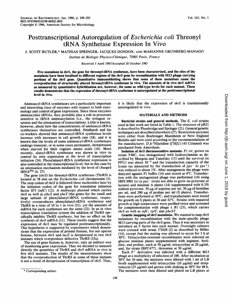
Construction of M13 probes. Plasmid pB1 (21), whichcarries all of thrS and infC, was digested with HpaI andadded to M13mp8 cut by Hindll. The DNAs were ligatedand used to transform strain JM101. White or light-blueplaques were picked and purified three times, and theidentity and orientation of the insert were determined byrestriction endonuclease digestion. All of the probes shownin Fig. 1, except plasmid M13mp8H54, were found in thisway and were the generous gift of G. Fayat and M. Panvert.Attempts to clone the EcoRI-HindIII fragment of plasmidM13mp8H45 into the EcoRI-HindIII site of M13mp9 to givethe insert in the opposite orientation relative to M13mp8H45were unsuccessful. Instead, we cloned the BamHI-HindIIIfragment of M13mp8H45 into the same sites in pBR322. Theonly clone (pSB1) found to contain the correct fragment alsocontains an approximately 250-base-pair HindIll fragment ofunknown origin in the HindlIl site. We tried -to clone theBamHI-HindIII fragment of pSB1 in the same sites inM13mp9, but we found no clones with the insert in thedesired orientation. Instead, we cloned the EcoRI-BamHIfragment of pSB1 into the same sites of M13mp8 to giveM13mp8H54. This recombinant M13mp8 carries the 250-base-pair fragment of unknown origin. We wanted to be surethat no RNA hybridizes to this HindIll fragment, so wedeleted the portion of M13mp8H54 corresponding to HpaI4-HpaIs (see Fig. 1) by digesting the plasmid with HindII andreligating it back together. The resulting recombinant,M13mp8H5441, carries only the 250-base-pair HindIII frag-
ment of unknown origin. In the hybridization assay de-scribed below we found no hybridization of RNA to single-stranded DNA probes derived from plasmid M13mp8H54A1.The M13mp8 probe M13mp8lacl4 was constructed by
inserting the 1.8-kilobase HpaI-EcoRI fragment of pMC871(1) into the EcoRI-HindII site of mp8. In ml3mp8 lac-14there is no complementarity between the portion of lacZcarried in M13mp8 and the 1.8-kilobase HpaI-EcoRI frag-ment of pMC871.
Isolation of pulse-labeled, [3HJU-labeled RNA. Mutant orwild-type strains were grown from A650 = 0.05 to A650 = 0.4in the MOPS glucose medium of Neidhardt et al. (17)supplemented with arginine, histidine, and proline (50 ,ug/ml)for all strains except IBPC4771, for which threonine (50,ug/ml) was also added. At A650 = 0.4, 0.50 ml of culture waslabeled with 500 R.Ci of [3H]U (43 Ci/mmol). Cell$ were thenlysed at the times indicated by the addition of 0.5 ml ofboiling SDS solution, and the [3H]U-RNA was extractedtwice with phenol saturated with 0.2 M sodium acetate (pH5.2), followed by three extractions with diethyl ether. The
H, 2
thrS
H3
infClr~-
!mp8H12!
,* lmp8H21 mp8H23
Mp8H32 :mp8I1
!np8H43 rp8H45i
mp8H54I1kb
FIG. 1. Physical map of the region of the E. coli chromosomecarrying the genes for ThrRS (thrS) and initiation factor IF3 (infC).Arrows represent the size of DNA inserts in phage M13mp8. Thedirection of the arrows represents the orientation of the insert in theHindIl site of the M13mp8 linker with left being towards the EcoRIsite and right being towards the HindlIl site. H, HpaI.
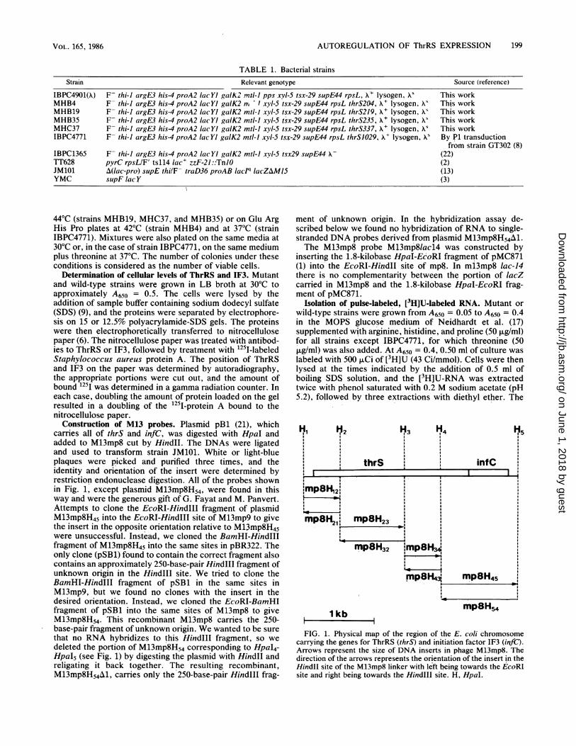
FIG. 2. Autoradiogram of immunoblots of protein extracts from wild-type and thrS mutant strains. Samples of 5 to 8 ,ug (even-numberedlanes) or 10 to 16 ,u.g (odd-numbered lanes) of protein were separated by electrophoresis on 12.5% polyacrylamide-SDS gels. The proteinswere transferred to nitrocellulose paper and treated as described in the text. Lanes 1 and 2, MHB19 (thrS219); lanes 3 and 4, MHB4 (thrS204);lanes 5 and 6, IBPC4901(A) (wild type); lanes 7 and 8, MHC37 (thrS337); lanes 9 and 10, IBPC4901(A) (wild type); lanes 11 and 12, IBPC4771(thrS1029); lanes 13 and 14, IBPC4901(X) (wild type).
[3H]U-RNA was precipitated with ethanol and stored insterile water at -20°C. The doubling times and labeling timesare, respectively, for each strain: IBPC4901(A), 70 min and105 s; MHB4, 90 min and 135 s; MHB19, 75 min and 110 s;
MHB35, 60 min and 90s; MHC37, 115 min and 170 s; andIBPC4771, 90 min and 135 s.
Hybridization of [3H]U-RNA to strand-specific M13 probes.Single-stranded phage M13mp8 DNAs were purified (13) andstored at -20°C in sterile water. The M13 single-strandedprobes were fixed to nitrocellulose filters (25-mm diameter)by filtration (2 ml/min) of 10 ,ug of each probe in 50 ml of 2 xSSC (lx SSC is 0.15 M NaCl plus 0.015 M sodium citrate).Each filter was cut into four smaller filters (6-mm diameter),and the filters were dried under vacuum at 80°C for 3 h.
Hybridizations were carried out in sterile 5-ml glass vialswith 167 ,ul of hybridization buffer (0.1 M Tris hydrochlo-ride, pH 8, 0.6 M NaCl, 0.02 M disodium EDTA, 0.1% SDS,0.02% Ficoll, 0.02% polyvinylpyrrolidone, 50% deionizedformamide) and 2 x 10⁶ dpm of [3H]U-RNA per filter at 41°Cfor 16 h with gentle shaking. In general, six filters wereincubated in each vial. Three of these filters carried a probewith the insert in one orientation, and the other three carrieda probe with the same insert in the opposite orientation. Thefourth 6-mm filter cut for each probe was incubated with[3H]U-RNA extracted from strain IBPC1365 carrying theplasmid pB21 (21). This plasmid produces approximately 10times more thrS and infC mRNA than the chromosome (seeFig. 2). The large amount of [3H]U-RNA hybridized to thefourth filter establishes that the other three filters carryenough DNA to saturate all of the specific [3H]U-RNAavailable for hybridization and that there are no significantdifferences between batches of filters.
After incubation, the filters were washed four times in 50ml of 2x SSC for 10 min at 30°C, treated with 0.75 ml ofRNase A (20 ,ug/ml; initially preincubated at 100°C for 2 min)per filter for 1 h at 30°C, and washed twice with 50 ml of 2xSSC for 10 min at 30°C. The filters were then washed in 100ml of 90% ethanol and dried before scintillation counting(twice, 10 min each, for each filter).
RESULTSGenetic mapping of thrS mutations. We decided to map the
mutations of a number of thrS thermosensitive mutantsisolated by P1 mutagenesis and of one additional mutation inthe hope that this might give us more insight into the
regulation of expression of thrS. First, we screened eachthrS thermosensitive mutant plus the one thrS auxotrophicmutant strain IBPC4771, using K Bi (23). We then chose forfurther study only those mutants complemented by K phagecarrying a complete thrS gene.The second method of genetic mapping that we employed
was recombination of F+ derivatives (see Materials andMethods) of each mutant by M13 phage carrying HpaIfragments of thrS inserted into the HindII site of theM13mp8 DNA (Fig. 1). After infection, cultures were grownfor 48 h to allow recombination to occur and then plated athigh temperature, or at low temperature in the absence ofthreonine in the case of strain IBPC4771. Table 2 lists thenumber of thermoresistant (or Thr+) colonies per 106 viablecells. For strains MHB19 F' and IBPC4771 F' the back-ground level of transduction was relatively high, probably as
a result of the relatively high reversion frequency of thesestrains. The values in Table 2 show that the thrS mutationsfall into two categories. First, there are three mutants(strains MHB4, MHB19, and MHC37) whose mutations arein the internal portion of thrS between the HpaI2 and HpaI3sites and thus are structural mutations in thrS. Second, thereare two mutants (strains MHB35 and IBPC4771) whosemutations are in the 5'-terminal region of thrS between theHpaIl and HpaI2 sites and so may be either structural or
regulatory mutations. Most importantly, these mapping ex-
periments demonstrate that the mutations in strains MHB4,MHB19, and MHC37 are in the thrS gene and thus allphenotypic irregularities in these strains must be a result ofmutations in thrS.Measurement of in vivo levels of ThrRS. It was possible
that some of these thrS mutations might result in variationsof ThrRS levels if the synthetase is involved in its own
TABLE 2. Recombination mapping of thrS mutationsNo. of transductants per 106 cells with M13
Strain phage:
mp8HI2 mp8H23 mp8H34 mp8H45
MHB4 F' (thrS204) 73 4.0 x 10⁴ 25 47MHB19 F' (thrS219) 200 1.3 x 10⁵ 167 666MHC37 F' (thrS337) <50 1.5 x 10⁴ <50 <50MHB35 F' (thrS235) 2 x 10³ 11 33 12IBPC4771 F' (thrS1029) 7 x 10⁴ <10 250 300
a Average ± SD of three separate experiments in which two differentconcentrations of protein were tested.bIn three strains ThrRS is recognized as degraded fragments.
regulation. To test this possibility, we determined ThrRSand IF3 protein levels in exponentially growing cells byquantitative immunoblotting (Fig. 2). We determined IF3levels in addition to ThrRS levels because there are onlythree nucleotides between the stop condon of thrS and theinitiation codon of infC (12). Since there are no transcriptiontermination signals between the two genes, they must becoexpressed. Table 3 lists the in vivo levels of ThrRS andIF3 for each mutant relative to the wild type. The level ofIF3 for each mutant is about the same as in the wild-typestrain. However, there is significant overproduction ofThrRS or fragments of ThrRS for a number of the mutants(strains MHB19, MHC37, and IBPC4771). In the case ofstrains MHB4, MHB19, and MHC37, ThrRS antibodiesrecognize lower-molecular-weight forms of ThrRS. Thus, itis likely that these mutant forms of ThrRS are susceptible toproteolysis. This fact, with the possibility that the affinity ofThrRS antibodies could be lower for these defective forms,suggests that the levels of ThrRS determined for the mutantsby this mnethod must represent the minimum amounts ofThrRS in vivo. The fact that IF3 is not overproduced inmutants which overproduce ThrRS suggests that IF3 expres-sion can be controlled independently of ThrRS.Measurement of in vivo levels of thrS mRNA. If the over-
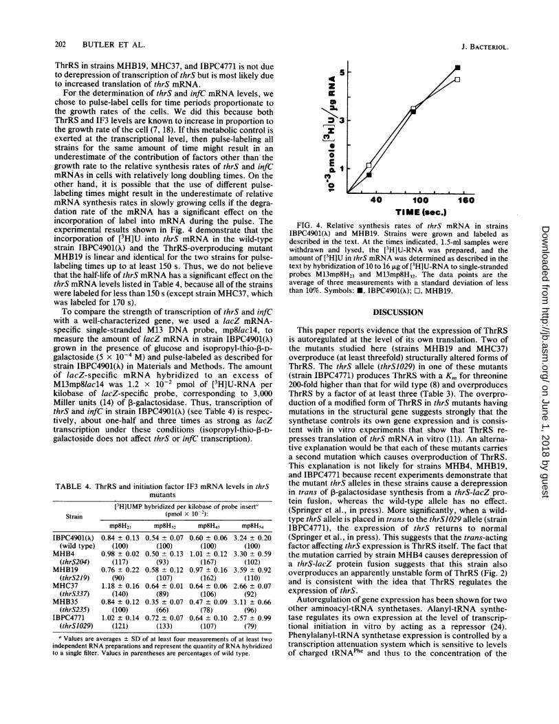
production of ThrRS is the result of derepression of tran-scription of thrS, then proportionate increases in thrSmRNA should be detected by hybridization of in vivo-labeled mRNA to single-stranded DNA probes. We mea-sured the amount of in vivo-labeled [3H]U-RNA transcribedfrom the thrS region by hybridization to single-strandedphage M13 DNA carrying different Hpal fragments from inand around thrS (Fig. 1). The data in Fig. 3 demonstrate thatthe hybridization system was sensitive to small changes in[3H]U-RNA levels. For each probe, increasing the amountsof specific [3H]U-RNAs resulted in proportionate increasesin the amounts of specifc [3H]U-RNAs hybridized to eachprobe. The amount of [3H]U-RNA hybridizing per kilobaseof probe along the thrS transcription unit was not constant,probably as a result of small differences in hybridizationefficiencies between each probe. More importantly, thelinear response between the total amount of [3H]U-RNAadded and the amounts of [3H]U-RNA found as hybridsguarantees that increases in in vivo levels of thrS or infCmRNA will be detected by hybridization to these probes.The in vivo levels of thrS and infC mRNA determined by
hybridization (Table 4) indicate two important features ofthrS and infC transcription. First, there was a three- tofivefold greater amount of hybridization to the M13mp8H54probe than to the preceding three probes (also compare Fig.
3A and B). This large increase in hybridization could reflectdifferences in hybridization efficiencies, but is most likelydue to a real increase in transcription near to the HpaI4 site.Significantly, this increase in transcription agrees quantita-tively with measurements showing that there is about five-fold more IF3 than ThrS in vivo (7, 16). It thus seems likelythat infC can be transcribed independently of thrS and thatthis transcription is initiated near to or after the HpaI4 site.This is consistent with in vivo experiments which demon-strate that infC can be expressed independently of thepromoter for thrS (26). There also seems to be a slightincrease in hybridization to probe M13mp8H43 for strainsMHB4 and MHB19. This is reflected by a slight elevation ofIF3 levels (Table 2) for each of these two strains. It ispossible that the sensitivity of the hybridization systemallows one to detect relatively minor changes in infC tran-scription initiated near the HpaI3 site.The second and most important feature of the hybridiza-
tion results is that for each thrS mutant the in vivo level ofthrS mRNA was not significantly different from that of thewild-type strain. Thus, the nearly fourfold overproduction of
16
12
8
E 1 3 5 72CMN
a B
a 4
z 3
2
1 3 5 7
INPUT (pmole[3H] UMP)FIG. 3. Relative amounts of [3H]U-RNA hybridizing to M13mp8
probes. Increasing amounts of [3H]U-RNA from strain IBPC1365carrying plasmid pB21 were hybridized to a fixed amount of eachM13mp8 probe as described in the text. The amount of [3H]U-RNAhybridizing to probes containing inserts in the opposite orientationhas been subtracted. Symbols: (A) *, M13mp8H54; (B) *,M13mp8H21; 0, M13mp8H32; O, M13mp8H43.
ThrRS in strains MHB19, MHC37, and IBPC4771 is not dueto derepression of transcription of thrS but is most likely dueto increased translation of thrS mRNA.For the determination of thrS and infC mRNA levels, we
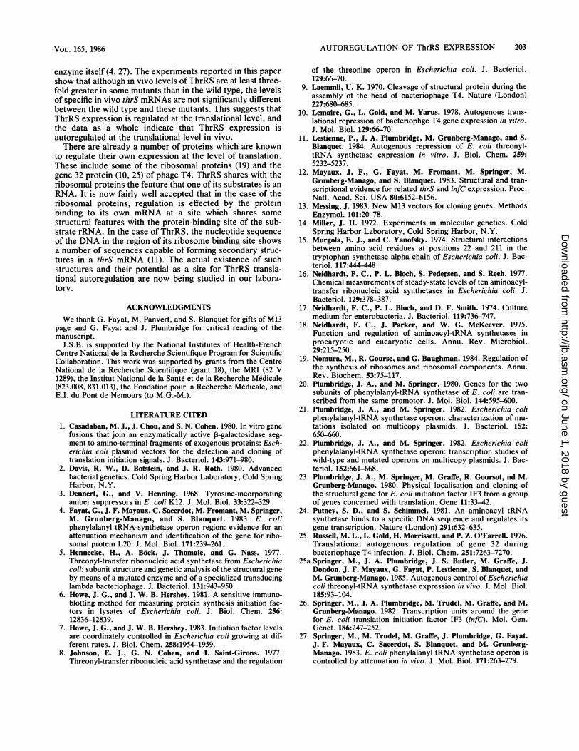
chose to pulse-label cells for time periods proportionate tothe groWth rates of the cells. We did this because bothThrRS and IF3 levels are known to increase in proportion tothe growth rate of the cell (7, 18). If this metabolic control isexerted at the transcriptional level, then pulse-labeling allstrains for the same amount of time might result in anunderestimate of the contribution of factors other than thegrowth rate to the relative synthesis rates of thrS and infCmRNAs in cells with relatively long doubling times. On theother hand, it is possible that the use of different pulse-labeling times might result in the underestimate of relativemRNA synthesis rates in slowly growing cells if the degra-dation rate of the mRNA has a significant effect on theincorporation of label into mRNA during the pulse. Theexperimental results shown in Fig. 4 demonstrate that theincorporation of [3H]U into thrS niRNA in the wild-typestrain IBPC4901(A) and the ThrRS-overproducing mutantMHJ319 is linear and identical for the two strains for pulse-labeling times up to at least 150 s. Thus, we do not believethat the half-life of thrS mRNA has a significant effect on thethrS mRNA levels listed in Table 4, because all of the strainswere labeled for less than 150 s (except strain MHC37, whichwas labeled for 170 s).To compare the strength of transcription of thrS and infC
with a well-characterized gene, we used a lacZ mRNA-specific single-stranded M13 DNA probe, mp8lacl4, tomeasure the amount of lacZ mRNA in strain IBPC4901(X)grown in the presence of glucose and isopropyl-thio-p-D-galactoside (5 x 10⁻⁴ M) and pulse-labeled as described forstrain IBPC4901(A) in Materials and Methods. The amountof lacZ-specific mRNA hybridized to an excess ofM13mp8lacl4 was 1.2 x 10⁻² pmol of [3H]U-RNA perkilobase of lacZ-specific probe, corresponding to 3,000Miller units (14) of P-galactosidase. Thus, transcripiion ofthrS and infC in strain IBPC4901(A) (see Table 4) is respec-tively, about one-half and three times as strong as lacZtranscription under these conditions (isopropyl-thio-p-D-galactoside does not affect thrS or infC transcription).
TABLE 4. ThrRS and initiation factor IF3 mRNA levels in thrSmutants
[3H]UMP hybridized per kilobase of probe insert"Strain (pmol x 10-2):
(thrSI029) (121) (133) (107) (79)"Values are averages ± SD of at least four measurements of at least two
independent RNA preparations and represent the quantity ofRNA hybridizedto a single filter. Values in parentheses are percentages of wild type.
5
z
4 3
ECL
40 100 160TIME (sec.)
FIG. 4. Relative synthesis rates of thrS mRNA in strainsIBPC4901(A) and MHB19. Strains were grown and labeled asdescribed in the text. At the times indicated, 1.5-ml samples werewithdrawn and lysed, the [3H]U-RNA was prepared, and theamount of [3H]U in thrS mRNA was determined as described in thetext by hybridization of 10 to 16 ,g of[PH]U-RNA to single-strandedprobes M13mp8H23 and M13mp8H32. The data points are theaverage of three measurements with a standard deviation of lessthan 10%. Symbols: *, IBPC4901(A); O, MHB19.
DISCUSSION
This paper reports evidence that the expression of ThrRSis autoregulated at the level of its own translation. Two ofthe mutants studied here (strains MHB19 and MHC37)overproduce (at least threefold) structurally altered forms ofThrRS. The thrS allele (thrS1029) in one of these mutants(strain IBPC4771) produces ThrRS with a Km for threonine200-fold higher than that for wild type (8) and overproducesThrRS by a factor of at least three (Table 3). The overpro-duction of a modified form of ThrRS in thrS mutants havingmutations in the structural gene suggests strongly that thesynthetase controls its own gene expression and is consis-tent with in vitro experiments that show that ThrRS re-presses translation of thrS mRNA in vitro (11). An alterna-tive explanation would be that each of these mutants carriesa second mutation which causes overproduction of ThrRS.This explanation is not likely for strains MHB4, MH1319,and IBPC4771 because recent experiments demonstrate thatthe mutant thrS alleles in these strains cause a derepressionin trans of ,-galactosidase synthesis from a thrS-lacZ pro-tein fusion, whereas the wild-type allele has no effect.(Springer et al., in press). More significantly, when a wild-type thrS allele is placed in trans to the thrS1029 allele (strainIBPC4771), the expression of thrS returns to normal(Springer et al., in press). This suggests that the trans-actingfactor affecting thrS expression is ThrRS itself. The fact thatthe mutation carried by strain MHB4 causes derepression ofa thrS-lacZ protein fusion suggests that this strain alsooverproduces an apparently unstable form of ThrRS (Fig. 2)and is consistent with the idea that ThrRS regulates theexpression of thrS.
Autoregulation of gene expression has been shown for twoother aminoacyl-tRNA synthetases. Alanyl-tRNA synthe-tase regulates its own expression at the level of transcrip-tional initiation in vitro by acting as a repressor (24).Phenylalanyl-tRNA synthetase expression is controlled by atranscription attenuation system which is sensitive to levelsof charged tRNAPhe and thus to the concentration of the
enzyme itself (4, 27). The experiments reported in this papershow that although in vivo levels of ThrRS are at least three-fold greater in some mutants than in the wild type, the levelsof specific in vivo thrS mRNAs are not significantly differentbetween the wild type and these mutants. This suggests thatThrRS expression is regulated at the translational level, andthe data as a whole indicate that ThrRS expression isautoregulated at the translational level in vivo.There are already a number of proteins which are known
to regulate their own expression at the level of translation.These include some of the ribosomal proteins (19) and thegene 32 protein (10, 25) of phage T4. ThrRS shares with theribosomal proteins the feature that one of its substrates is anRNA. It is now fairly well accepted that in the case of theribosomal proteins, regulation is effected by the proteinbinding to its own mRNA at a site which shares some
structural features with the protein-binding site of the sub-strate rRNA. In the case of ThrRS, the nucleotide sequence
of the DNA in the region of its ribosome binding site showsa number of sequences capable of forming secondary struc-tures in a thrS mRNA (11). The actual existence of suchstructures and their potential as a site for ThrRS transla-tional autoregulation are now being studied in our labora-tory.
ACKNOWLEDGMENTS
We thank G. Fayat, M. Panvert, and S. Blanquet for gifts of M13page and G. Fayat and J. Plumbridge for critical reading of themanuscript.
J.S.B. is supported by the National Institutes of Health-FrenchCentre National de la Recherche Scientifique Program for ScientificCollaboration. This work was supported by grants from the CentreNational de la Recherche Scientifique (grant 18), the MRI (82 V1289), the Institut National de la Sante et de la Recherche Medicale(823.008, 831.013), the Fondation pour la Recherche Medicale, andE.I. du Pont de Nemours (to M.G.-M.).
LITERATURE CITED
1. Casadaban, M. J., J. Chou, and S. N. Cohen. 1980. In vitro gene
fusions that join an enzymatically active ,B-galactosidase seg-
ment to amino-terminal fragments of exogenous proteins: Esch-erichia coli plasmid vectors for the detection and cloning oftranslation initiation signals. J. Bacteriol. 143:971-980.
2. Davis, R. W., D. Botstein, and J. R. Roth. 1980. Advancedbacterial genetics. Cold Spring Harbor Laboratory, Cold SpringHarbor, N.Y.
3. Dennert, G., and V. Henning. 1968. Tyrosine-incorporatingamber suppressors in E. coli K12. J. Mol. Biol. 33:322-329.
4. Fayat, G., J. F. Mayaux, C. Sacerdot, M. Fromant, M. Springer,M. Grunberg-Manago, and S. Blanquet. 1983. E. coliphenylalanyl tRNA-synthetase operon region: evidence for an
attenuation mechanism and identification of the gene for ribo-somal protein L20. J. Mol. Biol. 171:239-261.
5. Hennecke, H., A. Bock, J. Thomale, and G. Nass. 1977.Threonyl-transfer ribonucleic acid synthetase from Escherichiacoli: subunit structure and genetic analysis of the structural gene
by means of a mutated enzyme and of a specialized transducinglambda bacteriophage. J. Bacteriol. 131:943-950.
6. Howe, J. G., and J. W. B. Hershey. 1981. A sensitive immuno-blotting method for measuring protein synthesis initiation fac-tors in lysates of Escherichia coli. J. Biol. Chem. 256:12836-12839.
7. Howe, J. G., and J. W. B. Hershey. 1983. Initiation factor levelsare coordinately controlled in Escherichia coli growing at dif-ferent rates. J. Biol. Chem. 258:1954-1959.
8. Johnson, E. J., G. N. Cohen, and I. Saint-Girons. 1977.Threonyl-transfer ribonucleic acid synthetase and the regulation
of the threonine operon in Escherichia coli. J. Bacteriol.129:66-70.
9. Laemmli, U. K. 1970. Cleavage of structural protein during theassembly of the head of bacteriophage T4. Nature (London)227:680-685.
10. Lemaire, G., L. Gold, and M. Yarus. 1978. Autogenous trans-lational repression of bacteriophge T4 gene expression in vitro.J. Mol. Biol. 129:66-70.
11. Lestienne, P., J. A. Plumbridge, M. Grunberg-Manago, and S.Blanquet. 1984. Autogenous repression of E. coli threonyl-tRNA synthetase expression in vitro. J. Biol. Chem. 259:5232-5237.
12. Mayaux, J. F., G. Fayat, M. Fromant, M. Springer, M.Grunberg-Manago, and S. Blanquet. 1983. Structural and tran-scriptional evidence for related thrS and infC expression. Proc.Natl. Acad. Sci. USA 80:6152-6156.
13. Messing, J. 1983. New M13 vectors for cloning genes. MethodsEnzymol. 101:20-78.
14. Miller, J. H. 1972. Experiments in molecular genetics. ColdSpring Harbor Laboratory, Cold Spring Harbor, N.Y.
15. Murgola, E. J., and C. Yanofsky. 1974. Structural interactionsbetween amino acid residues at positions 22 and 211 in thetryptophan synthetase alpha chain of Escherichia coli. J. Bac-teriol. 117:444 448.
16. Neidhardt, F. C., P. L. Bloch, S. Pedersen, and S. Reeh. 1977.Chemical measurements of steady-state levels of ten aminoacyl-transfer ribonucleic acid synthetases in Escherichia coli. J.Bacteriol. 129:378-387.
17. Neidhardt, F. C., P. L. Bloch, and D. F. Smith. 1974. Culturemedium for enterobacteria. J. Bacteriol. 119:736-747.
18. Neidhardt, F. C., J. Parker, and W. G. McKeever. 1975.Function and regulation of aminoacyl-tRNA synthetases inprocaryotic and eucaryotic cells. Annu. Rev. Microbiol.29:215-250.
19. Nomura, M., R. Gourse, and G. Baughman. 1984. Regulation ofthe synthesis of ribosomes and ribosomal components. Annu.Rev. Biochem. 53:75-117.
20. Plumbridge, J. A., and M. Springer. 1980. Genes for the twosubunits of phenylalanyl-tRNA synthetase of E. coli are tran-scribed from the same promotor. J. Mol. Biol. 144:595-600.
21. Plumbridge, J. A., and M. Springer. 1982. Escherichia coliphenylalanyl-tRNA synthetase operon: characterization of mu-tations isolated on multicopy plasmids. J. Bacteriol. 152:650-660.
22. Plumbridge, J. A., and M. Springer. 1982. Escherichia coliphenylalanyl-tRNA synthetase operon: transcription studies ofwild-type and mutated operons on multicopy plasmids. J. Bac-teriol. 152:661-668.
23. Plumbridge, J. A., M. Springer, M. Graffe, R. Goursot, nd M.Grunberg-Manago. 1980. Physical localisation and cloning ofthe structural gene for E. coli initiation factor IF3 from a groupof genes concerned with translation. Gene 11:33-42.
24. Putney, S. D., and S. Schimmel. 1981. An aminoacyl tRNAsynthetase binds to a specific DNA sequence and regulates itsgene transcription. Nature (London) 291:632-635.
25. Russell, M. L., L. Gold, H. Morrissett, and P. Z. O'Farrell. 1976.Translational autogenous regulation of gene 32 duringbacteriophage T4 infection. J. Biol. Chem. 251:7263-7270.
25a.Springer, M., J. A. Plumbridge, J. S. Butler, M. Graffe, J.Dondon, J. F. Mayaux, G. Fayat, P. Lestienne, S. Blanquet, andM. Grunberg-Manago. 1985. Autogenous control of Escherichiacoli threonyl-tRNA synthetase expression in vivo. J. Mol. Biol.185:93-104.
26. Springer, M., J. A. Plumbridge, M. Trudel, M. Graffe, and M.Grunberg-Manago. 1982. Transcription units around the genefor E. coli translation initiation factor IF3 (infC). Mol. Gen.Genet. 186:247-252.
27. Springer, M., M. Trudel, M. Graffe, J. Plumbridge, G. Fayat.J. F. Mayaux, C. Sacerdot, S. Blanquet, and M. Grunberg-Manago. 1983. E. coli phenylalanyl tRNA synthetase operon iscontrolled by attenuation in vivo. J. Mol. Biol. 171:263-279.