Potential role for saccharopine reductase in swainsonine metabolism in endophytic fungus, Undifilum oxytropis Suman MUKHERJEE a, * ,1 , Angus L. DAWE a,c , Rebecca CREAMER a,b a Molecular Biology Program, New Mexico State University, Las Cruces, NM 88003, USA b Department of Entomology, Plant Pathology, and Weed Science, New Mexico State University, Las Cruces, NM 88003, USA c Biology Department, New Mexico State University, Las Cruces, NM 88003, USA article info Article history: Received 28 September 2011 Received in revised form 26 May 2012 Accepted 29 May 2012 Available online 16 June 2012 Corresponding Editor: Stephen W. Peterson Keywords: Gene disruption Saccharopine reductase Swainsonine Undifilum oxytropis abstract Locoweed plants in the southwestern United States often harbour a slow-growing endo- phytic fungus, Undifilum oxytropis (Phylum: Ascomycota; Order: Pleosporales), which produces a toxic alkaloid, swainsonine. Consumption of U. oxytropis by grazing animals induces a neurological disorder called locoism for which the toxic alkaloid swainsonine has been reported to be the causal agent. Little is known about the biosynthetic pathway of swain- sonine in endophytic fungi, but previous studies on non-endophytic ascomycetous fungi indicate that pipecolic acid and saccharopine are key intermediates. We have used degen- erate primers, Rapid amplification of cDNA ends (RACE)-PCR and inverse PCR to identify the gene sequence of U. oxytropis saccharopine reductase. To investigate the role of this gene product in swainsonine metabolism, we have developed a gene deletion system for this slow-growing endophyte based on our recently established transformation protocol. A strain of U. oxytropis lacking saccharopine reductase had decreased levels of saccharo- pine and lysine along with increased accumulation of pipecolic acid and swainsonine. Thus, saccharopine reductase influences the accumulation of swainsonine and its precur- sor, pipecolic acid, in U. oxytropis. ª 2012 The British Mycological Society. Published by Elsevier Ltd. All rights reserved. Introduction Locoweeds (Astragalus sp. and Oxytropis sericea) are perennial flowering plants found frequently in the rangelands of the western United States, Asia, and South America (Kingsbury 1964; Molyneux & James 1982; James & Nielson 1988; Cook et al. 2009). Consumption of locoweeds by cattle, sheep, and horses induces a neurological condition termed locoism (James & Panter 1989). The etiological agent of locoism, swain- sonine (1, 2, 8-trihydroxyindolizidine), is produced by fungal endophytes that reside within the locoweeds (Braun et al. 2003). The common endophyte of the O. sericea Nutt. Locoweed was recently classified as Undifilum oxytropis, belonging to the phylum Ascomycota and order Pleosporales (Cook et al. 2009; Graham et al. 2009; Pryor et al. 2009). Undifilum oxytropis can be isolated from stems, seeds, and leaves of locoweed plants (Ralphs et al. 2002; Braun et al. 2003). The fungus is transmitted from one generation to the next through the seed coat (James & Panter 1989)(Kingsbury 1964). When U. oxytropis grown in pure culture was fed to rats symptoms of locoism were induced (McLain-Romero et al. 2004). * Corresponding author. Laboratory of Biochemistry and Genetics, National Institute of Diabetes and Digestive and Kidney Diseases (NIDDK), National Institutes of Health (NIH), 8, Center Drive, Bethesda, MD 20892-0830, USA. Tel.: þ1 301 451 3771; fax: 1 301 402 0240. E-mail addresses: [email protected], [email protected], [email protected]1 Present address: Laboratory of Biochemistry and Genetics, National Institute of Diabetes and Digestive and Kidney Diseases, National Institutes of Health, Bethesda, MD 20892-0830, USA. journal homepage: www.elsevier.com/locate/funbio fungal biology 116 (2012) 902 e909 1878-6146/$ e see front matter ª 2012 The British Mycological Society. Published by Elsevier Ltd. All rights reserved. http://dx.doi.org/10.1016/j.funbio.2012.05.007
Transcript
f u n g a l b i o l o g y 1 1 6 ( 2 0 1 2 ) 9 0 2e9 0 9
journa l homepage : www.e lsev ier . com/ loca te / funb io
Potential role for saccharopine reductase in swainsoninemetabolism in endophytic fungus, Undifilum oxytropis
Suman MUKHERJEEa,*,1, Angus L. DAWEa,c, Rebecca CREAMERa,b
aMolecular Biology Program, New Mexico State University, Las Cruces, NM 88003, USAbDepartment of Entomology, Plant Pathology, and Weed Science, New Mexico State University, Las Cruces, NM 88003, USAcBiology Department, New Mexico State University, Las Cruces, NM 88003, USA
a r t i c l e i n f o
Article history:
Received 28 September 2011
Received in revised form
26 May 2012
Accepted 29 May 2012
Available online 16 June 2012
Corresponding Editor:
Stephen W. Peterson
Keywords:
Gene disruption
Saccharopine reductase
Swainsonine
Undifilum oxytropis
* Corresponding author. Laboratory of Bioch(NIDDK), National Institutes of Health (NIH),
E-mail addresses: suman.mukherjee@nih1 Present address: Laboratory of Biochemis
Institutes of Health, Bethesda, MD 20892-0831878-6146/$ e see front matter ª 2012 The Bhttp://dx.doi.org/10.1016/j.funbio.2012.05.007
a b s t r a c t
Locoweed plants in the southwestern United States often harbour a slow-growing endo-
phytic fungus, Undifilum oxytropis (Phylum: Ascomycota; Order: Pleosporales), which produces
a toxic alkaloid, swainsonine. Consumption of U. oxytropis by grazing animals induces
a neurological disorder called locoism for which the toxic alkaloid swainsonine has been
reported to be the causal agent. Little is known about the biosynthetic pathway of swain-
sonine in endophytic fungi, but previous studies on non-endophytic ascomycetous fungi
indicate that pipecolic acid and saccharopine are key intermediates. We have used degen-
erate primers, Rapid amplification of cDNA ends (RACE)-PCR and inverse PCR to identify
the gene sequence of U. oxytropis saccharopine reductase. To investigate the role of this
gene product in swainsonine metabolism, we have developed a gene deletion system for
this slow-growing endophyte based on our recently established transformation protocol.
A strain of U. oxytropis lacking saccharopine reductase had decreased levels of saccharo-
pine and lysine along with increased accumulation of pipecolic acid and swainsonine.
Thus, saccharopine reductase influences the accumulation of swainsonine and its precur-
sor, pipecolic acid, in U. oxytropis.
ª 2012 The British Mycological Society. Published by Elsevier Ltd. All rights reserved.
Introduction 2003). The common endophyte of theO. sericeaNutt. Locoweed
Locoweeds (Astragalus sp. and Oxytropis sericea) are perennial
flowering plants found frequently in the rangelands of the
western United States, Asia, and South America (Kingsbury
1964; Molyneux & James 1982; James & Nielson 1988; Cook
et al. 2009). Consumption of locoweeds by cattle, sheep, and
horses induces a neurological condition termed locoism
(James & Panter 1989). The etiological agent of locoism, swain-
sonine (1, 2, 8-trihydroxyindolizidine), is produced by fungal
endophytes that reside within the locoweeds (Braun et al.
emistry and Genetics, N8, Center Drive, Bethesd.gov, [email protected], c
try and Genetics, Nationa0, USA.ritish Mycological Societ
was recently classified as Undifilum oxytropis, belonging to the
phylum Ascomycota and order Pleosporales (Cook et al. 2009;
Graham et al. 2009; Pryor et al. 2009).
Undifilum oxytropis can be isolated from stems, seeds, and
leaves of locoweed plants (Ralphs et al. 2002; Braun et al.
2003). The fungus is transmitted from one generation to the
next through the seed coat (James & Panter 1989) (Kingsbury
1964). When U. oxytropis grown in pure culture was fed to
rats symptoms of locoism were induced (McLain-Romero
et al. 2004).
ational Institute of Diabetes and Digestive and Kidney Diseasesa, MD 20892-0830, USA. Tel.: þ1 301 451 3771; fax: 1 301 402 [email protected]
l Institute of Diabetes and Digestive and Kidney Diseases, National
y. Published by Elsevier Ltd. All rights reserved.
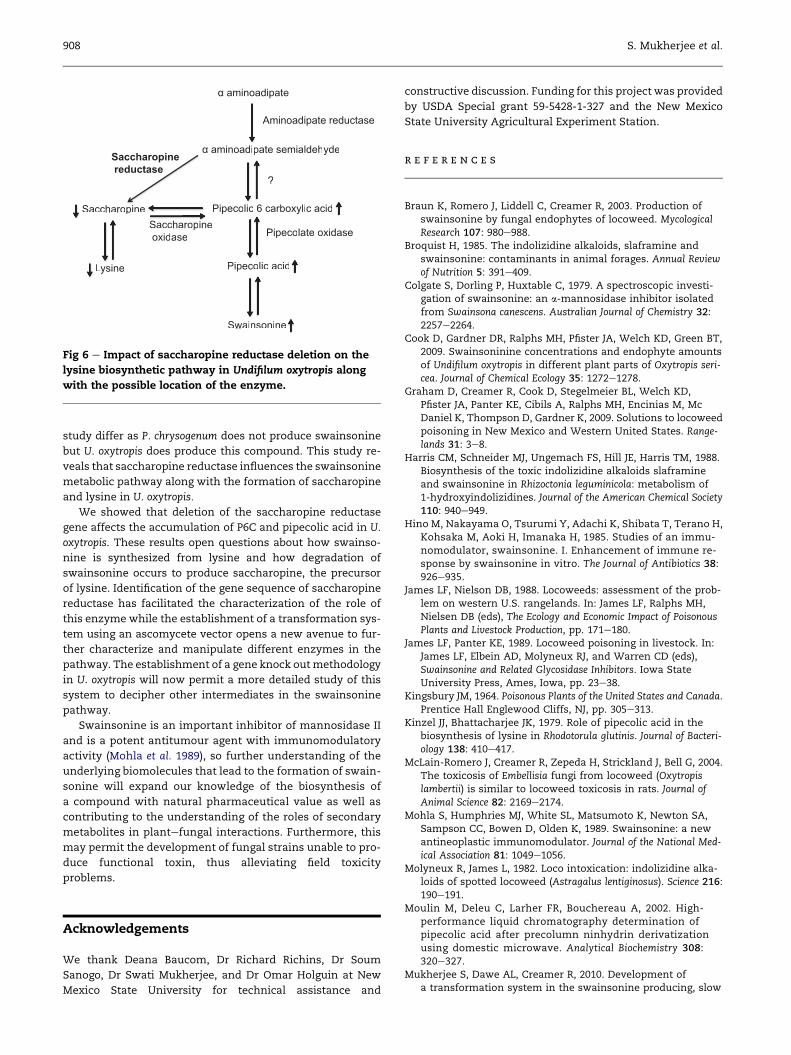
tant fungal strains exhibited an increase in concentration of
swainsonine and pipecolic acid and a decrease in saccharo-
pine and lysine level, but no difference in a-aminoadipic
acid betweenwild type andmutantwas detected. No swainso-
nine was detected in the growth media when tested. Twelve
individual saccharopine reductase disruption and wild type
U. oxytropis colonies were tested (Table 2). Due to the lack of
an available P6C chemical standard, only relative quantitative
data was obtained for this compound by mass-spectrometry
(Fig 5A). Chromatographic plots revealed a higher accumula-
tion of P6C in the disruption mutants (Fig 5C) as compared
to wild type strain where a low concentration of P6C was
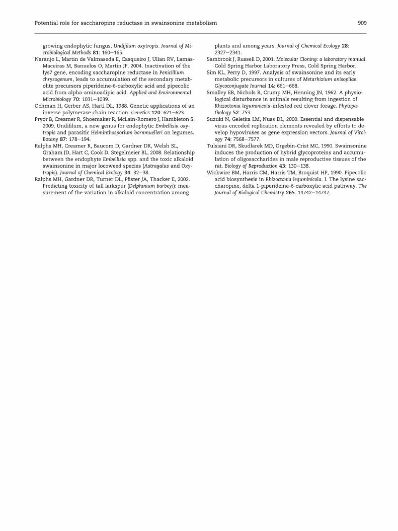
detected (Fig 5B). We propose a possible pathway (Fig 6) show-
ing increasing and decreasing intermediates in lysine-
swainsonine metabolic pathway in U. oxytropis based on our
chromatographic measurements.
Previous studies indicated that P6C might be formed by
non-enzymatic cyclization of a-aminoadipic acid semialde-
hyde, which is unstable for detection using chromatographic
methods (Sim & Perry 1997). The standard for a-aminoadipic
acid semialdehyde is not also available commercially, so this
intermediate compound was not measured.
Discussion
In order to examine the function of saccharopine reductase,
we have modified our recently developed transformation pro-
tocol for Undifilum oxytropis (Mukherjee et al. 2010) to develop
a specific gene deletion method for a slow-growing toxin-
Table 2 e Levels of biochemical intermediates in wildtype Undifilum oxytropis and saccharopine reductasedisruption mutant of Undifilum oxytropis.
Compound Wild type� 1SE (mg mL�1)a,b
Disruptionmutant� 1
SE (mg mL�1)a,b
Swainsonine 0.50� 0.02 4.2� 0.01
Pipecolic acid 0.30� 0.08 5.5� 0.02
Saccharopine 0.35� 0.08 >0.1� 0.03
Lysine 0.50� 0.05 0.32� 0.02
a-aminoadipic acid 0.45� 0.02 0.52� 0.05
a Starting tissue 10 mg of Undifilum oxytropis (dry weight).
b n¼ 12 for all sets of measurements.
producing endophyte. Disruption of saccharopine reductase
led to the accumulation of P6C, pipecolic acid (a precursor of
swainsonine), and swainsonine. However, the levels of sac-
charopine and lysine decreased upon disruption of saccharo-
pine reductase. The impact of inoculation of the saccharopine
reductase-deleted endophyte in plant host and the level of dif-
ferent biochemical intermediates were not tested because
a functional inoculation system is not yet available for this
system.
It has been previously reported that pipecolic acid is con-
verted to lysine through P6C and saccharopine (Kinzel &
Bhattacharjee 1979). Biochemical pathways for swainsonine
production have been partially characterized in Magnaporthe
anisopliae and Rhizoctonia leguminicola. As reported in an earlier
publication (Sim & Perry 1997), different pathways may be in-
volved or active during the formation of swainsonine, and sac-
charopine reductase has an impact on swainsonine synthesis
through this pathway. Naranjo et al. also reported that a lys7
(saccharopine reductase) disruption in Penicillium chrysogenum
caused elevated accumulation of pipecolic acid and P6C
(Naranjo et al. 2004). While these two studies investigated fun-
gal saccharopine reductase, the two fungal organisms in each
α aminoadipate
α aminoadipate semialdehyde
Aminoadipate reductase
?
Pipecolic 6 carboxylic acid
Pipecolic acid
Pipecolate oxidase
Swainsonine
Saccharopine
Lysine
Saccharopineoxidase
α aminoadipate semialdehyde
Aminoadipate redu
?
Pipecolic 6 carboxylic acid
Pipecolic acid
Pipecolate oxidas
Swainsonine
Saccharopine
Lysine
Saccharopineoxidase
Saccharopine
reductase
Fig 6 e Impact of saccharopine reductase deletion on the
lysine biosynthetic pathway in Undifilum oxytropis along
with the possible location of the enzyme.
908 S. Mukherjee et al.
study differ as P. chrysogenum does not produce swainsonine
but U. oxytropis does produce this compound. This study re-
veals that saccharopine reductase influences the swainsonine
metabolic pathway along with the formation of saccharopine
and lysine in U. oxytropis.
We showed that deletion of the saccharopine reductase
gene affects the accumulation of P6C and pipecolic acid in U.
oxytropis. These results open questions about how swainso-
nine is synthesized from lysine and how degradation of
swainsonine occurs to produce saccharopine, the precursor
of lysine. Identification of the gene sequence of saccharopine
reductase has facilitated the characterization of the role of
this enzyme while the establishment of a transformation sys-
tem using an ascomycete vector opens a new avenue to fur-
ther characterize and manipulate different enzymes in the
pathway. The establishment of a gene knock outmethodology
in U. oxytropis will now permit a more detailed study of this
system to decipher other intermediates in the swainsonine
pathway.
Swainsonine is an important inhibitor of mannosidase II
and is a potent antitumour agent with immunomodulatory
activity (Mohla et al. 1989), so further understanding of the
underlying biomolecules that lead to the formation of swain-
sonine will expand our knowledge of the biosynthesis of
a compound with natural pharmaceutical value as well as
contributing to the understanding of the roles of secondary
metabolites in plantefungal interactions. Furthermore, this
may permit the development of fungal strains unable to pro-
duce functional toxin, thus alleviating field toxicity
problems.
Acknowledgements
We thank Deana Baucom, Dr Richard Richins, Dr Soum
Sanogo, Dr Swati Mukherjee, and Dr Omar Holguin at New
Mexico State University for technical assistance and
constructive discussion. Funding for this project was provided
by USDA Special grant 59-5428-1-327 and the New Mexico
State University Agricultural Experiment Station.
r e f e r e n c e s
Braun K, Romero J, Liddell C, Creamer R, 2003. Production ofswainsonine by fungal endophytes of locoweed. MycologicalResearch 107: 980e988.
Broquist H, 1985. The indolizidine alkaloids, slaframine andswainsonine: contaminants in animal forages. Annual Reviewof Nutrition 5: 391e409.
Colgate S, Dorling P, Huxtable C, 1979. A spectroscopic investi-gation of swainsonine: an a-mannosidase inhibitor isolatedfrom Swainsona canescens. Australian Journal of Chemistry 32:2257e2264.
Cook D, Gardner DR, Ralphs MH, Pfister JA, Welch KD, Green BT,2009. Swainsoninine concentrations and endophyte amountsof Undifilum oxytropis in different plant parts of Oxytropis seri-cea. Journal of Chemical Ecology 35: 1272e1278.
Graham D, Creamer R, Cook D, Stegelmeier BL, Welch KD,Pfister JA, Panter KE, Cibils A, Ralphs MH, Encinias M, McDaniel K, Thompson D, Gardner K, 2009. Solutions to locoweedpoisoning in New Mexico and Western United States. Range-lands 31: 3e8.
Harris CM, Schneider MJ, Ungemach FS, Hill JE, Harris TM, 1988.Biosynthesis of the toxic indolizidine alkaloids slaframineand swainsonine in Rhizoctonia leguminicola: metabolism of1-hydroxyindolizidines. Journal of the American Chemical Society110: 940e949.
Hino M, Nakayama O, Tsurumi Y, Adachi K, Shibata T, Terano H,Kohsaka M, Aoki H, Imanaka H, 1985. Studies of an immu-nomodulator, swainsonine. I. Enhancement of immune re-sponse by swainsonine in vitro. The Journal of Antibiotics 38:926e935.
James LF, Nielson DB, 1988. Locoweeds: assessment of the prob-lem on western U.S. rangelands. In: James LF, Ralphs MH,Nielsen DB (eds), The Ecology and Economic Impact of PoisonousPlants and Livestock Production, pp. 171e180.
James LF, Panter KE, 1989. Locoweed poisoning in livestock. In:James LF, Elbein AD, Molyneux RJ, and Warren CD (eds),Swainsonine and Related Glycosidase Inhibitors. Iowa StateUniversity Press, Ames, Iowa, pp. 23e38.
Kingsbury JM, 1964. Poisonous Plants of the United States and Canada.Prentice Hall Englewood Cliffs, NJ, pp. 305e313.
Kinzel JJ, Bhattacharjee JK, 1979. Role of pipecolic acid in thebiosynthesis of lysine in Rhodotorula glutinis. Journal of Bacteri-ology 138: 410e417.
McLain-Romero J, Creamer R, Zepeda H, Strickland J, Bell G, 2004.The toxicosis of Embellisia fungi from locoweed (Oxytropislambertii) is similar to locoweed toxicosis in rats. Journal ofAnimal Science 82: 2169e2174.
Mohla S, Humphries MJ, White SL, Matsumoto K, Newton SA,Sampson CC, Bowen D, Olden K, 1989. Swainsonine: a newantineoplastic immunomodulator. Journal of the National Med-ical Association 81: 1049e1056.
Molyneux R, James L, 1982. Loco intoxication: indolizidine alka-loids of spotted locoweed (Astragalus lentiginosus). Science 216:190e191.
Moulin M, Deleu C, Larher FR, Bouchereau A, 2002. High-performance liquid chromatography determination ofpipecolic acid after precolumn ninhydrin derivatizationusing domestic microwave. Analytical Biochemistry 308:320e327.
Mukherjee S, Dawe AL, Creamer R, 2010. Development ofa transformation system in the swainsonine producing, slow
Potential role for saccharopine reductase in swainsonine metabolism 909
Naranjo L, Martin de Valmaseda E, Casqueiro J, Ullan RV, Lamas-Maceiras M, Banuelos O, Martin JF, 2004. Inactivation of thelys7 gene, encoding saccharopine reductase in Penicilliumchrysogenum, leads to accumulation of the secondary metab-olite precursors piperideine-6-carboxylic acid and pipecolicacid from alpha-aminoadipic acid. Applied and EnvironmentalMicrobiology 70: 1031e1039.
Pryor B, Creamer R, Shoemaker R, McLain-Romero J, Hambleton S,2009. Undifilum, a new genus for endophytic Embellisia oxy-tropis and parasitic Helminthosporium bornmuelleri on legumes.Botany 87: 178e194.
Ralphs MH, Creamer R, Baucom D, Gardner DR, Welsh SL,Graham JD, Hart C, Cook D, Stegelmeier BL, 2008. Relationshipbetween the endophyte Embellisia spp. and the toxic alkaloidswainsonine in major locoweed species (Astragalus and Oxy-tropis). Journal of Chemical Ecology 34: 32e38.
Ralphs MH, Gardner DR, Turner DL, Pfister JA, Thacker E, 2002.Predicting toxicity of tall larkspur (Delphinium barbeyi): mea-surement of the variation in alkaloid concentration among
plants and among years. Journal of Chemical Ecology 28:2327e2341.
Sambrook J, Russell D, 2001. Molecular Cloning: a laboratory manual.Cold Spring Harbor Laboratory Press, Cold Spring Harbor.
Sim KL, Perry D, 1997. Analysis of swainsonine and its earlymetabolic precursors in cultures of Metarhizium anisopliae.Glycoconjugate Journal 14: 661e668.
Smalley EB, Nichols R, Crump MH, Henning JN, 1962. A physio-logical disturbance in animals resulting from ingestion ofRhizoctonia leguminicola-infested red clover forage. Phytopa-thology 52: 753.
Suzuki N, Geletka LM, Nuss DL, 2000. Essential and dispensablevirus-encoded replication elements revealed by efforts to de-velop hypoviruses as gene expression vectors. Journal of Virol-ogy 74: 7568e7577.
Tulsiani DR, Skudlarek MD, Orgebin-Crist MC, 1990. Swainsonineinduces the production of hybrid glycoproteins and accumu-lation of oligosaccharides in male reproductive tissues of therat. Biology of Reproduction 43: 130e138.
Wickwire BM, Harris CM, Harris TM, Broquist HP, 1990. Pipecolicacid biosynthesis in Rhizoctonia leguminicola. I. The lysine sac-charopine, delta 1-piperideine-6-carboxylic acid pathway. TheJournal of Biological Chemistry 265: 14742e14747.