26
dr. Reza Yuridian Purwoko, Sp.KK Evah Luviah Komang Ardi Wahyuningsih Prof. dr. Jeanne Adiwinata Pawitan, PhD DR. Puji Lestari, M.S
| Date post: | 21-Dec-2015 |
| Category: |
Documents |
| Upload: | aditya-zulkarnain |
| View: | 9 times |
| Download: | 1 times |

dr. Reza Yuridian Purwoko, Sp.KK
Evah Luviah
Komang Ardi Wahyuningsih
Prof. dr. Jeanne Adiwinata Pawitan, PhDDR. Puji Lestari, M.S

Introduction
Regenerative medicine multidisciplinary science: - biomaterials,
- growth factors, &- stem cells to repair failing organs
ADSCs stem cell source w/ therapeutic applicability in diverse fields repair & regeneration of acute & chronically damaged tissues
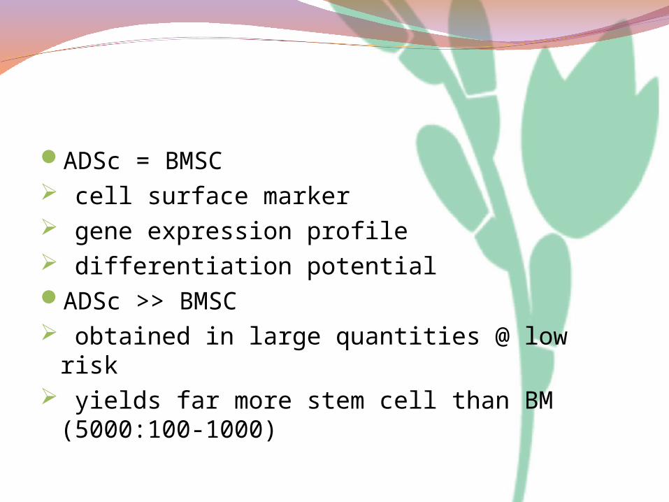
ADSc = BMSC cell surface marker gene expression profile differentiation potentialADSc >> BMSC obtained in large quantities @ low risk yields far more stem cell than BM (5000:100-
1000)

Obtained from liposuction aspirates & differentiate into multiple lineages of mesodermal or ectodermal origins.
It shown differentiation into - adipogenic, - osteogenic, - chondrogenic, - myogenic, - cardiomyogenic, &- neuro-genic lineages

Valuable source of ADSc WAT ASCs can also be harvested by a minimally
invasive procedure involving liposuction from subcutaneous depots.
Main depots of WAT :subcutaneous depots in the buttocks, thighs
& abdomen visceral/intrabdominal depots around the
omentum, intestines and perirenal areas.

The stem cell population derived from collagenase-digested human adipose tissue (SVF)
differentiate into multiple cell lineages, including the adipose tissue, cartilage, bone, skeletal muscle,neuronal cells, endothelial cells,cardiomyocytes, & smooth muscle cells

Isolation Method for ASCs
We obtained ADSc from liposuction aspirates collected using both manual canula & PAL (Microaire).
Lipoaspirate was obtained by tumescent liposuction and kept in a sterile bottle at 4°C for no more than 24 hours.

A. Manual canula, there are 4 type of canula with total 30 holes, 5 holes for each side, with 1 mm hole diameter, & length of range 200-230 mm.B. PAL-canula there are 10 type of canula with total 6 holes, 1-2 holes for each side, with 8-10x3-5 mm hole length x width, & length of range 220-300 mm
A B

All protocol for preparation of ADSc’s isolation based on method from Pawitan et al., 2012.
Lipoaspirate obtained from PAL and manual (1) were filtered through a fine mesh stainless steel tea or coffee filter (φ=7 cm) to separate adipose tissue fragments from tumescent, blood, or contaminants (2), and then wash the fat tissue fragments by soaking the filter in a sterile porcelain bowl filled with Phosphate Buffer Saline (PBS 1x) pH 7.4 and stirring using a small tea spoon to remove the other contaminants (3). This washing step is repeated several times. When fat tissue fragments appeared yellow and clean, then transferred into sterile 50 ml centrifuge tube (4). 0.075% collagenase type 1 (Sigma, USA) is added into the tube containing fat tissue fragments until tissue fragments : collagenase solution was 1:2 (f5). The tubes then incubated at 37oC, 5% CO2 for 1 hour with agitation every 5 minutes. After 1 hour, the appeared floating yellow free lipid were removed (6 & 7) and infranatant were poured into new centrifuge tube through nylon mesh filter and divided into sterile 15 ml centrifuge tube (8 & 9) and then centrifuge 1200 rpm for 10 minutes. After centrifugation step, the sedimented pellet were added lysis buffer if red blood cell still appeared (10 &11), then centrifuge step repeated, obtained pellet (12) then resuspended in culture medium. The number of viable cell were counted using Hemacytometer. The cells were cultured in 12-well plate with seeding number around 170.000 viable cells per well). The cultures cells were observed and medium changed every 2 – 3 days.

Description CANULA
MICROAIRE
Remark
Lipoaspiraate Dark red
Bright red
Isolation processing time/washing process in the lab
>> >>>
Sample after washing process with PBS several times
Bright, yelowwish
Yellowish, still seems reddish

Description CANULA
MICROAIRE
Remark
Sample after digestion with Collagenase
No RBC Still there is RBC (RBC still present while filtering infranatant through nylon mesh filter)
Cell pellet and after seeding > 2 days
Clearer the fat still present in pellet

Day-2 Day-4 Day-6 Day-9
Manual Canula
Microaire

Patient ID Age Sex BMI Harvest site
24 55 F 23.76 Flank
25 39 M 29.1 Abdomen
26 55 F 25.45 Flank
27 32 M 29.1 Abdomen
28 41 F 41.24 Abdomen
29 42 M 28.85 Abdomen

Patient ID
Method Canula type Lipoaspirate Volume
Cell yield count
24 PAL3.0 mm 300 ml 1,660,000
Manual Canula 11 G 300 ml 2,460,00025 PAL
4.0 mm 300 ml 900,000Manual Canula
11 G 300 ml 1,310,00026 PAL
4.0 mm 100 ml 855,000Manual Canula
11 G 300 ml 1,920,00027 PAL
4.0 mm 500 ml1,883,333
Manual Canula 11 G <500 ml
1,593,333
28 PAL5.00 mm >300 ml
2,885,000
Manual Canula 11 G >300 ml
2,380,000
29 PAL4.0 mm 300 ml
890,000
Manual Canula 11 G 250 ml
910,000
Patient ID Age Sex BMI Harvest site
24
55
F 23.76 Flank
25
39
M 29.1 Abdomen
26
55
F 25.45 Flank
27
32
M 29.1 Abdomen
28
41
F 41.24 Abdomen
29
42
M 28.85 Abdomen

Age : no differentiation between all ages varian
Sex : Female greater in number of cell yield count than male
Harvesting site : ADSc from flank greater in number of cell yield count than from abdomen
Canula type : cell yield from manual canula lipoaspirate greater in number than PAL lipoaspirate.


soft and stable small fat particle including many stromal cells
Highest density fat fraction contain more progenitor cells and increased concentrations of several angiogenic/vasculogenic and anti-inflammatory cytokines

Correction of dark circle on lower eyelid by structural high density fat graft,
Sung Min Kim, M.D.

Characterization of ADSc
Immunoprivileged << expression of MHC-II & co-stimulatory molecules on the cell surface allogeneic transplantation into immunocompetent recipients
Immunomodulator & can promote tissue repair prevent & treat acute GVHD in allogeneic stem cell transplantation, autoimmune diseases & inflammatory diseases.

Secrete an array of soluble factors promote tissue regeneration
The secretome includes angiogenic factors (VEGF), anti-apoptotic factors (IGF-1), hematopoietic factors (colony stimulating factors & interleukins), & HGF

Differentiate into several lineages of osteogenic, chondrogenic, adipogenic, cardiomyogenic, myogenic,& neurogenic cells.
Also into tissues of endo- & ectodermal lineages such as hepatocytes, pancreatic islet cells, endothelial cells, neural cells, & epithelial cells too

ADSc application on esthetics
Soft tissue augmentationThe augmentation of breast was successful &
had satisfactory clinical results w/out any major complications
depressed scarsWound healingdiabetes woundHair follicles regeneration

ConclusionAdipose-derived stem cells play an important role of
in regenerative medicineBecause adipose-derived stem cells demonstrate
several interesting properties, suggests potential clinical promise in many medical disciplines.
Protocol for adipose-derived stem cell use or clinical application in terms of type of cells used (stromal vascular fraction cells or cultured & purified adipose-derived stem cells) become easily available & no money & no time consuming.
Further basic science experimental studies still needed to be performed to ensure the safety & efficacy of ADSc

ReferenceGimble JM. Adipose tissue-derived therapeutics. Expert Opin Biol
Ther 2003;3:705–713Honga, S. J; Traktuev, D. O; and Marcha, K.L. Therapeutic potential
of adipose-derived stem cells in vascular growth and tissue repair. Current Opinion in Organ Transplantation 2010:15:86–91
Jin Jin, H. et al. Comparative Analysis of Human Mesenchymal Stem Cells from Bone Marrow, Adipose Tissue, and Umbilical Cord Blood as Sources of Cell Therapy. Int. J. Mol. Sci. 2013:14:17986-18001
Jeffrey M. Gimble, Adam J. Katz and Bruce A. Bunnell. Adipose-Derived Stem Cells for Regenerative Medicine. Journal of American Heart Association. 2007;100:1249-1260
Lindroos, B.; Suuronen, B & Miettinen, S. The Potential of Adipose Stem Cells in Regenerative Medicine. Stem Cell Rev and Rep, Springer Science media. 2010
Madonna, R., Geng, Y-G., and Caterina, R. Adipose Tissue-Derived Stem Cells : Characterization and Potential for Cardiovascular Repair. Journal of American Heart Association. 2009:29:1723-1729

Matsumoto, D. et al. Cell-Assisted Lipotransfer: Supportive Use of Human Adipose-or Soft Tissue Augmentation with Lipoinjection. Tissue Engineering. 2006:12:3375-3382
Mehrabani, D. et al Adipose-Derived Stem Cells (ADSC) and Aesthetic Surgery: A Mini Review. World Journal of Plastic Surgery. 2013:2:65-70
Ong, W. and Sugii, S. Adipose-derived stem cells: Fatty potentials for therapy. The International Journal of Biochemistry & Cell Biology. 2013:1-4
Mizuno, H. Adipose-derived Stem Cell for Tissue Repair and Regeneration: Ten Years of Research and a Literature Review. Journal Nippon Medical School. 2009:76:56-66
Pawitan, J.A et al. Simple lipoaspirate washing using a coffee filter. Asian Biomedicine. 2013:7:333-338
Zuk, P. Adipose-Derived Stem Cells in Tissue Regeneration. Hindawi Publishing Corporation ISRN Stem Cells. 2012:2013:1-35
Zuk, P. Human Adipose Tissue Is a Source of Multipotent Stem Cells. Molecular Biology of the Cell. 2002:13:4279–4295.
Zuk, P. Multilineage Cells from Human Adipose Tissue:Implications for Cell-Based Therapies. Tissue Engineering. 2001:7:2:211-228

Thankyou


