4 Prelemniscal Radiations Neuromodulation in Parkinson Disease´s Treatment José D. Carrillo-Ruiz 1,2 , Francisco Velasco 1 , Fiacro Jiménez 1 , Ana Luisa Velasco 1 , Guillermo Castro 1 , Julián Soto 1 and Victor Salcido 1 1 Unidad de Neurocirugía Funcional, Estereotaxia y Radiocirugía, Hospital General de México 2 Departamento de Neurociencias de la Universidad Anáhuac México Norte México 1. Introduction The early experience, in 80´s, of the use of electrical stimulation in thalamus (Vim and Voa/Vop nucleus) and Globus pallidus internus (GPi) to treat Parkinson´s disease (PD) promoted the well known performance in subthalamic nucleus (STN) neuromodulation. More recently, in 2000´s, the reutilization of old targets (utilized in lesions procedures) like Prelemniscal radiations (Raprl) and motor cortex, and new targets like Pedunculopontine nucleus (PPN) and Zona Incerta (Zi) complemented the tools to treat PD. The use of neuromodulation in thalamus, Gpi and STN in the treatment of Parkinson disease are spread around the world and strongly reinforced the electricity´s utilization in different brain nuclei, not only for clinical aspects but also in physiopathological basic research. By otherwise, the emergent targets need to demonstrate they use and effectiveness like a tool in the treatment of the illness. This chapter is focused in the study of Raprl neuromodulation to ameliorate the symptoms and signs of PD, analyzing the anatomical and physiological background in this area (Carrillo-Ruiz et al, 2007; Ito, 1975; Velasco F et al, 1972, 2009). Trough this article is demonstrated that exists clear evidence that Raprl is a good surgical point to treat PD patients. 2. Anatomy Subthalamic area is part of diencephalum. It is constructed like a pyramid with base in the bottom and has an upper trunked-vertex. This space is formed by nucleus and fibers in a small compact volume of few cubic milimeters. Nuclei are divided in two: 1) Subthalamic area nuclei that included STN, Zi and sustantia Q of Sano. 2) Extended nuclei from mesencephalum, that corresponded to Sustantia Nigra (SN) and Red Nucleus (RN or Ru). In the other side, fibers could be considered more complex and numerous. If it named from anterior to posterior, it could be described as follows: ansa lenticularis, Forel´s Fields (H, H1 and H2), Raprl, perirubral fibers, rubrothalamic fibers, among others (Velasco F, 2009). www.intechopen.com
Transcript
4
Prelemniscal Radiations Neuromodulation in Parkinson Disease´s Treatment
José D. Carrillo-Ruiz1,2, Francisco Velasco1, Fiacro Jiménez1, Ana Luisa Velasco1, Guillermo Castro1, Julián Soto1 and Victor Salcido1
1Unidad de Neurocirugía Funcional, Estereotaxia y Radiocirugía, Hospital General de México
2Departamento de Neurociencias de la Universidad Anáhuac México Norte México
1. Introduction
The early experience, in 80´s, of the use of electrical stimulation in thalamus (Vim and
Voa/Vop nucleus) and Globus pallidus internus (GPi) to treat Parkinson´s disease (PD)
promoted the well known performance in subthalamic nucleus (STN) neuromodulation.
More recently, in 2000´s, the reutilization of old targets (utilized in lesions procedures) like
Prelemniscal radiations (Raprl) and motor cortex, and new targets like Pedunculopontine
nucleus (PPN) and Zona Incerta (Zi) complemented the tools to treat PD. The use of
neuromodulation in thalamus, Gpi and STN in the treatment of Parkinson disease are
spread around the world and strongly reinforced the electricity´s utilization in different
brain nuclei, not only for clinical aspects but also in physiopathological basic research. By
otherwise, the emergent targets need to demonstrate they use and effectiveness like a tool in
the treatment of the illness.
This chapter is focused in the study of Raprl neuromodulation to ameliorate the symptoms
and signs of PD, analyzing the anatomical and physiological background in this area
(Carrillo-Ruiz et al, 2007; Ito, 1975; Velasco F et al, 1972, 2009). Trough this article is
demonstrated that exists clear evidence that Raprl is a good surgical point to treat PD
patients.
2. Anatomy
Subthalamic area is part of diencephalum. It is constructed like a pyramid with base in the
bottom and has an upper trunked-vertex. This space is formed by nucleus and fibers in a
small compact volume of few cubic milimeters. Nuclei are divided in two: 1) Subthalamic
area nuclei that included STN, Zi and sustantia Q of Sano. 2) Extended nuclei from
mesencephalum, that corresponded to Sustantia Nigra (SN) and Red Nucleus (RN or Ru). In
the other side, fibers could be considered more complex and numerous. If it named from
anterior to posterior, it could be described as follows: ansa lenticularis, Forel´s Fields (H, H1
and H2), Raprl, perirubral fibers, rubrothalamic fibers, among others (Velasco F, 2009).
www.intechopen.com
Topics in Neuromodulation Treatment
64
2.1 Nuclei
The nuclei extend from the midbrain area to midbrain diencephalon and involved SN and RN.
2.1.1 Substantia nigra
The substantia nigra is a great motor nucleus located between the tegmentum and the bases of the stem along the midbrain, extending into the subthalamic region of the diencephalon. The core is formed for medium-sized multipolar neurons. It is divided into two parts: one part compacta (SNc) and reticular portion (SNr). The portion is containing cytoplasmic inclusions in compact form of granules of melanin pigment, which is an area rich in dopamine, being more abundant in primates and especially in man. The granules are sparse at birth but rapidly increase in childhood and more slowly in the rest of life. In the reticular portion, the cells are not pigments but contain large amounts of iron demonstrable by histochemistry. Neurons in the pars reticulata are crossed by axons of neurons in the pars compacta. Afferents originate from the axons of caudate and lenticular found in the telencephalon and fewer of the subthalamic nucleus and midbrain raphe nuclei and the pontine reticular formation. The efferent fibers that originate in cells of the compact area go mainly to the caudate nucleus and putamen and some end up in the amygdala temporal lobe. The cells of the reticular portion are projecting into neostriatum, the ventral anterior nucleus and ventral lateral thalamus and superior colliculus.
2.1.2 Red nucleus
Ru is an important component of tegmental motor area. This nucleus has an ovoid shape (round is cross-shaped), extending from the caudal boundary of the superior colliculus to the subthalamic region of the diencephalon. The nucleus has a pinkish color in fresh specimens having a greater blood supply than the surrounding tissue. The core looks red dotted cuts with Weigert and Weil method, due to the myelinated fibers of the same. The red nucleus is divided into two regions: the caudal region that is phylogenetically the oldest, and consists of large cells and is known as magnocellular portion. The rostral is more recent and is especially developed in humans, is formed of small cells, so called parvicelular portion. As afferents can say that those in the cerebellum and cerebral cortex have been the best studied. The fibers that originate in the cerebellar nuclei (mainly the dentate nucleus) form the superior cerebellar peduncles and enter the midbrain. Some fibers end in the Ru and others around him on his way to thalamic nuclei (ventral lateral nucleus) and from this point toward the motor areas of the frontal lobe. These same areas give rise to numerous afferent fibers and there cortico-rubral pathways through the superior colliculus to the red nucleus. Efferent connections of the red nucleus are rubrospinal tract fibers that cross the median plane in the ventral tegmental decussation (Forel) and continue in the brainstem and the lateral funiculus of the spinal cord. Some fibers terminate in the facial motor nucleus and lateral reticular nucleus projecting to the cerebellum, some fibers end in the inferior olivary nuclei. There are also some other fibers involved like emboliform and globose nuclei of the cerebellum.
The other two nuclei are described in the next lines:
2.1.3 The subthalamic nucleus or body of Luys
STN or Sth is pink, is located at the junction of the cap of the midbrain and hypothalamus, below the thalamus. The upper and lower faces are convex. The outer edge is in contact with
www.intechopen.com
Prelemniscal Radiations Neuromodulation in Parkinson Disease´s Treatment
65
the internal capsule, and its rear end is above the locus niger, the upper surface is separated from the underside of the thalamus in the Zi and lenticular fasciculus. There are two types of neurons: a smaller than 10 μ and larger ones occupying the outside.
The Sth or STN is one of the motor nuclei and is best developed in advanced mammals. The connections of the subthalamic nucleus are reciprocal to the Gpi, these fibers are the subthalamic fasciculus that cuts through the internal capsule. The subthalamic nucleus also receives some pedunculopontine nucleus afferents and sends some to the SNr efferent pathways (Figure 1).
Fig. 1. Red nucleus and subthalamic nucleus. The position of the red nucleus is posterior to the back of subthalamus, however, the subthalamic nucleus is anterior. (Modified from England & Wakely, 1992).
2.1.4 Zona incerta
Zona incerta (Zi) was first described by Forel. It is a core derived from ventral thalamus, is a different heterogeneous nucleus that remains in the base of the thalamus. It is a very thin core of serpentine shape, starting from the base of the SN to the dorsal region of the diencephalon and ends in the posterior nuclei of the hypothalamus. It is located immediately above the STN, between the fiber bundles in the Forel´s fields and Raprl. The Zi is divided into four sectors: rostral, dorsal, ventral and caudal. The rostral component extends over the dorsal and medial STN, while it caudal or motor remains posterior to the STN. The Zi receives afferent exit points of the basal ganglia, which is the globus pallidus and substantia nigra pars reticulata, the ascending reticular activating system and motor areas, associative and limbic cortex. In contrast, the Zi sends efferent ways to the parafascicular and centromedian nucleus of the thalamus, ventral anterior nucleus, ventral lateral nuclei of the thalamus, midbrain extrapyramidal area, output nuclei of the basal ganglia and the cerebral cortex. Different sections have different functions Zi: rostral sector has been attributed to visceral control, dorsal sector in the wake, ventral area is under the guidance of the eye and head movements, and the posterior sector in the generation of axial flow and proximal members, including locomotion (Plaha, 2006).
2.1.5 Substance Q of Sano
Substance Q of Sano was described by the Japanese neurosurgeon Pr. Keiji Sano. It is located below the Zi and also immediately adjacent to the SN. Its function is not well elucidated; it may be part of the reticular formation.
www.intechopen.com
Topics in Neuromodulation Treatment
66
2.2 Fibers
The efferent fibers of the globus pallidus are contained in two tiny beams, different between them, the lenticular fasciculus and the lenticular loop. The lenticular fasciculus consists of fibers that cross the internal capsule to reach the subthalamus, where they form a band of white substance known as H2 Forel´s field. Most of the constituent fibers change direction in the area prerrubral or H (Haube= cap in german) Forel´s field, and penetrate the thalamic fasciculus or H1 Forel´s field, ending in the ventral lateral nucleus and ventral anterior thalamus. At a higher level, the handle forms a lenticular sharp curve around the medial border of the internal capsule and ends in the nuclei ventral lateral and ventral anterior thalamus. Only some fibers of the globus pallidus veer caudally and terminate in the pedunculopontine nucleus, which is one of the lateral group nuclei of the reticular formation located between the union between midbrain and pons. Mesencephalic reticular formation continues in the subthalamus where Zi appears between the lenticular and thalamic fascicles.
The subthalamus contains sensory tracts, extensions rostral midbrain nuclei (Ru and SN), and fibers´ bundles of the dentate nucleus of the cerebellum and globus pallidus, and STN. The sensory tracts are half lemniscus tract, and spinothalamic tracts that are extended immediately below the ventral intermediate nucleus, where the fibers ending.
Fig. 2. Subthalamic area includes nuclei and fibers. It shows the nuclei of the basal ganglia: substantia nigra, subthalamic nucleus, the motor thalamus and Zona Incerta. Among them, the respective fibers: subthalamic fascicles; H, H1 and H2 of Forel´s fields. The cut is rostral, therefore the Ru or the Raprl are not seen (modified from England & Wakely, 1992).
www.intechopen.com
Prelemniscal Radiations Neuromodulation in Parkinson Disease´s Treatment
67
Dentothalamic fibers that cross the median plane through the decussation of superior cerebellar peduncles surround and traverse the Ru and continue forward in the H Forel´s field or prerrubral area. The fibers help to form dentothalamic tracts and terminate in the thalamic nucleus ventral oral posterior (Vop) of the ventral lateral nucleus of the thalamus.
2.2.1 Prelemniscal radiations
Among the midbrain and diencephalon are found towards the back of subthalamus in the mesencephalic tegmentum (Figure 3), an area of white matter containing a bundle of fibers located and arranged so oblique and ventrolateral well the half lemniscus (Lm), this set of fibers are the most posterior and superior to the Zi and for being right in front of Lm are called prelemniscal Radiations or in latin Radiatio Praelemniscalis (Raprl).
Fig. 3. Mesencephalic tegmentum. On the left, the tegmentum is located between the
cerebral peduncles in front, and an imaginary horizontal line that crosses the aqueduct,
forming a triangle. On the right side, we see the red nucleus and radiation from the
tegmentum, which are Raprl (Modified from Testut, 1948).
Forel described in 1877, the position of Raprl. Originally he called them like BA Th, and
clearly distinguishing them from his H fields (more medial and anterior), and they shown in
the diagram below in Figure 4. The original name was aported by Cécile Vogt-Mugnier, in
1909, to refer to Raprl as radiation from the cerebellum in front of the lemniscus
(prelemniscal) and Hassler, as her pupil in 1959, included it in the description of his
stereotactic atlas. Also, in 1960, Talaraich included Raprl in his atlas.
2.2.1.1 Raprl definition
When it considered exclusively Raprl like a target structure, this could be understand like a
compact group of fibers located in the white posterior subthalamic area, in front of the
sensory lemniscus, that brings its own name. But in this moment, the real origin remains
www.intechopen.com
Topics in Neuromodulation Treatment
68
uncertain. Nevertheless, anatomic studies demonstrated that probably they come from three
different sites: 1) Axons growth from the cerebellar nuclei to the thalamus 2) Fibers crossing
from pallidum to motor thalamus, and 3) Neurons projecting from reticular formation
nuclei in ascendant pathway The whole fibers run obliquely from the posteroinferior to
anterosuperior way forming a funnel, beginning in mesencephalic reticular nuclei passing
through Substantia nigra to the thalamus (Vim and Voa/Vop) and ascending between Ru
and STN and Zi. (Modified from Testut, 1947). See figure 1.
Fig. 4. Forel's original description of the subthalamic region. Coronal cut with some
obliquity. In Fig. 6 (333), fig 8 (311) and fig. 10 (294), are located at Raprl as Bath, RK: Red
Nucleus. In the last figure, perfectly distinguishes H Forel´s field of Raprl (Forel, 1877).
1. Projections of the basal ganglia. Globus pallidus are connected to the motor thalamus,
by the lenticular bundle and Forel´s fields (H, H1 and H2), also interfacing with other
structures such as the STN and Zi. Importantly, the fields are above and rostral to
Raprl, and there is no absolute division between them: mainly with the fields H1 and H
in medial face, making their way to the thalamus are intertwined. In addition there are
fibers connecting the STN and SN by subthalamic fasciculus, which also passes through
this area. Ru nuclei have reciprocal connections with STN, which also are compacted
here. See figure 7.
www.intechopen.com
Prelemniscal Radiations Neuromodulation in Parkinson Disease´s Treatment
69
Fig. 5. Dentorubric and rubrothalamic-dentothalamic fibers of Raprl. This Flesching or horizontal cut, in a brain (left) and its schematization (right) form a funnel of Raprl fibers from different parts of the cerebellum and red nucleus. Note the anterior location of Raprl over the medial lemniscus (Modified from Testut, 1947 and England and Wakely, 1992).
Fig. 6. Component fibers of the basal ganglia on Raprl. Note to Raprl on arrival to the thalamus via rubrotahalamic and dentorubric pathways and, along with tracks from the lenticular loop and fibers of the Forel´s fields H, H1 and H2 that cross in the posterior subthalamic region (Adapted from Barr, 2005).
2. Projections of the reticular formation. The nuclei of the reticular formation (medulla, pons and midbrain) come down to two important centers in their connections with the thalamus. The first is Zi, in which reticular formation ends and the other is the thalamic reticular nucleus. Zi is adhered to the Raprl virtually its entire course, so leave this core fibers are directed towards the thalamus. There is also an important Zi connection with a motor nucleus of the reticular formation which is the pedunculopontine nucleus. Anatomical studies in cats have shown that, although they have the same name as in humans, this area belongs to the reticular formation of fibers emanating from the
www.intechopen.com
Topics in Neuromodulation Treatment
70
mesencephalic tegmentum and the ventral oral nucleus of the pons and terminate in the thalamus (Nauta and Kuypers, 1958). In the monkey, the same area corresponds to midbrain reticular nuclei (Ward et al, 1948). See Figure 8B.
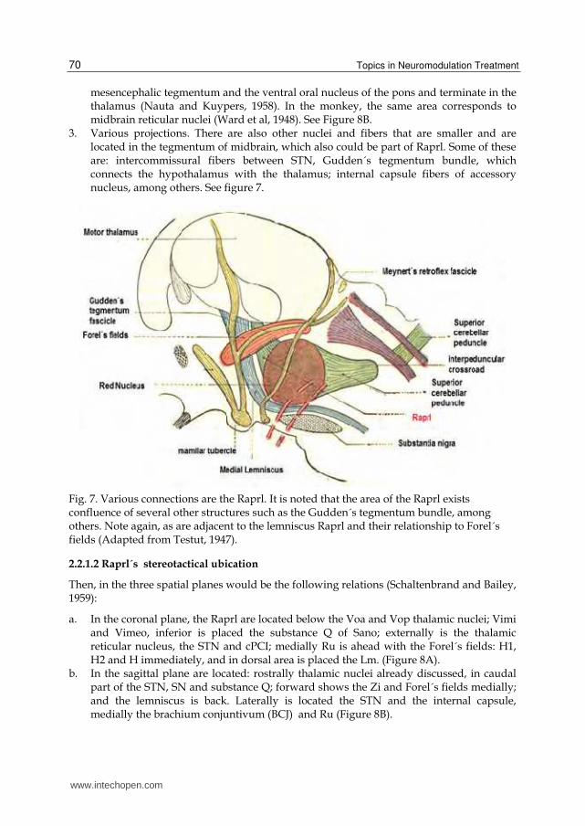
3. Various projections. There are also other nuclei and fibers that are smaller and are located in the tegmentum of midbrain, which also could be part of Raprl. Some of these are: intercommissural fibers between STN, Gudden´s tegmentum bundle, which connects the hypothalamus with the thalamus; internal capsule fibers of accessory nucleus, among others. See figure 7.
Fig. 7. Various connections are the Raprl. It is noted that the area of the Raprl exists confluence of several other structures such as the Gudden´s tegmentum bundle, among others. Note again, as are adjacent to the lemniscus Raprl and their relationship to Forel´s fields (Adapted from Testut, 1947).
2.2.1.2 Raprl´s stereotactical ubication
Then, in the three spatial planes would be the following relations (Schaltenbrand and Bailey, 1959):
a. In the coronal plane, the Raprl are located below the Voa and Vop thalamic nuclei; Vimi and Vimeo, inferior is placed the substance Q of Sano; externally is the thalamic reticular nucleus, the STN and cPCI; medially Ru is ahead with the Forel´s fields: H1, H2 and H immediately, and in dorsal area is placed the Lm. (Figure 8A).
b. In the sagittal plane are located: rostrally thalamic nuclei already discussed, in caudal part of the STN, SN and substance Q; forward shows the Zi and Forel´s fields medially; and the lemniscus is back. Laterally is located the STN and the internal capsule, medially the brachium conjuntivum (BCJ) and Ru (Figure 8B).
www.intechopen.com
Prelemniscal Radiations Neuromodulation in Parkinson Disease´s Treatment
71
c. Finally, in the axial plane, shows from top-down as follows: outside the Raprl the lenticular nucleus and thalamus divided both by the internal capsule. Thalamic nuclei are Voa, Vop and Vim, and behind the Vce. Laterally, it is located the STN and Zi, and medially the BCJ and Ru, and more anterior Ha, H, and H1 Forel´s field.(Figure 8C)
Fig. 8. Brain stereotactical sections to locate Raprl in humans. A) Coronal, where Zi, internal capsule and the STN stands externally, Ru stays medially; in cephalic area the thalamic nuclei are seen (Vimi and Vimeo, Ce); caudally substance Q, Ru and SN. B) Sagittal section shows where is the oblique arrangement of the fibers with the direction of thalamic nuclei (Vim, Voa and Vop). Ru and SN are observed caudally. Rostrally is finding the STN. C) Axial section puts the Ru and Raprl between the medial and posterior part of the Zi and the STN. (From Atlas for Stereotaxy of the Human Brain, Schaltenbrand y Bailey,1959).
www.intechopen.com
Topics in Neuromodulation Treatment
72
3. Physiology
If it is considered the anatomical aspects described below, Raprl´s functional aspects belong
to diverse systems. The anterior component, which is part of basal ganglia, regulates the
postural control. The inferior component has relation with reticular activation and in this
sense with selective attention and motor orienting response; and the cerebellar component is
involved into the dento-thalamo-cortical system to modulate muscular tone and coordinated
voluntary movements. Dysfunction of these systems originated postural abnormalities,
tremor, rigidity and probably bradykinesia (Bertrand, 1969; Velasco M, 1986).
Electrophysiological studies in Raprl that have previously been shown that intraoperative
microelectrode records under local anesthesia without sedation have reported that the area
2 to 3 mm below the output of motor thalamus has a unitary activity in the not show any
neuronal firing, just listening to background activity that is organized from time to time
with bursts of 4 to 6 cps, similar to the frequency of tremor parkinsonism (Velasco et al 1973,
1975), also reported by others ( Birk and Struppler, 1989; Luecking et al, 1971). These bursts
of rhythmic activity resemble those reported in Vim (Jasper, 1966) and Voa (Tasker, 1967) of
the thalamus, but are less frequent and prominent. Besides neurons were triggered with
morphology completely different to STN. See Figure 9.
Fig. 9. STN and Raprl register comparison. Voltage difference seen at the site where the
neurons firing in the STN when is compared to the Raprl. There is practically a
physiological noise corresponding to fibers and a few neurons firing.
www.intechopen.com
Prelemniscal Radiations Neuromodulation in Parkinson Disease´s Treatment
73
In addition, getting late evoked potentials already described previously (Velasco et al, 1988) confirms the involvement of the reticular formation in PD. Events related to somatic evoked potential (SEP) induced by median nerve stimulation during selective attention paradigm when an electrode located in the Raprl. In this area, only the late components (P200, P300), but not early (N20) were recorded. The late components vary significantly in amplitude for the effect of attention, so there is maximum amplitude during selective attention and new and there is minimal amplitude during habituation and distraction (Velasco et al, 1975, 1979, 1986; Jiménez et al, 2000). In contrast, the SEP recorded immediately posterior and medial lemniscus show a prominent N20, but not later P200 and P300 components. These findings place the Raprl as an extralemniscal system and are in contradiction with other reports (Birk and Struppler, 1989; Luecking et al, 1971, Momma et al, 1980). Similar components without component P200 were recorded early in the mesencephalic reticular formation (Velasco et al, 1979). Moreover, in monkeys with radiofrequency lesions made in an area equivalent to Raprl that corresponds to subthalamic and mesencephalic reticular formation, diminishing the contralateral limb tremor produced by lesions previously located in the SNc (Velasco et al, 1979). Also in humans (Andy et al, 1963, Velasco et al, 1980) and in experimental animals (Adey et al, 1962, Watson et al, 1974) lesions of this area can produce a "lack of spontaneous use" of contralateral extremities called neglect.
In humans, this appears to only occur when there is an additional subcortical atrophy in the thalamus (Velasco et al, 1986). In view of these observations, the existence of a reticulo-thalamic that mediates attention and tremor was proposed (Velasco et al, 1979).
4. Targeting and surgical technical aspects
Nowadays stereotactic surgery is refined in the precise identification of Raprl, since it is a very small target. In ancient times the ventriculography was commonly performed, and was changed by tomography scanner/magnetic resonance fusion with specific computational software. Patients are operated under local anesthesia, since patient collaboration and the commonly seen decrease or arrest of tremor in the moment of electrode´s insertion is an important clinical guide of the correct placement, and this is corroborated with macroestimulation when the electrode is connected to screening test machine. Coordinates for Raprl target are: lateral 11-13 mm, inferior to AC-PC line 4-5 mm and posterior to midline point in AC-PC line of 6-9 mm. Should electrodes’ contacts are anterior or superior to Raprl target, the results in controlling tremor and rigidity are incomplete or null; if the contacts are posterior, contralateral paresthesias are elicited, and if the contacts are displaced medially macrostimulation induces gaze deviation (Bertrand 1969, 2004; Velasco 1982).
Before discussing what the effect of stimulation on the sign in concrete, it is worth explaining that it reproduces what happens in the Raprl when the single insertion of the electrode produces a decrease or disappearance of the sign on the side contralateral to the site of electrode´s introduction. This happens dramatically with tremor and rigidity, and for bradykinesia also presented a decrease, and it is difficult to assess the other two signs (gait and posture) with the patient is supine. The explanation of this phenomenon has been described from thalamotomy or subthalamotomy, where the insertion of the radiofrequency electrode to the distance and the tip of the instrument on the nerve tissue, causes that the
www.intechopen.com
Topics in Neuromodulation Treatment
74
motor circuit the is blocked by the physical presence of the object. Once in the post-operative, clinical signs reappear in the patient. The same happens with the introduction of a single electrode first, by modifying the above signs, and introducing the second electrode there is also the same effect on the opposite side of implantation.
In some cases of a second (bilateral) electrode implantation, it is also worth discussing the transient impairment of consciousness after the introduction of it. It happens when it introduces in the left side, but when he gets on the contralateral side, the patient must be alert, is immobilized and has transient aphasia comprehension and expression. The patient presents an excessive sleepiness that lasts even after the surgery a few hours to 48 hours. The way that can explain this is to prevent access of information by ascending activating system, leading to a consequent loss of consciousness and awake. This effect is transitory and after this time the patient is recovered the alert state.
5. Surgery indications and contraindications (Bertrand 1969, Carrillo-Ruiz 2003, Velasco, 1972, 2001; Espinosa 2010)
The next are the indications to choose patients to Raprl neuromodulation:
1. Patients diagnosed with idiopathic PD.
2. Good response to Levodopa test.
3. Age between 35 to 80 years.
4. Intact cognition or discrete alterations of mood.
5. Tremor is the predominant symptom.
6. Rigidity accompanies tremor.
7. Acral bradykinesia.
On the other side, the issues below are contraindications:
1. Rigidity/bradykinetic signs exclusively.
2. Severe unbalance or gait disturbances.
3. Traditional surgical contraindications (coagulopathies, high anesthesic risk ASA > 3,
etc…).
4. Patients with previous brain lesions, mainly in subthalamus.
6. Results
6.1 Neuroimaging
The next images demonstrated the site of the electrodes between the Ru and Zi. Figure 10
shows an axial image where the electrodes are observed in Raprl. The electrodes are located
between STN and Ru nuclei, being one tenth below the line intercommissural. On the right
side is illustrated a diagram of the structures, with the names of the most important
structures.
Figure 11 shows coronal and axial sections of MRI in T2 sequence, where the electrodes are
observed bilaterally that are lateral to the midline, some of them displayed three to four
other contacts as rounded hypointense images with a white halo, which is located between
the Ru and STN.
www.intechopen.com
Prelemniscal Radiations Neuromodulation in Parkinson Disease´s Treatment
75
Fig. 10. Position of the electrodes. The image seen in an axial section of the atlas of Schaltenbrand and Bailey, the position where the electrode is located (red circle) between the STN and Ru which it corresponds to Raprl. It transposes the same level on MRI.
Fig. 11. Position of the electrodes in the MRI. Here is a coronal (left) and axial (right) sections. In the first corresponded to 2.5 to 5 mm anterior to the PC on both sides, the different electrode contacts. In the second, two ovoid hypotenses images 2 to 3 mm below the AC-PC line level. Note the electrodes are immediately lateral red nucleus.
www.intechopen.com
Topics in Neuromodulation Treatment
76
In figure 12, it shows a three dimensional representation of an electrode in Raprl target with different projections. In the first MR image, in a sagittal section; the second, third and fifth with an oblique/anterior and in the fourth image at an angle with a posterior projection, that shows very clearly as the electrode through the trephine reaches the stem brain to the midbrain, showing the location of the four electrodes contacts.
Fig. 12. Three dimensional reconstruction of an MRI demonstrated in a case position of the
electrodes in Raprl. It is noted as the electrode enters oblique anteroposterior manner,
following a midbrain-thalamic region. The tip is located in the midbrain tegmentum of the
patient. The rest of the contacts found in midbrain-subthalamic union in the posterior third
of subthalamus.
It has been described in several articles the position of the electrode when it was located in
Raprl target. It is interest to note that groups with implantation in STN had reported the best
points of stimulation with effectiveness in amelioration of the signs as outside of STN that
corresponded to the white posterior subthalamic zone (Yokohama et al, 2001; Yelnik et al,
2003; Hamel et al, 2003). Undoubtedly this is corresponded to Raprl and Raprl/Zi area.
6.2 Clinimetric results in lesional era
During stereotactic lesional era (60´s to 80´s), Raprl leucotomy showed an important effect to
arrest tremor with the simple insertion of the leucotome in the fibers that was optimized in
the moment of fibers´ coagulation itself. There is little information on the effect in other
Fig. 13. Clinimetric changes for Tremor. Longitudinal assessment of the tremor is seen by using the subitem 20 of the UPDRS part III. A and B corresponded to superior limbs. C and D for inferior limbs. The evaluation is presented in baseline, 3, 6, 9 and 12 months. Box plot represents the 75% of all patients; the bar illustrates the median and the outliers, maximum and minum values. Asterisks show statistical significance (*p<0.05, ** p<0.01,***p<0.001), (Carrillo-Ruiz, 2003).
www.intechopen.com
Prelemniscal Radiations Neuromodulation in Parkinson Disease´s Treatment
81
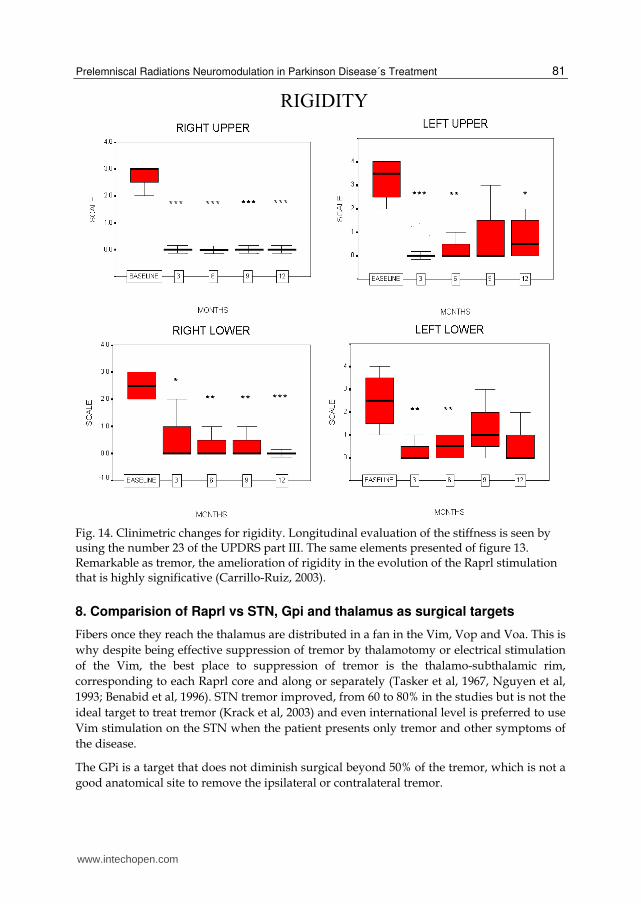
Fig. 14. Clinimetric changes for rigidity. Longitudinal evaluation of the stiffness is seen by using the number 23 of the UPDRS part III. The same elements presented of figure 13. Remarkable as tremor, the amelioration of rigidity in the evolution of the Raprl stimulation that is highly significative (Carrillo-Ruiz, 2003).
8. Comparision of Raprl vs STN, Gpi and thalamus as surgical targets
Fibers once they reach the thalamus are distributed in a fan in the Vim, Vop and Voa. This is
why despite being effective suppression of tremor by thalamotomy or electrical stimulation
of the Vim, the best place to suppression of tremor is the thalamo-subthalamic rim,
corresponding to each Raprl core and along or separately (Tasker et al, 1967, Nguyen et al,
1993; Benabid et al, 1996). STN tremor improved, from 60 to 80% in the studies but is not the
ideal target to treat tremor (Krack et al, 2003) and even international level is preferred to use
Vim stimulation on the STN when the patient presents only tremor and other symptoms of
the disease.
The GPi is a target that does not diminish surgical beyond 50% of the tremor, which is not a
good anatomical site to remove the ipsilateral or contralateral tremor.
www.intechopen.com
Topics in Neuromodulation Treatment
82
Fig. 15. Clinimetric evaluation uses for bradykinesia. Assessment of bradykinesia using subitem no. 32 of the UPDRS part III.The explanation is similar to figure 13 and 14 (Carrillo-Ruiz, 2003).
With respect to rigidity, Vim stimulation is not good (less than 40%), so it is not the best method to treat akinetic-rigid patients, this confirms the above that the tremor is more involved with the cerebellar pathway that the route of the basal ganglia. However, both the STN and the GPi are good targets for improved rigidity, but despite this, the percentage of Raprl has a greater efficiency.
Vim stimulation is not useful to improve the bradykinesia. The STN and Gpi alike improve these signs, being almost the same percentage held for the Raprl of about 65%.
In this sense the anatomical targets improve of gait, often the STN and GPi, as both through specific studies on the place have proven effective. The percentage is better than for Raprl, 50% vs. 45%, although the patients with Raprl are advanced (Hoeh & Yahr 5) and that STN patients have a varying degrees of Hoehn and Yahr, so no are fully comparable.
At last, the posture is a good example of improvement also mild level with the use of STN stimulation, but better than the Raprl neuromodulation. This may be due to connections
www.intechopen.com
Prelemniscal Radiations Neuromodulation in Parkinson Disease´s Treatment
83
with the brainstem to the STN and in the earliest stages of PD in these patients. The Vim and GPI are not good places to improve posture (Benabid et al, 1996 and 2000).
9. The future
The next point would be to establish what is the relationship of these findings with is currently known about the PD pathophysiology, both in animals and humans. If it refers to Alexander and DeLong classic diagram shows that none of the findings of this study fall in there. If it pays attention to the direct and indirect pathways have their starting point in the Gpi/ SNr and depending on their afferents may come from the putamen or the STN, respectively. However this scheme does not specify which routes are used from the STN or GPi. The pallidal and climbing pathways that reach the thalamus has been mentioned, are not Raprl as anatomically way is through the Forel´s fields that predate the site of interest, on the other side cannot be excluded that the fibers emanating from the STN may be added in the same Raprl but this is not contemplated in the scheme of Alexander (Alexander et al, 1986; Alexander & De Long, 1990).
It is relevant determinate exactly the anatomy of the white fibers, with the correlation to functional directions. In this effort, it is analyzing by different methods; tractography, potential unit neuron stimulation and other to determinate the role of brainstem with the subthalamus and basal ganglia.
10. Conclusions
Raprl neuromodulation in the treatment of PD is effective to improve tremor, rigidity and bradykinesia. The results over gait, posture and dyskinesia are less significant; results have been validated by different groups around the world.
11. References
[1] Adey W.R., Walter D.O., Lindsley D.F. Subthalamic lesions. Arch Neurol 6: 34-47, 1962. [2] Alexander G.E., DeLong M.R., Strick P.L. Parallel organization of functionally segregated
circuits linking basal ganglia and cortex. Annu Rev Neurosci 9:357-378, 1986. [3] Alexander G.E., Crutchter M.D. Functional architecture of basal ganglia circuits: Neural
substrates of parallel processing. Trends Neurosci 13:266-275, 1990. [4] Andy OJ, Jurko MF, Sias FRJr. Subthalamotomy in treatment of parkinsonism tremor. J
Neurosurg 20: 860-870, 1963. [5] Barr K. Neuroanatomía Humana. 8ª ed. McGraw Hill, Mc Graw Hill ed, 2005. [6] Benabid A.L., Pollak P., Gao D., Hoffmann D., Limousin P., Gay E., Payen I., Benazzouz
A. Chronic electrical stimulation of the ventralis intermedius nucleus of the thalamus as a treatment of movement disorders. J Neurosurg 84:203-214, 1996.
[7] Benabid A.L., Koudsié A., Benazzouz A., Fraix V., Ashraf A., Le Bas J.F, Chabardes S., Pollak P. Subthalamic Stimulation for Parkinson´s Disease. Arch Med Res. 31, 3:282-289, 2000.
[8] Bertrand CM, Hardy J, Molina-Negro P, Martínez N: Optimum physiological target for the arrest of tremor, in Gillingham FJ, Donaldson ML (eds): Third Symposium of Parkinson’s Disease. Edinburgh, E&S Livingston, 1969, pp 251–254.
www.intechopen.com
Topics in Neuromodulation Treatment
84
[9] Bertrand C., Molina-Negro, P., Martínez, N., Velasco, F.: Stereotaxic surgery in Parkinson’s disease. Progress in Neurological Surgery. Krayenbuhl, H., Maspes, P. And Sweet, W. (Eds.) Year Book Medical Publishers, Vol. V. 1974.
[10] Bertrand CM. Surgery of involuntary movements, particularly stereotactic surgery: reminiscences. Neurosurgery. 2004; 55(3):698-703.
[11] Birk P., Struppler A.: Functional neuroanatomy of the target area for the treatment of pathological tremor: an electrophysiological approach. Stereotact Funct Neurosurg 52:164-170, 1989.
[12] Bubnov AN [Neurosurgical anatomy of the zona incerta applicable to subthalamotomy]. Vopr Neirokhir. 1975; (1):36-40.
[13] Carrillo-Ruiz J., Velasco F., Jiménez F., Hernández-Silverio J.A., Arguëlles C. Bilateral Electrical Stimulation of Prelemniscal radiations in Advanced Parkinson´s disease. American Society for Stereotactic and Functional Neurosurgery 2003 Quadrennial Meeting, New York City, E.U.A., 18 to 21 of 2003. May 18 -21, 2003.
[14] Carrillo-Ruiz JD, Velasco F, Jiménez F, Velasco AL, Velasco M, Castro G. Neuromodulation of prelemniscal radiations in the treatment of Parkinson's disease. Acta Neurochir Suppl. 2007; 97(Pt 2):185-90.
[15] Carrillo-Ruiz JD, Velasco F, Jiménez F, Castro G, Velasco AL, Hernández JA, Ceballos J, Velasco M. Bilateral electrical stimulation of prelemniscal radiations in the treatment of advanced Parkinson's disease. Neurosurgery. 2008; 62 (2):347-57.
[16] Driollet R, Schvarcz JR, Orlando J. Optimum target for arrest tremor. Confin Neurol 36: 355, 1974.
[17] England MA and Wakely J. El cerebro y la médula espinal. Introducción a la neuroanatomía normal. Mosby Year Book, 1a ed., 1992.
[18] Espinosa J, Arango G, Fonseca M, Gálvez J, Atuesta J. Prelemniscal radiation deep brain stimulation: indications and results. In: Neuromodulation. Cukiert J (ed.) 2010: pp: 236-240.
[19] Espinosa J, Arango G. Surgical management of Parkinson’s disease. Deep brain stimulation of the prelemniscal radiation. Movement disorders. 2005; 20 Suppl 10:S159-60.
[20] Forel A. Untersuchungen über die Haubenregion und thre oberen Verknüpfungen im Gehirne des Menschen und der Saugetiere, mit Beitrágen zu den Metoden der Gehirn-Untersuchung. Archiv für Psychiatrie und Nervenkrankheiten,Berlin 7(3). 393-495, 1877.
[21] Fytagoridis A, Bloomstedt P. Complications and side effects of deep brain stimulation in the posterior subthalamic area. Stereotact Funct Neurosurg. 2010; 88:88-93.
[22] Hamel W, Fietzek U, Morsnowski A, Schrader B, Herzog J, Weinert D, Pfister G, Müller D, Volkmann J, Deuschl G, Mehdorn HM. Deep brain stimulation of the subthalamic nucleus in Parkinson's disease: evaluation of active electrode contacts J Neurol Neurosurg Psychiatry. 2003; 74(8):1036-46.
[23] Hamel W, Herzog J, Kopper F, Pinsker M, Weinert D, Müller D, Krack P, Deuschl G, Mehdorn HM. Deep brain stimulation in the subthalamic area is more effective than nucleus ventralis intermedius stimulation for bilateral intention tremor. Acta Neurochir (Wien). 2007; 149(8):749-58.
[24] Hassler R, Mundinger F, Riechert T. Correlations between clinical and autoptic findings in stereotaxic operations of parkinsonism. Confin Neurol 26: 282-290, 1965.
[25] Herzog J, Hamel W, Wenzelburger R, Pötter M, Pinsker MO, Bartussek J, Morsnowski A, Steigerwald F, Deuschl G, Volkmann J. Kinematic analysis of thalamic versus
www.intechopen.com
Prelemniscal Radiations Neuromodulation in Parkinson Disease´s Treatment
85
subthalamic neurostimulation in postural and intention tremor. Brain. 2007; 130 (Pt 6): 1608-25.
[26] Houdart R, Mamo H, Dondey M, Cophingnon J. Résultats des coagulations sous-thalamiques dans la maladie de Parkinson. Rev Neurol 112 (6): 521-529, 1965.
[27] Hullay J. Subthalamotomy in Parkinson's disease. Analysis of responses to electrostimulation. Acta Med Acad Sci Hung. 1971; 28(1):57-68.
[28] Ito Z. Stimulation and destruction of the pre-lemniscal radiations or its adjacent area in various extrapyramidal disorders. Confin Neurol, 37:41-48, 1975.
[29] Jasper, H.H., Bertrand C. Thalamic unit involved in somatic sensation and voluntary and involuntary movements in man. En: thalamus, Purpura D.P., y Yahr M.D. (eds) N.Y., Columbia University Press., pp 365-375, 1966.
[30] Jiménez F., Velasco F., Velasco M., Brito F., Morel C., Márquez I., Pérez M.L., Subthalamic prelemniscal radiation stimulation for the treatment of the Parkinson´s Disease: Electrophysiological characterization of the area. Arch Med Res 31, 3:270-281, 2000.
[31] Krack P, Batir A, Van Blercom N, Chabardes S, Fraix V, Ardouin C, Koudsie A, Limousin PD, Benazzouz A, LeBas JF, Benabid AL, Pollak P. Five-year follow-up of bilateral stimulation of the subthalamic nucleus in advanced Parkinson's disease. N Engl J Med 13; 349(20):1925-34, 2003.
[32] Kitagawa M, et al. Two-year follow-up of chronic stimulation of the posterior subthalamic white matter for tremor-dominant Parkinson’s disease. Neurosurgery. 2005; 56:281-9.
[33] Momma H., Sabin H.I., Branston N.M.: Clinical evidence supporting origin of P15 wave of the somatosensory evoked potentials to median nerve stimulation. Electroenceph Clin Neurophysiol 67:134, 1980.
[34]Mundinger F. Stereotaxic interventions on the zona incerta area for treatment of extrapyramidal motor disturbances and their results. Confin Neurol 26:222-230, 1965.
[35] Murata J. Electrical stimulation of the posterior subthalamic area for the treatment of intractable proximal tremor. J Neurosurg. 2003; 99: 708-15.
[36] Murata J, Kitagawa M, Uesugi H, Saito H, Iwasaki Y, Kikuchi S, Sawamura Y. [Deep brain stimulation of the posterior subthalamic area (Zi/Raprl) for intractable tremor]. No Shinkei Geka. 2007 Apr;35(4):355-62.
[37]Nauta, W.J.H., Kuypers, H.G.: Some ascending pathways in the brain stem reticular formation. Henry Ford Hospital Symposium of Reticular Formation of the Brain. Jasper H.H., Proctor L.D., Knighton, R.S (Eds), Boston, Little Brown Co, 1958, pp 3 15.
[38] Nguyen JP, Degos JD. Thalamic stimulation and proximal tremor. A specific target in the nucleus ventrointermedius thalami. Arch Neurol 50:498-500, 1993.
[39]Luecking C.H., Struppler A., Erbel F., Reiss W.: Spontaneus and evoked potentials in human thalamus and subthalamus. Electrenceph. Clin Neurophysiol 31:351-2, 1971.
[40] Plaha P, Gill SS. Bilateral deep brain stimulation of the pedunculopontine nucleus for Parkinson's disease. Neuroreport 28; 16(17):1883-7, 2005.
[41] Plaha P, Ben-Shlomo Y, Patel NK, Gill SS. Stimulation of the caudal zona incerta is superior to stimulation of the subthalamic nucleus in improving contralateral parkinsonism. Brain 129(Pt 7):1732-47, 2006.
[42] Schaltenbrand G., Bailey P.: Introduction to stereotaxis with an atlas of the human brain. Sttutgart Verlag-Thieme, Vol IV, 1959.
[44] Testut L. Tratado de Anatomía Humana. Segundo tomo: Angiología-Sistema Nervioso Central Ed. Salvat. pp: 875-1121, 1947.
[45] Velasco F, Molina-Negro P, Bertrand C, Hardy J. Further definition of the subthalamic target for arrest of tremor. J Neurosurg. 1972; 36(2):184-91.
[46] Velasco, F., Molina-Negro, P.: Electrophysiological topography of the human diencephalon. J. Neurosurg. 38:204-214, 1973.
[47] Velasco, M., Velasco, F., Maldonado, H., Machado, J.: Differential effect of thalamic and subthalamic lesions on early and late components of somatic evoked potentials in man. Electroenceph. Clin. Neurophysiol. 39:163-171, 1975.
[48] Velasco, M., Velasco, F., Maldonado, H., Machado, J.: Differential effect of thalamic and subthalamic lesions on early and late components of somatic evoked potentials in man. Electroenceph. Clin. Neurophysiol. 39:163-171, 1975.
[49] Velasco, F., Velasco, M. Maldonado, H.: Identificación y lesión de las radiaciones prelemniscales en el tratamiento quirúrgico del temblor. Arch. Invest. Méd. (Méx.) 7:29-42, 1976.
[50] Velasco, F. And Velasco, M.: A reticulo-thalamic system mediating propioceptive attention and tremor in man. Neurosurgery 4:30-36, 1979.
[51] Velasco, F., Velasco, M., Ogarrio, C.: Neglect induced by thalamotomy in man: a quantitative appraisal of the deficit. Neurosurgery 19:744-751, 1986.
[52]Velasco F, Jiménez F, Pérez ML, Carrillo-Ruiz JD, Velasco AL, Ceballos J, Velasco M. Electrical stimulation of the prelemniscal radiations in the treatment of Parkinson’s disease: An old target revised with new techniques. Neurosurgery, 44:293-306, 2001.
[53] Velasco F, Palfi S, Jiménez F, Carrillo-Ruiz JD, Castro G, Keravel Y. Other targets to treat Parkinson's disease (Posterior subthalamic targets and motor cortex). In: Lozano AM, Gildenberg PL, Tasker RR. Textbook os Stereotactic and Functional Neurosurgery. 2nd Edition. Vol. 2. Berlin-Heidelberg, Springer-Verlag 2009, pp 1665-1678.
[54] Watson R.T., Heilman K.M., Miller B.D., King F.A.: Neglect after mesencephalic reticular formation lesions. Neurology 24: 294-298, 1974.
[55] Ward, A.A., Maculloch, V.S., Magoun, H.W. Production of alternating tremor in monkeys. J Neurophysiol 11:317-328, 1948.
[56] Yelnik J, Damier P, Demeret S, Gervais D, Bardinet E, Bejjani BP, François C, Houeto JL, Arnule I, Dormont D, Galanaud D, Pidoux B, Cornu P, Agid Y. Localization of stimulating electrodes in patients with Parkinson disease by using a three-dimensional atlas-magnetic resonance imaging coregistration method. J Neurosurg. 2003; 99(1):89-99.
[57] Yokoyama T, Sugiyama K, Nishizawa S, Yokota N, Ohta S, Akamine S, Namba H: The optimal stimulation site for chronic stimulation of the subthalamic nucleus in Parkinson’s disease. Stereotact Funct Neurosurg 77:61–67, 2001.
www.intechopen.com
Topics in Neuromodulation TreatmentEdited by Dr. José Carrillo-Ruiz
ISBN 978-953-51-0395-0Hard cover, 190 pagesPublisher InTechPublished online 23, March, 2012Published in print edition March, 2012
InTech ChinaUnit 405, Office Block, Hotel Equatorial Shanghai No.65, Yan An Road (West), Shanghai, 200040, China
Phone: +86-21-62489820 Fax: +86-21-62489821
"Topics in Neuromodulation Treatment" is a book that invites to the reader to make an update in this importantand well-defined area involved in the Neuroscience world. The book pays attention in some aspects of theelectrical therapy and also in the drug delivery management of several neurological illnesses including theclassic ones like epilepsy, Parkinson's disease, pain, and other indications more recently incorporated to thisimportant tool like bladder incontinency, heart ischemia and stroke. The manuscript is dedicated not only to theexpert, but also to the scientist that begins in this amazing field. The authors are physicians of differentspecialties and they guarantee the clinical expertise to provide to the reader the best guide to treat the patient.
How to referenceIn order to correctly reference this scholarly work, feel free to copy and paste the following:
José D. Carrillo-Ruiz, Francisco Velasco, Fiacro Jiménez, Ana Luisa Velasco, Guillermo Castro, Julián Soto,Victor Salcido (2012). Prelemniscal Radiations Neuromodulation in Parkinson Disease´s Treatment, Topics inNeuromodulation Treatment, Dr. José Carrillo-Ruiz (Ed.), ISBN: 978-953-51-0395-0, InTech, Available from:http://www.intechopen.com/books/topics-in-neuromodulation-treatment/prelemniscal-radiations-neuromodulation-in-the-treatment-of-parkinson-s-disease