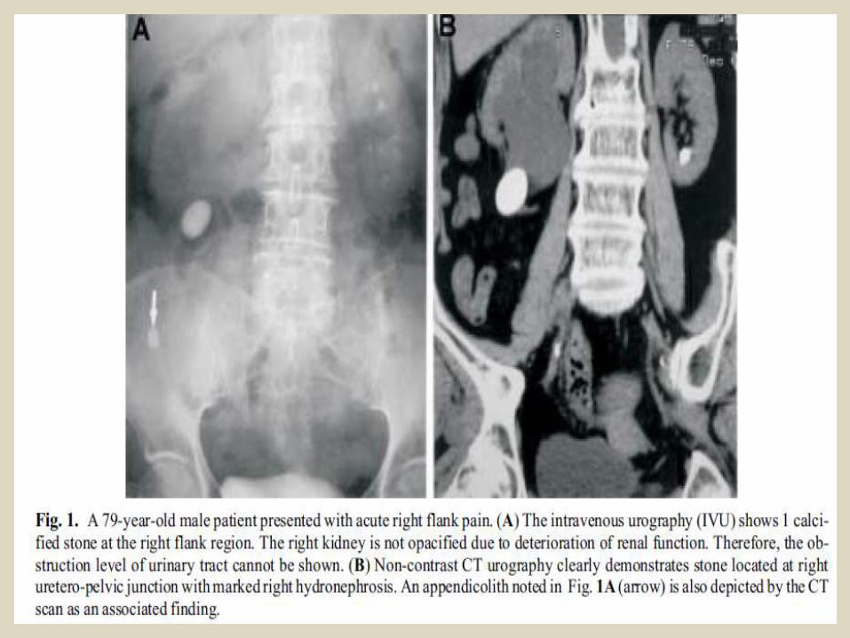
165
Abdominal film reading. Lecture 3. Dr/ ABD ALLAH NAZEER. MD.
| Date post: | 14-Jul-2015 |
| Category: |
Documents |
| Upload: | abdellah-nazeer |
| View: | 1,011 times |
| Download: | 3 times |

Abdominal film reading. Lecture 3.
Dr/ ABD ALLAH NAZEER. MD.

Radiological imaging ofthe urinary system.
PUTIVPUltrasoundCystoscopyCTMRIIsotope Scan

Purpose of the KUB Radiography:To determine the size, shape, and position of the kidneys and bladderTo detect obvious abnormalities of the urinary system, such as kidney stonesTo help differentiate between urologic and gastrointestinal diseases, which both produce abdominal painTo locate a foreign object (e.g., that has been swallowed)To detect air or fluid in the space surrounding the abdominal organs (peritoneal space).
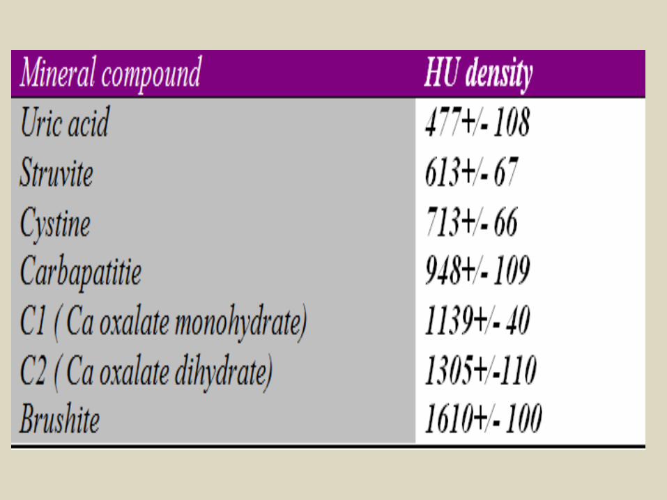
Plain filmCalcium containing stones are radiopaquecalcium oxalate +/- calcium phosphatestruvite (triple phosphate) - usually opaque but variablepure calcium phosphateLucent stones includeuric acidcystineIndinavir stonespure matrix stones.
KUB Radiography:

Upper and mid-ureteric stones.

Multiple renal and urinary bladder stones.
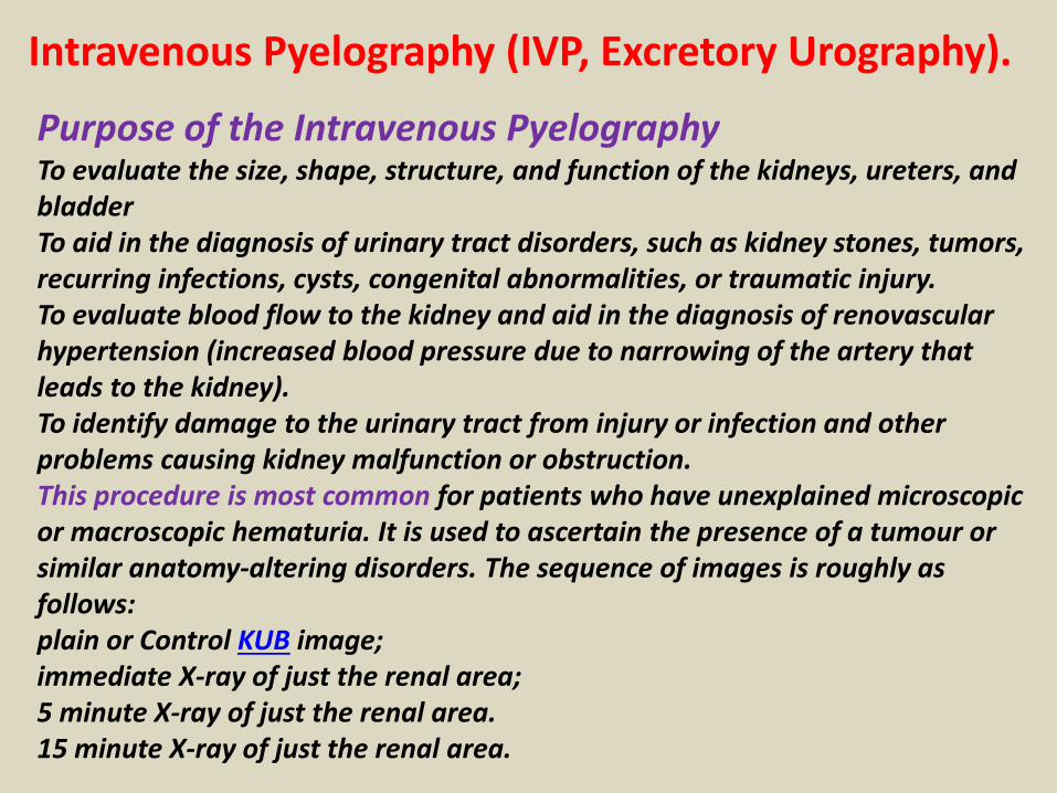
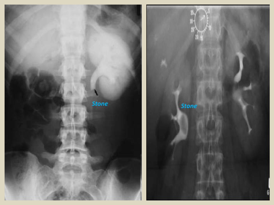
Intravenous Pyelography (IVP, Excretory Urography).
Purpose of the Intravenous PyelographyTo evaluate the size, shape, structure, and function of the kidneys, ureters, and bladderTo aid in the diagnosis of urinary tract disorders, such as kidney stones, tumors, recurring infections, cysts, congenital abnormalities, or traumatic injury.To evaluate blood flow to the kidney and aid in the diagnosis of renovascular hypertension (increased blood pressure due to narrowing of the artery that leads to the kidney).To identify damage to the urinary tract from injury or infection and other problems causing kidney malfunction or obstruction.This procedure is most common for patients who have unexplained microscopic or macroscopic hematuria. It is used to ascertain the presence of a tumour or similar anatomy-altering disorders. The sequence of images is roughly as follows:plain or Control KUB image;immediate X-ray of just the renal area;5 minute X-ray of just the renal area.15 minute X-ray of just the renal area.
Stone Stone
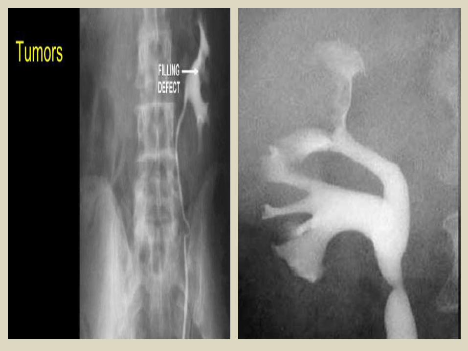
Cyst
Cyst.

UltrasonographyGenerally ultrasonography is an excellent imaging modality as it is noninvasive, reliable and affordable.It can be used to investigate the kidney, bladder, and prostate gland. It can also be combined with voiding, providing an indication of the residual volume. This gives an indirect measure of bladder function.

Stone
Stone.
Stone.

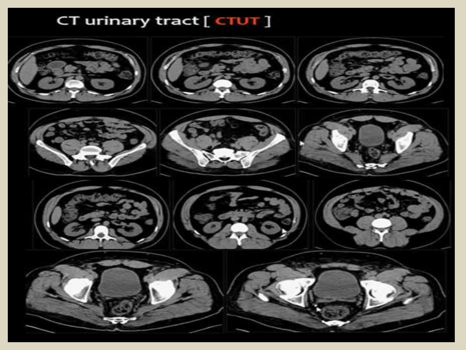
Computed tomography scanning of the urinary tract.
CT Advantage.
Detection of radio-opaque and radio-lucent calculi.Diagnosis of urinary obstruction and its level.Congenital anomalies.Detection and staging of urinary system neoplasm.Differentiation between cystic and solid urinary neoplasm.CT Urography.CT Angiography in renal stenosis and vascular anomalies.


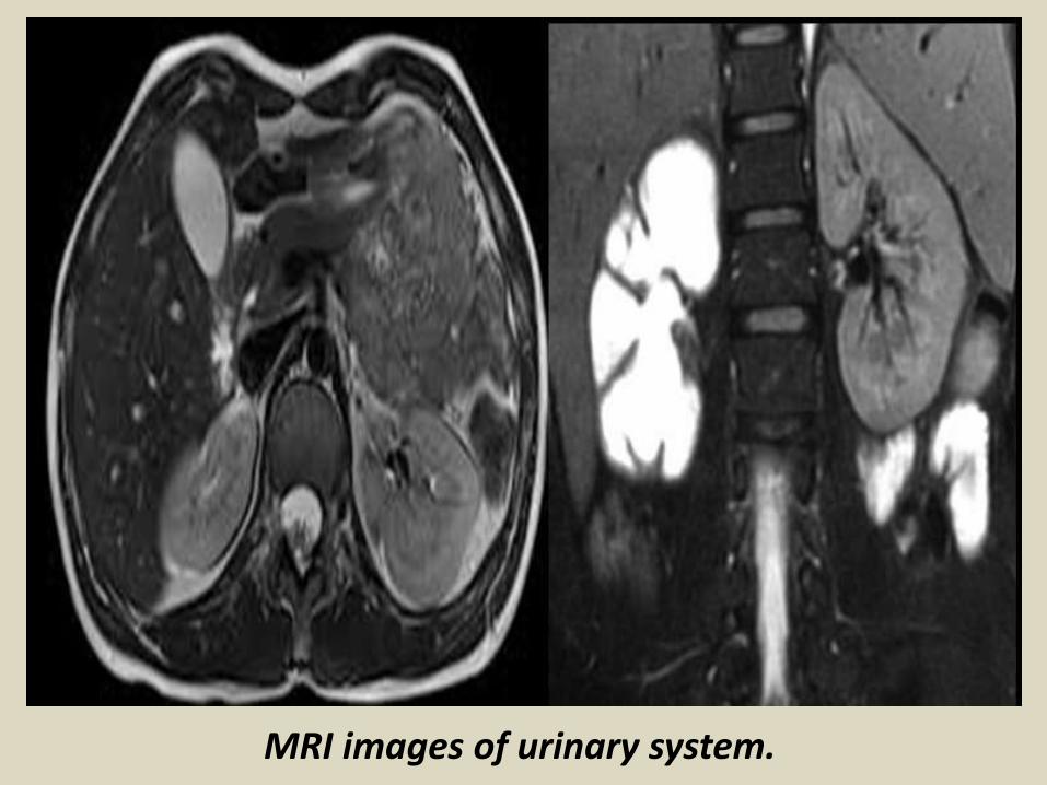
MRI images of urinary system.

MRU images.

Radioisotope renography is a form of kidney imaging involving radioisotopes.
The two most common radiolabelled pharmaceutical agents used are Tc99m-MAG3 (Mercaptoacetyltriglycine) and Tc99m-DTPA (Diethylene Triamine Pentacaetic Acid). Some other radiolabelled pharmaceuticals are EC (Ethyl Cysteine) and 131-Iodine labelled OIH (Ortho Iodo Hippurate). MAG3 is by far a better diagnostic agent than Tc-99m-DTPA, particularly in neonates, patients with impaired function, and patients with suspected obstruction. The MAG3 clearance is highly correlated with the effective renal plasma flow (ERPF), and the MAG3 clearance can be used as an independent measure of renal function. After intravenous administration, about 40-50% of the MAG3 in the blood is extracted by the proximal tubules with each pass through the kidneys; the proximal tubules then secrete the MAG3 into the tubular lumen. DTPA is the second most commonly used renal radiopharmaceutical in the United States, primarily because it is the least expensive. Tc-99m-DTPA is filtered by the glomerulus and may be used to measure the glomerular filtration rate (GFR). The extraction fraction of DTPA is approximately 20%, less than half that of MAG3. However, EC is preferred when the serum Creatinine is high.

Urinary system pathology.Congenital anomalies.Urinary stones.Inflammatory disease.Renal neoplasm(cystic and solid masses).Renal trauma.Miscellaneous lesions.

Congenital anomalies:1.Renal agenesis. Absence of the kidney
usually the left one
2.Renal hypoplasia.
3.Horse shoe kidney. Fused kidneys at their
poles.
4.Ectopic Kidney.5. Aberrant renal artery. 6-Persisteuce of fetal lobulation.7-Congenital polycystic kidney.8-Congenital solitary cyst.9-Double ureters and pelvis.10-Megaureter. Due to absence of ganglion cells11-Congenital stricture of the ureter.

CT Scan of right renal agenesis with non visualizationof the kidney and their blood vessels.
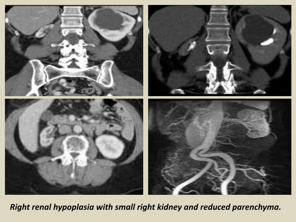
Right renal hypoplasia with small right kidney and reduced parenchyma.

Horse shoe kidneys (IVP and CT) with fusion of the kidney anterior to the spine.

Horse shoe kidneys (IVP and CT) with fusion of the kidney anterior to the spine.
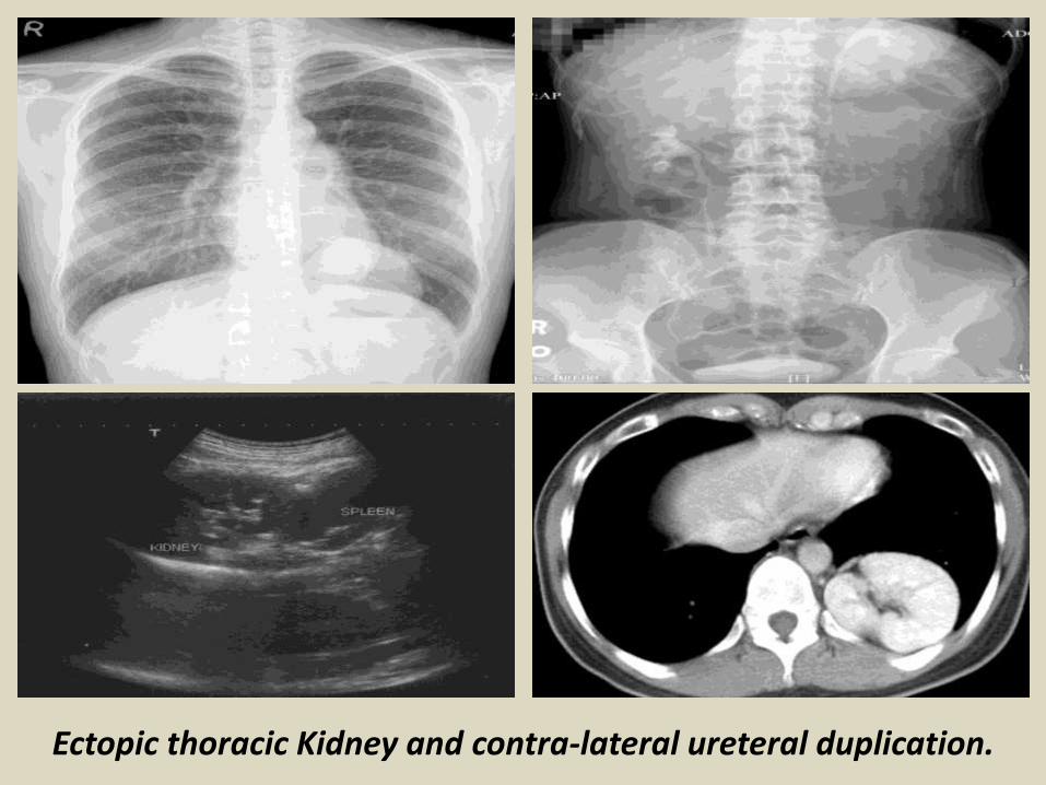
Ectopic thoracic Kidney and contra-lateral ureteral duplication.

Ectopic pelvic kidney.
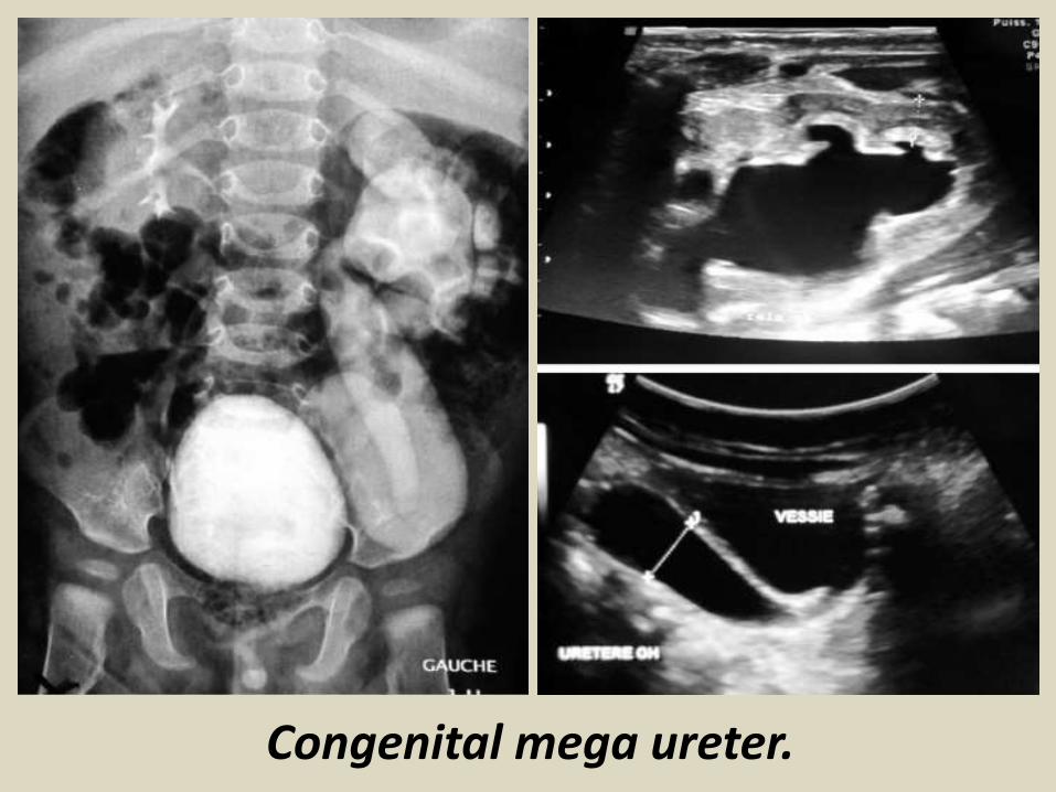
Congenital mega ureter.

Congenital polycystic kidney disease.

Congenital polycystic kidney disease.

Congenital Double left ureter and pelvis.
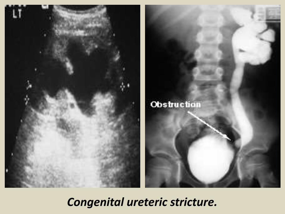
Congenital ureteric stricture.
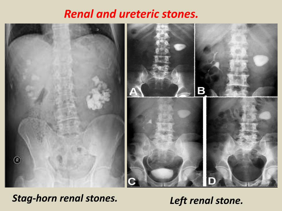
Renal and ureteric stones.
Stag-horn renal stones. Left renal stone.

IVP with ureteric stones.

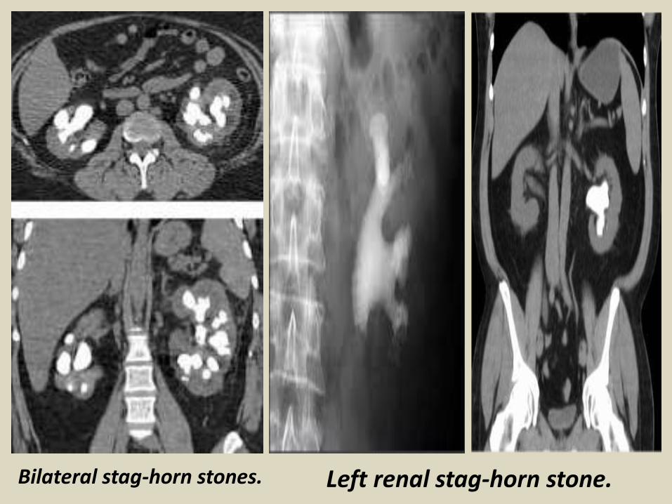
Bilateral stag-horn stones. Left renal stag-horn stone.

MRU images.
Ureteric stricture.
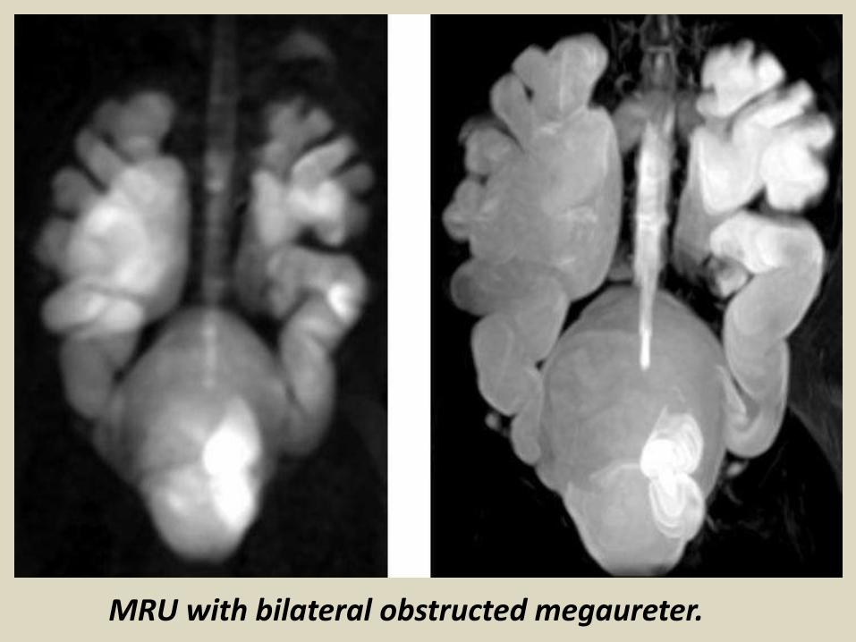
MRU with bilateral obstructed megaureter.

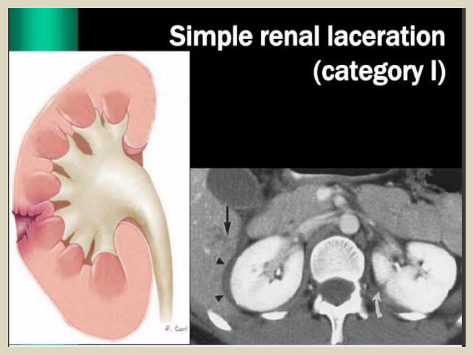
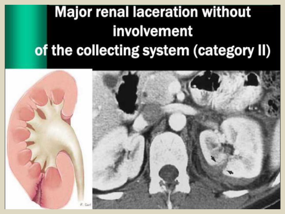
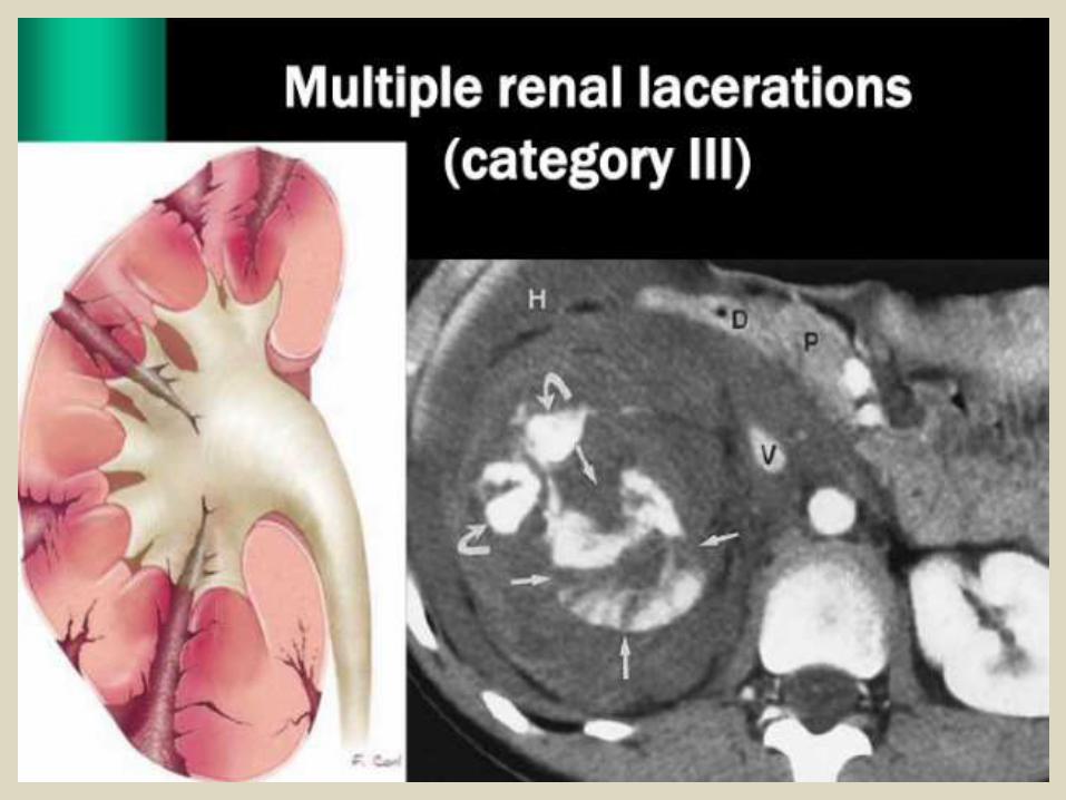
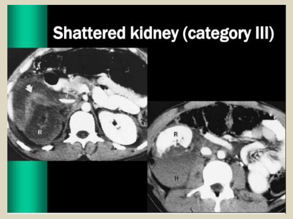
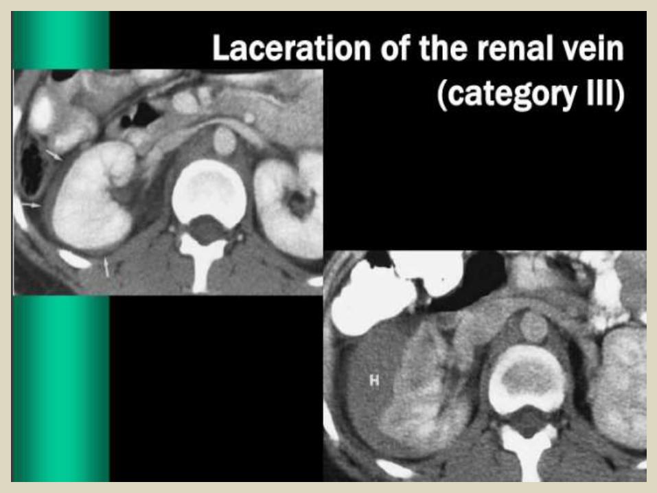
Renal Trauma.


Renal infection.

Acute Bacterial PyelonephritisInfection of collecting system and parenchymaAscending: Escherichia coliHematogenous: Staphylococcus aureusFlank pain and feverUsually a clinical diagnosis Imaging of Acute Pyelonephritis: ModalitiesIntravenous Urography.Ultrasound.Computed Tomography (CT Scan).Nuclear Scanning.

UltrasonographyLower sensitivity than CT or nuclear scansModality of choice in pregnant womenPositive Findings:Enlargement of entire kidneyHypoechoic cortex: edemaFocal hyperechoic areas: early abscess
Acute Pyelonephritis.
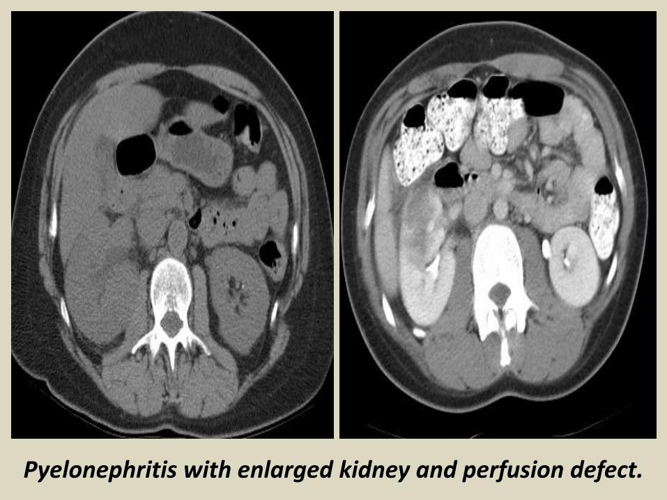
Pyelonephritis with enlarged kidney and perfusion defect.

Acute pyelonephritis with focal perfusion deficit
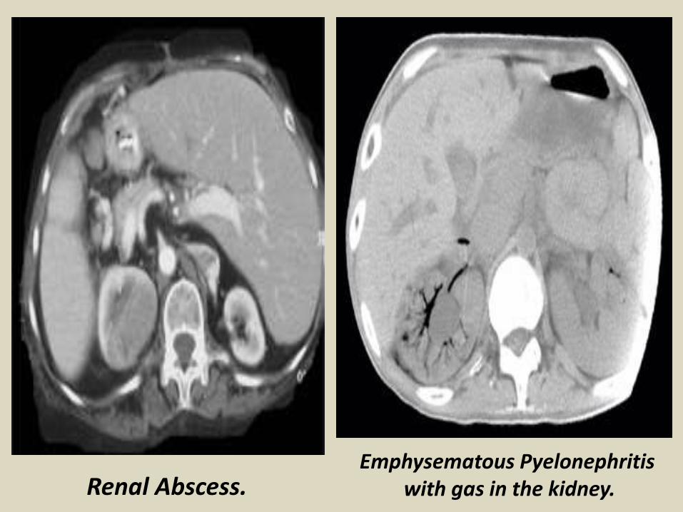
Renal Abscess.Emphysematous Pyelonephritis
with gas in the kidney.
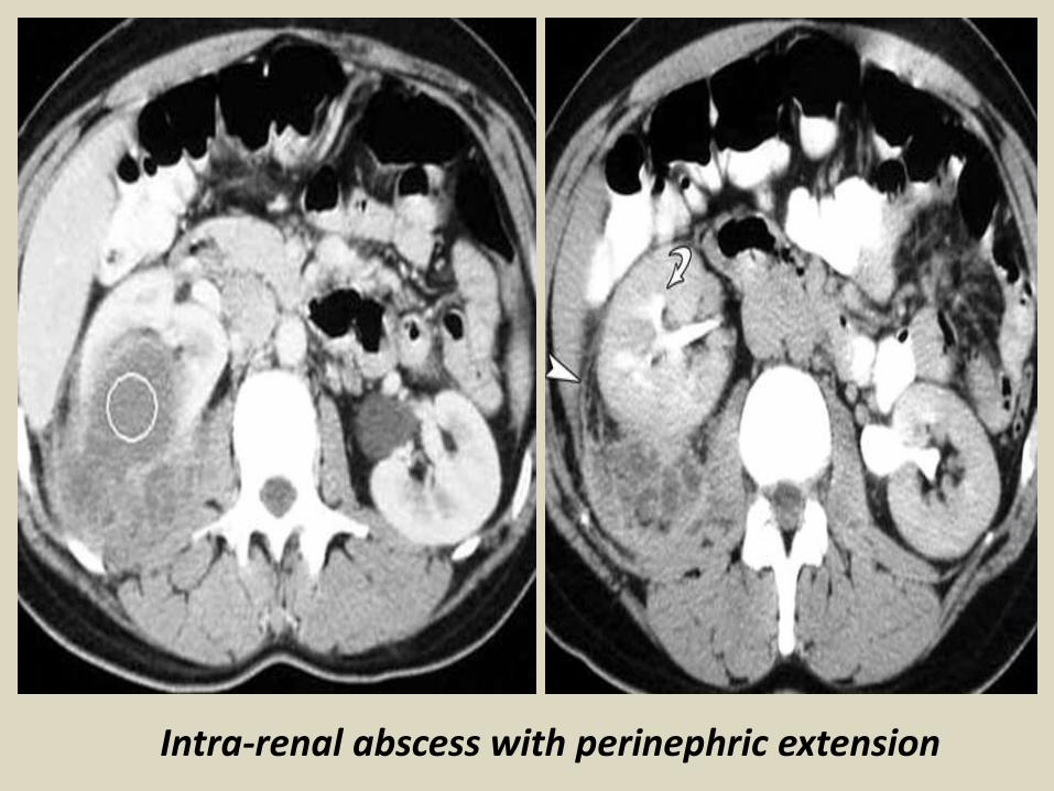
Intra-renal abscess with perinephric extension
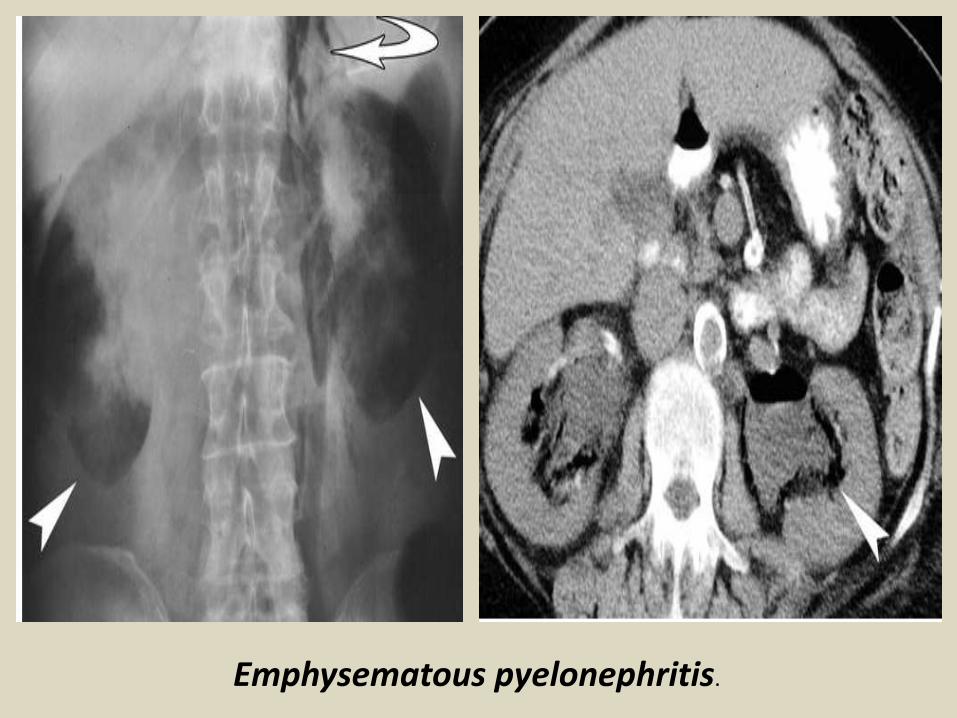
Emphysematous pyelonephritis.
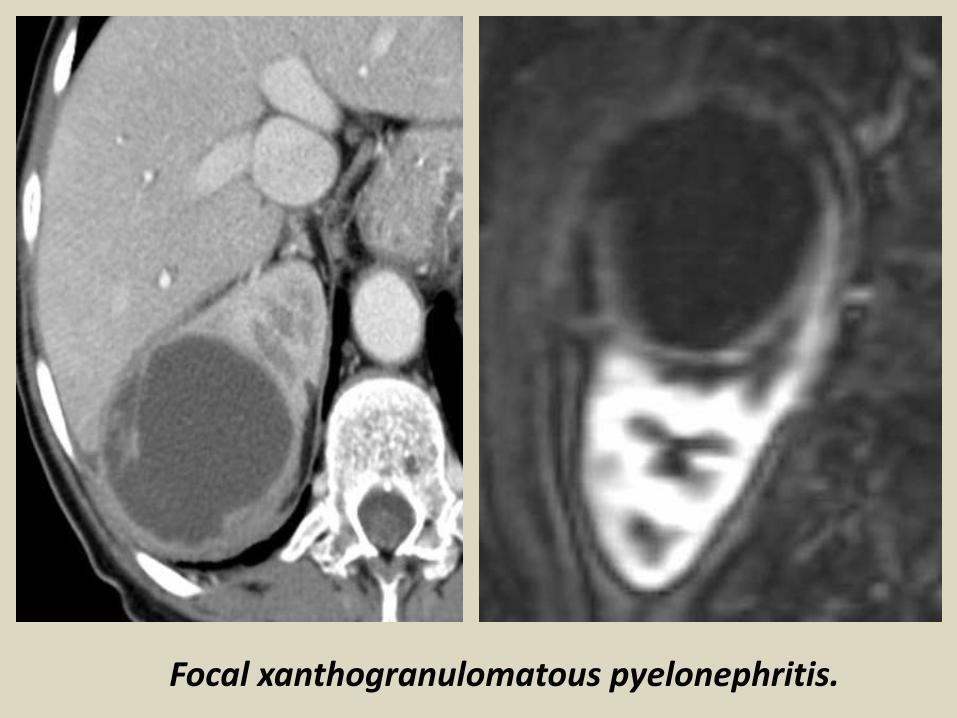
Focal xanthogranulomatous pyelonephritis.

Xanthogranulomatous pyelonephritis.
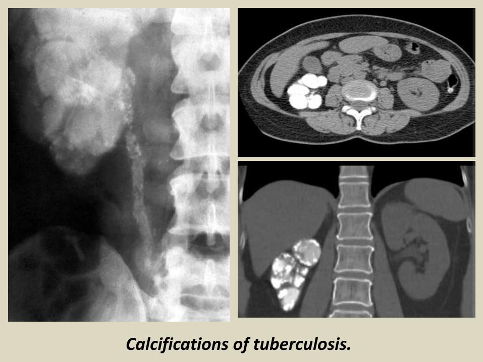
Calcifications of tuberculosis.
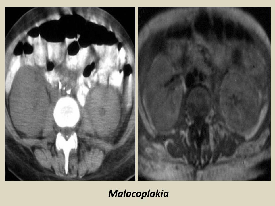
Malacoplakia
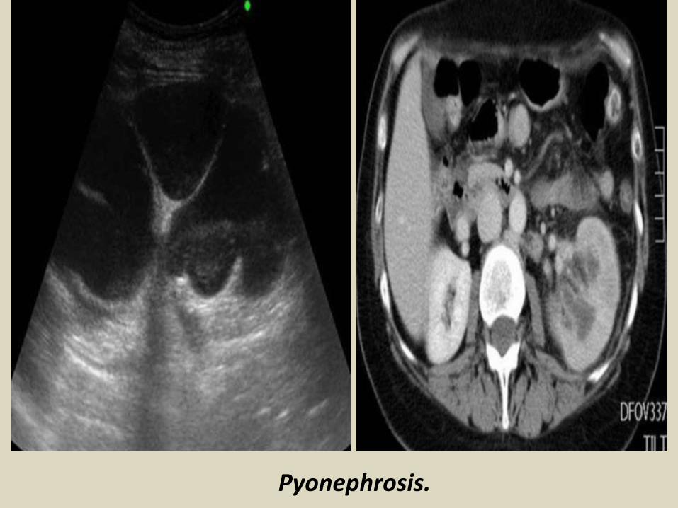
Pyonephrosis.
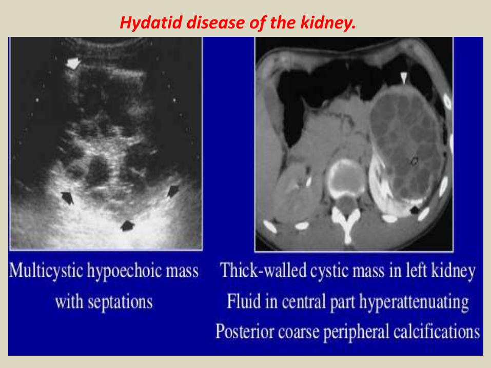
Hydatid disease of the kidney.


Renal cystic and solid masses.Kidney - Cystic masses.
Renal cysts can be classified according to the Bosniak classification depending on their features. Type I cysts are simple cysts.Type II are the minimally complicated cysts. Type I and II can be ignored. Type II F are probably benign, but need to be followed. Type III and IV both are surgical lesions. Type IV is inevitably malignant and in the type III group about 80-90% turn out to be malignant as well.

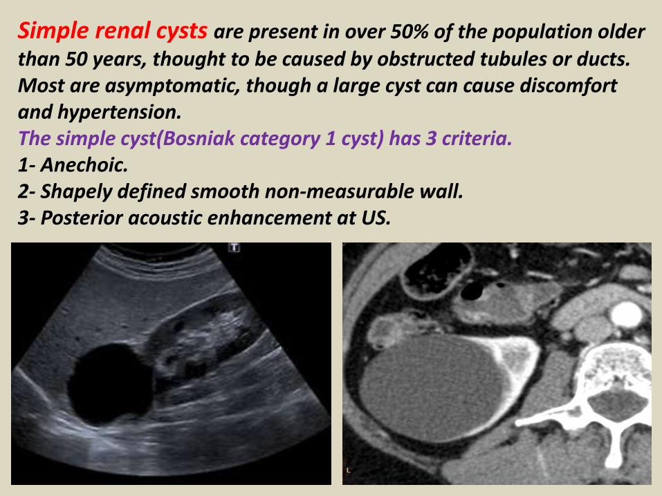
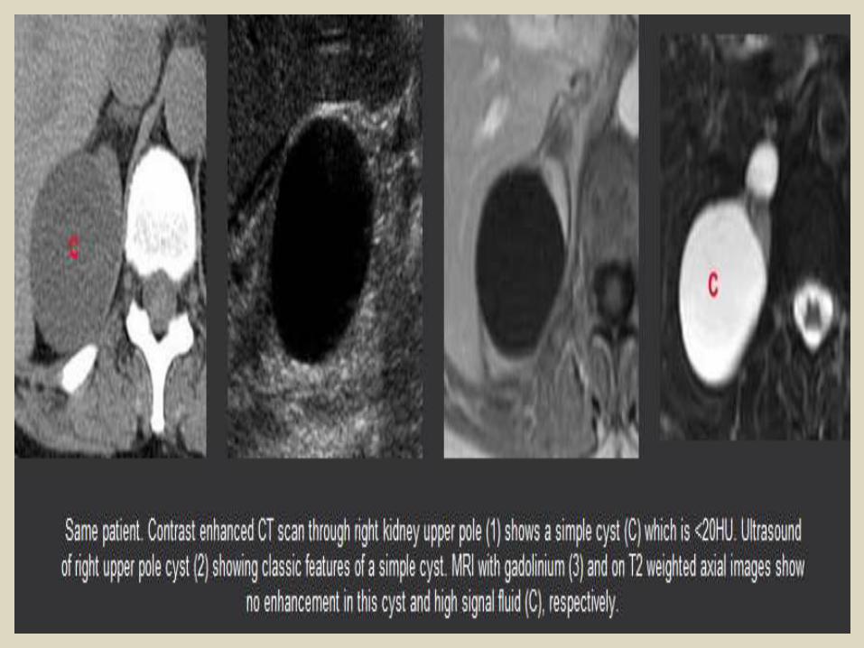
Simple renal cysts are present in over 50% of the population older than 50 years, thought to be caused by obstructed tubules or ducts. Most are asymptomatic, though a large cyst can cause discomfort and hypertension.The simple cyst(Bosniak category 1 cyst) has 3 criteria.1- Anechoic.2- Shapely defined smooth non-measurable wall.3- Posterior acoustic enhancement at US.
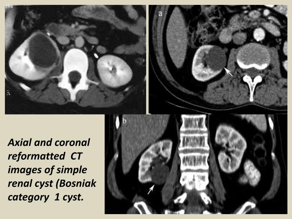
Axial and coronal reformatted CT images of simple renal cyst (Bosniak category 1 cyst.

Bosniak category I. MDCT image, sagittal reconstruction demonstrating the presence of homogeneous cystic lesions with fluid attenuation, without calcifications, septations or enhancement after intravenous contrast injection. Simple cyst. Note the presence of a major lesion (arrow) located in the superior renal pole.
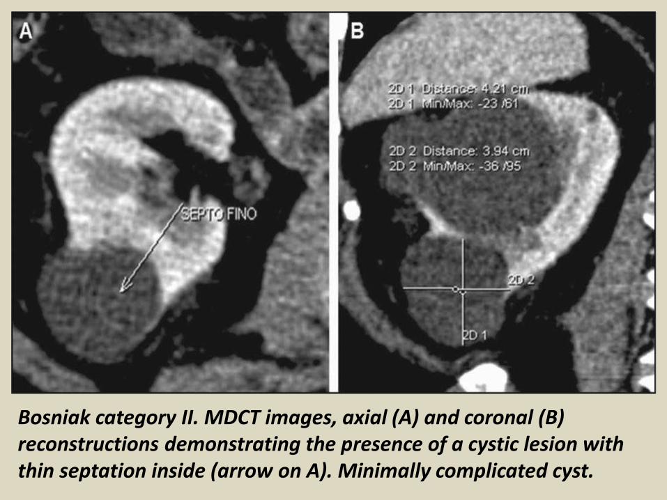
Bosniak category II. MDCT images, axial (A) and coronal (B) reconstructions demonstrating the presence of a cystic lesion with thin septation inside (arrow on A). Minimally complicated cyst.

Bosniak category IIF. MDCT, coronal (A), axial (B) and sagittal (C) reconstructions demonstrating the presence of a right renal cystic lesion with parietal thickening (arrow on B) and a thin septum (arrow on C). Minimally complicated cyst requiring follow-up.

Bosniak category III. MDCT images axial (A), coronal (B) and sagittal (C) reconstructions identifying a cystic lesion in the left kidney, with thin septum and septal calcifications (arrows on B). Indeterminate cyst.

Bosniak category IV. MDCT images, sagittal (A,D) and axial (B,C) reconstructions demonstrating a cystic lesion with gross and nodular parietal thickening.
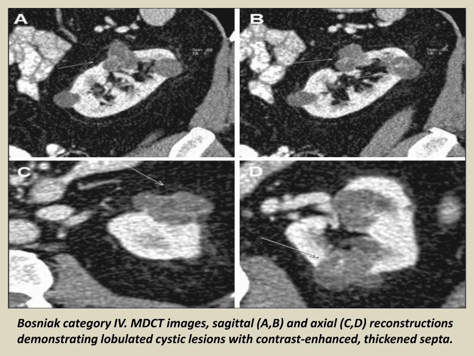
Bosniak category IV. MDCT images, sagittal (A,B) and axial (C,D) reconstructions demonstrating lobulated cystic lesions with contrast-enhanced, thickened septa.
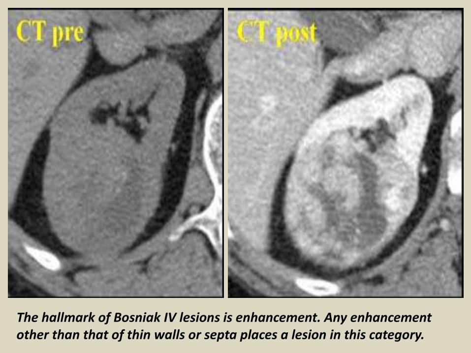
The hallmark of Bosniak IV lesions is enhancement. Any enhancement other than that of thin walls or septa places a lesion in this category.
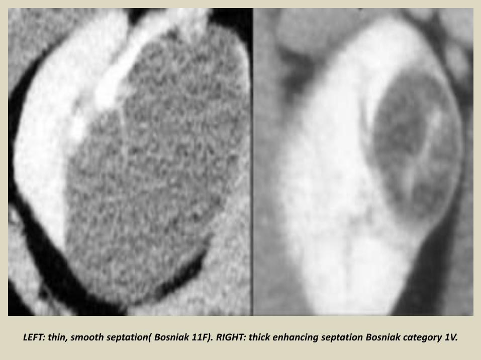
LEFT: thin, smooth septation( Bosniak 11F). RIGHT: thick enhancing septation Bosniak category 1V.
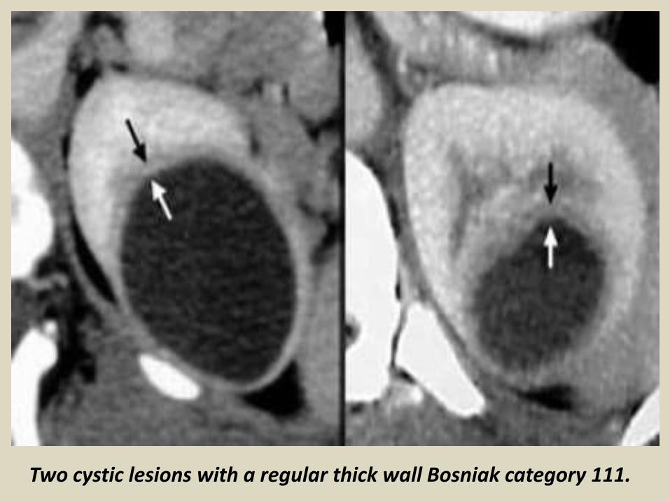
Two cystic lesions with a regular thick wall Bosniak category 111.

Multilocular cystic nephroma (MLCN).Benign hereditary cystic neoplasm.

Multilocular cystic nephroma (MLCN) with predominant cystic mass and internal septation inside.

Multilocular cystic nephroma.

Multiple renal cysts.

Bilateral congenital polycystic kidney.
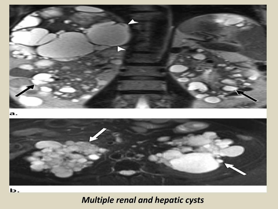
Multiple renal and hepatic cysts
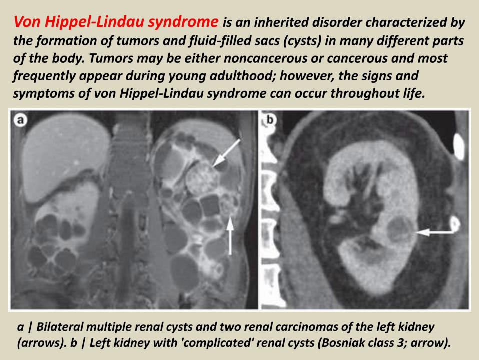
Von Hippel-Lindau syndrome is an inherited disorder characterized by
the formation of tumors and fluid-filled sacs (cysts) in many different parts of the body. Tumors may be either noncancerous or cancerous and most frequently appear during young adulthood; however, the signs and symptoms of von Hippel-Lindau syndrome can occur throughout life.
a | Bilateral multiple renal cysts and two renal carcinomas of the left kidney (arrows). b | Left kidney with 'complicated' renal cysts (Bosniak class 3; arrow).
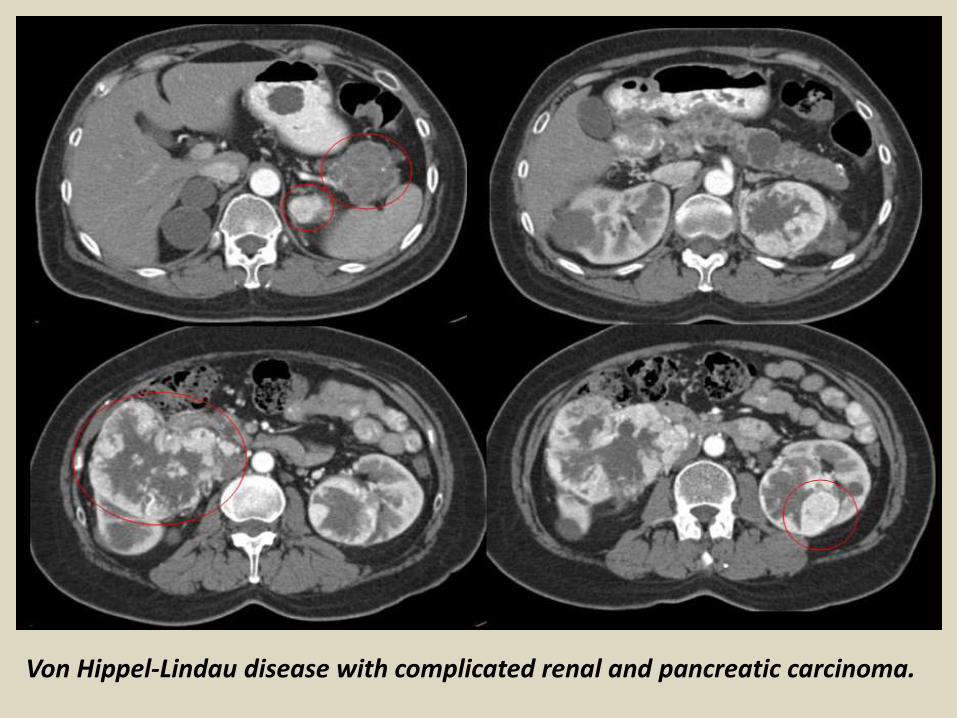
Von Hippel-Lindau disease with complicated renal and pancreatic carcinoma.

Multicystic dysplastic kidney(MCDK).
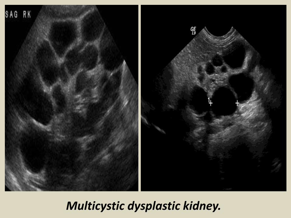
Multicystic dysplastic kidney.

MULTICYSTIC DYSPLASTIC RIGHT KIDNEY.
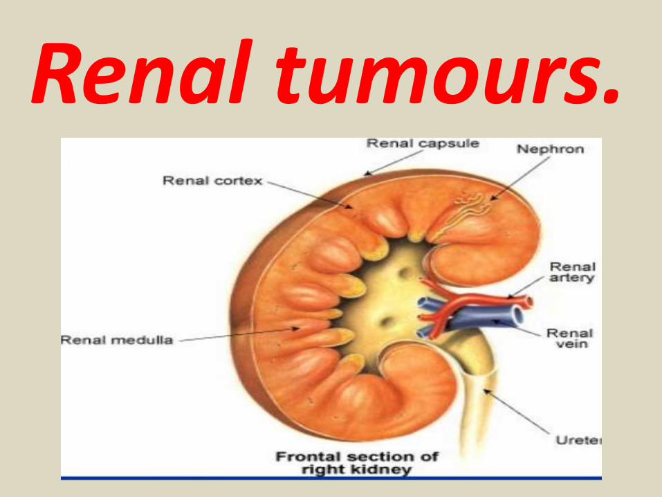
Renal tumours.


Renal oncocytoma is a type of relatively benign renal tumour. The main clinical
importance of this lesion is the difficulty in pre-operatively distinguishing it from renal cell carcinomas, as epidemiology, presentation, imaging and even histology can be very similar. Intravenous pyelogram: Oncocytomas appear as sharply demarcated, often large exophytic masses with enhancement during the nephrographic phase. Ultrasound: Ultrasound demonstrates a well circumscribed mass with echogenicity similar to the rest of the kidney. Occasionally a central scar may be visible. CT: Generally tend be large well demarcated tumours at presentation.Non contrast: if less than 3 cm - homogenous attenuation if more than 3 cm - heterogenous attenuationPost contrast: small tumours may enhance homogeneously, but usually enhancement is heterogenous and the mass is larger central stellate non-enhancing scar is seen in approximately a third of cases MRI: Typical signal characteristics includeT1: hypo-intense compared to renal cortex .T2: hyperintense compared to renal cortex
may demonstrate hypointense central stellate scarT1 C+ (Gd): usually demonstrates homogeneous enhancementAngiography: May demonstrate a spoke wheel pattern, of peripheral circumferential vessels penetrating towards the center of the lesion, which again, although characteristic
of oncocytomas, may also be seen in renal cell carcinomas.

Oncocytoma of right kidney. Triphasic study.
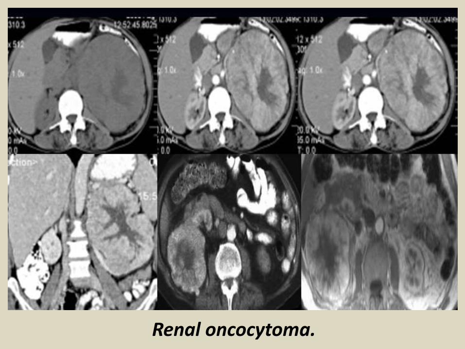
Renal oncocytoma.

Oncocytoma.
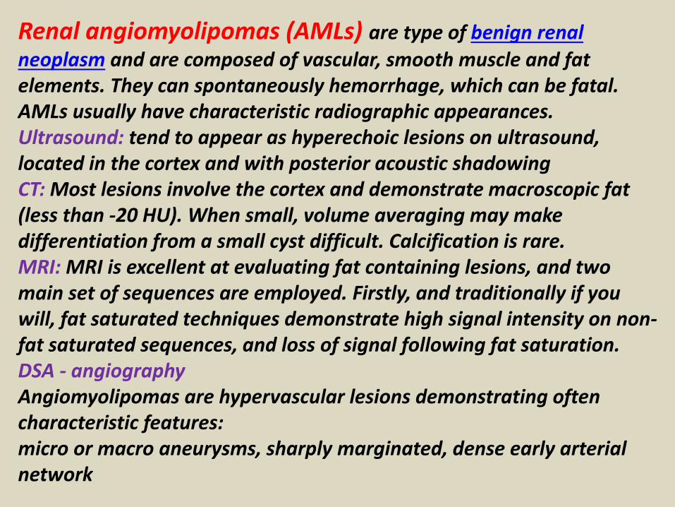
Renal angiomyolipomas (AMLs) are type of benign renal
neoplasm and are composed of vascular, smooth muscle and fat elements. They can spontaneously hemorrhage, which can be fatal. AMLs usually have characteristic radiographic appearances. Ultrasound: tend to appear as hyperechoic lesions on ultrasound, located in the cortex and with posterior acoustic shadowingCT: Most lesions involve the cortex and demonstrate macroscopic fat (less than -20 HU). When small, volume averaging may make differentiation from a small cyst difficult. Calcification is rare.MRI: MRI is excellent at evaluating fat containing lesions, and two main set of sequences are employed. Firstly, and traditionally if you will, fat saturated techniques demonstrate high signal intensity on non-fat saturated sequences, and loss of signal following fat saturation.DSA - angiographyAngiomyolipomas are hypervascular lesions demonstrating often characteristic features:micro or macro aneurysms, sharply marginated, dense early arterial network
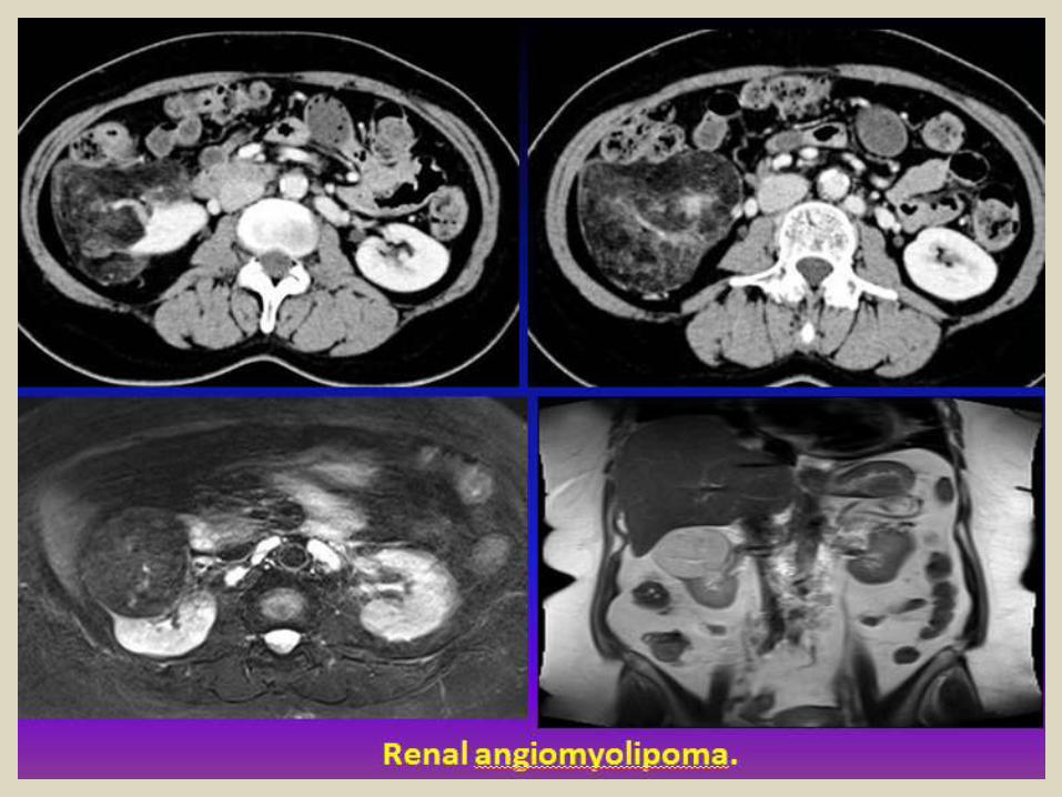
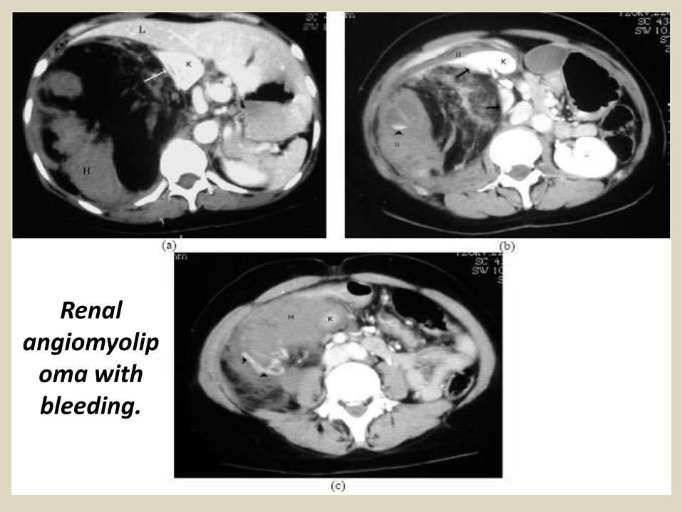
Renal angiomyolip
oma with bleeding.

Renal cell carcinomaRenal cell carcinoma (RCC), also known as renal cell cancer or renal celladenocarcinoma, is by far the most common type of kidney cancer. About 9 out of 10kidney cancers are renal cell carcinomas.Although RCC usually grows as a single tumor within a kidney, sometimes there are 2 or more tumors in one kidney or even tumors in both kidneys at the same time.There are several subtypes of RCC, based mainly on how the cancer cells look under a microscope. Knowing the subtype of RCC can be a factor in deciding treatment and can also help your doctor determine if your cancer might be due to an inherited genetic syndrome. Clear cell renal cell carcinomaThis is the most common form of renal cell carcinoma. About 7 out of 10 people with RCC have this kind of cancer. When seen under a microscope, the cells that make up clear cell RCC look very pale or clear.Papillary renal cell carcinoma: This is the second most common subtype – about 1 in 10 RCCs are of this type. These cancers form little finger-like projections (called papillae) in some, if not most, of the tumor. Some doctors call these cancers chromophilic because the cells take in certain dyes and look pink under the microscope.Chromophobe renal cell carcinoma: This subtype accounts for about 5% (5 cases in 100) of RCCs. The cells of these cancers are also pale, like the clear cells, but are much larger and have certain other features that can be recognized.


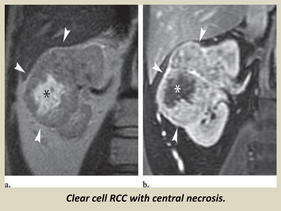
Clear cell RCC with central necrosis.
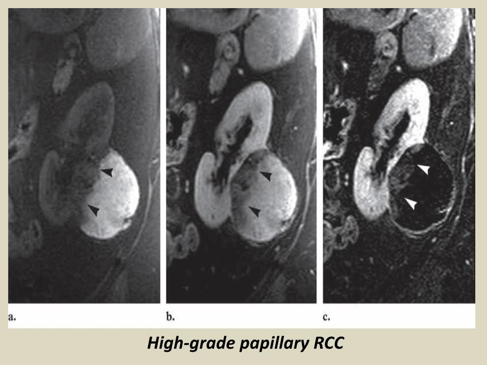
High-grade papillary RCC

Renal cell carcinoma with CT angiography.
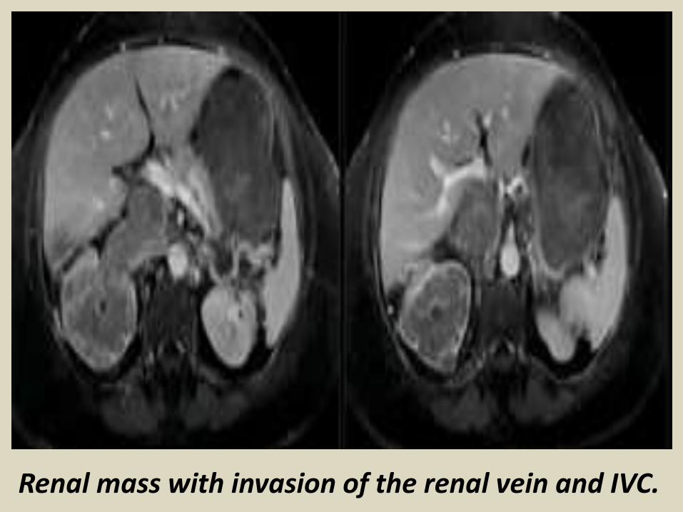
Renal mass with invasion of the renal vein and IVC.

Transitional cell carcinomas don’t start in the kidney itself,
but in the lining of the renal pelvis (where the urine goes before it enters the ureter). This lining is made up of cells called transitional cells that look like the cells that line the ureters and bladder. Cancers that develop from these cells look like other urothelial carcinomas, such as bladder cancer, under the microscope. Like bladder cancer, these cancers are often linked to cigarette smoking and being exposed to certain cancer-causing chemicals in the workplace.People with TCC often have the same signs and symptoms as people with renal cell cancer − blood in the urine and, sometimes, back pain.These cancers are usually treated by surgically removing the whole kidney and the ureter, as well as the portion of the bladder where the ureter attaches. Smaller, less aggressive cancers can sometimes be treated with less surgery. Chemotherapy (chemo) is sometimes given before or after surgery, depending on how much cancer is found. The chemo given is the same as that used for bladder cancer. It’s important to talk with your doctor to be aware of your options and the benefits and risks of each treatment.
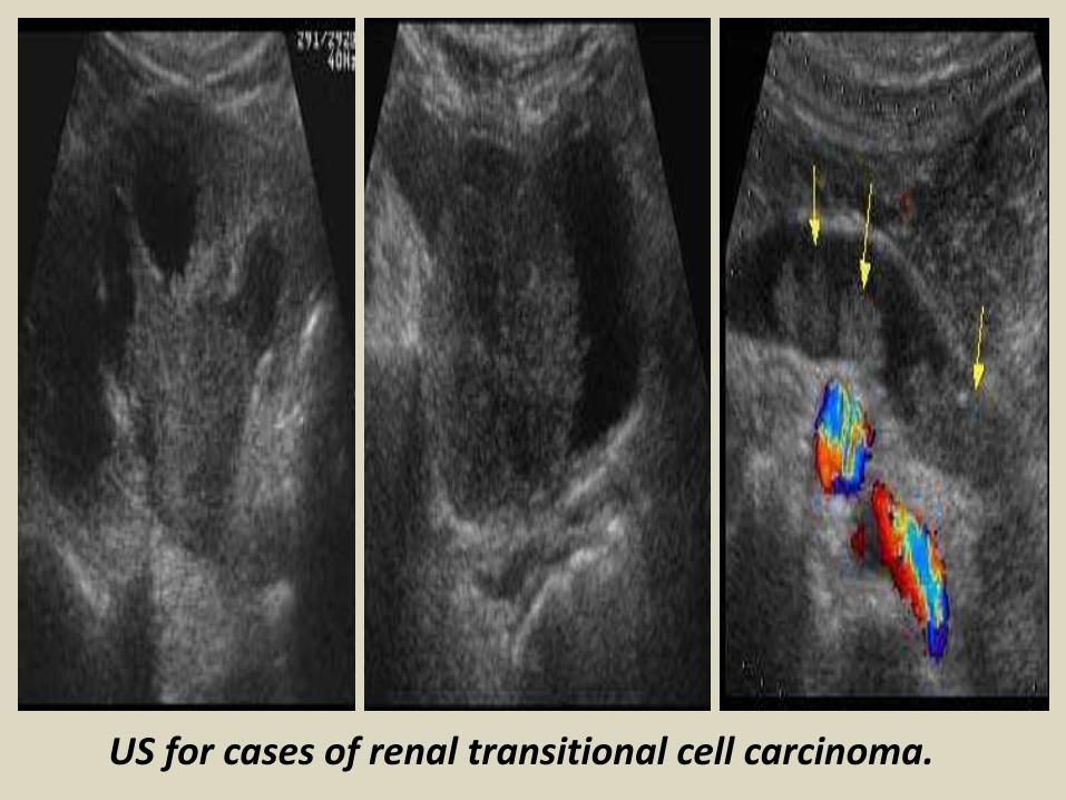
US for cases of renal transitional cell carcinoma.

Renal transitional cell carcinoma.

TCC at the renal pelvis and distal ureter.
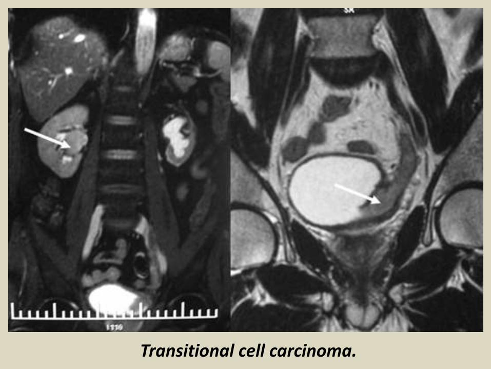
Transitional cell carcinoma.

Wilms' tumor (nephroblastoma) is a rare type of kidney cancer.
It causes a tumor on one or both kidneys. It usually affects children, but can happen in adults. Having certain genetic conditions or birth defects can increase the risk of getting it. Children that are at risk should be screened for Wilms' tumor every three months until they turn eight.Symptoms include a lump in the abdomen, blood in the urine, and a fever for no reason. Tests that examine the kidney and blood are used to find the tumor. Wilms' tumor is a malignant tumor containing metanephric blastema, stromal and epithelial derivatives. Characteristic is the presence of abortive tubules and glomeruli surrounded by a spindled cell stroma. The stroma may include striated muscle, cartilage, bone, fat tissue, fibrous tissue. The tumor is compressing the normal kidney parenchyma.The mesenchymal component may include cells showing rhabdomyoid differentiation. The rhabdomyoid component may itself show features of malignancy (rhabdomyosarcomatous Wilms).
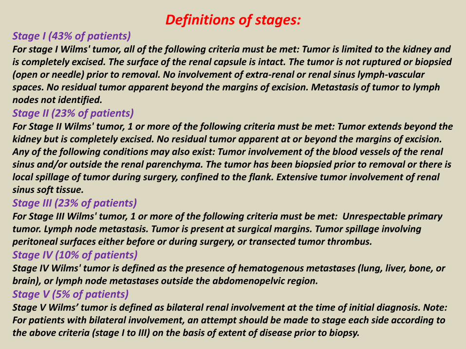
Definitions of stages:Stage I (43% of patients)For stage I Wilms' tumor, all of the following criteria must be met: Tumor is limited to the kidney and is completely excised. The surface of the renal capsule is intact. The tumor is not ruptured or biopsied (open or needle) prior to removal. No involvement of extra-renal or renal sinus lymph-vascular spaces. No residual tumor apparent beyond the margins of excision. Metastasis of tumor to lymph nodes not identified.
Stage II (23% of patients)For Stage II Wilms' tumor, 1 or more of the following criteria must be met: Tumor extends beyond the kidney but is completely excised. No residual tumor apparent at or beyond the margins of excision. Any of the following conditions may also exist: Tumor involvement of the blood vessels of the renal sinus and/or outside the renal parenchyma. The tumor has been biopsied prior to removal or there is local spillage of tumor during surgery, confined to the flank. Extensive tumor involvement of renal sinus soft tissue.
Stage III (23% of patients)For Stage III Wilms' tumor, 1 or more of the following criteria must be met: Unrespectable primary tumor. Lymph node metastasis. Tumor is present at surgical margins. Tumor spillage involving peritoneal surfaces either before or during surgery, or transected tumor thrombus.
Stage IV (10% of patients)Stage IV Wilms' tumor is defined as the presence of hematogenous metastases (lung, liver, bone, or brain), or lymph node metastases outside the abdomenopelvic region.
Stage V (5% of patients)Stage V Wilms’ tumor is defined as bilateral renal involvement at the time of initial diagnosis. Note: For patients with bilateral involvement, an attempt should be made to stage each side according to the above criteria (stage I to III) on the basis of extent of disease prior to biopsy.


Computed tomography showing the extent of a large, left-sided Wilms tumor in a 2-year-old girl with thrombus extending the full length of the inferior vena cava (IVC) and into the right atrium (between arrows).

Axial computed tomography scan showing bilateral Wilms' tumour with fullness of both flanks and the right renal mass is seen crossing the midline. Heterogenous contrast enhancement is noted. The bowel loops are displaced medially and sandwitched the two renal masses

Renal Lymphoma.
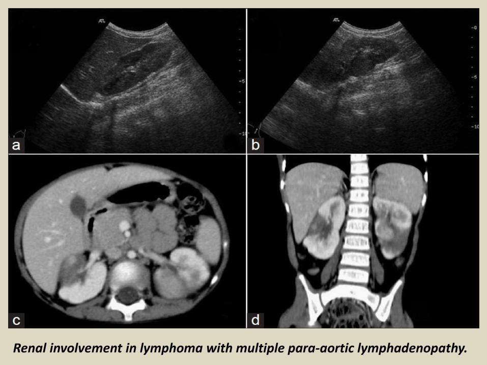
Renal involvement in lymphoma with multiple para-aortic lymphadenopathy.
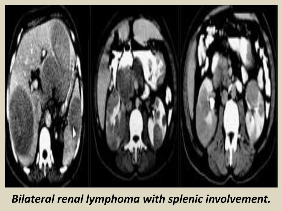
Bilateral renal lymphoma with splenic involvement.

Bilateral renal lymphoma.
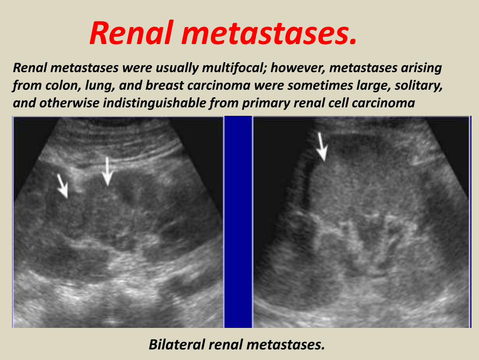
Renal metastases.Renal metastases were usually multifocal; however, metastases arising from colon, lung, and breast carcinoma were sometimes large, solitary, and otherwise indistinguishable from primary renal cell carcinoma
Bilateral renal metastases.
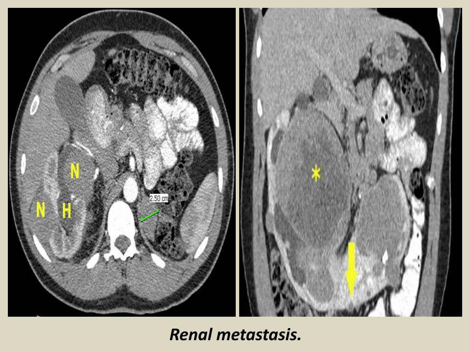
Renal metastasis.

The Urinary bladder.
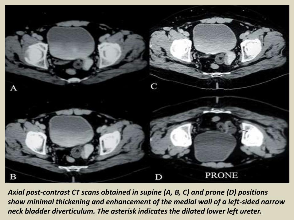
Axial post-contrast CT scans obtained in supine (A, B, C) and prone (D) positions show minimal thickening and enhancement of the medial wall of a left-sided narrow neck bladder diverticulum. The asterisk indicates the dilated lower left ureter.
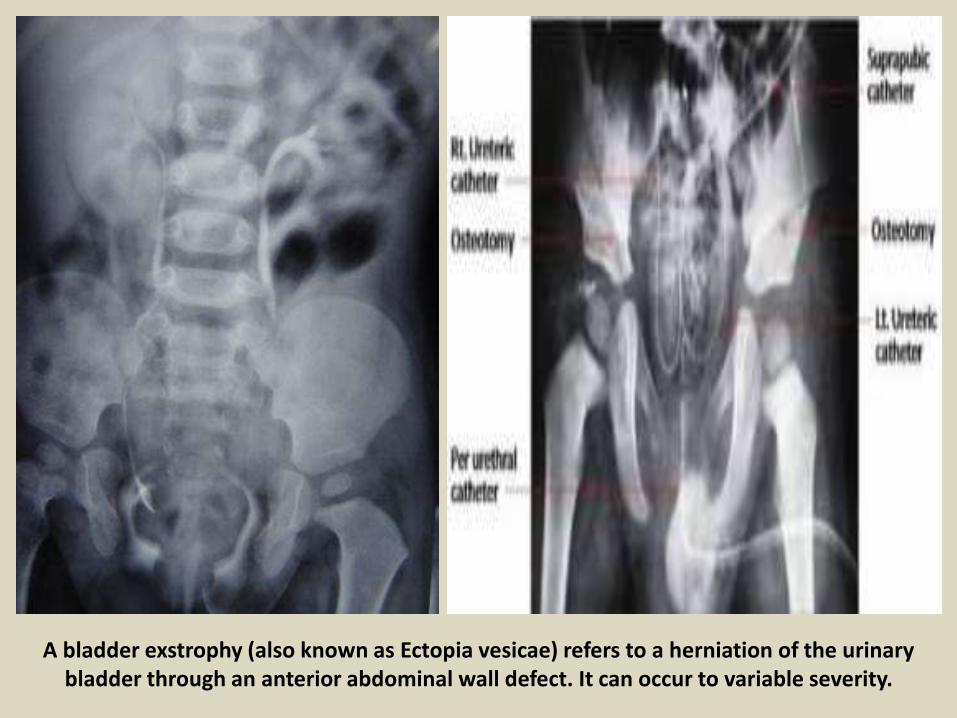
A bladder exstrophy (also known as Ectopia vesicae) refers to a herniation of the urinary bladder through an anterior abdominal wall defect. It can occur to variable severity.

Vesicoureteral Reflux. Primary: (Congenital) defect of UVJ (ureterovesical junction) – Most common –deficient tunnel / laterally displaced orificesSecondary (Acquired) increased intravesical pressure secondary to neurogenic problems or DES, bladder instability, bladder outlet obstruction (PUVs)UTIs (problem #1) do not cause reflux!!Reflux (problem #2) does not cause UTIs!!


Grading of VUR from 1-5.
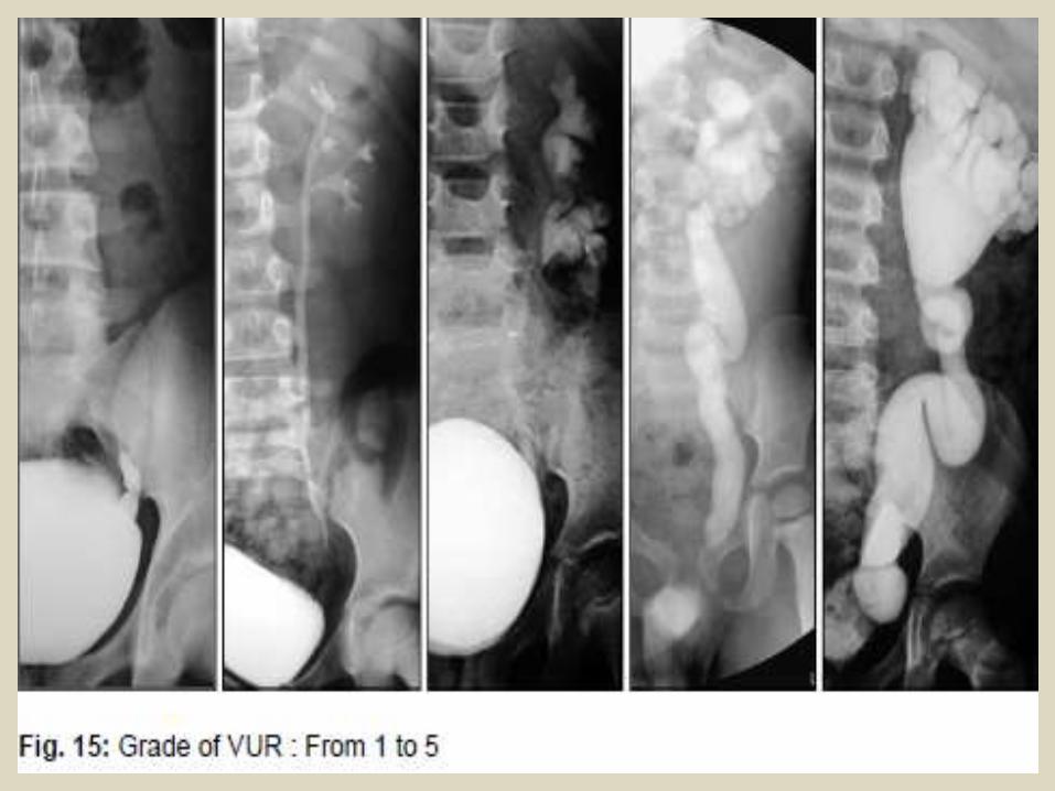

Grade 5 VUR.

Complete duplication of the urinary bladder and urethra.

MRI: A. Coronal, B. Sagittal T2-weighted view of the latero-lateral incomplete bladder duplication.

Fistula.Vesico-vaginal fistula (VVF) is a subtype of female urogenital fistula (UGF). VVF is an abnormal fistulous tract extending between the bladder and the vagina that allows the continuous involuntary discharge of urine into the vaginal vault.
Colovesical fistula (abnormal connection between colon and bladder) may develop in men or women with inflammatory bowel disease or diverticulitis and can result in passage of gas or stool in the urine, frequent UTI's and even sepsis (severe infection that enters the bloodstream).

Vesico-vaginal fistula.

Bladder stones.
Bladder stones are hard buildups of minerals that form in the urinary bladder.CausesBladder stones are usually caused by another urinary system problem, such as:Bladder diverticulumEnlarged prostateNeurogenic bladderUrinary tract infectionAlmost all bladder stones occur in men. Bladder stones are much less common than kidney stones.Bladder stones may occur when urine in the bladder is concentrated and materials crystallize. Bladder stones may also result from foreign objects in the bladder.

Urinary bladder stone.
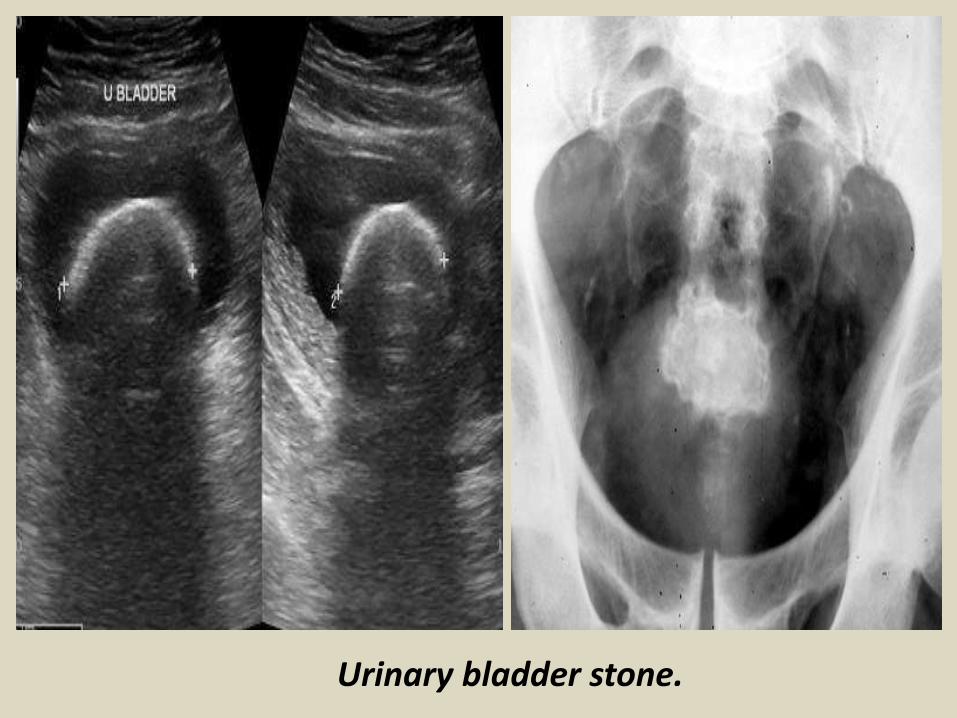
Urinary bladder stone.

Urinary tract infection.A urinary tract infection (UTI) (also known as acute cystitis or bladder infection) is an infection that affects part of the urinary tract. When it affects the lower urinary tract it is known as a simple cystitis (a bladder infection).Urinary tract infections occur more commonly in women
than men, with half of women having at least one infection at some point in their lives. Recurrences are common. Risk factors include female anatomy, sexual intercourse and family history. The main causal agent of both types is Escherichia coli, though other bacteria, viruses or fungi may rarely be the cause.

Acute cystitis.
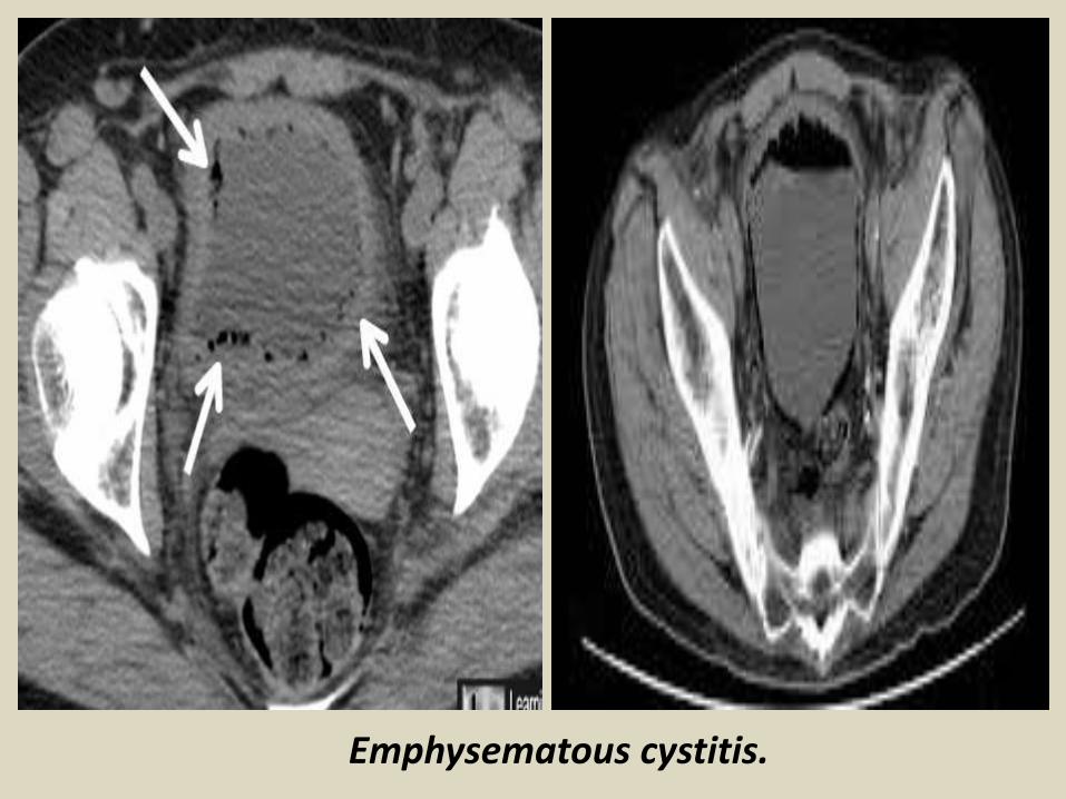
Emphysematous cystitis.

Acute cystitis.

Neoplasm of the urinary bladder.
Benign Tumors of the Bladder. There are numerous benign tumors of the bladder, but the more common ones include epithelial metaplasia, leukoplakia, inverted papilloma, nephrogenic adenoma, leiomyoma, cystitis cystica, cystitis glandularis and hemangioma .
Malignant tumour of the urinary bladder.Transitional cell carcinoma: Cancer that begins in cells in the innermost tissuelayer of the bladder. Most bladder cancers begin in the transitional cells. Transitional cell carcinoma can be low-grade or high-grade:
Low-grade transitional cell carcinoma often recurs (comes back) after treatment, but rarely spreads into the muscle layer of the bladder or to other parts of the body. High-grade transitional cell carcinoma often recurs (comes back) after treatment and often spreads into the muscle layer of the bladder, to other parts of the body, and to lymph nodes. Almost all deaths from bladder cancer are due to high-grade disease.
Squamous cell carcinoma: Cancer that begins in squamous cells, which are thin, flat cells that may form in the bladder after long-term infection or irritation. Adenocarcinoma: Cancer that begins in glandular (secretory) cells that are found in the lining of the bladder. This is a very rare type of bladder cancer.

Bladder cancer is any of several types of malignancy arising from the epithelial
lining (i.e., the urothelium) of the urinary bladder. Rarely the bladder is involved by non-epithelial cancers, such as lymphoma or sarcoma
Staging:T (Primary tumour)TX Primary tumour cannot be assessedT0 No evidence of primary tumourTa Non-invasive papillary carcinomaTis Carcinoma in situ (‘flat tumour’)T1 Tumour invades subepithelial connective tissueT2a Tumour invades superficial muscle (inner half)T2b Tumour invades deep muscle (outer half)T3 Tumour invades perivesical tissue:T3a MicroscopicallyT3b Macroscopically (extravesical mass)T4a Tumour invades prostate, uterus or vaginaT4b Tumour invades pelvic wall or abdominal wall
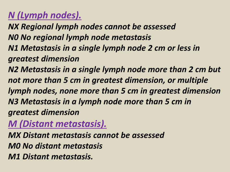
N (Lymph nodes).NX Regional lymph nodes cannot be assessedN0 No regional lymph node metastasisN1 Metastasis in a single lymph node 2 cm or less in greatest dimensionN2 Metastasis in a single lymph node more than 2 cm but not more than 5 cm in greatest dimension, or multiple lymph nodes, none more than 5 cm in greatest dimensionN3 Metastasis in a lymph node more than 5 cm in greatest dimension
M (Distant metastasis).MX Distant metastasis cannot be assessedM0 No distant metastasisM1 Distant metastasis.

Diagram shows the stages of tumor invasion in bladder cancer.
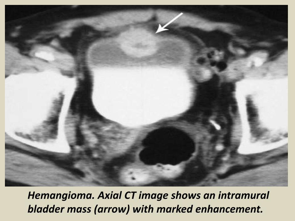
Hemangioma. Axial CT image shows an intramural bladder mass (arrow) with marked enhancement.
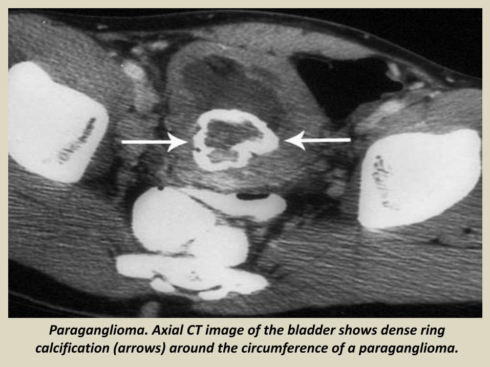
Paraganglioma. Axial CT image of the bladder shows dense ring calcification (arrows) around the circumference of a paraganglioma.
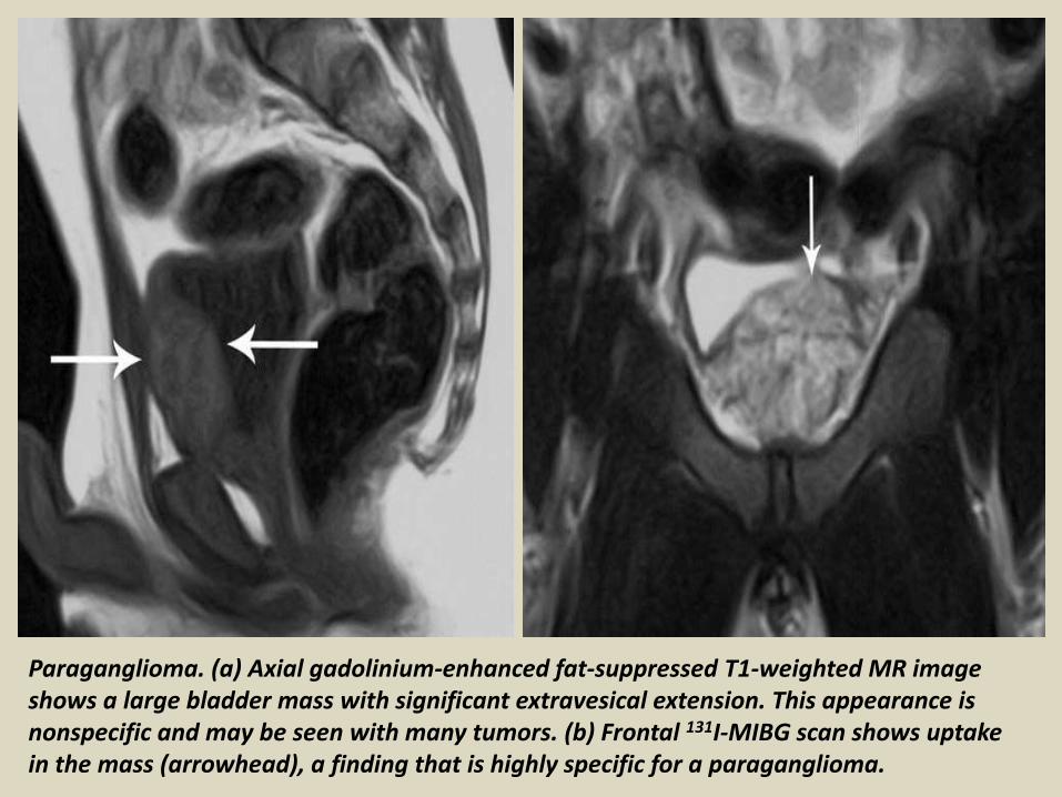
Paraganglioma. (a) Axial gadolinium-enhanced fat-suppressed T1-weighted MR image shows a large bladder mass with significant extravesical extension. This appearance is nonspecific and may be seen with many tumors. (b) Frontal 131I-MIBG scan shows uptake in the mass (arrowhead), a finding that is highly specific for a paraganglioma.

Leiomyoma. Sagittal T1-weighted (a) and T2-weighted (b) MR images of the bladder show a smooth, low-signal-intensity, intramural mass (arrows), an appearance typical of a leiomyoma.
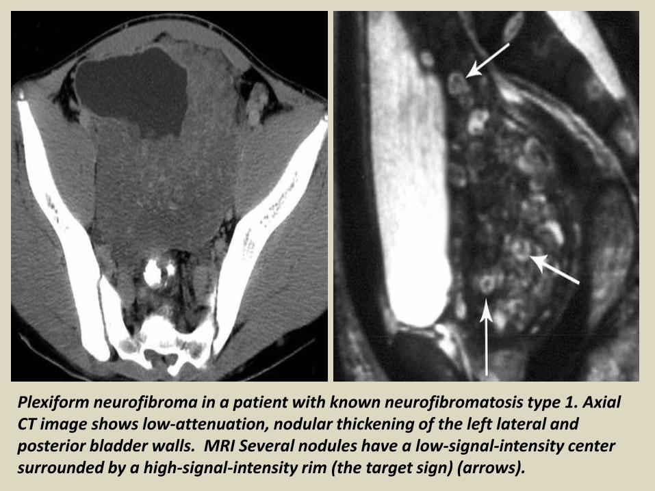
Plexiform neurofibroma in a patient with known neurofibromatosis type 1. Axial CT image shows low-attenuation, nodular thickening of the left lateral and posterior bladder walls. MRI Several nodules have a low-signal-intensity center surrounded by a high-signal-intensity rim (the target sign) (arrows).
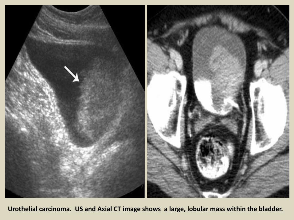
Urothelial carcinoma. US and Axial CT image shows a large, lobular mass within the bladder.

Noninvasive papillary urothelial tumor at MRI with intact bladder wall.

Invasive urothelial carcinoma. Axial gadolinium-enhanced fat-suppressed T1-weighted MR image of the bladder shows tumor invasion into the perivesical fat (arrows).
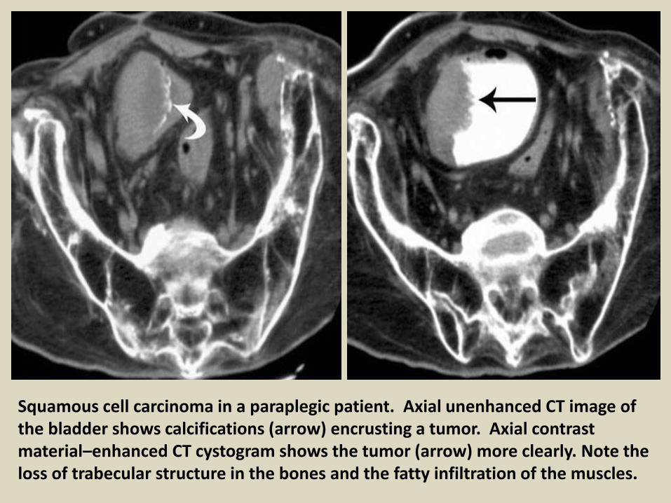
Squamous cell carcinoma in a paraplegic patient. Axial unenhanced CT image of the bladder shows calcifications (arrow) encrusting a tumor. Axial contrast material–enhanced CT cystogram shows the tumor (arrow) more clearly. Note the loss of trabecular structure in the bones and the fatty infiltration of the muscles.

Squamous cell carcinoma.

Invasive squamous cell carcinoma
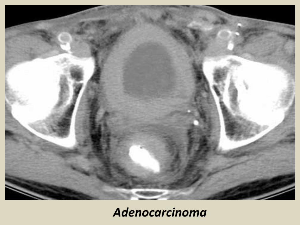
Adenocarcinoma

Small cell carcinoma.
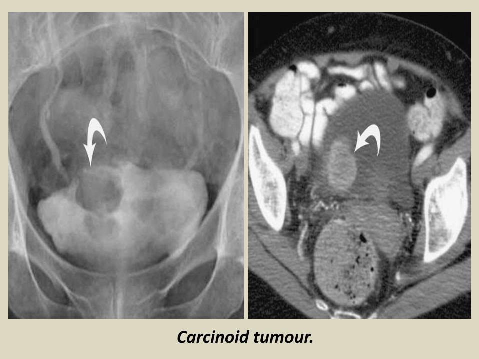
Carcinoid tumour.
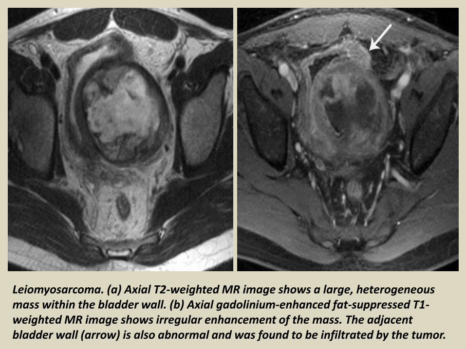
Leiomyosarcoma. (a) Axial T2-weighted MR image shows a large, heterogeneous mass within the bladder wall. (b) Axial gadolinium-enhanced fat-suppressed T1-weighted MR image shows irregular enhancement of the mass. The adjacent bladder wall (arrow) is also abnormal and was found to be infiltrated by the tumor.

B-cell lymphoma. (a) Longitudinal US image shows intraluminal bladder masses (white arrows) involving the posterior wall and ureteral orifice. The latter mass is causing obstruction in the form of a hydroureter (black arrows). (b) Axial CT image shows the thickening at the ureteral orifice (arrows).

Thank You.


