Prevalence and Histopathologic Characteristics of Pancreatitis in Cats H. E. V. DE COCK, M. A. FORMAN, T. B. FARVER, AND S. L. MARKS Department of Pathology, Microbiology and Immunology (HDC), Veterinary Medical Teaching Hospital (MAF), Department of Medicine and Epidemiology (SLM), and Department of Population Health and Reproduction (TBF), University of California, Davis, CA Abstract. Despite the high prevalence of feline pancreatic disease, no detailed description on the histopathologic nature of this disease is currently available in the literature. In this study we characterize the distribution and histopathologic changes commonly found in feline pancreases, correlate the lesions with age and gastrointestinal GI and extra-gastrointestinal disease, and compare the pancreatic lesions in cats with those in humans. The entire pancreas was removed and examined from 115 cats presented for necropsy irrespective of the cause of death. Histologic sections from left limb, right limb, and body were scored for lesions of acute (AP) and chronic pancreatitis (CP) with a scoring system based on similar systems used in human and veterinary literature. The lesions of CP in cats resemble CP in humans, with fibrosis being more prominent than inflammatory changes. Cystic degeneration gradually increased as other lesions of CP were more prominent. A distinct nodular change of zymogen depletion and acinar cell dysplasia not associated with pancreatitis was prominent in 15.6% of the pancreases. Histologically, AP consisted of neutrophilic inflammation associated with interstitial edema and necrosis of mesenteric fat. An overall prevalence of 67%, and 45% in clinically normal animals, was identified. CP was found in 69 (60.0%) pancreases, and 58 (50.4%) had CP only, with a significant correlation between age and occurrence of CP. There was a statistically significant higher prevalence of CP in the left limb in animals with gastrointestinal disease. AP was present in 18 animals (15.7%) of which 7 animals had AP only (6.1%). Key words: acute; chronic; feline; histopathology; pancreatitis; prevalence. Pancreatitis is an important and common disease of cats that may be associated with severe clinical disease and high mortality. 19,26,35,38 Acute, recurrent acute, and chronic pancreatitis are all recognized as clinical entities. 26 Antemortem diagnosis can be challenging owing to the vague nonspecific clinical signs including anorexia, lethargy, and weight loss, and the lack of sensitive and specific conventional diagnostic tests such as radiography and measure- ment of serum lipase and amylase concentrations. Recent advances in diagnostic testing, including abdominal ultrasound and measurement of serum pancreatic lipase concentrations, have facilitated the antemortem diagnosis of feline pancreatitis, although pancreatic histology is still felt to repre- sent the gold standard test for the diagnosis of feline pancreatitis. 19,21,30,35,37,42 Histopathology is still considered the most reliable diagnostic test for pancreatitis in the cat. 10,41,42 However, despite the high prevalence of feline pancreatic disease, no detailed description on the histopathologic nature of this disease is currently available in the literature. Several articles that discuss the clinical aspects of pancreatitis occasionally provide minimal information on the histopathology of pancreatitis in the cat. 19,26,30,39 The histopathology of experimentally induced chronic pancreatitis in the cat, either by intraductal or intraparenchymal injection of ethanol or by ligation of the pancreatic duct, has been de- scribed. 43,44 Total obstruction, caused by intraduc- tal damage by ethanol or ligation of the pancreatic duct, resulted initially in an acute necrotizing pancreatitis that progressed to a pronounced and progressive atrophy of both endocrine and exocrine tissues associated with prominent ductular dilation. Progressive chronic pancreatitis characterized by fibrosis, inflammatory cell infiltrate, and irregular stenosis and dilations of the pancreatic ductules was obtained in cats subjected to intraductal injections of alcohol and in all cats in which acinar and ductal damage was combined with partial obstruction. While diffuse acinar damage induced by multifocal ethanol injections resulted initially in severe acute hemorrhagic pancreatitis, it did not progress to chronic fibrosing lesions. 43,44 Lesions associated with chronic pancreatitis in humans are progressive fibrosis associated with Vet Pathol 44:39–49 (2007) 39
Transcript
Prevalence and Histopathologic Characteristics of Pancreatitis in Cats
H. E. V. DE COCK, M. A. FORMAN, T. B. FARVER, AND S. L. MARKS
Department of Pathology, Microbiology and Immunology (HDC), Veterinary Medical TeachingHospital (MAF), Department of Medicine and Epidemiology (SLM), and Department of PopulationHealth and Reproduction (TBF), University of California, Davis, CA
Abstract. Despite the high prevalence of feline pancreatic disease, no detailed description on thehistopathologic nature of this disease is currently available in the literature. In this study we characterizethe distribution and histopathologic changes commonly found in feline pancreases, correlate the lesionswith age and gastrointestinal GI and extra-gastrointestinal disease, and compare the pancreatic lesions incats with those in humans. The entire pancreas was removed and examined from 115 cats presented fornecropsy irrespective of the cause of death. Histologic sections from left limb, right limb, and body werescored for lesions of acute (AP) and chronic pancreatitis (CP) with a scoring system based on similarsystems used in human and veterinary literature. The lesions of CP in cats resemble CP in humans, withfibrosis being more prominent than inflammatory changes. Cystic degeneration gradually increased asother lesions of CP were more prominent. A distinct nodular change of zymogen depletion and acinarcell dysplasia not associated with pancreatitis was prominent in 15.6% of the pancreases. Histologically,AP consisted of neutrophilic inflammation associated with interstitial edema and necrosis of mesentericfat.
An overall prevalence of 67%, and 45% in clinically normal animals, was identified. CP was found in69 (60.0%) pancreases, and 58 (50.4%) had CP only, with a significant correlation between age andoccurrence of CP. There was a statistically significant higher prevalence of CP in the left limb in animalswith gastrointestinal disease. AP was present in 18 animals (15.7%) of which 7 animals had AP only(6.1%).
Pancreatitis is an important and common diseaseof cats that may be associated with severe clinicaldisease and high mortality.19,26,35,38 Acute, recurrentacute, and chronic pancreatitis are all recognized asclinical entities.26 Antemortem diagnosis can bechallenging owing to the vague nonspecific clinicalsigns including anorexia, lethargy, and weight loss,and the lack of sensitive and specific conventionaldiagnostic tests such as radiography and measure-ment of serum lipase and amylase concentrations.Recent advances in diagnostic testing, includingabdominal ultrasound and measurement of serumpancreatic lipase concentrations, have facilitatedthe antemortem diagnosis of feline pancreatitis,although pancreatic histology is still felt to repre-sent the gold standard test for the diagnosis offeline pancreatitis.19,21,30,35,37,42
Histopathology is still considered the mostreliable diagnostic test for pancreatitis in thecat.10,41,42 However, despite the high prevalence offeline pancreatic disease, no detailed descriptionon the histopathologic nature of this disease iscurrently available in the literature. Several articlesthat discuss the clinical aspects of pancreatitis
occasionally provide minimal information on thehistopathology of pancreatitis in the cat.19,26,30,39
The histopathology of experimentally inducedchronic pancreatitis in the cat, either by intraductalor intraparenchymal injection of ethanol or byligation of the pancreatic duct, has been de-scribed.43,44 Total obstruction, caused by intraduc-tal damage by ethanol or ligation of the pancreaticduct, resulted initially in an acute necrotizingpancreatitis that progressed to a pronounced andprogressive atrophy of both endocrine and exocrinetissues associated with prominent ductular dilation.Progressive chronic pancreatitis characterized byfibrosis, inflammatory cell infiltrate, and irregularstenosis and dilations of the pancreatic ductuleswas obtained in cats subjected to intraductalinjections of alcohol and in all cats in which acinarand ductal damage was combined with partialobstruction. While diffuse acinar damage inducedby multifocal ethanol injections resulted initially insevere acute hemorrhagic pancreatitis, it did notprogress to chronic fibrosing lesions.43,44
Lesions associated with chronic pancreatitis inhumans are progressive fibrosis associated with
Vet Pathol 44:39–49 (2007)
39
acinar atrophy and less prominent mononuclearinflammatory infiltrates, duct dysplasia with in-traductal mucoprotein plugs, and cysts.7,18,29 Themajor changes in the pancreas of humans associ-ated with aging are lipomatosis, fine intra-acinarfibrosis, and duct alterations.32,36 Fang et al. (1997)and Longnecker et al. (1980) also describe thepresence of atypical acinar nodules that wereeosinophilic, basophilic, or vacuolated as commonage-related findings.14,24,25
The objectives of the current study were to 1)characterize the distribution and histopathologicchanges commonly found in feline pancreases, 2)correlate the lesions with age and gastrointestinaland extra-gastrointestinal disease, and 3) comparethe pancreatic lesions in cats with those in humans.
Material and Methods
Animals and tissues
The entire pancreas was removed and examined from115 cats that were presented for necropsy betweenJanuary and December 2002 at the Veterinary MedicalTeaching Hospital, University of California, Davis. Theanimals were divided into 3 groups: group 1 includedapparently healthy cats at the time of death. Theanimals with clinical signs at the time of death werefurther divided based on necropsy findings into 2groups: group 2 included cats with gastrointestinaldisease (GI-disease) and group 3, cats with extra-GIdisease.
The entire pancreas was removed from each catwithin 4 hours of death and immersion-fixed in 10%neutral-buffered formalin. After fixation for 48 hours,sections were collected from each pancreatic division(left and right limb and body), and from the mesenterywith pancreatic-duodenal artery and gastroduodenalvein. All tissues were routinely processed for paraffinembedding and 4-mm-thick sections were cut and stainedwith hematoxylin and eosin (HE).
Histologic evaluation
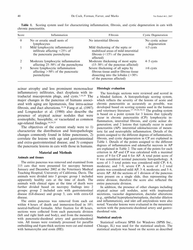
The histologic sections were reviewed and scored ina blinded fashion. A histopathologic scoring system,which reflects the severity of the lesions of acute andchronic pancreatitis as accurately as possible, wasdeveloped based on scoring systems used in the humanand veterinary literature.17–19,26,30,32 The grading systemis based on a point system for 3 lesions that typicallyoccur in chronic pancreatitis (CP): lymphocytic in-flammation, interstitial fibrosis, and cystic acinar de-generation; and 2 lesions that typically occur in acutepancreatitis (AP): interstitial edema/necrosis of mesen-teric fat and neutrophilic inflammation. Details of thepoints assigned to the different degrees of inflammation,fibrosis, and cystic degeneration in CP are explained inTable 1. Details of the points assigned to the differentdegrees of inflammation and edema/fat necrosis in APare explained in Table 2. The sum of the points for eachcriterion in AP and CP was calculated with a maximalscore of 9 for CP and 6 for AP. A total point score of0 was considered normal pancreatic histopathology. Ascore of 1–3 total points was considered mild CP; 4–6,moderate; and 7–9, severe CP. A score of 1–2 totalpoints was considered mild AP; 3–4, moderate; and 5–6,severe AP. All the sections of 1 division of the pancreaswere present on a single slide, thus representing theentire division; therefore the score also reflected theentire pancreatic division.
In addition, the presence of other changes includingatypical acinar cell nodules, acini with inspissatedsecretions, vacuolar degeneration, ductular alterations(e.g., epithelial hyperplasia/metaplasia, ductular dilationand inflammation), and islet cell amyloidosis were alsonoted. Vascular lesions were evaluated in the mesentericsection with the pancreatic-duodenal artery and gastro-duodenal vein.
Statistical analysis
Statistical software SPSS for Windows (SPSS Inc.,Chicago, IL) was used for the statistical analysis. Thestatistical analysis was based on the scores as described
Table 1. Scoring system used for characterizing inflammation, fibrosis, and cystic degeneration in cats withchronic pancreatitis.
Score Inflammation Fibrosis Cystic Degeneration
0 No or erratic small nests oflymphocytes
No interstitial fibrosis No cystic acinardegeneration
Mild thickening of the septa ormultifocal areas of mild interstitialfibrosis (,15% of the pancreasaffected)
#3 cysts
2 Moderate lymphocytic inflammationaffecting 25–50% of the parenchyma
Moderate thickening of most septa(15–30% of the pancreas affected)
4–5 cysts
3 Severe lymphocytic inflammationaffecting .50% of the pancreaticparenchyma
Severe thickening of all septa byfibrous tissue and/or fibrous tissuedissecting into the lobules (.30%of the pancreas affected)
$6 cysts
40 De Cock, Forman, Farver, and Marks Vet Pathol 44:1, 2007
in the grading system in the Histologic evaluationsection above. The analysis of variance for a repeatedmeasures experimental design was used to evaluate theeffect of study factors on AP and CP score. Clinicalgroup, sex, and breed were incorporated in the analysisas categorical between subject factors, while locationwas used as a repeated measures factor. Age wasa categorical covariate. Interaction effects were assessedin addition to main effects. The associations between APand CP score and age were estimated parametricallyusing the Pearson correlation coefficient and nonpar-ametrically using the Spearman rank correlation co-efficient. A P value ,.05 was considered statisticallysignificant.
Results
General
Of the 115 cats included in the study, 82 wereDomestic short hair; 13, Domestic long hair(DLH); 8, Domestic medium hair (DMH); 3,Siamese; 2, Abyssinian; 1, Burmese; 1, Bengal; 1,Maine Coon; 1, Himalayan; 1, Tonkinese; and 2,mixed breed. The age was known for 91 cats, witha mean of 102 months (SD 6 67 months; range 3–251 months). The other animals were all youngadults. The sex was known for 93 animals: 38 wereneutered males; 32, neutered females; 16, intactfemales; and 7, intact males. Forty-one animalswere considered healthy (group 1) at the time ofnecropsy, including 28 cats from clinical studiesthat did not undergo any treatment (controlanimals) and 13 cats that had no history of diseaseand died of trauma (e.g., hit by car, dog bites).Lesions identified at necropsy involved the di-gestive system of 22 cats (group 2), includinglymphoma (7) and other neoplastic processes (1hemangiosarcoma in caudal abdomen, 2 intestinalcarcinomas, 2 intestinal mast cell tumors, and 1liver lymphoma), intestinal obstruction (1), mega-esophagus (1), feline infectious peritonitis (FIP) (1),hepatic toxoplasmosis (1), intestinal mycobacter-iosis (1), Parvovirus enteritis (1), and peritonealactinomycosis (1). One cat had a pancreatic carci-noma. One cat had diffuse intravascular coagula-
tion (DIC) and had thrombi throughout the GI-tract including the pancreas. Fifty-two cats hadclinical conditions that did not involve the GI-tract(group 3): 10 cats had neoplasms (2 thoracicmesotheliomas, 1 transitional cell carcinoma, 1adrenal carcinoma, 1 nasal carcinoma, 1 chondro-sarcoma, 1 osteosarcoma, 1 leiomyosarcoma of thehead, 1 skin lymphoma, 1 skin squamous cellcarcinoma); 1 had a nasal polyp; 12 had lesions inthe central nervous system; 10 had cardiovascularlesions; 5 had endocrine disease; 8 had end-stagekidney disease; and 1 had pneumonia. Two cats hadmyelodysplasia. Three animals were strays in poorbody condition. Mean age for the animals for whichthe age was known was 86 months in the healthy(group 1), 104 months in the GI-disease (group 2),and 114 months in the extra-GI disease (group 3).
No lesions were identified in the left limb, rightlimb, or body in 38 of the 115 (33.0%) pancreasesevaluated. AP was present in 18 cats (15.7%) ofwhich 7 had AP only (6.1%). Both lobes and bodywere affected in 4 of these pancreases. In the other3 cats there was 1 pancreas with the left limb andbody affected, 1 with the right limb only, and 1with the body only. CP was found in 69 (60.0%)pancreases, and 58 (50.4%) had CP only. The leftlimb had CP in 51 (44.3%) cats, the right limb in 49(42.6%) cats, and the body in 57 (49.6%) cats. In 36(31.3%) of the pancreases, the lesions were presentin all divisions. In 4 cats, the left limb was affectedonly, in 4 the right limb only, and in 8 the bodyonly; the left limb and body were affected in 7cases, right limb and body in 6 cases, and left andright limbs in 4 cases. Eleven pancreases (9.6%) hadevidence of both AP and CP; 3 of these had lesionsof AP and CP in all divisions.
The mean score of AP in the left limb was 0.2832(60.9399; range 0–6), in the body 0.2124 (60.7494;range 0–5), and in the right limb 0.2174 (60.7106;range 0–4). The mean score of CP in the left limbwas 1.3805 (61.9925; range 0–9), in the body1.2035 (61.6211; range 0–7) and in the right limb1.0870 (61.6414; range 1–8). An overview of the
Table 2. Scoring system used for characterizing inflammation and edema in cats with acute pancreatitis.
Score Inflammation Edema and Fat Necrosis
0 No neutrophils present Not present1 Mild neutrophilic inflammatory infiltrate affecting maximum
25% of the pancreatic parenchymaMild, ,25% of the parenchyma
involved2 Moderate neutrophilic inflammation affecting 25–50% of the
parenchymaModerate, present in 25–50% of the
parenchyma3 Severe neutrophilic inflammation affecting .50% of the
pancreatic parenchymaSevere, .50% of the parenchyma
involved
Vet Pathol 44:1, 2007 Pancreatitis in Cats 41
mean scores for AP and CP in the differentdivisions of the pancreas for the different clinicalgroups is given in Table 3. Healthy animals alwayshad the lowest score for AP and CP compared withanimals from group 2 or group 3. Animals withGI-disease (group 2) had higher scores of APcompared with animals in group 3, whereas thereverse occurred with CP. The highest scoresfor CP occurred in animals with extra GI-disease(group 3). In animals of group 3, the CP scoreswere comparable in all divisions of the pancreas,while in animals with GI-disease the left limb hada much higher CP score compared with the rightlimb and body.
There was a statistically significant (P , .01)higher CP score in the left lobe compared with theright lobe in animals with GI-disease. There wasno statistically significant difference in AP or CPscore between the other clinical groups for theleft limb, right limb, and body when adjusted forage. There was a statistically significant correla-tion (P , .01) between the presence of CP andlocation in the pancreas. There was also a statisti-cally significant correlation (P , .01) between thepresence of AP and location in the pancreas. Astatistically significant correlation (P , .0005) wasfound between the age of the animal and thepresence of CP in the left limb, right limb, andbody unadjusted and when adjusted for other fac-tors. There was no correlation between age andAP score in any location, both unadjusted andwhen adjusted for sex and breed. Sex and breedwere shown to be nonsignificant factors in allanalyses.
Histopathology
In general, the lesions of CP were multifocal.Only in the end-stage were the lesions confluent,although small nests of normal-appearing aciniwere always present. Lesions of AP were alsorandomly scattered throughout the pancreas and
often extended into the surrounding mesentericadipose tissue.
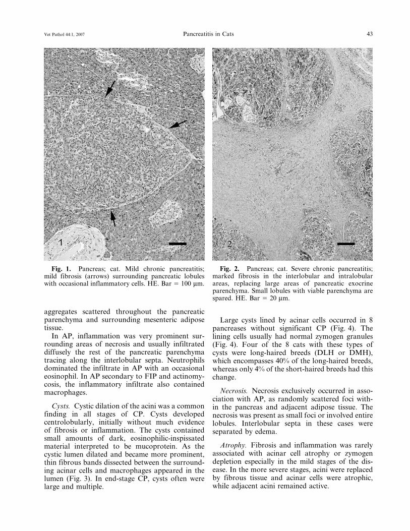
Fibrosis. Fibrosis was a very prominent featureof CP. In the mild stages of CP, fibrosis usually wasminimal and composed of a thin layer of densefibrous tissue that surrounded 1 or several pancre-atic lobules (Fig. 1). In the more severe CP, thefibrous tissue expanded uniformly around 1 ormore lobules and dissected into the lobule as thinstrands that separated the individual acini. In themost severe cases, the fibrosis was marked inthe interlobular and intralobular areas and oftenreplaced large areas of pancreatic exocrine paren-chyma (Fig. 2). In all cats with severe CP, smalllobules with viable parenchyma were spared(Fig. 2). Occasionally fibrosis started within pan-creatic lobules, individualizing the acinar structureswithout obvious interlobular fibrosis. The associ-ated acini in these cases often were atrophic, andinflammation was frequently more prominent.
Fibrosis was not a feature of AP.
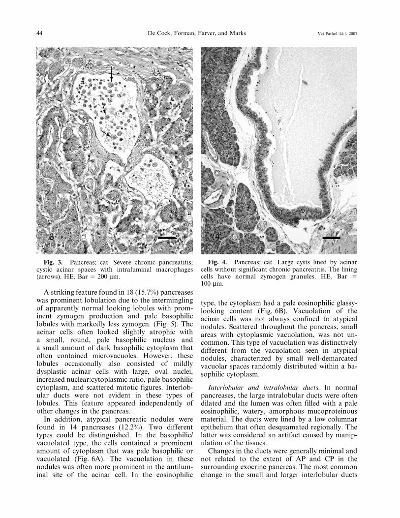
Inflammation. A few scattered lymphocyteswithin the pancreatic parenchyma was considerednormal. Inflammation in CP was variable but oftenrather mild in comparison to the fibrous response.In the mild stages, only occasional inflammatorycells were scattered within the fibrous tissue(Fig. 1). As the amount of fibrous tissue increased,inflammation also became more prominent (Fig. 2)and more widely distributed within the lobules.Lymphocytes were always the major inflammatorycomponent in CP, but occasionally eosinophils andmacrophages were also present. Macrophages wereespecially prominent in the lumen of cystic acinarstructures that developed in the more chronicstages of pancreatitis (see cysts) (Fig. 3).
In 29 pancreases (25%) there were nests oflymphocytes without associated signs of pancrea-titis. The lymphocytes were small and monomor-phous, closely arranged and located in small
Table 3. Histopathologic score for acute and chronic pancreatitis in the left and right limbs and body inapparently healthy cats, cats with gastrointestinal disease, and cats with extra-gastrointestinal disease.*
* LLAP 5 left limb acute pancreatitis; LLCP 5 left limb chronic pancreatitis; BAP 5 body acute pancreatitis; BCP 5 bodychronic pancreatitis; RLAP 5 right limb acute pancreatitis; RLCP 5 right limb chronic pancreatitis.
42 De Cock, Forman, Farver, and Marks Vet Pathol 44:1, 2007
aggregates scattered throughout the pancreaticparenchyma and surrounding mesenteric adiposetissue.
In AP, inflammation was very prominent sur-rounding areas of necrosis and usually infiltrateddiffusely the rest of the pancreatic parenchymatracing along the interlobular septa. Neutrophilsdominated the infiltrate in AP with an occasionaleosinophil. In AP secondary to FIP and actinomy-cosis, the inflammatory infiltrate also containedmacrophages.
Cysts. Cystic dilation of the acini was a commonfinding in all stages of CP. Cysts developedcentrolobularly, initially without much evidenceof fibrosis or inflammation. The cysts containedsmall amounts of dark, eosinophilic-inspissatedmaterial interpreted to be mucoprotein. As thecystic lumen dilated and became more prominent,thin fibrous bands dissected between the surround-ing acinar cells and macrophages appeared in thelumen (Fig. 3). In end-stage CP, cysts often werelarge and multiple.
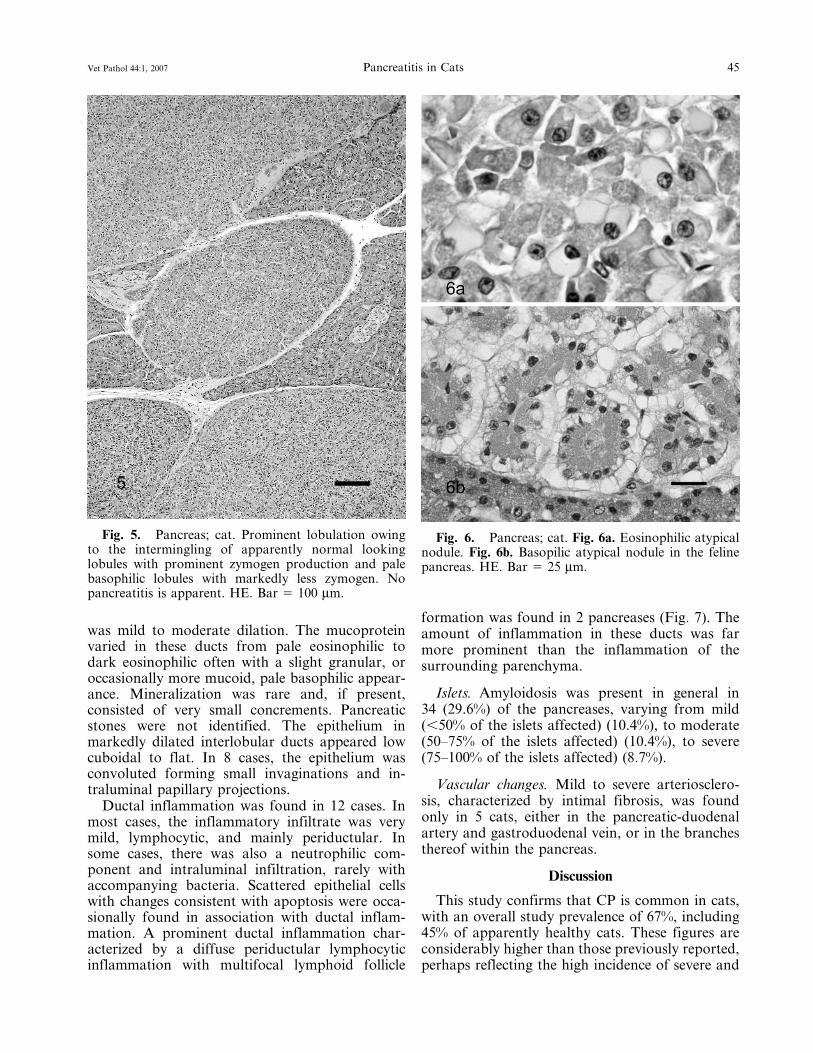
Large cysts lined by acinar cells occurred in 8pancreases without significant CP (Fig. 4). Thelining cells usually had normal zymogen granules(Fig. 4). Four of the 8 cats with these types ofcysts were long-haired breeds (DLH or DMH),which encompasses 40% of the long-haired breeds,whereas only 4% of the short-haired breeds had thischange.
Necrosis. Necrosis exclusively occurred in asso-ciation with AP, as randomly scattered foci with-in the pancreas and adjacent adipose tissue. Thenecrosis was present as small foci or involved entirelobules. Interlobular septa in these cases wereseparated by edema.
Atrophy. Fibrosis and inflammation was rarelyassociated with acinar cell atrophy or zymogendepletion especially in the mild stages of the dis-ease. In the more severe stages, acini were replacedby fibrous tissue and acinar cells were atrophic,while adjacent acini remained active.
Fig. 2. Pancreas; cat. Severe chronic pancreatitis;marked fibrosis in the interlobular and intralobularareas, replacing large areas of pancreatic exocrineparenchyma. Small lobules with viable parenchyma arespared. HE. Bar 5 20 mm.
Fig. 1. Pancreas; cat. Mild chronic pancreatitis;mild fibrosis (arrows) surrounding pancreatic lobuleswith occasional inflammatory cells. HE. Bar 5 100 mm.
Vet Pathol 44:1, 2007 Pancreatitis in Cats 43
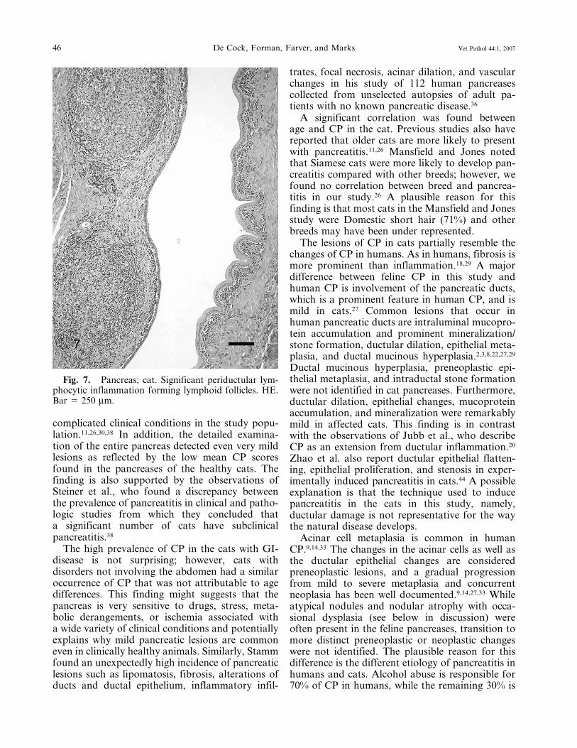
A striking feature found in 18 (15.7%) pancreaseswas prominent lobulation due to the interminglingof apparently normal looking lobules with prom-inent zymogen production and pale basophiliclobules with markedly less zymogen. (Fig. 5). Theacinar cells often looked slightly atrophic witha small, round, pale basophilic nucleus anda small amount of dark basophilic cytoplasm thatoften contained microvacuoles. However, theselobules occasionally also consisted of mildlydysplastic acinar cells with large, oval nuclei,increased nuclear:cytoplasmic ratio, pale basophiliccytoplasm, and scattered mitotic figures. Interlob-ular ducts were not evident in these types oflobules. This feature appeared independently ofother changes in the pancreas.
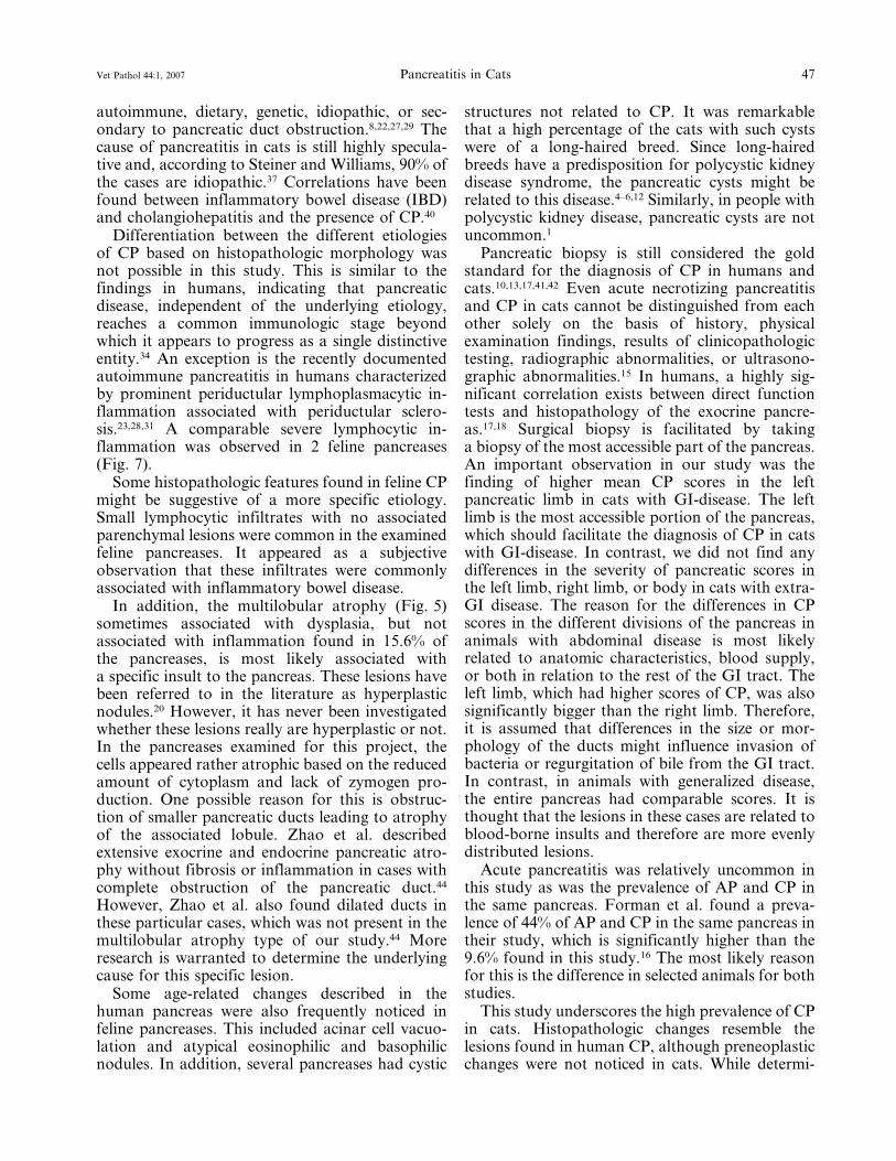
In addition, atypical pancreatic nodules werefound in 14 pancreases (12.2%). Two differenttypes could be distinguished. In the basophilic/vacuolated type, the cells contained a prominentamount of cytoplasm that was pale basophilic orvacuolated (Fig. 6A). The vacuolation in thesenodules was often more prominent in the antilum-inal site of the acinar cell. In the eosinophilic
type, the cytoplasm had a pale eosinophilic glassy-looking content (Fig. 6B). Vacuolation of theacinar cells was not always confined to atypicalnodules. Scattered throughout the pancreas, smallareas with cytoplasmic vacuolation, was not un-common. This type of vacuolation was distinctivelydifferent from the vacuolation seen in atypicalnodules, characterized by small well-demarcatedvacuolar spaces randomly distributed within a ba-sophilic cytoplasm.
Interlobular and intralobular ducts. In normalpancreases, the large intralobular ducts were oftendilated and the lumen was often filled with a paleeosinophilic, watery, amorphous mucoproteinousmaterial. The ducts were lined by a low columnarepithelium that often desquamated regionally. Thelatter was considered an artifact caused by manip-ulation of the tissues.
Changes in the ducts were generally minimal andnot related to the extent of AP and CP in thesurrounding exocrine pancreas. The most commonchange in the small and larger interlobular ducts
Fig. 3. Pancreas; cat. Severe chronic pancreatitis;cystic acinar spaces with intraluminal macrophages(arrows). HE. Bar 5 200 mm.
Fig. 4. Pancreas; cat. Large cysts lined by acinarcells without significant chronic pancreatitis. The liningcells have normal zymogen granules. HE. Bar 5100 mm.
44 De Cock, Forman, Farver, and Marks Vet Pathol 44:1, 2007
was mild to moderate dilation. The mucoproteinvaried in these ducts from pale eosinophilic todark eosinophilic often with a slight granular, oroccasionally more mucoid, pale basophilic appear-ance. Mineralization was rare and, if present,consisted of very small concrements. Pancreaticstones were not identified. The epithelium inmarkedly dilated interlobular ducts appeared lowcuboidal to flat. In 8 cases, the epithelium wasconvoluted forming small invaginations and in-traluminal papillary projections.
Ductal inflammation was found in 12 cases. Inmost cases, the inflammatory infiltrate was verymild, lymphocytic, and mainly periductular. Insome cases, there was also a neutrophilic com-ponent and intraluminal infiltration, rarely withaccompanying bacteria. Scattered epithelial cellswith changes consistent with apoptosis were occa-sionally found in association with ductal inflam-mation. A prominent ductal inflammation char-acterized by a diffuse periductular lymphocyticinflammation with multifocal lymphoid follicle
formation was found in 2 pancreases (Fig. 7). Theamount of inflammation in these ducts was farmore prominent than the inflammation of thesurrounding parenchyma.
Islets. Amyloidosis was present in general in34 (29.6%) of the pancreases, varying from mild(,50% of the islets affected) (10.4%), to moderate(50–75% of the islets affected) (10.4%), to severe(75–100% of the islets affected) (8.7%).
Vascular changes. Mild to severe arteriosclero-sis, characterized by intimal fibrosis, was foundonly in 5 cats, either in the pancreatic-duodenalartery and gastroduodenal vein, or in the branchesthereof within the pancreas.
Discussion
This study confirms that CP is common in cats,with an overall study prevalence of 67%, including45% of apparently healthy cats. These figures areconsiderably higher than those previously reported,perhaps reflecting the high incidence of severe and
Fig. 5. Pancreas; cat. Prominent lobulation owingto the intermingling of apparently normal lookinglobules with prominent zymogen production and palebasophilic lobules with markedly less zymogen. Nopancreatitis is apparent. HE. Bar 5 100 mm.
Fig. 6. Pancreas; cat. Fig. 6a. Eosinophilic atypicalnodule. Fig. 6b. Basopilic atypical nodule in the felinepancreas. HE. Bar 5 25 mm.
Vet Pathol 44:1, 2007 Pancreatitis in Cats 45
complicated clinical conditions in the study popu-lation.11,26,30,38 In addition, the detailed examina-tion of the entire pancreas detected even very mildlesions as reflected by the low mean CP scoresfound in the pancreases of the healthy cats. Thefinding is also supported by the observations ofSteiner et al., who found a discrepancy betweenthe prevalence of pancreatitis in clinical and patho-logic studies from which they concluded thata significant number of cats have subclinicalpancreatitis.38
The high prevalence of CP in the cats with GI-disease is not surprising; however, cats withdisorders not involving the abdomen had a similaroccurrence of CP that was not attributable to agedifferences. This finding might suggests that thepancreas is very sensitive to drugs, stress, meta-bolic derangements, or ischemia associated witha wide variety of clinical conditions and potentiallyexplains why mild pancreatic lesions are commoneven in clinically healthy animals. Similarly, Stammfound an unexpectedly high incidence of pancreaticlesions such as lipomatosis, fibrosis, alterations ofducts and ductal epithelium, inflammatory infil-
trates, focal necrosis, acinar dilation, and vascularchanges in his study of 112 human pancreasescollected from unselected autopsies of adult pa-tients with no known pancreatic disease.36
A significant correlation was found betweenage and CP in the cat. Previous studies also havereported that older cats are more likely to presentwith pancreatitis.11,26 Mansfield and Jones notedthat Siamese cats were more likely to develop pan-creatitis compared with other breeds; however, wefound no correlation between breed and pancrea-titis in our study.26 A plausible reason for thisfinding is that most cats in the Mansfield and Jonesstudy were Domestic short hair (71%) and otherbreeds may have been under represented.
The lesions of CP in cats partially resemble thechanges of CP in humans. As in humans, fibrosis ismore prominent than inflammation.18,29 A majordifference between feline CP in this study andhuman CP is involvement of the pancreatic ducts,which is a prominent feature in human CP, and ismild in cats.27 Common lesions that occur inhuman pancreatic ducts are intraluminal mucopro-tein accumulation and prominent mineralization/stone formation, ductular dilation, epithelial meta-plasia, and ductal mucinous hyperplasia.2,3,8,22,27,29
Ductal mucinous hyperplasia, preneoplastic epi-thelial metaplasia, and intraductal stone formationwere not identified in cat pancreases. Furthermore,ductular dilation, epithelial changes, mucoproteinaccumulation, and mineralization were remarkablymild in affected cats. This finding is in contrastwith the observations of Jubb et al., who describeCP as an extension from ductular inflammation.20
Zhao et al. also report ductular epithelial flatten-ing, epithelial proliferation, and stenosis in exper-imentally induced pancreatitis in cats.44 A possibleexplanation is that the technique used to inducepancreatitis in the cats in this study, namely,ductular damage is not representative for the waythe natural disease develops.
Acinar cell metaplasia is common in humanCP.9,14,33 The changes in the acinar cells as well asthe ductular epithelial changes are consideredpreneoplastic lesions, and a gradual progressionfrom mild to severe metaplasia and concurrentneoplasia has been well documented.9,14,27,33 Whileatypical nodules and nodular atrophy with occa-sional dysplasia (see below in discussion) wereoften present in the feline pancreases, transition tomore distinct preneoplastic or neoplastic changeswere not identified. The plausible reason for thisdifference is the different etiology of pancreatitis inhumans and cats. Alcohol abuse is responsible for70% of CP in humans, while the remaining 30% is
46 De Cock, Forman, Farver, and Marks Vet Pathol 44:1, 2007
autoimmune, dietary, genetic, idiopathic, or sec-ondary to pancreatic duct obstruction.8,22,27,29 Thecause of pancreatitis in cats is still highly specula-tive and, according to Steiner and Williams, 90% ofthe cases are idiopathic.37 Correlations have beenfound between inflammatory bowel disease (IBD)and cholangiohepatitis and the presence of CP.40
Differentiation between the different etiologiesof CP based on histopathologic morphology wasnot possible in this study. This is similar to thefindings in humans, indicating that pancreaticdisease, independent of the underlying etiology,reaches a common immunologic stage beyondwhich it appears to progress as a single distinctiveentity.34 An exception is the recently documentedautoimmune pancreatitis in humans characterizedby prominent periductular lymphoplasmacytic in-flammation associated with periductular sclero-sis.23,28,31 A comparable severe lymphocytic in-flammation was observed in 2 feline pancreases(Fig. 7).
Some histopathologic features found in feline CPmight be suggestive of a more specific etiology.Small lymphocytic infiltrates with no associatedparenchymal lesions were common in the examinedfeline pancreases. It appeared as a subjectiveobservation that these infiltrates were commonlyassociated with inflammatory bowel disease.
In addition, the multilobular atrophy (Fig. 5)sometimes associated with dysplasia, but notassociated with inflammation found in 15.6% ofthe pancreases, is most likely associated witha specific insult to the pancreas. These lesions havebeen referred to in the literature as hyperplasticnodules.20 However, it has never been investigatedwhether these lesions really are hyperplastic or not.In the pancreases examined for this project, thecells appeared rather atrophic based on the reducedamount of cytoplasm and lack of zymogen pro-duction. One possible reason for this is obstruc-tion of smaller pancreatic ducts leading to atrophyof the associated lobule. Zhao et al. describedextensive exocrine and endocrine pancreatic atro-phy without fibrosis or inflammation in cases withcomplete obstruction of the pancreatic duct.44
However, Zhao et al. also found dilated ducts inthese particular cases, which was not present in themultilobular atrophy type of our study.44 Moreresearch is warranted to determine the underlyingcause for this specific lesion.
Some age-related changes described in thehuman pancreas were also frequently noticed infeline pancreases. This included acinar cell vacuo-lation and atypical eosinophilic and basophilicnodules. In addition, several pancreases had cystic
structures not related to CP. It was remarkablethat a high percentage of the cats with such cystswere of a long-haired breed. Since long-hairedbreeds have a predisposition for polycystic kidneydisease syndrome, the pancreatic cysts might berelated to this disease.4–6,12 Similarly, in people withpolycystic kidney disease, pancreatic cysts are notuncommon.1
Pancreatic biopsy is still considered the goldstandard for the diagnosis of CP in humans andcats.10,13,17,41,42 Even acute necrotizing pancreatitisand CP in cats cannot be distinguished from eachother solely on the basis of history, physicalexamination findings, results of clinicopathologictesting, radiographic abnormalities, or ultrasono-graphic abnormalities.15 In humans, a highly sig-nificant correlation exists between direct functiontests and histopathology of the exocrine pancre-as.17,18 Surgical biopsy is facilitated by takinga biopsy of the most accessible part of the pancreas.An important observation in our study was thefinding of higher mean CP scores in the leftpancreatic limb in cats with GI-disease. The leftlimb is the most accessible portion of the pancreas,which should facilitate the diagnosis of CP in catswith GI-disease. In contrast, we did not find anydifferences in the severity of pancreatic scores inthe left limb, right limb, or body in cats with extra-GI disease. The reason for the differences in CPscores in the different divisions of the pancreas inanimals with abdominal disease is most likelyrelated to anatomic characteristics, blood supply,or both in relation to the rest of the GI tract. Theleft limb, which had higher scores of CP, was alsosignificantly bigger than the right limb. Therefore,it is assumed that differences in the size or mor-phology of the ducts might influence invasion ofbacteria or regurgitation of bile from the GI tract.In contrast, in animals with generalized disease,the entire pancreas had comparable scores. It isthought that the lesions in these cases are related toblood-borne insults and therefore are more evenlydistributed lesions.
Acute pancreatitis was relatively uncommon inthis study as was the prevalence of AP and CP inthe same pancreas. Forman et al. found a preva-lence of 44% of AP and CP in the same pancreas intheir study, which is significantly higher than the9.6% found in this study.16 The most likely reasonfor this is the difference in selected animals for bothstudies.
This study underscores the high prevalence of CPin cats. Histopathologic changes resemble thelesions found in human CP, although preneoplasticchanges were not noticed in cats. While determi-
Vet Pathol 44:1, 2007 Pancreatitis in Cats 47
nation of the underlying cause might not be pos-sible based on histopathologic examination of thepancreas in most cases, very typical lesions werenoticed in feline pancreases that warrant furtherresearch to determine the underlying cause.
Acknowledgements
Sincere thanks are due to all the pathologists,clinicians, students, and technicians of University ofCalifornia, Davis, who helped collect cases for thisproject. Specifically, we would like to thank MonicaKratochvil for her excellent assistance. The authorsappreciate the critical review of this manuscript by Dr.James N. MacLachlan, Department of Pathology,Microbiology, and Immunology, University of Califor-nia, Davis, School of Veterinary Medicine.
2 Allen-Mersh TG: What is the significance ofpancreatic ductal mucinous hyperplasia? Gut 26(8):825–833, 1985
3 Allen-Mersh TG: Pancreatic ductal mucinous hy-perplasia: distribution within the pancreas, andeffect of variation in ampullary and pancreatic ductanatomy. Gut 29(10): 1392–1396, 1988
4 Barrs VR, Gunew M, Foster SF, Beatty JA, MalikR: Prevalence of autosomal dominant polycystickidney disease in Persian cats and related breeds inSydney and Brisbane. Aust Vet J 79:257–259, 2001
5 Barthez PY, Rivier P, Begon D: Prevalence ofpolycystic kidney disease in Persian and Persianrelated cats in France. J Feline Med Surg 5:345–347,2003
6 Cannon MJ, MacKay AD, Barr FJ, Rudorf H,Bradley KJ, Gruffydd-Jones TJ: Prevalence of poly-cystic kidney disease in Persian cats in the UnitedKingdom. Vet Rec 149:409–411, 2001
7 Chari ST, Singer MV: The problem of classificationand staging of chronic pancreatitis. Proposals basedon current knowledge of its natural history.Scand J Gastroenterol 29:949–960, 1994
8 Cruickshank AH: Noninfective chronic pancreatitis.In: Pathology of the Pancreas, ed. Cruickshank AHand Benhow EW, pp. 233–258. Springer, London,UK, 1995
9 Cylwik B, Nowak HF, Puchalski Z, Barczyk J:Epithelial anomalies in chronic pancreatitis as a riskfactor of pancreatic cancer. Hepatogastroenterology45:528–532, 1998
10 Dill-Macky E: Pancreatic diseases of cats. CompendContin Educ Pract Vet 15:589–598, 1993
11 Duffel SJ: Some aspects of pancreatic disease in thecat. J Small Anim Pract 16:365–374, 1975
disease in Persian and Persian-cross cats. Vet Pathol34:117–126, 1997
13 Etemad B, Whitcomb DC: Chronic pancreatitis:diagnosis, classification, and new genetic develop-ments. Gastroenterology 120:682–707, 2001
14 Fang J, Hussong J, Roebuck BD, Talamonti MS,Rao MS: Atypical acinar cell foci in humanpancreas: morphological and morphometric analy-sis. Int J Pancreatol 22:127–130, 1997
15 Ferreri JA, Hardam E, Kimmel SE, Saunders HM,Van Winkle TJ, Drobatz KJ, Washabau RJ:Clinical differentiation of acute necrotizing fromchronic nonsuppurative pancreatitis in cats: 63 cases(1996–2001). J Am Vet Med Assoc 223:469–474,2003
16 Forman MA, Marks SL, De Cock HE, Hergesell EJ,Wisner ER, Baker TW, Kass PH, Steiner JM,Williams DA: Evaluation of serum feline pancreaticlipase immunoreactivity and helical computed to-mography versus conventional testing for the di-agnosis of feline pancreatitis. J Vet Intern Med18:807–815, 2004
17 Hayakawa T, Kondo T, Shibata T, Noda A, SuzukiT, Nakano S: Relationship between pancreaticexocrine function and histological changes in chronicpancreatitis. Am J Gastroenterol 87:1170–1174, 1992
18 Heij HA, Obertop H, van Blankenstein M, ten KateFW, Westbroek DL: Relationship between function-al and histological changes in chronic pancreatitis.Dig Dis Sci 31:1009–1013, 1986
19 Hill RC, Van Winkle TJ: Acute necrotizing pancre-atitis and acute suppurative pancreatitis in the cat. Aretrospective study of 40 cases (1976-1989). J VetIntern Med 7:25–33, 1993
20 Jubb KVF: The pancreas. In: Pathology of DomesticAnimals, ed. Jubb KVF, Kennedy PC, and PalmerN, pp. 407–424. Academic Press, San Diego, CA,1993
21 Kitchell BE, Strombeck DR, Cullen J, Harrold D:Clinical and pathologic changes in experimentallyinduced acute pancreatitis in cats. Am J Vet Res47:1170–1173, 1986
22 Kloppel G, Heitz, Ph: Exocrine pancreas. In:Pancreatic Pathology, ed. Kloppel G and Heitz,Ph, pp. 3–114. Churchill Livingstone, Edinburgh,UK, 1984
23 Kloppel G, Luttges J, Lohr M, Zamboni G, Long-necker D: Autoimmune pancreatitis: pathological,clinical, and immunological features. Pancreas27:14–19, 2003
24 Longnecker DS, Hashida Y, Shinozuka H: Relation-ship of age to prevalence of focal acinar celldysplasia in the human pancreas. J Natl Cancer Inst65:63–66, 1980
25 Longnecker DS, Shinozuka H, Dekker A: Focalacinar cell dysplasia in human pancreas. Cancer45:534–540, 1980
26 Mansfield CS, Jones BR: Review of feline pancre-atitis part two: clinical signs, diagnosis and treat-ment. J Feline Med Surg 3:125–132, 2001
48 De Cock, Forman, Farver, and Marks Vet Pathol 44:1, 2007
28 Okazaki K: Clinical relevance of autoimmune-re-lated pancreatitis. Best Pract Res Clin Gastroenterol16:365–378, 2002
29 Owen DA, Kelly JK: Embryology, normal anatomy,and histology of the pancreas. In: Pathology of theGallbladder, Biliary Tract and Pancreas, ed. OwenDA, pp. 1–14. WB Saunders, Philadelphia, PA, 2001
30 Owens JM: Pancreatic disease in the cat. J Am AnimHosp Assoc 11:83–89, 1975
32 Pitchumoni CS, Glasser M, Saran RM, Pancha-charam P, Thelmo W: Pancreatic fibrosis in chro-nic alcoholics and nonalcoholics without cli-nical pancreatitis. Am J Gastroenterol 79:382–388,1984
33 Shinozuka H, Lee RE, Dunn JL, Longnecker DS:Multiple atypical acinar cell nodules of the pancreas.Hum Pathol 11:389–391, 1980
34 Shrikhande SV, Martignoni ME, Shrikhande M,Kappeler A, Ramesh H, Zimmermann A, BuchlerMW, Friess H: Comparison of histological featuresand inflammatory cell reaction in alcoholic, idio-pathic and tropical chronic pancreatitis. Br J Surg90:1565–1572, 2003
35 Simpson KW, Shiroma JT, Biller DS: Antemortemdiagnosis of pancreatitis in four cats. J Small AnimPract 35:93–99, 1994
36 Stamm BH: Incidence and diagnostic significance ofminor pathologic changes in the adult pancreas atautopsy: a systematic study of 112 autopsies inpatients without known pancreatic disease. HumPathol 15:677–683, 1984
37 Steiner JM, Williams DA: Feline Trypsin-likeImmunoreactivity in Feline Exocrine PancreaticDisease. Comp Contin Educ Pract Vet 18:543–547,1996
38 Steiner JM, Williams DA, Davis A: Feline pancre-atitis. Comp Contin Educ Pract Vet 19:590–601,1997
39 Swift NC, Marks SL, MacLachlan NJ, Norris CR:Evaluation of serum feline trypsin-like immunoreac-tivity for the diagnosis of pancreatitis in cats. J AmVet Med Assoc 217:37–42, 2000
40 Weiss DJ, Gagne JM, Armstrong PJ: Relationshipbetween inflammatory hepatic disease and inflam-matory bowel disease, pancreatitis, and nephritis incats. J Am Vet Med Assoc 209:1114–1116, 1996
41 Whitney M: The laboratory assessment of canineand feline pancreatitis. Vet Med 88:1045–1052, 1993
42 Williams D: Diagnosis and management of pancre-atitis. J Small Anim Pract 35:445–454, 1993
43 Zhao P, Tu J, Martens A, Ponette E, Van Steenber-gen W, Oord JV, Fevery J: Radiologic investigationsand pathologic results of experimental chronicpancreatitis in cats. Acad Radiol 5:850–856, 1998
44 Zhao P, Tu J, van den Oord JJ, Fevery J: Damage toduct epithelium is necessary to develop progressinglesions of chronic pancreatitis in the cat. Hepatogas-troenterology 43:1620–1626, 1996
Request reprints from Dr. Hilde E. V. De Cock, University of Antwerp, Veterinary Pathology, Universiteitsplein 1,Wilrijk, 2610 (Belgium). E-mail: [email protected].


![arXiv:2006.02474v1 [cs.CV] 3 Jun 2020with hematoxylin and eosin (HE), periodic acidSchiff (PAS) or Jones. Samples were deidentified, and studies were approved by the Institutional](https://static.documents.pub/doc/80x56/5f26368d9adadc5c18232803/arxiv200602474v1-cscv-3-jun-2020-with-hematoxylin-and-eosin-he-periodic.jpg)


