Research ArticlePrevalence and Genetic Identification of Three EntamoebaSpecies in Pigs in Southeastern China
Ting Ji, Hao-Xuan Cao, Ran Wu, Lin-Lin Cui, Guo-Ming Su, Chang Niu, Ning Zhang,Shou-Kun Wang, and Dong-Hui Zhou
Key Laboratory of Fujian-Taiwan Animal Pathogen Biology, College of Animal Sciences (College of Bee Science),Fujian Agriculture and Forestry University, Fuzhou, Fujian 350002, China
Correspondence should be addressed to Dong-Hui Zhou; [email protected]
Received 1 August 2019; Revised 3 October 2019; Accepted 15 October 2019; Published 7 November 2019
Parasitic Entamoeba spp. can infect many classes of vertebrates including humans and pigs. Entamoeba suis and zoonoticEntamoeba polecki have been identified in pigs, and swine are implicated as potential reservoirs for Entamoeba histolytica.However, the prevalence of Entamoeba spp. in pigs in southeastern China has not been reported. In this study, 668 fecal samplescollected from 6 different regions in Fujian Province, southeastern China, were analyzed to identify three Entamoeba species bynested PCR and sequencing analysis. /e overall prevalence of Entamoeba spp. was 55.4% (370/668; 95% CI 51.6% to 59.2%), andthe infection rate of E. polecki ST1 was the highest (302/668; 45.2%, 95% CI 41.4% to 49.0%), followed by E. polecki ST3 (228/668;34.1%, 95% CI 30.5% to 37.7%) and E. suis (87/668; 13.0%, 95% CI 10.5% to 15.6%). E. histolytica was not detected in any samples.Moreover, the coinfection rate of E. polecki ST1 and ST3 was 25.1% (168/668; 95% CI 21.9% to 28.4%), the coinfection rate of E.polecki ST1 and E. suis was 3.7% (25/668; 95% CI 2.3% to 5.2%), the coinfection rate of E. polecki ST3 and E. suis was 0.3% (2/668),and the coinfection rate of E. polecki ST1, E. polecki ST3, and E. suis was 4.0% (27/668; 95% CI 2.5% to 5.5%). A representativesequence (MK347346) was identical to the sequence of E. suis (DQ286372). Two subtype-specific sequences (MK357717 andMK347347) were almost identical to the sequences of E. polecki ST1 (FR686383) and ST3 (AJ566411), respectively. /is is the firststudy to survey the occurrence and to conduct molecular identification of three Entamoeba species in southeastern China. /is isthe first report regarding mixed infections with E. suis, E. polecki ST1, and E. polecki ST3 in China. More research studies areneeded to better understand the transmission and zoonotic potential of Entamoeba spp.
1. Introduction
/e genus Entamoeba comprises many free-living andparasitic species and can infect all classes of vertebrates andsome invertebrates. Some Entamoeba species (e.g., E. his-tolytica, E. dispar, E. coli, E. moshkovskii, E. hartmanni, andE. polecki) have been identified in humans [1–4], and mostare considered harmless, but some of these species still causedisease. Amoebiasis caused by E. histolytica is the thirdleading parasitic disease causing morbidity and mortality inhumans, causing up to 50, 000 deaths per year, just behindmalaria and schistosomiasis [4–7]. /e disease is charac-terized as amebic colitis and liver abscess in humans andanimals [3, 8]. Although E. histolytica has not been detectedin farmed pigs thus far, and the susceptibility of swine to E.
histolytica infection was revealed only under experimentalconditions, swine have been considered as potential reser-voirs for E. histolytica [9–12].
Two species, E. suis [13] and E. polecki [14], have beenidentified in pigs. E. suis appears to be mostly restricted topigs [2, 3, 15, 16] and has been suggested to cause hem-orrhagic colitis by breaking down the lamina propria [10].Unlike E. suis, which infects pigs and potentially gorillas[17], E. polecki can infect many kinds of hosts, includinghumans, nonhuman primates, and pigs. /e intraspecificvariation of E. polecki was revealed by molecular analysis ofthe small-subunit ribosomal DNA, which showed that E.polecki could be divided into 4 subtypes (E. polecki ST1–ST4)[17, 18]. All the subtypes have been found in humans, E.polecki ST1 and E. polecki ST3 have also been found in pigs,
HindawiBioMed Research InternationalVolume 2019, Article ID 2824017, 8 pageshttps://doi.org/10.1155/2019/2824017
and E. polecki ST2 also exists in nonhuman primates [12, 17],while human cases of E. polecki primarily involve E. poleckiST4 [19]. For a long time, E. polecki ST4 was only knownfrom humans. Recently, however, ST4 was found in wildCelebes crested macaques (Macaca nigra) [20]. Although E.polecki is considered less pathogenic to humans or animalsin the case of solo infection, coinfections with other path-ogens, such as Lawsonia intracellularis, may increase theseverity of the disease [3].
Swine husbandry plays an indispensable role in theanimal husbandry in China. Because of the prosperity ofswine husbandry and the high population density in China,the risk of exposure to zoonotic Entamoeba spp. is inevitable.However, the molecular epidemiology of Entamoeba spp. inpigs in southeastern China has not been reported./is studydetermined the prevalence of three Entamoeba species inpigs in southeastern China using molecular detection, de-termined the genetic identity of these Entamoeba species byphylogenetic analysis, and evaluated the zoonotic potentialof Entamoeba spp.
2. Materials and Methods
2.1. Study Sampling. A total of 668 fecal samples werecollected from 6 regions in Fujian Province, southeasternChina (Figure 1). All specimens from pigs, including weanedpiglets, suckling piglets, sow, boars, nursery pigs, and fat-tening pigs, were collected directly from each pig’s rectum orwere immediately collected from the ground after defecationby the pigs. Fecal samples were marked with the corre-sponding sex, developmental stage, and origin of the pigsand then stored at 4°C until DNA extraction (generallywithin 48 hours).
2.2. Isolation of Genomic DNA. According to the manu-facturer’s instructions, genomic DNA was extracted fromapproximately 200mg of each fecal samples using a StoolDNA kit (OMEGA D4015-02), and the DNA was stored at− 20°C until use.
2.3. PCRAmplificationof Entamoeba spp. /e extracted fecalgenomic DNAwas used to determine the species/subtypes ofEntamoeba spp. by nested PCR targeting the small-subunitribosomal RNA (SSU rRNA) gene. /e first set of primers,E-1 and E-2, and the second set of primers, EH-1 and EH-2,were used to detect E. histolytica [1]. /e first round ofnested PCR used primers 764–RD3, and the second round ofnested PCR used primers 764–765, to identify E. suis [15]./e primary PCR for identifying E. polecki was performedusing primer set Epolec F6–Epolec R6, and then the sec-ondary PCR for subtype-specific characterization of E.polecki used primers Epolecki 1-Epolecki 2 (ST1) andEpST3F1-EpST3R2 (ST3) [2, 12].
An amplification reaction volume of 25 μL was used toperform nested PCR. For E. histolytica, the reaction mixtureof it contained 2.5 μL DNA, 0.4mM of each primers, 1mM10× buffer (Mg2+ free), 0.2mM dNTP, 1.5mM MgCl2, and0.375 U Taq DNA polymerase (TaKaRa, R001CM). /e
reaction mixture of E. suis contained 1 μL DNA, 0.5mM ofeach primers, 1mM 10× buffer (Mg2+ free), 0.2mM dNTP,1.5mMMgCl2, and 0.625 U Taq DNA polymerase (TaKaRa,R001CM). /e reaction mixture of E. polecki was similar tothe reaction mixture of E. suis, except that each primer wasused at 0.2mM.
2.4. Sequencing Analysis and Phylogenetic Analysis. PCRproducts were separated using 1.0% agarose gels, stainedwith GelStain, and visualized using a UV transilluminator./e positive PCR productions were sequenced with the BigDye Terminator v3.1 Cycle Sequencing Kit on an ABIPRISM™ 3730 XL DNA Analyzer (Applied Biosystems,Foster City, CA, USA). /e accuracy of the sequences wasverified with bidirectional sequencing. /e obtained se-quences were analyzed using the BLAST program at theNCBI website. Mega 7.0 (http://www.megasoftware.net/)software was used to perform phylogenetic analyses by theneighbor-joining method with the Kimura-2 parametermodel. Bootstrap analysis with 1000 replicates was used toassess the robustness of cluster formation.
2.5. Data Analysis. SPSS 22.0 (IBM Corp., New York, USA)was used to analyze the data. /e associations betweeninfection rates of different sampling areas and the associa-tions between infection rates of different developmentalstages of pigs were explored using the chi-square test.Differences were considered statistically significant whenP< 0.05.
3. Results
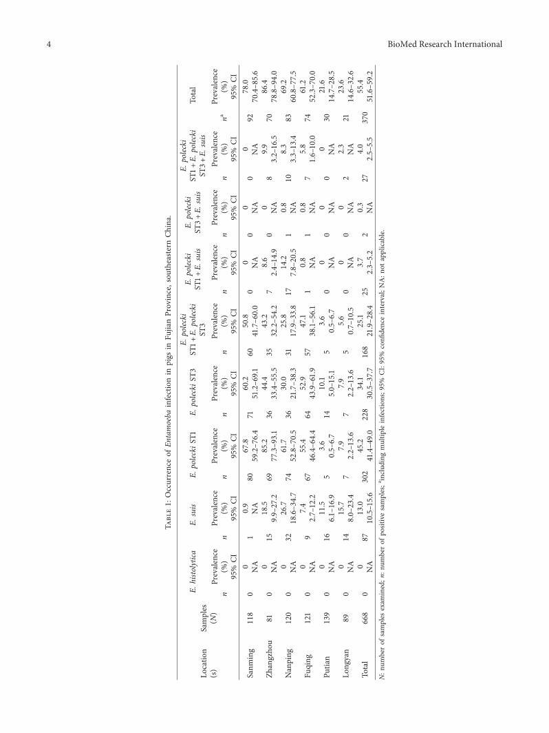
3.1. Prevalence of Entamoeba spp. A total of 370 of 668samples (55.4%, 95% CI 51.6% to 59.2%) were positive forEntamoeba spp. by nested PCR (Table 1). E. suis and E.polecki were identified in fecal samples, but samples with E.histolytica were not found in this study./e overall infectionrate of E. polecki ST1 was the highest (302/668, 45.2%, 95%CI 41.4% to 49.0%), while the overall infection rate of E. suiswas the lowest (87/668; 13.0%, 95% CI 10.5% to 15.6%). /ecoinfection rate of E. polecki ST1 and E. polecki ST3 was thehighest (168/668; 25.1%, 95% CI 21.9% to 28.4%), while thecoinfection rate of E. polecki ST3 and E. suis was the lowest(2/668; 0.3%).
Analysis of the infection rates of Entamoeba spp. indifferent sampling areas showed that there were regionaldifferences (χ2 �167.453, P< 0.05), with the rates beingmuch lower in Putian and Longyan than in other regions.
3.2. Distribution of Entamoeba spp. in Different De-velopmental Stages of Swine. /e detailed data of distribu-tion of Entamoeba spp. are shown in Table 2. Analysis of theinfection rates of Entamoeba spp. in different developmentalstages showed that there was a developmental stage pre-disposition to infection with Entamoeba spp. (χ2 � 50.362,P< 0.05), with the rates being much lower in suckling pigsthan in other developmental stages.
3.3. Phylogenetic and Sequencing Analysis of Entamoeba spp.�e positive product of E histolytica was not ampli ed bynested PCR in all samples. Representative sequences weresubmitted to GenBank under accession numbers MK347346(E. suis), MK347347 (E. polecki ST3), and MK357717 (E.polecki ST1). Meanwhile, the three representative sequencesdisplayed 100% sequence identity to other obtained se-quences of PCR-positive samples of E. suis and E. polecki ST1and ST3 in this study. �e sequence of E. suis (MK347346)was identical to the sequence isolated from pigs(DQ286372). �e representative sequences of E. polecki ST1(MK357717) and E. polecki ST3 (MK347347) were almostidentical to reference sequences of E. polecki ST1 (AF149913)and E. polecki ST3 (LC067574), respectively, and comparedwith the corresponding reference sequences, each currentsequence has 1 di�erent substitution. We chose knownsequences [12] to build the phylogenetic tree of the E. poleckisubtypes detected in the current study, and the resultsshowed that MK357717 shared a common clade withAF149913 (E. polecki ST1) and MK347347 shared a commonclade with LC067574 (E. polecki ST3) (Figure 2).
4. Discussion
Traditional microscopic examination is the most commonlyused clinical diagnostic tool for examining the presence ofEntamoeba organisms in fresh or xed stool samples[1, 7, 21, 22]. However, several distinct Entamoeba spp. with
similar morphological characteristics (for instance, the E.dispar, a nonpathogenic species, is morphologically identicalto E. histolytica) cannot be distinguished by microscopicexamination alone [1, 2, 4, 7]. �erefore, accurate identi -cation of species/subtypes of Entamoebawas performed withmolecular tools including PCR and nucleotide sequencing[1, 7, 12, 19, 23, 24].
In this study, the prevalence of Entamoeba spp. rangedfrom 21.6% to 86.4% in di�erent regions of Fujian Province,southeastern China, and there were signi cant di�erences inthe infection rates in the six areas (P< 0.05). �e causes ofthese di�erences may be related to managing technology,breeding conditions, health status, and the water sources onfarms. Moreover, the overall infection rate of Entamoebaspp. in this study is higher (55.4%) than the rate reported inKorea (5/136, 3.7%) [25], Iran (1/12, 8%; 2/12, 17%; 8% for E.suis and 17% for E. polecki) [26], Cambodian (24/76, 31.6%)[27], Germany (267/514, 52%) [28], and eastern China(45.8%) [12], but it is lower than that reported in Vietnam(11/12, 91.67%) [29]. �ese di�erences may be due to thedi�erent geographical variations, climates, and detectionprocedures.
�e phylogenetic analysis indicated that the isolates fromthe samples for E. polecki were E. polecki ST1 and E. poleckiST3. Infection with E. polecki ST1 was the most common(45.2%) in the present study, which was consistent with theobservations reported in Indonesia, Vietnam, and easternChina [12, 20, 29]. Mixed infections, including infection with
China
JiangxiNanping
Zhejiang
Fuqing
Taiwan
Putian
Sanming
Longyan
Zhangzhou
Guangdong
Miles
Hunan
0 20 40 120 16080
N
RiverSampling siteFujian Province
Figure 1: Sampling areas in Fujian Province, southeastern China.
BioMed Research International 3
Tabl
e1:
Occurrenceof
Entamoeba
infectionin
pigs
inFu
jianProvince,sou
theasternChina.
Locatio
n(s)
Samples
(N)
E.histolytica
E.suis
E.poleckiS
T1E.
poleckiS
T3E.
polecki
ST1+E.
polecki
ST3
E.polecki
ST1+E.
suis
E.polecki
ST3+E.
suis
E.polecki
ST1+E.
polecki
ST3+E.
suis
Total
nPrevalence
(%)
nPrevalence
(%)
nPrevalence
(%)
nPrevalence
(%)
nPrevalence
(%)
nPrevalence
(%)
nPrevalence
(%)
nPrevalence
(%)
naPrevalence
(%)
95%
CI
95%
CI
95%
CI
95%
CI
95%
CI
95%
CI
95%
CI
95%
CI
95%
CI
Sanm
ing
118
00
10.9
8067.8
7160.2
6050.8
00
00
00
9278.0
NA
NA
59.2–7
6.4
51.2–6
9.1
41.7–6
0.0
NA
NA
NA
70.4–8
5.6
Zhangzho
u81
00
1518.5
6985.2
3644.4
3543.2
78.6
00
89.9
7086.4
NA
9.9–
27.2
77.3–9
3.1
33.4–5
5.5
32.2–5
4.2
2.4–14.9
NA
3.2–16.5
78.8–9
4.0
Nanping
120
00
3226.7
7461.7
3630.0
3125.8
1714.2
10.8
108.3
8369.2
NA
18.6–3
4.7
52.8–7
0.5
21.7–3
8.3
17.9–3
3.8
7.8–
20.5
NA
3.3–13.4
60.8–7
7.5
Fuqing
121
00
97.4
6755.4
6452.9
5747.1
10.8
10.8
75.8
7461.2
NA
2.7–12.2
46.4–6
4.4
43.9–61.9
38.1–5
6.1
NA
NA
1.6–10.0
52.3–7
0.0
Putia
n139
00
1611.5
53.6
1410.1
53.6
00
00
00
3021.6
NA
6.1–
16.9
0.5–
6.7
5.0–15.1
0.5–
6.7
NA
NA
NA
14.7–2
8.5
Long
yan
890
014
15.7
77.9
77.9
55.6
00
00
22.3
2123.6
NA
8.0–
23.4
2.2–13.6
2.2–13.6
0.7–10.5
NA
NA
NA
14.6–3
2.6
Total
668
00
8713.0
302
45.2
228
34.1
168
25.1
253.7
20.3
274.0
370
55.4
NA
10.5–15.6
41.4–4
9.0
30.5–3
7.7
21.9–2
8.4
2.3–
5.2
NA
2.5–
5.5
51.6–5
9.2
N:n
umberof
samples
exam
ined;n
:num
berof
positivesamples;ainclud
ingmultip
leinfections;9
5%CI:95%
confi
denceinterval;N
A:n
otapplicable.
4 BioMed Research International
Tabl
e2:
Entamoeba
spp.
detected
amon
gdifferent
pigdevelopm
entalstage
grou
psin
FujianProvince,sou
theasternChina.
Growing
stage(s)
Samples
(N)
E.histolytica
E.suis
E.poleckiS
T1E.
poleckiS
T3E.
polecki
ST1+E.
polecki
ST3
E.polecki
ST1+E.
suis
E.polecki
ST3+E.
suis
E.polecki
ST1+E.
polecki
ST3+E.
suis
Total
nPrevalence
(%)
nPrevalence
(%)
nPrevalence
(%)
nPrevalence
(%)
nPrevalence
(%)
nPrevalence
(%)
nPrevalence
(%)
nPrevalence
(%)
naPrevalence
(%)
95%
CI
95%
CI
95%
CI
95%
CI
95%
CI
95%
CI
95%
CI
95%
CI
95%
CI
Weaned
piglet
116
00
21.7
6455.2
6455.2
4538.8
00
00
21.7
8169.8
NA
NA
46.0–6
4.4
46.0–6
4.4
29.8–4
7.8
NA
NA
NA
61.3–7
8.3
Sucking
piglet
105
00
32.9
2120.0
1211.4
76.7
11.0
11.0
00
2725.7
NA
NA
12.2–2
7.8
5.2–17.6
1.8–11.5
NA
NA
NA
17.2–3
4.2
Sow
280
00
6222.1
139
49.6
8630.7
6422.9
217.5
10.4
186.4
167
59.6
NA
17.2–2
7.0
43.8–5
5.5
25.3–3
6.2
17.9–2
7.8
4.4–10.6
NA
3.5–
9.3
53.9–6
5.4
Boar
280
00
1346.4
1242.9
1139.3
00
00
00
1450.0
NA
NA
26.7–6
6.1
23.3–6
2.4
20.0–5
8.6
NA
NA
NA
30.3–6
9.7
Nursery
pig
900
012
13.3
4550.0
4246.7
2932.2
22.2
00
77.8
5460.0
NA
6.2–
20.5
39.5–6
0.5
36.2–5
7.2
22.4–4
2.1
NA
NA
2.1–
13.4
49.7–7
0.3
Fatte
ning
pig
490
08
16.3
2040.8
1224.5
1224.5
12.0
00
00
2755.1
NA
5.6–
27.1
26.6–5
5.1
12.0–3
7.0
12.0–3
7.0
NA
NA
NA
40.7–6
9.5
Total
668
00
8713.0
302
45.2
228
34.1
168
25.1
253.7
20.3
274.0
370
55.4
NA
10.5–15.6
41.4–4
9.0
30.5–3
7.7
21.9–2
8.4
2.3–
5.2
NA
2.5–
5.5
51.6–5
9.2
N:n
umberof
samples
exam
ined;n
:num
berof
positivesamples;ainclud
ingmultip
leinfections;9
5%CI:95%
confi
denceinterval;N
A:n
otapplicable.
BioMed Research International 5
E. suis and E. polecki ST1, E. suis and E. polecki ST3, E. poleckiST1 and E. polecki ST3, and E. suis, E. polecki ST1, and E.polecki ST3, were observed in the study. /is result suggeststhat there is no competitive exclusion among these threespecies/subtypes (E. suis, E. polecki ST1, and E. polecki ST3). Inaddition, this is the first report regardingmixed infections withE. suis and E. polecki ST1 and ST3 in China. Infection with E.histolyticawas not observed in farmed pigs in this study, whichwas consistent with the previous research [10–12].
Traditionally, E. suis was considered to be mostly re-stricted to pigs [2, 3, 15, 16]. However, the sequence of anEntamoeba isolated from a gorilla (FR686456) was similar tothe sequence of E. suis (DQ286372) with one substitution[17], so whether E. suis only infects pigs should be verified bymore studies. /e results show that pig infection withEntamoeba spp. was related to the sampling areas and thedevelopmental stages of swine (P< 0.05), but this is not inagreement with the observation made in pigs by Li et al.(there was no age predisposition in pigs) [12]. /erefore,more research studies are needed to confirm whethersampling area and types of swine are risk factors for Ent-amoeba spp. infection. /ere were only detected threeEntamoeba species (E. histolytica, E. suis, and E. polecki ST1
and E. polecki ST3) in this study, and more research studiesare needed to determine prevalence and genetic identifi-cation of other species/subtypes in pigs in China in thefuture.
5. Conclusion
/epresent study conducted a prevalence survey andmolecularidentification of three Entamoeba species in pigs in southeasternChina. /e overall infection rate of Entamoeba spp. was 55.4%.E. suis and zoonotic E. polecki ST1 and E. polecki ST3 have beenfound in pigs. /us, further attention should be paid to the riskof the transmission of Entamoeba spp. between animal reser-voirs and humans. /e statistical analysis (SPSS) suggested thatsampling areas and developmental stages of swine are associatedwith swine infection with three Entamoeba species. /is is thefirst report ofmixed infectionswithE. suis,E. polecki ST1, andE.polecki ST3 in China.
Data Availability
/e data used to support the findings of this study are in-cluded within the article.
FN666249.1 E. bovis cattleFN666251.1 E. bovis cattle
AF149906.1 E. moshkovskii humanAF149907.1 E. hartmanni human
DQ286373.1 E. ecuadoriensis sewageAB282657.1 E. nuttalli monkey
X64142.1 E. histolytica humanZ49256.1 E. dispar human
AF149910.1 E. terrapinae turtleDQ286372.1 E. suis pig
D28490.1 E. gingivalis human
AF149914.1 E. coli humanFR686364.1 E. coli human
AF149913.1 E. polecki pigFR686383.1 E. polecki human
AJ566411.2 E. struthionis ostrich
95
94
93
48
100 99
8273
99
9346
38
83
43
3648
9776
75
99
LC067574.1 E. polecki pig
AF149912.1 E. chattoni monkey
FR686398.1 E. polecki humanFR686400.1 E. polecki humanFR686357.1 E. polecki humanFR686392.1 E. polecki human
AF149908.1 E. ranarum frogAF149905.1 E. invadens snake
MK357717
MK347347
ST1
ST3
ST4
ST2
0.020
Figure 2: Phylogenetic relationships of Entamoeba polecki isolates identified in this study. /e isolates obtained in this research areindicated by circles.
6 BioMed Research International
Conflicts of Interest
/e authors declare that there are no conflicts of interestregarding the publication of this article.
Authors’ Contributions
Ting Ji and Hao-Xuan Cao contributed equally to this work.
Acknowledgments
/is project was supported by the National Natural ScienceFoundation of China (Grant no. 31672549), the Open Fundof the Key Laboratory of Fujian Province Livestock Epi-demic Prevention, Control and Biological Technology(2018KF02), and the Undergraduate Innovation and En-trepreneurship Training Program of Fujian Province (no.201910389065).
References
[1] R. Ngui, L. Angal, S. Fakhrurrazi et al., “DifferentiatingEntamoeba histolytica, Entamoeba dispar and Entamoebamoshkovskii using nested polymerase chain reaction (PCR) inrural communities in Malaysia,” Parasites & Vectors, vol. 5,no. 1, p. 187, 2012.
[2] M. Matsubayashi, N. Murakoshi, T. Komatsu, M. Tokoro,M. Haritani, and T. Shibahara, “Genetic identification ofEntamoeba polecki subtype 3 from pigs in Japan and char-acterisation of its pathogenic role in ulcerative colitis,” In-fection, Genetics and Evolution, vol. 36, pp. 8–14, 2015.
[3] M. Matsubayashi, K. Kanamori, M. Sadahiro et al., “Firstmolecular identification of Entamoeba polecki in a piglet inJapan and implications for aggravation of ileitis by coinfectionwith Lawsonia intracellularis,” Parasitology Research, vol. 114,no. 8, pp. 3069–3073, 2015.
[4] H. M. Elsheikha, C. S. Regan, and C. G. Clark, “Novel Ent-amoeba findings in nonhuman primates,” Trends in Parasi-tology, vol. 34, no. 4, pp. 283–294, 2018.
[5] J. A. Walsh, “Problems in recognition and diagnosis of am-ebiasis: estimation of the global magnitude of morbidity andmortality,” Clinical Infectious Diseases, vol. 8, no. 2,pp. 228–238, 1986.
[6] J. Nath, S. K. Ghosh, B. Singha, and J. Paul, “Molecular ep-idemiology of amoebiasis: a cross-sectional study amongnorth east Indian population,” PLOS Neglected TropicalDiseases, vol. 9, no. 12, Article ID e0004225, 2015.
[7] H. Dong, J. Li, M. Qi et al., “Prevalence, molecular epide-miology, and zoonotic potential of Entamoeba spp. in non-human primates in China,” Infection, Genetics and Evolution,vol. 54, pp. 216–220, 2017.
[8] M. Martınez-Castillo, J. Pacheco-Yepez, N. Flores-Huertaet al., “Flavonoids as a natural treatment against Entamoebahistolytica,” Frontiers in Cellular and Infection Microbiology,vol. 8, p. 209, 2018.
[9] G.-Z. He, Y. Feng, and S.-X. Deng, “Evaluation of the in-testinal microbial diversity in miniature pig after orally in-fected with Entamoeba histolytica,” Parasitology Research,vol. 111, no. 2, pp. 939–941, 2012.
[10] M. Matsubayashi, F. Suzuta, Y. Terayama et al., “Ultra-structural characteristics and molecular identification ofEntamoeba suis isolated from pigs with hemorrhagic colitis:
implications for pathogenicity,” Parasitology Research,vol. 113, no. 8, pp. 3023–3028, 2014.
[11] Y. Hirashima, T.Manchanayake, T. Yano et al., “Developmentof molecular diagnostic protocols for detecting three types ofEntamoeba from diarrheal and asymptomatic pigs and en-vironmental moist soils,” Parasitology Research, vol. 116,no. 7, pp. 2001–2007, 2017.
[12] W.-C. Li, J.-Z. Geng, C. Chen et al., “First report on theoccurance of intestinal Entamoeba spp. in pigs in China,”ActaTropica, vol. 185, pp. 385–390, 2018.
[13] G. A. Noble and E. R. Noble, “Entamoebae in farmmammals,”=e Journal of Parasitology, vol. 38, no. 6, pp. 571–595, 1952.
[14] R. S. Desowitz and G. Barnish, “Entamoeba poleckiand otherintestinal protozoa in Papua New Guinea highland children,”Annals of Tropical Medicine & Parasitology, vol. 80, no. 4,pp. 399–402, 1986.
[15] C. G. Clark, F. Kaffashian, B. Tawari et al., “New insights intothe phylogeny of Entamoeba species provided by analysis offour new small-subunit rRNA genes,” International Journal ofSystematic and Evolutionary Microbiology, vol. 56, no. 9,pp. 2235–2239, 2006.
[16] Z. Cui, J. Li, Y. Chen, and L. Zhang, “Molecular epidemiology,evolution, and phylogeny of Entamoeba spp,” Infection, Ge-netics and Evolution, vol. 75, p. 104018, 2019.
[17] C. R. Stensvold, M. Lebbad, E. L. Victory et al., “Increasedsampling reveals novel lineages of Entamoeba: consequencesof genetic diversity and host specificity for taxonomy andmolecular detection,” Protist, vol. 162, no. 3, pp. 525–541,2011.
[18] J. J. Verweij, A. M. Polderman, and C. G. Clark, “Geneticvariation among human isolates of uninucleated cyst-pro-ducing Entamoeba species,” Journal of Clinical Microbiology,vol. 39, no. 4, pp. 1644–1646, 2001.
[19] C. R. Stensvold, J. Winiecka-Krusnell, T. Lier, and M. Lebbad,“Evaluation of a PCR method for detection of Entamoebapolecki, with an overview of its molecular epidemiology,”Journal of Clinical Microbiology, vol. 56, no. 5, Article IDe00154, 2018.
[20] J. Tuda, M. Feng, M. Imada, S. Kobayashi, X. Cheng, andH. Tachibana, “Identification ofEntamoeba poleckiwithunique 18S rRNA gene sequences from Celebes crestedmacaques and pigs in tangkoko nature reserve, north sulawesi,Indonesia,” Journal of Eukaryotic Microbiology, vol. 63, no. 5,pp. 572–577, 2016.
[21] R. Y. W. Kouassi, S. W. McGraw, P. K. Yao et al., “Diversityand prevalence of gastrointestinal parasites in seven non-human primates of the Taı National Park, Cote d’Ivoire,”Parasite, vol. 22, p. 1, 2015.
[22] M.-J. Kim, B.-K. Jung, J. Cho et al., “Prevalence of intestinalProtozoans among schoolchildren in suburban areas nearyangon, Myanmar,” =e Korean Journal of Parasitology,vol. 54, no. 3, pp. 345–348, 2016.
[23] M. Matsubayashi, Y. Sasagawa, T. Aita et al., “First report ofmixed Entamoeba polecki (ST1) and E. suis infection in pigletsshedding abnormalfeces by histopathological and molecularsurveys,” Acta Parasitologica, vol. 61, no. 4, pp. 665–670, 2016.
[24] M. Matsubayashi, Y. Matsuura, S. Nukata et al., “First de-tection and molecular identification of Entamoeba bovis fromJapanese cattle,” Parasitology Research, vol. 117, no. 1,pp. 339–342, 2018.
[25] H. A. H. A. Ismail, H.-K. Jeon, Y.-M. Yu, C. Do, and Y.-H. Lee,“Intestinal parasite infections in pigs and beef cattle in ruralareas of chungcheongnam-do, Korea,” =e Korean Journal ofParasitology, vol. 48, no. 4, pp. 347–349, 2010.
BioMed Research International 7
[26] S. Solaymani-Mohammadi, M. Rezaian, H. Hooshyar,G. R. Mowlavi, Z. Babaei, and M. A. Anwar, “Intestinalprotozoa in wild boars (Sus scrofa) in western Iran,” Journal ofWildlife Diseases, vol. 40, no. 4, pp. 801–803, 2004.
[27] F. Schar, T. Inpankaew, R. J. Traub et al., “/e prevalence anddiversity of intestinal parasitic infections in humans anddomestic animals in a rural Cambodian village,” ParasitologyInternational, vol. 63, no. 4, pp. 597–603, 2014.
[28] I. M. Damriyasa and C. Bauer, “Prevalence and age-dependentoccurrence of intestinal protozoan infections in sucklingpiglets,” Berl Munch Tierarztl Wochenschr, vol. 119, no. 7-8,pp. 287–290, 2006.
[29] A. S. Jacob, E. J. Busby, A. D. Levy, N. Komm, and C. G. Clark,“Expanding the Entamoeba Universe: new hosts yield novelribosomal lineages,” Journal of Eukaryotic Microbiology,vol. 63, no. 1, pp. 69–78, 2016.