26
Pediatric radiology Radiodiagnostická klinika Klinika dětského a dorostového lékařství VFN a 1.LF UK VFN
Pediatric radiology
Radiodiagnostická klinikaKlinika dětského a dorostového lékařství
VFN a 1.LF UK VFN
Child is not a small adult
• Limited cooperation and insight into theimportance of the examination
– Does not stay without moving
• MR, CT
• Presence of parents
• Different dif. dig. (newborn pneumopathy…)
• Radiosenzitivity
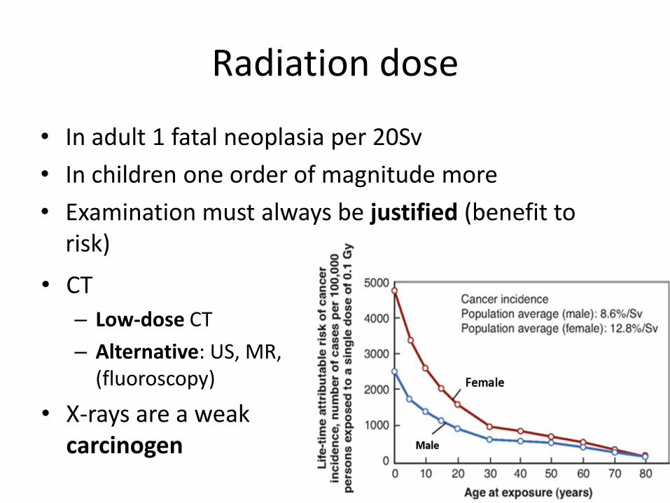
Radiation dose
• CT
– Low-dose CT
– Alternative: US, MR, (fluoroscopy)
• X-rays are a weakcarcinogen
• In adult 1 fatal neoplasia per 20Sv
• In children one order of magnitude more
• Examination must always be justified (benefit to risk)
Contrast
• Fluoroscopy:– Iodine based c.m. in newborns and small babies
– Barium• Never in susp. peforation, SBO, risk of inspisation
• Never <2 months of age
• CT:– Non-ionic
• 1 – 2 ml/kg
– Contrast nephropathy
– Allergy, preparation
Imaging methods
• US: echogenicity: an-, hypo-, izo-, hyper-echoic
• Radiograph: transparency
• MRI: a-, hypo-, izo-, hyper-signal
• CT: density: hypo-, izo-, hyper-dense
• Angiography: DSA, interventional
• Interventional radiology
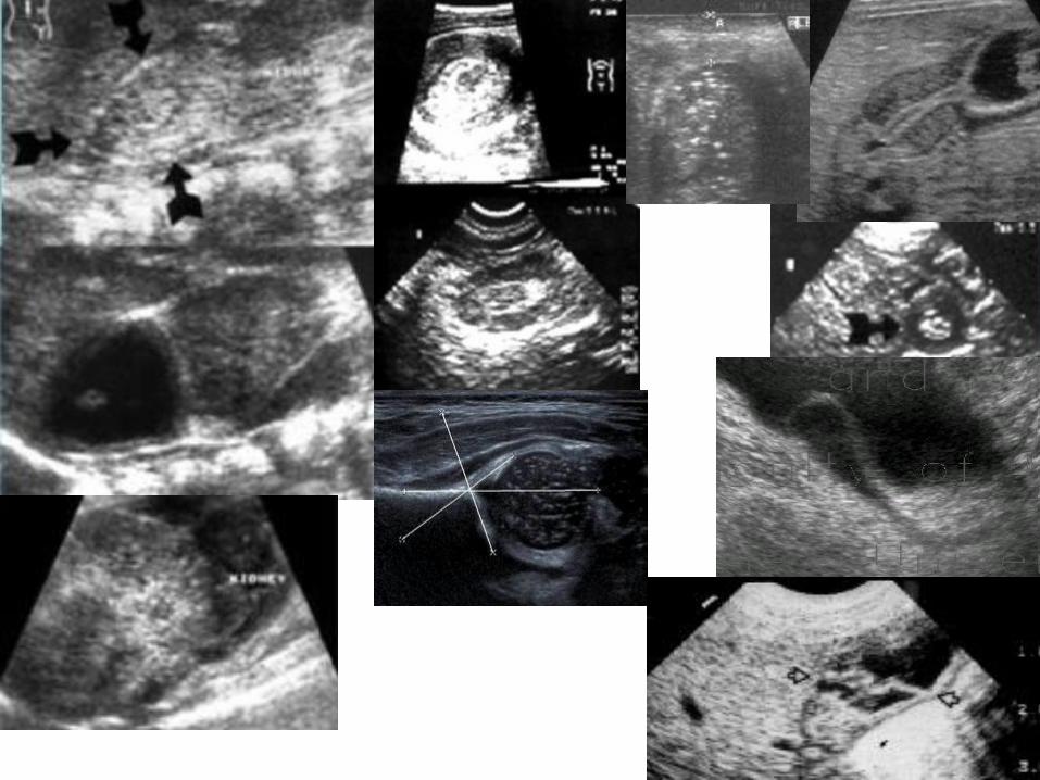
UZ• First-line examination method in many cases• Often final diagnosis
– Pylorostenosis (3mm, 18mm, sign of olive)– Prepyloric membrane– Adrenal lesions– Ureterocoele– Folcal liver lesions, bile ducts …– Abdominal tumours– Intussuception– Anorectal malformatiom– (Reflux)– Echocardiography– Enteritis, colitis (NEC)– Appendicitis– Testes – torsion, spermatocoele– Even ... SBO, pneumoperitoneum, pneumothorax, pleural fluid, joints, soft
tissue, LNN, thyroid ....– CN, UC, mesenteric lymfadenopathy– UZ brain in neonates
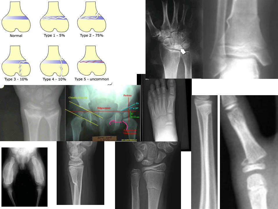
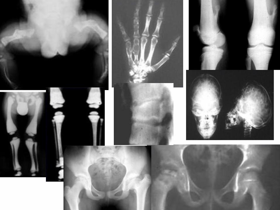
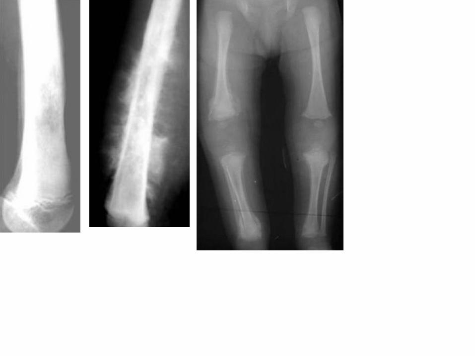
BONE
• Greenstick fracture• Epiphyseolysis• Bone tumours• Hip dysplasia• Cranial sutures• Aseptic nekrosis (Perthes, Osgood-Schlatter)• CRMO (chronic recurrent multifocal osteomyelitis)• Scoliosis, m. Scheuermann• Avitaminosis (Rachitis)• CAN – skeletal survey
– Avulsion, periostosis sec. to subperiost hematomas, multiple fractures (various degrees ofhealing), epiphyseolyses, avulsion of a bone edge (twisting), spiral fractures
• Bone age• Birth trauma – fracture of clavicle, femur• Systemic diseases – osteogenesis imperfecta, osteopetroza, dysostosis multiplex
(Mukopolysacharidosis)• Rheumatological disorders
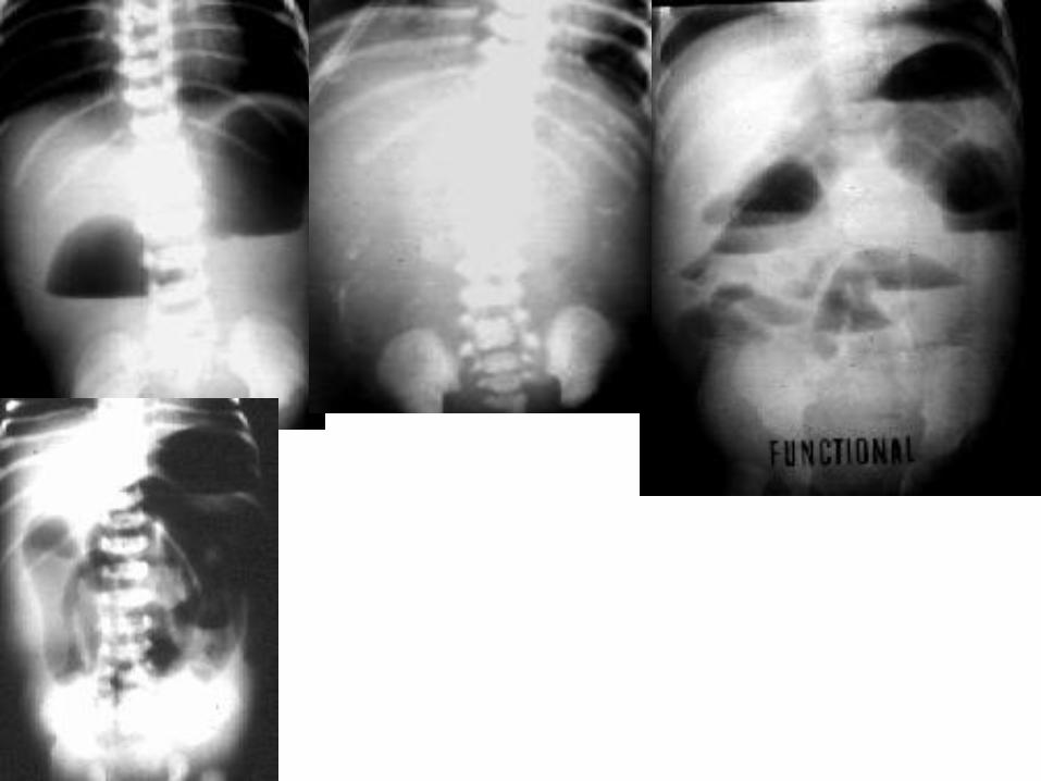
Abdominal radiograph
• Distribution of gas in bowel loops• SBO = distension and air-fluid levels
– Horizontal x-ray
• Meconium ileus = dry ileus – no air-fluid levels, CF (meconium < 48h)• Duodenal, jejunal atresia• Volvulus• Pneumoperitoneum• Foreign body• Catheter location• Nephrogram• Necrotizing enterocolitis – gas in portovenous system and bowel wall• Other organs are also visible on plain radiograph!
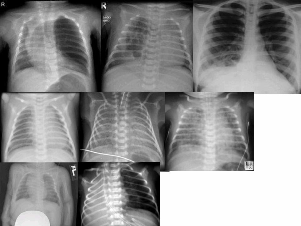
Chest x-ray• Newborn pneumopathy
– Transient tachpnea of newborn
• Delayed resorption of amniotic fluid
– Respiratory distress syndrom (RDS)
• Preterm, insufficient amount of surfactant
– Bronchopulmonary dysplasia
• Toxic O2 in long term ventilation
• Congenital lobar emphysema
• Sequestration
• Pneumonia
• Meconium aspiration
• Diaphragmatic hernia
• Heart vitia - cardiomegally, hyperemia due to recirculation
• Inflammatory changes of lung: lobar / alar pneumonia, pleuropneumonia, bronchopneumonia, atypicalpneumonia
• Aspiration of foreign body
• Asthma
• PNO, pneumomediastinum ...
• Thymus
• Thymic hyperplasia
• Lung agenesis, hypoplasia
• Fluidothorax
Other radiographs
• Semiaxial skull – Waters projection (paranasalsinuses)
– Development of paranasal sinuses in time
– Sinusitis
• air-fluid levels, decreased transparency
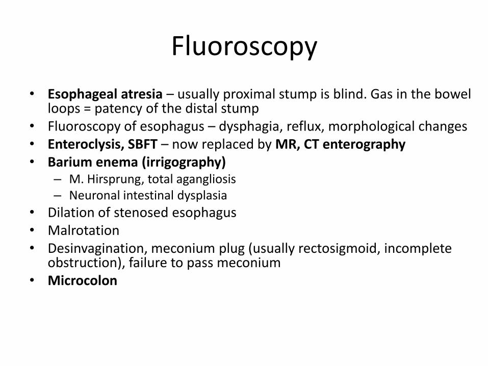
Fluoroscopy
• Esophageal atresia – usually proximal stump is blind. Gas in the bowel loops = patency of the distal stump
• Fluoroscopy of esophagus – dysphagia, reflux, morphological changes• Enteroclysis, SBFT – now replaced by MR, CT enterography• Barium enema (irrigography)
– M. Hirsprung, total agangliosis– Neuronal intestinal dysplasia
• Dilation of stenosed esophagus• Malrotation• Desinvagination, meconium plug (usually rectosigmoid, incomplete
obstruction), failure to pass meconium• Microcolon
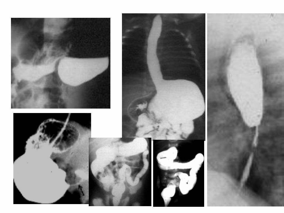
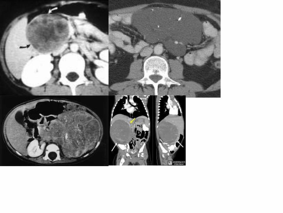
CT
• Judicious indication - radiation
• Special pediatric protocols– Decreased voltage and amps
• Uncooperative children – assitance of anesthesiologist
• Low dose protocols with iterative reconstruction
• CT can hardly be replaced in – HRCT of lungs
– Acute intracranial hemorrhage - CT of brain
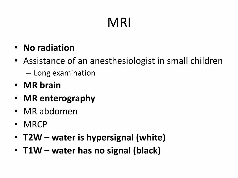
MRI
• No radiation
• Assistance of an anesthesiologist in small children– Long examination
• MR brain
• MR enterography
• MR abdomen
• MRCP
• T2W – water is hypersignal (white)
• T1W – water has no signal (black)
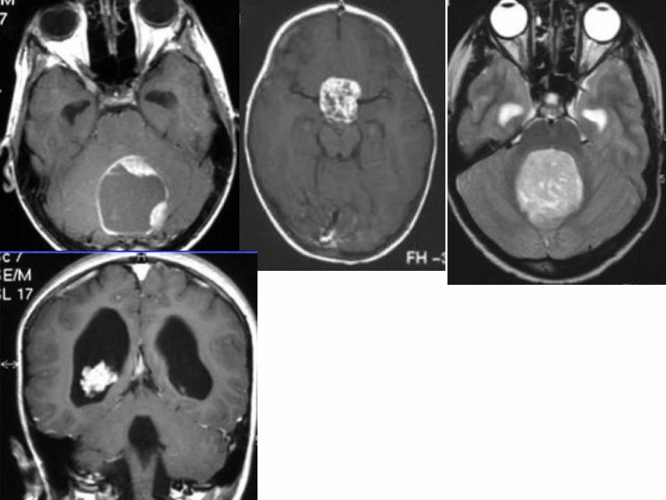
Brain tumors
• INTRAAXIAL x EXTRAAXIAL• Posterior fossa tumours more frequent• Pilocytic astrocytoma• other astrocytomas• ependymoma• meduloblastoma• kraniofaryngeoma• PNET• Papiloma of choroidal plexus
Uroradiology
• Voiding cystourethrogram– Anomally of urethra (valve, stenosis, diverticulum)
– Anomally of bladder (diverticulum, persistent urachus)
– Ureterocoele
– Vesicoureteral reflux (VUR)• Passive – when filling
• Active – when voiding
• Urolithiasis– Nephrogram (radiograph) – KUB (kidney-ureter-bladder)
– CT nephrogram
• Intravenous (excretory) urography, CT urography
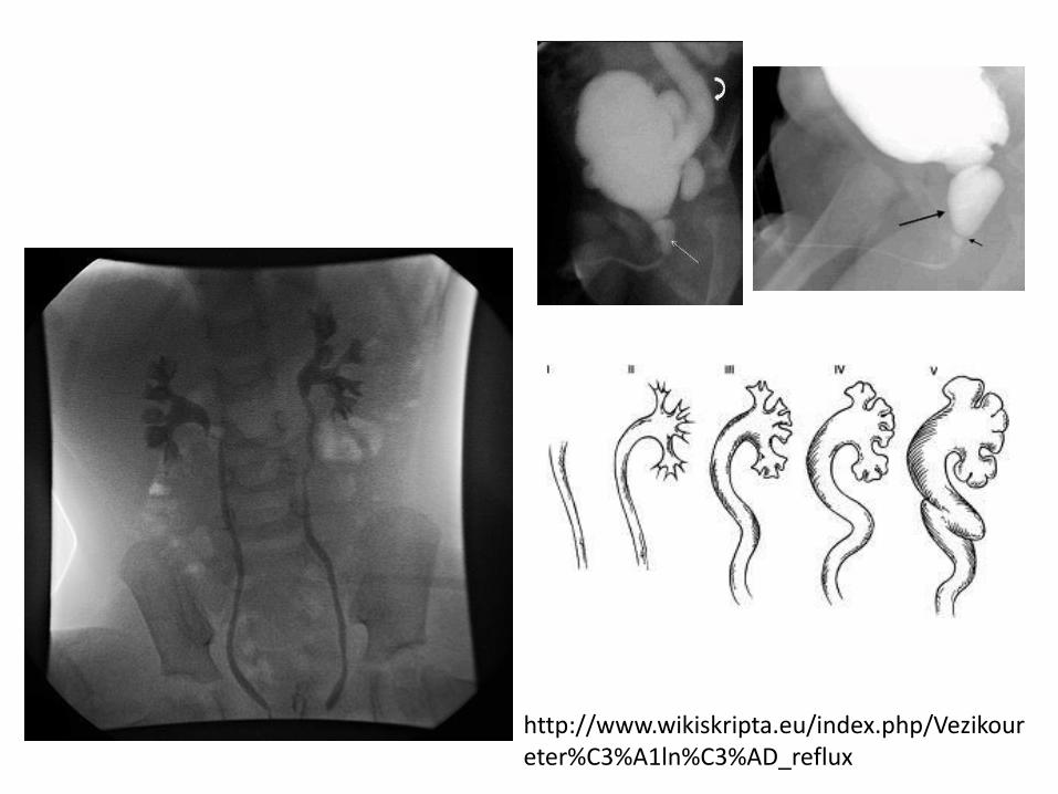
http://www.wikiskripta.eu/index.php/Vezikoureter%C3%A1ln%C3%AD_reflux
• www.mudr.org/web/prednasky


