395 Korean J Radiol 14(3), May/Jun 2013 kjronline.org INTRODUCTION Neuroendocrine tumors are derived from neuroendocrine cells and the most common site is the bronchopulmonary system. Theoretically, primary neuroendocrine tumors do not occur in the breast due to the lack of endocrine cells. Although there are many published studies regarding this rare pathological entity, there are a few reports that also include radiology findings. However, most of the published studies of patients with radiology findings are of the small cell type of tumor, and the characteristic imaging features are not described well (1-7). We report the imaging findings in a case of primary neuroendocrine tumor (large cell neuroendocrine carcinoma with atypical carcinoid features) Primary Neuroendocrine Tumor of the Breast: Imaging Features Eun Deok Chang, MD 1 , Min Kyun Kim, MD 2 , Jeong Soo Kim, MD 3 , In Yong Whang, MD 2 Departments of 1 Clinical Pathology, 2 Radiology and 3 Surgery, Uijeongbu St. Mary’s Hospital, College of Medicine, The Catholic University of Korea, Uijeongbu 480-717, Korea Focal neuroendocrine differentiation can be found in diverse histological types of breast tumors. However, the term, neuroendocrine breast tumor, indicates the diffuse expression of neuroendocrine markers in more than 50% of the tumor cell population. The imaging features of neuroendocrine breast tumor have not been accurately described due to extreme rarity of this tumor type. We present a case of a pathologically confirmed, primary neuroendocrine breast tumor in a 42-year-old woman, with imaging findings difficult to be differentiated from that of invasive ductal carcinoma. Index terms: Neuroendocrine tumor; Breast; Mammography; Sonography; Magnetic resonance Received July 13, 2012; accepted after revision December 5, 2012. Corresponding author: In Yong Whang, MD, Department of Radiology, Uijeongbu St. Mary’s Hospital, College of Medicine, The Catholic University of Korea, 271 Cheonbo-ro, Uijeongbu 480-717, Korea. • Tel: (8231) 820-3136 • Fax: (8231) 846-3080 • E-mail: [email protected]This is an Open Access article distributed under the terms of the Creative Commons Attribution Non-Commercial License (http://creativecommons.org/licenses/by-nc/3.0) which permits unrestricted non-commercial use, distribution, and reproduction in any medium, provided the original work is properly cited. Korean J Radiol 2013;14(3):395-399 found in the breast. We also review the relevant medical literature. CASE REPORT A 42-year-old female was presented with a lump in her right breast, which she had been aware of for several weeks. On physical examination, an approximately 5-cm, firm, movable lump was palpated in the upper outer subareolar region of her right breast. There was also a palpable, enlarged lymph node in the right axilla. Mammography revealed a high-density mass with an ill defined margin in the 11 o’clock direction of the right breast subareolar area. Right axillary lymphadenopathy was noted on the mediolateral oblique view. No microcalcification was detected on mammography (Fig. 1A). Targeted sonography of the right breast revealed an irregularly shaped, lobulated marginated, heterogeneous echotextured mass with posterior enhancement. Heterogeneous echotexture was noted, especially in the center of the mass, and increased vascular flow was noted in the mass on color Doppler scanning. There was also lymphadenopathy in the right axilla with cortical thickening and loss of fatty hilum (Fig. 1B-D). The mass was categorized as breast imaging http://dx.doi.org/10.3348/kjr.2013.14.3.395 pISSN 1229-6929 · eISSN 2005-8330 Case Report | Breast Imaging
Neuroendocrine tumors are derived from neuroendocrine cells and the most common site is the bronchopulmonary system. Theoretically, primary neuroendocrine tumors do not occur in the breast due to the lack of endocrine cells. Although there are many published studies regarding this rare pathological entity, there are a few reports that also include radiology findings. However, most of the published studies of patients with radiology findings are of the small cell type of tumor, and the characteristic imaging features are not described well (1-7). We report the imaging findings in a case of primary neuroendocrine tumor (large cell neuroendocrine carcinoma with atypical carcinoid features)
Primary Neuroendocrine Tumor of the Breast: Imaging FeaturesEun Deok Chang, MD1, Min Kyun Kim, MD2, Jeong Soo Kim, MD3, In Yong Whang, MD2
Departments of 1Clinical Pathology, 2Radiology and 3Surgery, Uijeongbu St. Mary’s Hospital, College of Medicine, The Catholic University of Korea, Uijeongbu 480-717, Korea
Focal neuroendocrine differentiation can be found in diverse histological types of breast tumors. However, the term, neuroendocrine breast tumor, indicates the diffuse expression of neuroendocrine markers in more than 50% of the tumor cell population. The imaging features of neuroendocrine breast tumor have not been accurately described due to extreme rarity of this tumor type. We present a case of a pathologically confirmed, primary neuroendocrine breast tumor in a 42-year-old woman, with imaging findings difficult to be differentiated from that of invasive ductal carcinoma.Index terms: Neuroendocrine tumor; Breast; Mammography; Sonography; Magnetic resonance
Received July 13, 2012; accepted after revision December 5, 2012.Corresponding author: In Yong Whang, MD, Department of Radiology, Uijeongbu St. Mary’s Hospital, College of Medicine, The Catholic University of Korea, 271 Cheonbo-ro, Uijeongbu 480-717, Korea. • Tel: (8231) 820-3136 • Fax: (8231) 846-3080• E-mail: [email protected] is an Open Access article distributed under the terms of the Creative Commons Attribution Non-Commercial License (http://creativecommons.org/licenses/by-nc/3.0) which permits unrestricted non-commercial use, distribution, and reproduction in any medium, provided the original work is properly cited.
Korean J Radiol 2013;14(3):395-399
found in the breast. We also review the relevant medical literature.
CASE REPORT
A 42-year-old female was presented with a lump in her right breast, which she had been aware of for several weeks. On physical examination, an approximately 5-cm, firm, movable lump was palpated in the upper outer subareolar region of her right breast. There was also a palpable, enlarged lymph node in the right axilla. Mammography revealed a high-density mass with an ill defined margin in the 11 o’clock direction of the right breast subareolar area. Right axillary lymphadenopathy was noted on the mediolateral oblique view. No microcalcification was detected on mammography (Fig. 1A). Targeted sonography of the right breast revealed an irregularly shaped, lobulated marginated, heterogeneous echotextured mass with posterior enhancement. Heterogeneous echotexture was noted, especially in the center of the mass, and increased vascular flow was noted in the mass on color Doppler scanning. There was also lymphadenopathy in the right axilla with cortical thickening and loss of fatty hilum (Fig. 1B-D). The mass was categorized as breast imaging
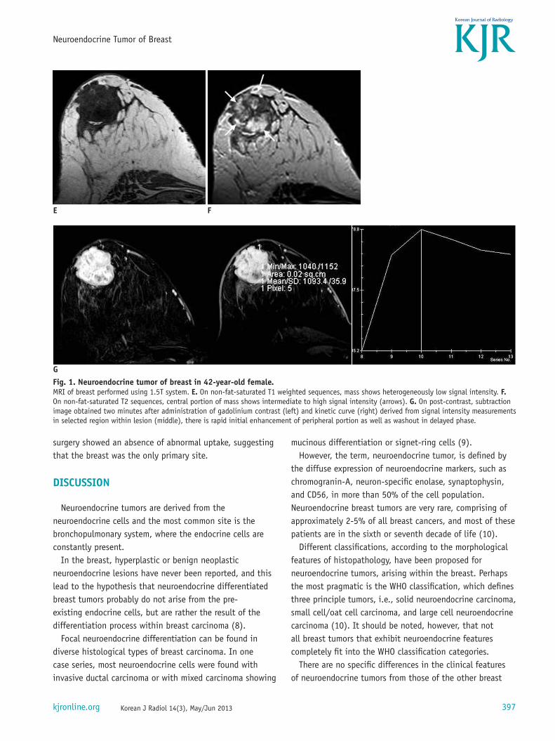
reporting and data system category 5, highly suggestive of malignancy. Fine needle aspiration cytology revealed highly cellular smears with monotonous hyperchromatic atypical small cells, suggesting high-grade invasive ductal carcinoma. MRI of the breast was performed using a 1.5T system. The mass appeared to be of heterogeneously low signal intensity on T1 weighted images and a few parts of the mass showed intermediate to high signal intensity on T2 weighted images. The T2 non-high signal area showed rapid enhancement within the first two minutes of gadolinium injection, and washout was noted (Fig. 1E-G).
The patient underwent a modified radical mastectomy and axillary dissection of the right breast. On the cut surface, the tumor appeared as a fairly well-defined but partially infiltrative, gray-white, solid mass with morphology similar to that of commonly seen, invasive ductal carcinoma.
Histopathologically, several, large, mucoid, necrotic areas with foamy macrophages were surrounded by dark, solid, and trabecular pattern tumor cells. The tumor was composed of a mostly solid growth of large to intermediate, polygonal or occasionally spindle cells, which showed organoid, nesting, trabecular, rosette-like, and palisading patterns. Immunohistochemically, the tumor cells were diffusely positive for synaptophysin and were focally positive for chromograin A, although they were negative for CD 56. E-cadherin was demonstrated in cytoplasmic membranes (Fig. 1H-K). Based on these pathology findings, we considered this tumor to be a large cell neuroendocrine carcinoma with atypical carcinoid features (histologic grade 3, T2N1M0, estrogen receptor [ER] [+], progesterone receptor [PR] [+], human epidermal growth factor receptor 2 [-]). Positron emission tomography - computed tomography after
A
B C D
Fig. 1. Neuroendocrine tumor of breast in 42-year-old female.A. Mammography shows high-density mass with ill defined margin in subareolar area of right breast. Enlarged axillary lymph node is noted in right mediolateral oblique view (arrow). B. Sonography reveals irregularly shaped, microlobulated marginated, heterogeneously echo-textured mass with posterior enhancement. C. Increased vascular flow is noted on color Doppler scan. D. There is enlarged lymph node with cortical thickening and loss of fatty hilum in right axilla.
397
Neuroendocrine Tumor of Breast
Korean J Radiol 14(3), May/Jun 2013kjronline.org
surgery showed an absence of abnormal uptake, suggesting that the breast was the only primary site.
DISCUSSION
Neuroendocrine tumors are derived from the neuroendocrine cells and the most common site is the bronchopulmonary system, where the endocrine cells are constantly present.
In the breast, hyperplastic or benign neoplastic neuroendocrine lesions have never been reported, and this lead to the hypothesis that neuroendocrine differentiated breast tumors probably do not arise from the pre-existing endocrine cells, but are rather the result of the differentiation process within breast carcinoma (8).
Focal neuroendocrine differentiation can be found in diverse histological types of breast carcinoma. In one case series, most neuroendocrine cells were found with invasive ductal carcinoma or with mixed carcinoma showing
mucinous differentiation or signet-ring cells (9).However, the term, neuroendocrine tumor, is defined by
the diffuse expression of neuroendocrine markers, such as chromogranin-A, neuron-specific enolase, synaptophysin, and CD56, in more than 50% of the cell population. Neuroendocrine breast tumors are very rare, comprising of approximately 2-5% of all breast cancers, and most of these patients are in the sixth or seventh decade of life (10).
Different classifications, according to the morphological features of histopathology, have been proposed for neuroendocrine tumors, arising within the breast. Perhaps the most pragmatic is the WHO classification, which defines three principle tumors, i.e., solid neuroendocrine carcinoma, small cell/oat cell carcinoma, and large cell neuroendocrine carcinoma (10). It should be noted, however, that not all breast tumors that exhibit neuroendocrine features completely fit into the WHO classification categories.
There are no specific differences in the clinical features of neuroendocrine tumors from those of the other breast
E
G
F
Fig. 1. Neuroendocrine tumor of breast in 42-year-old female.MRI of breast performed using 1.5T system. E. On non-fat-saturated T1 weighted sequences, mass shows heterogeneously low signal intensity. F. On non-fat-saturated T2 sequences, central portion of mass shows intermediate to high signal intensity (arrows). G. On post-contrast, subtraction image obtained two minutes after administration of gadolinium contrast (left) and kinetic curve (right) derived from signal intensity measurements in selected region within lesion (middle), there is rapid initial enhancement of peripheral portion as well as washout in delayed phase.
398
Chang et al.
Korean J Radiol 14(3), May/Jun 2013 kjronline.org
malignancy, and endocrine hormone-related syndromes are extremely rare. Of interest is the increase in neuroendocrine markers, such as chromogranin A, in the blood (10). As metastatic neuroendocrine tumors of the breast are more common than that of primary neuroendocrine tumors of the breast, it is, therefore, important to differentiate primary breast neuroendocrine tumor from metastatic disease to the breast because of the differences in treatment focus. Primary neuroendocrine tumor of the breast can be diagnosed if the presence of a non-mammary primary site can be clinically ruled out or if an in situ component is histologically detected, or both.
The treatment of primary neuroendocrine tumors is highly variable, although surgery should always be considered as the first line of treatment. The prognosis for this rare tumor remains controversial. Recent reports, which only deal with small cell neuroendocrine carcinoma, indicate that the size,
Fig. 1. Neuroendocrine tumor of breast in 42-year-old female.H. Gross specimen shows fairly well-defined, but partially infiltrative, gray-white, solid mass. I. Large, mucoid, necrotic area with foamy macrophages is surrounded by tumor cells (arrows) (H&E, x 12.5). J. Neuroendocrine carcinoma shows diffuse, solid growth patterns with organoid, nesting, trabecular, rosette-like, and palisading features. K. Carcinoma cells are diffusely positive for synaptophysin.
H
J
I
K
stage of disease at the time of diagnosis, expression of the ER and PR, and the Ki-67 index are important determinants of the prognosis (11-13). There is no relevant report about the prognosis of primary large cell neuroendocrine carcinoma seen in our case. However, the prognosis of our case would be poor, considering tumor size, axillary metastasis, and histologic grade.
Previously published case reports with radiology findings (1-7) have also described the imaging features of neuroendocrine tumors. Günhan et al. (1) described that the common radiological features of breast neuroendocrine tumor include, a high-density mass with predominantly spiculated or lobulated margins on mammography and mostly irregular or microlobulated, homogeneously hypoechoic masses with normal sound transmission on sonography. However, including the cases of Günhan et al., the number of cases with radiology findings has been too small to allow
399
Neuroendocrine Tumor of Breast
Korean J Radiol 14(3), May/Jun 2013kjronline.org
generalization of the imaging features, and there is no case report with radiology and pathology findings similar to ours.
In this report we present a case of primary neuroendocrine tumor (large cell neuroendocrine carcinoma with atypical carcinoid features) of the breast, together with its mammographic, sonographic, and MRI features. However, the radiologic findings are hard to differentiate from those of much more commonly seen invasive ductal carcinoma. Reports of new cases will be necessary in order to determine the radiologic presentation of primary neuroendocrine tumor of the breast.
AcknowledgmentsWe would like to thank Bonnie Hami, MA (USA) for her
editorial assistance in the preparation of the manuscript.
REFERENCES
1. Günhan-Bilgen I, Zekioglu O, Ustün EE, Memis A, Erhan Y. Neuroendocrine differentiated breast carcinoma: imaging features correlated with clinical and histopathological findings. Eur Radiol 2003;13:788-793
2. Mariscal A, Balliu E, Díaz R, Casas JD, Gallart AM. Primary oat cell carcinoma of the breast: imaging features. AJR Am J Roentgenol 2004;183:1169-1171
3. Kitakata H, Yasumoto K, Sudo Y, Minato H, Takahashi Y. A case of primary small cell carcinoma of the breast. Breast Cancer 2007;14:414-419
M, et al. Neuroendocrine tumor in the breast. Radiat Med 2008;26:28-32
5. Irshad A, Ackerman SJ, Pope TL, Moses CK, Rumboldt T, Panzegrau B. Rare breast lesions: correlation of imaging and histologic features with WHO classification. Radiographics 2008;28:1399-1414
6. Zhang JY, Chen WJ. Bilateral primary breast neuroendocrine carcinoma in a young woman: report of a case. Surg Today 2011;41:1575-1578
7. An JK, Woo JJ, Kang JH, Kim EK. Small-cell neuroendocrine carcinoma of the breast. J Korean Surg Soc 2012;82:116-119
8. Maluf HM, Koerner FC. Carcinomas of the breast with endocrine differentiation: a review. Virchows Arch 1994;425:449-457
9. Sapino A, Righi L, Cassoni P, Papotti M, Pietribiasi F, Bussolati G. Expression of the neuroendocrine phenotype in carcinomas of the breast. Semin Diagn Pathol 2000;17:127-137
10. Tavassoli FA, Devilee P. Tumours of the breast. In: Tavassoli FA, Devilee P, eds. Pathology and genetics of tumours of the breast and female genital organs. World Health Organization Classification of Tumours Series. Lyon: IARC Press, 2003:32-34
11. Adegbola T, Connolly CE, Mortimer G. Small cell neuroendocrine carcinoma of the breast: a report of three cases and review of the literature. J Clin Pathol 2005;58:775-778
12. Sapino A, Righi L, Cassoni P, Papotti M, Gugliotta P, Bussolati G. Expression of apocrine differentiation markers in neuroendocrine breast carcinomas of aged women. Mod Pathol 2001;14:768-776
13. Yamaguchi R, Furusawa H, Nakahara H, Inomata M, Namba K, Tanaka M, et al. Clinicopathological study of invasive ductal carcinoma with large central acellular zone: special reference to magnetic resonance imaging findings. Pathol Int 2008;58:26-30