Proc. Natl. Acad. Sci. USAVol. 95, pp. 13363–13383, November 1998Nobel Lecture
Prions
STANLEY B. PRUSINER†
Departments of Neurology and of Biochemistry and Biophysics, University of California, San Francisco, CA 94143
ABSTRACT Prions are unprecedented infectious patho-gens that cause a group of invariably fatal neurodegenerativediseases by an entirely novel mechanism. Prion diseases maypresent as genetic, infectious, or sporadic disorders, all ofwhich involve modification of the prion protein (PrP). Bovinespongiform encephalopathy (BSE), scrapie of sheep, andCreutzfeldt–Jakob disease (CJD) of humans are among themost notable prion diseases. Prions are transmissible parti-cles that are devoid of nucleic acid and seem to be composedexclusively of a modified protein (PrPSc). The normal, cellularPrP (PrPC) is converted into PrPSc through a posttransla-tional process during which it acquires a high b-sheet content.The species of a particular prion is encoded by the sequenceof the chromosomal PrP gene of the mammals in which it lastreplicated. In contrast to pathogens carrying a nucleic acidgenome, prions appear to encipher strain-specific propertiesin the tertiary structure of PrPSc. Transgenetic studies arguethat PrPSc acts as a template upon which PrPC is refolded intoa nascent PrPSc molecule through a process facilitated byanother protein. Miniprions generated in transgenic miceexpressing PrP, in which nearly half of the residues weredeleted, exhibit unique biological properties and should fa-cilitate structural studies of PrPSc. While knowledge aboutprions has profound implications for studies of the structuralplasticity of proteins, investigations of prion diseases suggestthat new strategies for the prevention and treatment of thesedisorders may also find application in the more commondegenerative diseases.
The torturous path of the scientific investigation that led to anunderstanding of familial Creutzfeldt–Jakob disease (CJD)chronicles a remarkable scientific odyssey. By 1930, the highincidence of familial (f) CJD in some families was known (1,2). Almost 60 years were to pass before the significance of thisfinding could be appreciated (3–5). CJD remained a curious,rare neurodegenerative disease of unknown etiology through-out this period of three score years (6). Only with transmissionof disease to apes after inoculation of brain extracts preparedfrom patients who died of CJD did the story begin to unravel(7).
Once CJD was shown to be an infectious disease, relativelylittle attention was paid to the familial form of the disease sincemost cases were not found in families. It is interesting tospeculate how the course of scientific investigation might haveproceeded had transmission studies not been performed untilafter the molecular genetic lesion had been identified. Hadthat sequence of events transpired, then the prion concept,which readily explains how a single disease can have a geneticor infectious etiology, might have been greeted with much lessskepticism (8).
Epidemiologic studies designed to identify the source of theCJD infection were unable to identify any predisposing riskfactors, although some geographic clusters were found (9–12).Libyan Jews living in Israel developed CJD about 30 times
more frequently than other Israelis (13). This findingprompted some investigators to propose that the Libyan Jewshad contracted CJD by eating lightly cooked brain fromscrapie-infected sheep when they lived in Tripoli prior toemigration. Subsequently, the Libyan Jewish patients were allfound to carry a mutation at codon 200 in their prion protein(PrP) gene (14–16).
My own interest in the subject began with a patient dying ofCJD in the fall of 1972. At that time, I was beginning aresidency in neurology and was most impressed by a diseaseprocess that could kill my patient in 2 months by destroying herbrain while her body remained unaffected by this process. Nofebrile response, no leukocytosis or pleocytosis, no humoralimmune response, and yet I was told that she was infected witha ‘‘slow virus.’’
Slow Viruses. The term ‘‘slow virus’’ had been coined byBjorn Sigurdsson in 1954 while he was working in Iceland onscrapie and visna of sheep (17). Five years later, WilliamHadlow had suggested that kuru, a disease of New Guineahighlanders, was similar to scrapie and thus, it, too, was causedby a slow virus (18). Seven more years were to pass before thetransmissibility of kuru was established by passaging thedisease to chimpanzees inoculated intracerebrally (19). Just asHadlow had made the intellectual leap between scrapie andkuru, Igor Klatzo made a similar connection between kuru andCJD (20). In both instances, these neuropathologists werestruck by the similarities in light microscopic pathology of thecentral nervous system (CNS) that kuru exhibited with scrapieor CJD. In 1968, the transmission of CJD to chimpanzees afterintracerebral inoculation was reported (7).
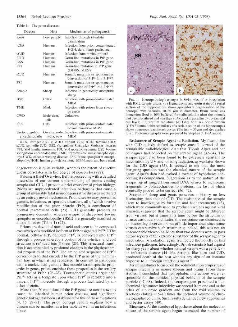
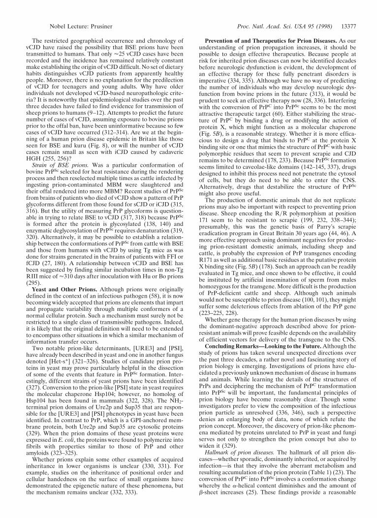
In scrapie, kuru, CJD, and all of the other disorders nowreferred to as prion diseases (Table 1), spongiform degener-ation and astrocytic gliosis is found upon microscopic exam-ination of the CNS (Fig. 1) (21). The degree of spongiform
Editor’s Note: This article is an abbreviated version of Stanley B.Prusiner’s Nobel Lecture, “Prions.” The 1997 Nobel Prize in Physi-ology or Medicine was awarded to Dr. Prusiner for his discovery ofprions, an entirely new genre of disease-causing agents, and forelucidating the fascinating principles that underline their mode ofaction. The Nobel Foundation graciously has granted us permission toreprint this article. The Nobel Lectures provide examples of successfulapproaches to major scientific problems as well as authoritativereviews. However, in recent years, these lectures have rarely been read,perhaps because of the difficulty in obtaining the collections. Byreprinting this lecture we hope to broaden their exposure.Abbreviations: Bo, bovine; BSE, bovine spongiform encephalopathy;CJD, Creutzfeldt–Jakob disease; sCJD, sporadic CJD; fCJD, familialCJD; iCJD, iatrogenic CJD; vCJD, (new) variant CJD; CNS, centralnervous system; CWD, chronic wasting disease; FFI, fatal familialinsomnia; FSE, feline spongiform encephalopathy; FSI, fatal sporadicinsomnia; GFAP, glial fibrillary acidic protein; GPI, glycosyl-phosphatidylinositol; GSS, Gerstmann–Straussler–Scheinker disease;HGH, human growth hormone; Hu, human; MBM, meat and bonemeal; Mo, mouse; r, recombinant; SHa, Syrian hamster; Tg, trans-genic; TME, transmissible mink encephalopathy; wt, wild-type.†To whom reprint requests should be addressed at: Department ofNeurology, University of California, San Francisco, CA 94143-0518.
‡Prions are defined as proteinaceous infectious particles that lacknucleic acid. PrPC is the cellular prion protein; PrPSc is the pathologicisoform. NH2-terminal truncation during limited proteolysis of PrPSc
produces PrP 27–30.
13363
degeneration is quite variable, whereas the extent of reactivegliosis correlates with the degree of neuron loss (22).
Prions: A Brief Overview. Before proceeding with a detaileddiscussion of our current understanding of prions causingscrapie and CJD, I provide a brief overview of prion biology.Prions are unprecedented infectious pathogens that cause agroup of invariably fatal neurodegenerative diseases mediatedby an entirely novel mechanism. Prion diseases may present asgenetic, infectious, or sporadic disorders, all of which involvemodification of the prion protein (PrP), a constituent ofnormal mammalian cells (23). CJD generally presents asprogressive dementia, whereas scrapie of sheep and bovinespongiform encephalopathy (BSE) are generally manifest asataxic illnesses (Table 1) (24).
Prions are devoid of nucleic acid and seem to be composedexclusively of a modified isoform of PrP designated PrPSc.‡ Thenormal, cellular PrP, denoted PrPC, is converted into PrPSc
through a process whereby a portion of its a-helical and coilstructure is refolded into b-sheet (25). This structural transi-tion is accompanied by profound changes in the physicochem-ical properties of the PrP. The amino acid sequence of PrPSc
corresponds to that encoded by the PrP gene of the mamma-lian host in which it last replicated. In contrast to pathogenswith a nucleic acid genome that encode strain-specific prop-erties in genes, prions encipher these properties in the tertiarystructure of PrPSc (26–28). Transgenetic studies argue thatPrPSc acts as a template upon which PrPC is refolded into anascent PrPSc molecule through a process facilitated by an-other protein.
More than 20 mutations of the PrP gene are now known tocause the inherited human prion diseases, and significantgenetic linkage has been established for five of these mutations(4, 16, 29–31). The prion concept readily explains how adisease can be manifest as a heritable as well as an infectiousillness.
Resistance of Scrapie Agent to Radiation. My fascinationwith CJD quickly shifted to scrapie once I learned of theremarkable radiobiological data that Tikvah Alper and hercolleagues had collected on the scrapie agent (32–34). Thescrapie agent had been found to be extremely resistant toinactivation by UV and ionizing radiation, as was later shownfor the CJD agent (35). It seemed to me that the mostintriguing question was the chemical nature of the scrapieagent; Alper’s data had evoked a torrent of hypotheses con-cerning its composition. Suggestions as to the nature of thescrapie agent ranged from small DNA viruses to membranefragments to polysaccharides to proteins, the last of whicheventually proved to be correct (36–42).
Scrapie of sheep and goats possesses a history no lessfascinating than that of CJD. The resistance of the scrapieagent to inactivation by formalin and heat treatments (43),which were commonly used to produce vaccines against viralillnesses, suggested that the scrapie agent might be differentfrom viruses, but it came at a time before the structure ofviruses was understood. Later, this resistance was dismissed asan interesting observation but of little importance since someviruses can survive such treatments; indeed, this was not anunreasonable viewpoint. More than two decades were to passbefore reports of the extreme resistance of the scrapie agent toinactivation by radiation again trumpeted the novelty of thisinfectious pathogen. Interestingly, British scientists had arguedfor many years about whether natural scrapie was a genetic oran infectious disease (44–46). Scrapie, like kuru and CJD,produced death of the host without any sign of an immuneresponse to a ‘‘foreign infectious agent.’’
My initial studies focused on the sedimentation properties ofscrapie infectivity in mouse spleens and brains. From thesestudies, I concluded that hydrophobic interactions were re-sponsible for the nonideal physical behavior of the scrapieparticle (47, 48). Indeed, the scrapie agent presented a bio-chemical nightmare: infectivity was spread from one end to theother of a sucrose gradient and from the void volume tofractions eluting at 5–10 times the included volume of chro-matographic columns. Such results demanded new approachesand better assays (49).
Bioassays. As the number of hypotheses about the molecularnature of the scrapie agent began to exceed the number of
a b
FIG. 1. Neuropathologic changes in Swiss mice after inoculationwith RML scrapie prions. (a) Hematoxylin and eosin stain of a serialsection of the hippocampus shows spongiform degeneration of theneuropil, with vacuoles 10–30 mm in diameter. Brain tissue wasimmersion fixed in 10% buffered formalin solution after the animalshad been sacrificed and was then embedded in paraffin. Py, pyramidalcell layer; SR, stratum radiatum. (b) Glial fibrillary acidic protein(GFAP) immunohistochemistry of a serial section of the hippocampusshows numerous reactive astrocytes. (Bar in b 5 50 mm and also appliesto a.) Photomicrographs were prepared by Stephen J. DeArmond.
Table 1. The prion diseases
Disease Host Mechanism of pathogenesis
Kuru Fore people Infection through ritualisticcannibalism
iCJD Humans Infection from prion-contaminatedHGH, dura mater grafts, etc.
vCJD Humans Infection from bovine prions?fCJD Humans Germ-line mutations in PrP geneGSS Humans Germ-line mutations in PrP geneFFI Humans Germ-line mutation in PrP gene
(D178N, M129)sCJD Humans Somatic mutation or spontaneous
conversion of PrPC into PrPSc?FSI Humans Somatic mutation or spontaneous
conversion of PrPC into PrPSc?Scrapie Sheep Infection in genetically susceptible
sheepBSE Cattle Infection with prion-contaminated
MBMTME Mink Infection with prions from sheep
or cattleCWD Mule deer,
elkUnknown
FSE Cats Infection with prion-contaminatedbovine tissues or MBM
13364 Nobel Lecture: Prusiner Proc. Natl. Acad. Sci. USA 95 (1998)
laboratories working on this problem, the need for newexperimental approaches became evident. Much of the avail-able data on the properties of the scrapie agent had beengathered on brain homogenates prepared from mice withclinical signs of scrapie. These mice had been inoculated 4–5months earlier with scrapie agent that originated in sheep buthad been passaged multiple times in mice. Once an experimentwas completed on these homogenates, an additional 12 monthswas required to obtain the results of an endpoint titration inmice (50). Typically, 60 mice were required to determine thetiter of a single sample. This slow, tedious, and expensivesystem discouraged systematic investigation.
Although the transmission of scrapie to mice had ushered ina new era of research, the 1.5- to 2-year intervals betweendesigning experiments and obtaining results discouraged se-quential studies. Infrequently, the results of one set of exper-iments were used as a foundation for the next and so on.Moreover, the large number of mice needed to measure theinfectivity in a single sample prevented studies where manyexperiments were performed in parallel. These problemsencouraged publication of inconclusive experimental results.
In 1972, when I became fascinated by the enigmatic natureof the scrapie agent, I thought that the most direct path todetermining the molecular structure of the scrapie agent waspurification. Fortunately, I did not appreciate the magnitudeof that task, although I had considerable experience andtraining in the purification of enzymes (51). Although manystudies had been performed to probe the physicochemicalnature of the scrapie agent by using the mouse endpointtitration system, few systematic investigations had been per-formed on the fundamental characteristics of the infectiousscrapie particle (42). In fact, 12 years after introduction of themouse bioassay, there were few data on the sedimentationbehavior of the scrapie particle. Since differential centrifuga-tion is frequently a useful initial step in the purification ofmany macromolecules, some knowledge of the sedimentationproperties of the scrapie agent under defined conditionsseemed mandatory. To perform such studies, Swiss mice wereinoculated intracerebrally with the Chandler isolate of scrapieprions and the mice were sacrificed about 30 and 150 days later,when the titers in their spleens and brains, respectively, wereexpected to be at maximal levels. The two tissues werehomogenized, extracted with detergent, and centrifuged forincreasing times and speeds (47, 52). The disappearance ofscrapie infectivity was measured in supernatant fractions byendpoint titration, which required 1 year to score.
Incubation time assays in hamsters. In view of these dauntinglogistical problems, the identification of an inoculum thatproduced scrapie in the golden Syrian hamster (SHa) in '70days after intracerebral inoculation proved to be an importantadvance (53, 54) once an incubation time assay was developed(55, 56). In earlier studies, SHa had been inoculated withprions, but serial passage with short incubation times was notreported (57). Development of the incubation time bioassayreduced the time required to measure prions in samples withhigh titers by a factor of 5: only 70 days were required insteadof the 360 days previously needed. Equally important, 4animals could be used in place of the 60 that were required forendpoint titrations, making possible a large number of parallelexperiments. With this bioassay, research on the nature of thescrapie agent was accelerated nearly 100-fold and the hamsterwith high prion titers in its brain became the experimentalanimal of choice for biochemical studies.
The incubation time assay enabled development of effectivepurification schemes for enriching fractions for scrapie infec-tivity. It provided a means to assess quantitatively thosefractions that were enriched for infectivity and those that werenot. Such studies led rather rapidly to the development of aprotocol for separating scrapie infectivity from most proteinsand nucleic acids. With a '100-fold purification of infectivity
relative to protein, .98% of the proteins and polynucleotideswere eliminated, permitting more reliable probing of theconstituents of these enriched fractions.
The Prion Concept. As reproducible data began to accu-mulate indicating that scrapie infectivity could be reduced byprocedures that hydrolyze or modify proteins but was resistantto procedures that alter nucleic acids, a family of hypothesesabout the molecular architecture of the scrapie agent began toemerge (58). These data established, for the first time, that aparticular macromolecule was required for infectivity and thatthis macromolecule was a protein. The experimental findingsextended earlier observations on resistance of scrapie infec-tivity to UV irradiation at 250 nm (33) in that the four differentprocedures used to probe for a nucleic acid are based onphysical principles that are independent of UV radiationdamage.
Once the requirement of protein for infectivity was estab-lished, I thought that it was appropriate to give the infectiouspathogen of scrapie a provisional name that would distinguishit from both viruses and viroids. After some contemplation, Isuggested the term ‘‘prion,’’ derived from proteinaceous andinfectious (58). At that time, I defined prions as proteinaceousinfectious particles that resist inactivation by procedures thatmodify nucleic acids. I never imagined the irate reaction ofsome scientists to the word ‘‘prion’’—it was truly remarkable!
Current definitions. Perhaps the best current working defi-nition of a prion is a proteinaceous infectious particle that lacksnucleic acid (28). Because a wealth of data supports thecontention that scrapie prions are composed entirely of aprotein that adopts an abnormal conformation, it is notunreasonable to define prions as infectious proteins (25, 27, 59,60). But I hasten to add that we still cannot eliminate a smallligand bound to PrPSc as an essential component of theinfectious prion particle. Learning how to renature PrPSc
accompanied by restoration of prion infectivity or to generateprion infectivity de novo by using a synthetic polypeptideshould help address this as-yet-unresolved issue (61). From abroader perspective, prions are elements that impart andpropagate conformational variability.
Although PrPSc is the only known component of the infec-tious prion particles, these unique pathogens share severalphenotypic traits with other infectious entities such as viruses.Because some features of the diseases caused by prions andviruses are similar, some scientists have difficulty accepting theexistence of prions despite a wealth of scientific data support-ing this concept (62–67).
Families of hypotheses. Once the requirement for a proteinwas established, it was possible to revisit the long list ofhypothetical structures that had been proposed for the scrapieagent and to eliminate carbohydrates, lipids, and nucleic acidsas the infective elements within a scrapie agent devoid ofprotein (58). No longer could structures such as a viroid-likenucleic acid, a replicating polysaccharide, or a small polynu-cleotide surrounded by a carbohydrate be entertained asreasonable candidates to explain the puzzling properties of thescrapie agent (58, 68).
The family of hypotheses that remained after identifying aprotein component was still large and required a continuedconsideration of all possibilities in which a protein was a criticalelement (49). The prion concept evolved from a family ofhypotheses in which an infectious protein was only one ofseveral possibilities. With the accumulation of experimentaldata on the molecular properties of the prion, it becamepossible to discard an increasing number of hypotheticalstructures. In prion research, as in many other areas ofscientific investigation, a single hypothesis is all too oftenchampioned at the expense of a reasoned approach thatrequires entertaining a series of complex arguments until oneor more can be discarded on the basis of experimental data(69).
Nobel Lecture: Prusiner Proc. Natl. Acad. Sci. USA 95 (1998) 13365
Genes and DNA. In some respects, the early development ofthe prion concept mirrors the story of DNA (70–72). Prior tothe acceptance of DNA as the genetic material of life (73, 74),many scientists asserted that the DNA preparations must becontaminated with protein that is the true genetic material(75). The prejudices of these scientists were similar in someways to those of investigators who have disputed the prionconcept. But the scientists who attacked the hypothesis thatgenes are composed of DNA had no well proven alternative;they had only a set of feelings derived from poorly substanti-ated data sets that genes are made of protein. In contrast, thosewho attacked the hypothesis that the prion is composed onlyof protein had more than 30 years of cumulative evidenceshowing that genetic information in all organisms on ourplanet is encoded in DNA and that biological diversity residesin DNA. Studies of viruses and eventually viroids extended thisconcept to these small infectious pathogens (76) and showedthat genes could also be composed of RNA (77, 78).
Discovery of the Prion Protein. The discovery of the prionprotein transformed research on scrapie and related diseases(79, 80). It provided a molecular marker that was subsequentlyshown to be specific for these illnesses as well as the major, andvery likely the only, constituent of the infectious prion.
PrP 27–30 was discovered by enriching fractions from SHabrain for scrapie infectivity (79, 80). This protein is theprotease-resistant core of PrPSc and has an apparent molecularmass of 27–30 kDa (81, 82). Although resistance to limitedproteolysis proved to be a convenient tool for many but not allstudies, use of proteases to enrich fractions for scrapie infec-tivity created a problem when the NH2-terminal sequence ofPrP 27–30 was determined (81). The ragged NH2 terminus ofPrP 27–30 yielded three sets of signals in almost every cycle ofthe Edman degradation. Only after these signals were properlyinterpreted and placed in correct register could a uniquesequence be assigned for the NH2 terminus of PrP 27–30. Thedetermination of the amino acid sequence of the NH2 terminusof PrP 27–30 made subsequent molecular cloning studies of thePrP gene possible (83, 84).
The finding that PrP mRNA levels were similar in normaluninfected and scrapie-infected tissues caused some investi-gators to argue that PrP 27–30 was not related to the infectiousprion particle (83). An alternate interpretation prompted asearch for a prion protein in uninfected animals that was foundto be protease sensitive and soluble in nondenaturing deter-gents, unlike PrP 27–30. This isoform was designated PrPC
(Fig. 2) (84, 85). Deduced amino acid sequences from PrPcDNA as well as immunoblotting studies revealed that PrP27–30 was NH2-terminally truncated and was derived from alarger molecule, designated PrPSc, that was unique to infectedanimals (81, 82, 84–86).
With the discovery of PrP 27–30 and the production ofantiserum (87), brains from humans and animals with putativeprion diseases were examined for the presence of this protein.In each case, PrP 27–30 was found, and it was absent in otherneurodegenerative disorders such as Alzheimer’s disease, Par-kinson’s disease, and amyotrophic lateral sclerosis (88–91).The extreme specificity of PrPSc for prion disease is animportant feature of the protein and is consistent with thepostulated role of PrPSc in both the transmission and patho-genesis of these illnesses (Table 2) (92).
The accumulation of PrPSc contrasts markedly with that ofglial fibrillary acidic protein (GFAP) in prion disease. Inscrapie, GFAP mRNA and protein levels rise as the diseaseprogresses (93), but the accumulation of GFAP is neitherspecific nor necessary for either the transmission or thepathogenesis of disease. Mice deficient for GFAP show noalteration in their incubation times (94, 95).
Except for PrPSc, no macromolecule has been found intissues of patients dying of the prion diseases that is specific forthese encephalopathies. In searches for a scrapie-specific
nucleic acid, cDNAs have been identified that are comple-mentary to mRNAs encoding other proteins with increasedexpression in prion disease (96–98). Yet none of the proteinshas been found to be specific for prion disease.
Attempts to Falsify the Prion Hypothesis. Numerous at-tempts to disprove the prion hypothesis over the past 15 yearshave failed. Such studies have tried unsuccessfully to separatescrapie infectivity from protein and more specifically fromPrPSc. No preparations of purified prions containing less thanone PrPSc molecule per ID50 unit have been reported (99), andno replication of prions in PrP-deficient (Prnp0/0) mice wasfound (100–104).
Copurification of PrP 27–30 and scrapie infectivity demandsthat the physicochemical properties as well as antigenicity ofthese two entities be similar (105) (Table 2). The results of awide array of inactivation experiments demonstrated the sim-ilarities in the properties of PrP 27–30 and scrapie infectivity(61, 106–109). To explain these findings in terms of the virushypothesis, it is necessary to postulate either a virus that hasa coat protein which is highly homologous with PrP or a virus
A
B
FIG. 2. Prion protein isoforms. (A) Western immunoblot of brainhomogenates from uninfected (lanes 1 and 2) and prion-infected(lanes 3 and 4) SHa. Samples in lanes 2 and 4 were digested with 50mgyml proteinase K for 30 min at 37°C. PrPC in lanes 2 and 4 wascompletely hydrolyzed under these conditions, whereas approximately67 amino acids were digested from the NH2 terminus of PrPSc togenerate PrP 27–30. After polyacrylamide gel electrophoresis (PAGE)and electrotransfer, the blot was developed with anti-PrP R073polyclonal rabbit antiserum. Molecular size markers are in kilodaltons(kDa). (B) Bar diagram of SHaPrP, which consists of 254 amino acids.Attached carbohydrate (CHO) and a glycosyl-phosphatidylinositol(GPI) anchor are indicated. After processing of the NH2 and COOHtermini, both PrPC and PrPSc consist of 209 residues. After limitedproteolysis, the NH2 terminus of PrPSc is truncated to form PrP 27–30,which is composed of approximately 142 amino acids.
13366 Nobel Lecture: Prusiner Proc. Natl. Acad. Sci. USA 95 (1998)
that binds tightly to PrPSc. In either case, the PrP-like coatprotein or the PrPScyvirus complex must display propertiesindistinguishable from PrPSc alone. With each species that the
putative virus invades, it must incorporate a new PrP sequenceduring replication.
Search for a scrapie-specific nucleic acid. The inability toinactivate preparations highly enriched for scrapie infectivityby procedures that modify nucleic acids militates against theexistence of a scrapie-specific nucleic acid (58, 110, 111). Toexplain the findings in terms of a virus, one must argue thatPrPSc or an as-yet-undetected PrP-like protein of viral originprotects the viral genome from inactivation. The notion thatthe putative scrapie virus encodes a PrP-like protein wasrefuted by nucleic acid hybridization studies using a PrP cDNAprobe. Less than 0.002 nucleic acid molecule encoding PrP perID50 unit was found in purified preparations of SHa prions(84). To circumvent this finding, it could be hypothesized thatthe genetic code used by the PrP gene differs so greatly fromthat found in the cell that a PrP cDNA probe failed to detectit in highly purified preparations.
If prions contained a genome with a unique genetic code,then it is likely that this genome would encode some special-ized proteins required for replication as well as some uniquetRNAs. But both UV and ionizing radiation inactivationstudies as well as physical studies have eliminated the possi-bility of a large nucleic acid hiding within purified preparationsof prions (110–112). Only oligonucleotides of fewer than 50bases were found at a concentration of one molecule per ID50unit in prion preparations highly enriched for scrapie infec-tivity (113, 114). These small nucleic acids were of variablelength and are thought to be degradation byproducts gener-ated during purification of prions. Failure to find a bona fidegenome was attributed to the unusual properties of the puta-tive viral nucleic acid or technical incompetence on the part ofthe investigators who were unable to find it (63, 115).
PrP amyloid. In preparations highly enriched for scrapieinfectivity and containing only PrP 27–30 by silver staining ofgels after SDSyPAGE, numerous rod-shaped particles wereseen by electron microscopy after negative staining (Fig. 3)(107). Each of the rods was slightly different, in contrast toviruses, which exhibit extremely uniform structures (116).These irregular rods, composed largely, if not entirely, of PrP27–30, were indistinguishable morphologically from manyother purified amyloids (117). Studies of the prion rods with
FIG. 3. Electron micrographs of negatively stained and ImmunoGold-labeled prion proteins. (A) PrPC. (B) PrPSc. Neither PrPC nor PrPSc formsrecognizable, ordered polymers. (C) Prion rods composed of PrP 27–30 were negatively stained. The prion rods are indistinguishable from manypurified amyloids. (Bar 5 100 nm.)
Table 2. Arguments for prions being composed largely, if notentirely, of PrPSc molecules and devoid of nucleic acid
1 PrPSc and scrapie infectivity copurify when biochemical andimmunologic procedures are used.
2 The unusual properties of PrPSc mimic those of prions. Manydifferent procedures that modify or hydrolyze PrPSc
inactivate prions.3 Levels of PrPSc are directly proportional to prion titers.
Nondenatured PrPSc has not been separated from scrapieinfectivity.
4 No evidence exists for a virus-like particle or a nucleic acidgenome.
5 Accumulation of PrPSc is invariably associated with thepathology of prion diseases, including PrP amyloid plaquesthat are pathognomonic.
6 PrP gene mutations are genetically linked to inherited priondisease and cause formation of PrPSc.
7 Overexpression of PrPC increases the rate of PrPSc formation,which shortens the incubation time. Knock out of the PrPgene eliminates the substrate necessary for PrPSc formationand prevents both prion disease and prion replication.
8 Species variations in the PrP sequence are responsible, at leastin part, for the species barrier that is found when prions arepassaged from one host to another.
9 PrPSc preferentially binds to homologous PrPC, resulting information of nascent PrPSc and prion infectivity.
10 Chimeric and partially deleted PrP genes change susceptibilityto prions from different species and support production ofartificial prions with novel properties that are not found innature.
11 Prion diversity is enciphered within the conformation of PrPSc.Strains can be generated by passage through hosts withdifferent PrP genes. Prion strains are maintained byPrPCyPrPSc interactions.
12 Human prions from fCJD(E200K) and FFI patients impartdifferent properties to chimeric MHu2M PrP in transgenicmice, which provides a mechanism for strain propagation.
Nobel Lecture: Prusiner Proc. Natl. Acad. Sci. USA 95 (1998) 13367
Congo red dye demonstrated that the rods also fulfilled thetinctorial criteria for amyloid (107), and immunostaining latershowed that PrP is a major component of amyloid plaques insome animals and humans with prion disease (118–120).Subsequently, it was recognized that the prion rods were notrequired for scrapie infectivity (121); furthermore, the rodswere shown to be an artifact of purification during whichlimited proteolysis of PrPSc generated PrP 27–30 that poly-merized spontaneously in the presence of detergent (Fig. 3)(122).
The idea that scrapie prions were composed of an amyloi-dogenic protein was truly heretical when it was introduced(107). Since the prevailing view at the time was that scrapie iscaused by an atypical virus, many argued that amyloid proteinsare mammalian polypeptides and not viral proteins!
Some investigators have argued that the prion rods aresynonymous with scrapie-associated fibrils (123–125) eventhough morphologic and tinctorial features of these fibrilsclearly differentiated them from amyloid and as such from theprion rods (126, 127). The scrapie-associated fibrils wereidentified by their unique ultrastructure in which two or foursubfilaments were helically wound around each other (126)and were proposed to represent the first example of a fila-mentous animal virus (128). After the argument for a fila-mentous animal virus causing scrapie faded, it was hypothe-sized that a virus induces the formation of PrP amyloid toexplain the accumulation of PrPSc in prion diseases (129).Besides the lack of evidence for a virus of any shape, nocompelling data have been offered in support of the idea thatprion diseases are caused by a filamentous bacterium called aspiroplasma (130).
Search for the ubiquitous “scrapie virus.” When PrP genemutations were discovered to cause familial prion diseases (4),it was postulated that PrPC is a receptor for the ubiquitousscrapie virus that binds more tightly to mutant than to wt PrPC
(131). A similar hypothesis was proposed to explain why thelength of the scrapie incubation time was found to be inverselyproportional to the level of PrP expression in transgenic (Tg)mice and why Prnp0/0 mice are resistant to scrapie (132). Thehigher the level of PrP expression, the faster the spread of theputative virus, which results in shorter incubation times;conversely, mice deficient for PrP lack the receptor requiredfor spread of the virus (63). The inability to find virus-likeparticles in purified preparations of PrPSc was attributed tothese particles being hidden (115) even though tobacco mosaicviruses could be detected when one virion was added per ID50unit of scrapie prions (121).
Recent studies on the transmission of mutant prions fromFFI and fCJD(E200K) to Tg(MHu2M) mice, which results inthe formation of two different PrPSc molecules (27), has forceda corollary to the ubiquitous virus postulate. To accommodatethis result, at least two different viruses must reside worldwide,each of which binds to a different mutant HuPrPC and each ofwhich induces a different MHu2M PrPSc conformer whentransferred to Tg mice. Even more difficult to imagine is howone ubiquitous virus might acquire different mutant PrPSc
molecules corresponding to FFI or fCJD(E200K) and theninduce different MHu2M PrPSc conformers upon transmissionto Tg mice.
Artificial prions. To explain the production of artificialprions from chimeric or mutant PrP transgenes in terms of avirus (133–135), mutated PrPSc molecules must be incorpo-rated into the virus. In the case of mice expressing chimeric PrPtransgenes, artificial prions are produced with host ranges notpreviously found in nature. Similarly, deleting specific regionsof PrP resulted in the formation of “miniprions” with a uniquehost range and neuropathology as described below. The pro-duction of artificial prions that were generated by modifyingthe PrP gene sequence and exhibit unique biological properties
is another compelling argument against the proposition thatscrapie and CJD are caused by viruses.
Skepticism once well justified. While the skepticism aboutprions was once well justified and formed the basis for avigorous scientific debate, the wealth of available data nowrenders such arguments moot. In summary, no single hypoth-esis involving a virus can explain the findings summarizedabove (Table 2); instead, a series of ad hoc hypotheses, virtuallyall of which can be refuted by experimental data, must beconstructed to accommodate a steadily enlarging body of data.
It is notable that the search for an infectious pathogen witha nucleic acid genome as the cause of scrapie and CJD has donelittle to advance our understanding of these diseases. Instead,studies of PrP have created a wealth of data that now explainalmost every aspect of these fascinating disorders. While nosingle experiment can refute the existence of the “scrapievirus,” all of the data taken together from numerous experi-mental studies present an impressive edifice which argues thatthe 50-year quest for a virus has failed because it does not exist!
Prions Defy Rules of Protein Structure. Once cDNA probesfor PrP became available, the PrP gene was found to beconstitutively expressed in adult, uninfected brain (83, 84).This finding eliminated the possibility that PrPSc stimulatedproduction of more of itself by initiating transcription of thePrP gene as proposed nearly two decades earlier (37). Deter-mination of the structure of the PrP gene eliminated a secondpossible mechanism that might explain the appearance ofPrPSc in brains already synthesizing PrPC. Since the entireprotein coding region was contained within a single exon, therewas no possibility for the two PrP isoforms to be the productsof alternatively spliced mRNAs (82). Next, a posttranslationalchemical modification that distinguishes PrPSc from PrPC wasconsidered, but none was found in an exhaustive study (59) andwe considered it likely that PrPC and PrPSc differed only intheir conformation, a hypothesis also proposed earlier (37).However, this idea was no less heretical than that of aninfectious protein.
For more than 25 years, it had been widely accepted that theamino acid sequence specifies one biologically active confor-mation of a protein (136). Yet in scrapie we were faced withthe possibility that one primary structure for PrP might adoptat least two different conformations to explain the existence ofboth PrPC and PrPSc. When the secondary structures of the PrPisoforms were compared by optical spectroscopy, they werefound to be markedly different (25). Fourier-transform infra-red (FTIR) and circular dichroism (CD) studies showed thatPrPC contains about 40% a-helix and little b-sheet, whereasPrPSc is composed of about 30% a-helix and 45% b-sheet (25,137). Nevertheless, these two proteins have the same aminoacid sequence!
Prior to comparative studies on the structures of PrPC andPrPSc, we found by metabolic labeling studies that the acqui-sition of PrPSc protease resistance is a posttranslational process(138). In our quest for a chemical difference that woulddistinguish PrPSc from PrPC, we found ethanolamine in hy-drolysates of PrP 27–30, which signaled the possibility that PrPmight contain a GPI anchor (139). Both PrP isoforms werefound to carry GPI anchors, and PrPC was found on the surfaceof cells where it could be released by cleavage of the anchor.Subsequent studies showed that PrPSc formation occurs afterPrPC reaches the cell surface (140, 141) and is localized tocaveolae-like domains (142–145).
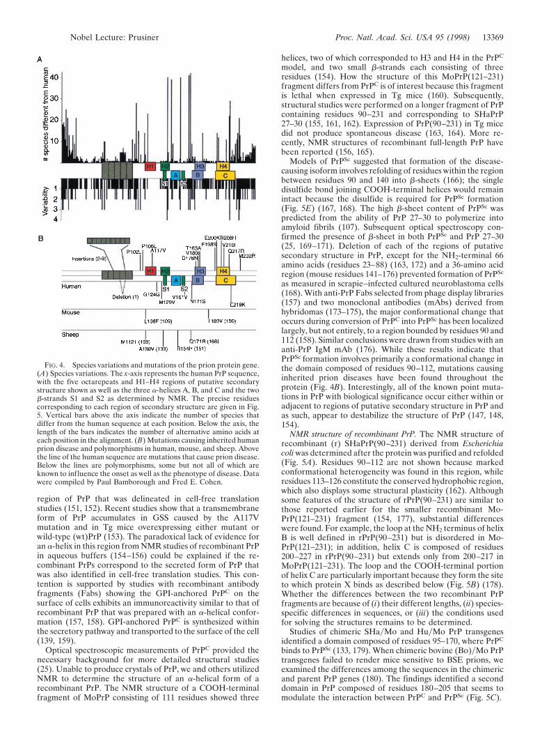
Modeling PrP structures. Molecular modeling studies pre-dicted that PrPC is a four-helix bundle protein containing fourregions of secondary structure denoted H1, H2, H3, and H4(Fig. 4) (146, 147). Subsequent NMR studies of a synthetic PrPpeptide containing residues 90–145 provided good evidencefor H1 (148). This peptide contains the residues 113–128,which are most highly conserved among all species studied(Fig. 4A) (147, 149, 150) and correspond to a transmembrane
13368 Nobel Lecture: Prusiner Proc. Natl. Acad. Sci. USA 95 (1998)
region of PrP that was delineated in cell-free translationstudies (151, 152). Recent studies show that a transmembraneform of PrP accumulates in GSS caused by the A117Vmutation and in Tg mice overexpressing either mutant orwild-type (wt)PrP (153). The paradoxical lack of evidence foran a-helix in this region from NMR studies of recombinant PrPin aqueous buffers (154–156) could be explained if the re-combinant PrPs correspond to the secreted form of PrP thatwas also identified in cell-free translation studies. This con-tention is supported by studies with recombinant antibodyfragments (Fabs) showing the GPI-anchored PrPC on thesurface of cells exhibits an immunoreactivity similar to that ofrecombinant PrP that was prepared with an a-helical confor-mation (157, 158). GPI-anchored PrPC is synthesized withinthe secretory pathway and transported to the surface of the cell(139, 159).
Optical spectroscopic measurements of PrPC provided thenecessary background for more detailed structural studies(25). Unable to produce crystals of PrP, we and others utilizedNMR to determine the structure of an a-helical form of arecombinant PrP. The NMR structure of a COOH-terminalfragment of MoPrP consisting of 111 residues showed three
helices, two of which corresponded to H3 and H4 in the PrPC
model, and two small b-strands each consisting of threeresidues (154). How the structure of this MoPrP(121–231)fragment differs from PrPC is of interest because this fragmentis lethal when expressed in Tg mice (160). Subsequently,structural studies were performed on a longer fragment of PrPcontaining residues 90–231 and corresponding to SHaPrP27–30 (155, 161, 162). Expression of PrP(90–231) in Tg micedid not produce spontaneous disease (163, 164). More re-cently, NMR structures of recombinant full-length PrP havebeen reported (156, 165).
Models of PrPSc suggested that formation of the disease-causing isoform involves refolding of residues within the regionbetween residues 90 and 140 into b-sheets (166); the singledisulfide bond joining COOH-terminal helices would remainintact because the disulfide is required for PrPSc formation(Fig. 5E) (167, 168). The high b-sheet content of PrPSc waspredicted from the ability of PrP 27–30 to polymerize intoamyloid fibrils (107). Subsequent optical spectroscopy con-firmed the presence of b-sheet in both PrPSc and PrP 27–30(25, 169–171). Deletion of each of the regions of putativesecondary structure in PrP, except for the NH2-terminal 66amino acids (residues 23–88) (163, 172) and a 36-amino acidregion (mouse residues 141–176) prevented formation of PrPSc
as measured in scrapie–infected cultured neuroblastoma cells(168). With anti-PrP Fabs selected from phage display libraries(157) and two monoclonal antibodies (mAbs) derived fromhybridomas (173–175), the major conformational change thatoccurs during conversion of PrPC into PrPSc has been localizedlargely, but not entirely, to a region bounded by residues 90 and112 (158). Similar conclusions were drawn from studies with ananti-PrP IgM mAb (176). While these results indicate thatPrPSc formation involves primarily a conformational change inthe domain composed of residues 90–112, mutations causinginherited prion diseases have been found throughout theprotein (Fig. 4B). Interestingly, all of the known point muta-tions in PrP with biological significance occur either within oradjacent to regions of putative secondary structure in PrP andas such, appear to destabilize the structure of PrP (147, 148,154).
NMR structure of recombinant PrP. The NMR structure ofrecombinant (r) SHaPrP(90–231) derived from Escherichiacoli was determined after the protein was purified and refolded(Fig. 5A). Residues 90–112 are not shown because markedconformational heterogeneity was found in this region, whileresidues 113–126 constitute the conserved hydrophobic region,which also displays some structural plasticity (162). Althoughsome features of the structure of rPrP(90–231) are similar tothose reported earlier for the smaller recombinant Mo-PrP(121–231) fragment (154, 177), substantial differenceswere found. For example, the loop at the NH2 terminus of helixB is well defined in rPrP(90–231) but is disordered in Mo-PrP(121–231); in addition, helix C is composed of residues200–227 in rPrP(90–231) but extends only from 200–217 inMoPrP(121–231). The loop and the COOH-terminal portionof helix C are particularly important because they form the siteto which protein X binds as described below (Fig. 5B) (178).Whether the differences between the two recombinant PrPfragments are because of (i) their different lengths, (ii) species-specific differences in sequences, or (iii) the conditions usedfor solving the structures remains to be determined.
Studies of chimeric SHayMo and HuyMo PrP transgenesidentified a domain composed of residues 95–170, where PrPC
binds to PrPSc (133, 179). When chimeric bovine (Bo)yMo PrPtransgenes failed to render mice sensitive to BSE prions, weexamined the differences among the sequences in the chimericand parent PrP genes (180). The findings identified a seconddomain in PrP composed of residues 180–205 that seems tomodulate the interaction between PrPC and PrPSc (Fig. 5C).
FIG. 4. Species variations and mutations of the prion protein gene.(A) Species variations. The x-axis represents the human PrP sequence,with the five octarepeats and H1–H4 regions of putative secondarystructure shown as well as the three a-helices A, B, and C and the twob-strands S1 and S2 as determined by NMR. The precise residuescorresponding to each region of secondary structure are given in Fig.5. Vertical bars above the axis indicate the number of species thatdiffer from the human sequence at each position. Below the axis, thelength of the bars indicates the number of alternative amino acids ateach position in the alignment. (B) Mutations causing inherited humanprion disease and polymorphisms in human, mouse, and sheep. Abovethe line of the human sequence are mutations that cause prion disease.Below the lines are polymorphisms, some but not all of which areknown to influence the onset as well as the phenotype of disease. Datawere compiled by Paul Bamborough and Fred E. Cohen.
Nobel Lecture: Prusiner Proc. Natl. Acad. Sci. USA 95 (1998) 13369
Recent NMR studies of full-length MoPrP(23–231) andSHaPrP(29–231) have shown that the NH2 termini are highlyflexible and lack identifiable secondary structure under theexperimental conditions employed (Fig. 5D) (156, 165). Stud-ies of SHaPrP(29–231) indicate transient interactions betweenthe COOH terminus of helix B and the highly flexible,NH2-terminal random-coil containing the octareapeats (resi-dues 29–125) (156); such interactions were not reported forMoPrP(23–231) (165).
PrP appears to bind copper. The highly flexible NH2 terminusof recombinant PrP may be more structured in the presence ofcopper. Each SHaPrP(29–231) molecule was found to bindtwo atoms of Cu21; other divalent cations did not bind to PrP(181). Earlier studies with synthetic peptides corresponding tothe octarepeat sequence demonstrated the binding of Cu21
ions (182, 183), and optical spectroscopy showed that Cu21
induced an a-helix formation in these peptides (184). Morerecently, PrP-deficient (Prnp0/0) mice were found to have lowerlevels of ZnyCu superoxide dismutase (SOD) activity than docontrols (185); SOD activity has been shown to mirror the state
of copper metabolism (186). Measurements of membraneextracts from brains of Prnp0/0 mice showed low levels of Cu,whereas Fe and Zn were unchanged suggesting PrPC mightfunction as a Cu21-binding protein (187).
Disturbances in Cu21 homeostasis leading to dysfunction ofthe CNS are well documented in humans and animals but arenot known to be due to abnormalities in PrP metabolism:Menkes disease is manifest at birth and is due to a mutationof the MNK gene on the X chromosome, whereas Wilson’sdisease appears in childhood and is due to a mutation of theWD gene on chromosome 13 (188–191). Both the MNK andWD genes encode copper-transporting ATPases. While bothMenkes and Wilson’s diseases are recessive disorders, onlyWilson’s disease can be treated with copper-chelating re-agents. Interestingly, cuprizone, a Cu21-chelating reagent, hasbeen used in mice to induce neuropathological changes similarto those found in the prion diseases (192, 193).
PrP Gene Structure and Expression. The entire openreading frame (ORF) of all known mammalian and avian PrPgenes resides within a single exon (4, 82, 194, 195). The mouse,
FIG. 5. Structures of prion proteins. (A) NMR structure of SHa recombinant (r) PrP(90–231). Presumably, the structure of the a-helical formof rPrP(90–231) resembles that of PrPC. rPrP(90–231) is viewed from the interface where PrPSc is thought to bind to PrPC. The color scheme isas follows: a-helices A (residues 144–157), B (172–193), and C (200–227) in pink; disulfide between Cys-179 and Cys-214 in yellow; conservedhydrophobic region composed of residues 113–126 in red; loops in gray; residues 129–134 in green encompassing strand S1 and residues 159–165in blue encompassing strand S2; the arrows span residues 129–131 and 161–163, as these show a closer resemblance to b-sheet (155). (B) NMRstructure of rPrP(90–231) is viewed from the interface where protein X is thought to bind to PrPC. Protein X appears to bind to the side chainsof residues that form a discontinuous epitope: some amino acids are in the loop composed of residues 165–171 and at the end of helix B (Gln-168and Gln-172 with a low-density van der Waals rendering), whereas others are on the surface of helix C (Thr-215 and Gln-219 with a high-densityvan der Waals rendering) (178). (C) PrP residues governing the transmission of prions (180). NMR structure of recombinant SHaPrP region 121–231(155) shown with the putative epitope formed by residues 184, 186, 203, and 205 highlighted in red. Residue numbers correspond to SHaPrP.Additional residues (138, 139, 143, 145, 148, and 155) that might participate in controlling the transmission of prions across species are depictedin green. Residues 168, 172, 215, and 219 that form the epitope for the binding of protein X are shown in blue. The three helices (A, B, and C)are highlighted in pink. (D) Schematic diagram showing the flexibility of the polypeptide chain for PrP(29–231) (156). The structure of the portionof the protein representing residues 90–231 was taken from the coordinates of PrP(90–231) (155). The remainder of the sequence was hand-builtfor illustration purposes only. The color scale corresponds to the heteronuclear {1H}-15N nuclear Overhauser enhancement data: red for the lowest(most negative) values, where the polypeptide is most flexible, to blue for the highest (most positive) values in the most structured and rigid regionsof the protein. (E) Plausible model for the tertiary structure of HuPrPSc (166). Color scheme is as follows: S1 b-strands are 108–113 and 116–122in red; S2 b-strands are 128–135 and 138–144 in green; a-helices H3 (residues 178–191) and H4 (residues 202–218) in gray; loop (residues 142–177)in yellow. Four residues implicated in the species barrier are shown in ball-and-stick form (Asn-108, Met-112, Met-129, Ala-133).
13370 Nobel Lecture: Prusiner Proc. Natl. Acad. Sci. USA 95 (1998)
sheep, cattle, and rat PrP genes contain three exons with theORFs in exon 3 (196–200) which is analogous to exon 2 of theSHa gene (82). The two exons of the SHaPrP gene areseparated by a 10-kb intron: exon 1 includes a portion of the59 untranslated leader sequence, while exon 2 includes theORF and 39 untranslated region (82). Recently, a low abun-dance SHaPrP mRNA containing an additional small exon inthe 59 untranslated region was discovered that is encoded bythe SHaPrP gene (201). Comparative sequencing of sheep andHu cosmid clones containing PrP genes revealed an additionalputative, small untranslated 59 exon in the HuPrP gene (202).The promoters of both the SHa- and MoPrP genes containmultiple copies of G1C-rich repeats and are devoid of TATAboxes. These G1C nonamers represent a motif that mayfunction as a canonical binding site for the transcription factorSp1 (203). Mapping of PrP genes to the short arm of Huchromosome 20 and to the homologous region of Mo chro-mosome 2 argues for the existence of PrP genes prior to thespeciation of mammals (204, 205).
Although PrP mRNA is constitutively expressed in thebrains of adult animals (83, 84), it is highly regulated duringdevelopment. In the septum, levels of PrP mRNA and cholineacetyltransferase were found to increase in parallel duringdevelopment (206). In other brain regions, PrP gene expres-sion occurred at an earlier age. In situ hybridization studiesshow that the highest levels of PrP mRNA are found in neurons(207).
PrPC expression in brain was defined by standard immuno-histochemistry (208) and by histoblotting in the brains ofuninfected controls (209). Immunostaining of PrPC in the SHabrain was most intense in the stratum radiatum and stratumoriens of the CA1 region of the hippocampus and was virtuallyabsent from the granule cell layer of the dentate gyrus and thepyramidal cell layer throughout Ammon’s horn. PrPSc stainingwas minimal in those regions that were intensely stained forPrPC. A similar relationship between PrPC and PrPSc was foundin the amygdala. In contrast, PrPSc accumulated in the medialhabenular nucleus, the medial septal nuclei, and the diagonalband of Broca; in contrast, these areas were virtually devoid ofPrPC. In the white matter, bundles of myelinated axons con-tained PrPSc but were devoid of PrPC. These findings suggestthat prions are transported along axons and are in agreementwith earlier findings in which scrapie infectivity was found tomigrate in a pattern consistent with retrograde transport(210–212).
Molecular Genetics of Prion Diseases. Independent ofenriching brain fractions for scrapie infectivity that led to thediscovery of PrPSc, the PrP gene was shown to be geneticallylinked to a locus controlling scrapie incubation times (213).Subsequently, mutation of the PrP gene was shown to begenetically linked to the development of familial prion disease(4). At the same time, expression of a SHaPrP transgene inmice was shown to render the animals highly susceptible toSHa prions, demonstrating that expression of a foreign PrPgene could abrogate the species barrier (214). Later, PrP-deficient (Prnp0/0) mice were found to be resistant to prioninfection and failed to replicate prions, as expected (100, 101).The results of these studies indicated PrP must play a centralrole in the transmission and pathogenesis of prion disease, butequally important, they argued that the abnormal isoform is anessential component of the prion particle (23).
PrP gene dosage controls length of incubation time. Scrapieincubation times in mice were used to distinguish prion strainsand to identify a gene controlling its length (135, 215). Thisgene was initially called Sinc on the basis of genetic crossesbetween C57BL and VM mice that exhibited short and longincubation times, respectively (215). Because of the restricteddistribution of VM mice, we searched for another mouse withlong incubation times. IyLn mice proved to be a suitablesubstitute for VM mice (216); eventually, IyLn and VM mice
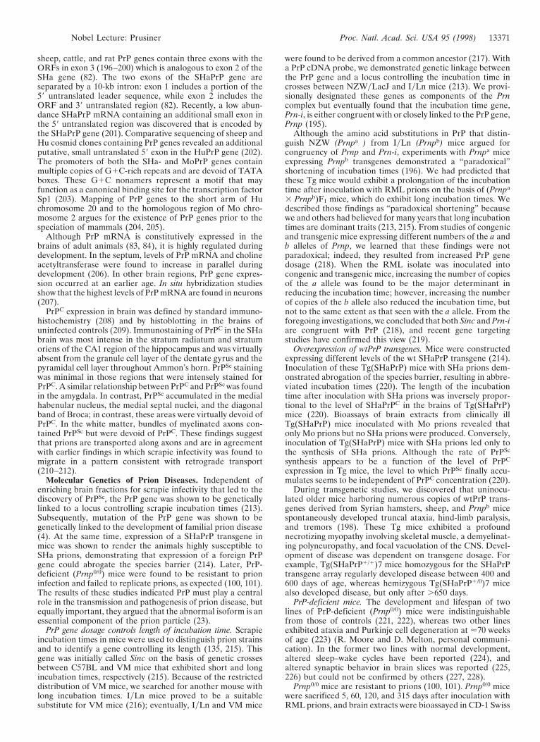
were found to be derived from a common ancestor (217). Witha PrP cDNA probe, we demonstrated genetic linkage betweenthe PrP gene and a locus controlling the incubation time incrosses between NZWyLacJ and IyLn mice (213). We provi-sionally designated these genes as components of the Prncomplex but eventually found that the incubation time gene,Prn-i, is either congruent with or closely linked to the PrP gene,Prnp (195).
Although the amino acid substitutions in PrP that distin-guish NZW (Prnpa ) from IyLn (Prnpb) mice argued forcongruency of Prnp and Prn-i, experiments with Prnpa miceexpressing Prnpb transgenes demonstrated a “paradoxical”shortening of incubation times (196). We had predicted thatthese Tg mice would exhibit a prolongation of the incubationtime after inoculation with RML prions on the basis of (Prnpa
3 Prnpb)F1 mice, which do exhibit long incubation times. Wedescribed those findings as “paradoxical shortening” becausewe and others had believed for many years that long incubationtimes are dominant traits (213, 215). From studies of congenicand transgenic mice expressing different numbers of the a andb alleles of Prnp, we learned that these findings were notparadoxical; indeed, they resulted from increased PrP genedosage (218). When the RML isolate was inoculated intocongenic and transgenic mice, increasing the number of copiesof the a allele was found to be the major determinant inreducing the incubation time; however, increasing the numberof copies of the b allele also reduced the incubation time, butnot to the same extent as that seen with the a allele. From theforegoing investigations, we concluded that both Sinc and Prn-iare congruent with PrP (218), and recent gene targetingstudies have confirmed this view (219).
Overexpression of wtPrP transgenes. Mice were constructedexpressing different levels of the wt SHaPrP transgene (214).Inoculation of these Tg(SHaPrP) mice with SHa prions dem-onstrated abrogation of the species barrier, resulting in abbre-viated incubation times (220). The length of the incubationtime after inoculation with SHa prions was inversely propor-tional to the level of SHaPrPC in the brains of Tg(SHaPrP)mice (220). Bioassays of brain extracts from clinically illTg(SHaPrP) mice inoculated with Mo prions revealed thatonly Mo prions but no SHa prions were produced. Conversely,inoculation of Tg(SHaPrP) mice with SHa prions led only tothe synthesis of SHa prions. Although the rate of PrPSc
synthesis appears to be a function of the level of PrPC
expression in Tg mice, the level to which PrPSc finally accu-mulates seems to be independent of PrPC concentration (220).
During transgenetic studies, we discovered that uninocu-lated older mice harboring numerous copies of wtPrP trans-genes derived from Syrian hamsters, sheep, and Prnpb micespontaneously developed truncal ataxia, hind-limb paralysis,and tremors (198). These Tg mice exhibited a profoundnecrotizing myopathy involving skeletal muscle, a demyelinat-ing polyneuropathy, and focal vacuolation of the CNS. Devel-opment of disease was dependent on transgene dosage. Forexample, Tg(SHaPrP1/1)7 mice homozygous for the SHaPrPtransgene array regularly developed disease between 400 and600 days of age, whereas hemizygous Tg(SHaPrP1/0)7 micealso developed disease, but only after .650 days.
PrP-deficient mice. The development and lifespan of twolines of PrP-deficient (Prnp0/0) mice were indistinguishablefrom those of controls (221, 222), whereas two other linesexhibited ataxia and Purkinje cell degeneration at '70 weeksof age (223) (R. Moore and D. Melton, personal communi-cation). In the former two lines with normal development,altered sleep–wake cycles have been reported (224), andaltered synaptic behavior in brain slices was reported (225,226) but could not be confirmed by others (227, 228).
Prnp0/0 mice are resistant to prions (100, 101). Prnp0/0 micewere sacrificed 5, 60, 120, and 315 days after inoculation withRML prions, and brain extracts were bioassayed in CD-1 Swiss
Nobel Lecture: Prusiner Proc. Natl. Acad. Sci. USA 95 (1998) 13371
mice. Except for residual infectivity from the inoculum de-tected at 5 days after inoculation, no infectivity was detectedin the brains of Prnp0/0 mice (101). One group of investigatorsfound that Prnp0/0 mice inoculated with RML prions andsacrificed 20 weeks later had 103.6 ID50 unitsyml of homoge-nate by bioassay (100). Others have used this report to arguethat prion infectivity replicates in the absence of PrP (67, 132).Neither we nor the authors of the initial report could confirmthe finding of prion replication in Prnp0/0 mice (101, 103).
Prion Species Barrier and Protein X. The passage of prionsbetween species is often a stochastic process, almost alwayscharacterized by prolonged incubation times during the firstpassage in the new host (36). This prolongation is oftenreferred to as the “species barrier” (36, 229). Prions synthe-sized de novo reflect the sequence of the host PrP gene and notthat of the PrPSc molecules in the inoculum derived from thedonor (90). On subsequent passage in a homologous host, theincubation time shortens to that recorded for all subsequentpassages. From studies with Tg mice, three factors have beenidentified that contribute to the species barrier: (i) the differ-ence in PrP sequences between the prion donor and recipient,(ii) the strain of prion, and (iii) the species specificity of proteinX, a factor defined by molecular genetic studies that binds toPrPC and facilitates PrPSc formation. This factor is likely to bea protein, hence the provisional designation protein X (134,178). The prion donor is the last mammal in which the prionwas passaged and its PrP sequence represents the “species” ofthe prion. The strain of prion, which seems to be encipheredin the conformation of PrPSc, conspires with the PrP sequence,which is specified by the recipient, to determine the tertiarystructure of nascent PrPSc. These principles are demonstratedby studies on the transmission of SHa prions to mice showingthat expression of a SHaPrP transgene in mice abrogated thespecies barrier (Table 3) (214). Besides the PrP sequence, thestrain of prion modified transmission of SHa prions to mice(Table 3) (135, 230, 231).
Transmission of Hu prions. Protein X was postulated toexplain the results on the transmission of Hu prions to Tg mice(Table 4) (134, 179). Mice expressing both Mo and HuPrPwere resistant to Hu prions, whereas those expressing onlyHuPrP were susceptible. These results argue that MoPrPC
inhibited transmission of Hu prions—i.e., the formation ofnascent HuPrPSc. In contrast to the foregoing studies, miceexpressing both MoPrP and chimeric MHu2MPrP were sus-ceptible to Hu prions and mice expressing MHu2MPrP alonewere only slightly more susceptible. These findings contendthat MoPrPC has only a minimal effect on the formation ofchimeric MHu2MPrPSc.
Genetic evidence for protein X. When the data on Hu priontransmission to Tg mice were considered together, they sug-gested that MoPrPC prevented the conversion of HuPrPC intoPrPSc but had little effect on the conversion of MHu2M intoPrPSc by binding to another Mo protein. We interpreted theseresults in terms of MoPrPC binding to this Mo protein with a
higher affinity than does HuPrPC. We postulated that MoPrPC
had little effect on the formation of PrPSc from MHu2M(Table 4) because MoPrP and MHu2M share the same aminoacid sequence at the COOH terminus. We hypothesized thatMoPrPC only weakly inhibited transmission of SHa prions toTg(SHaPrP) mice (Table 3) because SHaPrP is more closelyrelated to MoPrP than is HuPrP.
Using scrapie-infected Mo neuroblastoma cells transfectedwith chimeric HuyMo PrP genes, we extended our studies ofprotein X. Substitution of a Hu residue at position 214 or 218prevented PrPSc formation (Fig. 5B) (178). The side chains ofthese residues protrude from the same surface of the COOH-terminal a-helix, forming a discontinuous epitope with resi-dues 167 and 171 in an adjacent loop. Substitution of a basicresidue at position 167, 171, or 218 prevented PrPSc formation;these mutant PrPs appear to act as “dominant negatives” bybinding protein X and rendering it unavailable for prionpropagation. Our findings within the context of protein Xexplain the protective effects of basic polymorphic residues inPrP of humans and sheep (199, 232, 233).
Is protein X a molecular chaperone? Since PrP undergoes aprofound structural transition during prion propagation, itseems likely that other proteins such as chaperones participatein this process. Whether protein X functions as a molecularchaperone is unknown. Interestingly, scrapie-infected cells inculture display marked differences in the induction of heat-shock proteins (234, 235), and Hsp70 mRNA has been re-ported to increase in scrapie of mice (236). While attempts toisolate specific proteins that bind to PrP have been disappoint-ing (237), PrP has been shown to interact with Bcl-2 and Hsp60by two-hybrid analysis in yeast (238, 239). Although thesestudies are suggestive, no molecular chaperone involved inprion formation in mammalian cells has been identified.
FIG. 6. Miniprions produced by deleting PrP residues 23–89 and141–176. The deletion of residues 141–176 (green) containing helix Aand the S2 b-strand is shown. Side chains of residues 168, 172, 215, and219, which are thought to bind protein X, are shown in cyan.
Table 3. Influence of prion species and strains on transmissionacross a species barrier
Data are from refs. 101, 220, and 231 and unpublished data of D.Groth and S.B.P. Incubation times are given 6SEM. nyn0 5 numberof diseased animalsynumber of injected animals.
Table 4. Evidence for protein X from transmission studies ofhuman prions
Data with inoculum RG (134); incubation times are given 6SEM.
13372 Nobel Lecture: Prusiner Proc. Natl. Acad. Sci. USA 95 (1998)
Miniprions. By using the four-helix bundle model of PrPC
(Fig. 4A) (147), each region of proposed secondary structurewas systematically deleted and the mutant constructs wereexpressed in scrapie-infected neuroblastoma (ScN2a) cells andTg mice (164, 168). Deletion of any of the four putative helicalregions prevented PrPSc formation, whereas deletion of theNH2-terminal region containing residues 23–89 did not affectthe yield of PrPSc. In addition to the 67 residues at the NH2terminus, 36 residues from positions 141–176 could be deletedwithout altering PrPSc formation (Figs. 6 and 7). The resultingPrP molecule of 106 amino acids was designated PrP106. In thismutant PrP, helix A as well as the S2 b-strand were removed.When PrP106 was expressed in ScN2a cells, PrPSc106 wassoluble in 1% Sarkosyl. Whether the structure of PrPSc106 can
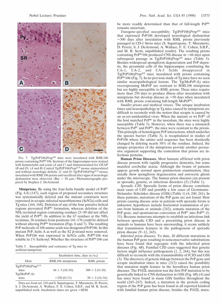
be more readily determined than that of full-length PrPSc
that expressed PrP106 developed neurological dysfunction'300 days after inoculation with RML prions previouslypassaged in CD-1 Swiss mice (S. Supattapone, T. Muramoto,D. Peretz, S. J. DeArmond, A. Wallace, F. E. Cohen, S.B.P.,and M. R. Scott, unpublished results). The resulting prionscontaining PrPSc106 produced CNS disease in '66 days uponsubsequent passage in Tg(PrP106)Prnp0/0 mice (Table 5).Besides widespread spongiform degeneration and PrP depos-its, the pyramidal cells of the hippocampus constituting theCA-1, CA-2, and CA-3 fields disappeared inTg(PrP106)Prnp0/0 mice inoculated with prions containingPrPSc106 (Fig. 7). In no previous study of Tg mice have we seensimilar neuropathological lesions. The Tg(MoPrP-A) miceoverexpressing MoPrP are resistant to RML106 miniprionsbut are highly susceptible to RML prions. These mice requiremore than 250 days to produce illness after inoculation withminiprions but develop disease in '50 days when inoculatedwith RML prions containing full-length MoPrPSc.
Smaller prions and mythical viruses. The unique incubationtimes and neuropathology in Tg mice caused by miniprions aredifficult to reconcile with the notion that scrapie is caused byan as-yet-unidentified virus. When the mutant or wt PrPC ofthe host matched PrPSc in the inoculum, the mice were highlysusceptible (Table 5). However, when there was a mismatchbetween PrPC and PrPSc, the mice were resistant to the prions.This principle of homologous PrP interactions, which underliesthe species barrier (Table 3), is recapitulated in studies ofPrP106 where the amino acid sequence has been drasticallychanged by deleting nearly 50% of the residues. Indeed, theunique properties of the miniprions provide another persua-sive argument supporting the contention that prions are in-fectious proteins.
Human Prion Diseases. Most humans afflicted with priondisease present with rapidly progressive dementia, but somemanifest cerebellar ataxia. Although the brains of patientsappear grossly normal upon postmortem examination, theyusually show spongiform degeneration and astrocytic gliosisunder the microscope. The human prion diseases can presentas sporadic, genetic, or infectious disorders (5) (Table 1).
Sporadic CJD. Sporadic forms of prion disease constitutemost cases of CJD and possibly a few cases of Gerstmann–Straussler–Scheinker disease (GSS) (Table 1) (4, 240, 241). Inthese patients, mutations of the PrP gene are not found. Howprions causing disease arise in patients with sporadic forms isunknown; hypotheses include horizontal transmission of pri-ons from humans or animals (242), somatic mutation of thePrP gene, and spontaneous conversion of PrPC into PrPSc (5,15). Because numerous attempts to establish an infectious linkbetween sporadic CJD and a preexisting prion disease inanimals or humans have been unrewarding, it seems unlikelythat transmission features in the pathogenesis of sporadicprion disease (9–12, 243).
Inherited prion diseases. To date, 20 different mutations inthe human PrP gene resulting in nonconservative substitutionshave been found that segregate with the inherited priondiseases (Fig. 4B). Familial CJD cases suggested that geneticfactors might influence pathogenesis (1, 2, 244), but this wasdifficult to reconcile with the transmissibility of fCJD and GSS(3). The discovery of genetic linkage between the PrP gene andscrapie incubation times in mice (213) raised the possibilitythat mutation might feature in the hereditary human priondiseases. The P102L mutation was the first PrP mutation to begenetically linked to CNS dysfunction in GSS (Fig. 4B) (4) andhas since been found in many GSS families throughout theworld (245–247). Indeed, a mutation in the protein codingregion of the PrP gene has been found in all reported kindredwith familial human prion disease; besides the P102L muta-
FIG. 7. Tg(PrP106)Prnp0/0 mice were inoculated with RML106prions containing PrPSc106. Sections of the hippocampus were stainedwith hematoxylin and eosin ~A and C! and immunostained for GFAP~B and D!. ~A and B! Control Tg~PrP106!Prnp0y0 mouse uninoculatedand without neurologic deficits. ~C and D! Tg~PrP106!Prnp0y0 mouseinoculated with RML106 prions and sacrificed after signs of neurologicdysfunction were observed. ~Bar 5 50 mm.! Photomicrographs pre-pared by Stephen J. DeArmond.
Table 5. Susceptibility and resistance of Tg mice toartificial miniprions
Host
Incubation time, days (nyn0)
RML106 miniprions RML prions
Tg(PrP106)Prnp0y0
mice 66 6 3 (10y10) 300 6 2 (9y10)Tg(MoPrP-A)
mice .250 (0y11) 50 6 2 (16y16)
Data are from ref. 218 and S. Supattapone, T. Muramoto, D. Peretz,S. J. DeArmond, A. Wallace, F. E. Cohen, S.B.P., and M. R. Scott,unpublished work. Incubation times are given 6SEM.
Nobel Lecture: Prusiner Proc. Natl. Acad. Sci. USA 95 (1998) 13373
tion, genetic linkage has been established for four othermutations (16, 29–31).
Tg mouse studies confirmed that mutations of the PrP genecan cause neurodegeneration. The P102L mutation of GSS wasintroduced into the MoPrP transgene, and five lines of Tg-(MoPrP-P101L) mice expressing high levels of mutant PrPdeveloped spontaneous CNS degeneration consisting of wide-spread vacuolation of the neuropil, astrocytic gliosis, andnumerous PrP amyloid plaques similar to those seen in thebrains of humans who die from GSS(P102L) (248–250). Brainextracts prepared from spontaneously ill Tg(MoPrP-P101L)mice transmitted CNS degeneration to Tg196 mice but con-tained no protease-resistant PrP (249, 250). The Tg196 mice donot develop spontaneous disease but express low levels of themutant transgene MoPrP-P101L and are deficient for mousePrP (Prnp0/0) (221). These studies, combined with the trans-mission of prions from patients who died of GSS to apes andmonkeys (3) or to Tg(MHu2M-P101L) mice (134), demon-strate that prions are generated de novo by mutations in PrP.Additionally, brain extracts from patients with some otherinherited prion diseases, fCJD(E200K) or FFI, transmit dis-ease to Tg(MHu2M) mice (27). An artificial set of mutationsin a PrP transgene consisting of A113V, A115V, and A118Vproduced neurodegeneration in neonatal mice; these Valsubstitutions were selected for their propensity to formb-sheets (153, 251). In preliminary studies, brain extracts fromtwo of these mice transmitted disease to hamsters and to Tgmice expressing a chimeric SHayMo PrP.
Genetic disease that is transmissible. Had the PrP gene beenidentified in families with prion disease by positional cloningor through the purification and sequencing of PrP in amyloidplaques before brain extracts were shown to be transmissible,the prion concept might have been more readily accepted.Within that scenario, it seems likely that we would haveexplored the possibility that the mutant protein, upon inocu-lation into a susceptible host, stimulated production of more ofa similar protein. Postulating an infectious pathogen with aforeign genome would have been the least likely candidate toexplain how a genetic disease could be experimentally trans-missible.
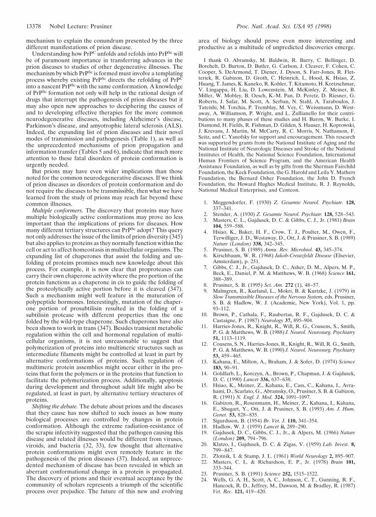
Infectious prion diseases. The infectious prion diseases in-clude kuru of the Fore people in New Guinea, where prionswere transmitted by ritualistic cannibalism (242, 252, 253).With the cessation of cannibalism at the urging of missionaries,kuru began to decline long before it was known to be trans-missible (Fig. 8). Sources of prions causing infectious CJD onseveral different continents include improperly sterilizeddepth electrodes, transplanted corneas, human growth hor-mone (HGH) and gonadotropin derived from cadaveric pitu-itaries, and dura mater grafts (254). Over 90 young adults havedeveloped CJD after treatment with cadaveric HGH; theincubation periods range from 3 to more than 20 years (255,256). Dura mater grafts implanted during neurosurgical pro-cedures seem to have caused more than 60 cases of CJD; theseincubation periods range from 1 to more than 14 years(257–259).
Prion Diversity. The existence of prion strains raises thequestion of how heritable biological information can be enci-phered in a molecule other than nucleic acid (131, 215,260–264). Strains or varieties of prions have been defined byincubation times and the distribution of neuronal vacuolation(215, 265). Subsequently, the patterns of PrPSc deposition werefound to correlate with vacuolation profiles, and these patternswere also used to characterize strains of prions (231, 266, 267).
The typing of prion strains in C57BL, VM, and (C57BL 3VM)F1 inbred mice began with isolates from sheep withscrapie. The prototypic strains called Me7 and 22A gaveincubation times of '150 and '400 days in C57BL mice,respectively (215, 268, 269). The PrP genes of C57BL and IyLn
(and later VM) mice encode proteins differing at two residuesand control scrapie incubation times (195, 213, 217–219, 270).
Until recently, support for the hypothesis that the tertiarystructure of PrPSc enciphers strain-specific information (23)was minimal except for the DY strain isolated from mink withtransmissible encephalopathy by passage in Syrian hamsters(26, 271, 272). PrPSc in DY prions showed diminished resis-tance to proteinase K digestion as well as a peculiar site ofcleavage. The DY strain presented a puzzling anomaly, sinceother prion strains exhibiting similar incubation times did notshow this altered susceptibility to proteinase K digestion ofPrPSc (135). Also notable was the generation of new strainsduring passage of prions through animals with different PrPgenes (135, 230).
PrPSc conformation enciphers diversity. Persuasive evidencethat strain-specific information is enciphered in the tertiarystructure of PrPSc comes from transmission of two differentinherited human prion diseases to mice expressing a chimericMHu2M PrP transgene (27). In FFI, the protease-resistantfragment of PrPSc after deglycosylation has a mass of 19 kDa,whereas in fCJD(E200K) and most sporadic prion diseases itis 21 kDa (Table 6) (273, 274). This difference in molecular sizewas shown to be due to different sites of proteolytic cleavageat the NH2 termini of the two human PrPSc molecules reflect-ing different tertiary structures (273). These distinct confor-mations were understandable because the amino acid se-quences of the PrPs differ.
Extracts from the brains of FFI patients transmitted diseaseto mice expressing a chimeric MHu2M PrP gene about 200days after inoculation and induced formation of the 19-kDaPrPSc, whereas fCJD(E200K) and sCJD produced the 21-kDa
FIG. 8. Disappearance of the kuru and BSE epidemics. (A) Annualnumber of cases of BSE in cattle in Great Britain. (B) Biannualnumber of cases of kuru in Papua New Guinea. Data were compiledfor BSE by John Wilesmith and for kuru by Michael Alpers.
13374 Nobel Lecture: Prusiner Proc. Natl. Acad. Sci. USA 95 (1998)
PrPSc in mice expressing the same transgene (27). On secondpassage, Tg(MHu2M) mice inoculated with FFI prions showedan incubation time of '130 days and a 19-kDa PrPSc, whereasthose inoculated with fCJD(E200K) prions exhibited an incu-bation time of '170 days and a 21-kDa PrPSc (28). Theexperimental data demonstrate that MHu2MPrPSc can exist intwo different conformations based on the sizes of the protease-resistant fragments, yet the amino acid sequence ofMHu2MPrPSc is invariant.
The results of our studies argue that PrPSc acts as a templatefor the conversion of PrPC into nascent PrPSc. Imparting thesize of the protease-resistant fragment of PrPSc through con-formational templating provides a mechanism for both thegeneration and propagation of prion strains.
Interestingly, the protease-resistant fragment of PrPSc afterdeglycosylation with a mass of 19 kDa has been found in apatient who developed a sporadic case of prion disease similarto FFI but with no family history. Because both PrP allelesencoded the wt sequence and a Met at position 129, we labeledthis case fatal sporadic insomnia (FSI). At autopsy, the spon-giform degeneration, reactive astrogliosis, and PrPSc deposi-tion were confined to the thalamus (275). These findings arguethat the clinicopathologic phenotype is determined by theconformation of PrPSc in accord with the results of thetransmission of human prions from patients with FFI to Tgmice (27).
Selective neuronal targeting. Besides incubation times, pro-files of spongiform change (Fig. 1) have been used to charac-terize prion strains (276), but recent studies argue that suchprofiles are not an intrinsic feature of strains (277, 278). Themechanism by which prion strains modify the pattern ofspongiform degeneration was perplexing, since earlier inves-tigations had shown that PrPSc deposition precedes neuronalvacuolation and reactive gliosis (212, 231). When FFI prionswere inoculated into Tg(MHu2M) mice, PrPSc was confinedlargely to the thalamus (Fig. 9A) as is the case for FFI inhumans (27, 279). In contrast, fCJD(E200K) prions inoculatedinto Tg(MHu2M) mice produced widespread deposition ofPrPSc throughout the cortical mantle and many of the deepstructures of the CNS (Fig. 9B) as is seen in fCJD(E200K) ofhumans. To examine whether the diverse patterns of PrPSc
deposition are influenced by Asn-linked glycosylation of PrPC,we constructed Tg mice expressing PrPs mutated at one orboth of the Asn-linked glycosylation consensus sites (278).These mutations resulted in aberrant neuroanatomic topolo-gies of PrPC within the CNS, whereas pathologic point muta-tions adjacent to the consensus sites did not alter the distri-bution of PrPC. Tg mice with mutation of the second PrPglycosylation site exhibited prion incubation times of .500days and unusual patterns of PrPSc deposition. These findingsraise two possible scenarios. First, glycosylation can modify theconformation of PrPC and affect its affinity for a particularconformer of PrPSc, which results in specific patterns of PrPSc
deposition; such interactions between PrPSc and PrPC are likelyto determine the rate of nascent PrPSc formation. Second,glycosylation modifies the stability of PrPSc and, hence, the rateof PrPSc clearance. This latter explanation is consistent with
the proposal that the binding of PrPC to protein X is therate-limiting step in PrPSc formation under most circumstances(280, 345).
BSE. Prion strains and the species barrier are of paramountimportance in understanding the BSE epidemic in GreatBritain, in which it is estimated that almost one million cattlewere infected with prions (281, 282). The mean incubationtime for BSE is about 5 years. Most cattle therefore did notmanifest disease because they were slaughtered between 2 and3 years of age (283). Nevertheless, more than 160,000 cattle,primarily dairy cows, have died of BSE over the past decade(Fig. 8A) (281). BSE is a massive common-source epidemiccaused by meat and bone meal (MBM) fed primarily to dairycows (282, 284). The MBM was prepared from the offal ofsheep, cattle, pigs, and chickens as a high-protein nutritionalsupplement. In the late 1970s, the hydrocarbon-solvent extrac-tion method used in the rendering of offal began to beabandoned, resulting in MBM with a much higher fat content(284). It is now thought that this change in the renderingprocess allowed scrapie prions from sheep to survive renderingand to be passed into cattle. Alternatively, bovine prions werepresent at low levels prior to modification of the renderingprocess and with the processing change survived in sufficientnumbers to initiate the BSE epidemic when inoculated backinto cattle orally through MBM. Against the latter hypothesisis the widespread geographical distribution throughout En-gland of the initial 17 cases of BSE, which occurred almostsimultaneously (282, 285, 286). Furthermore, there is noevidence of a preexisting prion disease of cattle, either in GreatBritain or elsewhere.
Origin of BSE prions? The origin of the bovine prions causingBSE cannot be determined by examining the amino acidsequence of PrPSc in cattle with BSE because the PrPSc in theseanimals has the bovine sequence whether the initial prions inMBM came from cattle or sheep. The bovine PrP sequencediffers from that of sheep at 7 or 8 positions (287–289). Incontrast to the many PrP polymorphisms found in sheep, onlyone PrP polymorphism has been found in cattle. Though mostbovine PrP alleles encode five octarepeats, some encode six.PrP alleles encoding six octarepeats do not seem to beoverrepresented in BSE (Fig. 4B) (290).
Brain extracts from BSE cattle cause disease in cattle, sheep,mice, pigs, and mink after intracerebral inoculation (291–295),but prions in brain extracts from sheep with scrapie fed tocattle produced illness substantially different from BSE (296).However, no exhaustive effort has been made to test differentstrains of sheep prions or to examine the disease after bovine-to-bovine passage. The annual incidence of sheep with scrapiein Britain over the past two decades has remained relativelylow (J. Wilesmith, personal communication). In July 1988, thepractice of feeding MBM to sheep and cattle was banned.Recent statistics argue that the epidemic is now disappearingas a result of this ruminant feed ban (Fig. 8A) (281), reminis-cent of the disappearance of kuru in the Fore people of NewGuinea (242, 253) (Fig. 8B).
Monitoring cattle for BSE prions. Although many plans havebeen offered for the culling of older cattle to minimize the
Table 6. Distinct prion strains generated in humans with inherited prion diseases and transmitted toTg mice
InoculumHost
species Host PrP genotypeIncubationtime, days (nyn0)
Data are from ref. 27 and G. Telling and S.B.P., unpublished work. Incubation times are given 6SEM.
Nobel Lecture: Prusiner Proc. Natl. Acad. Sci. USA 95 (1998) 13375
spread of BSE (281), it seems more important to monitor thefrequency of prion disease in cattle as they are slaughtered forhuman consumption. No reliable, specific test for prion diseasein live animals is available, but immunoassays for PrPSc in thebrainstems of cattle might provide a reasonable approach toestablishing the incidence of subclinical BSE in cattle enteringthe human food chain (176, 209, 289, 297–299, 345). Deter-mining how early in the incubation period PrPSc can bedetected by immunological methods is now possible, since areliable bioassay has been created by expressing the BoPrP
gene in Tg mice (180). Prior to development of Tg(BoPrP-)Prnp0/0 mice, non-Tg mice inoculated intracerebrally withBSE brain extracts required more than 300 days to developdisease (67, 295, 300, 301). Depending on the titer of theinoculum, the structures of PrPC and PrPSc, and the structureof protein X, the number of inoculated animals developingdisease can vary over a wide range (Table 3). Some investi-gators have stated that transmission of BSE to mice is quitevariable with incubation periods exceeding 1 year (67),whereas others report low prion titers in BSE brain homoge-nates (300, 301) compared with rodent brain scrapie (54, 56,302, 303).
Have bovine prions been transmitted to humans? In 1994, thefirst cases of CJD in teenagers and young adults that wereeventually labeled new variant (v) CJD occurred in Britain(304). Besides these patients being young (305, 306), theirbrains showed numerous PrP amyloid plaques surrounded bya halo of intense spongiform degeneration (Fig. 10) (307).Later, one French case meeting these criteria followed (308).These unusual neuropathologic changes have not been seen inCJD cases in the United States, Australia, or Japan (307, 309).Both macaque monkeys and marmosets developed neurologicdisease several years after inoculation with bovine prions(310), but only the macaques exhibited numerous PrP plaquessimilar to those found in vCJD (311) (R. Ridley and H. Baker,personal communication).
FIG. 9. Regional distribution of PrPSc deposition inTg(MHu2M)Prnp0/0 mice inoculated with prions from humans whodied of inherited prion diseases (Table 5). Histoblot of PrPSc deposi-tion in a coronal section a Tg(MHu2M)Prnp0/0 mouse through thehippocampus and thalamus (27). (A) The Tg mouse was inoculatedwith brain extract prepared from a patient who died of FFI. (B) TheTg mouse was inoculated with extract from a patient withfCJD(E200K). Cryostat sections were mounted on nitrocellulose andtreated with proteinase K to eliminate PrPC (209). To enhance theantigenicity of PrPSc, the histoblots were exposed to 3 M guanidiniumisothiocyanate before immunostaining using anti-PrP 3F4 mAb (174).(C) Labeled diagram of a coronal sections of the hippocampusythalamus region. NC, neocortex; Hp, hippocampus; Hb, habenula; Th,thalamus; vpl, ventral posterior lateral thalamic nucleus; Hy, hypo-thalamus; Am, amygdala. Photomicrographs were prepared by Ste-phen J. DeArmond.
FIG. 10. Histopathology of vCJD in Great Britain. (a) Section fromfrontal cortex stained by the periodic acid–Schiff method, showing afield with aggregates of plaques surrounded by spongiform degener-ation. (393.) (b) Multiple plaques and amorphous deposits are PrPimmunopositive. (3460.) Specimens were provided by James Ironside,Jeanne Bell, and Robert Will; photomicrographs were prepared byStephen J. DeArmond.
13376 Nobel Lecture: Prusiner Proc. Natl. Acad. Sci. USA 95 (1998)
The restricted geographical occurrence and chronology ofvCJD have raised the possibility that BSE prions have beentransmitted to humans. That only '25 vCJD cases have beenrecorded and the incidence has remained relatively constantmake establishing the origin of vCJD difficult. No set of dietaryhabits distinguishes vCJD patients from apparently healthypeople. Moreover, there is no explanation for the predilectionof vCJD for teenagers and young adults. Why have olderindividuals not developed vCJD-based neuropathologic crite-ria? It is noteworthy that epidemiological studies over the pastthree decades have failed to find evidence for transmission ofsheep prions to humans (9–12). Attempts to predict the futurenumber of cases of vCJD, assuming exposure to bovine prionsprior to the offal ban, have been uninformative because so fewcases of vCJD have occurred (312–314). Are we at the begin-ning of a human prion disease epidemic in Britain like thoseseen for BSE and kuru (Fig. 8), or will the number of vCJDcases remain small as seen with iCJD caused by cadavericHGH (255, 256)?
Strain of BSE prions. Was a particular conformation ofbovine PrPSc selected for heat resistance during the renderingprocess and then reselected multiple times as cattle infected byingesting prion-contaminated MBM were slaughtered andtheir offal rendered into more MBM? Recent studies of PrPSc
from brains of patients who died of vCJD show a pattern of PrPglycoforms different from those found for sCJD or iCJD (315,316). But the utility of measuring PrP glycoforms is question-able in trying to relate BSE to vCJD (317, 318) because PrPSc
is formed after the protein is glycosylated (138, 140) andenzymatic deglycosylation of PrPSc requires denaturation (319,320). Alternatively, it may be possible to establish a relation-ship between the conformations of PrPSc from cattle with BSEand those from humans with vCJD by using Tg mice as wasdone for strains generated in the brains of patients with FFI orfCJD (27, 180). A relationship between vCJD and BSE hasbeen suggested by finding similar incubation times in non-TgRIII mice of '310 days after inoculation with Hu or Bo prions(295).
Yeast and Other Prions. Although prions were originallydefined in the context of an infectious pathogen (58), it is nowbecoming widely accepted that prions are elements that impartand propagate variability through multiple conformers of anormal cellular protein. Such a mechanism must surely not berestricted to a single class of transmissible pathogens. Indeed,it is likely that the original definition will need to be extendedto encompass other situations in which a similar mechanism ofinformation transfer occurs.
Two notable prion-like determinants, [URE3] and [PSI],have already been described in yeast and one in another fungusdenoted [Het-s*] (321–326). Studies of candidate prion pro-teins in yeast may prove particularly helpful in the dissectionof some of the events that feature in PrPSc formation. Inter-estingly, different strains of yeast prions have been identified(327). Conversion to the prion-like [PSI] state in yeast requiresthe molecular chaperone Hsp104; however, no homolog ofHsp104 has been found in mammals (322, 328). The NH2-terminal prion domains of Ure2p and Sup35 that are respon-sible for the [URE3] and [PSI] phenotypes in yeast have beenidentified. In contrast to PrP, which is a GPI-anchored mem-brane protein, both Ure2p and Sup35 are cytosolic proteins(329). When the prion domains of these yeast proteins wereexpressed in E. coli, the proteins were found to polymerize intofibrils with properties similar to those of PrP and otheramyloids (323–325).
Whether prions explain some other examples of acquiredinheritance in lower organisms is unclear (330, 331). Forexample, studies on the inheritance of positional order andcellular handedness on the surface of small organisms havedemonstrated the epigenetic nature of these phenomena, butthe mechanism remains unclear (332, 333).
Prevention of and Therapeutics for Prion Diseases. As ourunderstanding of prion propagation increases, it should bepossible to design effective therapeutics. Because people atrisk for inherited prion diseases can now be identified decadesbefore neurologic dysfunction is evident, the development ofan effective therapy for these fully penetrant disorders isimperative (334, 335). Although we have no way of predictingthe number of individuals who may develop neurologic dys-function from bovine prions in the future (313), it would beprudent to seek an effective therapy now (28, 336). Interferingwith the conversion of PrPC into PrPSc seems to be the mostattractive therapeutic target (60). Either stabilizing the struc-ture of PrPC by binding a drug or modifying the action ofprotein X, which might function as a molecular chaperone(Fig. 5B), is a reasonable strategy. Whether it is more effica-cious to design a drug that binds to PrPC at the protein Xbinding site or one that mimics the structure of PrPC with basicpolymorphic residues that seem to prevent scrapie and CJDremains to be determined (178, 233). Because PrPSc formationseems limited to caveolae-like domains (142–145, 337), drugsdesigned to inhibit this process need not penetrate the cytosolof cells, but they do need to be able to enter the CNS.Alternatively, drugs that destabilize the structure of PrPSc
might also prove useful.The production of domestic animals that do not replicate
prions may also be important with respect to preventing priondisease. Sheep encoding the RyR polymorphism at position171 seem to be resistant to scrapie (199, 232, 338–344);presumably, this was the genetic basis of Parry’s scrapieeradication program in Great Britain 30 years ago (44, 46). Amore effective approach using dominant negatives for produc-ing prion-resistant domestic animals, including sheep andcattle, is probably the expression of PrP transgenes encodingR171 as well as additional basic residues at the putative proteinX binding site (Fig. 5B) (178). Such an approach can be readilyevaluated in Tg mice, and once shown to be effective, it couldbe instituted by artificial insemination of sperm from maleshomozygous for the transgene. More difficult is the productionof PrP-deficient cattle and sheep. Although such animalswould not be susceptible to prion disease (100, 101), they mightsuffer some deleterious effects from ablation of the PrP gene(223–225, 228).
Whether gene therapy for the human prion diseases by usingthe dominant-negative approach described above for prion-resistant animals will prove feasible depends on the availabilityof efficient vectors for delivery of the transgene to the CNS.
Concluding Remarks—Looking to the Future. Although thestudy of prions has taken several unexpected directions overthe past three decades, a rather novel and fascinating story ofprion biology is emerging. Investigations of prions have elu-cidated a previously unknown mechanism of disease in humansand animals. While learning the details of the structures ofPrPs and deciphering the mechanism of PrPC transformationinto PrPSc will be important, the fundamental principles ofprion biology have become reasonably clear. Though someinvestigators prefer to view the composition of the infectiousprion particle as unresolved (336, 346), such a perspectivedenies an enlarging body of data, none of which refute theprion concept. Moreover, the discovery of prion-like phenom-ena mediated by proteins unrelated to PrP in yeast and fungiserves not only to strengthen the prion concept but also towiden it (329).
Hallmark of prion diseases. The hallmark of all prion dis-eases—whether sporadic, dominantly inherited, or acquired byinfection—is that they involve the aberrant metabolism andresulting accumulation of the prion protein (Table 1) (23). Theconversion of PrPC into PrPSc involves a conformation changewhereby the a-helical content diminishes and the amount ofb-sheet increases (25). These findings provide a reasonable
Nobel Lecture: Prusiner Proc. Natl. Acad. Sci. USA 95 (1998) 13377
mechanism to explain the conundrum presented by the threedifferent manifestations of prion disease.
Understanding how PrPC unfolds and refolds into PrPSc willbe of paramount importance in transferring advances in theprion diseases to studies of other degenerative illnesses. Themechanism by which PrPSc is formed must involve a templatingprocess whereby existing PrPSc directs the refolding of PrPC
into a nascent PrPSc with the same conformation. A knowledgeof PrPSc formation not only will help in the rational design ofdrugs that interrupt the pathogenesis of prion diseases but itmay also open new approaches to deciphering the causes ofand to developing effective therapies for the more commonneurodegenerative diseases, including Alzheimer’s disease,Parkinson’s disease, and amyotrophic lateral sclerosis (ALS).Indeed, the expanding list of prion diseases and their novelmodes of transmission and pathogenesis (Table 1), as well asthe unprecedented mechanisms of prion propagation andinformation transfer (Tables 5 and 6), indicate that much moreattention to these fatal disorders of protein conformation isurgently needed.
But prions may have even wider implications than thosenoted for the common neurodegenerative diseases. If we thinkof prion diseases as disorders of protein conformation and donot require the diseases to be transmissible, then what we havelearned from the study of prions may reach far beyond thesecommon illnesses.
Multiple conformers. The discovery that proteins may havemultiple biologically active conformations may prove no lessimportant than the implications of prions for diseases. Howmany different tertiary structures can PrPSc adopt? This querynot only addresses the issue of the limits of prion diversity (345)but also applies to proteins as they normally function within thecell or act to affect homeostasis in multicellular organisms. Theexpanding list of chaperones that assist the folding and un-folding of proteins promises much new knowledge about thisprocess. For example, it is now clear that proproteases cancarry their own chaperone activity where the pro portion of theprotein functions as a chaperone in cis to guide the folding ofthe proteolytically active portion before it is cleaved (347).Such a mechanism might well feature in the maturation ofpolypeptide hormones. Interestingly, mutation of the chaper-one portion of prosubtilisin resulted in the folding of asubtilisin protease with different properties than the onefolded by the wild-type chaperone. Such chaperones have alsobeen shown to work in trans (347). Besides transient metabolicregulation within the cell and hormonal regulation of multi-cellular organisms, it is not unreasonable to suggest thatpolymerization of proteins into multimeric structures such asintermediate filaments might be controlled at least in part byalternative conformations of proteins. Such regulation ofmultimeric protein assemblies might occur either in the pro-teins that form the polymers or in the proteins that function tofacilitate the polymerization process. Additionally, apoptosisduring development and throughout adult life might also beregulated, at least in part, by alternative tertiary structures ofproteins.
Shifting the debate. The debate about prions and the diseasesthat they cause has now shifted to such issues as how manybiological processes are controlled by changes in proteinconformation. Although the extreme radiation-resistance ofthe scrapie infectivity suggested that the pathogen causing thisdisease and related illnesses would be different from viruses,viroids, and bacteria (32, 33), few thought that alternativeprotein conformations might even remotely feature in thepathogenesis of the prion diseases (37). Indeed, an unprece-dented mechanism of disease has been revealed in which anaberrant conformational change in a protein is propagated.The discovery of prions and their eventual acceptance by thecommunity of scholars represents a triumph of the scientificprocess over prejudice. The future of this new and evolving
area of biology should prove even more interesting andproductive as a multitude of unpredicted discoveries emerge.
I thank O. Abramsky, M. Baldwin, R. Barry, C. Bellinger, D.Borchelt, D. Burton, D. Butler, G. Carlson, J. Cleaver, F. Cohen, C.Cooper, S. DeArmond, T. Diener, J. Dyson, S. Farr-Jones, R. Flet-terick, R. Gabizon, D. Groth, C. Heinrich, L. Hood, K. Hsiao, Z.Huang, T. James, K. Kaneko, R. Kohler, T. Kitamoto, H. Kretzschmar,V. Lingappa, H. Liu, D. Lowenstein, M. McKinley, Z. Meiner, B.Miller, W. Mobley, B. Oesch, K.-M. Pan, D. Peretz, D. Riesner, G.Roberts, J. Safar, M. Scott, A. Serban, N. Stahl, A. Taraboulos, J.Tateishi, M. Torchia, P. Tremblay, M. Vey, C. Weissmann, D. West-away, A. Williamson, P. Wright, and L. Zullianello for their contri-butions to many phases of these studies and H. Baron, W. Burke, I.Diamond, H. Fields, R. Fishman, D. Gilden, S. Hauser, H. Koprowski,J. Krevans, J. Martin, M. McCarty, R. C. Morris, N. Nathanson, F.Seitz, and C. Yanofsky for support and encouragement. This researchwas supported by grants from the National Institute of Aging and theNational Institute of Neurologic Diseases and Stroke of the NationalInstitutes of Health, the National Science Foundation, InternationalHuman Frontiers of Science Program, and the American HealthAssistance Foundation, as well as by gifts from the Sherman FairchildFoundation, the Keck Foundation, the G. Harold and Leila Y. MathersFoundation, the Bernard Osher Foundation, the John D. FrenchFoundation, the Howard Hughes Medical Institute, R. J. Reynolds,National Medical Enterprises, and Centeon.
1. Meggendorfer, F. (1930) Z. Gesamte Neurol. Psychiatr. 128,337–341.
2. Stender, A. (1930) Z. Gesamte Neurol. Psychiatr. 128, 528–543.3. Masters, C. L., Gajdusek, D. C. & Gibbs, C. J., Jr. (1981) Brain
104, 559–588.4. Hsiao, K., Baker, H. F., Crow, T. J., Poulter, M., Owen, F.,
Terwilliger, J. D., Westaway, D., Ott, J. & Prusiner, S. B. (1989)Nature (London) 338, 342–345.
5. Prusiner, S. B. (1989) Annu. Rev. Microbiol. 43, 345–374.6. Kirschbaum, W. R. (1968) Jakob-Creutzfeldt Disease (Elsevier,
Amsterdam), p. 251.7. Gibbs, C. J., Jr., Gajdusek, D. C., Asher, D. M., Alpers, M. P.,
Beck, E., Daniel, P. M. & Matthews, W. B. (1968) Science 161,388–389.
8. Prusiner, S. B. (1995) Sci. Am. 272 (1), 48–57.9. Malmgren, R., Kurland, L., Mokri, B. & Kurtzke, J. (1979) in
Slow Transmissible Diseases of the Nervous System, eds. Prusiner,S. B. & Hadlow, W. J. (Academic, New York), Vol. 1, pp.93–112.
10. Brown, P., Cathala, F., Raubertas, R. F., Gajdusek, D. C. &Castaigne, P. (1987) Neurology 37, 895–904.
11. Harries-Jones, R., Knight, R., Will, R. G., Cousens, S., Smith,P. G. & Matthews, W. B. (1988) J. Neurol. Neurosurg. Psychiatry51, 1113–1119.
12. Cousens, S. N., Harries-Jones, R., Knight, R., Will, R. G., Smith,P. G. & Matthews, W. B. (1990) J. Neurol. Neurosurg. Psychiatry53, 459–465.
13. Kahana, E., Milton, A., Braham, J. & Sofer, D. (1974) Science183, 90–91.
14. Goldfarb, L., Korczyn, A., Brown, P., Chapman, J. & Gajdusek,D. C. (1990) Lancet 336, 637–638.
15. Hsiao, K., Meiner, Z., Kahana, E., Cass, C., Kahana, I., Avra-hami, D., Scarlato, G., Abramsky, O., Prusiner, S. B. & Gabizon,R. (1991) N. Engl. J. Med. 324, 1091–1097.
16. Gabizon, R., Rosenmann, H., Meiner, Z., Kahana, I., Kahana,E., Shugart, Y., Ott, J. & Prusiner, S. B. (1993) Am. J. Hum.Genet. 53, 828–835.
17. Sigurdsson, B. (1954) Br. Vet. J. 110, 341–354.18. Hadlow, W. J. (1959) Lancet ii, 289–290.19. Gajdusek, D. C., Gibbs, C. J., Jr., & Alpers, M. (1966) Nature
(London) 209, 794–796.20. Klatzo, I., Gajdusek, D. C. & Zigas, V. (1959) Lab. Invest. 8,
799–847.21. Zlotnik, I. & Stamp, J. L. (1961) World Neurology 2, 895–907.22. Masters, C. L. & Richardson, E. P., Jr. (1978) Brain 101,
333–344.23. Prusiner, S. B. (1991) Science 252, 1515–1522.24. Wells, G. A. H., Scott, A. C., Johnson, C. T., Gunning, R. F.,
Hancock, R. D., Jeffrey, M., Dawson, M. & Bradley, R. (1987)Vet. Rec. 121, 419–420.
13378 Nobel Lecture: Prusiner Proc. Natl. Acad. Sci. USA 95 (1998)
25. Pan, K.-M., Baldwin, M., Nguyen, J., Gasset, M., Serban, A.,Groth, D., Mehlhorn, I., Huang, Z., Fletterick, R. J., Cohen,F. E. & Prusiner, S. B. (1993) Proc. Natl. Acad. Sci. USA 90,10962–10966.
26. Bessen, R. A. & Marsh, R. F. (1994) J. Virol. 68, 7859–7868.27. Telling, G. C., Parchi, P., DeArmond, S. J., Cortelli, P., Mon-
tagna, P., Gabizon, R., Mastrianni, J., Lugaresi, E., Gambetti, P.& Prusiner, S. B. (1996) Science 274, 2079–2082.
28. Prusiner, S. B. (1997) Science 278, 245–251.29. Dlouhy, S. R., Hsiao, K., Farlow, M. R., Foroud, T., Conneally,
P. M., Johnson, P., Prusiner, S. B., Hodes, M. E. & Ghetti, B.(1992) Nat. Genet. 1, 64–67.
30. Petersen, R. B., Tabaton, M., Berg, L., Schrank, B., Torack,R. M., Leal, S., Julien, J., Vital, C., Deleplanque, B., Pendle-bury, W. W., et al. (1992) Neurology 42, 1859–1863.
31. Poulter, M., Baker, H. F., Frith, C. D., Leach, M., Lofthouse, R.,Ridley, R. M., Shah, T., Owen, F., Collinge, J., Brown, G., et al.(1992) Brain 115, 675–685.
32. Alper, T., Haig, D. A. & Clarke, M. C. (1966) Biochem. Biophys.Res. Commun. 22, 278–284.
33. Alper, T., Cramp, W. A., Haig, D. A. & Clarke, M. C. (1967)Nature (London) 214, 764–766.
34. Latarjet, R., Muel, B., Haig, D. A., Clarke, M. C. & Alper, T.(1970) Nature (London) 227, 1341–1343.
35. Gibbs, C. J., Jr., Gajdusek, D. C. & Latarjet, R. (1978) Proc.Natl. Acad. Sci. USA 75, 6268–6270.
36. Pattison, I. H. (1965) in Slow, Latent and Temperate VirusInfections, NINDB Monograph 2, eds. Gajdusek, D. C., Gibbs,C. J., Jr., & Alpers, M. P. (U.S. Gov. Printing Office, Wash-ington, DC), pp. 249–257.
37. Griffith, J. S. (1967) Nature (London) 215, 1043–1044.38. Pattison, I. H. & Jones, K. M. (1967) Vet. Rec. 80, 1–8.39. Gibbons, R. A. & Hunter, G. D. (1967) Nature (London) 215,
1041–1043.40. Hunter, G. D., Kimberlin, R. H. & Gibbons, R. A. (1968) J.
Theor. Biol. 20, 355–357.41. Field, E. J., Farmer, F., Caspary, E. A. & Joyce, G. (1969) Nature
(London) 222, 90–91.42. Hunter, G. D. (1972) J. Infect. Dis. 125, 427–440.43. Gordon, W. S. (1946) Vet. Res. 58, 516–520.44. Parry, H. B. (1962) Heredity 17, 75–105.45. Dickinson, A. G., Young, G. B., Stamp, J. T. & Renwick, C. C.
(1965) Heredity 20, 485–503.46. Parry, H. B. (1983) in Scrapie Disease in Sheep, ed. Oppenhei-
mer, D. R. (Academic, New York), pp. 31–59.47. Prusiner, S. B. (1978) J. Biol. Chem. 253, 916–921.48. Prusiner, S. B., Garfin, D. E., Baringer, J. R., Cochran, S. P.,
Hadlow, W. J., Race, R. E. & Eklund, C. M. (1978) in PersistentViruses, eds. Stevens, J., Todaro, G. & Fox, C. F. (Academic,New York), pp. 591–613.
49. Prusiner, S. B. (1984) Sci. Am. 251 (4), 50–59.50. Chandler, R. L. (1963) Res. Vet. Sci. 4, 276–285.51. Prusiner, S., Davis, J. N. & Stadtman, E. R. (1976) J. Biol. Chem.
251, 3447–3456.52. Prusiner, S. B., Hadlow, W. J., Eklund, C. M. & Race, R. E.
(1977) Proc. Natl. Acad. Sci. USA 74, 4656–4660.53. Marsh, R. F. & Kimberlin, R. H. (1975) J. Infect. Dis. 131,
104–110.54. Kimberlin, R. & Walker, C. (1977) J. Gen. Virol. 34, 295–304.55. Prusiner, S. B., Groth, D. F., Cochran, S. P., Masiarz, F. R.,
McKinley, M. P. & Martinez, H. M. (1980) Biochemistry 19,4883–4891.
56. Prusiner, S. B., Cochran, S. P., Groth, D. F., Downey, D. E.,Bowman, K. A. & Martinez, H. M. (1982) Ann. Neurol. 11,353–358.
57. Zlotnik, I. (1963) Lancet ii, 1072.58. Prusiner, S. B. (1982) Science 216, 136–144.59. Stahl, N., Baldwin, M. A., Teplow, D. B., Hood, L., Gibson,
B. W., Burlingame, A. L. & Prusiner, S. B. (1993) Biochemistry32, 1991–2002.
60. Cohen, F. E., Pan, K.-M., Huang, Z., Baldwin, M., Fletterick,R. J. & Prusiner, S. B. (1994) Science 264, 530–531.
61. Prusiner, S. B., Groth, D., Serban, A., Stahl, N. & Gabizon, R.(1993) Proc. Natl. Acad. Sci. USA 90, 2793–2797.
62. Carp, R. I., Kascsak, R. J., Rubenstein, R. & Merz, P. A. (1994)Trends Neurosci. 17, 148–149.
63. Manuelidis, L. & Fritch, W. (1996) Virology 216, 46–59.
64. Narang, H. (1996) Proc. Soc. Exp. Biol. Med. 212, 208–224.65. Ozel, M., Baldauf, E., Beekes, M. & Diringer, H. (1996) in
Transmissible Subacute Spongiform Encephalopathies: Prion Dis-eases, eds. Court, L. & Dodet, B. (Elsevier, Paris), pp. 369–373.
66. Caughey, B. & Chesebro, B. (1997) Trends Cell Biol. 7, 56–62.67. Lasmezas, C. I., Deslys, J.-P., Robain, O., Jaegly, A., Beringue,
V., Peyrin, J.-M., Fournier, J.-G., Hauw, J.-J., Rossier, J. &Dormont, D. (1997) Science 275, 402–405.
68. Diener, T. O., McKinley, M. P. & Prusiner, S. B. (1982) Proc.Natl. Acad. Sci. USA 79, 5220–5224.
69. Chamberlin, T. C. (1890) Science [Old Series] 15, 92–97.70. Avery, O. T., MacLeod, C. M. & McCarty, M. (1944) J. Exp.
Med. 79, 137–157.71. Stanley, W. M. (1970) Arch. Environ. Health 21, 256–262.72. McCarty, M. (1985) The Transforming Principle: Discovering that
Genes Are Made of DNA (Norton, New York).73. Hershey, A. D. & Chase, M. (1952) J. Gen. Physiol. 36, 39–56.74. Watson, J. D. & Crick, F. H. C. (1953) Nature (London) 171,
964–967.75. Mirsky, A. E. & Pollister, A. W. (1946) J. Gen. Physiol. 30,
134–135.76. Diener, T. O. (1979) Viroids and Viroid Diseases (Wiley, New
York).77. Fraenkel-Conrat, H. & Williams, R. C. (1955) Proc. Natl. Acad.
Sci. USA 41, 690–698.78. Gierer, A. & Schramm, G. (1956) Nature (London) 177, 702–
703.79. Bolton, D. C., McKinley, M. P. & Prusiner, S. B. (1982) Science
218, 1309–1311.80. Prusiner, S. B., Bolton, D. C., Groth, D. F., Bowman, K. A.,
Cochran, S. P. & McKinley, M. P. (1982) Biochemistry 21,6942–6950.
81. Prusiner, S. B., Groth, D. F., Bolton, D. C., Kent, S. B. & Hood,L. E. (1984) Cell 38, 127–134.
82. Basler, K., Oesch, B., Scott, M., Westaway, D., Walchli, M.,Groth, D. F., McKinley, M. P., Prusiner, S. B. & Weissmann, C.(1986) Cell 46, 417–428.
83. Chesebro, B., Race, R., Wehrly, K., Nishio, J., Bloom, M.,Lechner, D., Bergstrom, S., Robbins, K., Mayer, L., Keith, J. M.,et al. (1985) Nature (London) 315, 331–333.
84. Oesch, B., Westaway, D., Walchli, M., McKinley, M. P., Kent,S. B. H., Aebersold, R., Barry, R. A., Tempst, P., Teplow, D. B.,Hood, L. E., et al. (1985) Cell 40, 735–746.
85. Meyer, R. K., McKinley, M. P., Bowman, K. A., Braunfeld,M. B., Barry, R. A. & Prusiner, S. B. (1986) Proc. Natl. Acad.Sci. USA 83, 2310–2314.
86. Locht, C., Chesebro, B., Race, R. & Keith, J. M. (1986) Proc.Natl. Acad. Sci. USA 83, 6372–6376.
87. Bendheim, P. E., Barry, R. A., DeArmond, S. J., Stites, D. P. &Prusiner, S. B. (1984) Nature (London) 310, 418–421.
88. Bockman, J. M., Kingsbury, D. T., McKinley, M. P., Bendheim,P. E. & Prusiner, S. B. (1985) N. Engl. J. Med. 312, 73–78.
89. Brown, P., Coker-Vann, M., Pomeroy, K., Franko, M., Asher,D. M., Gibbs, C. J., Jr., & Gajdusek, D. C. (1986) N. Engl. J. Med.314, 547–551.
90. Bockman, J. M., Prusiner, S. B., Tateishi, J. & Kingsbury, D. T.(1987) Ann. Neurol. 21, 589–595.
91. Manuelidis, L., Valley, S. & Manuelidis, E. E. (1985) Proc. Natl.Acad. Sci. USA 82, 4263–4267.
92. Prusiner, S. B. (1987) N. Engl. J. Med. 317, 1571–1581.93. Manuelidis, L., Tesin, D. M., Sklaviadis, T. & Manuelidis, E. E.
(1987) Proc. Natl. Acad. Sci. USA 84, 5937–5941.94. Gomi, H., Yokoyama, T., Fujimoto, K., Ikeda, T., Katoh, A.,
Itoh, T. & Itohara, S. (1995) Neuron 14, 29–41.95. Tatzelt, J., Maeda, N., Pekny, M., Yang, S.-L., Betsholtz, C.,
Eliasson, C., Cayetano, J., Camerino, A. P., DeArmond, S. J. &Prusiner, S. B. (1996) Neurology 47, 449–453.
96. Duguid, J. R., Rohwer, R. G. & Seed, B. (1988) Proc. Natl. Acad.Sci. USA 85, 5738–5742.
97. Duguid, J. R., Bohmont, C. W., Liu, N. & Tourtellotte, W. W.(1989) Proc. Natl. Acad. Sci. USA 86, 7260–7264.
98. Diedrich, J. F., Carp, R. I. & Haase, A. T. (1993) Microb. Pathog.15, 1–6.
99. Akowitz, A., Sklaviadis, T. & Manuelidis, L. (1994) NucleicAcids Res. 22, 1101–1107.
100. Bueler, H., Aguzzi, A., Sailer, A., Greiner, R.-A., Autenried, P.,Aguet, M. & Weissmann, C. (1993) Cell 73, 1339–1347.
Nobel Lecture: Prusiner Proc. Natl. Acad. Sci. USA 95 (1998) 13379
101. Prusiner, S. B., Groth, D., Serban, A., Koehler, R., Foster, D.,Torchia, M., Burton, D., Yang, S.-L. & DeArmond, S. J. (1993)Proc. Natl. Acad. Sci. USA 90, 10608–10612.
102. Manson, J. C., Clarke, A. R., McBride, P. A., McConnell, I. &Hope, J. (1994) Neurodegeneration 3, 331–340.
103. Sailer, A., Bueler, H., Fischer, M., Aguzzi, A. & Weissmann, C.(1994) Cell 77, 967–968.
104. Sakaguchi, S., Katamine, S., Shigematsu, K., Nakatani, A.,Moriuchi, R., Nishida, N., Kurokawa, K., Nakaoke, R., Sato, H.,Jishage, K., et al. (1995) J. Virol. 69, 7586–7592.
105. Gabizon, R., McKinley, M. P., Groth, D. F. & Prusiner, S. B.(1988) Proc. Natl. Acad. Sci. USA 85, 6617–6621.
106. McKinley, M. P., Bolton, D. C. & Prusiner, S. B. (1983) Cell 35,57–62.
107. Prusiner, S. B., McKinley, M. P., Bowman, K. A., Bolton, D. C.,Bendheim, P. E., Groth, D. F. & Glenner, G. G. (1983) Cell 35,349–358.
108. Bolton, D. C., McKinley, M. P. & Prusiner, S. B. (1984)Biochemistry 23, 5898–5906.
109. Riesner, D., Kellings, K., Post, K., Wille, H., Serban, H., Groth,D., Baldwin, M. A. & Prusiner, S. B. (1996) J. Virol. 70,1714–1722.
110. Bellinger-Kawahara, C., Cleaver, J. E., Diener, T. O. &Prusiner, S. B. (1987) J. Virol. 61, 159–166.
111. Bellinger-Kawahara, C., Diener, T. O., McKinley, M. P., Groth,D. F., Smith, D. R. & Prusiner, S. B. (1987) Virology 160,271–274.
112. Bellinger-Kawahara, C. G., Kempner, E., Groth, D. F., Gabizon,R. & Prusiner, S. B. (1988) Virology 164, 537–541.
113. Kellings, K., Meyer, N., Mirenda, C., Prusiner, S. B. & Riesner,D. (1992) J. Gen. Virol. 73, 1025–1029.
114. Kellings, K., Prusiner, S. B. & Riesner, D. (1994) Phil. Trans. R.Soc. London B 343, 425–430.
115. Manuelidis, L., Sklaviadis, T., Akowitz, A. & Fritch, W. (1995)Proc. Natl. Acad. Sci. USA 92, 5124–5128.
116. Williams, R. C. (1954) Adv. Virus Res. 2, 183–239.117. Cohen, A. S., Shirahama, T. & Skinner, M. (1982) in Electron
Microscopy of Proteins, ed. Harris, J. R. (Academic, New York),Vol. 3, pp. 165–206.
118. DeArmond, S. J., McKinley, M. P., Barry, R. A., Braunfeld,M. B., McColloch, J. R. & Prusiner, S. B. (1985) Cell 41,221–235.
119. Kitamoto, T., Tateishi, J., Tashima, I., Takeshita, I., Barry,R. A., DeArmond, S. J. & Prusiner, S. B. (1986) Ann. Neurol.20, 204–208.
120. Roberts, G. W., Lofthouse, R., Brown, R., Crow, T. J., Barry,R. A. & Prusiner, S. B. (1986) N. Engl. J. Med. 315, 1231–1233.
121. Gabizon, R., McKinley, M. P. & Prusiner, S. B. (1987) Proc.Natl. Acad. Sci. USA 84, 4017–4021.
122. McKinley, M. P., Meyer, R., Kenaga, L., Rahbar, F., Cotter, R.,Serban, A. & Prusiner, S. B. (1991) J. Virol. 65, 1440–1449.
123. Diringer, H., Gelderblom, H., Hilmert, H., Ozel, M., Edelbluth,C. & Kimberlin, R. H. (1983) Nature (London) 306, 476–478.
124. Merz, P. A., Kascsak, R. J., Rubenstein, R., Carp, R. I. &Wisniewski, H. M. (1987) J. Virol. 61, 42–49.
125. Diener, T. O. (1987) Cell 49, 719–721.126. Merz, P. A., Somerville, R. A., Wisniewski, H. M. & Iqbal, K.
(1981) Acta Neuropathol. (Berlin) 54, 63–74.127. Merz, P. A., Wisniewski, H. M., Somerville, R. A., Bobin, S. A.,
Masters, C. L. & Iqbal, K. (1983) Acta Neuropathol. (Berlin) 60,113–124.
128. Merz, P. A., Rohwer, R. G., Kascsak, R., Wisniewski, H. M.,Somerville, R. A., Gibbs, C. J., Jr., & Gajdusek, D. C. (1984)Science 225, 437–440.
129. Diringer, H. (1991) Eur. J. Epidemiol. 7, 562–566.130. Bastian, F. O. (1993) ASM News 59, 235–240.131. Kimberlin, R. H. (1990) Semin. Virol. 1, 153–162.132. Chesebro, B. & Caughey, B. (1993) Curr. Biol. 3, 696–698.133. Scott, M., Groth, D., Foster, D., Torchia, M., Yang, S.-L.,
DeArmond, S. J. & Prusiner, S. B. (1993) Cell 73, 979–988.134. Telling, G. C., Scott, M., Mastrianni, J., Gabizon, R., Torchia,
M., Cohen, F. E., DeArmond, S. J. & Prusiner, S. B. (1995) Cell83, 79–90.
135. Scott, M. R., Groth, D., Tatzelt, J., Torchia, M., Tremblay, P.,DeArmond, S. J. & Prusiner, S. B. (1997) J. Virol. 71, 9032–9044.
136. Anfinsen, C. B. (1973) Science 181, 223–230.
137. Pergami, P., Jaffe, H. & Safar, J. (1996) Anal. Biochem. 236,63–73.
138. Borchelt, D. R., Scott, M., Taraboulos, A., Stahl, N. & Prusiner,S. B. (1990) J. Cell Biol. 110, 743–752.
139. Stahl, N., Borchelt, D. R., Hsiao, K. & Prusiner, S. B. (1987) Cell51, 229–240.
140. Caughey, B. & Raymond, G. J. (1991) J. Biol. Chem. 266,18217–18223.
141. Borchelt, D. R., Taraboulos, A. & Prusiner, S. B. (1992) J. Biol.Chem. 267, 16188–16199.
142. Taraboulos, A., Scott, M., Semenov, A., Avrahami, D., Laszlo,L. & Prusiner, S. B. (1995) J. Cell Biol. 129, 121–132.
143. Vey, M., Pilkuhn, S., Wille, H., Nixon, R., DeArmond, S. J.,Smart, E. J., Anderson, R. G., Taraboulos, A. & Prusiner, S. B.(1996) Proc. Natl. Acad. Sci. USA 93, 14945–14949.
144. Kaneko, K., Vey, M., Scott, M., Pilkuhn, S., Cohen, F. E. &Prusiner, S. B. (1997) Proc. Natl. Acad. Sci. USA 94, 2333–2338.
145. Naslavsky, N., Stein, R., Yanai, A., Friedlander, G. & Tarabou-los, A. (1997) J. Biol. Chem. 272, 6324–6331.
146. Gasset, M., Baldwin, M. A., Lloyd, D., Gabriel, J.-M., Holtz-man, D. M., Cohen, F., Fletterick, R. & Prusiner, S. B. (1992)Proc. Natl. Acad. Sci. USA 89, 10940–10944.
147. Huang, Z., Gabriel, J.-M., Baldwin, M. A., Fletterick, R. J.,Prusiner, S. B. & Cohen, F. E. (1994) Proc. Natl. Acad. Sci. USA91, 7139–7143.
148. Zhang, H., Kaneko, K., Nguyen, J. T., Livshits, T. L., Baldwin,M. A., Cohen, F. E., James, T. L. & Prusiner, S. B. (1995) J. Mol.Biol. 250, 514–526.
149. Schatzl, H. M., Da Costa, M., Taylor, L., Cohen, F. E. &Prusiner, S. B. (1995) J. Mol. Biol. 245, 362–374.
150. Bamborough, P., Wille, H., Telling, G. C., Yehiely, F., Prusiner,S. B. & Cohen, F. E. (1996) Cold Spring Harbor Symp. Quant.Biol. 61, 495–509.
151. Hay, B., Barry, R. A., Lieberburg, I., Prusiner, S. B. & Lingappa,V. R. (1987) Mol. Cell. Biol. 7, 914–920.
152. DeFea, K. A., Nakahara, D. H., Calayag, M. C., Yost, C. S.,Mirels, L. F., Prusiner, S. B. & Lingappa, V. R. (1994) J. Biol.Chem. 269, 16810–16820.
153. Hegde, R. S., Mastrianni, J. A., Scott, M. R., DeFea, K. A.,Tremblay, P., Torchia, M., DeArmond, S. J., Prusiner, S. B. &Lingappa, V. R. (1998) Science 279, 827–834.
154. Riek, R., Hornemann, S., Wider, G., Billeter, M., Glockshuber,R. & Wuthrich, K. (1996) Nature (London) 382, 180–182.
155. James, T. L., Liu, H., Ulyanov, N. B., Farr-Jones, S., Zhang, H.,Donne, D. G., Kaneko, K., Groth, D., Mehlhorn, I., Prusiner,S. B. & Cohen, F. E. (1997) Proc. Natl. Acad. Sci. USA 94,10086–10091.
156. Donne, D. G., Viles, J. H., Groth, D., Mehlhorn, I., James, T. L.,Cohen, F. E., Prusiner, S. B., Wright, P. E. & Dyson, H. J. (1997)Proc. Natl. Acad. Sci. USA 94, 13452–13457.
157. Williamson, R. A., Peretz, D., Smorodinsky, N., Bastidas, R.,Serban, H., Mehlhorn, I., DeArmond, S. J., Prusiner, S. B. &Burton, D. R. (1996) Proc. Natl. Acad. Sci. USA 93, 7279–7282.
158. Peretz, D., Williamson, R. A., Matsunaga, Y., Serban, H.,Pinilla, C., Bastidas, R., Rozenshteyn, R., James, T. L.,Houghten, R. A., Cohen, F. E., et al. (1997) J. Mol. Biol. 273,614–622.
159. Caughey, B., Race, R. E., Ernst, D., Buchmeier, M. J. &Chesebro, B. (1989) J. Virol. 63, 175–181.
160. Shmerling, D., Hegyi, I., Fischer, M., Blattler, T., Brandner, S.,Gotz, J., Rulicke, T., Flechsig, E., Cozzio, A., von Mering, C.,et al. (1998) Cell 93, 203–214.
161. Mehlhorn, I., Groth, D., Stockel, J., Moffat, B., Reilly, D.,Yansura, D., Willett, W. S., Baldwin, M., Fletterick, R., Cohen,F. E., et al. (1996) Biochemistry 35, 5528–5537.
162. Zhang, H., Stockel, J., Mehlhorn, I., Groth, D., Baldwin, M. A.,Prusiner, S. B., James, T. L. & Cohen, F. E. (1997) Biochemistry36, 3543–3553.
163. Fischer, M., Rulicke, T., Raeber, A., Sailer, A., Moser, M.,Oesch, B., Brandner, S., Aguzzi, A. & Weissmann, C. (1996)EMBO J. 15, 1255–1264.
164. Muramoto, T., DeArmond, S. J., Scott, M., Telling, G. C.,Cohen, F. E. & Prusiner, S. B. (1997) Nat. Med. 3, 750–755.
165. Riek, R., Hornemann, S., Wider, G., Glockshuber, R. & Wuth-rich, K. (1997) FEBS Lett 413, 282–288.
166. Huang, Z., Prusiner, S. B. & Cohen, F. E. (1996) Fold. Des. 1,13–19.
13380 Nobel Lecture: Prusiner Proc. Natl. Acad. Sci. USA 95 (1998)
167. Turk, E., Teplow, D. B., Hood, L. E. & Prusiner, S. B. (1988)Eur. J. Biochem. 176, 21–30.
168. Muramoto, T., Scott, M., Cohen, F. & Prusiner, S. B. (1996)Proc. Natl. Acad. Sci. USA 93, 15457–15462.
169. Caughey, B. W., Dong, A., Bhat, K. S., Ernst, D., Hayes, S. F.& Caughey, W. S. (1991) Biochemistry 30, 7672–7680.
170. Gasset, M., Baldwin, M. A., Fletterick, R. J. & Prusiner, S. B.(1993) Proc. Natl. Acad. Sci. USA 90, 1–5.
171. Safar, J., Roller, P. P., Gajdusek, D. C. & Gibbs, C. J., Jr. (1993)J. Biol. Chem. 268, 20276–20284.
172. Rogers, M., Yehiely, F., Scott, M. & Prusiner, S. B. (1993) Proc.Natl. Acad. Sci. USA 90, 3182–3186.
173. Barry, R. A. & Prusiner, S. B. (1986) J. Infect. Dis. 154, 518–521.174. Kascsak, R. J., Rubenstein, R., Merz, P. A., Tonna-DeMasi, M.,
Fersko, R., Carp, R. I., Wisniewski, H. M. & Diringer, H. (1987)J. Virol. 61, 3688–3693.
175. Rogers, M., Serban, D., Gyuris, T., Scott, M., Torchia, T. &Prusiner, S. B. (1991) J. Immunol. 147, 3568–3574.
176. Korth, C., Stierli, B., Streit, P., Moser, M., Schaller, O., Fischer,R., Schulz-Schaeffer, W., Kretzschmar, H., Raeber, A., Braun,U., et al. (1997) Nature (London) 389, 74–77.
177. Billeter, M., Riek, R., Wider, G., Hornemann, S., Glockshuber,R. & Wuthrich, K. (1997) Proc. Natl. Acad. Sci. USA 94,7281–7285.
178. Kaneko, K., Zulianello, L., Scott, M., Cooper, C. M., Wallace,A. C., James, T. L., Cohen, F. E. & Prusiner, S. B. (1997) Proc.Natl. Acad. Sci. USA 94, 10069–10074.
179. Telling, G. C., Scott, M., Hsiao, K. K., Foster, D., Yang, S.-L.,Torchia, M., Sidle, K. C. L., Collinge, J., DeArmond, S. J. &Prusiner, S. B. (1994) Proc. Natl. Acad. Sci. USA 91, 9936–9940.
180. Scott, M. R., Safar, J., Telling, G., Nguyen, O., Groth, D.,Torchia, M., Koehler, R., Tremblay, P., Walther, D., Cohen,F. E., et al. (1997) Proc. Natl. Acad. Sci. USA 94, 14279–14284.
181. Stockel, J., Safar, J., Wallace, A. C., Cohen, F. E. & Prusiner,S. B. (1998) Biochemistry 37, 7185–7193.
182. Hornshaw, M. P., McDermott, J. R., Candy, J. M. & Lakey, J. H.(1995) Biochem. Biophys. Res. Commun. 214, 993–999.
183. Hornshaw, M. P., McDermott, J. R. & Candy, J. M. (1995)Biochem. Biophys. Res. Commun. 207, 621–629.
184. Miura, T., Hori-i, A. & Takeuchi, H. (1996) FEBS Lett. 396,248–252.
185. Brown, D. R., Schulz-Schaeffer, W. J., Schmidt, B. &Kretzschmar, H. A. (1997) Exp. Neurol. 146, 104–112.
186. Harris, E. D. (1992) J. Nutr. 122, 636–640.187. Brown, D. R., Qin, K., Herms, J. W., Madlung, A., Manson, J.,
Strome, R., Fraser, P. E., Kruck, T., von Bohlen, A., Schulz-Schaeffer, W., et al. (1997) Nature (London) 390, 684–687.
189. Chelly, J., Tumer, Z., Tønnesen, T., Petterson, A., Ishikawa-Brush, Y., Tommerup, N., Horn, N. & Monaco, A. P. (1993)Nat. Genet. 3, 14–19.
190. Bull, P. C., Thomas, G. R., Rommens, J. M., Forbes, J. R. & Cox,D. W. (1993) Nat. Genet. 5, 327–337.
191. Petrukhin, K., Fischer, S. G., Pirastu, M., Tanzi, R. E., Chernov,I., Devoto, M., Brzustowicz, L. M., Cayanis, E., Vitale, E.,Russo, J. J., et al. (1993) Nat. Genet. 5, 338–343.
192. Pattison, I. H. & Jebbett, J. N. (1971) Res. Vet. Sci. 12, 378–380.193. Kimberlin, R. H., Millson, G. C., Bountiff, L. & Collis, S. C.
(1974) J. Comp. Path. 84, 263–270.194. Gabriel, J.-M., Oesch, B., Kretzschmar, H., Scott, M. &
Prusiner, S. B. (1992) Proc. Natl. Acad. Sci. USA 89, 9097–9101.195. Westaway, D., Goodman, P. A., Mirenda, C. A., McKinley,
M. P., Carlson, G. A. & Prusiner, S. B. (1987) Cell 51, 651–662.196. Westaway, D., Mirenda, C. A., Foster, D., Zebarjadian, Y.,
Scott, M., Torchia, M., Yang, S.-L., Serban, H., DeArmond,S. J., Ebeling, C., et al. (1991) Neuron 7, 59–68.
197. Yoshimoto, J., Iinuma, T., Ishiguro, N., Horiuchi, M., Imamura,M. & Shinagawa, M. (1992) Virus Genes 6, 343–356.
198. Westaway, D., Cooper, C., Turner, S., Da Costa, M., Carlson,G. A. & Prusiner, S. B. (1994) Proc. Natl. Acad. Sci. USA 91,6418–6422.
199. Westaway, D., Zuliani, V., Cooper, C. M., Da Costa, M.,Neuman, S., Jenny, A. L., Detwiler, L. & Prusiner, S. B. (1994)Genes Dev. 8, 959–969.
200. Saeki, K., Matsumoto, Y., Hirota, Y., Matsumoto, Y. & On-odera, T. (1996) Virus Genes 12, 15–20.
201. Li, G. & Bolton, D. C. (1997) Brain Res. 751, 265–274.202. Lee, I., Westaway, D., Smit, A., Wang, K., Cooper, C., Yao, H.,
Prusiner, S. B. & Hood, L. (1998) Genome Res. 8, in press.203. McKnight, S. & Tjian, R. (1986) Cell 46, 795–805.204. Robakis, N. K., Devine-Gage, E. A., Kascsak, R. J., Brown,
W. T., Krawczun, C. & Silverman, W. P. (1986) Biochem.Biophys. Res. Commun. 140, 758–765.
205. Sparkes, R. S., Simon, M., Cohn, V. H., Fournier, R. E. K., Lem,J., Klisak, I., Heinzmann, C., Blatt, C., Lucero, M., Mohandas,T., et al. (1986) Proc. Natl. Acad. Sci. USA 83, 7358–7362.
206. Mobley, W. C., Neve, R. L., Prusiner, S. B. & McKinley, M. P.(1988) Proc. Natl. Acad. Sci. USA 85, 9811–9815.
207. Kretzschmar, H. A., Prusiner, S. B., Stowring, L. E. & DeAr-mond, S. J. (1986) Am. J. Pathol. 122, 1–5.
208. DeArmond, S. J., Mobley, W. C., DeMott, D. L., Barry, R. A.,Beckstead, J. H. & Prusiner, S. B. (1987) Neurology 37, 1271–1280.
209. Taraboulos, A., Jendroska, K., Serban, D., Yang, S.-L., DeAr-mond, S. J. & Prusiner, S. B. (1992) Proc. Natl. Acad. Sci. USA89, 7620–7624.
210. Kimberlin, R. H., Field, H. J. & Walker, C. A. (1983) J. Gen.Virol. 64, 713–716.
211. Fraser, H. & Dickinson, A. G. (1985) Brain Res. 346, 32–41.212. Jendroska, K., Heinzel, F. P., Torchia, M., Stowring, L.,
Kretzschmar, H. A., Kon, A., Stern, A., Prusiner, S. B. &DeArmond, S. J. (1991) Neurology 41, 1482–1490.
213. Carlson, G. A., Kingsbury, D. T., Goodman, P. A., Coleman, S.,Marshall, S. T., DeArmond, S., Westaway, D. & Prusiner, S. B.(1986) Cell 46, 503–511.
214. Scott, M., Foster, D., Mirenda, C., Serban, D., Coufal, F.,Walchli, M., Torchia, M., Groth, D., Carlson, G., DeArmond,S. J., et al. (1989) Cell 59, 847–857.
215. Dickinson, A. G., Meikle, V. M. H. & Fraser, H. (1968) J. Comp.Pathol. 78, 293–299.
216. Kingsbury, D. T., Kasper, K. C., Stites, D. P., Watson, J. D.,Hogan, R. N. & Prusiner, S. B. (1983) J. Immunol. 131, 491–496.
217. Carlson, G. A., Goodman, P. A., Lovett, M., Taylor, B. A.,Marshall, S. T., Peterson-Torchia, M., Westaway, D. & Prusiner,S. B. (1988) Mol. Cell. Biol. 8, 5528–5540.
218. Carlson, G. A., Ebeling, C., Yang, S.-L., Telling, G., Torchia, M.,Groth, D., Westaway, D., DeArmond, S. J. & Prusiner, S. B.(1994) Proc. Natl. Acad. Sci. USA 91, 5690–5694.
219. Moore, R. C., Hope, J., McBride, P. A., McConnell, I., Selfridge,J., Melton, D. W. & Manson, J. C. (1998) Nat. Genet. 18,118–125.
220. Prusiner, S. B., Scott, M., Foster, D., Pan, K.-M., Groth, D.,Mirenda, C., Torchia, M., Yang, S.-L., Serban, D., Carlson,G. A., et al. (1990) Cell 63, 673–686.
221. Bueler, H., Fischer, M., Lang, Y., Bluethmann, H., Lipp, H.-P.,DeArmond, S. J., Prusiner, S. B., Aguet, M. & Weissmann, C.(1992) Nature (London) 356, 577–582.
222. Manson, J. C., Clarke, A. R., Hooper, M. L., Aitchison, L.,McConnell, I. & Hope, J. (1994) Mol. Neurobiol. 8, 121–127.
223. Sakaguchi, S., Katamine, S., Nishida, N., Moriuchi, R., Shige-matsu, K., Sugimoto, T., Nakatani, A., Kataoka, Y., Houtani, T.,Shirabe, S., et al. (1996) Nature (London) 380, 528–531.
224. Tobler, I., Gaus, S. E., Deboer, T., Achermann, P., Fischer, M.,Rulicke, T., Moser, M., Oesch, B., McBride, P. A. & Manson,J. C. (1996) Nature (London) 380, 639–642.
225. Collinge, J., Whittington, M. A., Sidle, K. C., Smith, C. J.,Palmer, M. S., Clarke, A. R. & Jefferys, J. G. R. (1994) Nature(London) 370, 295–297.
226. Whittington, M. A., Sidle, K. C. L., Gowland, I., Meads, J., Hill,A. F., Palmer, M. S., Jefferys, J. G. R. & Collinge, J. (1995) Nat.Genet. 9, 197–201.
227. Herms, J. W., Kretzschmar, H. A., Titz, S. & Keller, B. U. (1995)Eur. J. Neurosci. 7, 2508–2512.
228. Lledo, P.-M., Tremblay, P., DeArmond, S. J., Prusiner, S. B. &Nicoll, R. A. (1996) Proc. Natl. Acad. Sci. USA 93, 2403–2407.
229. Pattison, I. H. & Jones, K. M. (1968) Res. Vet. Sci. 9, 408–410.230. Kimberlin, R. H., Walker, C. A. & Fraser, H. (1989) J. Gen.
Virol. 70, 2017–2025.231. Hecker, R., Taraboulos, A., Scott, M., Pan, K.-M., Torchia, M.,
Jendroska, K., DeArmond, S. J. & Prusiner, S. B. (1992) GenesDev. 6, 1213–1228.
232. Hunter, N., Goldmann, W., Benson, G., Foster, J. D. & Hope,J. (1993) J. Gen. Virol. 74, 1025–1031.
Nobel Lecture: Prusiner Proc. Natl. Acad. Sci. USA 95 (1998) 13381
234. Tatzelt, J., Zuo, J., Voellmy, R., Scott, M., Hartl, U., Prusiner,S. B. & Welch, W. J. (1995) Proc. Natl. Acad. Sci. USA 92,2944–2948.
235. Tatzelt, J., Prusiner, S. B. & Welch, W. J. (1996) EMBO J. 15,6363–6373.
236. Kenward, N., Hope, J., Landon, M. & Mayer, R. J. (1994)J. Neurochem. 62, 1870–1877.
237. Oesch, B., Teplow, D. B., Stahl, N., Serban, D., Hood, L. E. &Prusiner, S. B. (1990) Biochemistry 29, 5848–5855.
238. Kurschner, C. & Morgan, J. I. (1996) Mol. Brain Res. 37,249–258.
239. Edenhofer, F., Rieger, R., Famulok, M., Wendler, W., Weiss, S.& Winnacker, E.-L. (1996) J. Virol. 70, 4724–4728.
240. Gerstmann, J., Straussler, E. & Scheinker, I. (1936) Z. Neurol.154, 736–762.
241. Masters, C. L., Harris, J. O., Gajdusek, D. C., Gibbs, C. J., Jr.,Bernouilli, C. & Asher, D. M. (1978) Ann. Neurol. 5, 177–188.
242. Gajdusek, D. C. (1977) Science 197, 943–960.243. Bobowick, A. R., Brody, J. A., Matthews, M. R., Roos, R. &
Gajdusek, D. C. (1973) Am. J. Epidemiol. 98, 381–394.244. Rosenthal, N. P., Keesey, J., Crandall, B. & Brown, W. J. (1976)
Arch. Neurol. 33, 252–259.245. Doh-ura, K., Tateishi, J., Sasaki, H., Kitamoto, T. & Sakaki, Y.
(1989) Biochem. Biophys. Res. Commun. 163, 974–979.246. Goldgaber, D., Goldfarb, L. G., Brown, P., Asher, D. M., Brown,
W. T., Lin, S., Teener, J. W., Feinstone, S. M., Rubenstein, R.,Kascsak, R. J., et al. (1989) Exp. Neurol. 106, 204–206.
247. Kretzschmar, H. A., Honold, G., Seitelberger, F., Feucht, M.,Wessely, P., Mehraein, P. & Budka, H. (1991) Lancet 337, 1160.
248. Hsiao, K. K., Scott, M., Foster, D., Groth, D. F., DeArmond,S. J. & Prusiner, S. B. (1990) Science 250, 1587–1590.
249. Hsiao, K. K., Groth, D., Scott, M., Yang, S.-L., Serban, H.,Rapp, D., Foster, D., Torchia, M., DeArmond, S. J. & Prusiner,S. B. (1994) Proc. Natl. Acad. Sci. USA 91, 9126–9130.
250. Telling, G. C., Haga, T., Torchia, M., Tremblay, P., DeArmond,S. J. & Prusiner, S. B. (1996) Genes Dev. 10, 1736–1750.
251. Scott, M. R., Nguyen, O., Stockel, J., Tatzelt, J., DeArmond,S. J., Cohen, F. E. & Prusiner, S. B. (1997) Protein Sci. 6, Suppl.1, 84.
252. Alpers, M. P. (1968) in The Central Nervous System, SomeExperimental Models of Neurological Diseases, eds. Bailey, O. T.& Smith, D. E. (Williams and Wilkins, Baltimore), pp. 234–251.
253. Alpers, M. (1987) in Prions—Novel Infectious Pathogens CausingScrapie and Creutzfeldt-Jakob Disease, eds. Prusiner, S. B. &McKinley, M. P. (Academic, Orlando), pp. 451–465.
254. Brown, P., Preece, M. A. & Will, R. G. (1992) Lancet 340, 24–27.255. Billette de Villemeur, T., Deslys, J.-P., Pradel, A., Soubrie, C.,
Alperovitch, A., Tardieu, M., Chaussain, J.-L., Hauw, J.-J.,Dormont, D., Ruberg, M. & Agid, Y. (1996) Neurology 47,690–695.
256. Public Health Service Interagency Coordinating Committee(1997) Report on Human Growth Hormone and Creutzfeldt-Jakob Disease 14, 1–11.
257. Esmonde, T., Lueck, C. J., Symon, L., Duchen, L. W. & Will,R. G. (1993) J. Neurol. Neurosurg. Psychiatry 56, 999–1000.
258. Lane, K. L., Brown, P., Howell, D. N., Crain, B. J., Hulette,C. M., Burger, P. C. & DeArmond, S. J. (1994) Neurosurgery 34,737–740.
259. Centers for Disease Control and Prevention (1997) Morbid.Mortal. Wkly. Rep. 46, 1066–1069.
260. Kimberlin, R. H. (1982) Trends Biochem. Sci. 7, 392–394.261. Dickinson, A. G. & Outram, G. W. (1988) in Novel Infectious
Agents and the Central Nervous System, CIBA FoundationSymposium, eds. Bock, G. & Marsh, J. (Wiley, Chichester,U.K.), Vol. 135, pp. 63–83.
262. Bruce, M. E., McConnell, I., Fraser, H. & Dickinson, A. G.(1991) J. Gen. Virol. 72, 595–603.
263. Weissmann, C. (1991) Nature (London) 352, 679–683.264. Ridley, R. M. & Baker, H. F. (1996) Neurodegeneration 5,
219–231.265. Fraser, H. & Dickinson, A. G. (1973) J. Comp. Pathol. 83, 29–40.266. Bruce, M. E., McBride, P. A. & Farquhar, C. F. (1989) Neurosci.
Lett. 102, 1–6.
267. DeArmond, S. J., Yang, S.-L., Lee, A., Bowler, R., Taraboulos,A., Groth, D. & Prusiner, S. B. (1993) Proc. Natl. Acad. Sci. USA90, 6449–6453.
268. Dickinson, A. G. & Meikle, V. M. (1969) Genet. Res. 13,213–225.
269. Bruce, M. E. & Dickinson, A. G. (1987) J. Gen. Virol. 68, 79–89.270. Hunter, N., Hope, J., McConnell, I. & Dickinson, A. G. (1987)
J. Gen. Virol. 68, 2711–2716.271. Bessen, R. A. & Marsh, R. F. (1992) J. Gen. Virol. 73, 329–334.272. Bessen, R. A., Kocisko, D. A., Raymond, G. J., Nandan, S.,
Lansbury, P. T. & Caughey, B. (1995) Nature (London) 375,698–700.
273. Monari, L., Chen, S. G., Brown, P., Parchi, P., Petersen, R. B.,Mikol, J., Gray, F., Cortelli, P., Montagna, P., Ghetti, B., et al.(1994) Proc. Natl. Acad. Sci. USA 91, 2839–2842.
274. Parchi, P., Castellani, R., Capellari, S., Ghetti, B., Young, K.,Chen, S. G., Farlow, M., Dickson, D. W., Sima, A. A. F.,Trojanowski, J. Q., Petersen, R. B. & Gambetti, P. (1996) Ann.Neurol. 39, 767–778.
275. Mastrianni, J., Nixon, F., Layzer, R., DeArmond, S. J. &Prusiner, S. B. (1997) Neurology 48, Suppl., A296 (abstr.).
276. Fraser, H. & Dickinson, A. G. (1968) J. Comp. Pathol. 78,301–311.
277. Carp, R. I., Meeker, H. & Sersen, E. (1997) J. Gen. Virol. 78,283–290.
278. DeArmond, S. J., Sanchez, H., Yehiely, F., Qiu, Y., Ninchak-Casey, A., Daggett, V., Camerino, A. P., Cayetano, J., Rogers,M., Groth, D., et al. (1997) Neuron 19, 1337–1348.
279. Medori, R., Tritschler, H.-J., LeBlanc, A., Villare, F., Manetto,V., Chen, H. Y., Xue, R., Leal, S., Montagna, P., Cortelli, P., etal. (1992) N. Engl. J. Med. 326, 444–449.
280. Prusiner, S. B., Scott, M. R., DeArmond, S. J. & Cohen, F. E.(1998) Cell 93, 1–20.
281. Anderson, R. M., Donnelly, C. A., Ferguson, N. M., Woolhouse,M. E. J., Watt, C. J., Udy, H. J., MaWhinney, S., Dunstan, S. P.,Southwood, T. R. E., Wilesmith, J. W., et al. (1996) Nature(London) 382, 779–788.
282. Nathanson, N., Wilesmith, J. & Griot, C. (1997) Am. J. Epide-miol. 145, 959–969.
283. Stekel, D. J., Nowak, M. A. & Southwood, T. R. E. (1996) Nature(London) 381, 119.
284. Wilesmith, J. W., Ryan, J. B. M. & Atkinson, M. J. (1991) Vet.Rec. 128, 199–203.
285. Wilesmith, J. W. (1991) Semin. Virol. 2, 239–245.286. Kimberlin, R. H. (1996) in Bovine Spongiform Encephalopathy:
The BSE Dilemma, ed. Gibbs, C. J., Jr. (Springer, New York),pp. 155–175.
287. Goldmann, W., Hunter, N., Manson, J. & Hope, J. (1990)Proceedings of the VIIIth International Congress of Virology,Berlin, Aug. 26–31, p. 284 (abstr.).
288. Goldmann, W., Hunter, N., Martin, T., Dawson, M. & Hope, J.(1991) J. Gen. Virol. 72, 201–204.
289. Prusiner, S. B., Fuzi, M., Scott, M., Serban, D., Serban, H.,Taraboulos, A., Gabriel, J.-M., Wells, G., Wilesmith, J., Bradley,R., et al. (1993) J. Infect. Dis. 167, 602–613.
290. Hunter, N., Goldmann, W., Smith, G. & Hope, J. (1994) Vet.Rec. 135, 400–403.
291. Fraser, H., McConnell, I., Wells, G. A. H. & Dawson, M. (1988)Vet. Rec. 123, 472.
292. Dawson, M., Wells, G. A. H. & Parker, B. N. J. (1990) Vet. Rec.126, 112–113.
293. Dawson, M., Wells, G. A. H., Parker, B. N. J. & Scott, A. C.(1990) Vet. Rec. 127, 338.
294. Bruce, M., Chree, A., McConnell, I., Foster, J. & Fraser, H.(1993) Proceedings of the IXth International Congress of Virology,Glasgow, Aug. 9–12, p. 93 (abstr.).
295. Bruce, M. E., Will, R. G., Ironside, J. W., McConnell, I.,Drummond, D., Suttie, A., McCardle, L., Chree, A., Hope, J.,Birkett, C., et al. (1997) Nature (London) 389, 498–501.
296. Robinson, M. M., Hadlow, W. J., Knowles, D. P., Huff, T. P.,Lacy, P. A., Marsh, R. F. & Gorham, J. R. (1995) J. Comp. Path.113, 241–251.
297. Hope, J., Reekie, L. J. D., Hunter, N., Multhaup, G.,Beyreuther, K., White, H., Scott, A. C., Stack, M. J., Dawson,M. & Wells, G. A. H. (1988) Nature (London) 336, 390–392.
298. Serban, D., Taraboulos, A., DeArmond, S. J. & Prusiner, S. B.(1990) Neurology 40, 110–117.
13382 Nobel Lecture: Prusiner Proc. Natl. Acad. Sci. USA 95 (1998)
299. Grathwohl, K.-U. D., Horiuchi, M., Ishiguro, N. & Shinagawa,M. (1997) J. Virol. Methods 64, 205–216.
300. Taylor, K. C. (1991) Vet. Rec. 129, 522–526.301. Fraser, H., Bruce, M. E., Chree, A., McConnell, I. & Wells,
G. A. H. (1992) J. Gen. Virol. 73, 1891–1897.302. Hunter, G. D., Millson, G. C. & Chandler, R. L. (1963) Res. Vet.
Sci. 4, 543–549.303. Eklund, C. M., Kennedy, R. C. & Hadlow, W. J. (1967) J. Infect.
Dis. 117, 15–22.304. Will, R. G., Ironside, J. W., Zeidler, M., Cousens, S. N.,
Estibeiro, K., Alperovitch, A., Poser, S., Pocchiari, M., Hofman,A. & Smith, P. G. (1996) Lancet 347, 921–925.
305. Bateman, D., Hilton, D., Love, S., Zeidler, M., Beck, J. &Collinge, J. (1995) Lancet 346, 1155–1156.
306. Britton, T. C., Al-Sarraj, S., Shaw, C., Campbell, T. & Collinge,J. (1995) Lancet 346, 1155.
307. Ironside, J. W. (1997) Amyloid: Int. J. Exp. Clin. Invest. 4, 66–69.308. Chazot, G., Broussolle, E., Lapras, C. I., Blattler, T., Aguzzi, A.
& Kopp, N. (1996) Lancet 347, 1181.309. Centers for Disease Control and Prevention (1996) Morbid.
Mortal. Wkly. Rep. 45, 665–668.310. Baker, H. F., Ridley, R. M. & Wells, G. A. H. (1993) Vet. Rec.
132, 403–406.311. Lasmezas, C. I., Deslys, J.-P., Demaimay, R., Adjou, K. T.,
Lamoury, F., Dormont, D., Robain, O., Ironside, J. & Hauw,J.-J. (1996) Nature (London) 381, 743–744.
312. Collinge, J., Palmer, M. S., Sidle, K. C., Hill, A. F., Gowland, I.,Meads, J., Asante, E., Bradley, R., Doey, L. J. & Lantos, P. L.(1995) Nature (London) 378, 779–783.
313. Cousens, S. N., Vynnycky, E., Zeidler, M., Will, R. G. & Smith,P. G. (1997) Nature (London) 385, 197–198.
314. Raymond, G. J., Hope, J., Kocisko, D. A., Priola, S. A.,Raymond, L. D., Bossers, A., Ironside, J., Will, R. G., Chen,S. G., Petersen, R. B., et al. (1997) Nature (London) 388,285–288.
315. Collinge, J., Sidle, K. C. L., Meads, J., Ironside, J. & Hill, A. F.(1996) Nature (London) 383, 685–690.
316. Hill, A. F., Desbruslais, M., Joiner, S., Sidle, K. C. L., Gowland,I., Collinge, J., Doey, L. J. & Lantos, P. (1997) Nature (London)389, 448–450.
317. Parchi, P., Capellari, S., Chen, S. G., Petersen, R. B., Gambetti,P., Kopp, P., Brown, P., Kitamoto, T., Tateishi, J., Giese, A. &Kretzschmar, H. (1997) Nature (London) 386, 232–233.
318. Somerville, R. A., Chong, A., Mulqueen, O. U., Birkett, C. R.,Wood, S. C. E. R. & Hope, J. (1997) Nature (London) 386, 564.
319. Haraguchi, T., Fisher, S., Olofsson, S., Endo, T., Groth, D.,Tarantino, A., Borchelt, D. R., Teplow, D., Hood, L., Burlin-game, A., et al. (1989) Arch. Biochem. Biophys. 274, 1–13.
320. Endo, T., Groth, D., Prusiner, S. B. & Kobata, A. (1989)Biochemistry 28, 8380–8388.
321. Wickner, R. B. (1994) Science 264, 566–569.
322. Chernoff, Y. O., Lindquist, S. L., Ono, B., Inge-Vechtomov,S. G. & Liebman, S. W. (1995) Science 268, 880–884.
323. Glover, J. R., Kowal, A. S., Schirmer, E. C., Patino, M. M., Liu,J.-J. & Lindquist, S. (1997) Cell 89, 811–819.
324. King, C.-Y., Tittman, P., Gross, H., Gebert, R., Aebi, M. &Wuthrich, K. (1997) Proc. Natl. Acad. Sci. USA 94, 6618–6622.
325. Paushkin, S. V., Kushnirov, V. V., Smirnov, V. N. & Ter-Avanesyan, M. D. (1997) Science 277, 381–383.
326. Coustou, V., Deleu, C., Saupe, S. & Begueret, J. (1997) Proc.Natl. Acad. Sci. USA 94, 9773–9778.
327. Derkatch, I. L., Chernoff, Y. O., Kushnirov, V. V., Inge-Vechtomov, S. G. & Liebman, S. W. (1996) Genetics 144,1375–1386.
328. Patino, M. M., Liu, J.-J., Glover, J. R. & Lindquist, S. (1996)Science 273, 622–626.
329. Wickner, R. B. (1997) Proc. Natl. Acad. Sci. USA 94, 10012–10014.
330. Sonneborn, T. M. (1948) Proc. Natl. Acad. Sci. USA 34, 413–418.331. Landman, O. E. (1991) Annu. Rev. Genetics 25, 1–20.332. Beisson, J. & Sonneborn, T. M. (1965) Proc. Natl. Acad. Sci.
USA 53, 275–282.333. Frankel, J. (1990) J. Cell Sci. 97, 205–211.334. Chapman, J., Ben-Israel, J., Goldhammer, Y. & Korczyn, A. D.
(1994) Neurology 44, 1683–1686.335. Spudich, S., Mastrianni, J. A., Wrensch, M., Gabizon, R.,
Meiner, Z., Kahana, I., Rosenmann, H., Kahana, E. & Prusiner,S. B. (1995) Mol. Med. 1, 607–613.
336. Aguzzi, A. & Weissmann, C. (1997) Nature (London) 389,795–798.
337. Gorodinsky, A. & Harris, D. A. (1995) J. Cell Biol. 129, 619–627.338. Goldmann, W., Hunter, N., Smith, G., Foster, J. & Hope, J.
(1994) J. Gen. Virol. 75, 989–995.339. Belt, P. B. G. M., Muileman, I. H., Schreuder, B. E. C., Ruijter,
J. B., Gielkens, A. L. J. & Smits, M. A. (1995) J. Gen. Virol. 76,509–517.
340. Clousard, C., Beaudry, P., Elsen, J. M., Milan, D., Dussaucy, M.,Bounneau, C., Schelcher, F., Chatelain, J., Launay, J. M. &Laplanche, J. L. (1995) J. Gen. Virol. 76, 2097–2101.
341. Ikeda, T., Horiuchi, M., Ishiguro, N., Muramatsu, Y., Kai-Uwe,G. D. & Shinagawa, M. (1995) J. Gen. Virol. 76, 2577–2581.
342. Hunter, N., Moore, L., Hosie, B. D., Dingwall, W. S. & Greig,A. (1997) Vet. Rec. 140, 59–63.
343. Hunter, N., Cairns, D., Foster, J. D., Smith, G., Goldmann, W.& Donnelly, K. (1997) Nature (London) 386, 137.
344. O’Rourke, K. I., Holyoak, G. R., Clark, W. W., Mickelson, J. R.,Wang, S., Melco, R. P., Besser, T. E. & Foote, W. C. (1997)J. Gen. Virol. 78, 975–978.
345. Safar, J., Wille, H., Itri, V., Groth, D., Serban, H., Torchia, M.,Cohen, F. E. & Prusiner, S. B. (1998) Nat. Med. 4, 1157–1165.
346. Chesebro, B. (1998) Science 279, 42–43.347. Shinde, U. P., Liu, J. J. & Inouye, M. (1997) Nature (London)
389, 520–522.
Nobel Lecture: Prusiner Proc. Natl. Acad. Sci. USA 95 (1998) 13383