1 Seite 1 RUPRECHT-KARLS- UNIVERSITY HEIDELBERG Computer Assisted Clinical Medicine Prof. Dr. Lothar Schad 12/9/2008 | Page 1 Master‘s Program in Medical Physics Chair in Computer Assisted Clinical Medicine Faculty of Medicine Mannheim University of Heidelberg Theodor-Kutzer-Ufer 1-3 D-68167 Mannheim, Germany [email protected]www.ma.uni-heidelberg.de/inst/cbtm/ckm/ Physics of Imaging Systems Basic Principles of Magnetic Resonance Imaging III Prof. Dr. Lothar Schad RUPRECHT-KARLS- UNIVERSITY HEIDELBERG Computer Assisted Clinical Medicine Prof. Dr. Lothar Schad 12/9/2008 | Page 2 Relaxation Relaxation
Transcript
1
Seite 1
RUPRECHT-KARLS-UNIVERSITY HEIDELBERG
Computer Assisted Clinical MedicineProf. Dr. Lothar Schad
12/9/2008 | Page 1Master‘s Program in Medical Physics
Chair in Computer Assisted Clinical MedicineFaculty of Medicine Mannheim University of HeidelbergTheodor-Kutzer-Ufer 1-3D-68167 Mannheim, GermanyLothar.Schad@MedMa.Uni-Heidelberg.dewww.ma.uni-heidelberg.de/inst/cbtm/ckm/
Physics of Imaging Systems
Basic Principles of Magnetic Resonance Imaging III
Prof. Dr. Lothar Schad
RUPRECHT-KARLS-UNIVERSITY HEIDELBERG
Computer Assisted Clinical MedicineProf. Dr. Lothar Schad
12/9/2008 | Page 2
Relaxation
Relaxation
2
Seite 2
RUPRECHT-KARLS-UNIVERSITY HEIDELBERG
Computer Assisted Clinical MedicineProf. Dr. Lothar Schad
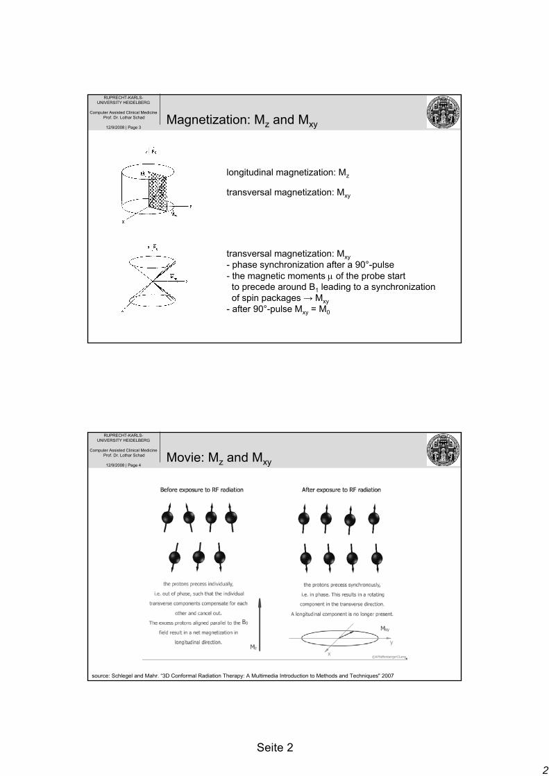
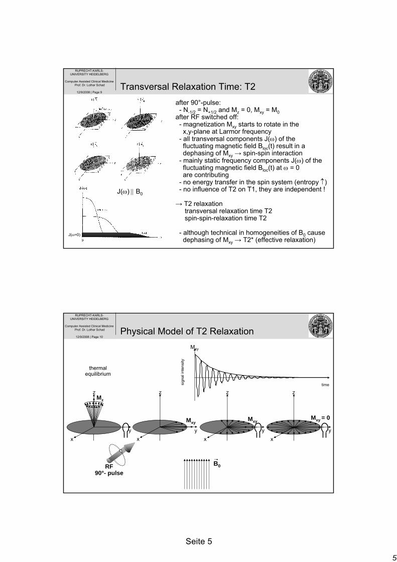
12/9/2008 | Page 3Magnetization: Mz and Mxy
longitudinal magnetization: Mz
transversal magnetization: Mxy
transversal magnetization: Mxy- phase synchronization after a 90°-pulse- the magnetic moments μ of the probe startto precede around B1 leading to a synchronizationof spin packages → Mxy
- after 90°-pulse Mxy = M0
RUPRECHT-KARLS-UNIVERSITY HEIDELBERG
Computer Assisted Clinical MedicineProf. Dr. Lothar Schad
12/9/2008 | Page 4Movie: Mz and Mxy
source: Schlegel and Mahr. “3D Conformal Radiation Therapy: A Multimedia Introduction to Methods and Techniques" 2007
3
Seite 3
RUPRECHT-KARLS-UNIVERSITY HEIDELBERG
Computer Assisted Clinical MedicineProf. Dr. Lothar Schad
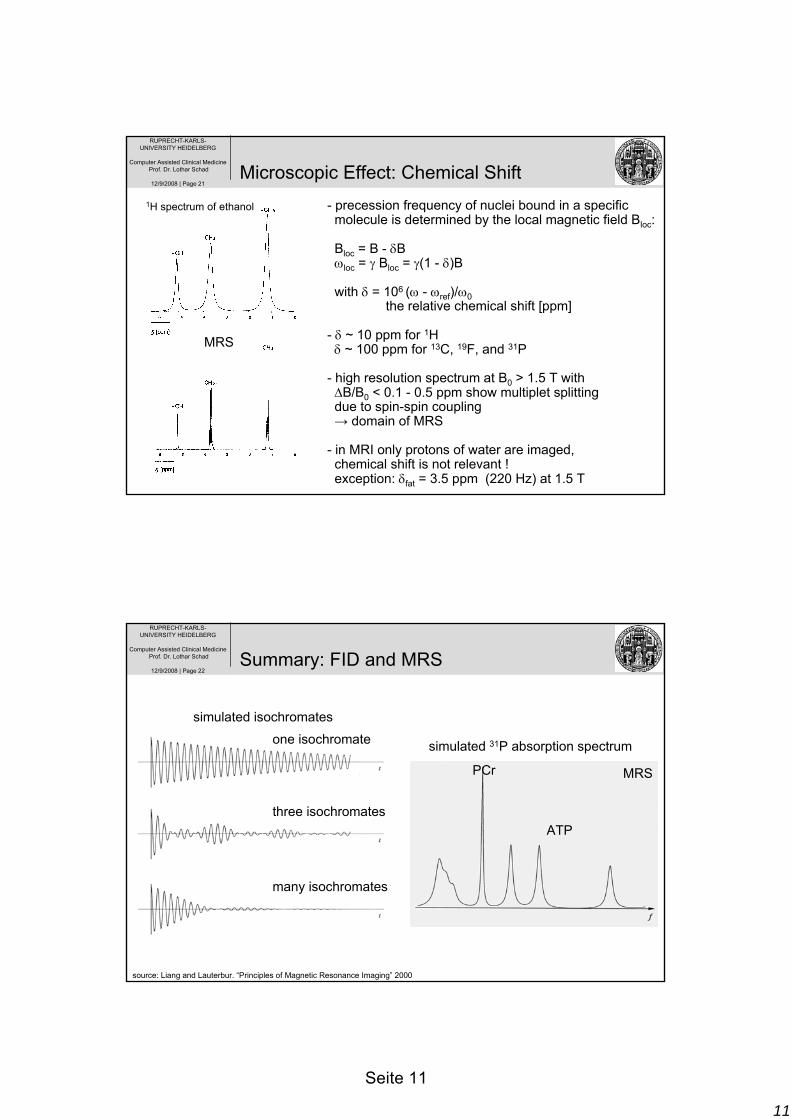
- Lenz’s law: the induced current produces an own magnetic moment μ in a conductor opposite to B0
- most of biological tissues have diamagnetic propertiessince the electron magnetization Me of the electron sheath is opposite to B0 due to Lenz’s law:B = μ0(H + Me)Me = χ H with μ0 = 1.257 10-6 Vs/A magnetic field constant
χH2O = -0.72 10-6 magnetic susceptibility
- weaker B-field inside a diamagnetic sphere due toe--shielding which is very effective since γe- = 658 γp
- intersection of different tissues creates additionallocal field inhomogeneities of B0can be “homogenized” by additional shim coils
11
Seite 11
RUPRECHT-KARLS-UNIVERSITY HEIDELBERG
Computer Assisted Clinical MedicineProf. Dr. Lothar Schad
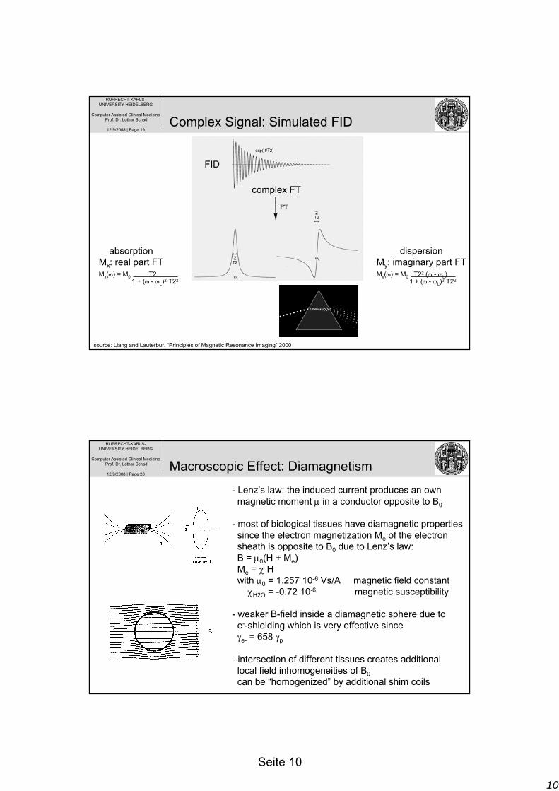
12/9/2008 | Page 21Microscopic Effect: Chemical Shift
- precession frequency of nuclei bound in a specific molecule is determined by the local magnetic field Bloc:
Bloc = B - δBωloc = γ Bloc = γ(1 - δ)B
with δ = 106 (ω - ωref)/ω0the relative chemical shift [ppm]
- δ ~ 10 ppm for 1Hδ ~ 100 ppm for 13C, 19F, and 31P
- high resolution spectrum at B0 > 1.5 T with ΔB/B0 < 0.1 - 0.5 ppm show multiplet splitting due to spin-spin coupling→ domain of MRS
- in MRI only protons of water are imaged,chemical shift is not relevant !exception: δfat = 3.5 ppm (220 Hz) at 1.5 T
1H spectrum of ethanol
MRS
RUPRECHT-KARLS-UNIVERSITY HEIDELBERG
Computer Assisted Clinical MedicineProf. Dr. Lothar Schad
12/9/2008 | Page 22
one isochromate
three isochromates
many isochromates
Summary: FID and MRS
simulated 31P absorption spectrum
PCr
ATP
MRS
simulated isochromates
source: Liang and Lauterbur. “Principles of Magnetic Resonance Imaging” 2000
12
Seite 12
RUPRECHT-KARLS-UNIVERSITY HEIDELBERG
Computer Assisted Clinical MedicineProf. Dr. Lothar Schad
12/9/2008 | Page 23
FID signal is the transient response of a spin system after RF excitation;FID is a complex signal with amplitude and phase
FID amplitude is dependent on many parameters like: flip angle, number of spins, and magnetic field strength
FID timing is dependent on the grade of local magnetic field inhomogeneitiescharacterized by T2*:
1/T2* = 1/T2 + γΔBz with T2* < T2
T2*: the effective (local) T2 relaxation timeT2 : the true T2 relaxation time
Summary: FID and MRI
RUPRECHT-KARLS-UNIVERSITY HEIDELBERG
Computer Assisted Clinical MedicineProf. Dr. Lothar Schad
12/9/2008 | Page 24
Saturation-Recovery Sequence
Inversion-Recovery Sequence
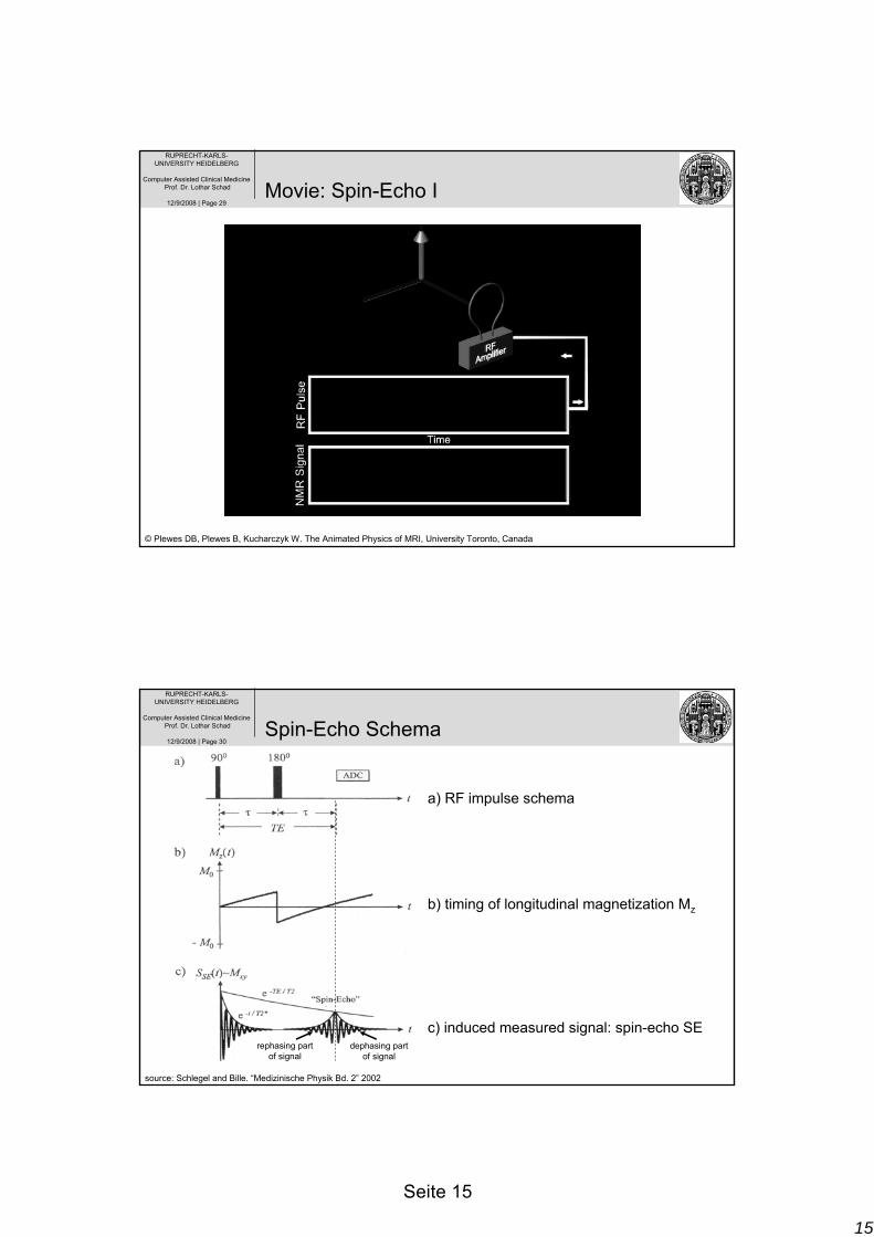
Spin-Echo Sequence
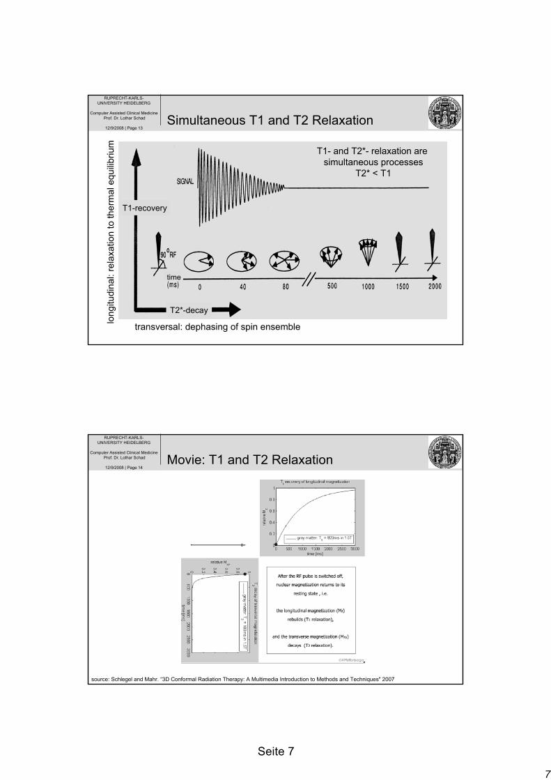
Standard Techniques for T1 and T2
13
Seite 13
RUPRECHT-KARLS-UNIVERSITY HEIDELBERG
Computer Assisted Clinical MedicineProf. Dr. Lothar Schad
12/9/2008 | Page 25
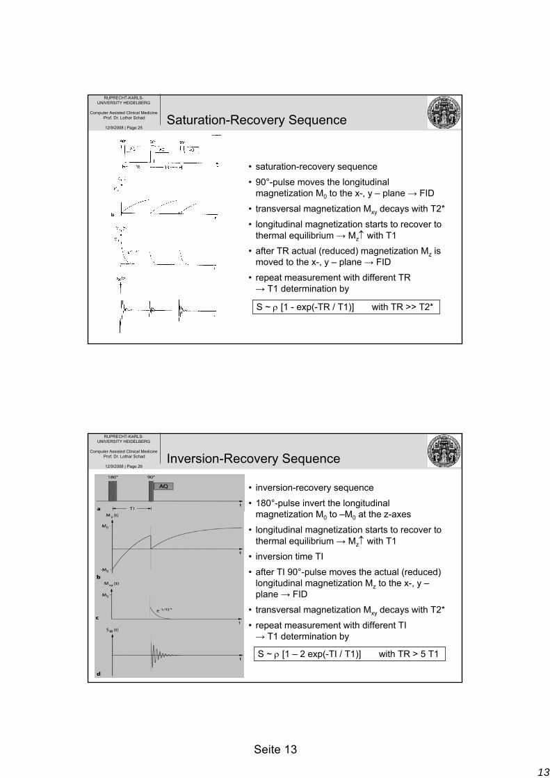
• saturation-recovery sequence
• 90°-pulse moves the longitudinal magnetization M0 to the x-, y – plane → FID
• transversal magnetization Mxy decays with T2*
• longitudinal magnetization starts to recover to thermal equilibrium → Mz↑ with T1
• after TR actual (reduced) magnetization Mz is moved to the x-, y – plane → FID
• repeat measurement with different TR→ T1 determination by
Saturation-Recovery Sequence
S ~ ρ [1 - exp(-TR / T1)] with TR >> T2*
RUPRECHT-KARLS-UNIVERSITY HEIDELBERG
Computer Assisted Clinical MedicineProf. Dr. Lothar Schad
12/9/2008 | Page 26
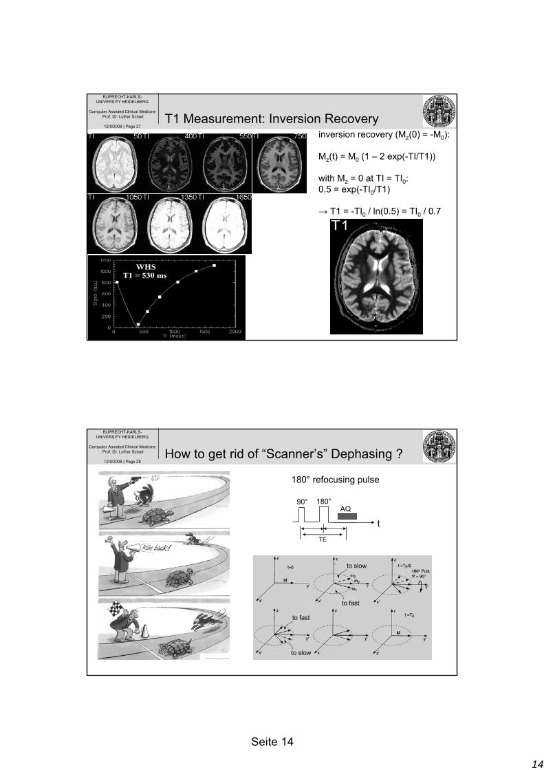
• inversion-recovery sequence
• 180°-pulse invert the longitudinal magnetization M0 to –M0 at the z-axes
• longitudinal magnetization starts to recover to thermal equilibrium → Mz↑ with T1
• inversion time TI
• after TI 90°-pulse moves the actual (reduced) longitudinal magnetization Mz to the x-, y –plane → FID
• transversal magnetization Mxy decays with T2*
• repeat measurement with different TI→ T1 determination by
Inversion-Recovery Sequence
S ~ ρ [1 – 2 exp(-TI / T1)] with TR > 5 T1
14
Seite 14
RUPRECHT-KARLS-UNIVERSITY HEIDELBERG
Computer Assisted Clinical MedicineProf. Dr. Lothar Schad
Computer Assisted Clinical MedicineProf. Dr. Lothar Schad
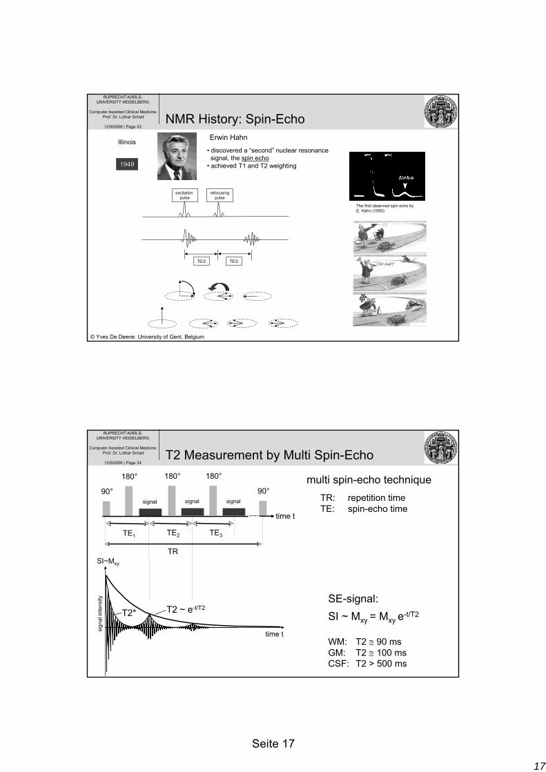
12/9/2008 | Page 32
1/T2* = 1/T2 + 1/T2´T2* : „effective“ relaxation with T2* < T2T2 : „true“ relaxation due to irreversible dephasingT2‘ : „scanner“ relaxation due to static and constant
field inhomogeneitiessource: Dössel. “Bildgebende Verfahren in der Medizin” 2000
Multi Spin-Echoes
RFexcitation
MTsignal
17
Seite 17
RUPRECHT-KARLS-UNIVERSITY HEIDELBERG
Computer Assisted Clinical MedicineProf. Dr. Lothar Schad
12/9/2008 | Page 33
19491949
Erwin Hahn
• discovered a “second” nuclear resonance signal, the spin echo
Computer Assisted Clinical MedicineProf. Dr. Lothar Schad
12/9/2008 | Page 34T2 Measurement by Multi Spin-Echo
SE-signal:SI ~ Mxy = Mxy e-t/T2
WM: T2 ≅ 90 msGM: T2 ≅ 100 msCSF: T2 > 500 ms
time t
90°
180°
TE2
180°
signal signal
180°
signal
TE3TE1
90°
TRSI~Mxy
time t
sign
al in
tens
ity
T2 ~ e-t/T2T2*
multi spin-echo techniqueTR: repetition timeTE: spin-echo time
18
Seite 18
RUPRECHT-KARLS-UNIVERSITY HEIDELBERG
Computer Assisted Clinical MedicineProf. Dr. Lothar Schad
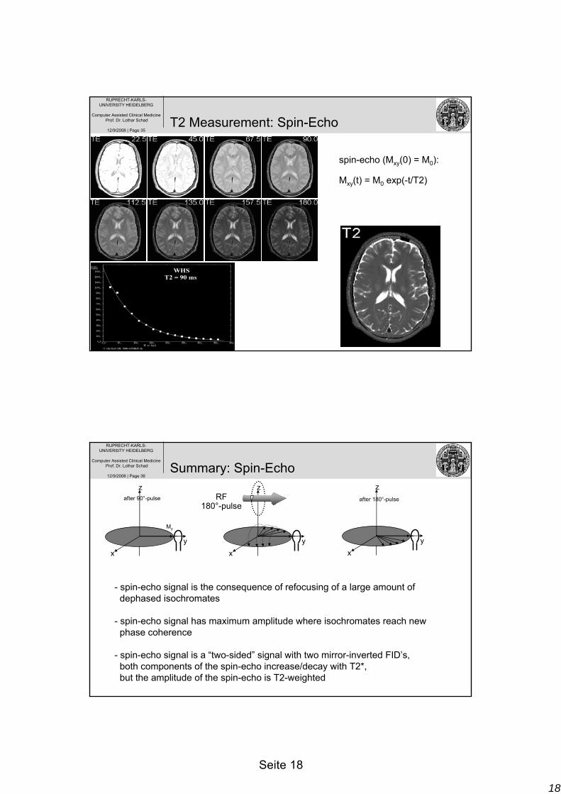
12/9/2008 | Page 35T2 Measurement: Spin Echo
spin-echo (Mxy(0) = M0):
Mxy(t) = M0 exp(-t/T2)
T2 Measurement: Spin-Echo
RUPRECHT-KARLS-UNIVERSITY HEIDELBERG
Computer Assisted Clinical MedicineProf. Dr. Lothar Schad
12/9/2008 | Page 36
- spin-echo signal is the consequence of refocusing of a large amount ofdephased isochromates
- spin-echo signal has maximum amplitude where isochromates reach new phase coherence
- spin-echo signal is a “two-sided” signal with two mirror-inverted FID’s,both components of the spin-echo increase/decay with T2*,but the amplitude of the spin-echo is T2-weighted
xy
z
My
xy
z
xy
zafter 90°-pulse after 180°-pulseRF
180°-pulse
Summary: Spin-Echo
19
Seite 19
RUPRECHT-KARLS-UNIVERSITY HEIDELBERG
Computer Assisted Clinical MedicineProf. Dr. Lothar Schad