+ All Categories
Home > Documents > Project: Ghana Emergency Medicine Collaborative Document Title: Advanced Cardiac Life Support...

Project: Ghana Emergency Medicine Collaborative Document Title: Advanced Cardiac Life Support...

Date post: 22-Dec-2015
Category:
Upload: melvyn-burke
View: 216 times
Download: 0 times
Share this document with a friend
Popular Tags:
79
Project: Ghana Emergency Medicine Collaborative Document Title: Advanced Cardiac Life Support Author(s): Rocky Oteng (University of Michigan), MD 2012 License: Unless otherwise noted, this material is made available under the terms of the Creative Commons Attribution Share Alike-3.0 License: http://creativecommons.org/licenses/by-sa/3.0/ We have reviewed this material in accordance with U.S. Copyright Law and have tried to maximize your ability to use, share, and adapt it. These lectures have been modified in the process of making a publicly shareable version. The citation key on the following slide provides information about how you may share and adapt this material. Copyright holders of content included in this material should contact [email protected] with any questions, corrections, or clarification regarding the use of content. For more information about how to cite these materials visit http://open.umich.edu/privacy-and-terms-use. Any medical information in this material is intended to inform and educate and is not a tool for self-diagnosis or a replacement for medical evaluation, advice, diagnosis or treatment by a healthcare professional. Please speak to your physician if you have questions about your medical condition. Viewer discretion is advised: Some medical content is graphic and may not be suitable for all viewers. 1
Transcript

Project: Ghana Emergency Medicine Collaborative

Document Title: Advanced Cardiac Life Support

Author(s): Rocky Oteng (University of Michigan), MD 2012

License: Unless otherwise noted, this material is made available under the terms of the Creative Commons Attribution Share Alike-3.0 License: http://creativecommons.org/licenses/by-sa/3.0/

We have reviewed this material in accordance with U.S. Copyright Law and have tried to maximize your ability to use, share, and adapt it. These lectures have been modified in the process of making a publicly shareable version. The citation key on the following slide provides information about how you may share and adapt this material.

Copyright holders of content included in this material should contact [email protected] with any questions, corrections, or clarification regarding the use of content.

For more information about how to cite these materials visit http://open.umich.edu/privacy-and-terms-use.

Any medical information in this material is intended to inform and educate and is not a tool for self-diagnosis or a replacement for medical evaluation, advice, diagnosis or treatment by a healthcare professional. Please speak to your physician if you have questions about your medical condition.

Viewer discretion is advised: Some medical content is graphic and may not be suitable for all viewers.

1

Attribution Key

for more information see: http://open.umich.edu/wiki/AttributionPolicy

Use + Share + Adapt

Make Your Own Assessment

Creative Commons – Attribution License

Creative Commons – Attribution Share Alike License

Creative Commons – Attribution Noncommercial License

Creative Commons – Attribution Noncommercial Share Alike License

GNU – Free Documentation License

Creative Commons – Zero Waiver

Public Domain – Ineligible: Works that are ineligible for copyright protection in the U.S. (17 USC § 102(b)) *laws in your jurisdiction may differ

Public Domain – Expired: Works that are no longer protected due to an expired copyright term.

Public Domain – Government: Works that are produced by the U.S. Government. (17 USC § 105)

Public Domain – Self Dedicated: Works that a copyright holder has dedicated to the public domain.

Fair Use: Use of works that is determined to be Fair consistent with the U.S. Copyright Act. (17 USC § 107) *laws in your jurisdiction may differ

Our determination DOES NOT mean that all uses of this 3rd-party content are Fair Uses and we DO NOT guarantee that your use of the content is Fair.

To use this content you should do your own independent analysis to determine whether or not your use will be Fair.

{ Content the copyright holder, author, or law permits you to use, share and adapt. }

{ Content Open.Michigan believes can be used, shared, and adapted because it is ineligible for copyright. }

{ Content Open.Michigan has used under a Fair Use determination. }

2

ACLS

• Systematic approach to assessment and management of cardiopulmonary emergencies

• Continuation of Basic Life Support• Resuscitation efforts aimed at

restoring spontaneous circulation and retaining intact neurologic function ABCD

3

The AAA’s

• Assess the patient– Establish unresponsiveness– Check pulse, respirations

• Activate EMS– Call for help

• AED– Get an AED (automated external

defibrillator)

4

Primary Survey (BLS)

• Airway• Breathing• Circulation• Defibrillation

Always assess and manage before moving on to the next step!

5

Airway

• Open the airway– Head tilt-chin lift– Jaw thrust

Wellcome Photo Library, Wellcome Photos

Wellcome Photo Library, Wellcome Photos

6

Breathing

• Look, Listen and Feel

• Give 2 rescue breaths

• Watch for appropriate chest rise and fall

U.S. Navy photo by Photographer's Mate 3rd Class Jesse Praino, Wikimedia Commons7

Circulation

• Check for a pulse• Start CPR

– 30 compressions/ 2 respirations

• Compressions more important than respirations!

U.S. Navy photo by Mass Communication Specialist Seaman Gabriel S. Weber, Wikimedia Commons8

Defibrillation

• Know your AED

• Universal steps:1. Power ON2. Attach electrode pads3. Analyze the rhythm4. Shock (if advised)

Ernstl, Wikimedia Commons 9

Defibrillation

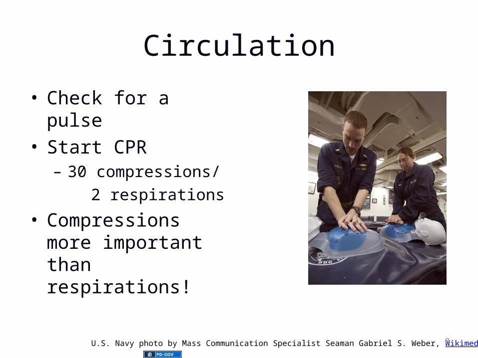
• Most frequent initial rhythm in witnessed sudden cardiac arrest is ventricular fibrillation (VF) or pulseless ventricular tachycardia (VT) which rapidly deteriorates into VF

• The only effective treatment for VF is electrical defibrillation

• Probability of successful defibrillation diminishes rapidly over time

• VF rapidly converts to asystole if not treated

10

Early Defibrillation = Increased Survival

Source unknown 11

Outcomes of Rapid Defibrillation by Security Officers after Cardiac

Arrest in Casinos

• NEJM Vol 343 (17) October 26, 2000• Used AEDs on 105 patients with

Ventricular Fibrillation• 53% survived to discharge (back to

casino)• Previously, less than 5% survive

12

Public-Access Defibrillation and Survival after Out-of-Hospital Cardiac

Arrest

• NEJM 2004• Community based trial of AED

deployment and layperson training.• 30 in AED group versus 15 survivors in

CPR only group to hospital discharge• Average age of survivor - 69.8 years• Study cost - $9.5 million

13

Secondary Survey (ACLS)

• Airway• Breathing• Circulation• Differential Diagnosis

• Assess and manage at each step before moving on!

14

Airway

• Maintain airway patency– Head tilt-chin lift/jaw thrust– Oro- or nasopharyngeal airway

• Advanced airway management– ETT– Combitube– LMA

Ignis, Wikimedia Commons 15

Breathing

• Assess adequacy of oxygenation and ventilation

• Provide supplemental oxygen• Confirm proper airway placement• Secure tube

16

Circulation

• Assess/monitor cardiac rhythm• Establish IV access• Give medications as appropriate for

rhythm and BP• Fluid resuscitation• Minimize interruption of

compressions to maximize survival.

17

Differential Diagnosis

• Look for and treat any reversible cause of arrest

18

Basic Rhythm Analysis

19

Basic Rhythm Analysis

• Rate – too fast or too slow?• Rhythm – regular or irregular?• Is there a normal looking QRS? Is it

wide or narrow?• Are P waves present?• What is the relationship of the P

waves to the QRS complex?

20

Rhythm Analysis

Lethal vs non-lethal?

Shockable vs. non-shockable? Too fast vs too slow?

Symptomatic vs. asymptomatic?

21

Lethal Rhythms

• Shockable (Defibrillation)– Ventricular fibrillation– Pulseless ventricular tachycardia

• Non-shockable– Asystole– Pulseless electrical activity

22

Non-Lethal Rhythms

• Too fast (tachycardias)– Sinus– Supraventricular (including a-fib/flutter)– Ventricular

• Too slow (bradycardias)– Sinus– Heart block (1°, 2°, 3° AV block)

23

What is a Symptomatic Dysrhythmia?

• Any abnormal rhythm that produces signs or symptoms of hypoperfusion– Chest Pain/ischemic EKG changes– Shortness of Breath– Decreased level of consciousness– Syncope/pre-syncope– Hypotension– Shock - decreased Uop, cool

extremities, etc.– Pulmonary Congestion/CHF

24

Name that rhythm…

25

63 yo man with a witnessed collapse while mowing the lawn

What is the rhythm?What is the management?

Chikumaya, Wikimedia Commons 26

Ventricular Fibrillation

• Rapid and irregular• No normal P waves or QRS complexes

Jer5150, Wikimedia Commons 27

VF / Pulseless VT

Primary Survey - ABC

Secondary Survey - ABC

Source unknown 28

ACLS Algorithm

• Primary Survey• Shock – 360 J• Secondary Survey• Vasopressor - Epi or Vasopressin IV• Shock 360J• Antiarrhythmic – Amiodarone,

Lidocaine or Magnesium Sulfate IV• Shock 360J

29

79yo man s/p NSTEMI

What is the rhythm?What is the management?

Glenlarson, Wikimedia Commons 30

Ventricular Tachycardia

• Rapid and regular• No P waves• Wide QRS complexes

Ksheka, Wikimedia Commons 31

Ventricular Tachycardia

• Monomorphic VT

• Polymorphic VT

32

Ksheka, Wikimedia Commons

Displaced, Wikimedia Commons

Ventricular Tachycardia

• Assume any wide complex tachycardia is VT until proven otherwise– SVT with aberrant conduction may also

have wide QRS complexes • Attempt to establish the diagnosis

– Ischemia risk and VT go together

33

Treatment of VT

• If pulseless - follow VF algorithm• If stable try anti-arrhythmics

– Amiodarone– Lidocaine– Procainamide?

• If patient has a pulse, but is unstable or not responding to meds - shock

34

Treatment of VT

• Anti-arrhythmics are also pro-arrhythmic

• One antiarrhythmic may help, more than one may harm

• Anti-arrhythmics can impair an already impaired heart

• Electrical cardioversion should be the second intervention of choice

35

60yo diabetic man with chest pain

What is the rhythm?What is the management?

Knutux, Wikimedia Commons 36

Normal Sinus Rhythm

• Regular rate and rhythm• Normal P waves and QRS• Evaluate for cause of chest pain and

monitor for change in rhythm

Knutux, Wikimedia Commons 37

40 yo woman found down, pulseless and apneic

What is the rhythm?What is the management?

Masur, Wikimedia Commons 38

Pulseless Electrical Activity

• Any organized (or semi-organized) electrical activity in a patient without a detectable pulse

• Non-perfusing

• Treat the patient NOT the monitor

• Find and treat the cause!!!!!39

PEA and AsystoleSecondary Survey - ABCD

Primary Survey - ABCSource unknown 40

PEA

A trop in e 1 m g IV Pif P E A is slow

E p in ep h rin e 1 m g IV Prep ea t every 3 -5 m in u tes

S earch fo r an d Trea t C au ses

S econ d ary S u rvey

P rim ary S u rvey

41

Find and Treat the Cause

• Non-shockable rhythm • The most effective treatment is to

find and fix the underlying problem

Rama, Wikimedia Commons 42

So what causes PEA?

• #1 cause of PEA in adults is hypovolemia

• #1 cause in children is hypoxia/respiratory arrest

• Other causes?

43

The H’s and T’s

• Hypovolemia• Hypoxia• Hydrogen ion

(acidosis)• Hyper-/

hypokalemia• Hypothermia • Hypoglycemia

(rare)

• Toxins• Tamponade• Tension

pneumothorax• Thrombosis

(coronary or pulmonary)

• Trauma

44

Treat the H’s and T’s• Hypovolemia

– Volume – IVF, PRBC’s• Hypoxia

– Oxygenate/Ventilate• Hydrogen ion (acidosis)

– Sodium bicarbonate– Hyperventilation

• Hyper-/hypokalemia– Sodium bicarbonate– Insulin/glucose– Calcium

• Hypothermia – Warm -- invasive

• Hypoglycemia – Dextrose

• Toxins– Check levels– Charcoal– Antidotes

• Tamponade– pericardiocentesis

• Tension pneumothorax– Needle decompression– Tube thoracostomy

• Thrombosis (coronary or pulmonary)– Thrombolytics– OR/cath lab

• Trauma

45

19yo man with palpitations

What is the rhythm?What is the management?

Displaced, Wikimedia Commons 46

Supraventricular Tachycardia

• Rapid (usually 150-250 bpm) and regular• P waves cannot be positively identified• QRS narrow

Displaced, Wikimedia Commons 47

Treatment of Stable SVT

• Consider vagal maneuvers– Carotid sinus massage– Valsalva– Eyeball massage– Ice water to face– Digital rectal exam

• Adenosine– 6 mg, 12 mg, 12 mg

48

Treatment of Unstable SVT

• Electrical Cardioversion• Cardioversion is not defibrillation• Use defibrillator in “sync” mode

– prevents delivering energy in the wrong part of the cardiac cycle (R on T phenomenon)

49

Electrical Cardioversion

• Energy level – somewhat controversial

• 100 J→200J→300J→360J• Atrial flutter may convert with lower

energy– 50J

• For polymorphic VT – start with 200J• The EP guys tend to start with 360J

50

Electrical Cardioversion

• Be prepared– Patient on monitor, IV, Oxygen– Suction ready and working– Airway supplies ready

• Pre-medicate whenever possible– Conscious sedation– Electrical shocks are painful!

51

Tachycardia

Lots of optionsbased on rhythm

Stable?

Shock

Unstable?

Evaluate Patien t

• Treat the patient NOT the monitor!!!

52

Stable Tachycardias

• Narrow complex?– Regular rhythm

• Sinus tachycardia• SVT• AV nodal reentry

– Irregular rhythm• Atrial fibrillation • Atrial flutter

• Wide complex?– Uncertain rhythm –

assume VT– Narrow complex

tachycardia with aberrancy

– Ventricular tachycardia

• Monomorphic or polymorphic

53

56 yo woman with shortness of breath and chest pain

What is the rhythm?What is the management?

J. Heuser, Wikimedia Commons 54

Atrial fibrillation/flutter

• May be rapid• Irregular (fib) or more regular (flutter)• No P waves, narrow QRS

James Heilman, MD, Wikimedia Commons

J. Heuser, Wikimedia Commons

55

Atrial fibrillation/flutter• Treatment based on patient’s clinical

picture– Unstable = Immediate electrical

cardioversion– Stable

• Control the rate– Diltiazem– Esmolol (not if EF < 40%)– Digoxin

• Provide anticoagulation

• Treat the patient NOT the monitor!!! 56

78yo man found down, pulseless and apneic, unknown duration

What is the rhythm?What is the management?

D Dinneen, Wikimedia Commons 57
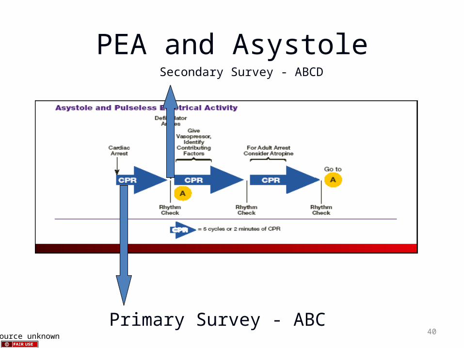
Asystole

• Is it really asystole?• Check lead and cable connections.• Is everything turned on?• Verify asystole in another lead.• Maybe it is really fine v-fib?

D Dinneen, Wikimedia Commons 58

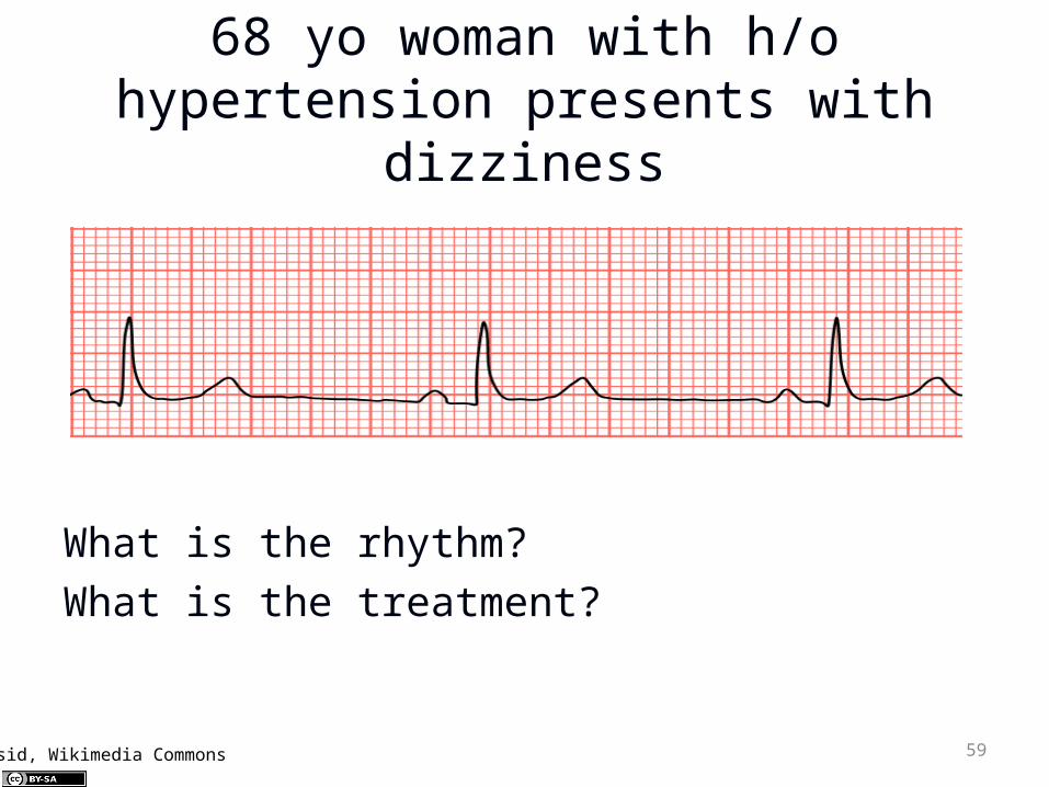
68 yo woman with h/o hypertension presents with

dizziness

What is the rhythm?What is the treatment?

Mysid, Wikimedia Commons 59
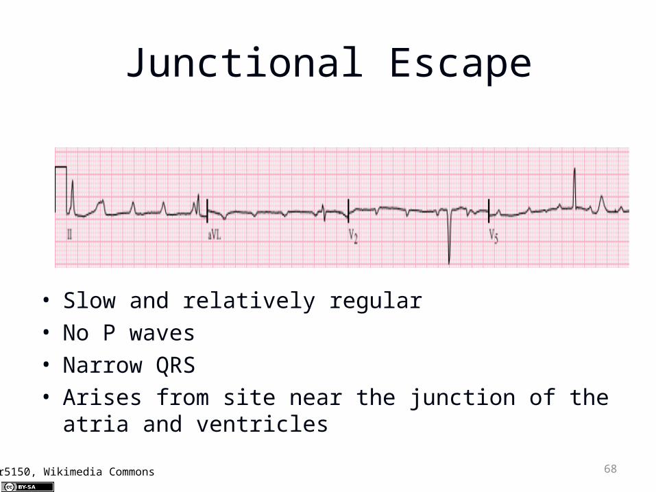
Sinus Bradycardia

• Slow and regular• Normal P waves and QRS complexes

Mysid, Wikimedia Commons 60

Bradycardias

• Many possible causes– Enhanced parasympathetic tone – Increased ICP. – Hypothyroidism – Hypothermia – Hyperkalemia – Hypoglycemia – Drug therapy

61

Bradycardias

• Treat only symptomatic bradycardias– Ask if the bradycardia causing the

symptoms

• Recognize the red flag bradycardias– Second degree type II block– Third degree block

62

Source unknown 63

Transcutaneous pacing

• Class I for all symptomatic bradycardias

• Always appropriate• Doesn’t always work• Technique

– Attach pacer pads– Set a rate to 80 bpm– Turn up the juice (amps) until you get

capture• Painful – may need sedation / analgesia

64

Transvenous Pacing

• Invasive• Time-consuming to establish• Skilled procedure• Better long-term than

transcutaneous• May have better capture than

transcutaneous pacing

65

Bradycardia Treatment

• Medications– Vagolytic

• Atropine

– Adrenergic• Epinephrine• Dopamine

66

What if the same patient had this rhythm?

What is the rhythm?What is the treatment?

Jer5150, Wikimedia Commons 67

Junctional Escape

• Slow and relatively regular• No P waves• Narrow QRS• Arises from site near the junction of the atria

and ventricles

Jer5150, Wikimedia Commons 68

29 yo asymptomatic female

What is the rhythm?What is the management?

69Steven Fruitsmaak, Wikimedia Commons

1° AV block

• Regular rate and rhythm• Normal P wave with long PR interval

(>0.2msec/1 big box)• Normal QRS

70Steven Fruitsmaak, Wikimedia Commons

58yo asymptomatic woman

What is the rhythm?What is the management?

Jer5150, Wikimedia Commons 71

2° AV Block - Type I

• aka Wenckebach• Regular rate and rhythm• Normal P waves and QRS complexes• Increasing PR interval until QRS dropped

Jer5150, Wikimedia Commons 72

80 yo man with syncope

What is the rhythm?What is the management?

Jer5150, Wikimedia Commons 73

2° AV Block – Mobitz Type II

• Regular atrial rate with normal P wave• Consistent PR interval• Random QRS dropped

Jer5150, Wikimedia Commons 74

Another 80 yo man with syncope

What is the rhythm?What is the management?

MoodyGroove, Wikimedia Commons 75

3° AV Block

• Normal P waves• Normal QRS• No relationship between P and QRS• aka complete heart block

MoodyGroove, Wikimedia Commons 76

Know When To Stop

• With return of spontaneous circulation

• No ROSC during or after 20 minutes of resuscitative efforts– Possible exceptions include near-

drowning, severe hypothermia, known reversible cause, some overdoses

• DNR orders presented• Obvious signs of irreversible death

77

Take Home Points

• Assess and manage at every step before moving on to the next step

• Rapid defibrillation is the ONLY effective treatment for VF/VT

• Search for and treat the cause• Treat the patient not the monitor• Reassess frequently• Minimize interruptions to chest

compressions78

Special thanks to:Steve Kronick, MD andSuzanne Dooley-Hash, MDfor contributing slides and content for this lecture.

79


Recommended