LiNaBioFLuid_D2.3_Public summary of biological fluid transport mechanisms on website Page 1 of 4 Project LiNaBioFluid Deliverable D2.3: Public summary of biological fluid transport mechanism on website Reporting period from 01.07.2015 to 30.06.2016 Report completed and released 13.09.2016 1. Objectives and Detailed Description The status of understanding of the biological mechanism for the fluid transport was reported in milestone M2 (month 12). Despite the directional water transport in capillary channels on the integument of horned lizards, the transport of water on the cuticle of flat bugs was focused. The transport of water out of the bug capillaries onto the cuticle plates is of special interest, in particular in view of the addressed potential fields of application, such as lubrication. The total understanding of this process is still not completely understood though – the research is ongoing. However, the principles of the directional, passive fluid transport in the scent gland channels of flat bugs have been identified and successfully transferred to hot working tool steel as well as polymer samples. Deliverable D2.3 is partially based on deliverables D2.1 and D2.2. A summary of these findings was published on the public section of the LiNaBioFluid website ( www.laserbiofluid.eu ) as the following content: Directional water transport in skin capillary channels of horned lizards Moisture harvesting lizards such as species of the genera Phrynosoma (Fig. 1) have remarkable adaptations for inhabiting arid regions. Special skin structures enable them to access water sources such as moist sand and dew: their skin is capable of collecting and transporting water. Even small amounts of water were observed to be transported in capillary channels between the imbricate overlapping scales (Fig. 1 B-C). That transport is even preferentially towards the mouth where the water is drunk by the lizard (Fig. 1 D).
Transcript
LiNaBioFLuid_D2.3_Public summary of biological fluid transport mechanisms on website Page 1 of 4
Project LiNaBioFluid Deliverable D2.3: Public summary of biological fluid transport mechanism on website Reporting period from 01.07.2015 to 30.06.2016 Report completed and released 13.09.2016
1. Objectives and Detailed Description The status of understanding of the biological mechanism for the fluid transport was reported in milestone M2 (month 12). Despite the directional water transport in capillary channels on the integument of horned lizards, the transport of water on the cuticle of flat bugs was focused. The transport of water out of the bug capillaries onto the cuticle plates is of special interest, in particular in view of the addressed potential fields of application, such as lubrication. The total understanding of this process is still not completely understood though – the research is ongoing. However, the principles of the directional, passive fluid transport in the scent gland channels of flat bugs have been identified and successfully transferred to hot working tool steel as well as polymer samples. Deliverable D2.3 is partially based on deliverables D2.1 and D2.2. A summary of these findings was published on the public section of the LiNaBioFluid website ( www.laserbiofluid.eu ) as the following content: Directional water transport in skin capillary channels of horned lizards Moisture harvesting lizards such as species of the genera Phrynosoma (Fig. 1) have remarkable adaptations for inhabiting arid regions. Special skin structures enable them to access water sources such as moist sand and dew: their skin is capable of collecting and transporting water. Even small amounts of water were observed to be transported in capillary channels between the imbricate overlapping scales (Fig. 1 B-C). That transport is even preferentially towards the mouth where the water is drunk by the lizard (Fig. 1 D).
LiNaBioFLuid_D2.3_Public summary of biological fluid transport mechanisms on website Page 2 of 4
Figure 1: Phenomenon of directional water transport on the skin of Phrynosoma cornutum. A) Texas horned lizard P. cornutum. B) SEM-image of capillary opening between dorsal scales. C) Skin cross section analysed by Optical Coherence Tomography (OCT). Capillaries are indicated by arrows. D) Image sequence of a dyed water droplet on the dorsal integument. Analyses of the lizard skin enabled the identification of two principles underlying the observed directional water transport. A specific geometry of capillary channels results in a periodic narrowing of the capillary channels (asymmetry) as well as a channel network (interconnection) in between the scales (Fig. 2).
Figure 2: Analyses of capillary structure. A) Skin cross section of Phrynosoma cornutum from single OCT-slide. Capillaries are indicated by arrows. B) OCT-rendering of skin topography. C) Histological slices of the dorsal integument, illustrating the cross sectional view of longitudinal capillary channels (arrows). D) Model of skin capillary network. The principle of asymmetry enables a directional transport in a single channel, whereas the principle of interconnection prohibits a stopping of the water transport within the capillary network. Abstracting these principles to saw-tooth geometry of single capillaries that are interconnected, the mechanism is as follows (Fig. 3): Applied droplets are soaked into the surface channel structure by capillary forces. The liquid front stops at the sharp edges in capillary I, while it is transported farther into capillary II. As the liquid front in capillary II reaches the nearby interconnection the liquid is transported into capillary I. The liquid coming through the interconnection picks up the stopped liquid in capillary I and forms a new free liquid front. Thereafter the liquid is transported through a second interconnection into capillary II, where the stopped liquid is picked up. This mechanism was successfully validated with a biomimetic prototype laser cut into PMMA (Fig. 3 C).
LiNaBioFLuid_D2.3_Public summary of biological fluid transport mechanisms on website Page 3 of 4
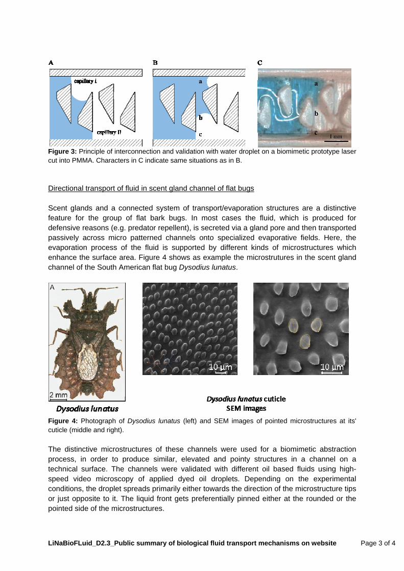
Figure 3: Principle of interconnection and validation with water droplet on a biomimetic prototype laser cut into PMMA. Characters in C indicate same situations as in B. Directional transport of fluid in scent gland channel of flat bugs Scent glands and a connected system of transport/evaporation structures are a distinctive feature for the group of flat bark bugs. In most cases the fluid, which is produced for defensive reasons (e.g. predator repellent), is secreted via a gland pore and then transported passively across micro patterned channels onto specialized evaporative fields. Here, the evaporation process of the fluid is supported by different kinds of microstructures which enhance the surface area. Figure 4 shows as example the microstrutures in the scent gland channel of the South American flat bug Dysodius lunatus.
Figure 4: Photograph of Dysodius lunatus (left) and SEM images of pointed microstructures at its' cuticle (middle and right). The distinctive microstructures of these channels were used for a biomimetic abstraction process, in order to produce similar, elevated and pointy structures in a channel on a technical surface. The channels were validated with different oil based fluids using high-speed video microscopy of applied dyed oil droplets. Depending on the experimental conditions, the droplet spreads primarily either towards the direction of the microstructure tips or just opposite to it. The liquid front gets preferentially pinned either at the rounded or the pointed side of the microstructures.
LiNaBioFLuid_D2.3_Public summary of biological fluid transport mechanisms on website Page 4 of 4
Fluid transport from capillary channels onto the cuticle plates of flat bugs The South American flat bug Dysodius lunatus shows also the fascinating behaviour of colour change when wetted by water. This camouflage behaviour requires the insect to have a hydrophilic surface and passive surface structures which facilitate the liquid spreading. SEM-analysis of the surface showed, that D. lunatus is covered with pillar-like microstructures. This roughness, and a chemical hydrophilicity of the cuticle waxes, renders the bug almost superhydrophilic: Water spreads immediately across the surface. Besides the fluid spreading across the surface, water is also transported in capillary channels between the body segments. However, water can exit the capillaries at interconnection points between the segments. Then, water starts spreading across the surface. We propose that this process is related to chemical gradients within the distribution of amphiphilic substances in the capillaries (e.g. more hydrophobic chemistry of the capillaries at interconnection points would “push” the water out). Investigations are still ongoing. This includes different chemical and spectrometric analyses.
2. Evaluation of Goals and Resulting Actions Having published the summary of biological fluid transport mechanisms on the project website, we accomplished D2.3. Further information can be obtained from the following publications: Comanns, P., Buchberger, G., Buchsbaum, A., Baumgartner, R., Kogler, A., Bauer, S., Baumgartner, W. (2015). Directional, passive liquid transport: the Texas horned lizard as a model for a biomimetic ‘liquid diode’. J. R. Soc. Interface 12: 20150415. http://dx.doi.org/10.1098/rsif.2015.0415 Plamadeala, C., F. Hischen, R. Friesenecker, R. Wollhofen, J. Jacak, G. Buchberger, E. Heiss, T.A. Klar, W. Baumgartner, J. Heitz. Bio-inspired polymer microstructures for directional transport of oily liquids. Under review in J. R. Soc. Interface. Hischen, F., G. Buchberger, C. Plamadeala, E. Heiss, K. Winands, W. Baumgartner. Scent gland channels of true bugs: A biomimetic role model for passive, directional fluid transport. In preparation.